Coriell Institute For Medical Research
Mouse Embryonic Stem Cell Culturing Protocols
Form 1301-05 Rev B-072214 1 of 6
General Guidelines for Handling Mouse ES cells
• mES cells are cryopreserved in plastic cryovials and shipped on dry ice. If storing the mES
cells before thawing, store in liquid nitrogen vapor. Storage directly in liquid nitrogen may
result in cracking of the o-rings.
• It is highly recommended that a small number of vials are cryopreserved as a master stock
before beginning any experimentation
Coriell Institute For Medical Research
Mouse Embryonic Stem Cell Culturing Protocols
2 of 6
Form 1301-05 Rev B-072214
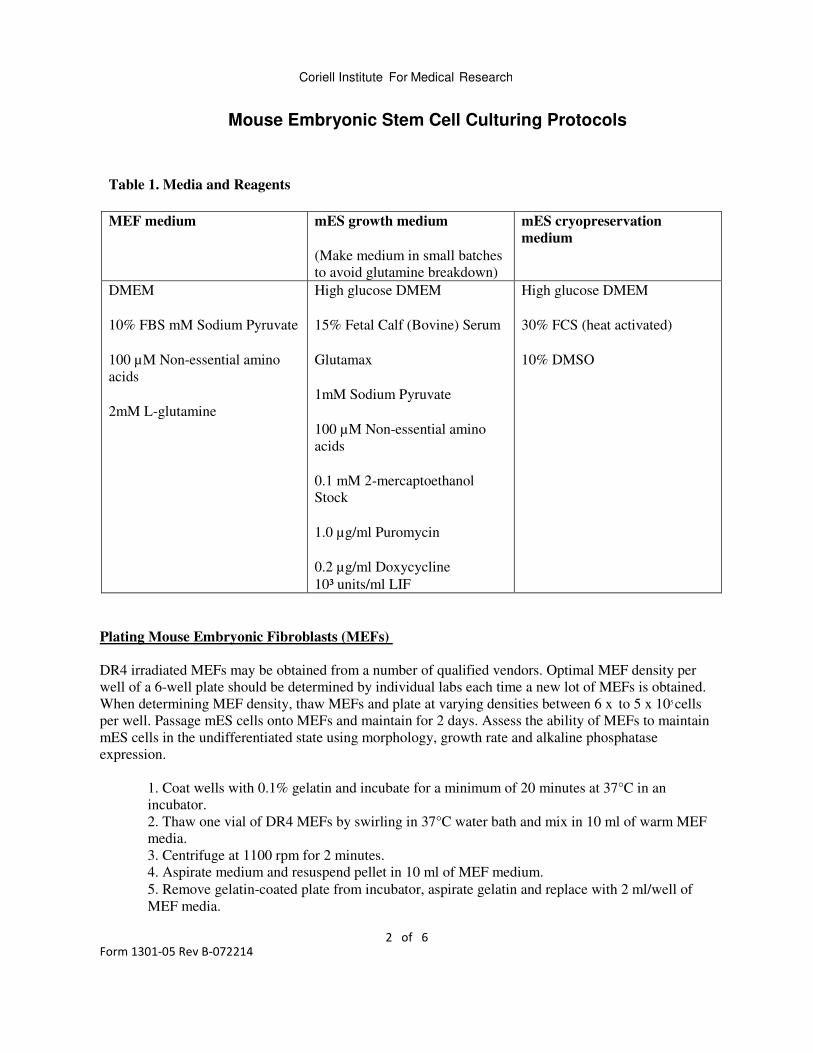
Table 1. Media and Reagents
MEF medium mES growth medium
(Make medium in small batches
to avoid glutamine breakdown)
mES cryopreservation
medium
DMEM
10% FBS mM Sodium Pyruvate
100 µM Non-essential amino
acids
2mM L-glutamine
High glucose DMEM
15% Fetal Calf (Bovine) Serum
Glutamax
1mM Sodium Pyruvate
100 µM Non-essential amino
acids
0.1 mM 2-mercaptoethanol
Stock
1.0 µg/ml Puromycin
0.2 µg/ml Doxycycline
10³ units/ml LIF
High glucose DMEM
30% FCS (heat activated)
10% DMSO
Plating Mouse Embryonic Fibroblasts (MEFs)
DR4 irradiated MEFs may be obtained from a number of qualified vendors. Optimal MEF density per
well of a 6-well plate should be determined by individual labs each time a new lot of MEFs is obtained.
When determining MEF density, thaw MEFs and plate at varying densities between 6 x to 5 x 10⁵ cells
per well. Passage mES cells onto MEFs and maintain for 2 days. Assess the ability of MEFs to maintain
mES cells in the undifferentiated state using morphology, growth rate and alkaline phosphatase
expression.
1. Coat wells with 0.1% gelatin and incubate for a minimum of 20 minutes at 37°C in an
incubator.
2. Thaw one vial of DR4 MEFs by swirling in 37°C water bath and mix in 10 ml of warm MEF
media.
3. Centrifuge at 1100 rpm for 2 minutes.
4. Aspirate medium and resuspend pellet in 10 ml of MEF medium.
5. Remove gelatin-coated plate from incubator, aspirate gelatin and replace with 2 ml/well of
MEF media.
Coriell Institute For Medical Research
Mouse Embryonic Stem Cell Culturing Protocols
Form 1301-05 Rev B-072214 5 of 6
6. Mix cell suspension and count viable cells by Trypan blue dye exclusion.
7. Using viable cell number, calculate and aliquot appropriate number of cells to yield
predetermined optimal cell density into each well of a 6-well plate (be sure to mix cell
suspension several times during plating to avoid settling of cells).
8. Place in incubator and shake plate back/forth and left/right.
NOTE: Failure to gently shake plate back and forth may result in uneven seeding.
9. Incubate overnight or up to 5 days before using as feeder layer for mES cells.
Thawing murine ES cells
For optimal thawing result of murine ES cells, MEFs should be plated on gelatin-coated plates at
least 1 day prior to use to allow adherence and flattening prior to mES cell plating. Seed mES cells
up to 5 days after plating MEFs.
1. Remove ES cells from liquid nitrogen/dry ice and thaw quickly in 37°C water bath.
2. Transfer cell suspension to sterile 15 ml tube containing 10 ml warm growth medium and mix
cells.
3. Centrifuge conical tube containing cells at 1100 rpm for 2 minutes at room temperature.
4. Aspirate freeze medium and resuspend cells into 2 ml of warm ES growth medium.
5. Plate cells in 1- 2 wells of a 6-well plate (1.0 X 10⁶ to 1.8 x 10⁶ cells per well) containing MEF
feeder cells.
6. Maintain cells by daily medium exchange.
7. Passage cells at 75%-90% confluence (Image 1).
Passaging of murine ES cells
Passage cells every 2-3 days depending upon the growth rate of cells. The optimal condition is to
maintain cells at approximately 80% confluency on day 2 or 3 (Image 1). To avoid spontaneous
differentiation, do not allow cells to become confluent (Image 2). Split ratios range from 1:4 to 1:10.
Optimal cell number for seeding in a 6 well plate is between 1.0 X 10⁶ and 1.8 x 10⁶ cells per well.
(approximately 1.0 x 10⁵ cells/cm2).
1. Remove spent medium from culture and rinse with PBS.
2. Add 1 ml/well accutase and place in incubator for 5 minutes or until cells begin to dissociate
from plate.
3. Triturate accutase solution 2-3 times to dissociate cells from plate.
4. Transfer the detached cell aggregates to a 15 ml conical tube containing 5 ml mES growth
medium.
5. Rinse each well with an additional 1 ml of growth medium to collect any remaining
aggregates. Add the rinse to conical tube containing cells.
6. Take small aliquot of cells for cell count.
Coriell Institute For Medical Research
Mouse Embryonic Stem Cell Culturing Protocols
4 of 6
Form 1301-05 Rev B-072214
7. Centrifuge conical tube containing cells at 1100 rpm for 2 minutes at room temperature.
NOTE: When using accutase, cells can be plated directly into culture vessel without
centrifuging to pellet cells as long as accutase is inactivated by the addition of the
growth medium at a 3:1 ratio (medium to accutase).
8. Remove the supernatant from conical tubes and resuspend cells in appropriate volume of
growth medium such that there is an appropriate cell density for cell culture vessel (1.0 X 10⁶ to
1.8 x 10⁶ cells per well of a 6-well plate (approximately 1.0 x 10⁵ cells/cm2).
9. Seed cells onto prepared MEF-containing plates. Rock plates gently back and forth, sideways
and diagonally to achieve uniform cell distribution.
NOTE: Failure to gently shake plate back and forth may result in uneven seeding.
Cyropreservation of murine ES cells
1. Remove spent medium from culture and rinse with PBS.
2. Add 1 ml/well accutase and place in incubator for 5 minutes or until cells begin to dissociate
from plate.
3. Triturate accutase solution 2-3 times to dissociate cells from plate.
4. Transfer the detached cell aggregates to a 15 ml conical tube containing 5 ml mES growth
medium.
5. Rinse each well with an additional 1 ml of growth medium to collect any remaining
aggregates. Add the rinse to conical tube containing cells.
6. Centrifuge conical tube containing cells at 1100 rpm for 2 minutes at room temperature.
7. Remove supernatant and resuspend cells in appropriate volume of pre-cooled freeze medium
(1 ml per well of a 6-well plate).
8. Transfer 1 ml of cell suspension to cryovials on ice.
9. Place cryovials in isopropanol freezing container and store at -80°C overnight.
10. Transfer vials to liquid nitrogen vapor for permanent storage.
Coriell Institute For Medical Research
Mouse Embryonic Stem Cell Culturing Protocols
Form 1301-05 Rev B-072214 5 of 6
Table 2. Troubleshooting Tips
Problem Observation of problem Possible Causes and solutions
Spontaneous
differentiation within
culture (Image 2)
Differentiation can look different
depending on culture and cause of
differentiation:
1. flattened cells that have
dark and spiky boundaries
2. colonies appear as individual
cells instead of one large
syncial mass
3. flattened colonies
1. Low feeder layer quality- this
can be prevented by testing
optimal density for feeder
layers prior to using in an
experiment
2. Inappropriate feeder layer
density- when testing feeder
layers for trophic support of
ES cells, test at varying
densities to determine
appropriate density per lot of
feeder layers
3. Low LIF concentration
4. Over confluence (allowing
cultures to exceed 90%
confluence by day 3
Dying or differentiating
cells
Large colonies with necrotic centers
Cells will appear healthy and then appear
to round up and lift off plate
Cells have not been passaged for 4 or
more days
• Passage cells and plate at
higher density to ensure
70-90% confluence by day
3
Non-uniform distribution
of colonies with culture
vessel
Areas within culture vessel that are
highly confluent mixed with areas that
do not contain colonies
Cells were not distributed evenly
throughout culture vessel or culture
vessel was disturbed following plating
• Rock plates following
plating- usually rocking
plates back and forth and
then side to side produces
a fairly uniform
distribution
Cells are growing but do
not appear to be doubling
as expected
Cells are not reaching 70-90%
confluence by day 3
Plating at lower than optimal cell
density
Suboptimal culturing conditions;
check the following reagents
1. FCS lot
2. Glutamine
3. MEF Feeders
Coriell Institute For Medical Research
Mouse Embryonic Stem Cell Culturing Protocols
6 of 6
Form 1301-05 Rev B-072214
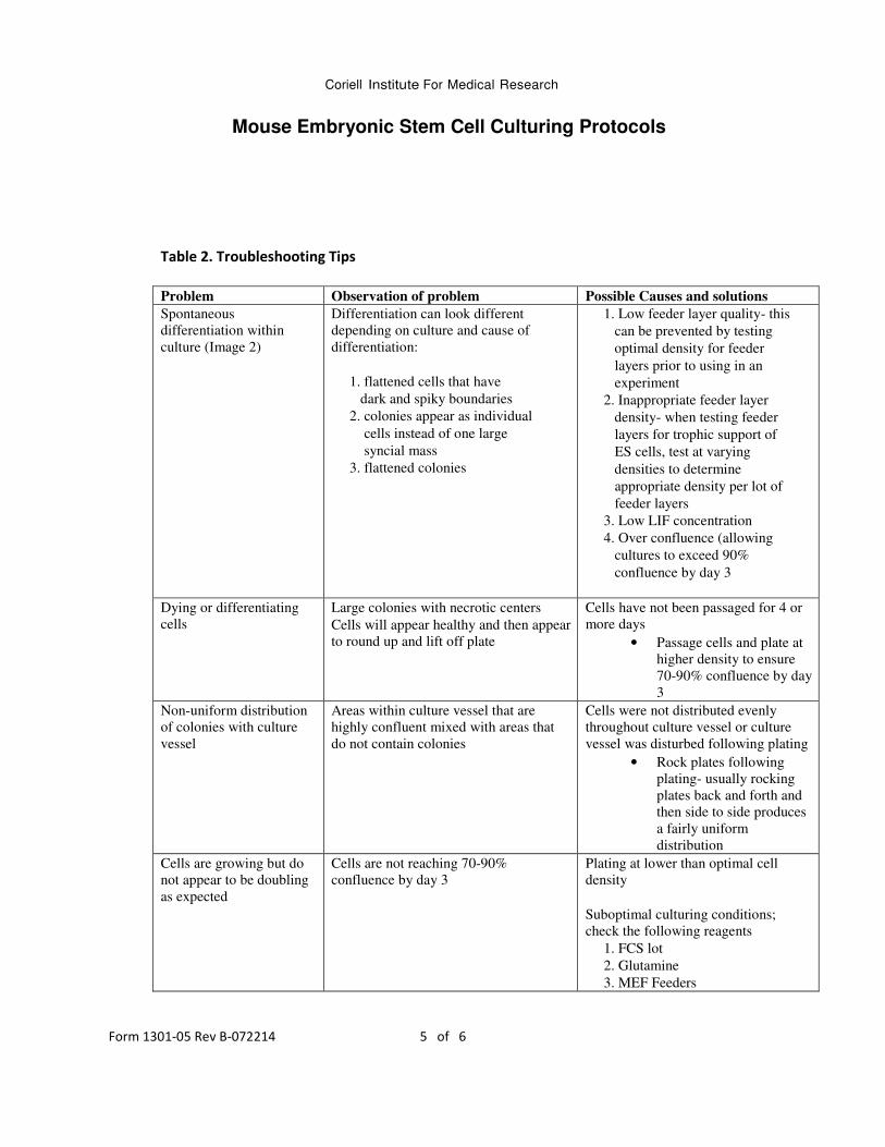
Image 1: mES cultures at 60 to 80% confluence. Note round morphology with distinct borders.
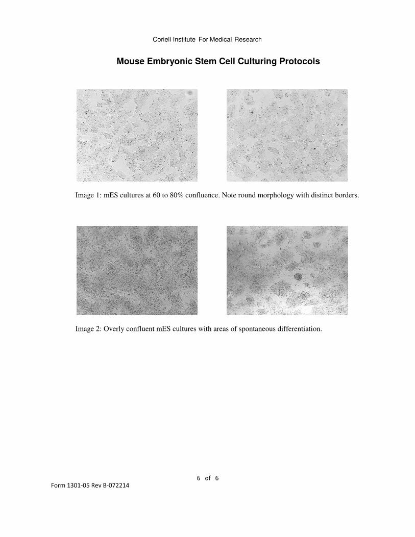
Image 2: Overly confluent mES cultures with areas of spontaneous differentiation.


