American Journal of Computer Science and Technology 2018; 1(1): 1-7
http://www.sciencepublishinggroup.com/j/ajcst
doi: 10.11648/j.ajcst.20180101.11
MR Brain Image Edge Detection Guided with Distribution of Noise and Artifact
Yuchou Chang
Computer Science and Engineering Technology Department, University of Houston-Downtown, Houston, USA
Email address: [email protected]
To cite this article: Yuchou Chang. MR Brain Image Edge Detection Guided with Distribution of Noise and Artifact. American Journal of Computer Science
and Technology. Vol. 1, No. 1, 2017, pp. 1-7. doi: 10.11648/j.ajcst.20180101.11
Received: October 26, 2017; Accepted: November 13, 2017; Published: December 20, 2017
Abstract: Magnetic resonance imaging (MRI) has revolutionized radiology in past four decades. MR image edge detection
can identify anatomy boundaries and extract features for image analysis applications like segmentation and recognition of
anatomy structures. Traditional MR image edge detection methods directly identify discontinuities in MR image domain
without considering distribution of noise and aliasing artifact produced from MR scanner and reconstruction. It is difficult to
suppress effects of noise and aliasing artifact during the edge detection process. In this project, a novel MR brain image edge
detection method is proposed, which is based on parallel MRI reconstruction method. Distribution of noise and aliasing artifact
is characterized by geometry factor map that also guides edge detection process for avoiding detection of noise and aliasing
artifact. A collaborative learning strategy is applied on voting edges for producing the final edge detection. Experimental
results show that the proposed method not only keep anatomy structure boundaries without missing edge components, but also
avoid detection of noise and artifact with wrong edges.
Keywords: Edge Detection, Magnetic Resonance Imaging, Geometry Factor, Canny Edge Detector, Aliasing Artifact
1. Introduction
Edge detection is a basic digital image processing
technique [1]. It contains a variety of mathematical
models to identify points or segments in images where
image intensities change or discontinue sharply. Magnetic
resonance (MR) images [2, 3] enable radiologists to
observe anatomical or physiology structures inside the
human body. A number of MRI pulse sequences [4] can be
grouped to form a protocol with changing magnetic
gradients and other parameters. Different types of
anatomy or physiology tissues display different intensities
on MR images, which are produced by a group of MR
scan protocols. For this reason, MR image edge detection
is not only dependent on traditional digital image
processing techniques, but also related to imaging
protocols and MR physics [5].
MRI noise [2, 3] may deteriorate edge detection
performance, since many edge detection algorithms are
sensitive to noise in the image. Noise can also degrade edge
detection accuracy in MR images. Noise generation in MR
scan is complicated, which is related to electrical current,
coils, and magnetic field. Noise should be considered in MR
image edge detection for suppressing noises and producing
accurate edges. For traditional methods, filtering or
smoothing is applied on detecting edges. It not only
suppresses noises in the MR images, but also blurs anatomy
tissue boundaries. For this reason, it may miss existing edges
among different types of tissues.
Furthermore, aliasing artifact [2, 3] is caused when the
field of view (FOV) is smaller than anatomy part during
imaging. The artifact is wrapped around on the other side of
the image. Aliasing artifact deteriorates edge detection
performance, since its values change abruptly in neighbored
pixels. It is easily detected by edge detection algorithms as
the fake edges in image. Aliasing artifact is not true edges of
different types of tissues, unlike edges around anatomy
structure. Therefore, aliasing artifact should be eliminated,
suppressed, or avoided in edge detection algorithms for MR
images.
In this paper, an edge detection algorithm is proposed for
a parallel MR imaging technique – Sensitivity Encoding
(SENSE) [6], which has been implemented on almost all
commercial MR scanners and widely used for clinical
applications as shown in the Table 1. The method is

2 Yuchou Chang: MR Brain Image Edge Detection Guided with Distribution of Noise and Artifact
different from traditional methods which denoise images to
avoid edge detection accuracy caused by noise degradation.
It also measures locations of aliasing artifact and eliminates
wrong edge detection caused by aliasing artifact. The
proposed method investigates noise generation in a parallel
MR imaging technique and noise distribution characterized
by geometry factor map. The introduction and background
are presented at first. The proposed method will be
illustrated in the third section. Results are evaluated and
presented in the fourth part. Conclusion is given in the final
section.
2. Background
Edge detection algorithms can be divided into gradient and
Laplacian categories. The first category measures minimum
and maximum in the first derivative of pixels in image, and
the second category focuses on the second derivative of
pixels in image. The Canny edge detector was proposed in
1986 [7], which extract structural information in visual
objects. The typical Canny edge detection algorithm contains
the following five steps [7]:
(1). Smoothing image for eliminating noise;
(2). Calculating intensity gradients of pixels;
(3). Applying non-maximum suppression to get rid of
spurious response to edge detection;
(4). Determining potential edges by double thresholds;
(5). Tracking edge by hysteresis.
A number of methods have been proposed for MR
image edge detection. SUSAN and Soble [17] edge
detection were compared in MRI images for feature
extraction [8]. It provided a guide to choose SUSAN or
Soble edge detection in MRI feature extraction. In
addition, optimal threshold is optimized for edge detection
in MR brain images [9]. The adaptive threshold is
determined by Ant Colony Optimization (ACO). The ACO
based threshold outperforms other adaptive method.
Different types of edge detection techniques were
compared for brain MR images [10]. These edge detectors
include Prewitt [17], Canny [7], Laplacian [18], and so
forth. Results show that Canny edge detection technique
extracts edges well from a brain MR image. On the other
hand, edge detectors including Prewitt, Canny, Sobel, and
Laplacian detect only local brightness transition with
varying contrast or strength. From the global perspective
of the entire image, when the threshold is too low, the
detection result using these operators will produce more
incorrect edges due to noise, or the correct edge cannot be
detected when the threshold is too high. A novel algorithm
for edge detection is proposed, in which global constraint
and local contrast information are introduced to solve this
problem [11]. It employed an ensemble of edge detectors
that consider both local and global information. Results
showed that the proposed method outperformed any single
edge detector. This multi-view ensemble is also used in
the proposed method of MR brain image edge detection.
Furthermore, MR image can be produced by fast parallel
MR imaging technique to accelerate imaging speed. SENSE
technique is one of widely used parallel MR image
reconstruction technique. The fast speed of SENSE can make
patients more comfortable and reduce clinical costs.
Although, more noise and aliasing artifacts are produced as a
tradeoff of imaging acceleration in SENSE method, it has
been widely used in clinical applications. Table 1 shows
acronyms [12] of SENSE implementation on five MR
scanner vendors. For arbitrary trajectories, the general
SENSE equation is
Ef = d (1)
, where d is the vector formed from the k-space data across
all channels, f is the full FOV image needed to be solved, and
E is the sensitivity encoding matrix. Using the unfolding
operation, signal is separated for each pixel in the reduce
FOV to produce non-aliased full-FOV image. In addition,
geometry factor, a priori signal-to-noise (SNR) estimates and
an important criterion for designing coil arrays, is provided
by
���� = ���S��S��� �,��S��S��,� (2)
where � is pixel, S is the sensitivity matrix, and Ψ is the
receiver noise covariance matrix. Geometry related noise
enhancement increases rapidly when reduction factor
increases during parallel MR imaging. The proposed method
identifies distribution of noise and aliasing artifacts by using
geometry factor for enhanced edge detection of MR brain
images.
Table 1. Acronyms of SENSE implemented on major MR scanner vendors.
MR Scanner Vendors Acronyms of SENSE Implementation
Siemens mSENSE
General Electric (GE) ASSET
Philips SENSE
Hitachi RAPID
Toshiba SPEEDER
3. Proposed Method
The flowchart of the proposed method is presented in
Figure 1. The proposed edge detection method is based on
SENSE reconstruction and geometry factor map. SENSE
reconstructs undersampled k-space signals faster than
traditional full sampled data acquisition from MR scanners.
Due to production of noise and aliasing artifacts,
conventional edge detection methods identify inappropriate
boundaries as anatomy edges. A multi-view edge detection
strategy is proposed, which uses ensemble learning to detect
distribution of noise and aliasing artifacts from multiple
windows. The details of multi-view mechanism are presented
in the next section.

American Journal of Computer Science and Technology 2018; 1(1): 1-7 3
Figure 1. Flowchart of the proposed method.
3.1. SENSE Reconstruction and Geometry Factor Map
SENSE is a typical image-based parallel MRI
reconstruction method. It makes a un-aliasing process in the
image domain. After inverse Fourier transform of the k-space
data, SENSE reconstructs aliased coil images. In the inverse
Fourier transform process, the field of view (FOV) is reduced
by the 1/R (R is the reduction factor for undersampling k-
space). This means that the same information is contained in
a smaller area which leads to fold over type aliasing artifacts.
Following the equation (1), a brain MR image is
reconstructed by SENSE reconstruction as shown in Figure 2
(d). It is seen that the reconstructed image deteriorates by
noise and aliasing artifacts due to trade-off acceleration by
undersampling signals on k-space. Furthermore, a geometry
factor map is also produced as shown in Figure 1 (e). It can
characterize distribution of noise and aliasing artifacts
presented in SENSE image in Figure 1 (d). The higher values
of geometry factor represents lower SNR in the
corresponding pixel position in the image. The geometry
factor is able to guide noise strength in the reconstructed
SENSE image. The proposed method exploits geometry
factor values for guiding canny edge detector in the image
domain. Noise and aliasing artifacts should not be detected
by edge detector.
3.2. Edge Detection Via Multiple Views Ensemble
The noise level of each pixel on reconstructed MR
images can be characterized by geometry factor value. The
higher value represents noise is stronger and SNR is
lower. Each pixel is considered as the central point of the
local window for edge detection calculation. However,
windows size is generally fixed for different types of edge
detectors. In the proposed method, Canny edge detector is
executed in a window with random size and random pixel
positions for detecting local edges. The proposed
algorithm comes from collaborative learning of knowledge
creation communities. The use of collaborative learning
[19, 20] helps to determine the noise level of each pixel
from multiple perspectives. This is similar to the previous
work on image enhancement collaborative learning via
multiple views [13].
Collaborative learning is firstly studied in education,
which contains a group of approaches involving joint
intellectual effort by students, teachers, and scholars [14].
Learners coordinate and adjust their methods and activities to
enhance the knowledge construction and problem solving
ability. The theory of collaborative learning is extended to
machine learning research. For example, a refinement of
classification hierarchies was proposed to use different kinds
of clustering results for ensemble learning [15]. This method
associates multiple instances of learning methods-clustering
algorithms to achieve an optimally accurate classification.
Inspired by the collaborative learning idea applied in
machine learning research, we propose a MR brain image
edge detection algorithm based on identifying distribution of
noise and aliasing artifacts.
For a reconstructed MR image I with the size of W×H, the
central point of the ith
randomly chosen window is selected as
following.
CenX (i)=rand × W (3)
and
CenY (i)=rand × H (4)
where rand is a random real number generated between 0 and
1. The center of any window can be pixel position in the
image. The width and height of the ith
window are calculated
as following.
( ) 12
2 +
××= WrandiWinW (5)
and
( ) 12
2 +
××= HrandiWinH (6)
�∙� is the operation rounds the element to the nearest
integer of the original value. Note that rand in Equations (3)
to (6) produces a random number for each equation and for
each window. Based on the center (CenX (i), CenY (i)) of the
ith
window, a window S(i)
WinW(i)×WinH(i) can be extracted from
the original image I. For each window whose boundary
exceeds the border of the image I, the portion of the window
outside the original image is cut off.
4 Yuchou Chang: MR Brain Image Edge Detection Guided with Distribution of Noise and Artifact
Since a pixel with higher geometry factor value
represents higher level of noise, the window center pixel’s
geometry factor value serves as an indicator of the local
window noise levels. For a local window whose center
pixel has higher geometry factor value, a higher threshold is
used for Canny edge detection to suppress noise. On the
other hand, for a window whose center pixel has lower
geometry factor value, a lower threshold values is applied
on Canny edge detector for detecting local edges within
each window.
3.3. Edge Detection Ensemble Via Local Detectors’ Voting
Classifier ensembles and clustering ensembles have
been widely studied in machine learning research. Robust
and accurate results can be obtained by ensemble of
individual classifiers or clusters. Voting has been proven
to be effective in many classification or clustering
applications. For example, cumulative voting was
proposed to solve the problem of cluster label alignment
[16, 21]. Accuracy can be improved in compared to using
individual clustering algorithms. The voting is also
applied in the proposed method for detecting true edges
generated from multiple windows.
4. Results
The proposed algorithm is evaluated on two MR brain
image datasets. One is axial brain image dataset and
another one is sagittal brain image dataset. The axial
image dataset contains 8 channels acquired from 8
receiver coils and the sagittal dataset includes 4 channels
acquired from 4 receiver coils. The sensitivity map is
calculated based on the central k-space data, which is fully
sampled. The reduction factor is 4 for undersamplilng k-
space data for both brain image datasets. The number of
windows is set up as 200 and threshold of Canny edge
operator is fixed as 0.05 for both of reference image and
SENSE image. For the proposed method, the threshold of
Canny edge operator ranges from 0.05 to 0.3. Each
window’s threshold is determined by geometry factor
value on the center pixel of that window.
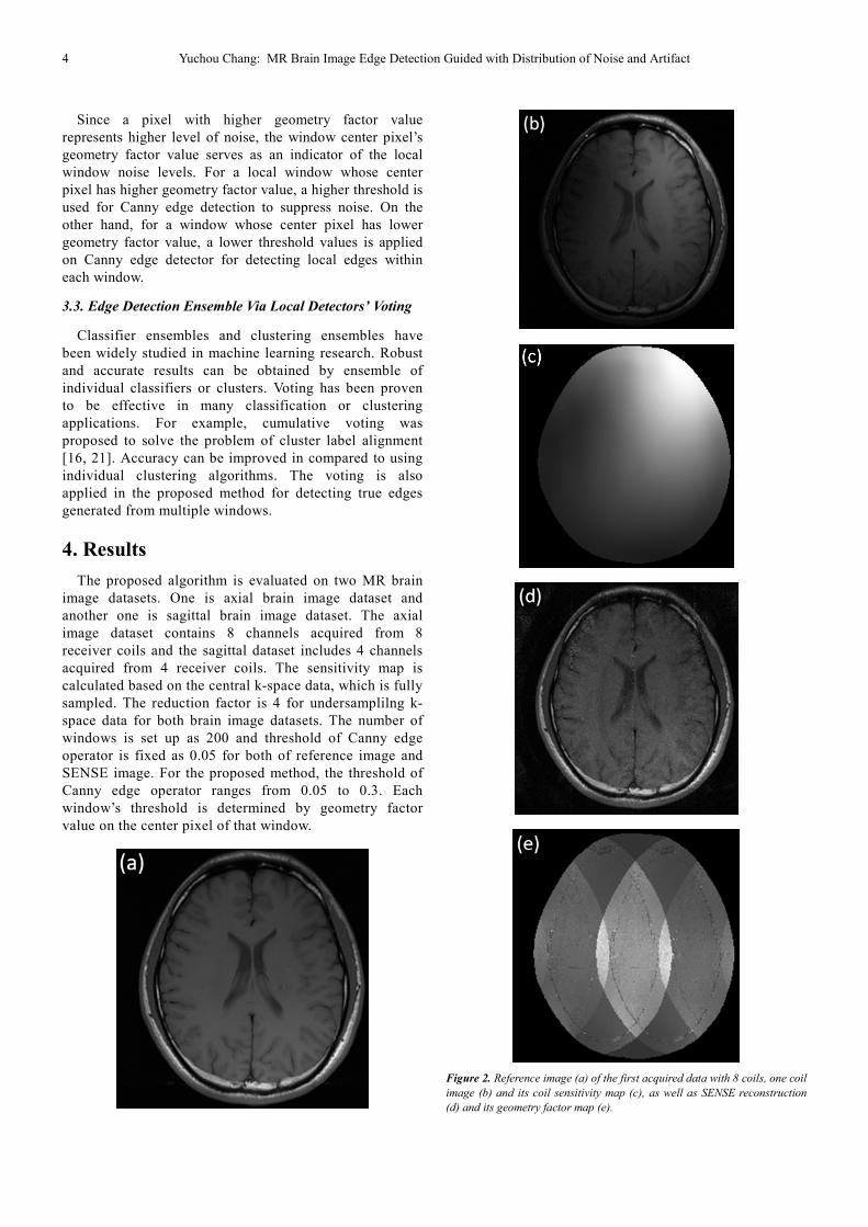
Figure 2. Reference image (a) of the first acquired data with 8 coils, one coil
image (b) and its coil sensitivity map (c), as well as SENSE reconstruction
(d) and its geometry factor map (e).

American Journal of Computer Science and Technology 2018; 1(1): 1-7 5
The reference is calculated from traditional canny edge
detection on fully sampled reconstruction. As shown in the
Figure 2 (a), the reference image has few noise and aliasing
artifacts. It is fully sampled but required longer acquisition
time on MR scanner. One coil image and its coil sensitivity
map is presented in Figure 2 (b) and (c). The coil sensitivity
produces noise and aliasing artifacts in the reconstructed
SENSE image, thereby reducing image quality. It can be seen
that edge detection on the reference image can detect most of
the boundaries between white matter, gray matter and
cerebrospinal fluid (CSF). Traditional canny detector is also
applied on brain MR image with noise and aliasing artifacts,
which is reconstructed by SENSE. The corresponding
geometry factor map indicates distribution of noise and
aliasing artifacts. The traditional canny detector on SENSE
image presents contradiction of missing edges and wrong
edges. As shown in Figure 3 (c), brain boundaries indicated
by green arrow are missed by canny detector, but aliasing
artifact indicated by red arrow is wrongly detected. For the
proposed algorithm, aliasing artifact is suppressed without
detection in Figure 3 (d). It is closer to Figure 3 (b) of edge
detection on reference image without noise and aliasing
artifact. Furthermore, brain boundaries are detected correctly,
which are similar to edges detected in the reference image.
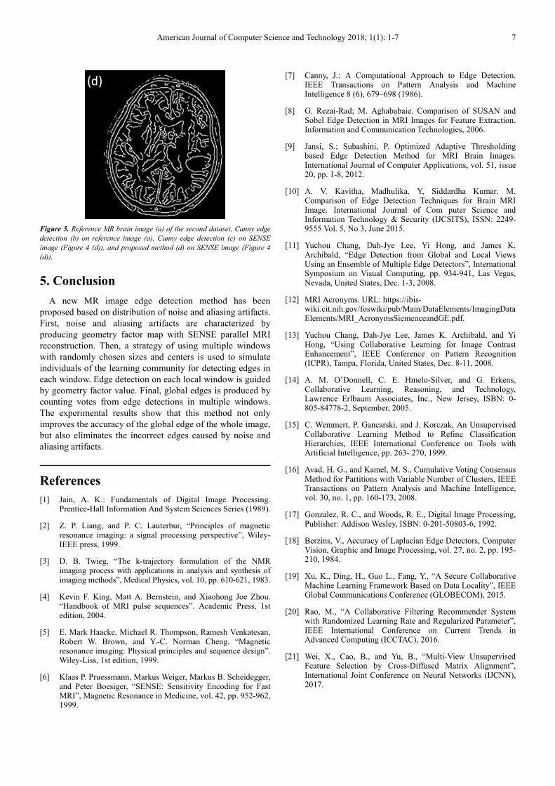
Figure 3. Reference MR brain image (a) of the first dataset, Canny edge
detection (b) on reference image (a), Canny edge detection (c) on SENSE
image (Figure 2 (d)), and proposed method (d) on SENSE image (Figure 2
(d)).
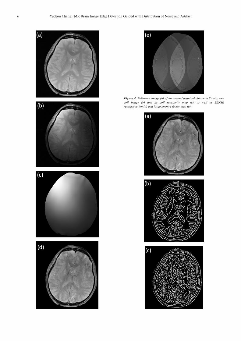
The proposed method is applied on another MR brain image
as shown in Figure 4. The reference is also calculated from the
traditional canny edge detection of the complete sampling
reconstruction in Figure 4 (a). As shown in Figure 4 (a), the
reference image has little noise and aliasing artifacts since it is
fully sampled. A coil image and its coil sensitivity diagram
shown in Figure 4 (b) and (c). The SENSE image and its
geometry factor map are shown in Figure 4 (d) and (e). It can
be seen that edge detection on the reference image can detect
most of the margins between anatomy structures. Conventional
canny detectors are also applied to brain MR images with
noise and aliasing artifacts reconstructed by SENSE. The
corresponding geometric factor assists edge detection of the
proposed method. SENSE images on the traditional canny
detectors exist missing edges and edges of the error. As shown
in Figure 5, the proposed method (Figure 5 (d)) avoids noise
and aliasing artifacts such that the edges cause the brain
boundary to be correctly detected, which is similar to the edge
detected in the reference image (Figure 5 (b)). On the other
hand, as shown in Figure 5 (c), the conventional canny
operator cannot suppress noise and aliasing artifacts, thus
producing more extraneous edges.
6 Yuchou Chang: MR Brain Image Edge Detection Guided with Distribution of Noise and Artifact
Figure 4. Reference image (a) of the second acquired data with 8 coils, one
coil image (b) and its coil sensitivity map (c), as well as SENSE
reconstruction (d) and its geomentry factor map (e).
American Journal of Computer Science and Technology 2018; 1(1): 1-7 7
Figure 5. Reference MR brain image (a) of the second dataset, Canny edge
detection (b) on reference image (a), Canny edge detection (c) on SENSE
image (Figure 4 (d)), and proposed method (d) on SENSE image (Figure 4
(d)).
5. Conclusion
A new MR image edge detection method has been
proposed based on distribution of noise and aliasing artifacts.
First, noise and aliasing artifacts are characterized by
producing geometry factor map with SENSE parallel MRI
reconstruction. Then, a strategy of using multiple windows
with randomly chosen sizes and centers is used to simulate
individuals of the learning community for detecting edges in
each window. Edge detection on each local window is guided
by geometry factor value. Final, global edges is produced by
counting votes from edge detections in multiple windows.
The experimental results show that this method not only
improves the accuracy of the global edge of the whole image,
but also eliminates the incorrect edges caused by noise and
aliasing artifacts.
References
[1] Jain, A. K.: Fundamentals of Digital Image Processing. Prentice-Hall Information And System Sciences Series (1989).
[2] Z. P. Liang, and P. C. Lauterbur, “Principles of magnetic resonance imaging: a signal processing perspective”, Wiley-IEEE press, 1999.
[3] D. B. Twieg, “The k-trajectory formulation of the NMR imaging process with applications in analysis and synthesis of imaging methods”, Medical Physics, vol. 10, pp. 610-621, 1983.
[4] Kevin F. King, Matt A. Bernstein, and Xiaohong Joe Zhou. “Handbook of MRI pulse sequences”. Academic Press, 1st edition, 2004.
[5] E. Mark Haacke, Michael R. Thompson, Ramesh Venkatesan, Robert W. Brown, and Y.-C. Norman Cheng. “Magnetic resonance imaging: Physical principles and sequence design”. Wiley-Liss, 1st edition, 1999.
[6] Klaas P. Pruessmann, Markus Weiger, Markus B. Scheidegger, and Peter Boesiger, “SENSE: Sensitivity Encoding for Fast MRI”, Magnetic Resonance in Medicine, vol. 42, pp. 952-962, 1999.
[7] Canny, J.: A Computational Approach to Edge Detection. IEEE Transactions on Pattern Analysis and Machine Intelligence 8 (6), 679–698 (1986).
[8] G. Rezai-Rad; M. Aghababaie. Comparison of SUSAN and Sobel Edge Detection in MRI Images for Feature Extraction. Information and Communication Technologies, 2006.
[9] Jansi, S.; Subashini, P. Optimized Adaptive Thresholding based Edge Detection Method for MRI Brain Images. International Journal of Computer Applications, vol. 51, issue 20, pp. 1-8, 2012.
[10] A. V. Kavitha, Madhulika. Y, Siddardha Kumar. M. Comparison of Edge Detection Techniques for Brain MRI Image. International Journal of Com puter Science and Information Technology & Security (IJCSITS), ISSN: 2249-9555 Vol. 5, No 3, June 2015.
[11] Yuchou Chang, Dah-Jye Lee, Yi Hong, and James K. Archibald, “Edge Detection from Global and Local Views Using an Ensemble of Multiple Edge Detectors”, International Symposium on Visual Computing, pp. 934-941, Las Vegas, Nevada, United States, Dec. 1-3, 2008.
[12] MRI Acronyms. URL: https://ibis-wiki.cit.nih.gov/foswiki/pub/Main/DataElements/ImagingDataElements/MRI_AcronymsSiemenceandGE.pdf.
[13] Yuchou Chang, Dah-Jye Lee, James K. Archibald, and Yi Hong, “Using Collaborative Learning for Image Contrast Enhancement”, IEEE Conference on Pattern Recognition (ICPR), Tampa, Florida, United States, Dec. 8-11, 2008.
[14] A. M. O’Donnell, C. E. Hmelo-Silver, and G. Erkens, Collaborative Learning, Reasoning, and Technology, Lawrence Erlbaum Associates, Inc., New Jersey, ISBN: 0-805-84778-2, September, 2005.
[15] C. Wemmert, P. Gancarski, and J. Korczak, An Unsupervised Collaborative Learning Method to Refine Classification Hierarchies, IEEE International Conference on Tools with Artificial Intelligence, pp. 263- 270, 1999.
[16] Avad, H. G., and Kamel, M. S., Cumulative Voting Consensus Method for Partitions with Variable Number of Clusters, IEEE Transactions on Pattern Analysis and Machine Intelligence, vol. 30, no. 1, pp. 160-173, 2008.
[17] Gonzalez, R. C., and Woods, R. E., Digital Image Processing, Publisher: Addison Wesley, ISBN: 0-201-50803-6, 1992.
[18] Berzins, V., Accuracy of Laplacian Edge Detectors, Computer Vision, Graphic and Image Processing, vol. 27, no. 2, pp. 195-210, 1984.
[19] Xu, K., Ding, H., Guo L., Fang, Y., “A Secure Collaborative Machine Learning Framework Based on Data Locality”, IEEE Global Communications Conference (GLOBECOM), 2015.
[20] Rao, M., “A Collaborative Filtering Recommender System with Randomized Learning Rate and Regularized Parameter”, IEEE International Conference on Current Trends in Advanced Computing (ICCTAC), 2016.
[21] Wei, X., Cao, B., and Yu, B., “Multi-View Unsupervised Feature Selection by Cross-Diffused Matrix Alignment”, International Joint Conference on Neural Networks (IJCNN), 2017.


