NEW PERSPECTIVES OF INTRAOPERATIVE
US GUIDANCE Enzo Durante
Head General Surgery Unit University of Ferrara, Italy
I n te r n a tio n a l B r e a s t Ultr a s o u n d S c h o o l F o u n d in g M e m b e r a n d S e c r e ta r y

In the second century B.C. Leonidas of Alexandria established the identity breast cancer as typical neoplasia of
female sex and described nipple retraction as sign of breast cancer
He also described treatment with breast excision and cauterization for better
restrain the haemorrage. Moreover He recommended to cauterize tissues for
eradication of disease

• Early diagnosis and echographic staging as fundamental step for adequate conservative surgery in the treatment of breast carcinoma.
Argomenti di Chirurgia 1-2, 11:117-130, 1982.
• Surgical echography as diagnostic and staging tool in breast pathology.
Thoracic Surgery , Monduzzi Ed., 1988, pp.301-308.
Background of our experience based on the lobar anatomy and radial
echographic scanning

We must stress the concept for breast surgery:
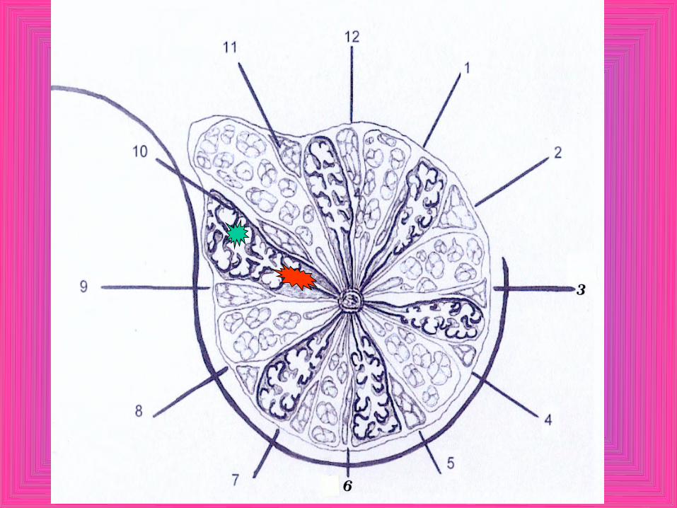
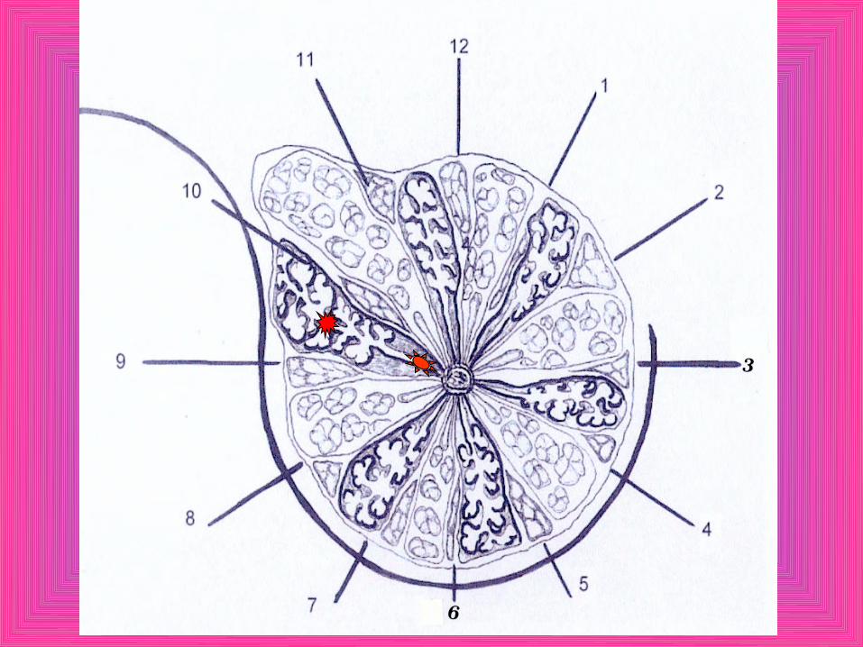
• Breast composed of 15-20 lobes as many as the ducts
• each lobe is a sector or a segment
• major ducts come from periphery to the nipple
BREAST DISEASES ARE DISEASES OF DUCTAL SYSTEM

More recently the principle of lobar disease has been recognized by others authors

Primary objective of breast surgeons is to remove lesions with adequate margins but in radical way and preserving the patient’s
aesthetic good looking.
Another objective for the surgeons is to perform surgical intervention and axillary
staging in one single definitive procedure.
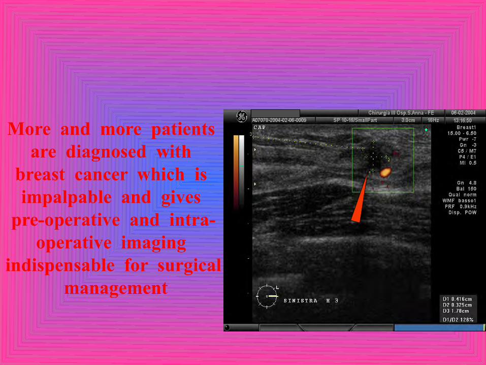
More and more patients are diagnosed with
breast cancer which is impalpable and gives
pre-operative and intra-operative imaging
indispensable for surgical management

The size of tumor plays a critical role in determining both the stage and the treatment
of breast cancer
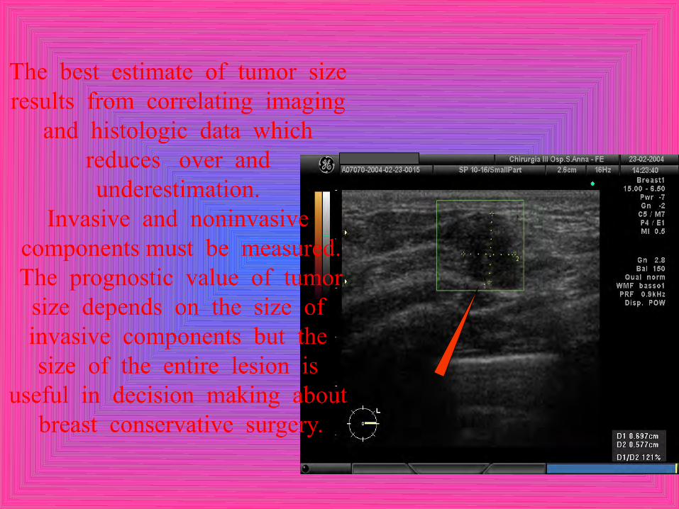
The best estimate of tumor size results from correlating imaging
and histologic data which reduces over and underestimation.
Invasive and noninvasive components must be measured.
The prognostic value of tumor size depends on the size of invasive components but the size of the entire lesion is
useful in decision making about breast conservative surgery.

Breast US is a valuable tool for cancer staging but an adequate training is mandatory. Trained physician
should be allowed and encouraged to use this technique without arbitrary limitations due to medical
specialty
SMALL DUCTAL I CA + DCIS
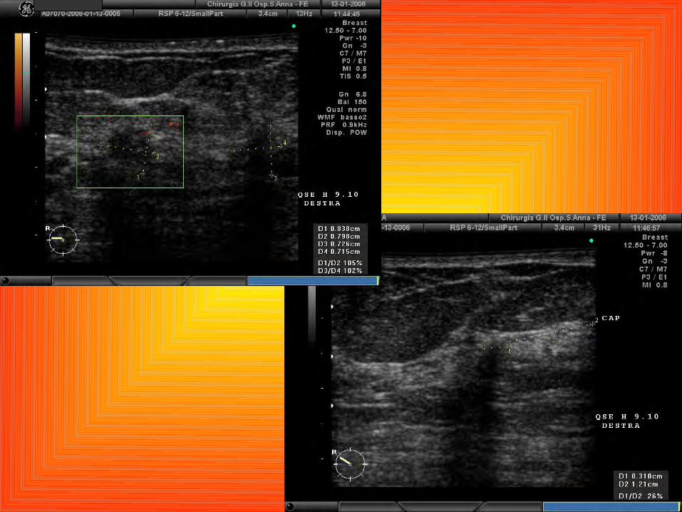
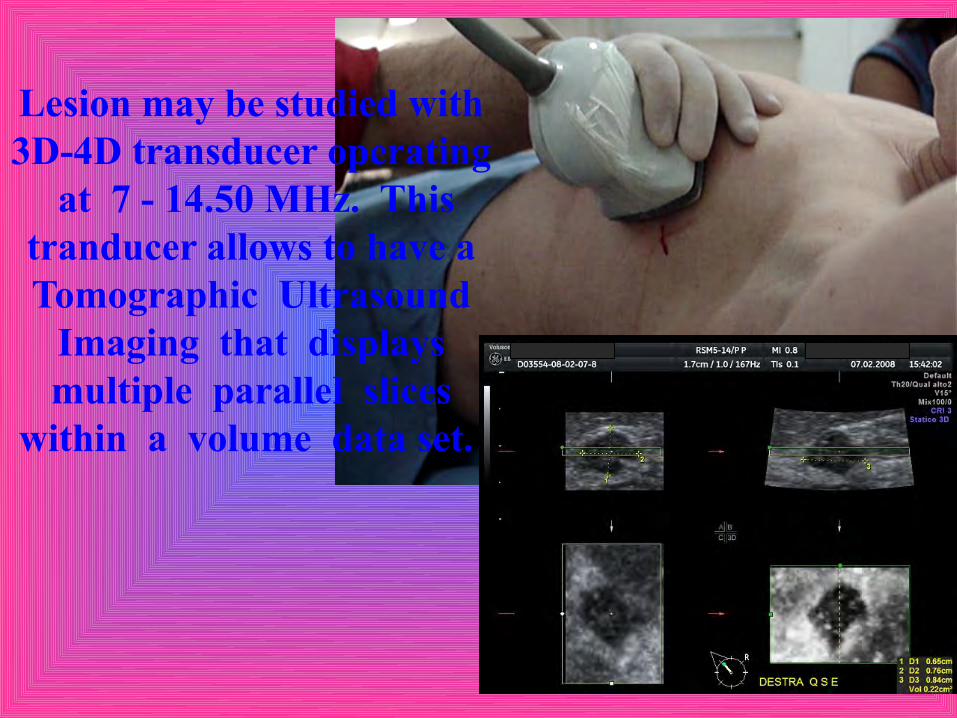
Lesion may be studied with 3D-4D transducer operating
at 7 - 14.50 MHz. This tranducer allows to have a Tomographic Ultrasound
Imaging that displays multiple parallel slices
within a volume data set.

TUI is important tool in order to reduce the resection of healthy tissue
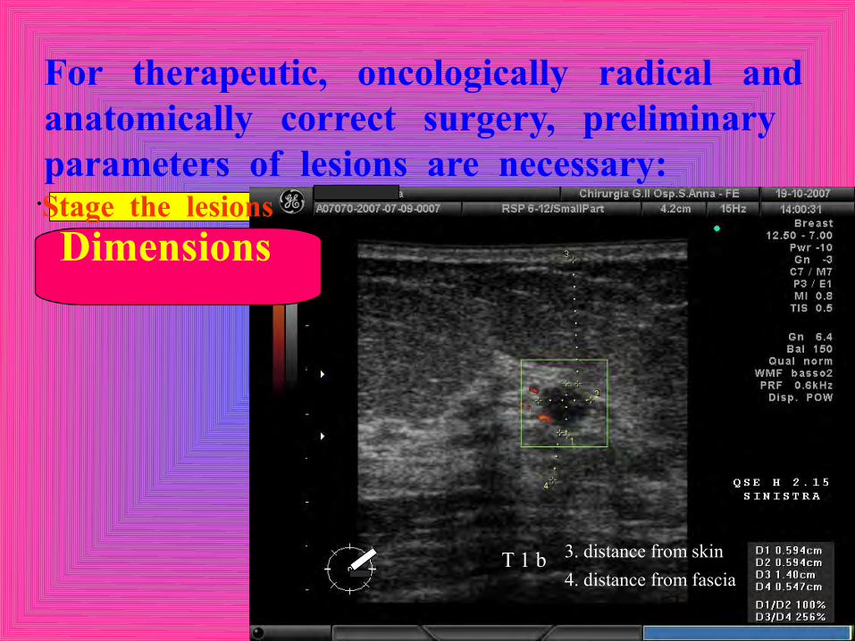
For therapeutic, oncologically radical and anatomically correct surgery, preliminary parameters of lesions are necessary:
Dimensions
T 1 b 3. distance from skin4. distance from fascia
•Stage the lesions

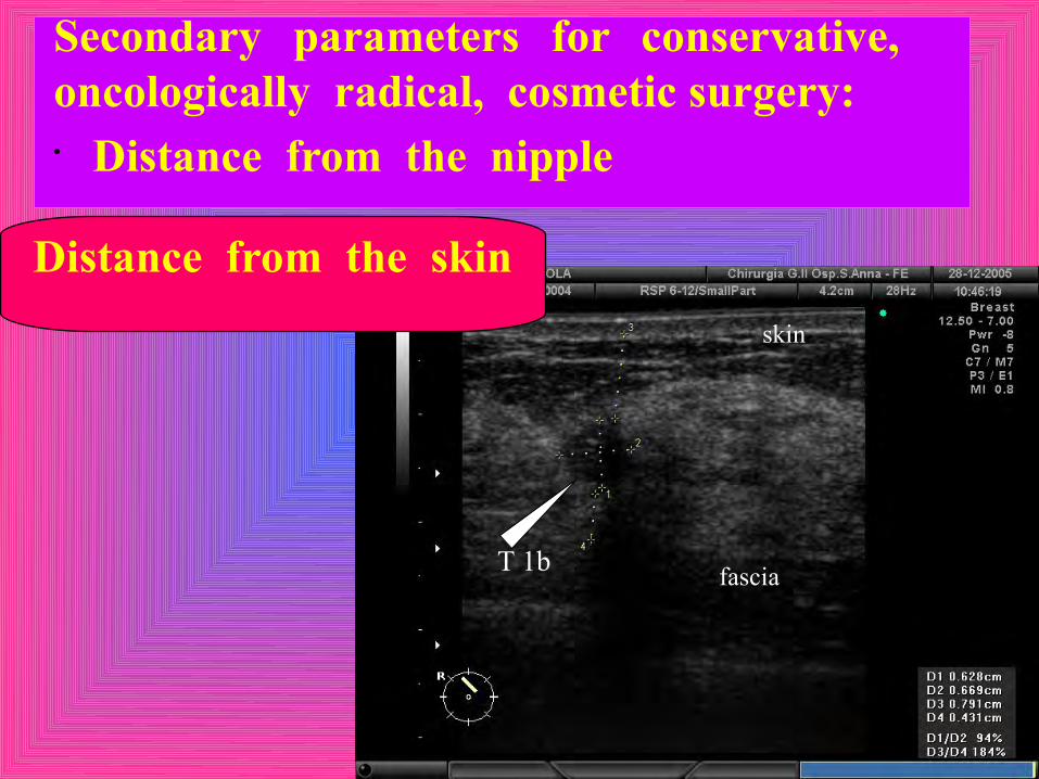
Secondary parameters for conservative, oncologically radical, cosmetic surgery :
Distance from the nipple
Nipple
1. Distance from
Secondary parameters for conservative, oncologically radical, cosmetic surgery:• Distance from the nipple
Distance from the skin skin
fasciaT 1b

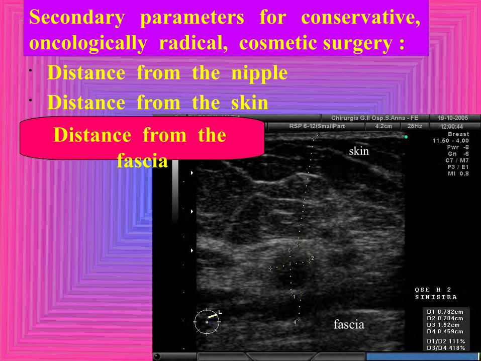
Secondary parameters for conservative, oncologically radical, cosmetic surgery :• Distance from the nipple• Distance from the skin
ID extension
skin

Secondary parameters for conservative, oncologically radical, cosmetic surgery :• Distance from the nipple• Distance from the skin
Next to the skin
When the skin is very near to the tumor we must remove the skin in front of the tumor using mostly a double curvilinear incision according the Langer lines.
Secondary parameters for conservative, oncologically radical, cosmetic surgery :• Distance from the nipple• Distance from the skin
Distance from the fascia skin
fascia

Secondary parameters for conservative, oncologically radical, cosmetic surgery :• Distance from the nipple• Distance from the skin• Distance from the pectoralis fascia
Pectoralis fascia is a different anatomic entity from the deeper layer of superficial fascia that
envelop the breast tissue. Behind this there is a retromammary fat
layer and than the pectoralis fascia
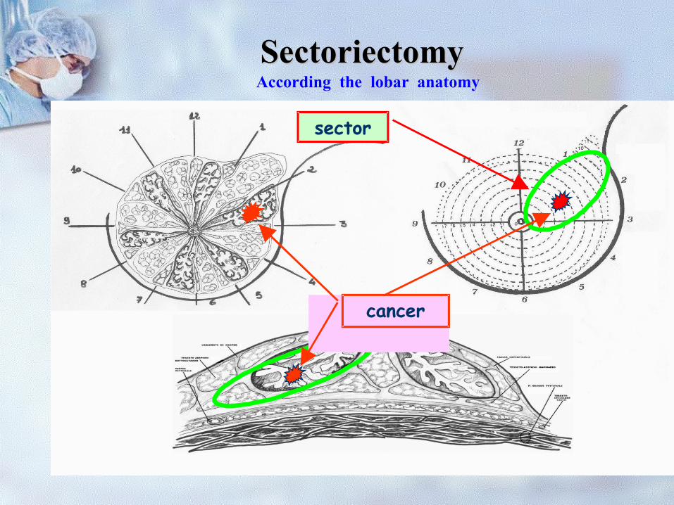
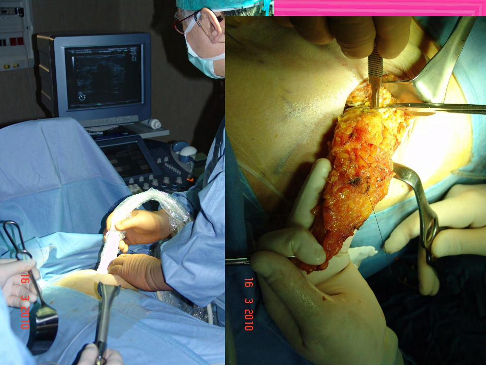
SectoriectomySectoriectomy
sector
cancer
According the lobar anatomy

Intraoperative US guided localization of non-palpable lesions is a method of
choice

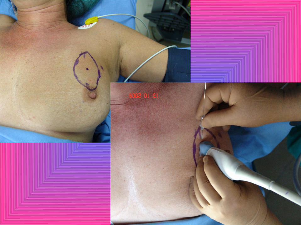


US sentinel node is localized at the beginning of surgical procedure by
hook-wire

Surgical planning is based on the echographic assessment of lesion and adjacent tissue in radial scans with multifrequency transducer ope- rating at 8-18MHz and with 3D-4D scans with transducer operating at 7.10-14.50 MHz. we draw on the skin the extension of the lobe and plan the most advantageous incision always according the Langer lines and the resection of breast tissue according the lobar anatomy described by Craig and Towsend.
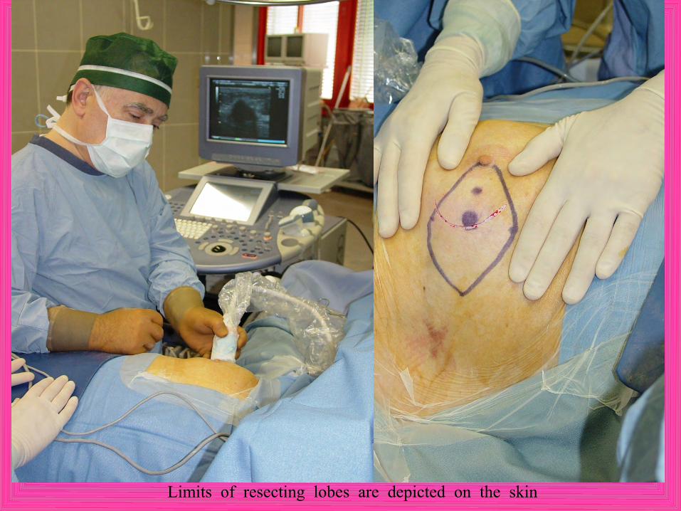
Limits of resecting lobes are depicted on the skin

Skin incision is made by curvilinear incision according the Langer’s lines parallel to the
periareolar line

Single or double curvilinear incision depends on the distance of tumor from the skin. If the tumor is far from the skin more than 5mm and the superficial layer of the
superficial fascia is free of distortion or disruption we don’t remove the skin so that we perform a single curvilinear incision and always when it is possible we perform a peri-areolar incision even if this requires more time to dissect the tissue until the periphery.

If the tumor is near to the skin or the fascia superficial layer is altered we perform a double curvilinear incision and remove
the skin in front of the tumor.

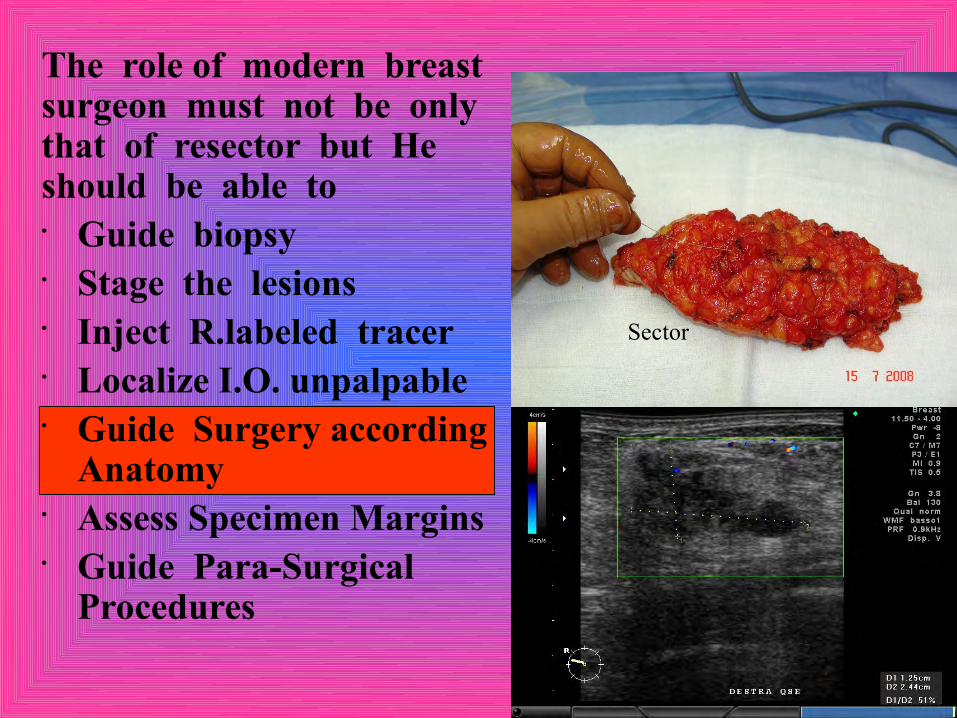
The role of modern breast surgeon must not be only that of resector but He should be able to• Guide biopsy• Stage the lesions• Inject R.labeled tracer• Localize I.O. unpalpable• Guide Surgery according
Anatomy• Assess Specimen Margins• Guide Para-Surgical
Procedures
Sector
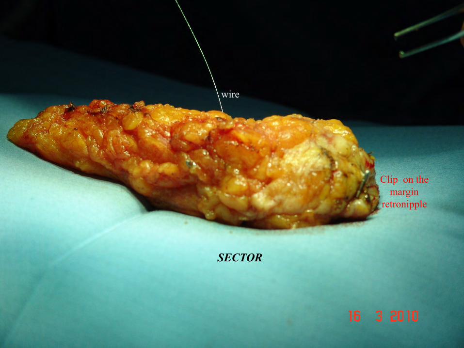
SECTOR
wire
Clip on the margin
retronipple

An US image of the resected specimen immediately allows the surgeon to visualize the presence of lesion, the adequate lateral margins that may benefit, eventually, from immediate reexcision. This does not exclude the option of specimen radiography that we perform with
Faxitron equipment side to the operating room in case of microcalcifications.
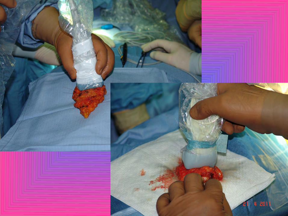
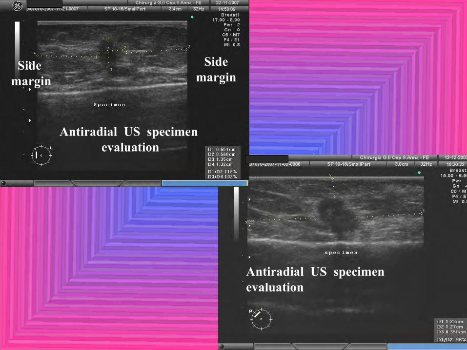
Antiradial US specimen evaluation
Side margin
Side margin
Antiradial US specimen evaluation

The role of modern breast surgeon must not be only that of resector but He should be able to• Guide biopsy• Stage the lesions• Inject R.labeled tracer• Localize I.O. unpalpable• Guide Surgery according
Anatomy• Assess Specimen Margins• Guide Para-Surgical
Procedureswith Faxitron in case of microcalcifications or clip
after VAB
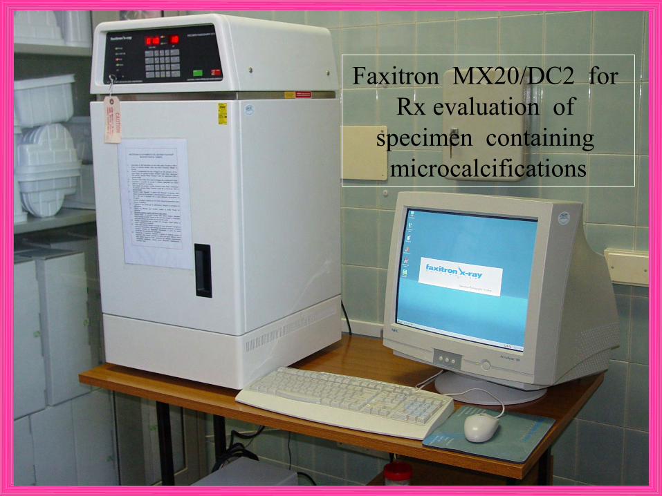
Faxitron MX20/DC2 for Rx evaluation of
specimen containing microcalcifications

If indicated by specimen US or X-Ray the lateral margins are extended
intraoperatively but in our experience this comes very few times only for
diffuse microcalcifications
Residual microcalcifications near to
the margin Clip after VAB
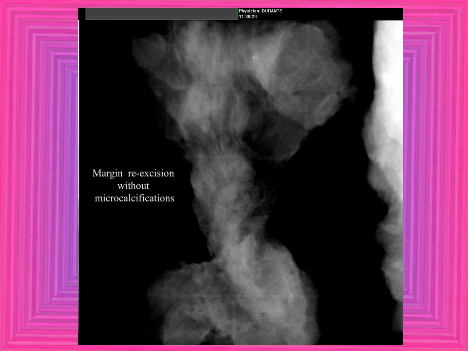
Margin re-excision without
microcalcifications

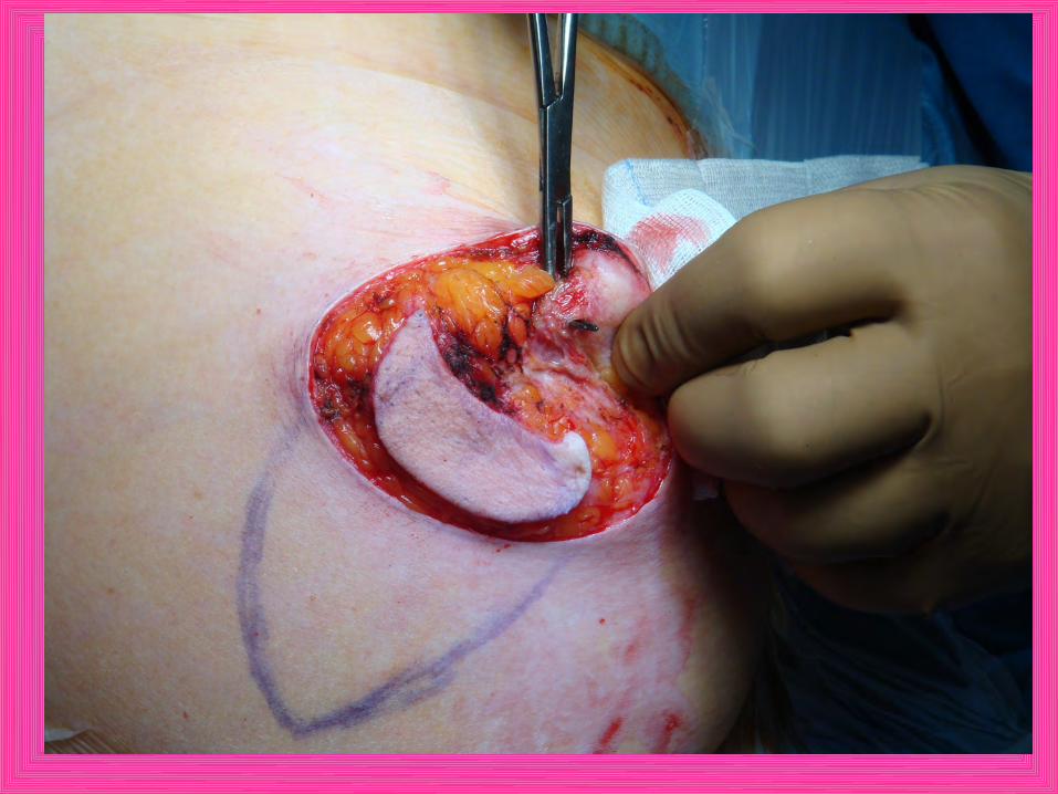
Surgical bed of resected sector is US evaluated by 8-18 MHz transducer

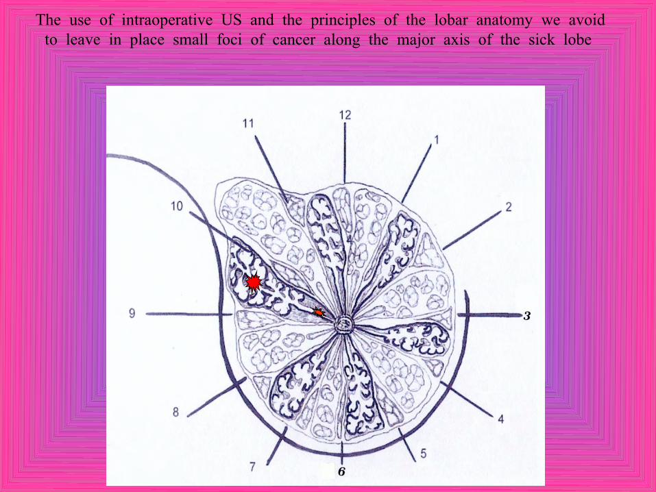
The use of intraoperative US and the principles of the lobar anatomy we avoid to leave in place small foci of cancer along the major axis of the sick lobe
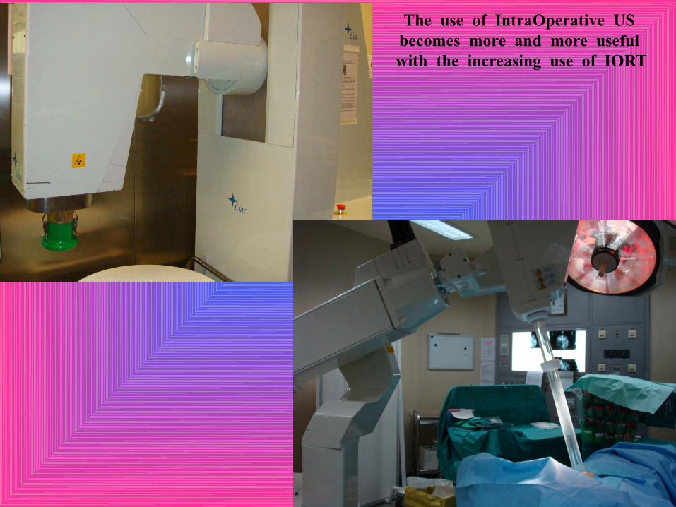
The use of IntraOperative US becomes more and more useful with the increasing use of IORT
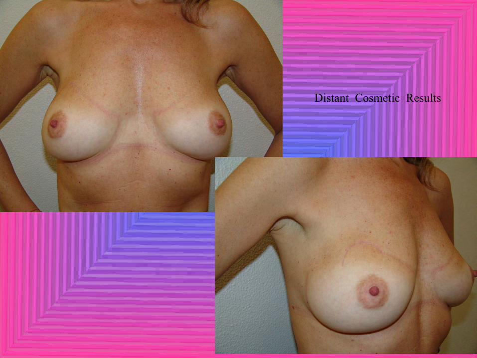
Distant Cosmetic Results

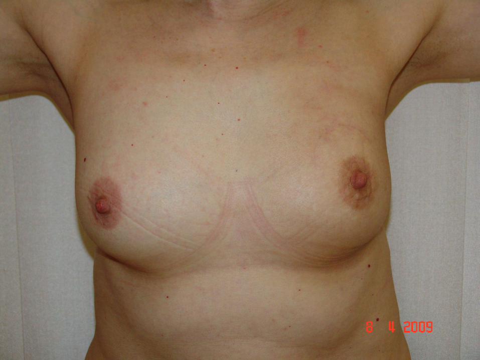
Distant Cosmetic Results

Distant Cosmetic Results
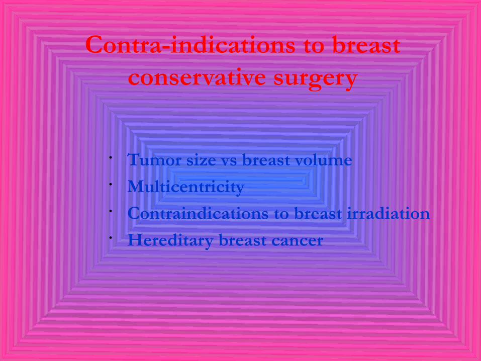
Contra-indications to breast conservative surgery
• Tumor size vs breast volume• Multicentricity• Contraindications to breast irradiation• Hereditary breast cancer
Fare clic per modificare lo stile del sottotitolo dello schema
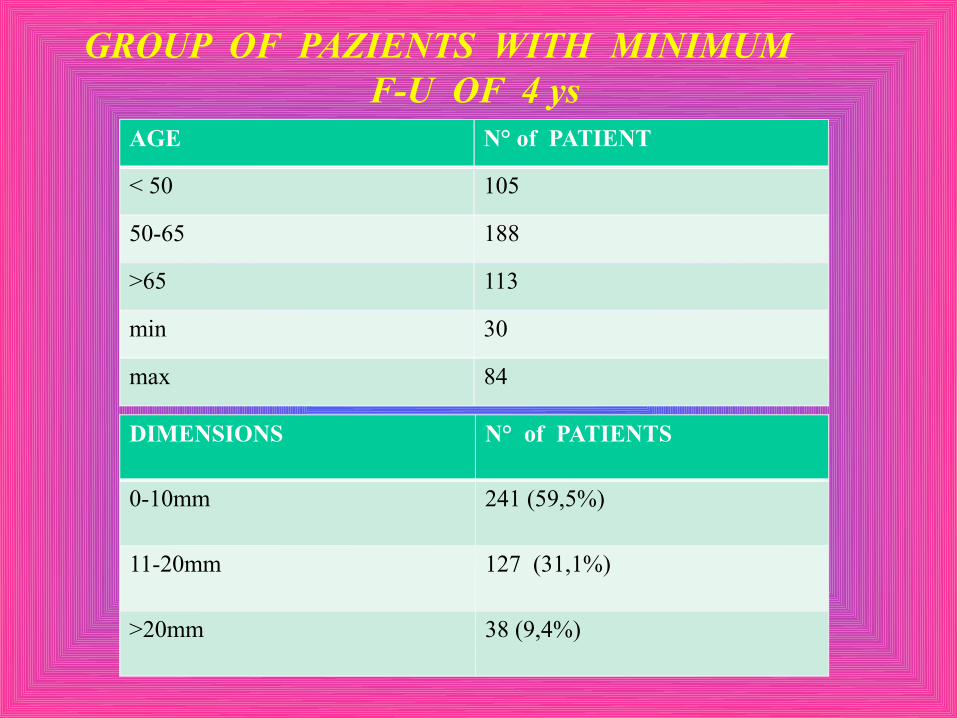
AGE N° of PATIENT
< 50 105
50-65 188
>65 113
min 30
max 84
DIMENSIONS N° of PATIENTS
0-10mm 241 (59,5%)
11-20mm 127 (31,1%)
>20mm 38 (9,4%)
GROUP OF PAZIENTS WITH MINIMUM F-U OF 4 ys

Fare clic per modificare lo stile del sottotitolo dello schema
HISTOLOGY IN THE SAME GROUP
INF. CA
DCIS

RISULTATIRecidive con un minimo di 3 anni di Follow Up
0
5
10
RESULTS
RECURRENCES IN ONE GROUP WITH MINIMUM F-U OF 4 ys

• Indipendent planning of surgery according the lobar anatomy
• IO Localization• Absence of needle dislocation• Precise planning of incision • Better anatomic orientation
• Less resection of breast tissue• Less reintervention for axillary dissection (6%)
• Fewer recurrences (<1% absolute in 21 ys f-u)• Less hospitalization (24 hours)
• Patient return sooner to a normal lifestyle• Cosmesis is improved
• Better ratio cost/benefit
ADVANTAGES OF US GUIDED SURGERY
Surgical ultrasound in breast is still underused even if the high-end equipment
used in the operating room is able to visualize the anatomy and architecture, to accurately localize lesions and guide the
better planning
Ultrasound technology migrates very quickly and surgeons should be able to get the innovations for better treat an
increasing number of patient
Surgeons should be well-educated and opened to a relatively innovative use of
ultrasound in the operating room for breast surgery

INTERNATIONAL BREAST ULTRASOUND COURSE
September 7 - 10, 2011
http://www.ibus.org
FERRARA, ITALY


