Download - Newsletter Spring 2014 - FAU

newsLetter Fall spring 2014
Dear colleagues and friends,
An Interdiscipl inary Center of the Friedrich-Alexander-Universität Erlangen-Nürnberg
Editorial
On March 14, which was Paul Ehrlich’s 160th birthday, Michael Reth, Professor of Molecular Immunology at the Albert-Ludwig-University and research group leader at the Max Planck Institute for Immunobiology and Epigenetics in Freiburg, received the Paul Ehrlich and Ludwig Darmstaedter-Prize 2014. The prize, which is worth € 100,000 and sponsored by the Paul-Ehrlich Foundation, the Federal Ministry of Health and by donations from companies, was
awarded to Michael Reth for his fundamental contributions in anti-body and B cell research. Michael Reth discovered the “immunore-ceptor tyrosine activation motif” (ITAM). He and his group also proved that the activation of B cells via their receptor does not result from the aggregation of individual B cell receptor molecules, but rather from the dissociation of B cell receptor aggregates. As spokesman of the excellence cluster BIOSS in Freiburg Michael Reth has been the driving force for a research approach termed “synthetic biology”, which aims to understand signalling events by engineering receptors and signalling molecules in model organisms. Michael Reth is a member of the Transregio Collaborative Research Center 130 on B cell immunity and autoimmunity run by the universities of Erlangen, Freiburg and Berlin and has long-lasting scientific relationships with several immunologists at our university. We therefore wish to express our heartiest congratulations to him for this well-deserved honour.
Recently, Georg Schett and Markus Neurath have submitted a pre-application for a new Collaborative Research Center (CRC) to the DFG. The focus of the consortium, which consists of 25 researchers from 12 institutions at the UKER and the FAU and contains 19 re-search proposals and a central imaging platform, will be the analysis of molecular mechanisms underlying the chronification and resolution of inflammation in mice and man. On April 16, the application will be evaluated by a panel of reviewers in Bonn and discussed to-gether with a delegation of researchers from Erlangen. We all hope that this new CRC initiative will receive a positive vote so that a full application can be handed in.
A couple of weeks ago, members of the steering committee of the Medical Immunology Campus Erlangen met together with Jochen Kalden and Martin Röllinghoff in the “Kulisse” to discuss a proposal by Hans-Martin Jäck that Erlangen should consider to apply to be the organizer of the Annual Conference of the German Society of Immunology (DGFI) in 2017, the year of the 50th anniversary of the DGFI. After a fruitful exchange of ideas and a critical evaluation of aspects of reputation, science, infrastructure and geographic location we came to the conclusion that Erlangen meets all principal requirements for such an application. The respective bid will have to be compiled by the end of this summer. In case of success, it would be the first time ever that Erlangen hosts a DGFI annual conference.
prof. Christian BogdanChairman of the Medical Immunology Campus Erlangen
NewsletterSpring 2014C O n t e n t
Scientific Highlights p. 2 – 4
•Fra-2 expression in Osteoblasts regulates metabolism
•On the molecular mechanism of a novel glycolipid adjuvant
NEW! Methods p. 5
•Generation of moDCs and lymphocytes from LRSCs
People p. 6
•PD Dr. Dimitrios Mougiakakosr Funding by the European Hematology Association (EHA)
• Prof. Dr. Michael Stürzl Support by Wolfgang-Lutz-Foundation
• Prof. Dr. Jens Titze Awards by two separate societies
News and Updates p. 7
•Joachim Kalden Lecture 2013
Upcoming Events p. 8
controls Fosl2 ob-tet
BV/TV TbTh TbN NOb ObS/BS NOc OcS/BS
36.19±0.88***47.17±1.54***3.72±0.03***29.91±1.4131.35±1.17***0.72±0.05*1.82±0.09*
19.08±0.3037.57±0.402.90±0.0728.08±1.6222.66±0.521.36±0.053.18±0.10 controls Fosl2 ob-tet
0
0.01
0.02
0.03
0.04
0.05
Fat p
ad/b
ody
weig
ht
ND HFD0
10
20
30
40
Bod
y w
eigh
t (g)
*** ***
* **
*** ***
*
ND HFD
a b
SciEntific HigHligHtS
Fra-2 expression in Osteoblasts regulates metabolism
Osteoblast-specific expression of Fra-2/AP-1 affects metabo-lism by regulating adiponectin and osteocalcin
alinE BOZECDEparTMEnT OF MEDiCinE 3, UniVErsiTÄTsKliniKUM ErlangEn
spring 2014 2
… genetic manipulation of Fra-2 in osteoblasts
specifically affects the bone-forming
activity of the osteo-blast lineage which regulates endocrine
function …
…. Adipoq expression is controlled by Fra-2 in a cell-autonomous
manner in osteoblasts …
… osteoblast-specific Fra-2 expression can
regulate Adipoq produced by osteoblasts,
which contributes to the defects in
glucose and insulin handling. …
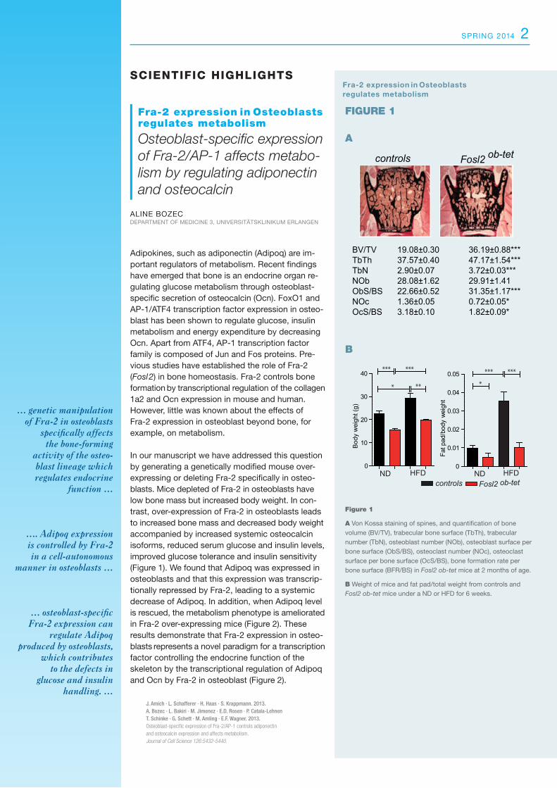
Adipokines, such as adiponectin (Adipoq) are im-portant regulators of metabolism. Recent findings have emerged that bone is an endocrine organ re-gulating glucose metabolism through osteoblast-specific secretion of osteocalcin (Ocn). FoxO1 and AP-1/ATF4 transcription factor expression in osteo-blast has been shown to regulate glucose, insulin metabolism and energy expenditure by decreasing Ocn. Apart from ATF4, AP-1 transcription factor family is composed of Jun and Fos proteins. Pre-vious studies have established the role of Fra-2 (Fosl2) in bone homeostasis. Fra-2 controls bone formation by transcriptional regulation of the collagen 1a2 and Ocn expression in mouse and human. However, little was known about the effects of Fra-2 expression in osteoblast beyond bone, for example, on metabolism.
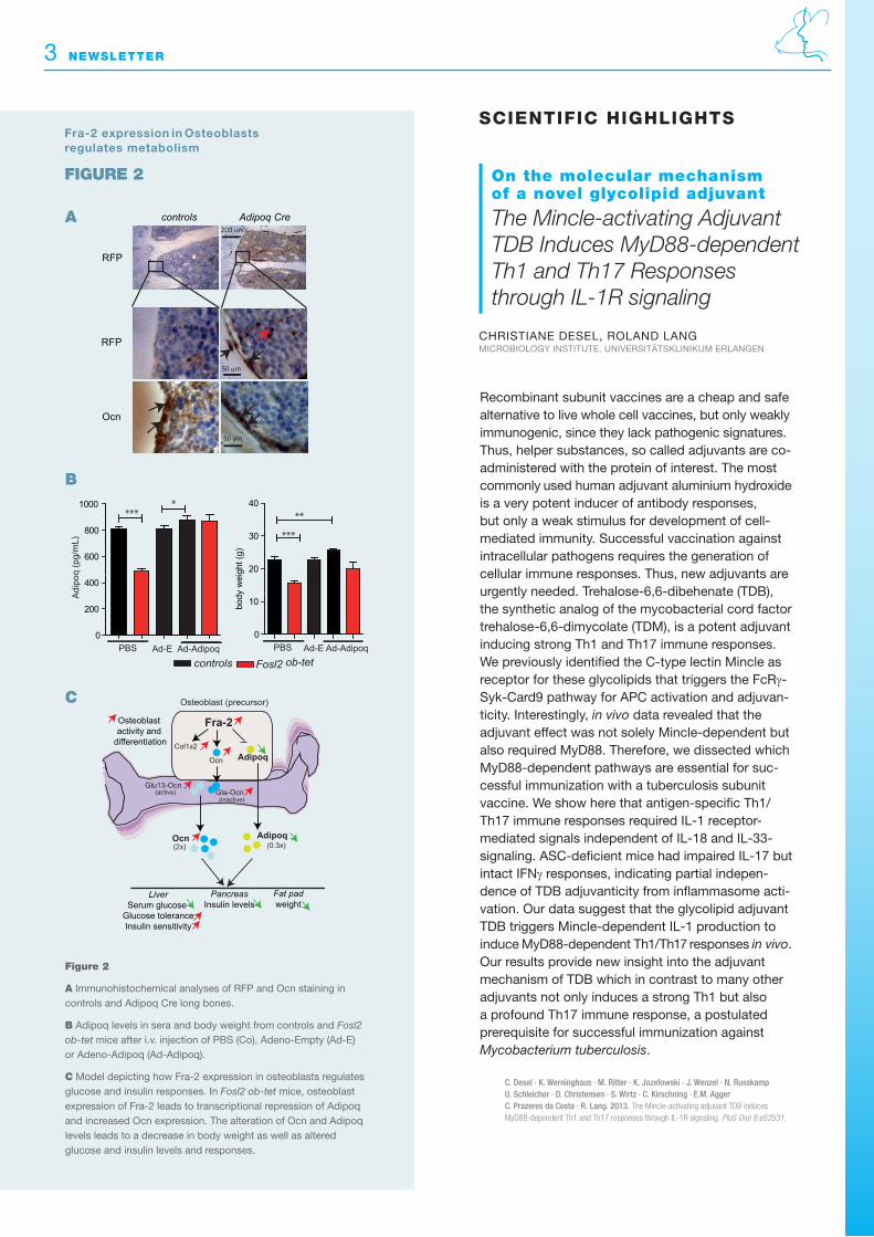
In our manuscript we have addressed this question by generating a genetically modified mouse over-expressing or deleting Fra-2 specifically in osteo-blasts. Mice depleted of Fra-2 in osteoblasts have low bone mass but increased body weight. In con-trast, over-expression of Fra-2 in osteoblasts leads to increased bone mass and decreased body weight accompanied by increased systemic osteocalcin isoforms, reduced serum glucose and insulin levels, improved glucose tolerance and insulin sensitivity (Figure 1). We found that Adipoq was expressed in osteoblasts and that this expression was transcrip-tionally repressed by Fra-2, leading to a systemic decrease of Adipoq. In addition, when Adipoq level is rescued, the metabolism phenotype is ameliorated in Fra-2 over-expressing mice (Figure 2). These results demonstrate that Fra-2 expression in osteo-blasts represents a novel paradigm for a transcription factor controlling the endocrine function of the skeleton by the transcriptional regulation of Adipoq and Ocn by Fra-2 in osteoblast (Figure 2).
J. Amich · L. Schafferer · H. Haas · S. Krappmann. 2013. A. Bozec · L. Bakiri · M. Jimenez · E.D. Rosen · P. Catala-LehnenT. Schinke · G. Schett · M. Amling · E.F. Wagner. 2013.Osteoblast-specific expression of Fra-2/AP-1 controls adiponectin and osteocalcin expression and affects metabolism. Journal of Cell Science 126:5432-5440.
Figure 1 A Von Kossa staining of spines, and quantification of bone volume (BV/TV), trabecular bone surface (TbTh), trabecular number (TbN), osteoblast number (NOb), osteoblast surface per bone surface (ObS/BS), osteoclast number (NOc), osteoclast surface per bone surface (OcS/BS), bone formation rate per bone surface (BFR/BS) in Fosl2 ob-tet mice at 2 months of age.
B Weight of mice and fat pad/total weight from controls and Fosl2 ob-tet mice under a ND or HFD for 6 weeks.
A
B
FIGUre 1
controls Fosl2 ob-tet
BV/TV TbTh TbN NOb ObS/BS NOc OcS/BS
36.19±0.88***47.17±1.54***3.72±0.03***29.91±1.4131.35±1.17***0.72±0.05*1.82±0.09*
19.08±0.3037.57±0.402.90±0.0728.08±1.6222.66±0.521.36±0.053.18±0.10 controls Fosl2 ob-tet
0
0.01
0.02
0.03
0.04
0.05
Fat p
ad/b
ody
wei
ght
ND HFD0
10
20
30
40
Bod
y w
eigh
t (g)
*** ***
* **
*** ***
*
ND HFD
a b
Fra-2 expression in Osteoblasts regulates metabolism
controls Adipoq Cre
RFP
RFP
Ocn
50 μm
50 μm
200 μm
Adi
poq
(pg/
mL)
0
200
400
600
800
1000***
*
PBS Ad-AdipoqAd-Econtrols Fosl2 ob-tet
0
10
20
30
40
***
**
PBS Ad-AdipoqAd-E
body
wei
ght (
g)
Glu13-Ocn
Osteoblast (precursor)
Fra-2
Ocn Adipoq
Gla-Ocn
AdipoqOcn
(active)(inactive)
LiverSerum glucose
Glucose toleranceInsulin sensitivity
Col1a2
Osteoblast activity and
differentiation
Fat padweight
PancreasInsulin levels
(0.3x)(2x)
a
b
ccontrols Adipoq Cre
RFP
RFP
Ocn
50 μm
50 μm
200 μm
Adi
poq
(pg/
mL)
0
200
400
600
800
1000***
*
PBS Ad-AdipoqAd-Econtrols Fosl2 ob-tet
0
10
20
30
40
***
**
PBS Ad-AdipoqAd-E
body
wei
ght (
g)
Glu13-Ocn
Osteoblast (precursor)
Fra-2
Ocn Adipoq
Gla-Ocn
AdipoqOcn
(active)(inactive)
LiverSerum glucose
Glucose toleranceInsulin sensitivity
Col1a2
Osteoblast activity and
differentiation
Fat padweight
PancreasInsulin levels
(0.3x)(2x)
a
b
c
3 newsLetter
Figure 2
A Immunohistochemical analyses of RFP and Ocn staining in controls and Adipoq Cre long bones.
B Adipoq levels in sera and body weight from controls and Fosl2 ob-tet mice after i.v. injection of PBS (Co), Adeno-Empty (Ad-E) or Adeno-Adipoq (Ad-Adipoq).
C Model depicting how Fra-2 expression in osteoblasts regulates glucose and insulin responses. In Fosl2 ob-tet mice, osteoblast expression of Fra-2 leads to transcriptional repression of Adipoq and in creased Ocn expression. The alteration of Ocn and Adipoq levels leads to a decrease in body weight as well as altered glucose and insulin levels and responses.
A
B
C
FIGUre 2
SciEntific HigHligHtS
On the molecular mechanism of a novel glycolipid adjuvant
The Mincle-activating Adjuvant TDB Induces MyD88-dependent Th1 and Th17 Responses through IL-1R signaling
CHrisTianE DEsEl, rOlanD langMiCrOBiOlOgY insTiTUTE, UniVErsiTÄTsKliniKUM ErlangEn
Recombinant subunit vaccines are a cheap and safe alternative to live whole cell vaccines, but only weakly immunogenic, since they lack pathogenic signatures. Thus, helper substances, so called adjuvants are co-administered with the protein of interest. The most commonly used human adjuvant aluminium hydroxide is a very potent inducer of antibody responses, but only a weak stimulus for development of cell-mediated immunity. Successful vaccination against intracellular pathogens requires the generation of cellular immune responses. Thus, new adjuvants are urgently needed. Trehalose-6,6-dibehenate (TDB), the synthetic analog of the mycobacterial cord factor trehalose-6,6-dimycolate (TDM), is a potent adjuvant inducing strong Th1 and Th17 immune responses. We previously identified the C-type lectin Mincle as receptor for these glycolipids that triggers the FcRγ-Syk-Card9 pathway for APC activation and adjuvan-ticity. Interestingly, in vivo data revealed that the adjuvant effect was not solely Mincle-dependent but also required MyD88. Therefore, we dissected which MyD88-dependent pathways are essential for suc-cessful immunization with a tuberculosis subunit vaccine. We show here that antigen-specific Th1/Th17 immune responses required IL-1 receptor- mediated signals independent of IL-18 and IL-33- signaling. ASC-deficient mice had impaired IL-17 but intact IFNγ responses, indicating partial indepen-dence of TDB adjuvanticity from inflammasome acti-vation. Our data suggest that the glycolipid adjuvant TDB triggers Mincle-dependent IL-1 production to induce MyD88-dependent Th1/Th17 responses in vivo. Our results provide new insight into the adjuvant mechanism of TDB which in contrast to many other adjuvants not only induces a strong Th1 but also a profound Th17 immune response, a postulated prerequisite for successful immunization against Mycobacterium tuberculosis.
C. Desel · K. Werninghaus · M. Ritter · K. Jozefowski · J. Wenzel · N. RusskampU. Schleicher · D. Christensen · S. Wirtz · C. Kirschning · E.M. Agger C. Prazeres da Costa · R. Lang. 2013. The Mincle-activating adjuvant TDB induces MyD88-dependent Th1 and Th17 responses through IL-1R signaling. PloS One 8:e53531.
controls Adipoq Cre
RFP
RFP
Ocn
50 μm
50 μm
200 μm
Adi
poq
(pg/
mL)
0
200
400
600
800
1000***
*
PBS Ad-AdipoqAd-Econtrols Fosl2 ob-tet
0
10
20
30
40
***
**
PBS Ad-AdipoqAd-E
body
wei
ght (
g)
Glu13-Ocn
Osteoblast (precursor)
Fra-2
Ocn Adipoq
Gla-Ocn
AdipoqOcn
(active)(inactive)
LiverSerum glucose
Glucose toleranceInsulin sensitivity
Col1a2
Osteoblast activity and
differentiation
Fat padweight
PancreasInsulin levels
(0.3x)(2x)
a
b
c
Fra-2 expression in Osteoblasts regulates metabolism
spring 2014 4
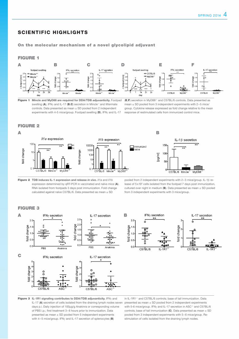
Figure 2 TDB induces IL-1 expression and release in vivo. Il1a and Il1b expression determined by qRT-PCR in vaccinated and naïve mice (A). RNA isolated from footpads 3 days post immunization. Fold change calculated against naïve C57BL/6. Data presented as mean ± SD
pooled from 2 independent experiments with 2–3 mice/group. IL-1β re-lease of 5 x 10⁵ cells isolated from the footpad 7 days post immunization, cultured over night in medium (B). Data presented as mean ± SD pooled from 3 independent experiments with 3 mice/group.
A B
FIGUre 2
A B
C
FIGUre 3
FIGUre 1
A B C D e F
Figure 1 Mincle and MyD88 are required for DDA/TDB adjuvanticity. Footpad swelling (A), IFNγ and IL-17 (B,C) secretion in Mincle-/- and littermate controls. Data presented as mean ± SD pooled from 2 independent experiments with 4-5 mice/group. Footpad swelling (D), IFNγ and IL-17
(e,F) secretion in MyD88-/- and C57BL/6 controls. Data presented as mean ± SD pooled from 3 independent experiments with 2–5 mice/group. Cytokine release expressed as fold change relative to the mean response of restimulated cells from immunized control mice.
On the molecular mechanism of a novel glycolipid adjuvant
SciEntific HigHligHtS
IL-1R1 signaling contributes to DDA/TDB adjuvanticity. IFNγ and IL-17 (A) secretion of cells isolated from the draining lymph nodes seven days p.i. Daily injection of 100µg/g Anakinra or corresponding volume of PBS i.p.; first treatment 3 – 6 hours prior to immunization. Data presented as mean ± SD pooled from 5 independent experiments with 4–5 mice/group. IFNγ and IL-17 secretion of splenocytes (B)
in IL-1R1-/- and C57BL/6 controls; base of tail immunization. Data presented as mean ± SD pooled from 2 independent experiments with 5-6 mice/group. IFNγ and IL-17 secretion in ASC-/- and C57BL/6 controls; base of tail immunization (C). Data presented as mean ± SD pooled from 3 independent experiments with 5–6 mice/group. Re- stimulation of cells isolated from the draining lymph nodes.
Figure 3
5 newsLetter
MEtHodS
Generation of moDCs and lymphocytes from LrsCs
Leukoreduction system chambers (LRSCs) as a novel and highly economic source of viable and functional monocyte-derived dendritic cells and lymphocytes for research purposes
ilKa KnippErTZ
DEparTMEnT OF iMMUnE MODUlaTiOn aT THE DEparTMEnT OF DErMaTOlOgY, UniVErsiTÄTsKliniKUM ErlangEn
The need of primary human monocyte-derived dendritic cells (moDCs) and lymphocytes for research purposes has steadily increased over the last decade. However, it is still a challenge to obtain them in adequate quantity, quality, and at reasonable costs. Today, the gold standard to obtain peripheral blood mononuclear cells (PBMCs) is leukapheresis. However, the use of these products for research purposes is not only highly expensive and time-consuming, but leukapheresis products are not available from every blood bank either. Blood samples from healthy donors are limited in volume and difficult to obtain for ethical and regulatory reasons. Leukoreduction system chambers (LRSCs), on the other hand, arise after routine donor plateletpheresis procedures and are usually discarded.
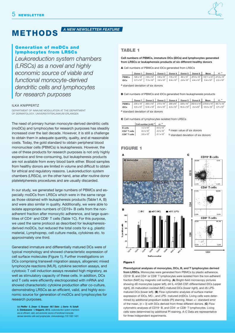
In our study, we generated large numbers of PBMCs and es-pecially moDCs from LRSCs which were in the same range as those obtained with leukapheresis products (Table 1 A, B) and were also similar in quality. Additionally, we were able to isolate appropriate numbers of CD19+ B cells from the non-adherent fraction after monocytic adherence, and large quan-tities of CD4+ and CD8+ T cells (Table 1C). For this purpose, we used the same protocol as described for leukapheresis-derived moDCs, but reduced the total costs for e.g. plastic material, Lymphoprep, cell culture media, cytokines etc. to approximately one third.
Generated immature and differentially matured DCs were of typical morphology and showed characteristic expression of cell surface molecules (Figure 1). Further investigations on DCs comprising transwell migration assays, allogeneic mixed lymphocyte reactions (MLR), cytokine secretion assays, and cytotoxic T cell induction assays revealed high migratory, as well as stimulatory capacity of these cells. In addition, DCs and T cells were efficiently electroporated with mRNA and showed characteristic cytokine production after co-culture, demonstrating LRSCs as an efficient, valid, and highly eco-nomic source for generation of moDCs and lymphocytes for research purposes.
Figure 1. Phenotypical analyses of monocytes, DCs, B, and T lymphocytes derived from LRSCs. Monocytes were generated from PBMCs by plastic adherence. CD19+ B, and CD4+ or CD8+ T lymphocytes were isolated from the non-‐adherent fraction (NAF) by magnetic cell sorting. (A) Bright-‐field microscopy pictures showing d0 monocytes (upper left), d4 IL-‐4/GM-‐CSF-‐differentiated iDCs (upper right), d5 maturation cocktail (MC) matured DCs (lower right), and d5 LPS-‐ matured DCs (lower left). (B) Flow cytometric analyses of surface marker expression of iDCs, MC-‐, and LPS-‐ matured (m)DCs. Living cells were determined by additional propidium iodide (PI) staining. Mean +/-‐ standard error of the mean, (n = 3) with DCs derived from three different donors. (C) Flow cytometric analyses of CD19+ B, and CD4+ or CD8+ T lymphocytes. Living cells were determined by additional PI staining. A-‐C Data are representative for three independent experiments. Pfeiffer, I.A., E. Zinser, E. Strasser, M.F. Stein, J. Dorrie, N. Schaft, A. Steinkasserer, and I. Knippertz. 2013. Leukoreduction system chambers are an efficient, valid, and economic source of functional monocyte-‐derived dendritic cells and lymphocytes. Immunobiology 218:1392-‐1401.
Figure 1. Phenotypical analyses of monocytes, DCs, B, and T lymphocytes derived from LRSCs. Monocytes were generated from PBMCs by plastic adherence. CD19+ B, and CD4+ or CD8+ T lymphocytes were isolated from the non-‐adherent fraction (NAF) by magnetic cell sorting. (A) Bright-‐field microscopy pictures showing d0 monocytes (upper left), d4 IL-‐4/GM-‐CSF-‐differentiated iDCs (upper right), d5 maturation cocktail (MC) matured DCs (lower right), and d5 LPS-‐ matured DCs (lower left). (B) Flow cytometric analyses of surface marker expression of iDCs, MC-‐, and LPS-‐ matured (m)DCs. Living cells were determined by additional propidium iodide (PI) staining. Mean +/-‐ standard error of the mean, (n = 3) with DCs derived from three different donors. (C) Flow cytometric analyses of CD19+ B, and CD4+ or CD8+ T lymphocytes. Living cells were determined by additional PI staining. A-‐C Data are representative for three independent experiments. Pfeiffer, I.A., E. Zinser, E. Strasser, M.F. Stein, J. Dorrie, N. Schaft, A. Steinkasserer, and I. Knippertz. 2013. Leukoreduction system chambers are an efficient, valid, and economic source of functional monocyte-‐derived dendritic cells and lymphocytes. Immunobiology 218:1392-‐1401.
I.A. Pfeiffer · E. Zinser · E. Strasser · M.F. Stein · J. Dorrie · N. Schaft A. Steinkasserer · I. Knippertz. 2013. Leukoreduction system chambers are an efficient, valid, and economic source of functional monocyte-derived dendritic cells and lymphocytes. Immunobiology 218:1392-1401.
Figure 1
Phenotypical analyses of monocytes, DCs, B, and T lymphocytes derived from LRSCs. Monocytes were generated from PBMCs by plastic adherence. CD19+ B, and CD4+ or CD8+ T lymphocytes were isolated from the non-adherent fraction (NAF) by magnetic cell sorting. (A) Bright-field microscopy pictures showing d0 monocytes (upper left), d4 IL-4/GM-CSF-differentiated iDCs (upper right), d5 maturation cocktail (MC) matured DCs (lower right), and d5 LPS- matured DCs (lower left). (B) Flow cytometric analyses of surface marker expression of iDCs, MC-, and LPS- matured (m)DCs. Living cells were deter-mined by additional propidium iodide (PI) staining. Mean +/- standard error of the mean, (n = 3) with DCs derived from three different donors. (C) Flow cytometric analyses of CD19+ B, and CD4+ or CD8+ T lymphocytes. Living cells were determined by additional PI staining. A-C Data are representative for three independent experiments.
…Large numbers of PBMCs and moDCs similar in quality to those from leukapheresis products….
…Reduced total costs for consumable material to approximately one third…
Table 1. Cell numbers of PBMCs, immature DCs (iDCs) and lymphocytes generated from LRSCs
or leukapheresis products of six different healthy donors
A) Cell numbers of PBMCs and iDCs generated from LRSCs
a standard deviation of six donors
B) Cell numbers of PBMCs and iDCs generated from leukapheresis products
a standard deviation of six donors
C) Cell numbers of lymphocytes isolated from LRSCs
a mean value of six donors b standard deviation of six donors Figure 1
Donor 1 Donor 2 Donor 3 Donor 4 Donor 5 Donor 6 Mean +/- a
PBMCs 120 x 107 130 x 107 130 x 107 115 x 107 60 x 107 87.5 x 10⁷ 107.1 x 10⁷ 27.9 x 10⁷
iDCs 5.7 x 10⁷ 7.3 x 10⁷ 3.8 x 10⁷ 8.4 x 10⁷ 2.8 x 10⁷ 4.4 x 10⁷ 5.4 x 10⁷ 2.2 x 10⁷
Donor 1 Donor 2 Donor 3 Donor 4 Donor 5 Donor 6 Mean +/- a
PBMCs 235 x 107 350 x 107 275 x 107 200 x107 325 x 107 292.5 x 10⁷ 279.6 x 10⁷ 55.7 x 10⁷
iDCs 8.5 x 10⁷ 12.6 x 10⁷ 8.4 x 10⁷ 6.0 x 10⁷ 16.2 x 10⁷ 8.4 x10⁷ 10.0 x 10⁷ 3.7 x 10⁷
Cell number (n=6) a +/- b
B cells 1.2 x 10⁷ 0.4 x 10⁷
CD4+ T cells 8.3 x 10⁷ 2.0 x 10⁷
CD8+ T cells 5.6 x 10⁷ 0.1 x 10⁷
Cell numbers of PBMCs, immature DCs (iDCs) and lymphocytes generated from LRSCs or leukapheresis products of six different healthy donors
A Cell numbers of PBMCs and iDCs generated from LRSCs
B Cell numbers of PBMCs and iDCs generated from leukapheresis products
C Cell numbers of lymphocytes isolated from LRSCs
A C
a standard deviation of six donors
a standard deviation of six donors
a mean value of six donorsb standard deviation of six donors
A NEW NEWSLETTER FEATURE
tABLe 1
FIGUre 1
B

PeOPLe
spring 2014 6
PD Dr. med. Dimitrios Mougiakakos
Dimitrios Mougiakakos received research funding by the European Hematology Association (EHA) for the next three years.
Dr. Mougiakakos, head of the Max-Eder Junior Research Group “Translationale Tumor- und Trans-plantationsimmunologie” sponsored by the German Cancer Aid at the Department of Medicine 5 since 2013, will be supported by the European Hema-tology Association with 240.000 Euro for the next three years to perform research on the mitochon-drial metabolism of lymphocytes during chronic lymphocytic leukemia (CLL).
Dimitrios Mougiakakos and his group observed drastic alterations in the mitochondrial metabolism of CLL-cells. Most strikingly, the mitochondria in CLL-cells were identified as the main source for abundant reactive oxygen species (ROS). Dr. Mougiakakos intends to further investigate the bioenergetics and redox characteristics in-volved during CLL in order to develop new thera-peutic strategies against this type of leukemia.
Prof. Dr. Dr. Michael Stürzl
Wolfgang-Lutz-Foundation supports Prof. Michael Stürzl’s cancer research
Michael Stürzl, head of the Division for Molecular and Experimental Surgery at the Department of Surgery since 2003, received 70.000 Euro from a private benefactor, the Wolfgang-Lutz-Foundation, located in the Nürnberger Land. This foundation supports medical research especially in the field of cancer research. Prof. Stürzl will use this donation to develop innovative models to investigate the molecular processes involved in the development of colorectal cancers.
Prof. Dr. med. Jens Titze
Jens Titze was awarded by two separate socie-ties for his research in electrolyte homeostasis
The American Heart Association and the American Stroke Association (AHA/ASA) have honored Jens Titze, Professor for Electrolyte and Circulatory Research at the IZKF, with the HBPR Mid Career Award for Research Excellence worth 1000 Euro. Later last year, Jens Titze received another award, the Franz-Vollhard-Prize worth 10.000 Euro at the Annual Meeting of the Deutsche Gesellschaft für Nephrologie. Professor Titze and his colleagues discovered that brain, kidneys and vessels were not the only actors during electrolyte homeostasis regulation. The skin was shown to also regulate its own sodium reservoir with the help of immune cells that control skin lymphatic electrolyte homeostasis and blood pressure. During high-salt diet in mice, Titze and his group observed a recruitment of phagocytes to the skin, where they sense the hypertonic electrolyte accumulation and activate a cascade of proteins which in the end enhances electrolyte clearance via cutaneous lymph vessels.

7 newsLetter
news AnD UPDAtes
Joachim Kalden Lecture 2013The Medical Immunology Campus Erlangen honors Prof. Dr. rer. nat. Andreas Radbruch
The Joachim Kalden Lecture 2013, which took place on January 8, 2014, was delivered by Professor Andreas Radbruch, Scientific Director of the Deut-sches Rheumaforschungszentrum (DRFZ) Berlin. Andreas Radbruch´s main research interests focus on the molecular understanding of the control of immune reactions and immunological memory in vaccination as well as autoimmune and allergic inflammation. After his studies of biology at the universities of Bonn and Cologne, he was a PhD scholar at the lab of Prof. Klaus Rajewsky at the University of Cologne, where he finished his thesis in 1980. He soon led his own junior research group and became professor of genetics and immunology, before being appointed as scientific director of the DRFZ Berlin in 1996. In his early years, besides demonstrating the role of switch recombination in vivo, he also demonstrated that switch recombination is controlled by cytokines from T lymphocytes. To-day, he focusses more on the pathogenic cytokines secreted by memory T cells and long-lived plasma cells. Currently, he is working on a method to spe-cifically eliminate pathogenic T memory cells with-out disturbing protective immunity.
Amongst immunologists, Professor Radbruch is most famous for his groundbreaking co-invention of the magnetic-activated cell sorting (MACS) method, a standard procedure to separate specific cell popu-lations from mixed cell suspension using magnetic nano-beads. He has received numerous high-ran-king prizes and honors. For example, in 2011, his outstanding research results were rewarded with the Carol-Nachman-Prize, one of the highest medi-cal honors in Germany as well as the internationally highest-paying award in the field of rheumatology.
Andreas Radbruch delivered a captivating lecture entitled “The resting and the restless immunological memory”. He presented new and exciting data on the bone marrow niche of CD4+ memory T cells, CD8+ memory T cells, and of memory plasma cells. Each of these memory lymphocyte populations appear to interact with distinct types of bone marrow stromal cells that express different ligands (e.g. collagen II and XI, fibronectin, VCAM1) for lympho-cyte retention molecules (e.g. CD69, CD49b).
The Medical Immunology Campus Erlangen honors Andreas Radbruch
Prof. Andreas Radbruch (middle) holding
the Certificate of the Joachim Kalden
Lecture 2013 with Prof. Joachim Kalden (left)
and Prof. Christian Bogdan (right).
8 newsLetter
Medical Immunology Campus Erlangen Executive Boardprof. Dr. med. Christian Bogdan (Chairman) prof. Dr. rer. nat. Diana Dudziak prof. Dr. med. Kai-Uwe Eckardt prof. Dr. med. Bernhard Fleckenstein prof. Dr. rer. nat. Hans-Martin Jäck prof. Dr. med. andreas Mackensen prof. Dr. med. Markus neurath (Deputy Chairman) Dr. rer. nat. sonja pötzsch (scientific Coordinator)prof. Dr. med. georg schett prof. Dr. med. gerold schuler prof. Dr. rer. nat. alexander steinkasserer prof. Dr. rer. nat. Thomas Winkler (Deputy Chairman)
PublisherMedical immunology Campus Erlangenan interdisciplinary Center of the Friedrich-alexander-Universität Erlangen-nürnberg
Dr. rer. nat. sonja pötzsch (scientific Coordinator)
Mikrobiologisches institut – Klinische Mikrobiologie, immunologie und Hygiene
Universitätsklinikum ErlangenFriedrich-alexander-Universität Erlangen-nürnbergWasserturmstraße 3/5 · 91054 Erlangen
Phone +49. 9131. 85. 225 71Fax +49. 9131. 85. 225 73
Mail [email protected]
Conceptual Design and EditorDr. rer. nat. sonja pötzsch V.i.s.d.p.
Subscription via Email to:[email protected]
please note that the authors are responsible for the content of their contributions.
We are looking forward to suggestions for the next MiCE newsletter. please send material to:[email protected]
An Interdiscipl inary Center of the Friedrich-Alexander-Universität Erlangen-Nürnberg
MICE Immunological Colloquium – Summer 2014, Tuesdays, 5.15 pm
29. 04. 2014Prof. Victor TybulewiczMrC national institute for Medical research, london, UK
“B cell survival and tonic signalling from the BC”
06. 05. 2014 Prof. Manfred Kopfinstitute of Molecular Health sciences ETH Zürich, switzerland
“Alveolar Macrophage Development and Function in Respiratory Viral Infection”
13. 05. 2014Prof. Joachim SchultzeliMEs institute, genomics and immunoregulation, Bonn
“The spectrum of macrophage activation. Beyond the M1/M2 model”
20. 05. 2014Prof. Felix RandowMrC laboratory of Molecular Biology, Cambridge, UK
“Autophagy in host-pathogen interactions”
27. 05. 2014 Prof. Jan Ruppinstitut für Med. Mikrobiologie und Hygiene, Med. Klinik iii, lübeck
“Title to be announced”
03. 06. 2014Dr. Gijs VersteegMax F. perutz laboratories (MFpl), Vienna, austria
“TRIMmunity: The roles of the TRIM E3-ubiquitin ligase family in innate antiviral immunity”
17. 06. 2014Prof. Sai ReddyETH Zürich, Technology Department of Biosystems science & Engineering, Basel, switzerland
“Title to be announced”
24. 06. 2014Prof. Wolfgang Kastenmüller, MDinstitutes of Molecular Medicine and Experimental immunology (iMMEi), University of Bonn
“Title to be announced”
01. 07. 2014Vera Martins, PhDinstitute for immunology, University Hospital Ulm
“Title to be announced”
08. 07. 2014Prof. Dr. med. Stephan EhlCCi - Centrum für Chronische immun-defizienz, Universitätsklinikum Freiburg
“New lessons from human genetic disorders of T cell immunity”
UPCOmInG events
Further Conferences of Interest
may 2 – 6, 2014Immunology 20144pittsburgh, Usawww.immunology2014.org
June 25 – 27, 20148th German Meeting on Immune RegulationBerlin-schmöckwitz
July 27 – August 1, 2014International Union of Microbiological Societies (IUMS) 2014Montreal, Canadawww.montrealiums2014.org
september 14 – 18, 2014
13th International Symposium on Dendritic CellsTourswww.dc-2014.com
september 17 – 20, 201444. Jahrestagung der Deutschen Gesellschaft für Immunologie DGfI 2014Bonnwww.immunology-conference.de
september 14 – 18, 201413th International Symposium on Dendritic CellsTours, France www.dc-2014.com
september 17 – 20, 201444. Jahrestagung der Deutschen Gesell-schaft für Immunologie DGfI 2014Bonn www.immunology-conference.de
October 2 – 4, 201428th Annual Meeting of the European Dendritic Cell Society (EMDS)Vienna, austriawww.macrophage.de/meetings.htm
October 5 – 8, 20144. Gemeinsame Jahrestagung der Deutschen Gesellschaft für Hygiene und Mikrobiologie (DGHM)Dresdenwww.dghm-vaam-kongress.de


