8/3/2019 Optical Probing of a Dynamic Membrane Interaction
http://slidepdf.com/reader/full/optical-probing-of-a-dynamic-membrane-interaction 1/6
Optical probing of a dynamic membrane interactionthat regulates the TREK1 channelGuillaume Sandoza,b,1, Sarah C. Bellc,1, and Ehud Y. Isacoffa,c,d,2
aDepartment of Molecular and Cell Biology and Helen Wills Neuroscience Institute, University of California, Berkeley, CA 94720; bInstitut de PharmacologieMoléculaire et Cellulaire, Centre National de la Recherche Scientifique and Université de Nice Sophia-Antipolis, Sophia-Antipolis, 06560 Valbonne, France;c
Chemical Biology Graduate Program, University of California, Berkeley, CA 94720; and
d
Physical Bioscience Division, Lawrence Berkeley National Laboratory,Berkeley, CA 94720
Edited* by Lily Yeh Jan, University of California, San Francisco, and approved December 23, 2010 (received for review October 21, 2010)
TREK channels produce background currents that regulate cellexcitability. These channels aresensitiveto a wide variety of stimuli
including polyunsaturated fatty acids (PUFAs), phospholipids,
mechanical stretch, and intracellular acidification. They are
inhibited by neurotransmitters, hormones, and pharmacologicalagents such as the antidepressantfluoxetine. TREK1 knockout mice
have impaired PUFA-mediated neuroprotection to ischemia, re-
duced sensitivity to volatile anesthetics, altered perception of pain,
and a depression-resistant phenotype. Here, we investigate TREK1
regulation by Gq-coupled receptors (GqPCR) and phospholipids.
Several reports indicate that the C-terminal domain of TREK1 is
a key regulatory domain. We developed a fluorescent-based tech-nique that monitors the plasma membrane association of the
C terminus of TREK1 in real time. Our fluorescence and functional
experiments link the modulation of TREK1 channel function by in-
ternal pH, phospholipid, and GqPCRs to TREK1–C-terminal domainassociation to the plasma membrane, where increased association
results in greater activity.In keeping with this relation, inhibition of
TREK1 current by fluoxetine is found to be accompanied by disso-
ciation of the C-terminal domain from the membrane.
ion channel | two-pore-domain K+ | phosphatidylinositol-4,5-biphosphate|Prozac
T
REK1 is a two-pore-domain K + (K 2P) channel that produces
a nearly time- and voltage-independent background current.This current drives the membrane potential toward the K +
equilibrium potential and thus affects input resistance. TREK1displays low basal activity when expressed alone (1) but can bestrongly stimulated by temperature (2), mechanical stretch (3),external alkalization (4), intracellular acidification (5), poly-unsaturated fatty acids (PUFAs) (6), lysophospholipids (7),phosphatidylinositol-4,5-bisphosphate [PI(4,5)P2] (8, 9), andpharmacological agents such as volatile anesthetics (10) andriluzole (11). TREK1 is inhibited by neurotransmitters and hor-mones that activate Gq and Gs pathways (3, 12–14) and phar-macological agents such as the antidepressant drug fluoxetine(15). TREK1 gene inactivation produces mice with decreasedsensitivity to volatile anesthetics, impaired PUFA-mediatedneuroprotection (16), and altered perception of pain (17). Thesemice also display a depression-resistant phenotype (18). Thisphenotype is in agreement with the sensitivity of TREK1 to flu-oxetine, a widely used antidepressant drug.
In the last decade, much effort has been made to elucidate thegating mechanism of the TREK1 channel. The cytosolic carboxyl-terminal domain of TREK1 that follows its fourth transmembranedomain (post-M4) has been implicated in its function and regula-tion. A glutamate residue, E306, has been shown to be a key ele-ment for stimulation by intracellular acidification (19) and a clusterofbasicresiduesinthesameregionhasbeenshowntobeinvolvedinTREK1 regulation by phospholipid (8). However, the mechanismof regulation of the channel by Gq-coupled receptors(GqPCR)andby pharmacological agents such as fluoxetine remains unclear.
In this study, we developed a fluorescence-based technique toaddress regulation of TREK1 by GqPCRs and fluoxetine by monitoring in real time the association of an EGFP-tagged
TREK1 C-terminal domain with the plasma membrane. We in- vestigated the roles of key residues in the TREK1 C terminus andfound that mutants with increased or decreased channel activity have, respectively, increased or decreased membrane association.Moreover, GqPCR inhibition of TREK1 channel activity wasfound to be accompanied by C-terminal domain dissociationfrom the membrane. This regulatory effect was found to be in-dependent of PKC but to depend on the breakdown of PI(4,5)P2.Strikingly, the inhibitory effect of fluoxetine was also attributed toC-terminal domain dissociation from the membrane, providingboth insight into the molecular mechanism of this modulation and
a possible route for identifying novel antidepressants.Results
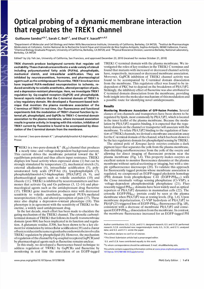
Monitoring Membrane Association of GFP-Fusion Proteins. Severalclasses of ion channels and transporters have been shown to beregulated by lipids, most commonly by PI(4,5)P2, which is locatedin the inner leaflet of the plasma membrane. Because the modu-lation by PI(4,5)P2 requires binding, it is expected that the regu-latory domain on the channel or transporter will associate with themembrane. To relate PI(4,5)P2 binding to the regulation of func-tion of TREK1channels, we devised a membrane association assay for the C-terminal domain of the channel, which has been shown tobe the key regulatory domain for phospholipid sensing by TREK1.
The animal pole of Xenopus laevis oocytes contains a dark pigment layer that separates the yolk from the plasma membrane,thus shielding autofluorescence from the yolk of the oocyte andallowing for direct imaging of fluorescent proteins near theplasma membrane (Fig. 1 A). This property makes oocytes anexcellent system to monitor fluorescence dynamics at the plasmamembrane without optical sectioning or imaging, but with a sim-ple epifluorescence microscope (20). To validate this approachfor fluorescently tagged proteins whose membrane association isregulated, we coexpressed an EGFP-tagged pleckstrin homology (PH) domain from phospholipase C-δ1 (EGFP-PHPLC) withthe Ciona intestinalis voltage sensing phosphatase (Ci-VSP), a
voltage-dependent phosphoinositide phosphatase (21). Fluo-rescently tagged PHPLC domains have been widely used as opticalreporters of PI(4,5)P2 dynamics in mammalian cells (22). Thecytosolic EGFP-PHPLC protein could be seen at the plasmamembrane when PI(4,5)P2 was at resting levels. (Fig. 1 A). Uponmembrane depolarization, Ci-VSP hydrolysis of PI(4,5)P2 to
PI(4)P (23) triggered loss of EGFP-PHPLC fluorescence (Fig. 1 B),consistent with a decrease of membrane PI(4,5)P2 and conse-quent EGFP-PHPLC dissociation from the membrane. In contrast,the membrane fluorescence increased for an EGFP-tagged PH
Author contributions: G.S., S.C.B., and E.Y.I. designed research; G.S. and S.C.B. performed
research; S.C.B. contributed new reagents/analytic tools; G.S., S.C.B., and E.Y.I. analyzed
data; and G.S. and E.Y.I. wrote the paper.
The authors declare no conflict of interest.
*This Direct Submission article had a prearranged editor.
1G.S. and S.C.B. have contributed equally to this work.
2To whom correspondence should be addressed. E-mail: [email protected].
This article contains supporting information online at www.pnas.org/lookup/suppl/doi:10.
1073/pnas.1015788108/-/DCSupplemental.
www.pnas.org/cgi/doi/10.1073/pnas.1015788108 PNAS Early Edition | 1 of 6
P H Y S I O L O G
Y
8/3/2019 Optical Probing of a Dynamic Membrane Interaction
http://slidepdf.com/reader/full/optical-probing-of-a-dynamic-membrane-interaction 2/6
domain of yeast oxysterol binding protein homolog (EGFP-
PHOSH1), a protein that binds PI(4)P (24) (Fig. 1C), consistent with conversion of PI(4,5)P2 to PIP(4) by Ci-VSP. Indeed, PI(4,5)P2 depletion and PI(4)P production could be detected in a singlecell coexpressing tagRFP-PHPLC and EGFP-PHOSH1 (Fig. 1C).
We extended this technique to see whether we can track changes in phospholipids induced by activation of G proteincoupled receptors (GPCRs). We coexpressed EGFP-PHPLC withthe 5HT2c receptor, a GPCR coupled to the Gq α-subunit of Gprotein (i.e., a GqPCR), whose receptor activation induces thehydrolysis of PI(4,5)P2 to DAG (25). As expected, activation of the 5HT2c receptor decreased EGFP-PHPLC fluorescence at theplasma membrane (Fig. 1 D). Success of this simple translocationassay for known membrane-interacting proteins encouraged usto use the approach to study the membrane association of theregulatory TREK1 C-terminal domain.
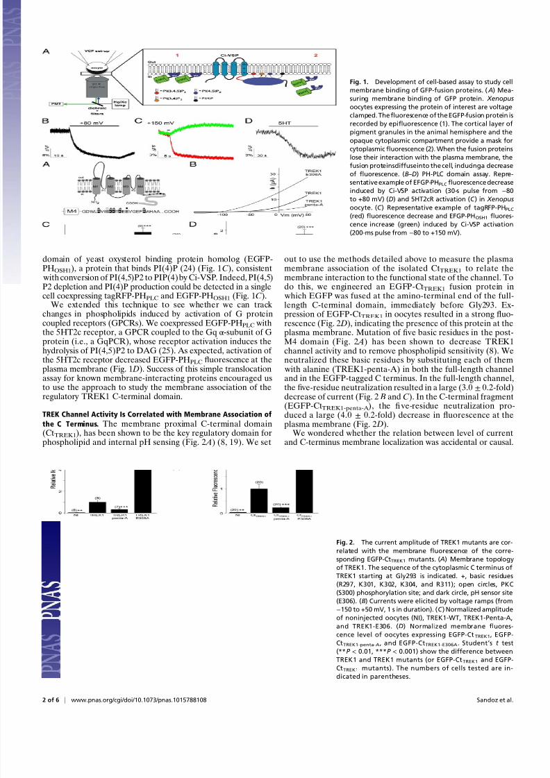
TREK Channel Activity Is Correlated with Membrane Association of
the C Terminus. The membrane proximal C-terminal domain(CtTREK1), has been shown to be the key regulatory domain forphospholipid and internal pH sensing (Fig. 2 A) (8, 19). We set
out to use the methods detailed above to measure the plasma
membrane association of the isolated CtTREK1 to relate themembrane interaction to the functional state of the channel. Todo this, we engineered an EGFP-CtTREK1 fusion protein in
which EGFP was fused at the amino-terminal end of the full-length C-terminal domain, immediately before Gly293. Ex-pression of EGFP-CtTREK1 in oocytes resulted in a strong fluo-rescence (Fig. 2 D), indicating the presence of this protein at theplasma membrane. Mutation of fi ve basic residues in the post-M4 domain (Fig. 2 A) has been shown to decrease TREK1channel activity and to remove phospholipid sensitivity (8). Weneutralized these basic residues by substituting each of them
with alanine (TREK1-penta-A) in both the full-length channeland in the EGFP-tagged C terminus. In the full-length channel,the fi ve-residue neutralization resulted in a large (3.0 ± 0.2-fold)decrease of current (Fig. 2 B and C). In the C-terminal fragment
(EGFP-CtTREK1-penta-A ), thefi ve-residue neutralization pro-duced a large (4.0 ± 0.2-fold) decrease in fluorescence at the
plasma membrane (Fig. 2 D).We wondered whether the relation between level of current
and C-terminus membrane localization was accidental or causal.
Fig. 1. Development of cell-based assay to study cell
membrane binding of GFP-fusion proteins. ( A) Mea-
suring membrane binding of GFP protein. Xenopus
oocytes expressing the protein of interest are voltage
clamped. Thefluorescence of the EGFP-fusion protein is
recorded by epifluorescence (1). The cortical layer of
pigment granules in the animal hemisphere and the
opaque cytoplasmic compartment provide a mask for
cytoplasmicfluorescence (2). When the fusion proteins
lose their interaction with the plasma membrane, the
fusion proteinsdiffuseinto the cell, inducinga decrease
of fluorescence. (B–D) PH-PLC domain assay. Repre-
sentative example of EFGP-PHPLCfluorescence decrease
induced by Ci-VSP activation (30-s pulse from −80
to +80 mV) (D) and 5HT2cR activation (C ) in Xenopus
oocyte. (C ) Representative example of tagRFP-PHPLC(red) fluorescence decrease and EFGP-PHOSH1 fluores-
cence increase (green) induced by Ci-VSP activation
(200-ms pulse from −80 to +150 mV).
Fig. 2. The current amplitude of TREK1 mutants are cor-related with the membrane fluorescence of the corre-
sponding EGFP-CtTREK1 mutants. ( A) Membrane topology
of TREK1. The sequence of the cytoplasmic C terminus of
TREK1 starting at Gly293 is indicated. +, basic residues
(R297, K301, K302, K304, and R311); open circles, PKC
(S300) phosphorylation site; and dark circle, pH sensor site
(E306). (B) Currents were elicited by voltage ramps (from
−150 to +50 mV, 1 s in duration). (C ) Normalized amplitude
of noninjected oocytes (NI), TREK1-WT, TREK1-Penta-A,
and TREK1-E306. (D) Normalized membrane fluores-
cence level of oocytes expressing EGFP-CtTREK1, EGFP-
CtTREK1-penta-A, and EGFP-CtTREK1-E306A. Student’s t test
(**P < 0.01, ***P < 0.001) show the difference between
TREK1 and TREK1 mutants (or EGFP-CtTREK1 and EGFP-
CtTREK1 mutants). The numbers of cells tested are in-
dicated in parentheses.
2 of 6 | www.pnas.org/cgi/doi/10.1073/pnas.1015788108 Sandoz et al.
8/3/2019 Optical Probing of a Dynamic Membrane Interaction
http://slidepdf.com/reader/full/optical-probing-of-a-dynamic-membrane-interaction 3/6
We therefore turned to another mutant of the channel that isknown to have the opposite effect on activity. The mutation wechose was of glutamate 306 to alanine (E306A). E306 is located inthe post-M4 region of CtTREK1, embedded in the region con-taining the fi ve basic residues that we mutated above (Fig. 2 A).This mutation has been proposed to mimic internal acidificationand has been shown to induce a large increase in the current (19).Indeed, we found that E306A current was 4.3 ± 0.4-fold largerthan wild type (Fig.2 B and C). We found that EGFP-CtTREK1-E306A
was higher in basal membranefl
uorescence by 1.9±
0.2-foldcompared with wild-type EGFP-CtTREK1 (Fig. 2 D).The differences in basal membrane fluorescence between
EGFP-CtTREK1, EGFP-CtTREK1-penta-A , and EGFP-CtTREK1-E306A could be due to differences in membrane targeting, but they couldalso be due to differences in protein expression levels. Because theinside of the oocyte is opaque and the yolk highly fluorescent, wecould not image the EGFP-tagged TREK1 C-terminal domain inthe oocyte interior. We therefore turned to transient expression inHEK cells to distinguish between these possibilities. The cells werefi xed with either paraformaldehyde (PFA), tofi x all of the proteinsthroughout the cell, or else they were fi xed with ice-cold ethanol, in
which case only membrane-associated proteins are fi xed to themembrane proteins and unbound proteins leak out of the cells(26). Although the PFA fi xation showed that the proteins wereexpressed at comparable levels (Fig. S1 B), the fluorescence in theethanol fi xation was 2.6± 0.6-fold lower for EGFP-CtTREK1-penta-A than for the wild-type EGFP-CtTREK1, and EGFP-CtTREK1-E306A
was more fluorescent by 1.4 ± 0.1-fold compared with wild-typeEGFP-CtTREK1 (Fig. S1 A and B). Thus, the C-terminal domainsof the two mutants express at the same level, but associate todifferent degrees with the membrane in agreement with the basalfluorescence measurements in oocytes.
We next asked whether the effect of the mutations on currentamplitude in oocytes could be attributed to differences in mem-brane targeting of the full-length channel. We quantified the ex-pression levels of the EGFP-tagged full-length channelfor the wildtype and the two mutations by injecting the same amount of RNA at low enough levels to space channels far enough apart to becounted as individual spots in total internal reflection fluorescence(TIRF) microscopy (27, 28). We found the expression levels of the
wild-type and mutant channels to be the same (Fig. S1 C and D).Thus, the effect of the mutants could be attributed to changes
in C-terminus membrane association and channel activity, and anincrease in plasma membrane binding (E306A) is associated withincreased current, whereas a decrease in membrane binding(penta-A) is associated with decreased current.
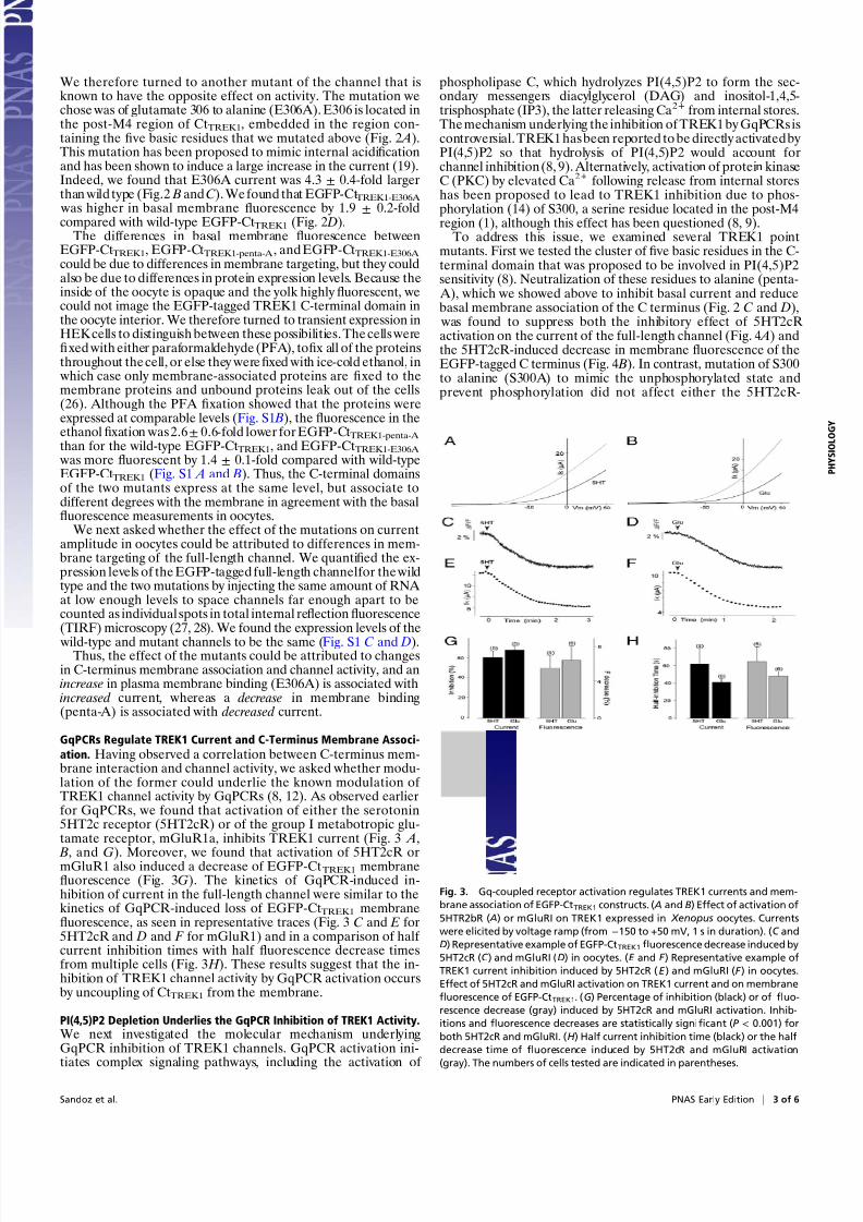
GqPCRs Regulate TREK1 Current and C-Terminus Membrane Associ-
ation. Having observed a correlation between C-terminus mem-brane interaction and channel activity, we asked whether modu-lation of the former could underlie the known modulation of TREK1 channel activity by GqPCRs (8, 12). As observed earlierfor GqPCRs, we found that activation of either the serotonin5HT2c receptor (5HT2cR) or of the group I metabotropic glu-tamate receptor, mGluR1a, inhibits TREK1 current (Fig. 3 A,
B, and G). Moreover, we found that activation of 5HT2cR ormGluR1 also induced a decrease of EGFP-CtTREK1 membranefluorescence (Fig. 3G). The kinetics of GqPCR-induced in-hibition of current in the full-length channel were similar to thekinetics of GqPCR-induced loss of EGFP-CtTREK1 membranefluorescence, as seen in representative traces (Fig. 3 C and E for5HT2cR and D and F for mGluR1) and in a comparison of half current inhibition times with half fluorescence decrease timesfrom multiple cells (Fig. 3 H ). These results suggest that the in-hibition of TREK1 channel activity by GqPCR activation occursby uncoupling of CtTREK1 from the membrane.
PI(4,5)P2 Depletion Underlies the GqPCR Inhibition of TREK1 Activity.We next investigated the molecular mechanism underlyingGqPCR inhibition of TREK1 channels. GqPCR activation ini-tiates complex signaling pathways, including the activation of
phospholipase C, which hydrolyzes PI(4,5)P2 to form the sec-ondary messengers diacylglycerol (DAG) and inositol-1,4,5-trisphosphate (IP3), the latter releasing Ca2+ from internal stores.The mechanism underlying the inhibition of TREK1 by GqPCRs iscontroversial. TREK1 has been reported to be directly activated by PI(4,5)P2 so that hydrolysis of PI(4,5)P2 would account forchannel inhibition (8, 9). Alternatively, activation of protein kinaseC (PKC) by elevated Ca2+ following release from internal storeshas been proposed to lead to TREK1 inhibition due to phos-
phorylation (14) of S300, a serine residue located in the post-M4region (1), although this effect has been questioned (8, 9).To address this issue, we examined several TREK1 point
mutants. First we tested the cluster of fi ve basic residues in the C-terminal domain that was proposed to be involved in PI(4,5)P2sensitivity (8). Neutralization of these residues to alanine (penta-
A), which we showed above to inhibit basal current and reducebasal membrane association of the C terminus (Fig. 2 C and D),
was found to suppress both the inhibitory effect of 5HT2cRactivation on the current of the full-length channel (Fig. 4 A) andthe 5HT2cR-induced decrease in membrane fluorescence of theEGFP-tagged C terminus (Fig. 4 B). In contrast, mutation of S300to alanine (S300A) to mimic the unphosphorylated state andprevent phosphorylation did not affect either the 5HT2cR-
Fig. 3. Gq-coupled receptor activation regulates TREK1 currents and mem-
brane association of EGFP-CtTREK1 constructs. ( A and B) Effect of activation of
5HTR2bR ( A) or mGluRI on TREK1 expressed in Xenopus oocytes. Currents
were elicited by voltage ramp (from −150 to +50 mV, 1 s in duration). (C and
D) Representative example of EGFP-CtTREK1fluorescence decrease induced by
5HT2cR (C ) and mGluRI (D) in oocytes. (E and F ) Representative example of
TREK1 current inhibition induced by 5HT2cR (E ) and mGluRI (F ) in oocytes.
Effect of 5HT2cR and mGluRI activation on TREK1 current and on membrane
fluorescence of EGFP-CtTREK1. (G) Percentage of inhibition (black) or of fluo-
rescence decrease (gray) induced by 5HT2cR and mGluRI activation. Inhib-
itions and fluorescence decreases are statistically significant (P < 0.001) for
both 5HT2cR and mGluRI. (H ) Half current inhibition time (black) or the half
decrease time of fluorescence induced by 5HT2cR and mGluRI activation
(gray). The numbers of cells tested are indicated in parentheses.
Sandoz et al. PNAS Early Edition | 3 of 6
P H Y S I O L O G
Y

8/3/2019 Optical Probing of a Dynamic Membrane Interaction
http://slidepdf.com/reader/full/optical-probing-of-a-dynamic-membrane-interaction 4/6
induced inhibition of channel current or reduction of C-terminal
membrane association (Fig. 4 A and B). Moreover, overnightpreincubation of oocytes with 5 μM of the PKC inhibitor cal-phostatin C had no effect on the 5HT2cR-induced inhibition of current or fluorescence decrease of EGFP-CtTREK1. To explorethe specificity of the disruption in modulation by 5HT2cR, wealso tested the neutralization of glutamate 306 (E306A), the siteimplicated in acid sensing (19), which we showed above to aug-ment basal channel activity and C-terminus membrane association(Fig. 2 C and D). This mutation did not alter the 5HT2cR-in-duced inhibition of channel current and reduction of C-terminusmembrane association (Fig. 4 A and B).
Taken together, these results show the inhibition of channelactivity by 5HT2cR depends specifically on the post-M4 poly-
basic motif and not on either PKC activation or on a generalsensitivity to mutations in the post-M4 region. This suggests thatthe key to GqPCR modulation of TREK1 is PI(4,5)P2 break-down and consequent loss of membrane interaction of theTREK1 C-terminal polybasic motif. We tested this notion fur-ther in the next set of experiments.
Is TREK1 Directly Regulated by PI(4,5)P2 in the Cell? Quenching thecharge of membrane phospholipids by polycationic agents such
as polylysine reduces TREK1 current (8, 9), and PI(4,5)P2 ap-plication to the intracellular surface restores TREK1 channelactivity (8). Therefore, it has been proposed that PI(4,5)P2 isa direct and specific regulator of TREK1 channels (8). To testthis model, we used two inducible enzymatic systems that spe-cifically deplete PI(4,5)P2, but which, unlike GqPCR activation,do not generate other signaling lipids such as DAG and IP3.First, we used Ci-VSP to hydrolyze PI(4,5)P2 to PI(4)P (Fig. 1 A–C). We found that, although Ci-VSP activation by membranedepolarization inhibited the PI(4,5)P2-dependent IRK1Q cur-rent, as shown earlier (29, 30), it did not affect TREK1 current(Fig. 5 A). In keeping with the selective inhibition of IRK1Q, theactivation of Ci-VSP decreased the membrane fluorescence of the C terminus of IRK1Q, but not of TREK1.
In a second approach, we used a chemically inducible PI(4,5)P2
depletion system based on the rapamycin-induced translocationof a phosphoinositide 5-phosphatase enzyme to the plasmamembrane, to evoke the rapid hydrolysis of (PI(4,5)P2 to PI(4)P)(31, 32). Whereas TREK1 was not inhibited by the application of 5 μM of rapamycin, IRK1Q current was strongly inhibited (Fig. 5 D, E, and G). One possible explanation for the difference in be-havior of TREK1 and IRK1Q is that TREK1 might have a higherPI(4,5)P2 af finity than this lower-af finity mutant of IRK1 and,therefore, TREK1 may be insensitive to the degree PI(4,5)P2depletion induced by rapamycin system. We therefore examined
wild-type IRK1 (IRK1-WT), which has a very high PI(4,5)P2 af-finity. As shown in Fig. 5 F and G, IRK1-WT current was signif-icantly inhibited by the rapamycin-inducible 5-phosphatase ( P <0.05) (Fig. 5 F and G). The IRK1-WT inhibition was smaller than
Fig. 4. Point mutations in C-terminal domain regulate TREK1 response to
GPCR activation. ( A) Percentage of current inhibition by 5HT2cR activation for
TREK1-WT, TREK1-Penta-A, TREK1-E306, TREK1-S300A, and TREK1-WT pre-
incubated with calphostatin C. (B) Relative fluorescence decrease induced by
5HT2cR activation for EGFP-CtTREK1WT, EGFP-CtTREK1-Penta-A, EGFP-CtTREK1-E306,
EGFP-CtTREK1-S300A, and TREK1-WT preincubated with calphostatin C (over-
night 5 μM). The asterisks indicate the significance (P < 0.05) of the difference
between TREK1 mutants and TREK1-WT (or EGFP-CtTREK1 mutants and EGFP-
CtTREK1). Student’s t test: ***P < 0.001. The numbers of cells tested are in-
dicated in parentheses.
Fig. 5. TREK1 is not directly regulated by PI(4,5)P2. ( A) TREK1 current inhibition induced by the activation of Ci-VSP by a prepulse to +150 mV (200 ms).
Currents were measured 15 s after the prepulse. ( B) Representative examples of EGFP-CtTREK1 and EFGP-PHPLC fluorescence decrease induced by Ci-VSP ac-
tivation (pulse at +150 mV from −80 mV, 200-ms duration). (C ) Relative fluorescence decrease of EFGP-PHPLC and EGFP-CtTREK1 and EGFP-CtIRK-R228Q induced by
Ci-VSP. (D–G) PI(4,5)P2 depletion by rapamycin-inducible 5-phosphatase [PI(4,5)P2→ PI(4)P] failed to regulate TREK current. (D–F ) Effect of activation of
5-phosphatase by rapamycin (5 μM) on TREK1 (D), IRK1-R228Q (E ), and IRK1-WT (F ). Currents before (dashed lines) and after rapamycin (solid lines) were
elicited by voltage ramps (from −50 to +50 mV, 1 s in duration). ( G) Inhibition induced by PI(4,5)P2 depletion by the rapamycin system on TREK1, IRK1-R228Q,
and IRK1-WT. Student’s t test: **P < 0.01, ***P < 0.001. The numbers of cells tested are indicated in parentheses.
4 of 6 | www.pnas.org/cgi/doi/10.1073/pnas.1015788108 Sandoz et al.
8/3/2019 Optical Probing of a Dynamic Membrane Interaction
http://slidepdf.com/reader/full/optical-probing-of-a-dynamic-membrane-interaction 5/6
inhibition seen in the IRK1Q mutant (40± 10% versus 87.5± 4%,respectively), consistent with a higher af finity of IRK1-WT forPI(4,5)P2 (29). These data show that the rapamycin-inducible 5-phosphatase can reveal specific PI(4,5)P2 interactions even fora high-af finity PI(4,5)P2 interaction and, therefore, suggests thatTREK1 is regulated differently from IRK1.
Thus, TREK1 is inhibited by PI(4,5)P2 conversion to DAG andIP3 by GqPCR activation, as shown above, but not by PI(4,5)P2hydrolysis to PI(4)P. In others words, the modulation of TREK1
activity appears to be compatible with membrane association of the C terminus with either PI(4,5)P2 or PI(4)P, suggesting that it ismediated by a nonspecific electrostatic interaction with the post-M4 polybasic motif of the C terminus. This kind of interactiondiffers from the stereo-specific binding of PI(4,5)P2 to PHPLC andis more like the nonspecific electrostatic membrane binding of theMARKS peptide (33).
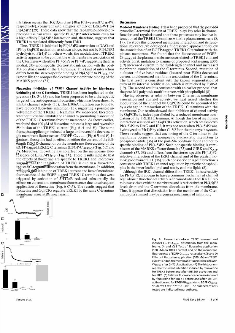
Fluoxetine Inhibition of TREK1 Channel Activity by Membrane
Unbinding of the C Terminus. TREK1 has been implicated in de-pression (18, 34, 35) and has been proposed to be a therapeutictarget of the antidepressant fluoxetine, which has been shown toinhibit channel activity (15). The E306A mutation was found tohave reduced fluoxetine inhibition (15), suggesting a possible in-
volvement of the TREK1 C terminus. We wondered, therefore,
whether fluoxetine inhibits the channel by promoting dissociationof the TREK1 C terminus from the membrane. As shown earlier, we found that 100 μM of fluoxetine induced a large and reversibleinhibition of the TREK1 current (Fig. 6 A and E). The samefluoxetine application induced a large and reversible decrease inthe membrane fluorescence of EGFP-CtTREK1 (Fig. 6 B and F ).Incontrast, fluoxetine had no effect on either the current of the full-length IRK1Q channel or on the membrane fluorescence of theEGFP-tagged IRK1Q C terminus (EFGP-CtIRK1Q) (Fig. 6 E and F ). Moreover, fluoxetine has no effect on the membrane fluo-rescence of EFGP-PHPLC (Fig. 6 F ). These results indicate thatthe effects of fluoxetine are specific to TREK1 and, moreover,suggest that the inhibition of TREK1 is due to a fluoxetine-induced C-terminus dissociation from the membrane. In addition,
we found that inhibition of TREK1 current and loss of membrane
fluorescence of the EGFP-tagged TREK1 C terminus that weretriggered by activation of 5HT2cR reduced substantially theeffects on current and membrane fluorescence due to subsequentapplication of fluoxetine (Fig. 6 C– F ). The results suggest thatfluoxetine and GqPCRs regulate TREK1 by the same C-terminusmembrane association mechanism.
Discussion
Model of Membrane Binding. It has been proposed that the post-M4cytosolic C-terminal domain of TREK1 plays key roles in channelfunction and regulation and that these processes may involve in-teraction of the TREK1 C terminus with the plasma membrane (8,19). To study this proposed membrane interaction and its func-tional relevance, we developed a fluorescence approach to followthe association of an EGFP-tagged TREK1 C terminus with theplasma membrane. We found that the fluorescence of EGFP-
CtTREK1 at the plasma membrane is correlated to TREK1 channelactivity. First, mutation to alanine of proposed acid sensing E306(19) increased current in the full-length channel and increasedmembrane association of the C terminus. Second, mutation of a cluster of fi ve basic residues (located near E306) decreasedcurrent and decreased membrane association of the C terminus.The first result is consistent with the known augmentation of current by internal acidification, which is mimicked by E306A (19). The second result is consistent with an earlier proposal thatthe post-M4 polybasic motif interacts with phospholipid (8).
Having observed a relation between C-terminus membraneassociation and channel activity, we next asked whether themodulation of the channel by GqPCRs could be accounted forby a change in interaction of the TREK1 C terminus with themembrane. Our results showed that inhibition of channel activity by GqPCRs is, indeed paralleled by, a reduced membrane asso-ciation of the TREK1 C terminus. Although this loss of membraneinteraction was seen with GqPCRs activation, which breaks downPI(4,5)P2 to DAG and IP3, it was not seen when PI(4,5)P2 washydrolyzed to PI(4)P by either Ci-VSP or the rapamycin system.These results suggest that anchoring of the C terminus to themembrane occurs via a nonspecific electrostatic interaction tophosphoinositide (36) of the post-M4 polybasic motif and not tospecific binding of PI(4,5)P2. Such nonspecific binding is remi-niscent of the MARKS effector domain (33) and GIRK and K ATPchannels (37, 38) and differs from the stereo-specific PI(4,5)P2-selective interaction of the IRK1 channel and of the plextrin ho-mology domain of PLC (36). Such nonspecific charge interaction isconsistent with TREK1 channel regulation by anionic phospholi-pids in the inner leaflet lipid and not by cationic lipids (8).
Although the IRK1 channel differs from TREK1 in its selectivity
for PI(4,5)P2, it appears to have a common mechanism of channelregulation in that channel activity is enhanced when theIRK1 C ter-minus associates with the membrane and is reduced when PI(4,5)P2levels drop and the C terminus dissociates from the membrane.Thus, it appears that dissociation from the membrane of the C ter-minus of a channel may be a general mechanism of inhibition.
Fig. 6. Fluoxetine reduces TREK1 current and
induces EGFP-CtTREK1 dissociation from the mem-
brane. ( A and C ) Effect of fluoxetine application
(100 μM) on TREK1 current and on the membrane
fluorescence of EGFP-CtTREK1, respectively.(B and D)
Effect of fluoxetine application (100 μM) on TREK1
current andon themembranefluorescence of EGFP-
CtTREK1 after 5HT2cR activation. (E ) The histograms
represent current inhibition induced by fluoxetine
for TREK1 before and after 5HT2cR activation and
for IRK1. (F ) Relative fluorescence decrease induced
by fluoxetine for TREK1 before and after 5HT2cR
activation and for EFGP-PHPLCandand EGFP-CtIRK1Q.
Student’s t test: ***P < 0.001. The numbers of cells
tested are indicated in parentheses.
Sandoz et al. PNAS Early Edition | 5 of 6
P H Y S I O L O G
Y
8/3/2019 Optical Probing of a Dynamic Membrane Interaction
http://slidepdf.com/reader/full/optical-probing-of-a-dynamic-membrane-interaction 6/6
Mechanism of Modulation by Fluoxetine. TREK1 has been impli-cated in mood regulation. TREK1 knockout mice (TREK1− / −)display a depression-resistant phenotype (18). Recently, it wasreported that spadin, a sortilin-derived peptide, targets TREK1 togenerate an antidepressive effect (35). In addition, fluoxetine, thechemical name of the widely used antidepressant Prozac, has beenshown to inhibit TREK1 at clinical concentrations (15). Moreover,fluoxetine administration to TREK1− / − mice does not affect theirbehavior (18). These results suggest that TREK1 is an importanttarget for antidepressive drugs,includingfluoxetine. It is importantto determine the mechanism of fluoxetine inhibition. Fluoxetineinhibition of TREK1 is not voltage dependent and is less effectiveon the E306A mutant, which has a higher open probability (15).This argues that the binding site is unlikely to be in the pore or
within the span of the membrane, but, rather, is on an internal- orexternal-facing portion of the channel. Our experiments show thatfluoxetine induces dissociation of the TREK1 C terminus from theplasma membrane. This provides a mechanistic explanation forhow fluoxetine inhibits TREK1 channel activity.
In summary, we describe a unique technique for monitoring thereversible plasma membrane association of protein domains usingepifluorescence without scanning, optical slicing, or imaging. Themethod can be used to follow changes in lipid composition thatresult from membrane signaling events or to study the binding of membrane by cytoplasmic regulatory domains of ion channels. We
find that dynamic changes in the association of the C-terminalcytoplasmic domain of the TREK1 channel with the plasmamembrane that result from activation of GqPCRs arecorrelated tochanges in channel activity, with reduced membrane binding beingassociated with inhibition of current. The membrane associationappears to depend on interaction between a conserved polybasicmotif in the C terminus and the most abundant phosphoinositol in
the plasma membrane, PI(4,5)P2, although this interaction is notas phospholipid specific as in IRK1 channels. The inhibitory effectof fluoxetine on TREK1 channel activity, which has been proposedto underlie its antidepressant effects, is associated with C-terminusdissociation from the membrane. We propose that this fluores-cence assay for C-terminus membrane association could be used toscreen for alternative small molecule inhibitors of TREK1, whichmay be potent and specific antidepressants.
Materials and MethodsElectrophysiology and Fluorometry. Defolliculated Xenopus oocytes were
injected with 50 nL of cRNA at 0.02–0.4 μg/ μL and recorded 2–4 d later. For
electrophysiology, single oocytes were placed in a 0.3-mL perfusion chamber
and impaled with two standard microelectrodes (1–2.5 MΩ resistance) filled
with 3 M KCl and voltage clamped with a Dagan CA-1 amplifier, in ND96
solution (96 mM NaCl, 2 mM KCl, 1.8 mM CaCl2, 2 mM MgCl2, 5 mM Hepes,
pH 7.4 with NaOH). Stimulation of the preparation, data acquisition, and
analysis were performed using pClamp software (Axon Instruments).
For fluorometry, a Diaphot inverted microscope with a 20× 0.75 NA
fluorescence objective (Nikon) was used with illumination by a 150-W xenon
lamp. Fluorescence was measured with a Hamamatsu HC120-05 photo-
multiplier tube. The amplifier, photomultiplier and Uniblitz shutter (Vincent
Associates) were controlled by a Digidata-1440 board and pClamp10 soft-
ware package (Axon Instruments). Filter sets were: tagRFP (HQ535/50ex,
HQ610/75em, Q565LP dichroic) and EGFP (HQ480/40ex, HQ535/50em,
Q505LP dichroic) (Chroma Technology). Fluorescence signals were low-pass
filtered at 2 kHz through an eight-pole Bessel filter (Frequency Devices).
For additional methods, see SI Materials and Methods.
ACKNOWLEDGMENTS. We thank S. Wiese and T. Kim for excellent technicalassistance; J. Levitz, R. Arant, J. Patti, and S. Kohout for helpful discussion;and the Fulbright Foundation (G.S.), Philippe Foundation (G.S.), and theNational Institutes of Health (R01 NS35549) (to E.Y.I.) for support.
1. Fink M, et al. (1996) Cloning, functional expression and brain localization of a novel
unconventional outward rectifier K+ channel. EMBO J 15:6854–6862.
2. Maingret F, et al. (2000) TREK-1 is a heat-activated background K(+) channel. EMBO J
19:2483–2491.
3. Patel AJ, et al. (1998) A mammalian two pore domain mechano-gated S-like K+
channel. EMBO J 17:4283–4290.
4. Sandoz G, Douguet D, Chatelain F, Lazdunski M, Lesage F (2009) Extracellular
acidification exerts opposite actions on TREK1 and TREK2 potassium channels via
a single conserved histidine residue. Proc Natl Acad Sci USA 106:14628–14633.5. Maingret F, Patel AJ, Lesage F, Lazdunski M, Honoré E (1999) Mechano- or acid
stimulation, two interactive modes of activation of the TREK-1 potassium channel.
J Biol Chem 274:26691–26696.
6. Fink M, et al. (1998) A neuronal two P domain K+ channel stimulated by arachidonic
acid and polyunsaturated fatty acids. EMBO J 17:3297–3308.
7. Maingret F, Patel AJ, Lesage F, Lazdunski M, Honoré E (2000) Lysophospholipids open
the two-pore domain mechano-gated K(+) channels TREK-1 and TRAAK. J Biol Chem
275:10128–10133.
8. Chemin J, et al. (2005) A phospholipid sensor controls mechanogating of the K+
channel TREK-1. EMBO J 24:44–53.
9. Lopes CM, et al. (2005) PIP2 hydrolysis underlies agonist-induced inhibition and
regulates voltage gating of two-pore domain K+ channels. J Physiol 564:117–129.
10. Patel AJ, et al. (1999) Inhalational anesthetics activate two-pore-domain background
K+ channels. Nat Neurosci 2:422–426.
11. Duprat F, et al. (2000) The neuroprotective agent riluzole activates the two P domain
K(+) channels TREK-1 and TRAAK. Mol Pharmacol 57:906–912.
12. Sandoz G, et al. (2006) AKAP150, a switch to convert mechano-, pH- and arachidonic
acid-sensitive TREK K(+) channels into open leak channels. EMBO J 25:5864–5872.13. Sandoz G, et al. (2008) Mtap2 is a constituent of the protein network that regulates
twik-related K+ channel expression and trafficking. J Neurosci 28:8545–8552.
14. Murbartián J, Lei Q, Sando JJ, Bayliss DA (2005) Sequential phosphorylation mediates
receptor- and kinase-induced inhibition of TREK-1 background potassium channels.
J Biol Chem 280:30175–30184.
15. Kennard LE, et al. (2005) Inhibition of thehuman two-poredomain potassiumchannel,
TREK-1, by fluoxetine and its metabolite norfluoxetine. Br J Pharmacol 144:821–829.
16. Heurteaux C, et al. (2004) TREK-1, a K+ channel involved in neuroprotection and
general anesthesia. EMBO J 23:2684–2695.
17. Alloui A, et al. (2006) TREK-1, a K+ channel involved in polymodal pain perception.
EMBO J 25:2368–2376.
18. Heurteaux C, et al. (2006) Deletion of the background potassium channel TREK-1
results in a depression-resistant phenotype. Nat Neurosci 9:1134–1141.
19. Honoré E, Maingret F, Lazdunski M, Patel AJ (2002) An intracellular proton sensor
commandslipid-andmechano-gatingofthe K(+)channelTREK-1. EMBOJ 21:2968–2976.
20. Mannuzzu LM, Moronne MM, Isacoff EY (1996) Direct physical measure of confor-
mational rearrangement underlying potassium channel gating. Science 271:213–216.
21. Murata Y, Iwasaki H, Sasaki M, Inaba K, Okamura Y (2005) Phosphoinositide
phosphatase activity coupled to an intrinsic voltage sensor. Nature 435:1239–1243.
22. Várnai P, Balla T (2007) Visualization and manipulation of phosphoinositide dynamics
in live cells using engineered protein domains. P fl ugers Arch 455:69–82.
23. Halaszovich CR, Schreiber DN, Oliver D (2009) Ci-VSP is a depolarization-activated
phosphatidylinositol-4,5-bisphosphate and phosphatidylinositol-3,4,5-trisphosphate
5′-phosphatase. J Biol Chem 284:2106
–2113.
24. Balla T, Varnai P (2009) Visualization of cellular phosphoinositide pools with GFP-
fused protein-domains. Curr Protoc Cell Biol , Chapter 24:Unit 24.4.
25. Kohout SC, et al. (2010) Electrochemical coupling in the voltage-dependent
phosphatase Ci-VSP. Nat Chem Biol 6:369–375.
26. Kalejta RF, Shenk T, Beavis AJ (1997) Use of a membrane-localized green fluorescent
protein allows simultaneous identification of transfected cells and cell cycle analysis
by flow cytometry. Cytometry 29:286–291.
27. Ulbrich MH, Isacoff EY (2008) Rules of engagement for NMDA receptor subunits. Proc
Natl Acad Sci USA 105:14163–14168.
28. Tombola F, Ulbrich MH, I sacoff EY (2008) The voltage-gated proton channel Hv1 has
two pores, each controlled by one voltage sensor. Neuron 58:546–556.
29. Zhang H, He C, Yan X, Mirshahi T, Logothetis DE (1999) Activation of inwardly
rectifying K+ channels by distinct PtdIns(4,5)P2 interactions. Nat Cell Biol 1:183–188.
30. Kohout SC, Ulbrich MH, Bell SC, Isacoff EY (2008) Subunit organization and functional
transitions in Ci-VSP. Nat Struct Mol Biol 15:106–108.
31. Suh BC, Inoue T, Meyer T, Hille B (2006) Rapid chemically induced changes of PtdIns
(4,5)P2 gate KCNQ ion channels. Science 314:1454–1457.
32. Varnai P, Thyagarajan B, Rohacs T, Balla T (2006) Rapidly inducible changes in
phosphatidylinositol 4,5-bisphosphate levels influence multiple regulatory functions
of the lipid in intact living cells. J Cell Biol 175:377–382.
33. McLaughlin S, Hangyás-Mihályné G, Zaitseva I, Golebiewska U (2005) Reversible—
through calmodulin—electrostatic interactions between basic residues on proteins
and acidic lipids in the plasma membrane. Biochem Soc Symp 72):189–198.
34. Perlis RH, et al. (2008) Pharmacogenetic analysis of genes implicated in rodent models
of antidepressant response: Association of TREK1 and treatment resistance in the
STAR(*)D study. Neuropsychopharmacology 33:2810–2819.
35. Mazella J, et al. (2010) Spadin, a sortilin-derived peptide, targeting rodent TREK-1
channels: A new concept in the antidepressant drug design. PLoS Biol 8:e1000355.
36. DiNitto JP, Cronin TC, Lambright DG (2003) Membrane recognition and targeting by
lipid-binding domains. Sci STKE 2003:re16.
37. Rohács T, Chen J, Prestwich GD, Logothetis DE (1999) Distinct specificities of inwardly
rectifying K(+) channels for phosphoinositides. J Biol Chem 274:36065–36072.
38. Rohács T, et al. (2003) Specificity of activation by phosphoinositides determines lipid
regulation of Kir channels. Proc Natl Acad Sci USA 100:745–750.
6 of 6 | www.pnas.org/cgi/doi/10.1073/pnas.1015788108 Sandoz et al.


