
Ovarian Serous Borderline Ovarian Serous Borderline TumorsTumors
Rouba Ali-Fehmi,MD
The Karmanos Cancer Institute, Wayne State University School of Medicine


omentum
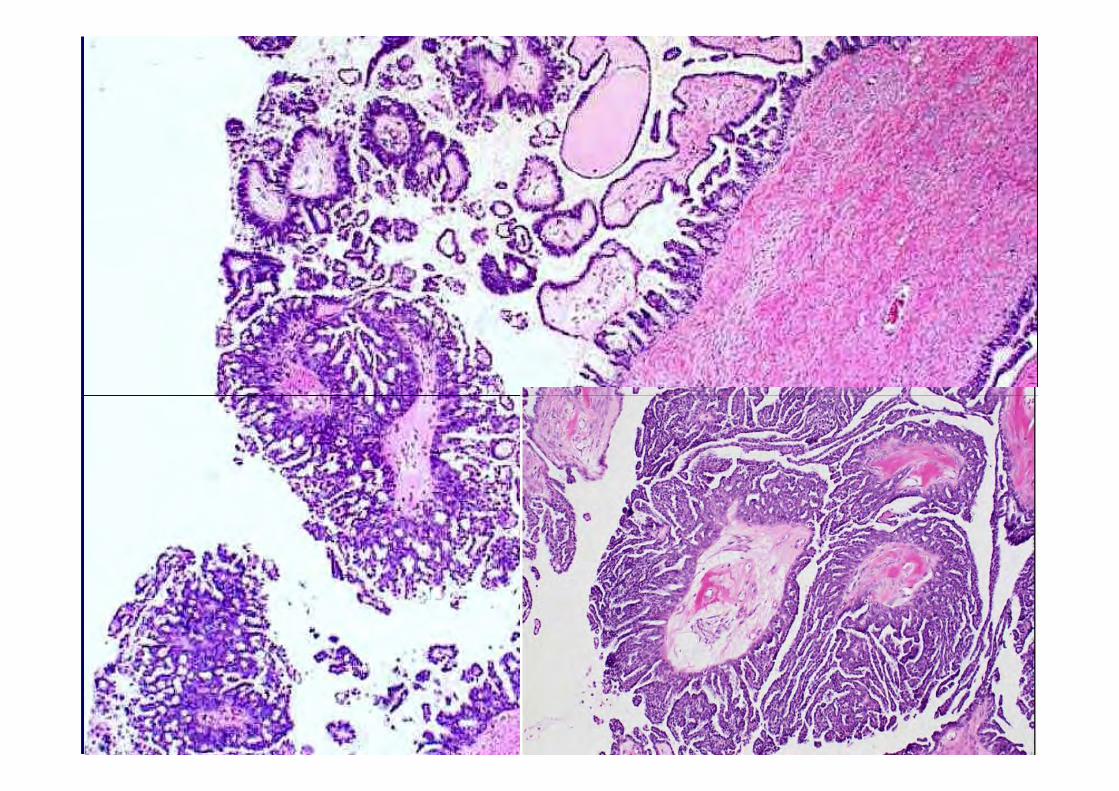
Pathology:Pathology:
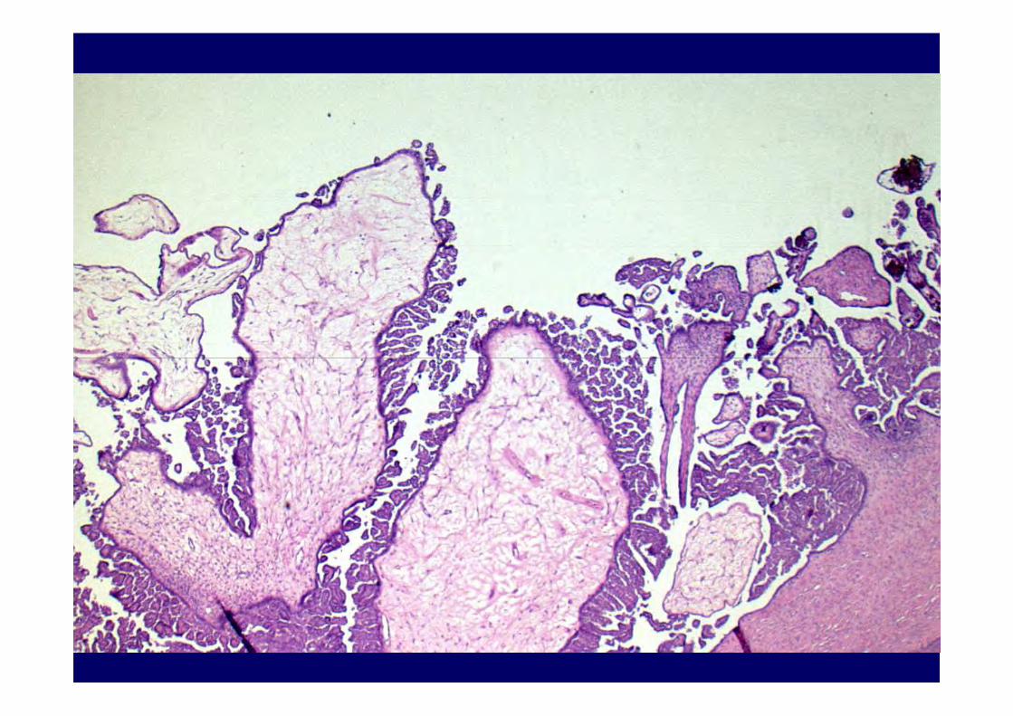
• Left ovary: Serous borderline tumor, with micropapillary pattern, – endophytic and exophytic components are
seen, seen,
• Omentum & uterine serosa: positive for invasive implants.
• Lymph nodes: negative• Cytology: positive for serous
neoplasm.

Ovarian Serous Borderline TumorsOvarian Serous Borderline Tumors
• Identified more than 100 years ago
• Popularized in 1960s-70s
• Initially controversial
• Adopted in classifications of international organizations (FIGO, WHO, ISGP)
• Continues to be challenged
Ovarian Serous Borderline Tumors Ovarian Serous Borderline Tumors Historical Synonyms,Historical Synonyms,
• Semimalignant tumor
• Low potential malignancy• Low potential malignancy
• Borderline malignancy (carcinoma of LMP)
• Tumor of LMP
• Atypical proliferative tumor
WHO Classification of Ovarian TumorsWHO Classification of Ovarian TumorsSurface EpithelialSurface Epithelial--Stromal TumorsStromal Tumors
Degree of Epithelial ProliferationBenignBenignBorderlineMalignant
World Health Organization World Health Organization Classification of Ovarian TumorsClassification of Ovarian Tumors
Borderline Tumors Borderline Tumors -- CriteriaCriteria
• Epithelial proliferation greater than that seen in benign tumors of the same cell seen in benign tumors of the same cell type
• Destructive or obvious stromal invasion is not present

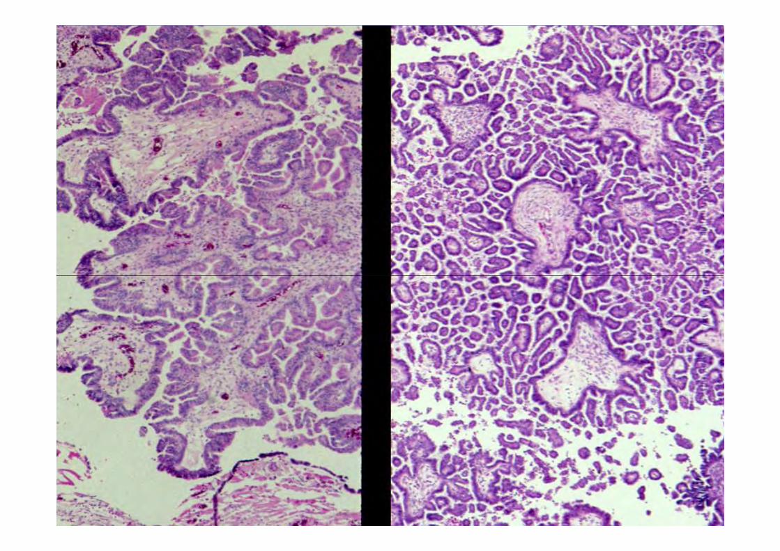
Borderline TumorsBorderline TumorsMicroscopic CriteriaMicroscopic Criteria
Cellular stratification Detached cell clustersDetached cell clustersMitotic activityNuclear atypia ABSENT of destructive stromal invasion
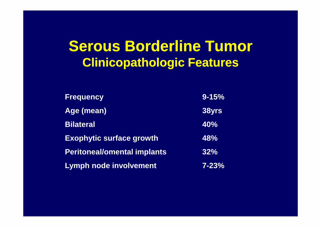
Serous Borderline TumorSerous Borderline TumorClinicopathologic FeaturesClinicopathologic Features
Frequency 9-15%
Age (mean) 38yrsAge (mean) 38yrs
Bilateral 40%
Exophytic surface growth 48%
Peritoneal/omental implants 32%
Lymph node involvement 7-23%
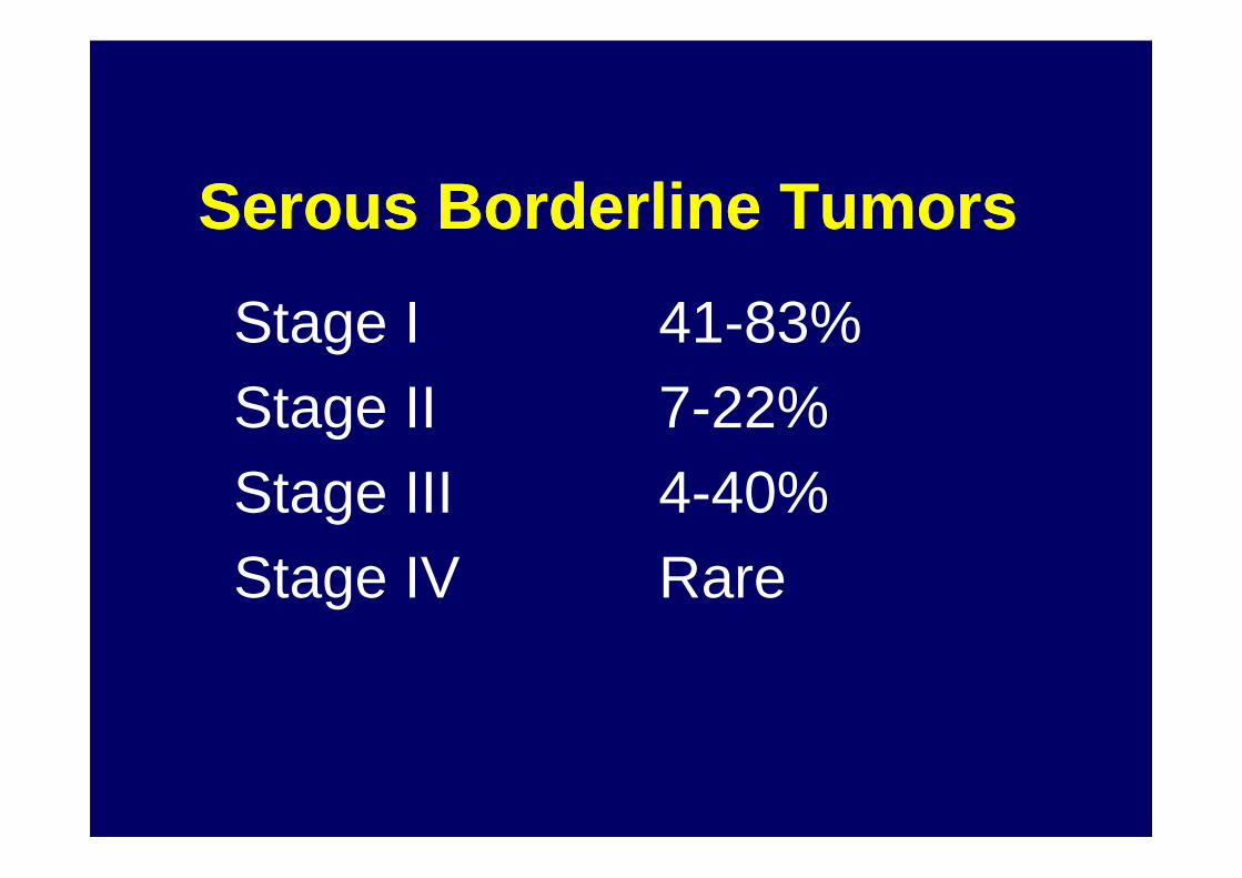
Serous Borderline TumorsSerous Borderline Tumors
Stage I 41-83%Stage II 7-22%Stage II 7-22%Stage III 4-40%Stage IV Rare
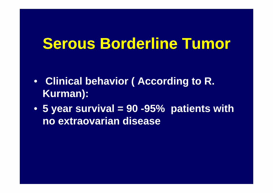
Serous Borderline TumorSerous Borderline Tumor
• Clinical behavior ( According to R. Kurman): Kurman):
• 5 year survival = 90 -95% patients with no extraovarian disease

Serous Borderline Tumors SurvivalSerous Borderline Tumors Survival
Review of the Literature
2,104 cases
Surveillance, Epidemiology and End Result (SEER)
2818 women 1988-1997
Stage I 99.5%Stage II and III 70%
10 yr relative survival by stageStage I 97%Stage II 90%Stage III 88%Stage IV 69%Total II-IV 82%
Seidman JD, Kurman RJ Hum Pathol 31:539-577, 2000 Trimble CL et al Gynecol Oncol 86:34, 2002
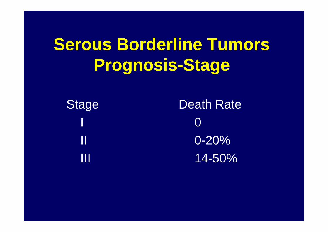
Serous Borderline TumorsSerous Borderline TumorsPrognosisPrognosis--StageStage
Stage Death RateI 0I 0II 0-20%III 14-50%
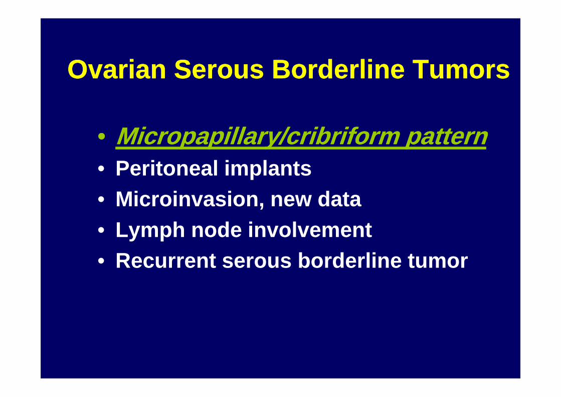
Ovarian Serous Borderline TumorsOvarian Serous Borderline Tumors
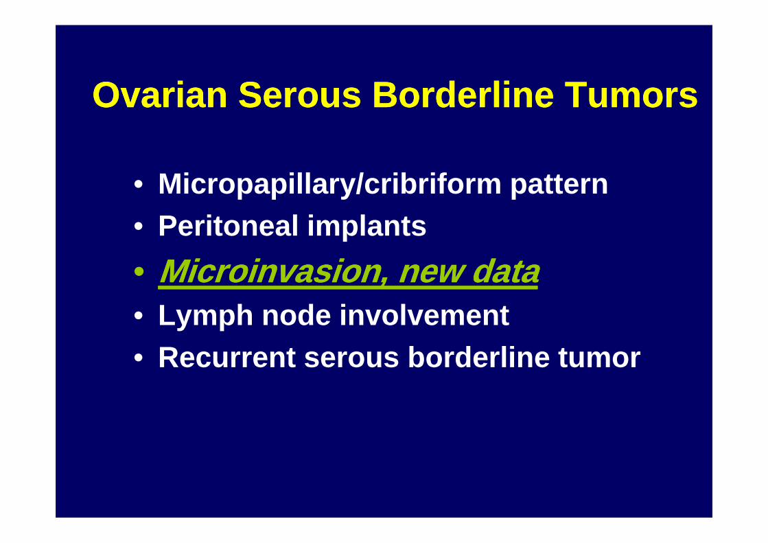
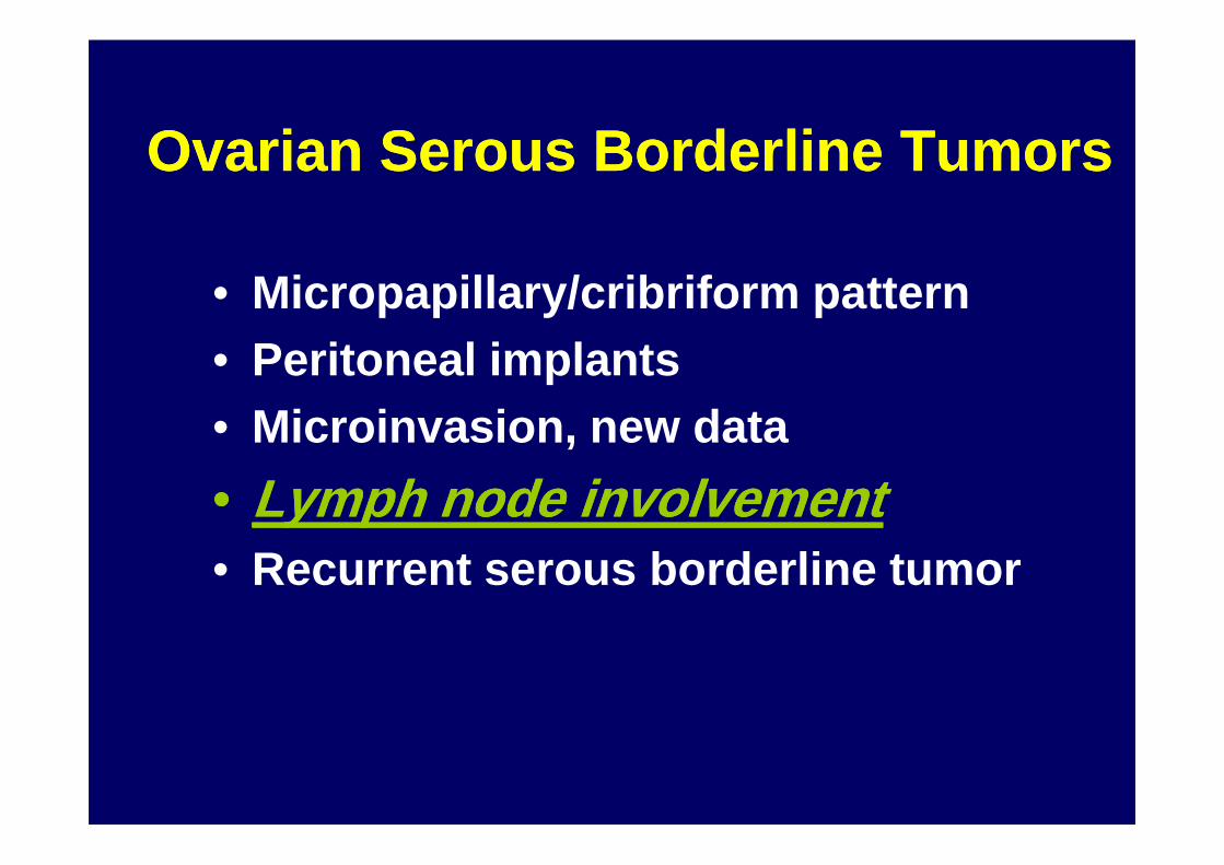
•• Micropapillary/cribriform patternMicropapillary/cribriform pattern• Peritoneal implants• Microinvasion, new data• Microinvasion, new data• Lymph node involvement• Recurrent serous borderline tumor
Serous Borderline TumorsSerous Borderline Tumors
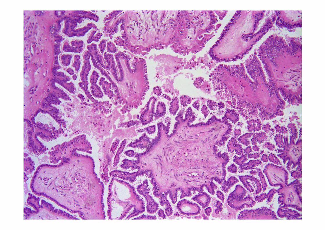
• Typical
• Micropapillary/cribriform
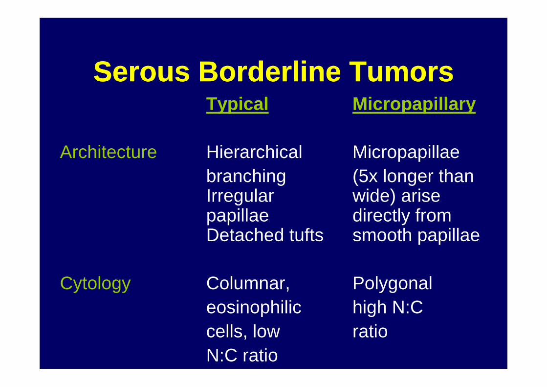
Serous Borderline TumorsSerous Borderline TumorsTypical Micropapillary
Architecture Hierarchical Micropapillaebranching (5x longer than Irregular wide) arise Irregular wide) arise papillae directly from Detached tufts smooth papillae
Cytology Columnar, Polygonaleosinophilic high N:Ccells, low ratioN:C ratio


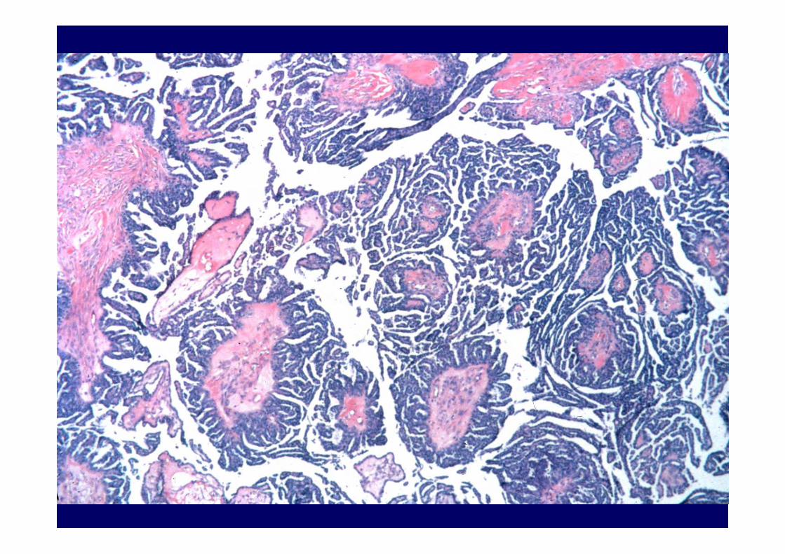

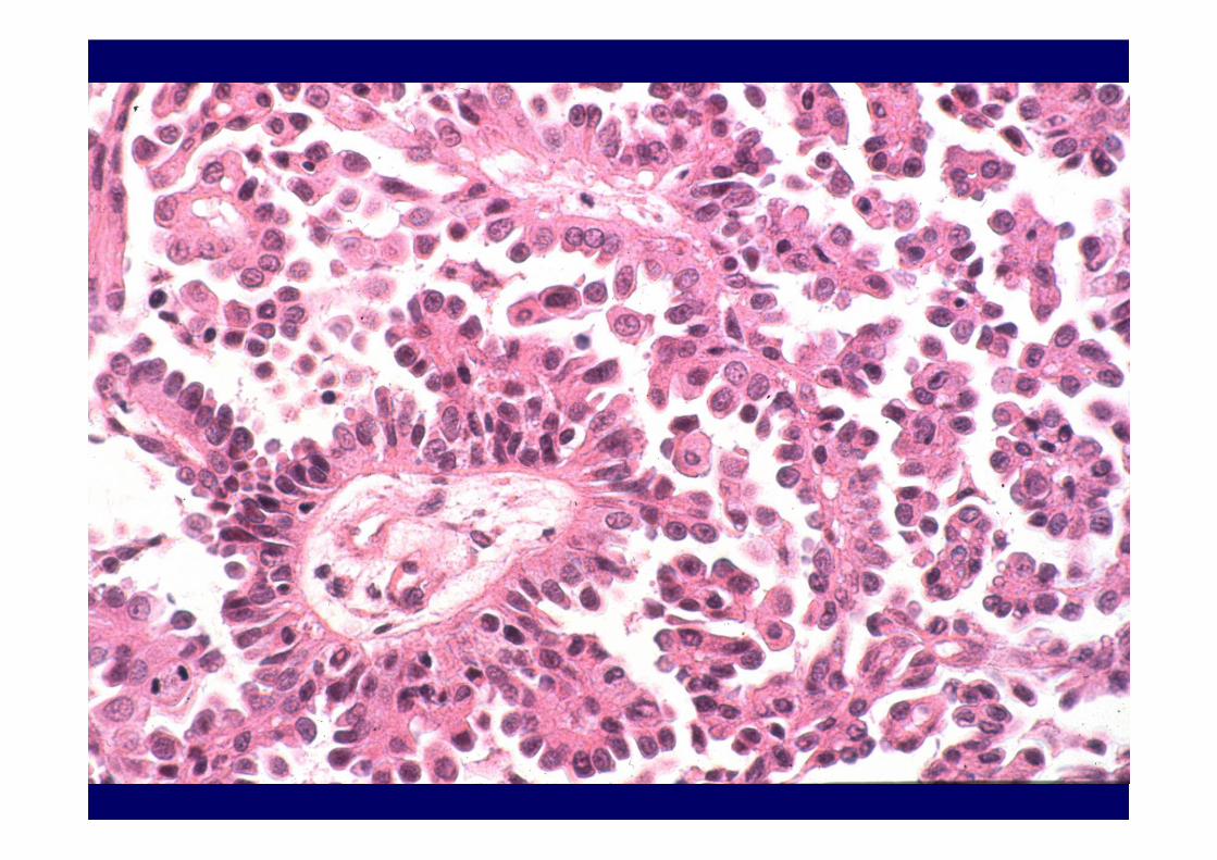

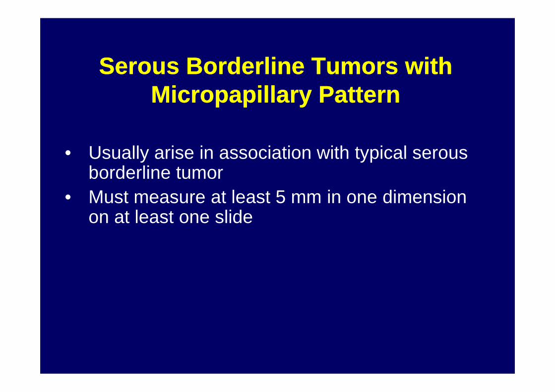
Serous Borderline Tumors withSerous Borderline Tumors withMicropapillary PatternMicropapillary Pattern
• Usually arise in association with typical serous borderline tumor
• Must measure at least 5 mm in one dimension • Must measure at least 5 mm in one dimension on at least one slide

Johns Hopkins Group Johns Hopkins Group
• Low grade serous carcinoma• Micropapillary serous carcinoma
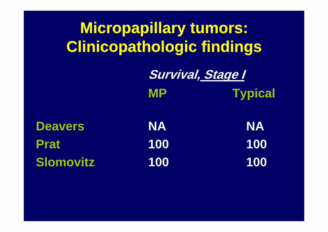
Micropapillary tumors: Micropapillary tumors: Clinicopathologic findingsClinicopathologic findings
Survival, Stage IMP Typical
Deavers NA NAPrat 100 100Slomovitz 100 100

Micropapillary tumors: Micropapillary tumors: Survival Stage II+Survival Stage II+
Sig? MP Typical
Deavers No 72 85Noninv 67 87Inv Impl 33 60Inv Impl 33 60
Prat NoNoninv 100 100Inv Impl 0 75
Slomovitz NoNoninv 100 100
Longacre No
If there is no difference in If there is no difference in survival stage by stagesurvival stage by stage-- -- -- --
Should micropapillary tumors be Should micropapillary tumors be separated from typical serous borderline tumors?
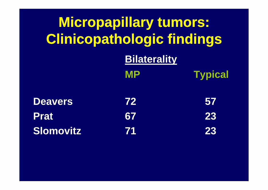
Micropapillary tumors: Micropapillary tumors: Clinicopathologic findingsClinicopathologic findings
Bilaterality
MP Typical
Deavers 72 57Prat 67 23Slomovitz 71 23
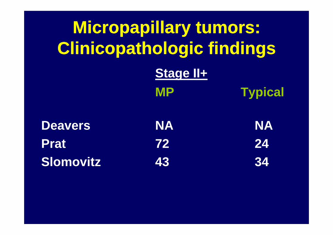
Micropapillary tumors: Micropapillary tumors: Clinicopathologic findingsClinicopathologic findings
Stage II+
MP Typical
Deavers NA NAPrat 72 24Slomovitz 43 34
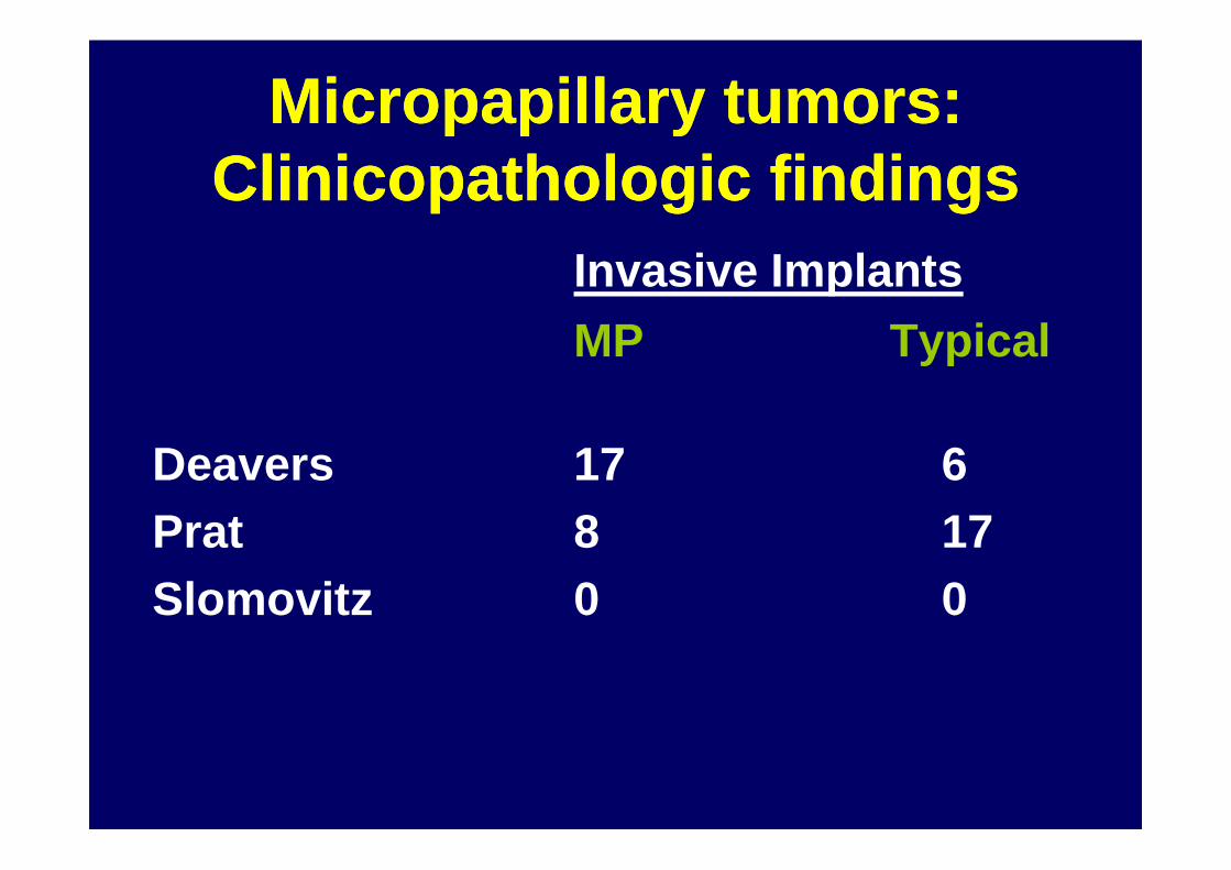
Micropapillary tumors: Micropapillary tumors: Clinicopathologic findingsClinicopathologic findings
Invasive Implants
MP Typical
Deavers 17 6Prat 8 17Slomovitz 0 0
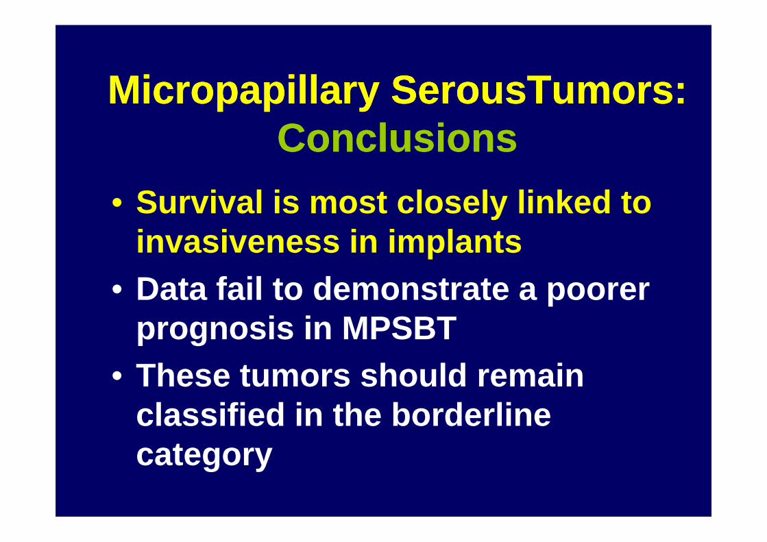
Micropapillary SerousTumors: Micropapillary SerousTumors: ConclusionsConclusions
• Survival is most closely linked to invasiveness in implants
• Data fail to demonstrate a poorer • Data fail to demonstrate a poorer prognosis in MPSBT
• These tumors should remain classified in the borderline category
Micropapillary Serous Tumors: Micropapillary Serous Tumors: ConclusionsConclusions
• MPSBT have distinct features (age, bilaterality, stage, invasive implants) implants)
• Warrant clear distinction from typical SBT in pathology report

Ovarian Serous Borderline TumorsOvarian Serous Borderline Tumors
• Micropapillary/cribriform pattern
•• Peritoneal implantsPeritoneal implants• Microinvasion, new data• Microinvasion, new data• Lymph node involvement• Recurrent serous borderline tumor

Serous Borderline TumorsSerous Borderline TumorsPeritoneal ImplantsPeritoneal Implants
•• NoninvasiveNoninvasiveNoninvasive of underlying tissue
•• InvasiveInvasive•• InvasiveInvasiveIrregular infiltration of underlying tissue
– Bell, DA Hum Pathol 22:750, 1991
Serous Borderline TumorsSerous Borderline TumorsPeritoneal ImplantsPeritoneal Implants
• Outcome of patients with advanced stage disease (implants) correlates withType of implantType of implant– Noninvasive– Invasive

Implant Status and Outcome Implant Status and Outcome 467 467 PatientsP atients
No. of Series Total Stage II/III Noninvasive Invasive Fllow up MeanPatient Deaths/Total Deaths/Total Mean, months
22 467 17/363 35/104 89
Survival 95.3% 66%Survival 95.3% 66%
Seidman JD, Kurman RJ Hum Pathol 31:539-577, 2000

Tissue Invasion Criteria and PrognosisTissue Invasion Criteria and PrognosisSurvival (%)
Author Noninvasive InvasiveMcCaughey 11/13 (85) 0/5 (0)D. Bell 47/50 (94) 1/6 (17)De Nictolis 10/10 (100) 5/9 (56)Eichhorn 13/13 (100) 0/2 (0)Eichhorn 13/13 (100) 0/2 (0)Prat 34/34 (100) 0/5 (0)Gilks 39/42 (93) 3/6 (50)Seidman 43/51(84) 1/3 (33)Deavers 78/91 (84) 4/8 (50)Longacre Highly sig, uni, multivariate

Non-invasive implant
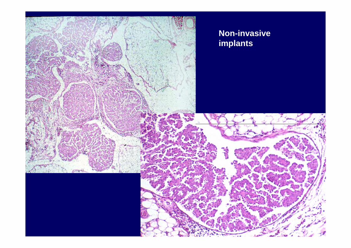
Non-invasive implants
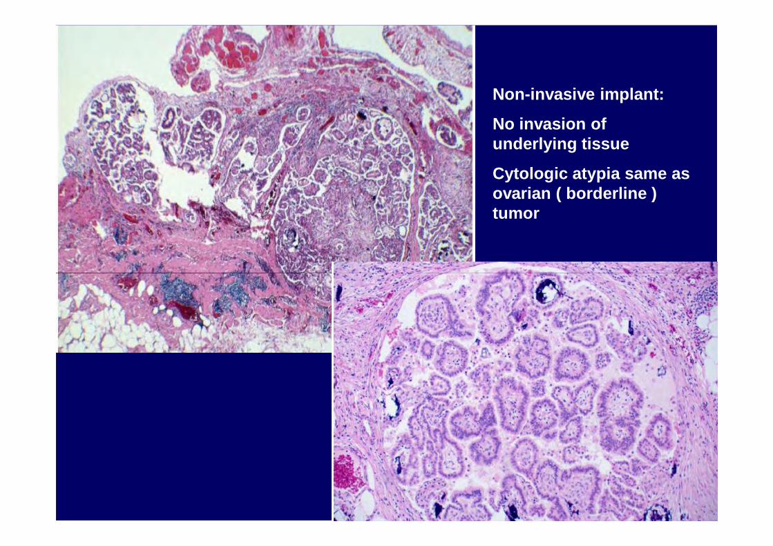
Non-invasive implant:
No invasion of underlying tissue
Cytologic atypia same as ovarian ( borderline ) tumor

Non-invasive implant: Papillae with fibrous or hyalinized coresEpithelium of implants merges with stroma

Non-invasive implant:
Stroma has granulation tissue tissue appearance
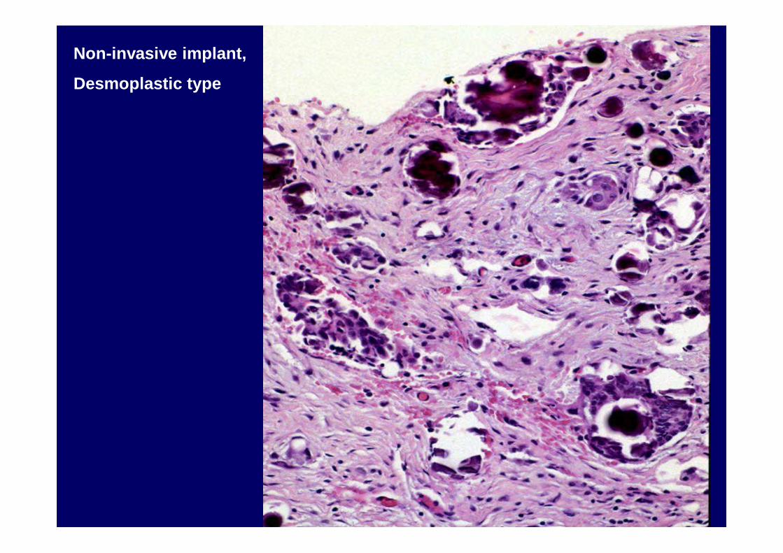
Non-invasive implant,
Desmoplastic type
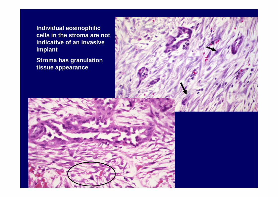
Individual eosinophilic cells in the stroma are not indicative of an invasive implant
Stroma has granulation tissue appearance
Noninvasive ImplantNoninvasive Implant
• Ratio of epithelium to stroma is less than in invasive implants
• Epithelial cells often larger than those in invasive implantsinvasive implants– Nuclei can be more atypical– Cytoplasm often abundant and eosinophilic
Invasive ImplantInvasive Implant
• Invasion of Normal Tissue
• “Micropapillary” Architecture
• Small Solid Epithelial Nests Surrounded By Clefts
• Cytologic atypia resembles that of a grade 1 serous carcinoma

Non-invasive desmoplastic implant
Invasive implant

Invasive Implant
Invasion Of Normal Tissue

Invasive Implant

Serous Borderline TumorsSerous Borderline TumorsPeritoneal ImplantsPeritoneal Implants--Subtyping CriteriaSubtyping Criteria
• Relationship of the tumor and its surrounding stroma to underlying tissue-tissue invasion• Characteristics of the tumor itself:• Characteristics of the tumor itself:
• Micropapillarity• Solid nests of cells with clefts• Glands with extensive bridging• Marked cytologic atypia• Single cells

Invasive Implant,“Micropapillary” Architecture
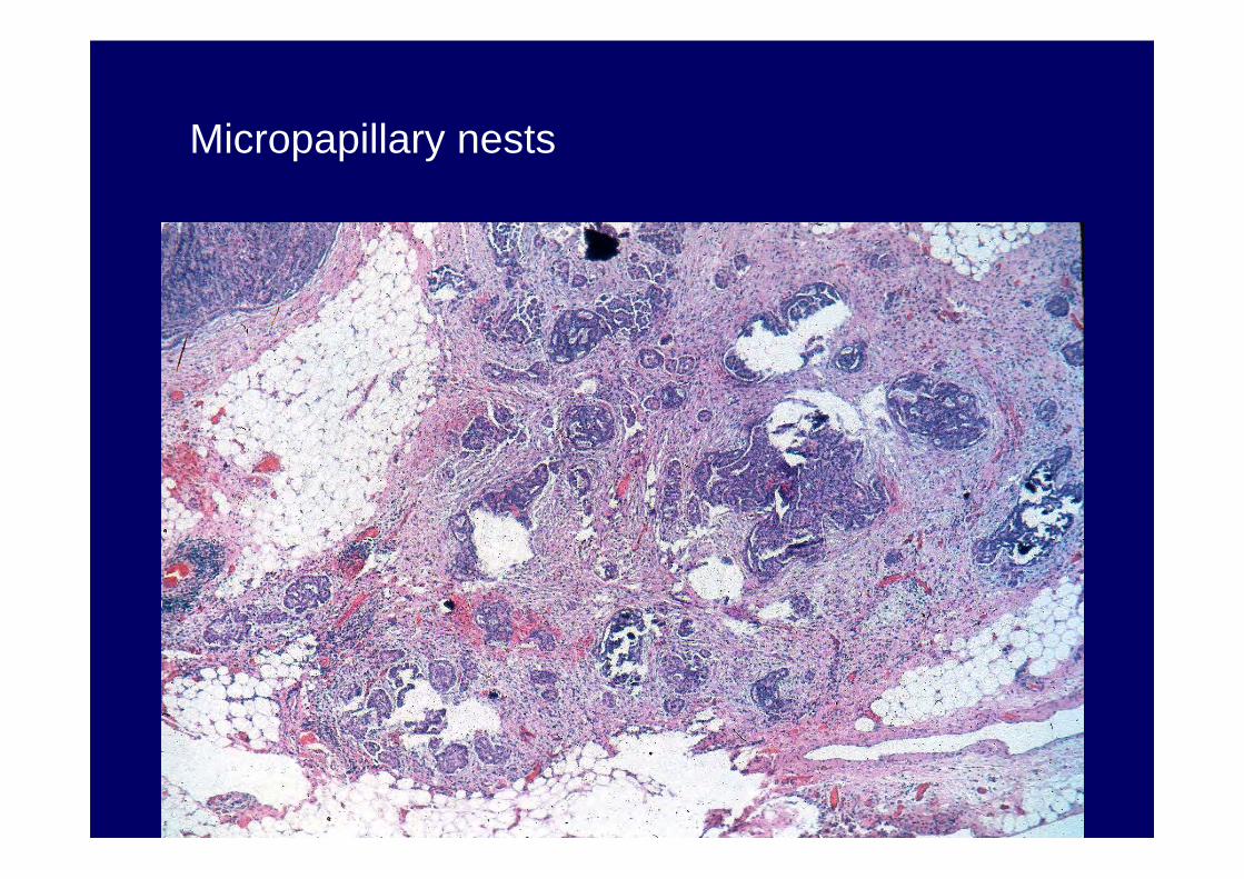
Micropapillary nests
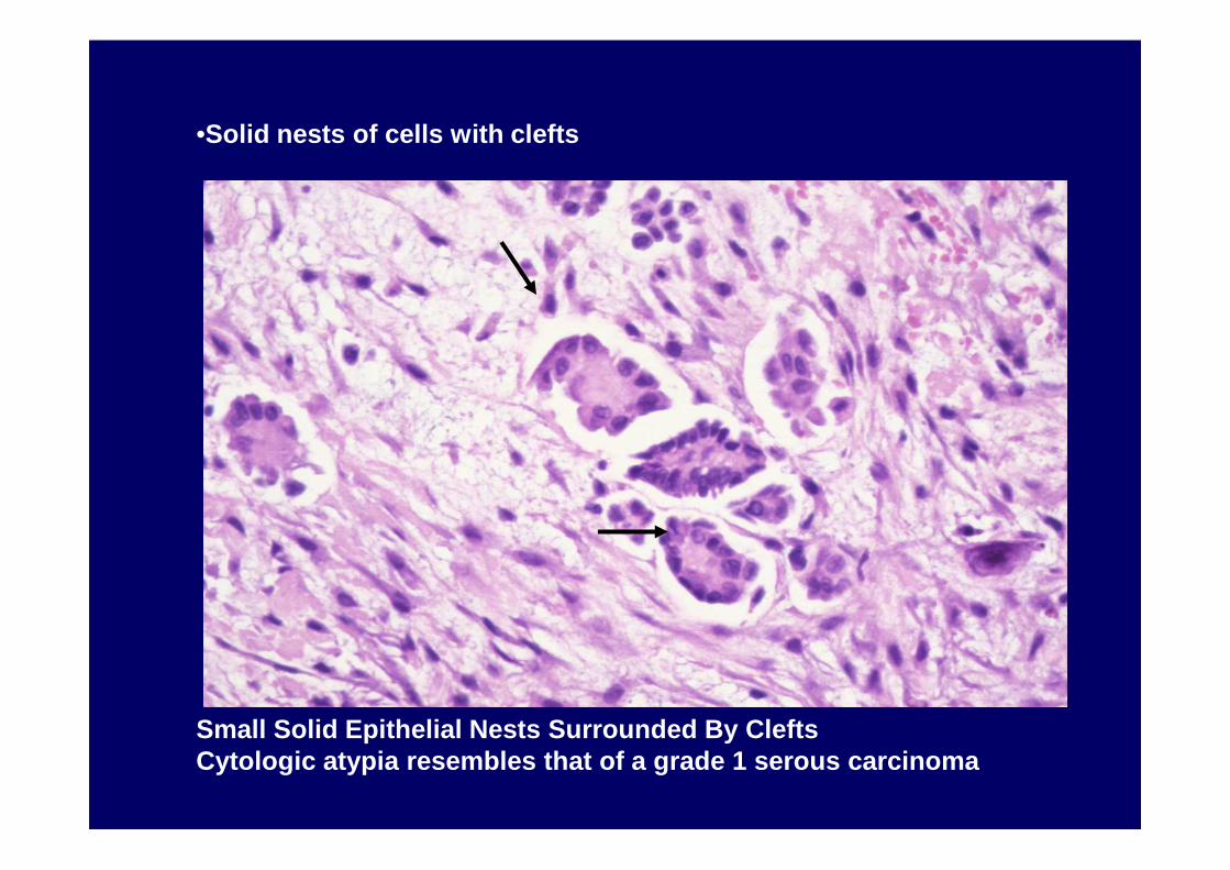
•Solid nests of cells with clefts
Small Solid Epithelial Nests Surrounded By CleftsCytologic atypia resembles that of a grade 1 serous carcinoma
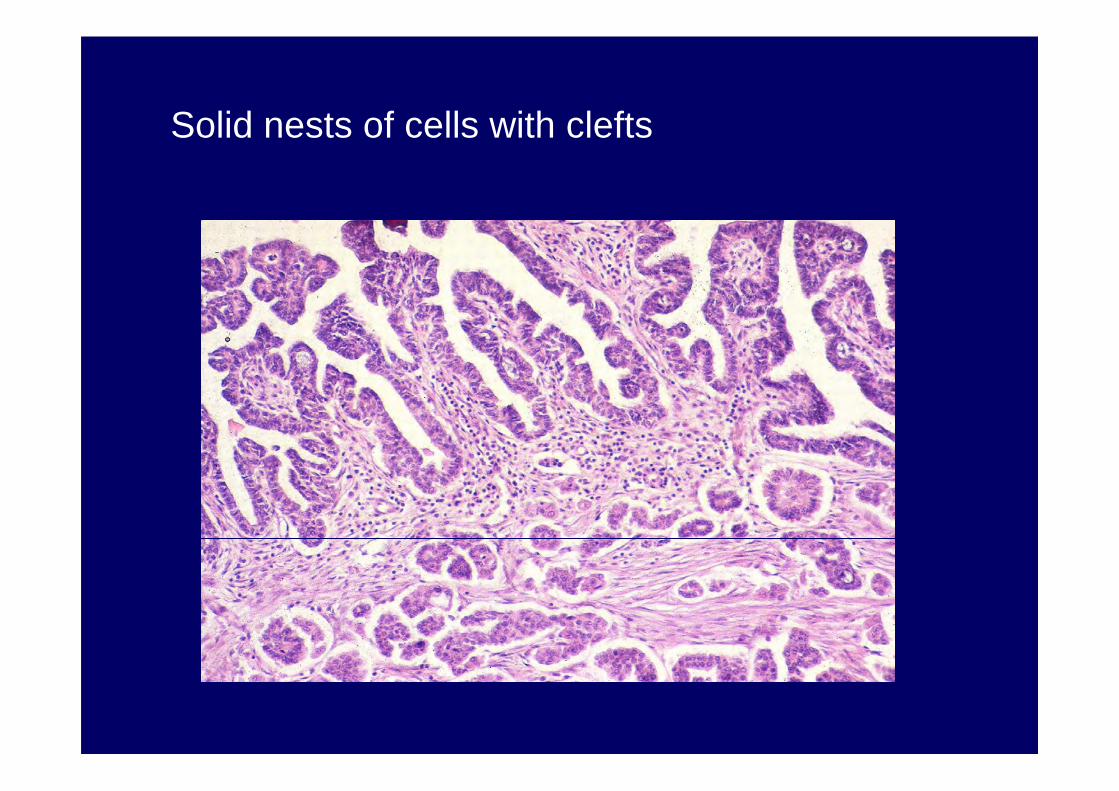
Solid nests of cells with clefts


Peritoneal Implants Peritoneal Implants –– Reproducibility of Reproducibility of Various CriteriaVarious Criteria
Gilks, McKenney, Kalloger, Longacre(USCAP 2005)Feature KappaInvasiveness (Tissue inv)Invasiveness (Tissue inv) 00..8484Micropapillarity 0.71Single stromal cells 0.50Single stromal cells 0.50Mitotic activity 0.46Periglandular Clefts 0.34Prominent Nucleoli 0.20Nuclear pleomorphism 0.03Invasiveness (Inv, micrpap, clefts) 0.72

Peritoneal Implants Peritoneal Implants ConclusionsConclusions
• Most reproducible feature is tissue invasion
• Micropapillarity and solid nests with • Micropapillarity and solid nests with clefts co-vary with invasion and are also reproducible

Peritoneal ImplantsPeritoneal ImplantsConclusionsConclusions
• Peritoneal tumor associated with SBT should be termed:
“noninvasive and invasive implants” “noninvasive and invasive implants” • invasion of underlying tissue is the
most important adverse prognostic factor
Ovarian Serous Borderline TumorsOvarian Serous Borderline Tumors
• Micropapillary/cribriform pattern• Peritoneal implants
•• Microinvasion, new dataMicroinvasion, new data•• Microinvasion, new dataMicroinvasion, new data• Lymph node involvement• Recurrent serous borderline tumor
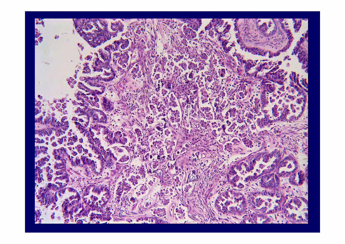
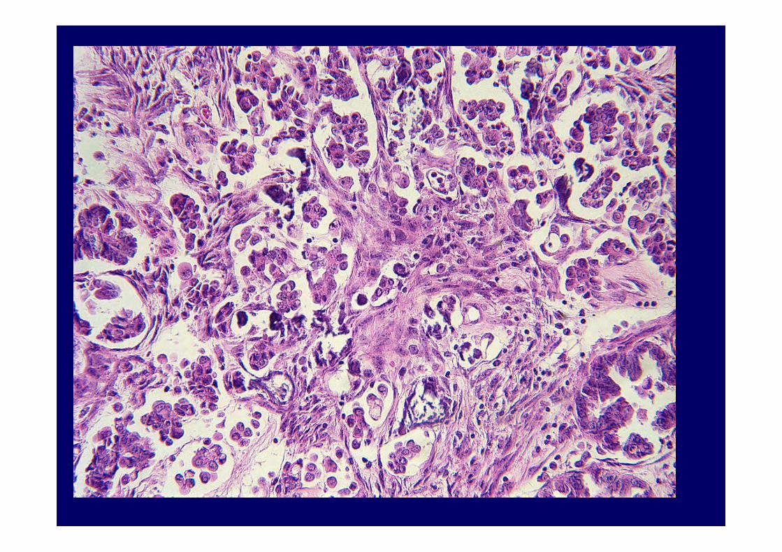
Serous Borderline Tumors Serous Borderline Tumors with Stromal Microinvasionwith Stromal Microinvasion
10-15% of tumors have small foci of invasion 10-15% of tumors have small foci of invasion in tumor stroma with a minimal stromal reaction
Definition of microinvasionDefinition of microinvasion
• Less than 3 mm in diameter• Less than 10 sq mm in area• Less than 5 mm in greatest • Less than 5 mm in greatest dimension

Serous Borderline Tumors Serous Borderline Tumors with Microinvasionwith Microinvasion
Microinvasive foci present in:
• 10% of Stage I SBTs• 10% of Stage I SBTs
• 56% of Stage II and III SBTs
• Pregnant women
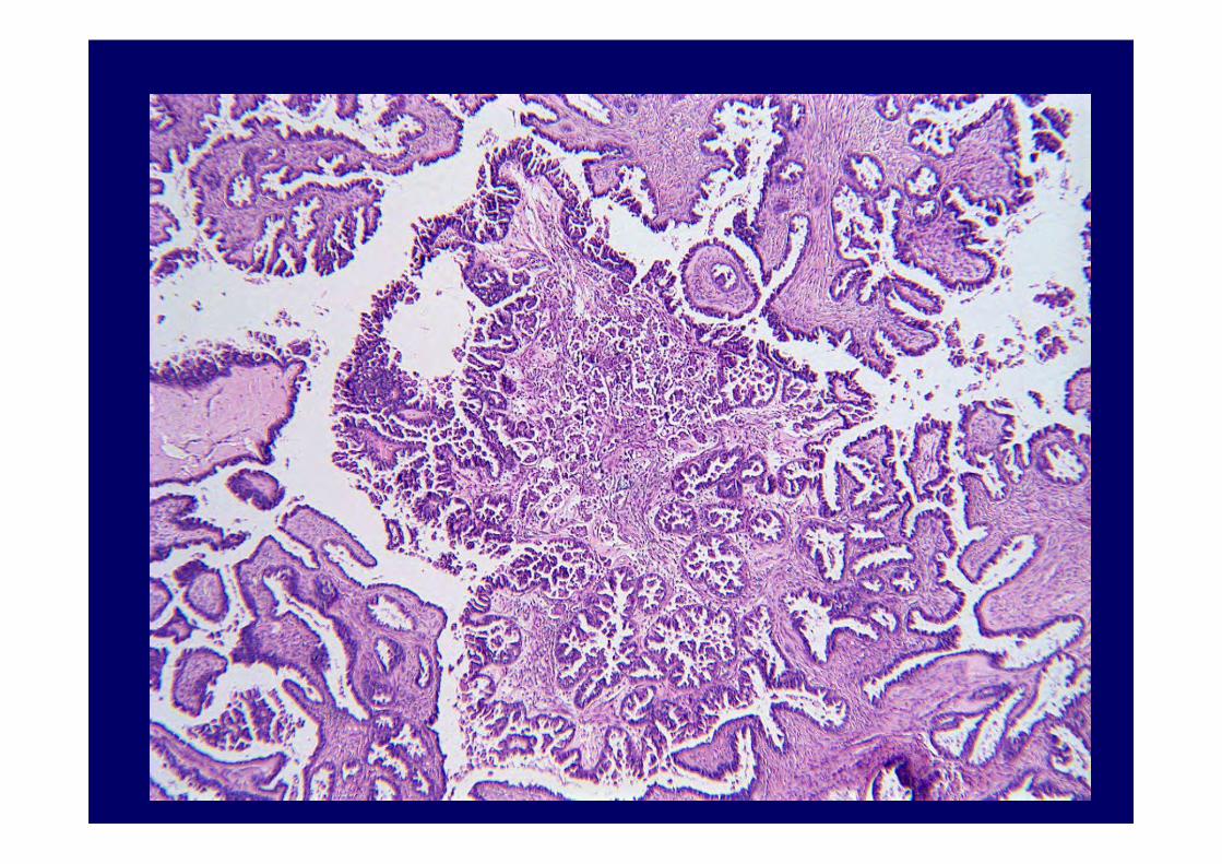
Serous Borderline Tumors Serous Borderline Tumors with Stromal Microinvasionwith Stromal Microinvasion
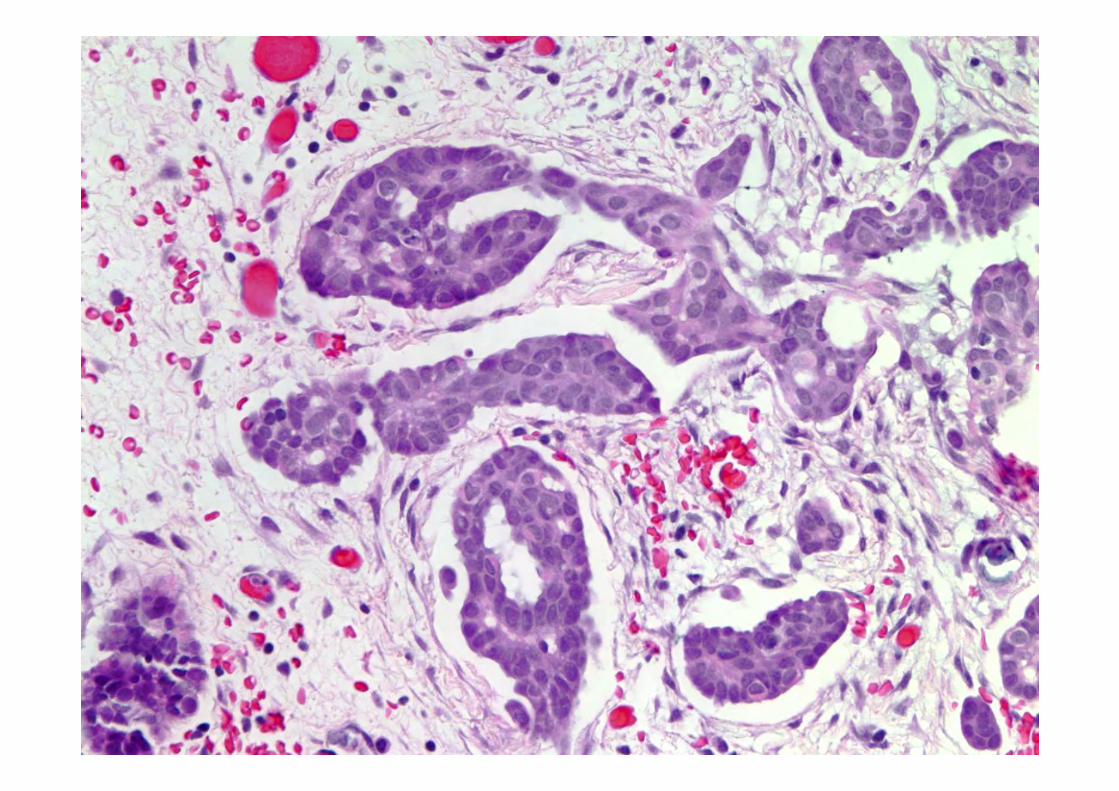
Morphologic patterns:Common pattern: Eosinophilic cell patternCommon pattern: Eosinophilic cell patternUnusual patterns: Micropapillae, solid
nests, cribriform nests-rare

Serous Borderline Tumors Serous Borderline Tumors with Stromal Microinvasionwith Stromal Microinvasion


Microinvasion and OutcomeMicroinvasion and Outcome94 94 PatientsPatients
No. of Series No. of Casses Last Known Status
15 101 94 pts, 6,7 yrs means FLU
1 recurrence, pt AWDSurvival-100%
Seidman JD, Kurman RJ
Hum Pathol 31:539-577, 2000
Microinvasion: Microinvasion: 20032003
Eosinophilic cell pattern • Does not convey a worse prognosis• Does not convey a worse prognosis• Should be designated “serous
borderline tumor with microinvasion”
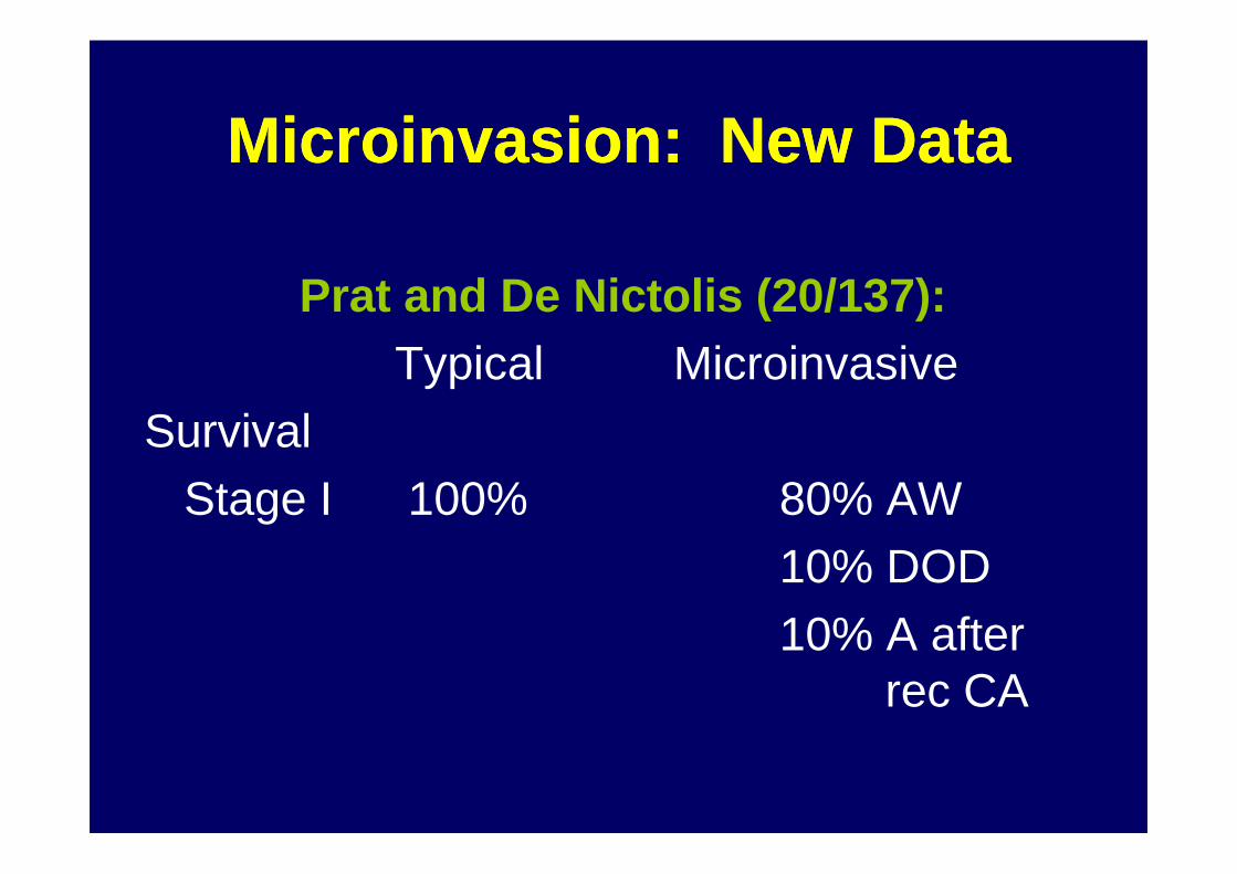
Microinvasion: New DataMicroinvasion: New Data
Prat and De Nictolis (20/137):Typical Microinvasive
SurvivalSurvivalStage I 100% 80% AW
10% DOD10% A after
rec CA
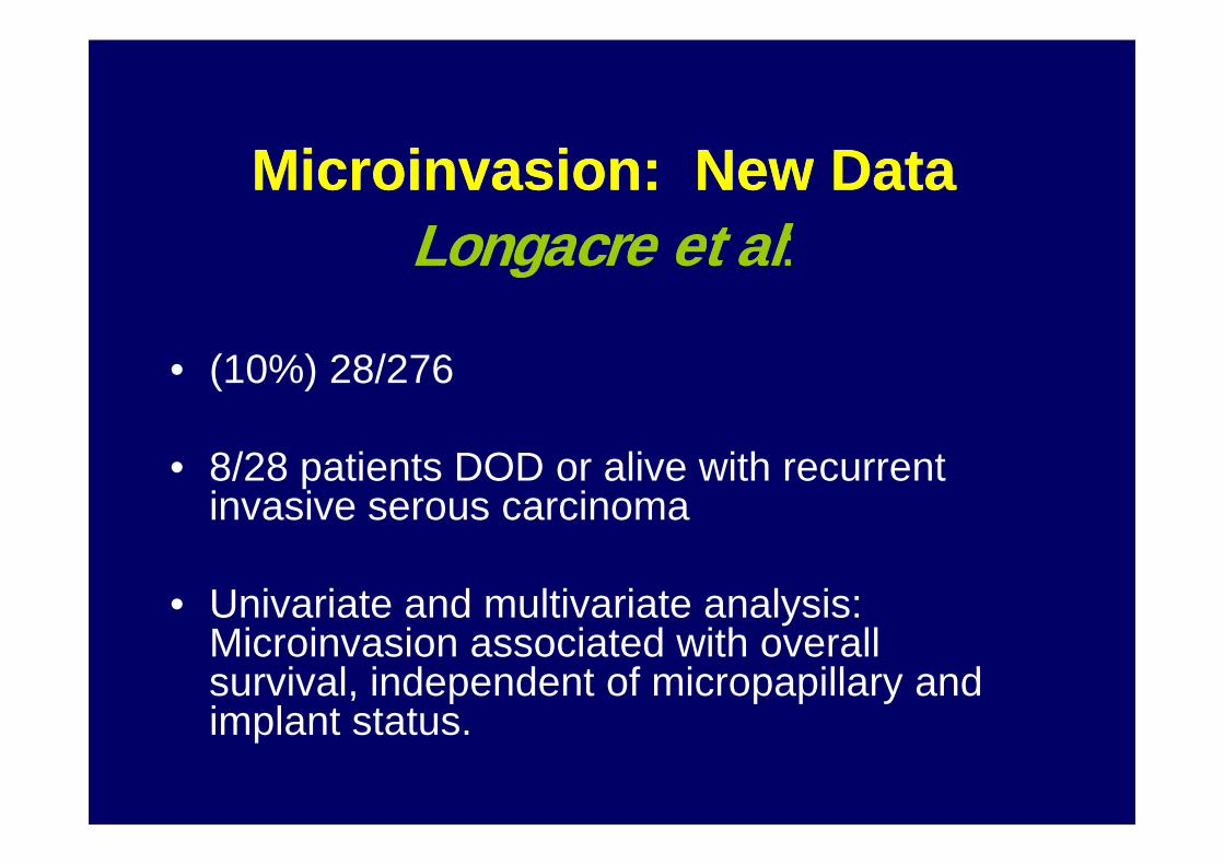
Microinvasion: New DataMicroinvasion: New DataLongacre et alLongacre et al::
• (10%) 28/276
• 8/28 patients DOD or alive with recurrent invasive serous carcinoma
• Univariate and multivariate analysis: Microinvasion associated with overall survival, independent of micropapillary and implant status.
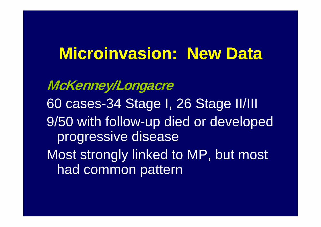
Microinvasion: New DataMicroinvasion: New Data
McKenney/Longacre60 cases-34 Stage I, 26 Stage II/III9/50 with follow-up died or developed
progressive diseaseMost strongly linked to MP, but most
had common pattern
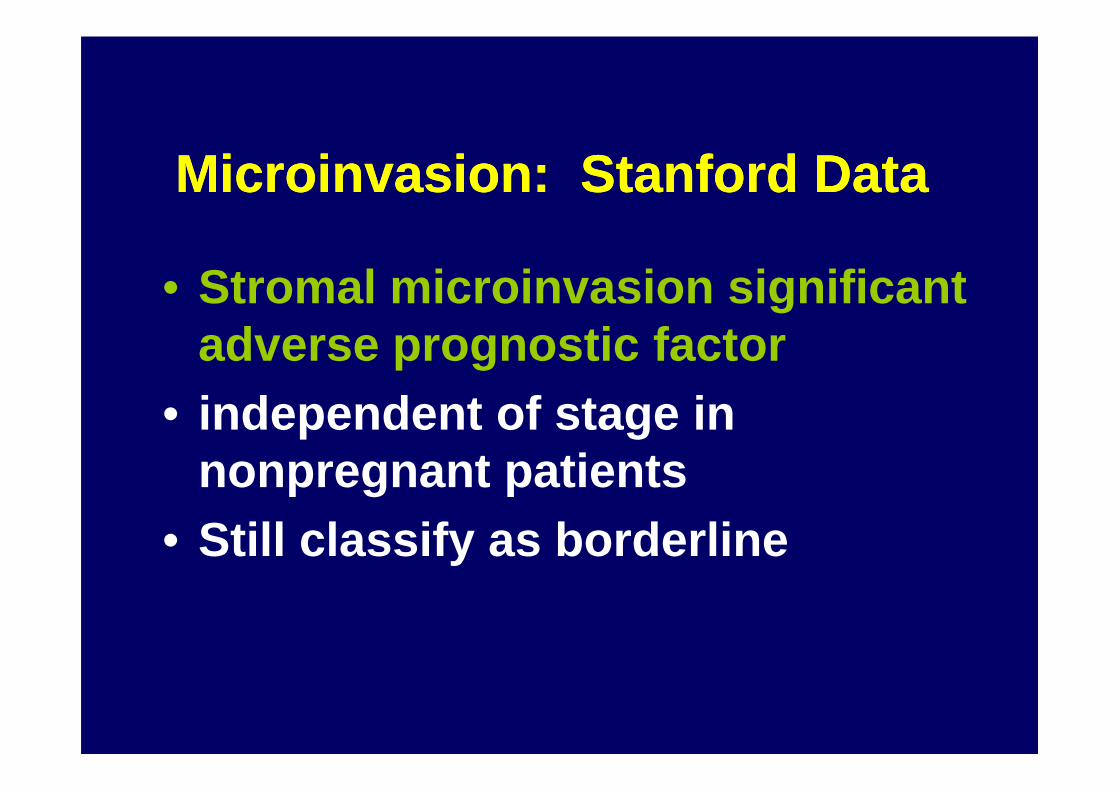
Microinvasion: Stanford DataMicroinvasion: Stanford Data
• Stromal microinvasion significant adverse prognostic factor
• independent of stage in • independent of stage in nonpregnant patients
• Still classify as borderline
Ovarian Serous Borderline TumorsOvarian Serous Borderline Tumors
• Micropapillary/cribriform pattern• Peritoneal implants• Microinvasion, new data• Microinvasion, new data
•• Lymph node involvementLymph node involvement• Recurrent serous borderline tumor
Serous Borderline Tumors Serous Borderline Tumors Lymph Node InvolvementLymph Node Involvement
• Patients who undergo lymph node samplingsampling
• 21-25% of pelvic or paraaortic lymph nodes involvement
Serous Borderline Tumors Serous Borderline Tumors Lymph Node InvolvementLymph Node Involvement
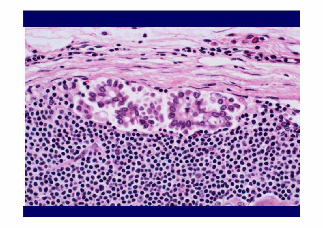
• Tumor confined to lymph nodes • Present in sinuses, • Less frequently in sinuses and parenchyma• Less frequently in sinuses and parenchyma
Two morphologic types• Eosinophilic cells, singly or in papillae• Columnar serous cells in clusters, papillae


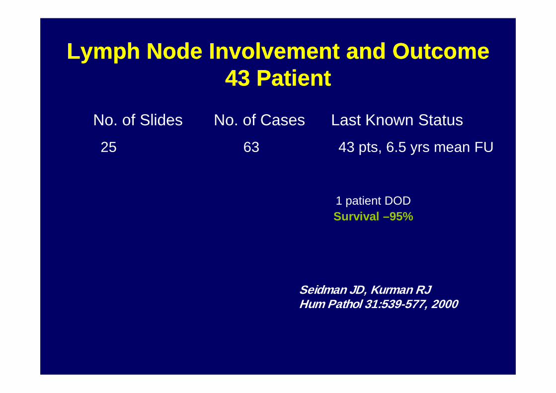
Lymph Node Involvement and OutcomeLymph Node Involvement and Outcome43 43 PatientPatient
No. of Slides No. of Cases Last Known Status
25 63 43 pts, 6.5 yrs mean FU
1 patient DOD1 patient DODSurvival –95%
Seidman JD, Kurman RJ Hum Pathol 31:539-577, 2000

Lymph Node Involvement Lymph Node Involvement ConclusionsConclusions
Lymph node involvement at presentation is not associated with an adverse outcome in patients with peritoneal implants, as long as the tumor aggregates are not >1mm
Am J Surg Pathol, Vol 30, May 2006
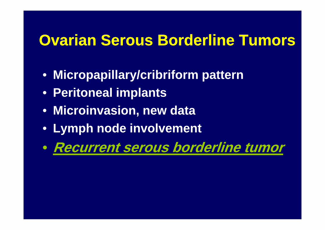
Ovarian Serous Borderline TumorsOvarian Serous Borderline Tumors
• Micropapillary/cribriform pattern• Peritoneal implants• Microinvasion, new data• Microinvasion, new data• Lymph node involvement
•• Recurrent serous borderline tumorRecurrent serous borderline tumor
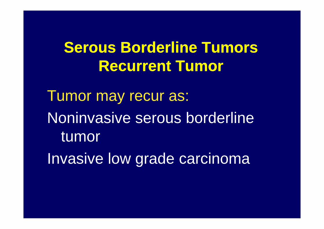
Serous Borderline Tumors Serous Borderline Tumors Recurrent TumorRecurrent Tumor
Tumor may recur as:Noninvasive serous borderline Noninvasive serous borderline
tumorInvasive low grade carcinoma
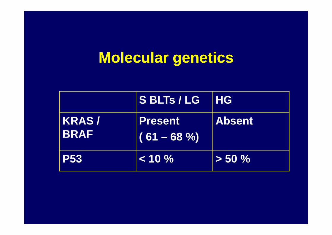
Molecular geneticsMolecular genetics
S BLTs / LG HG
KRAS / Present AbsentKRAS / BRAF
Present ( 61 – 68 %)
Absent
P53 < 10 % > 50 %
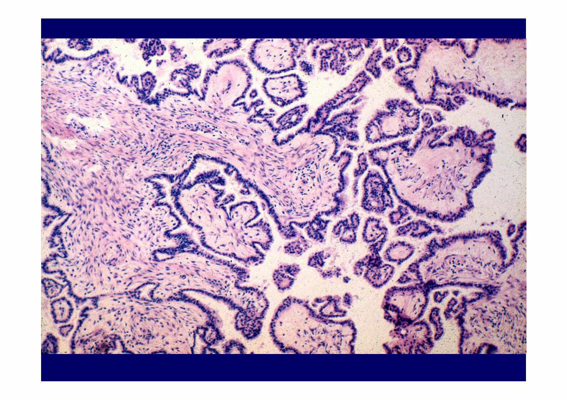


Serous Borderline Tumors Serous Borderline Tumors Recurrent TumorRecurrent Tumor
Tumor may recur as:Noninvasive serous borderline Noninvasive serous borderline
tumor-good prognosisInvasive low grade carcinoma-
patients who develop clinically apparent progressive disease
Serous Borderline Tumors Serous Borderline Tumors Recurrent Tumor Recurrent Tumor
ConclusionsConclusions
Recurrent tumor should be histologically examined and histologically examined and classified as borderline or carcinoma

ConclusionsConclusionsOvarian Serous Borderline TumorsOvarian Serous Borderline Tumors
•• MicroinvasionMicroinvasion•• Peritoneal implantsPeritoneal implants•• Micropapillary/cribriform patternMicropapillary/cribriform pattern•• Micropapillary/cribriform patternMicropapillary/cribriform pattern•• Recurrent serous borderline tumorRecurrent serous borderline tumor•• Lymph node involvementLymph node involvement


