Download - Parturition

PARTURITION
MEDIANA SUTOPO LIEDAPRAJA
PPDS TAHAP 1A
OBSTETRI DAN GINEKOLOGI FAKULTAS KEDOKTERANUNIVERSITAS INDONESIA – RSUPN CIPTOMANGUNKUSOMO

• The timing of birth development of placenta
gene expression of CRH (corticotropin releasing hormone)
• Maternal plasma CRH increase as pregnancy advances peak at time of delivery
• Human produce CRHBP for CRH end of pregnancy CRHBP falls CRH rise.
Birth Placenta CRH
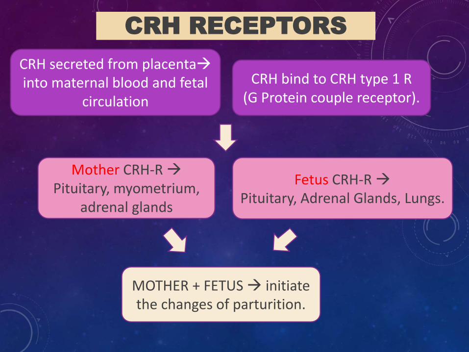
CRH RECEPTORS
CRH secreted from placentainto maternal blood and fetal
circulation
CRH bind to CRH type 1 R (G Protein couple receptor).
Mother CRH-R Pituitary, myometrium,
adrenal glands
Fetus CRH-R Pituitary, Adrenal Glands, Lungs.
MOTHER + FETUS initiate the changes of parturition.

stimulate placenta release CRH
Glucocorticoids stimulate CRH gene and production CRH CRH stimulate pituitary
produce corticotropinAdrenal cortex to release cortisol +DHEAS
Estrogen synthesis.
“CRH level has a relatively specific association with risk of preterm birth Maternal CRH levels is the most accurate predictor.”
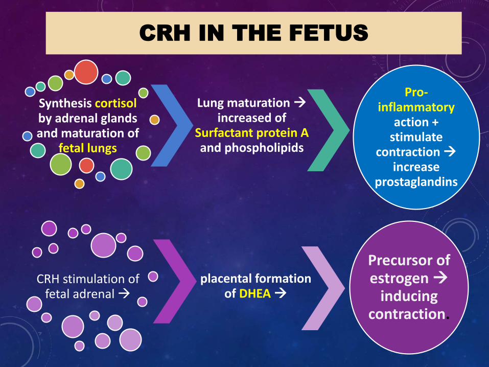
CRH IN THE FETUS
Synthesis cortisolby adrenal glands and maturation of
fetal lungs
Lung maturation increased of
Surfactant protein A and phospholipids
Pro-inflammatory
action + stimulate
contraction increase
prostaglandins
CRH stimulation of fetal adrenal
placental formation of DHEA
Precursor of estrogen
inducing contraction.

IN CONCLUSION
as gestation advances Systems in mother and fetus increase in placental CRH
change in fetal cortisol concentration, fetal lung maturations, amniotic fluid protein, phospholipids and myometrial receptor expression.


COX -2

ACTIVATION OF THE MYOMETRIUM AT TERM
Important event “ Contraction associated proteins”
Relax ?? Or contraction ??
3 types:
1. Interaction between actin and myosin proteins
2. Excitability of myometrial cells
3. Intercellular connectivity

Physical connection by multimer connexin 43.
Connection formed by paracrine Prostaglandin F2α and local release of calcium.
Depolarization
Contraction

• Action and Myosin interaction = Contraction
• Actin converted Globular to Filamentous
• Actin partner Myosin activated by M-light chain kinase activated by Calmodulin and Intracellular Calcium.
• Myocyte depolarizes Influx extracellular Ca2+
contraction.
Example :
Nifedipine Tocolytic block voltage-regulated Ca2+ channel.


1• Stretching of myometrium (fetal growth)mitogen
activated protein kinase
2
• increase intracellular CAMP
• activating protein kinase A.
3• inactivate myosin light chain
4•Contraction

FETAL MEMBRANE ACTIVATION
Production of surfactant proteins, phospholipids and inflammatory
cytokines in amniotic fluid
Increase as COX-2 activity and PGE2 in amnion.
Mediators of inflammation in the
amnion

Chorion underlies the amnion
Produce PDGH (prostaglandin dehydrogenase)
As Potent “Inactivator” of Prostaglandins.
Release of Metalloproteases
Weaken placental membrane
CRH
MMP-9
DegradateCollagen
Cervix Structure


A Better Understanding of the pathway to normal birth should provide a pathological process.
The goal is to predict which pregnancies carry a risk of preterm,
Reduce the incidences of cerebral palsy and cognitive impairment associated with preterm birth.

THANK YOU


