
Plant medicine and veterinary potential of antimicrobial peptides
produced by entomopathogenicnematode symbiotic bacteria
András Fodor1
J. Racsko1, J. Hogan1, D. Vozik2
L. Makrai3, F., Olasz4,
H. Goodrich Blair5
Fodor et al., BABE_2014 1
• Antibiotics-resistance of pathogenic organisms
emerged as a new challenge to plant protection,
veterinary and even human clinical practice.
• The antibiotic multidrug-resistance can be
overcome by anti-microbial compounds of totally
INTRODUCTION
overcome by anti-microbial compounds of totally
different mode of action.
• Biocontrol potential of antimicrobial peptides
produced by EPB), Xenorhabdus budapestensis
(EMA) and X. szentirmaii (EMC) is discussed in
this presentation.
Fodor et al., BABE_2014 2

é
Fodor et al., BABE_2014 3
• This EPN bacterium (EPNB) is carried into the blood cavity (hemocoel) of insect hosts by a specific transmission (infective dauer juvenile, IJ) stage of the nematode.
• These bacteria could easily be isolated • These bacteria could easily be isolated and cultured in the laboratory both in solid and liquid media, both in small and larger scale and test for antagonistic antimicrobial actibities.
Fodor et al., BABE_2014 4
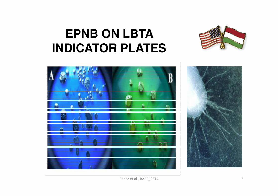
EPNB ON LBTA INDICATOR PLATES
Fodor et al., BABE_2014 5

• Once there, the nematode releases the bacteria,
which then express immune suppressive and
virulence factors that kill insects.
• Then, within the insect cadaver, bacterial activity
promotes degradation of insect tissues and
deters competitors (including opportunistic non-
host nematodes). host nematodes).
• Thus, as part of their lifecycle, EPNB obligatorily
interact with and influence the physiologies of
competing micro-organisms, nematode
parasites, and insects, largely through the
production of bioactive proteins and small molecules.
Fodor et al., BABE_2014 6

MATERIALS AND METHODS
• After detaliled comparison of EPNB species,
strains and isolates from the aspect of their
antagonistic activity toward different test
tarhets, we found that two species, tarhets, we found that two species,
Xenorhabdus budapestensis and X.
szentirmaii have outstandind antimicrobial
potential.
Fodor et al., BABE_2014 7

Figure 5 Interspecific differences between Xenorhabdus strains tested
on the Klebsiella pneumoniae (mastitis isolate, #696) (Photo: A., M.,
Fodor; 2006)
Fodor et al., BABE_2014 8

Figure 3 Xenorhabdus budapestensis (left) and X.
budapestensis (right) inhibition zone on E. coli (Photo:
Andrea Máthé-Fodor, 2010)
Fodor et al., BABE_2014 9

MATRIALS: Antimicrobial producers and test organisms
• This study focuses mainly on thebacterium Xenorhabdus budapestensiswhich is the obligate symbiont of the EPN Steinernema bicornutum and pathogen of Steinernema bicornutum and pathogen of insects.
• Some data on X. szentirmaii, a strain which had already been sequenced are also presented.
Fodor et al., BABE_2014 10
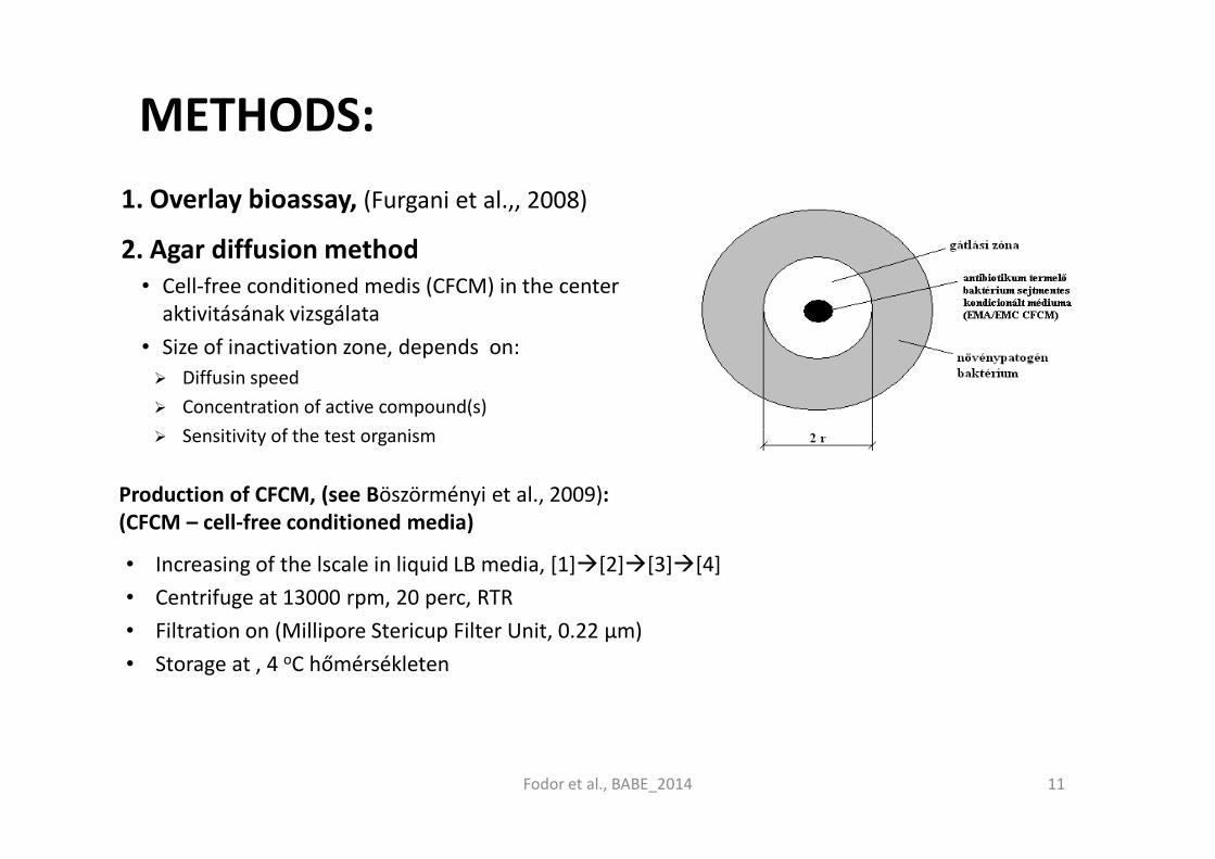
METHODS:
1. Overlay bioassay, (Furgani et al.,, 2008)
2. Agar diffusion method• Cell-free conditioned medis (CFCM) in the center
aktivitásának vizsgálata
• Size of inactivation zone, depends on:
� Diffusin speed
� Concentration of active compound(s)
� Sensitivity of the test organism� Sensitivity of the test organism
Fodor et al., BABE_2014
Production of CFCM, (see Böszörményi et al., 2009):(CFCM – cell-free conditioned media)
• Increasing of the lscale in liquid LB media, [1]�[2]�[3]�[4]
• Centrifuge at 13000 rpm, 20 perc, RTR
• Filtration on (Millipore Stericup Filter Unit, 0.22 µm)
• Storage at , 4 oC hőmérsékleten
11

METHODS: BIOASSAYS USED BY US
(OVERLAY) BIOSSAY
X. budapestensis (EMA, left),X. szentirmaii (EMC, right)
AGARDIFFUSION TEST:Foto: Fodorné Máthé Andrea
• A. Curtobacterium flaccumfaciens pv. betae NCAIM B 01612: EMA („Észak”) EMC („dél”).
• B.: Xanthomonas axonopodis pv. phaseoli NCAIM 1523;
• C.: Dyckeya chrysanthemi NCAIM B 01839;
• D.: Erwinia amylovora (Ea1) Hevesi Mária.
• Foto: Dr. Mária Hevesi.
Foto: Fodorné Máthé Andrea
Fodor et al., BABE_2014 12
RESULTS
• Xenorhabdus budapestensis (AF 2013), a strain with impressive antimicrobial potential.
• X. budapestensis culture cell free supernatant has antimicrobial activity against mastitis isolates [1], wild type and antibiotic resistant strains of the wild type and antibiotic resistant strains of the plant pathogen Erwinia amylovora [2],
• the eukaryotic potato pathogen Phytophthorainfestans, [2],
• multi-drug resistant Staphylococcus aureus(MRSA) (Fodor and McGwire, in prep.), and closely related EPNB strains [3].
Fodor et al., BABE_2014 13
X. budapestensis culture cell free supernatant has antimicrobial activity against mastitis isolates [1],
Fodor et al., BABE_2014 14

SUCCESFUL EXPERIMENTS ON
EPB strains
(Lengyel et
al., 2005)
• X. budapestensis
• Escherichia coli B (OF-323);
• E. coli K12 (OF-319;
• E. coli TG1 (OF-290);
• E. coli TG90 (OF-630);• X. budapestensis
EMA
• X. szentirmaii
• EMC*
• E. coli TG90 (OF-630);
• Salmonella enteritis (OF- MA-1504)
• Salmonella enteritis NCAIM B 02186;
• Salmonella typhimurium NCAIM B 02212;
• Campylobacter coli NCAIM B 02255;
• Campylobacter jejuni NCAIM B 02254;
• Clostridium perfrigens NCAIM B 01417;
• Ralstonia solanacearum 1226 és 879Fodor et al., BABE_2014 15
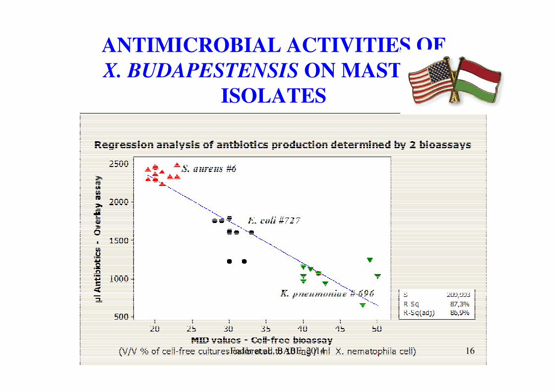
ANTIMICROBIAL ACTIVITIES OF
X. BUDAPESTENSIS ON MASTITIS
ISOLATES
Fodor et al., BABE_2014 16

Inhibition diameter in mm of Xenorhabdus budapestensis
on the phytopathogenic bacteria overlaid on LBA media
plates with four replications
Fodor et al., BABE_2014 17

RESULTS OF AGAR DIFFUSION BIOASSAYS OF
SOME PLANT PATHOGENIC BACTERIA
Fodor et al., BABE_2014 18
• Abstract• Rastonia solanacearum is a pathogen causes bacterial wilt in potato and other
Solanaceum species, which has been considered as one of the most significant
epidemic disease in plant medicine. The potential of using antibacterial substances
from entomopathogenic nematode-symbiotic bacterium strains Xenorhabdus
budapestensis (EMA) and X. szentirmaii (EMC) in Ralstonia control has been
Xenorhabdus budapestensis (AF2013, EMA) as a potential tool of
controlling Ralstonia. Vozik, D., J. Bélafi-Bakó, K., Hogan, J. Racsko, A.
Fodor at al., 2014)
budapestensis (EMA) and X. szentirmaii (EMC) in Ralstonia control has been
studied. We have elaborated reproducible methodology to quantitate (1) optimum
inoculum size needed for successful Ralstonia infection in plant experiments; (2)
the minimum phytotoxic concentration and (3) the minimal Ralstonia inhibiting
concentration of EMA cell free conditioned medium (CFCM) in vitro. At the light
of the results we consider the antibacterial component(s) of EMA CFCM potential
tool(s) of Ralstonia control.
•
• Keywords: X. budapestensis, R. solanacearum, antimicrobial activity, antibiotic
resistance, phytotoxicity
Fodor et al., BABE_2014 19

SENSITIVITY OF
Ralstonia solanacearum
1226 virulent strain on
EMA CFCM substances in
overlay bioassay (Photó: overlay bioassay (Photó:
Dr. Mária Hevesi
Fodor et al., BABE_2014 20
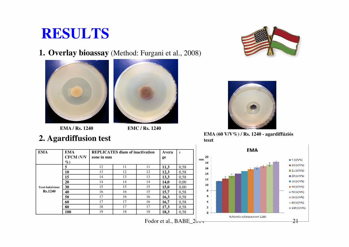
RESULTS1. Overlay bioassay (Method: Furgani et al., 2008)
2. Agardiffusion test
Fodor et al., BABE_2014
EMA EMA
CFCM (V/V
%)
REPLICATES diam of inactivation
zone in mm
Avera
ge
s
Teszt-baktérium:
Rs.1240
5 12 11 11 11,3 0,58
10 13 12 12 12,3 0,58
15 14 13 13 13,3 0,58
20 14 14 14 14,0 0,00
30 15 15 15 15,0 0,00
40 16 16 15 15,7 0,58
50 17 16 16 16,3 0,58
60 17 17 16 16,7 0,58
80 18 17 17 17,3 0,58
100 19 18 18 18,3 0,58
EMA / Rs. 1240 EMC / Rs. 1240
EMA (60 V/V%) / Rs. 1240 - agardiffúziós
teszt
21

3. Antibiotikum-sensitivities of R. solanacearum 1240
4. In planta infection test (R. solanacearum 1240 – on potato seedlings)
RESULTS
DILUTION: 1 0.5 0.25 10(-1) 10(-2) 10(-3) 10(-4) 0O/N Rs.1240
Baktérium
szuszpenzió (ml)
(OD: 1,050 ;
8 4 2 1
� Dilution: autokclaved tap water
S = érzékenyR = rezisztens
Fodor et al., BABE_2014
(OD: 1,050 ;
CFU/ml: 6*108)
10X hígított bakt.
szuszp. (ml)0.9
100X hígított bakt.
szuszp. (ml)0.9 0.08
autoklávozott
csapvíz (ml)0 4 6 9 8.1 8.1 7.92 8
Abszorbancia
(λ = 620 nm)1,050 0,585 0,306 0,126 0,014 0,005 0,002 0,000
� Dilution: autokclaved tap water
� 2 replicates (test tubes) in each doses
� 2-3 plants / test tube
� 1-5 stages of sickness (visual observation)
� 1=healthy plant; 5= destroyed plant
DILUTION: 1 0.5 0.25 10(-1) 10(-2) 10(-3) 10(-4) 0
DAY 0 1 1 1 1 1 1 1 1
DAY 3 3 2 2 1 1 1 1 1
DAY 6 4 3 3 2 1 1 1 1
DAY 18 5 5 5 5 2 1 1 1
22

6. Bactericid effect of EMA CFCM on R/ solanacearum RS 1240
RESULTS
EMA CFCM (V/V%) 0 5 10 15 20 25O/N Bacterium Suspension
(OD: 0.866 ; CFU/ml: 4,9*108)
2.0 2.0 2.0 2.0 2.0 2.0
Autoclaved tap water (ml) 6.0 5.6 5.2 4.8 4.4 4.0
EMA CFCM (ml) 0 0.4 0.8 1.2 1.6 2.0
CFU/ml (0) 1,2*108 1,2*108 1,2*108 1,2*108 1,2*108 1,2*108
CFU/ml (2 óra után) lawn 465000 250000 220000 45000 20000
CFU/ml (4 óra után) lawn 405000 75000 60000 25000 15000
CFU/ml (6 óra után) lawn 340000 70000 40000 16500 fertőzött
CFU/ml (8 óra után) lawn 80000 10000 16000 4500 fertőzött
CFU/ml (24 óra után) lawn 25000 - - - fertőzött
EMA CFCM (V/V%) 30 35 40 45 50 75O/N Bacterium Suspension
(OD: 0.866 ; CFU/ml: 4,9*108)
2.0 2.0 2.0 2.0 2.0 2.0
EMA CFCM (ml) 3.6 3.2 2.8 2.4 2.0 0
EMA CFCM (ml) 2.4 2.8 3.2 3.6 4.0 6.0
CFU/ml (0) 1,2*108 1,2*108 1,2*108 1,2*108 1,2*108 1,2*108
CFU/ml (2 óra után) 10000 10000 - 5000 5000 5000
CFU/ml (4 óra után) 5000 - - - - -
CFU/ml (6 óra után) 2500 - - 2500 500 -
CFU/ml (8 óra után) - - - - - -
CFU/ml (24 óra után) - - - - - -
• Dilution: with autoclaved tapwater
• Incubation> 110 rpm; 28 oC-on
• Plating and colony counting on LBA plates (at 2nd,; 4th; 6th; 8th; 24 th hrs
Fodor et al., BABE_2014 23
DAY 0 DAY 6, INFECTEDDAY 0 DAY 6, INFECTED
DAY 18, INFECTED
DAY 18, NOT INFECTED
Fodor et al., BABE_2014 24
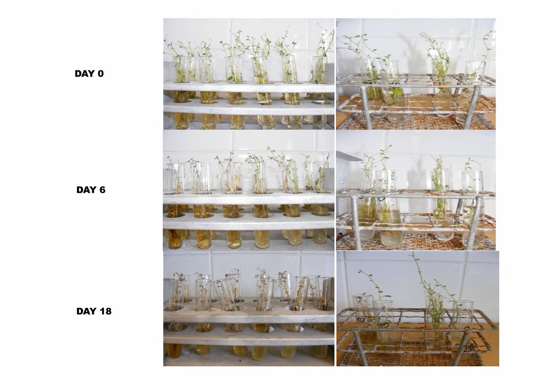
DAY 0
DAY 6
Fodor et al., BABE_2014
DAY 6
DAY 18
25

5. PHYTOTOXICITY OF EMA CFCM
RESULTS
EMA CFCM (V/V%) 100 75 50 40 30 20 10 0
EMA CFCM (ml) 8 6.0 4.0 3.2 2.4 1.6 0.8 0
AUTOCLAVED TAP
WATER (ml)
0 2.0 4.0 4.8 5.6 6.4 7.2 8
� Dilution: autokclaved tap water
EMA CFCM (V/V%) 100 75 50 40 30 20 10 0
DILUTION: 1111 1111 1111 1111 1111 1111 1111 1111
DAY 0 2222 1111 1111 1111 1111 1111 1111 1111
DAY 3 3333 3333 2222 1111 1111 1111 1111 1111
DAY 6 5555 5555 5555 5555 5555 5555 3333 1111
� Dilution: autokclaved tap water
� 2 replicates (test tubes) in each doses
� 2-3 plants / test tube
� 1-5 stages of sickness (visual observation)
� 1=healthy plant; 5=totally destroyed plant
Fodor et al., BABE_2014 26

6. Bactericid effect of EMA CFCM on R/ solanacearum RS 1240
RESULTS
EMA CFCM (V/V%) 0 5 10 15 20 25O/N Bacterium Suspension
(OD: 0.866 ; CFU/ml: 4,9*108)
2.0 2.0 2.0 2.0 2.0 2.0
Autoclaved tap water (ml) 6.0 5.6 5.2 4.8 4.4 4.0
EMA CFCM (ml) 0 0.4 0.8 1.2 1.6 2.0
CFU/ml (0) 1,2*108 1,2*108 1,2*108 1,2*108 1,2*108 1,2*108
CFU/ml (2 óra után) lawn 465000 250000 220000 45000 20000
CFU/ml (4 óra után) lawn 405000 75000 60000 25000 15000
CFU/ml (6 óra után) lawn 340000 70000 40000 16500 fertőzött
CFU/ml (8 óra után) lawn 80000 10000 16000 4500 fertőzött
CFU/ml (24 óra után) lawn 25000 - - - fertőzött
EMA CFCM (V/V%) 30 35 40 45 50 75O/N Bacterium Suspension
(OD: 0.866 ; CFU/ml: 4,9*108)
2.0 2.0 2.0 2.0 2.0 2.0
EMA CFCM (ml) 3.6 3.2 2.8 2.4 2.0 0
EMA CFCM (ml) 2.4 2.8 3.2 3.6 4.0 6.0
CFU/ml (0) 1,2*108 1,2*108 1,2*108 1,2*108 1,2*108 1,2*108
CFU/ml (2 óra után) 10000 10000 - 5000 5000 5000
CFU/ml (4 óra után) 5000 - - - - -
CFU/ml (6 óra után) 2500 - - 2500 500 -
CFU/ml (8 óra után) - - - - - -
CFU/ml (24 óra után) - - - - - -
• Dilution: with autoclaved tapwater
• Incubation> 110 rpm; 28 oC-on
• Plating and colony counting on LBA plates (at 2nd,; 4th; 6th; 8th; 24 th hrs
Fodor et al., BABE_2014 27

Experiments in Columbus with Dr. Bradford McGwire.
Effects of CFCM of EMA, EMC és X. innexii on different
clinical pathogens
Fodor et al., BABE_2014 28
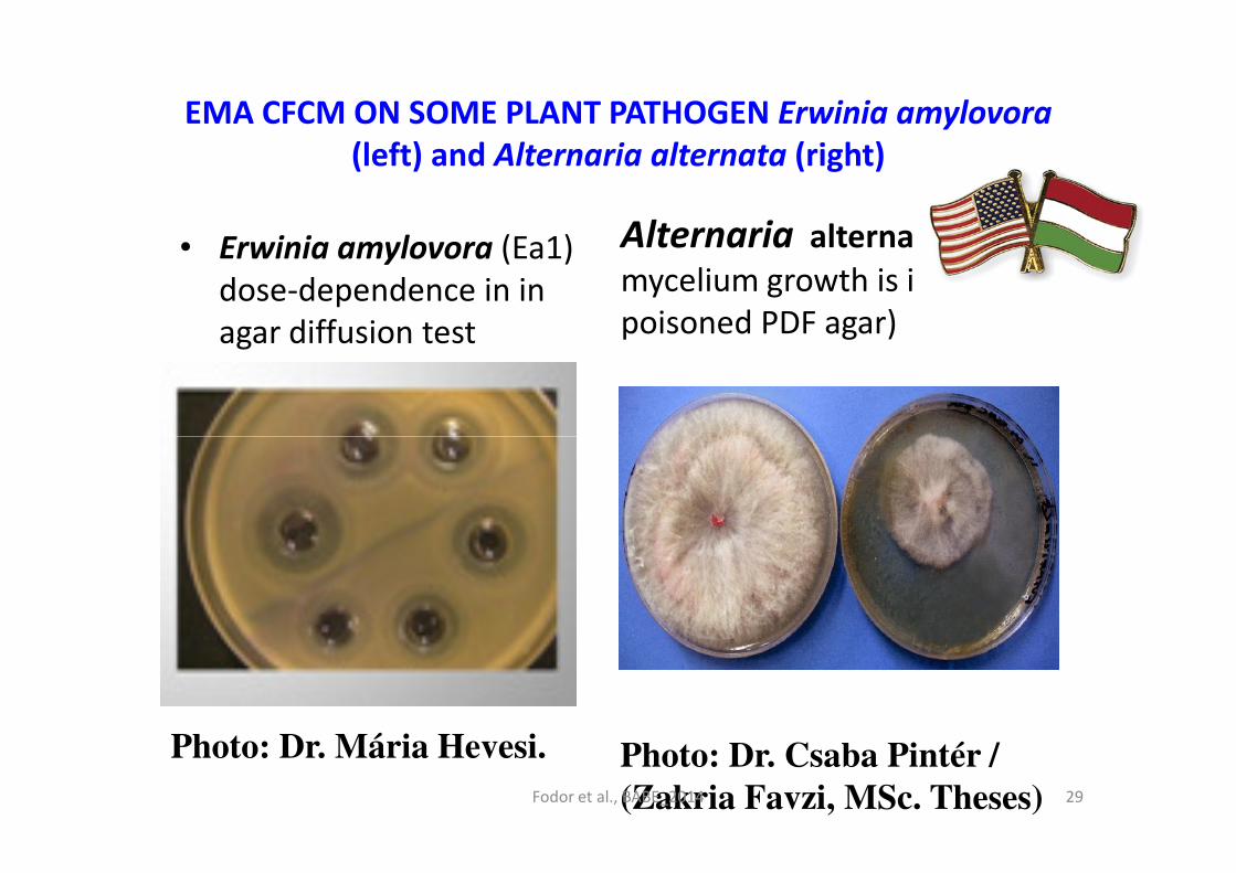
EMA CFCM ON SOME PLANT PATHOGEN Erwinia amylovora
(left) and Alternaria alternata (right)
• Erwinia amylovora (Ea1)
dose-dependence in in
agar diffusion test
Alternaria alternata gombamycelium growth is inhibited, in
poisoned PDF agar)
Photo: Dr. Mária Hevesi. Photo: Dr. Csaba Pintér /
(Zakria Favzi, MSc. Theses)Fodor et al., BABE_2014 29

P. citricola
Effect of cell-free conditioned media (CFCM) of X. szentirmaii
on the mycelial growthSejtmentes X. szentirmaii médium hatás myceliumok növekedésére
Biological Control of Plant pathogenic fungi
30
Kontrol(Sárgarépa- agar)
Sejtmentes (25 V/V% )EMC-fermentlé
(Fotó: Pintér, Cs., 2011)Fodor et al., BABE_2014 30

EREDMÉNYEK - 2
• A Clostridium perfringensNCAIM 1417 törzse rárétegzéses és agardiffúziós tesztekben egyaránt érzékeny a
• X. budapestensis és a • X. budapestensis és a
• X. szentirmaiantibakteriális aktivitásával szemben.
• Fotó: X. budapestensisagar-difffúziós tesztben C. perfringens NCAIM 1417ellen. Dr. Pintér Csaba
Fodor et al., BABE_2014 31
DISCUSSION
• Our goal has been to exploit the potential of the active antmicrobial compounds of X. budapestensis against plant and animal pathogens.
• One of the discovered very active microbial compound in X. budapestensis is the hexapeptid Bicornutin A
• Let us make a short comparison with our finding with the literature of AMPs.
Fodor et al., BABE_2014 32

Antimicrobial peptides – a general discussion
• Organisms of different taxa produce antimicrobial peptides. Their general biological role is self-defense.
• Each peptide of known antibacterial activity (AMP) is of cationic (positively charged) and amphipathic nature. It is also true for most antiviral, anti-parasitic nature. It is also true for most antiviral, anti-parasitic and antifungal peptides.
• The targets of these peptides as well as of their mode of actions are rather different.
• There are great differences between their targetspectra of different AMPs.
Fodor et al., BABE_2014 33

Molecular structures of antimicrobial peptides
Jenssen, et al. Clin Microbiol Rev. (2006 )19:491-511.
(A)Human β-defensin-2 (PDB code
1FQQ)
(B) (B) Loop-like structure of thanatin
(PDB code 8TFV) ;
(C) (C) β-laminal structure of (C) (C) β-laminal structure of
polyphemusin (PDB code 1RKK)
(D) (D) Rabbit defensin-1 (PDB code
1EWS) 165); [65]
(E) (E) α-helical structure of magainin-2
(PDB code 2MAG) (76);
(F) (F) Relaxed structure of indolicidin
(PDB code 1G89) [66] . The disulfide
bridges are yellow. The pictures were
created by a Mol Mol 2K.1 [67]
graphic program.
Fodor et al., BABE_2014 34
STRUCTURE – ACTIVITY RELATIONS
• The molecular basis of the mode of action of
antimicrobial peptides is based on non-specific
structural changes in the target membranes followed
by rapture.
• The mode of action of AMP has largely been
determined by the membrane structure of the target
cells.
• At present 5 models of mode of action has
unambiguously confirmed while others are
hypothetical
Fodor et al., BABE_2014 35

Fodor et al., BABE_2014 36

MODE OF ACTION OF AMPs
• The target cell membrane (yellow) is a lipid double layer.
• The peptide molecules are labeled as little cylinders.
• Hydrophilic parts of the molecules are labeled as red, while the hydrophobic ones are labeled as blue. while the hydrophobic ones are labeled as blue.
• The peptide-glucose membrane components are labeled as purple cylinders.
• Models A-D described the consequences of the changes in the permeability of the target cell membranes.
• Models E-I show the effects on the biosynthesis and structural rearrangements (protein folding, for instance) of the macromolecules in the target cells
Fodor et al., BABE_2014 37
• In the last years have been focusing on EPB
virulence factors of EPB [16-23] with special attention
to exploit the antimicrobial potential of two EPB
(Xenorhabdus budapestensis and X. szentirmaii [24]
species identified by us and use them for controlling
OUR RESULTS AT THE LIGHT OF THE LITERATURE
species identified by us and use them for controlling
plant pathogenic purposes).
• Gram positive pathogens, such as Staphylococcus
aureus and Clostridium perfringens pathogens proved
even more sensitive to EPB antimicrobials than the
Gram-negative ones (Fodor et al., in preparation).
Fodor et al., BABE_2014 38

OUR RESULTS AT THE LIGHT OF THE LITERATURE
• There are not many publications related to EPBantimicrobial peptides could be found in the literature.This provides an advantage but also a great challenge andneeds an inventive approach and scientific creativity.needs an inventive approach and scientific creativity.
• The fact that the Helix BioMedix Company hasintensively working on hexapeptide (putative) drugs inorder to use them in lipid dense environment such asblood sera against bacterial and fungal pathogens. Severalof theses hexa-peptides are in pre-clinical stage
Fodor et al., BABE_2014 39

THE ANTIMICROBIAL COMPOUNDS OF X.
BUDAPESTENSIS ARE ANTIMICROBIAL
PEPTIDES
• In our previous experiments in the last decade these compounds proved active both in Gram-positive and Gram-negative bacterium pathogens (Clostridium perfringens, multi-resistant pathogenic E. coli and Salmonella strains as well as in eukaryotic pathogens such as Eimeria tenella (Dr. Klaus Teichmann et al., such as Eimeria tenella (Dr. Klaus Teichmann et al., Biomin, unpublished), Alternaria alternata, Phytophthora infestans (Fodor et al., in preparation).
• In the nature the bacterial partner of the entomopathogenic nematode (EPN) / bacterium (EPNB) symbiotic complexes produces antimicrobial peptides (AP) to protect the monoxenic EPN/EPB system in the cadaver in polyxenic (soil) conditions.
Fodor et al., BABE_2014 40
FINAL CONCLUSIONS
• We described several important plant and veterinary pathogenic organisms, (belonging to Gram (+) and (-) bacterium, oomycetal, fungal, and Protista species) susceptible to the native cell-free conditioned media (CFCM) of X. budapestensis in vitro.
• We discovered a different activity of CFCM on • We discovered a different activity of CFCM on closely related species [31]
• Whether the AMPs would or would not be developed to plant protective products capable of controlling the most harmful eukaryotic and prokaryotic plant, - and animal pathogens, and overcome multiple antibiotics resistance will be decided at the end of the project.
Fodor et al., BABE_2014 41

• One active component (bicornutin A,) produced by X.
budapestensis had been identified [[[[26-28]]]] by us. Other
researchers also found antibacterial peptides in X.
budapestensis [[[[30]]]] and others in X. szentirmaii [[[[31]]]].
• Each AMP published so far has a larger molecular weight
and more amino-acid (AA) residua than the Bicornutin A
BICORNUTIN A: A NATURAL COMPOUND
OF STRONG ANTIMICROBIAL ACTIVITY
and more amino-acid (AA) residua than the Bicornutin A
(discovered by us). The recent interest toward antimicrobial
sexta-peptides (see below) however, indicates the
perspectives of our project proposal.
• Many antimicrobials are synthesized through the action of non-ribosomal peptide synthetases (NRPS) with modular structures. We intend to dicover the the
genes responsible its biosynthesis
Fodor et al., BABE_2014 42
• The cell-free conditioned culture media (CFCM) of
both species exerted cytotoxic activity on to mastitis
isolates [25], prokaryotic (E. amylovora) and on
eukaryotic (Phytophthora sp.) plant pathogens [26],
coliform Gram-negative pathogens of veterinary
POTENTIAL TO OVERCOME ANTI-
BIOTICS POLY-RESITANCE
coliform Gram-negative pathogens of veterinary
importance independently of their antibiotics resistant
profiles (Fodor et al., in preparation).
• The wild type and antibiotics resistant variants of the
target species (E. emylovora, E. Coli, Salmonella,
Agrobacterium) are equally sensitive to them [26].
They proved poly-resistant pathogens (S. aureus
MRSA) (Fodor and McGwire, in preparation). Fodor et al., BABE_2014 43

PLANT PROTECTION POTENTIAL
We propose further efforts toward developing application
technology of EPB antimicrobial peptides against
• Fire blight (Erwinia amylovora)
• Potato blight (Phytophthora infestans);
• Plant diseases caused by
• Clavibacter,
• Curtobacter,
• Xanthomonas and
• Ralstonia species.
Fodor et al., BABE_2014 44
VETERINARY POTENTIAL
• As for veterinary application, we found that all studied strains of independently of their resistance to other antibiotics
• Aeromonas hydrophila
• Bacillus cereus
• Corynebacterium pseudotuberculosis
• E. coli
• Salmonella
• Listeria monocytagenes • Listeria monocytagenes
• Pasteurella multicida
• Rhodococcus equi
• Streptococcus equi
• Staphylococccus areus
• Bordatella bronchoseptica
• Klebsiella pneumoniae –
• Proved sensitive to CFCM (BicornutinA) of X. budapestensis.
Fodor et al., BABE_2014 45

God as an artist: polyiodinin exo-crystals produced by X. szentirmaii
• X. szentirmaii colony • X. szentirmaii colony surfacs felszín (Foto: Dr. Pintér Csaba) and exocrystal (Fotó: F. Máthé Andrea)
Fodor et al., BABE_2014 46

THANKS TO VALENT BIOSCIENCES FOR FINANCIAL SUPPORT
• This work is a result of 11-years cooperation of scientists from Hungary and the USA. Experiments have been executed at the following laboratories:
• Ohio State University, Department of Animal Sciences, Wooster OH 44691 USA
• University of Pannonia, Georgikon Faculty, Keszthely and Veszprém, Hungary
• Szent István University, Department of Microbiology, Budapest, Hungary
Fodor et al., BABE_2014 47


