Download - Platelet Rich Plasma (PRP)

Platelet Rich Plasma (PRP)
Dania Segreti, SPT Vanguard In-service - July 31, 2013

What is PRP?
• In medicine since 1970s
– First uses in bone healing began in late 1990s
– Gained popularity for tissue healing in early 2000s
• Blood derived, autologous substance with high plasma concentration
– Normal blood sample: 93% RBC, 6% platelets, 1% WBC
– PRP blood sample: 6% RBC, 93% platelets, 1% WBC
PRP Applications
• Commercial preparation of PRP result in various versions
– Leukocyte rich vs. leukocyte poor
– Ideal makeup not yet determined
• Direct application
• Single or multiple injections
• Gel
• Collagen sponge

Creating PRP
• Can only be made from anticoagulated blood
• Citrate is added to whole blood
• 2 centrifugation steps:
1. Separation of RBC & WBC from plasma & platelets
2. Further concentration of platelets
• Substance added to activate clotting mechanism once injected*
– Thrombin
– CaCl2
– Type 1 collagen

Clotting Mechanism Substances
• Thrombin
– Allows approx. 70% growth factors released within 10 minutes
• 100% within 1 hour
– Platelets may produce additional small amounts of growth factors during the rest of lifespan
– Can cause complications formation of antibodies
Clotting Mechanism Substances
• CaCl2 – platelet rich fibrin matrix (PRFM)
– Added during 2nd centrigugation step
– Prothrombin -----CaCl2 -----> autologous thrombin
– Results in a dense fibrin matrix = slower platelet activation
• ~ 7 days

Clotting Mechanism Substances
• Type I collagen
– Allows injection of PRP not yet activated
• Followed by collagen injection
– Equally effective as thrombin in stimulating growth factors
– Decreased risk of clot formation compared to thrombin
How PRP works
• 3 stages:
1. Activation
• Thrombin
• CaCl2
• type 1 collagen
2. Secretion
• Alpha and dense granules stimulate 3 stages of healing
3. Aggregation of WBC
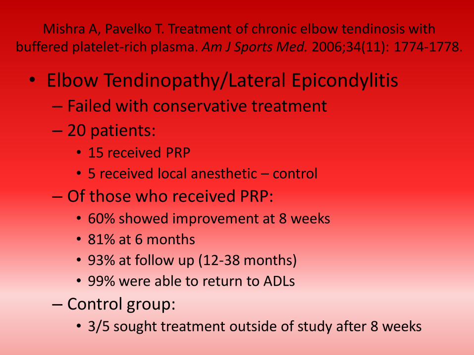
Mishra A, Pavelko T. Treatment of chronic elbow tendinosis with buffered platelet-rich plasma. Am J Sports Med. 2006;34(11): 1774-1778.
• Elbow Tendinopathy/Lateral Epicondylitis – Failed with conservative treatment
– 20 patients: • 15 received PRP
• 5 received local anesthetic – control
– Of those who received PRP: • 60% showed improvement at 8 weeks
• 81% at 6 months
• 93% at follow up (12-38 months)
• 99% were able to return to ADLs
– Control group: • 3/5 sought treatment outside of study after 8 weeks

• Strengths – No adverse effects or complications reported – One of few studies to have a control group – Used human subjects
• Critiques – Small sample size (n=20) – 60% attrition rate – Not randomized – Not blinded
• Rehab protocol: – Standard eccentric strengthening and functional
progressions – Return to activities over 6-8 week when full ROM is
achieved and localized pain or tenderness has diminished – Not necessary to immobilize elbow post injection
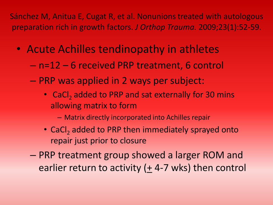
• Acute Achilles tendinopathy in athletes
– n=12 – 6 received PRP treatment, 6 control
– PRP was applied in 2 ways per subject:
• CaCl2 added to PRP and sat externally for 30 mins allowing matrix to form – Matrix directly incorporated into Achilles repair
• CaCl2 added to PRP then immediately sprayed onto repair just prior to closure
– PRP treatment group showed a larger ROM and earlier return to activity (+ 4-7 wks) then control
Sánchez M, Anitua E, Cugat R, et al. Nonunions treated with autologous preparation rich in growth factors. J Orthop Trauma. 2009;23(1):52-59.
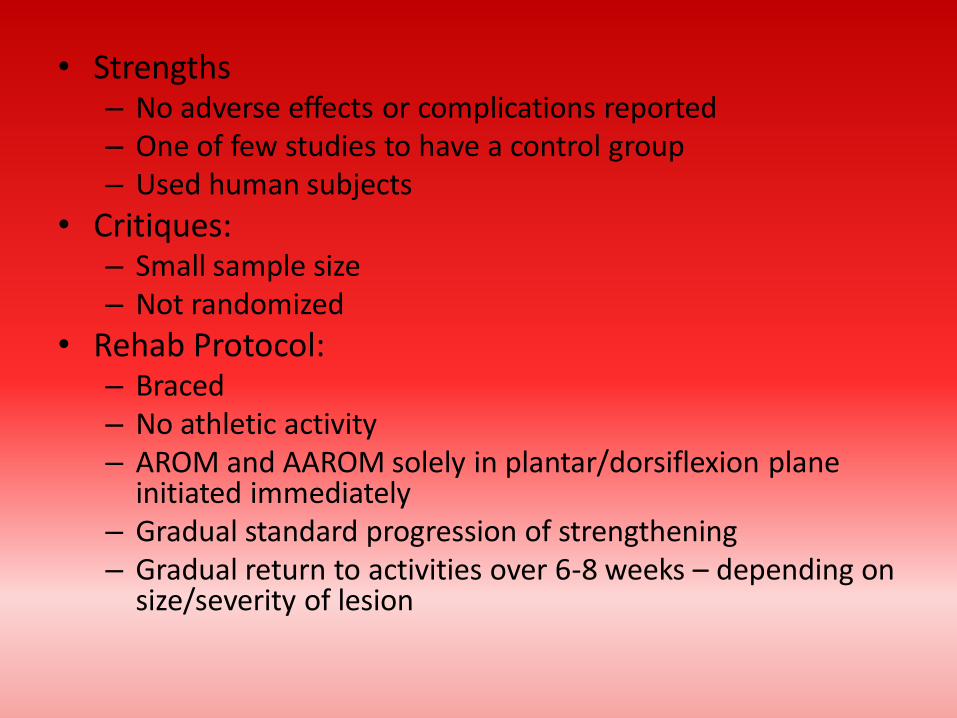
• Strengths – No adverse effects or complications reported – One of few studies to have a control group – Used human subjects
• Critiques: – Small sample size – Not randomized
• Rehab Protocol: – Braced – No athletic activity – AROM and AAROM solely in plantar/dorsiflexion plane
initiated immediately – Gradual standard progression of strengthening – Gradual return to activities over 6-8 weeks – depending on
size/severity of lesion

Barrett S, Erredge S. Growth factors for chronic plantar fasciitis. Podiatry Today. 2004;17:37-42.
• Chronic refractory plantar fasciitis – n=9
– Participants underwent “washout” 90-day period prior to PRP injection: • No brace
• No NSAIDS
• No corticosteroids
– 6 of 9 patients were asymptomatic after 2 months • The other 3 achieved this same status after a second PRP
injection
– At 1 year: 77.9% were still asymptomatic

• Strengths – No adverse effects or complications reported
– Used human subjects
• Critiques – No control group
– Small sample size
• Rehab protocol: – Immediate WB
– Standard rehab program for strengthening
– Gradual return to activities over 6-8 weeks
– More of a gradual training schedule for running athletes

Kajikawa Y, Morihara T, Sakamoto H, et al. Platelet-rich plasma enhances the initial mobilization of circulation-derived cells for
tendon healing. J cell Physiol. 2008;215(3):837-845
• Patellar Tendinopathy AKA Jumper’s knee – Histologically the tendon attained increased levels
of types I and III collagen and macrophages
• Strengths: – No adverse effects
• Critiques: – Not performed on human subjects
– Small sample size (n=3)
– No control group
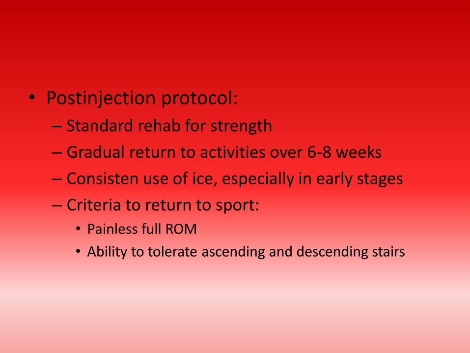
• Postinjection protocol:
– Standard rehab for strength
– Gradual return to activities over 6-8 weeks
– Consisten use of ice, especially in early stages
– Criteria to return to sport:
• Painless full ROM
• Ability to tolerate ascending and descending stairs

Berghoff W, Pietrzak W, Rhodes R. Platelet-rich plasma application during closure following total knee arthroplasty.
Orthopedics. 2006;29(7):590-598.
• Total Knee Arthroplasty – N=137
• 71 received PRP
• 66 were control
– PRP mixed with thrombin and Cacl2 sprayed into knee just prior to closure
– Intervention group demonstrated: • Higher post-op hemoglobin levels
• Shorter hospital stays
• At 6-week follow up had significantly greater knee ROM
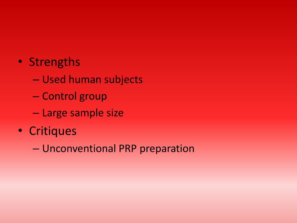
• Strengths
– Used human subjects
– Control group
– Large sample size
• Critiques
– Unconventional PRP preparation
Murray MM, Spindler KP, Abreu E, et al. Collagen-platelet rich plasma hydrogel enhances primary repair of the porcine anterior
cruciate ligament. J Orthop Res. 2007;25(1):81-91.
• ACL Reconstruction
– Treated with collagen-PRP hydrogel at ACL transection site
– Showed improvement in:
• Load at yield
• Maximum load
• Stiffness at 4 weeks post-op

• Strengths
– No adverse effects
• Critiques
– No human subjects
– Small sample size
Sánchez M, Anitua E, Azofra J, Aguirre JJ, Andia I. Intra-articular injection of an autologous preparation rich in growth factors for the treatment of knee OA: a retrospective cohort study. Clin Exp
Rheumatol. 2008;26(5):910-913.
• Osteoarthritis
– n=40
– Treated with 3 separate intra-articular PRP injection
– Outcome measures showed significant improvement in visual analog pain scale and subjective functional scales at 6-month follow up
– Patients under 60 years showed 85% satisfaction
– Patients over 60 years showed 33% satisfaction
• Strengths
– Human subjects
• Critiques
– Small sample size
– Not randomized
Bottom Line
• PRP is a promising, but not yet proven treatment for joint, tendon, ligament, and muscle repair
• PRP is autologous – simple administration • Current studies suggest an excellent safety
profile • No standardized method of producing PRP in
place conflicting results in studies
Bottom Line
• Local anesthetics may change pH environment which could change effects of PRP
• Multiplaner injection technique recommended to accommodate larger surface area
• 24-48 hours post injection patients are encouraged to ice and elevate limb for pain and inflammation control
• No NSAIDS within first 2 weeks after injection – may inhibit prostaglandin pathway

References
Ahmad Z, Howard D, Brooks RA, Wardale J, Henson FMD, Getgood A, Rushton N. The role of platelet rich plasma in musculoskeletal science. J R Soc Med Sh Rep. 2012;3:40
Barrett S, Erredge S. Growth factors for chronic plantar fasciitis. Podiatry Today. 2004;17:37-42.
Berghoff W, Pietrzak W, Rhodes R. Platelet-rich plasma application during closure following total knee arthroplasty. Orthopedics. 2006;29(7):590-598
Kajikawa Y, Morihara T, Sakamoto H, et al. Platelet-rich plasma enhances the initial mobilization of circulation-derived cells for tendon healing. J cell Physiol. 2008;215(3):837-845

References
Mishra A, Pavelko T. Treatment of chronic elbow tendinosis with buffered platelet-rich plasma. Am J Sports Med. 2006;34(11): 1774-1778 Murray MM, Spindler KP, Abreu E, et al. Collagen-platelet rich plasma hydrogel enhances primary repair of the porcine anterior cruciate ligament. J Orthop Res. 2007;25(1):81-91. Sánchez M, Anitua E, Azofra J, Aguirre JJ, Andia I. Intra-articular injection of an autologous preparation rich in growth factors for the treatment of knee OA: a retrospective cohort study. Clin Exp Rheumatol. 2008;26(5):910-913. Sánchez M, Anitua E, Cugat R, et al. Nonunions treated with autologous preparation rich in growth factors. J Orthop Trauma. 2009;23(1):52-59.


