OCTOBER JOGC OCTOBRE 2014 z 927
SOGC CLINICAL PRACTICE GUIDELINE
Abstract
Objective: To provide obstetrical and genetic health care practitioners
with guidelines and recommendations for prenatal screening,
diagnosis, and obstetrical management of fetal open and closed
QHXUDO�WXEH�GHIHFWV��2&17'��
Options: This review includes prenatal screening and diagnostic
WHFKQLTXHV�FXUUHQWO\�EHLQJ�XVHG�IRU�WKH�GHWHFWLRQ�RI�2&17'�including maternal serum alpha fetoprotein screening, ultrasound,
fetal magnetic resonance imaging, and amniocentesis.
Outcomes: To improve prenatal screening, diagnosis, and obstetrical
PDQDJHPHQW�RI�2&17'�ZKLOH�WDNLQJ�LQWR�FRQVLGHUDWLRQ�SDWLHQW�FDUH��HI¿FDF\��FRVW��DQG�FDUH�SURFHGXUHV�
Evidence: Published literature was retrieved through searches
RI�3XE0HG�RU�0('/,1(��&,1$+/��DQG�7KH�&RFKUDQH�/LEUDU\�LQ�1RYHPEHU��������XVLQJ�DSSURSULDWH�FRQWUROOHG�YRFDEXODU\�DQG�NH\�ZRUGV��H�J���SUHQDWDO�VFUHHQLQJ��FRQJHQLWDO�DQRPDOLHV��neural tube defects, alpha fetoprotein, ultrasound scan, magnetic
UHVRQDQFH�LPDJLQJ���5HVXOWV�ZHUH�UHVWULFWHG�WR�V\VWHPDWLF�reviews, randomized control trials/controlled clinical trials, and
observational studies published in English from 1977 to 2012.
Searches were updated on a regular basis and incorporated in
WKH�JXLGHOLQH�WR�1RYHPEHU�����������*UH\��XQSXEOLVKHG��OLWHUDWXUH�ZDV�LGHQWL¿HG�WKURXJK�VHDUFKLQJ�WKH�ZHEVLWHV�RI�KHDOWK�WHFKQRORJ\�DVVHVVPHQW�DQG�KHDOWK�WHFKQRORJ\�UHODWHG�DJHQFLHV��FOLQLFDO�practice guideline collections, clinical trial registries, and national
DQG�LQWHUQDWLRQDO�PHGLFDO�VSHFLDOW\�VRFLHWLHV��$Q�RQOLQH�VXUYH\�RI�health care practitioners was also reviewed.
Values:�7KH�TXDOLW\�RI�HYLGHQFH�LQ�WKLV�GRFXPHQW�ZDV�UDWHG�XVLQJ�WKH�criteria described in the Report of the Canadian Task Force on
3UHYHQWLYH�+HDOWK�&DUH��7DEOH�.
%HQH¿WV��KDUPV��DQG�FRVWV� This review will provide health
care practitioners with a better understanding of the available
SUHQDWDO�VFUHHQLQJ�PHWKRGV�IRU�2&17'�DQG�WKH�EHQH¿WV�DQG�risks associated with each technique to allow evidenced-based
GHFLVLRQV�RQ�2&17'�VFUHHQLQJ��GLDJQRVLV��DQG�REVWHWULFDO�management.
No. 314, October 2014 (Replaces No. 261, July 2011)
SOGC CLINICAL PRACTICE GUIDELINE
Prenatal Screening, Diagnosis, and Pregnancy Management of Fetal Neural Tube Defects
This document reflects emerging clinical and scientific advances on the date issued and is subject to change. The information should not be construed as dictating an exclusive course of treatment or procedure to be followed. Local institutions can dictate amendments to these opinions. They should be well documented if modified at the local level. None of these contents may be reproduced in any form without prior written permission of the SOGC.
This clinical practice guideline has been prepared by the Genetics Committee, reviewed by Family Physician Advisory and Diagnostic Imaging Committees, and approved by the Executive and Council of the Society of Obstetricians and Gynaecologists of Canada.
PRIMARY AUThOR
5��'RXJODV�:LOVRQ��0'��&DOJDU\�$%
SOGC GENETICS COMMITTEE
5��'RXJODV�:LOVRQ��&KDLU���0'��&DOJDU\�$%
Francois Audibert, MD, Montreal QC
-R�$QQ�%URFN��0'��+DOLID[�16
&DUOD�&DPSDJQROR��06F��&&*&��/RQGRQ�21
-XQH�&DUUROO��0'��7RURQWR�21
Lola Cartier, MSc, CCGC, Montreal QC
'DYLG�&KLWD\DW��0'��7RURQWR�21
$ODLQ�*DJQRQ��0'��9DQFRXYHU�%&
-R�$QQ�-RKQVRQ��0'��&DOJDU\�$%
6\OYLH�/DQJORLV��0'��9DQFRXYHU�%&
:��.LP�0DF'RQDOG��0'��+DOLID[�16
/\QQ�0XUSK\�.DXOEHFN��0'��0RQFWRQ�1%
1DQHWWH�2NXQ��0'��7RURQWR�21
0HODQLH�3DVWXFN��51��&RFKUDQH�$%
SPECIAL CONTRIBUTOR
9DQHVVD�3RSD��&DOJDU\�$%
Key words: prenatal screening, congenital anomalies, neural tube
defects, alpha fetoprotein, ultrasound scan, magnetic resonance
LPDJLQJ��DPQLRFHQWHVLV��P\HORPHQLQJRFHOH��VSLQD�EL¿GD��DQHQFHSKDO\
-�2EVWHW�*\QDHFRO�&DQ����������������±���
928 z OCTOBER JOGC OCTOBRE 2014
SOGC CLINICAL PRACTICE GUIDELINE
RECOMMENDATIONS
Screening Evaluation
� ���� 7KH�SULPDU\�VFUHHQLQJ�WHVW�IRU�WKH�GHWHFWLRQ�RI�IHWDO�VWUXFWXUDO�abnormalities including open/closed neural tube defects
�DQHQFHSKDO\��HQFHSKDORFHOH��VSLQD�EL¿GD��LV�D�VHFRQG�WULPHVWHU�anatomical ultrasound with detailed fetal intracranial and spinal
LPDJLQJ�DQG�DVVHVVPHQW���,,��$�
� ���� 7KH�SULPDU\�XVH�RI�PDWHUQDO�VHUXP�DOSKD�IHWRSURWHLQ�IRU�RSHQ�closed neural tube defects screening should be discontinued
with the limited clinical exceptions of pregnant women with a
SUH�SUHJQDQW�ERG\�PDVV�LQGH[������NJ�P2 or when geographical
RU�FOLQLFDO�DFFHVV�IDFWRUV�OLPLW�WLPHO\�DQG�JRRG�TXDOLW\�XOWUDVRXQG�VFUHHQLQJ�DW����WR����ZHHNV¶�JHVWDWLRQ���,,��$�
� ���� :KHQ�XVHG�DV�D�FRPSRQHQW�LQ�PDWHUQDO�VHUXP�JHQHWLF�DQHXSORLG\�screening, maternal serum alpha fetoprotein can be used as a
VHFRQGDU\�VFUHHQLQJ�WRRO�LQ�WKH�VHFRQG�WULPHVWHU���,,��$�
4. Positive screening results for open/closed neural tube defect
�XOWUDVRXQG���PDWHUQDO�VHUXP�DOSKD�IHWRSURWHLQ��UHTXLUH�WLPHO\�referral to appropriate experienced providers for genetic review,
GLDJQRVLV��DQG�FRXQVHOOLQJ���,,��$�
Diagnostic Evaluation
5. If the second trimester screening fetal ultrasound indicates a
probable diagnosis of neural tube defects, the women should be
UHIHUUHG�WR�D�WHUWLDU\�RU�UHJLRQDO�FHQWUH�ZLWK�XOWUDVRXQG�H[SHUWLVH�for a more detailed ultrasound examination looking for the
IHDWXUHV�DVVRFLDWHG�ZLWK�D�QHXUDO�WXEH�GHIHFW�VHTXHQFH���,,��$�
6. Prenatal magnetic resonance imaging can be considered as
an additional fetal imaging technique if further detailed fetal
FHQWUDO�QHUYRXV�V\VWHP�DVVHVVPHQW�LV�UHTXLUHG�IRU�GLDJQRVWLF�RU�PDQDJHPHQW�FRXQVHOOLQJ���,,��$�
Invasive Prenatal Diagnostic Methods
� ���� 7KH�DPQLRWLF�ÀXLG�VSHFLPHQ�IURP�D�GLDJQRVWLF�DPQLRFHQWHVLV��IROORZLQJ�WKH�XOWUDVRXQG�GHWHFWLRQ�RI�IHWDO�DQRPDOLHV�LQFOXGLQJ�FRQ¿UPHG�RU�VXVSHFWHG�RSHQ�FORVHG�QHXUDO�WXEH�GHIHFW���VKRXOG�EH�HYDOXDWHG�IRU�D�IHWDO�NDU\RW\SH��DQG��LI�LQGLFDWHG�DQG�
DYDLODEOH��D�FKURPRVRPDO�PLFURDUUD\���DPQLRWLF�ÀXLG�DOSKD�IHWRSURWHLQ��DQG�DPQLRWLF�ÀXLG�DFHW\OFKROLQHVWHUDVH��7KHVH�WHVW�UHVXOWV�ZLOO�DOORZ�FRPSUHKHQVLYH�HYDOXDWLRQ�RI�WKH�HWLRORJ\��estimated risk of recurrence, and prediction of long-term
neonatal and childhood outcomes of open/closed neural tube
GHIHFW�IRU�IDPLO\�FRXQVHOOLQJ���,,��$�
� ���� :KHQ�D�URXWLQH�GLDJQRVWLF�DPQLRFHQWHVLV�LQGLFDWHV�RQO\�D�ULVN�of
DQHXSORLG\��DQG�QR�LGHQWL¿HG�IHWDO�DQRPDOLHV��LW�LV�QRW�QHFHVVDU\�WR�WDNH�DQ�DPQLRWLF�ÀXLG�VSHFLPHQ�RU�WR�RUGHU�DPQLRWLF�ÀXLG�DOSKD�IHWRSURWHLQ�DQG�DFHW\OFKROLQHVWHUDVH�WHVWLQJ�WR�VFUHHQ�IRU�RSHQ�QHXUDO�WXEH�GHIHFWV���,,��(��
� ���� 7KH�GLDJQRVWLF�LGHQWL¿FDWLRQ�RI�D�SUHJQDQF\�ZLWK�DQ�RSHQ�FORVHG�QHXUDO�WXEH�GHIHFW��LVRODWHG�RU�LQ�D�PRUH�FRPSOH[�PXOWLSOH�DQRPDO\�JURXSLQJ��UHTXLUHV�UHIHUUDO�IRU�FRPSUHKHQVLYH�JHQHWLF��maternal–fetal medicine, and pediatric neurosurgical counselling
IRU�FRPSOHWH�SDWLHQW�IRFXVHG�FDUH���,,��$�
Pregnancy Management
10. Following the detection of an isolated open/closed neural tube
GHIHFW��IDPLOLHV�VKRXOG�EH�RIIHUHG�D�FKRLFH�RI���REVWHWULFDO�FDUH�management options after diagnostic and genetic testing results
are available. Options should include information about SUHQDWDO P\HORPHQLQJRFHOH�UHSDLU�DQG�SURJQRVLV��LI�WKHUH�DUH�QR�PDWHUQDO�or fetal contraindications for prenatal repair at 20–26 weeks’
JHVWDWLRQ���SRVWQDWDO�P\HORPHQLQJRFHOH�VXUJLFDO�UHSDLU�DQG�prognosis, and SUHJQDQF\�WHUPLQDWLRQ�ZLWK�DXWRSV\��%HFDXVH�DQHQFHSKDO\�LV�D�OHWKDO�FRQGLWLRQ��SUHJQDQF\�ZLWK�DQHQFHSKDO\�PD\�EH�LQWHUUXSWHG�DW�DQ\�JHVWDWLRQDO�DJH�RQ�WKH�ZRPDQ¶V�request. For an encephalocele, individualized counselling is
UHFRPPHQGHG�EHFDXVH�RI�WKH�SRVVLEO\�XQLTXH�FLUFXPVWDQFHV�RI�WKH�DQRPDO\���,,��$�
����� &DHVDUHDQ�VHFWLRQ�LV�WKH�PRVW�FRPPRQ�PHWKRG�RI�GHOLYHU\�IRU�D�IHWXV�ZLWK�D�P\HORPHQLQJRFHOH��0&&��LQ�HLWKHU�YHUWH[�RU�EUHHFK�SUHVHQWDWLRQ��EXW�LV�LW�PDQGDWRU\�IRU�EUHHFK�SUHVHQWDWLRQ��9DJLQDO�GHOLYHU\�ZLWK�LQWUDSDUWXP�IHWDO�KHDUW�UDWH�PRQLWRULQJ�FDQ�EH�considered in selected MMC vertex presentation cases that have
QR�PDFURFHSKDO\�UHODWHG�WR�JHVWDWLRQDO�DJH�DQG�D�VPDOO�RU�QR�00&�VDF���,,��$�
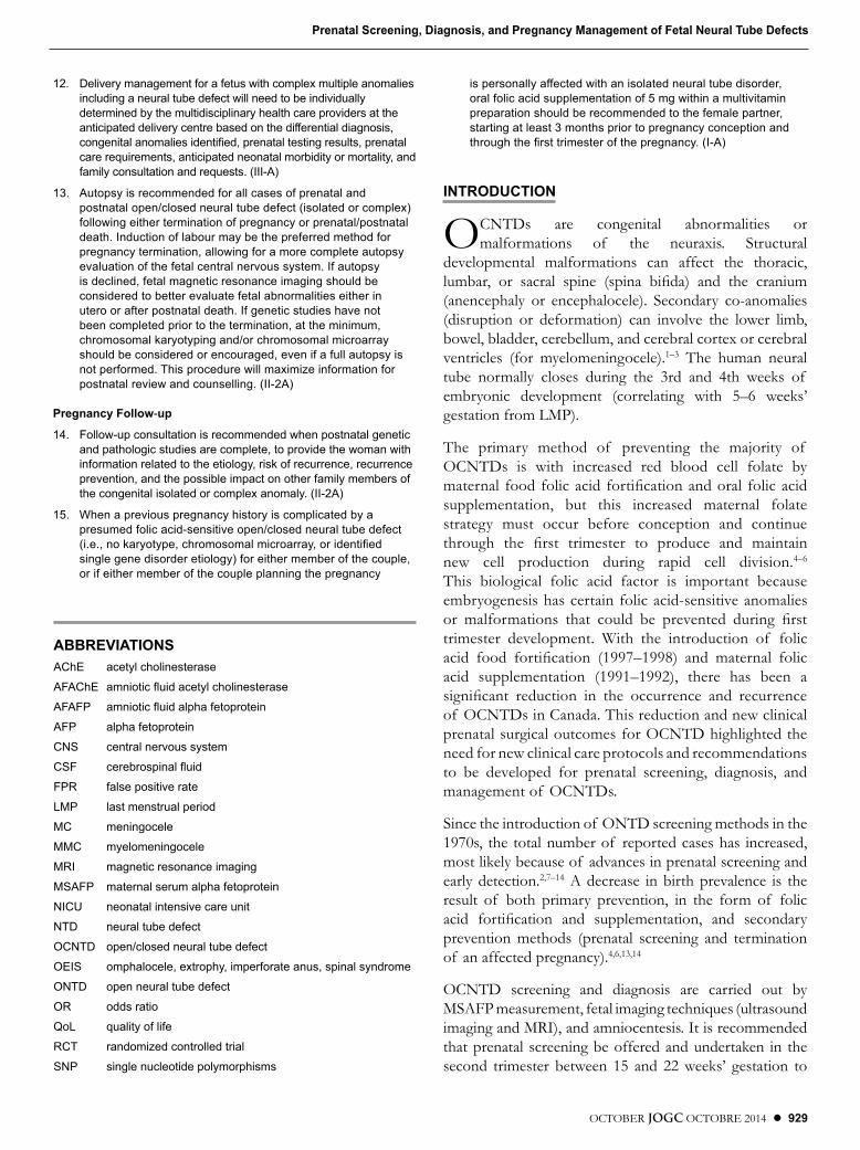
Key to evidence statements and grading of recommendations, using the ranking of the Canadian Task Force on Preventive health Care4XDOLW\�RI�HYLGHQFH�DVVHVVPHQW &ODVVL¿FDWLRQ�RI�UHFRPPHQGDWLRQV�
,��������(YLGHQFH�REWDLQHG�IURP�DW�OHDVW�RQH�SURSHUO\�UDQGRPL]HG� controlled trial
A. There is good evidence to recommend the clinical preventive action
II-1: Evidence from well-designed controlled trials without
randomization
B. There is fair evidence to recommend the clinical preventive action
,,������(YLGHQFH�IURP�ZHOO�GHVLJQHG�FRKRUW��SURVSHFWLYH�RU�� UHWURVSHFWLYH��RU�FDVH±FRQWURO�VWXGLHV��SUHIHUDEO\�IURP�� more than one centre or research group
&���7KH�H[LVWLQJ�HYLGHQFH�LV�FRQÀLFWLQJ�DQG�GRHV�QRW�DOORZ�WR�PDNH�D�UHFRPPHQGDWLRQ�IRU�RU�DJDLQVW�XVH�RI�WKH�FOLQLFDO�SUHYHQWLYH�DFWLRQ��KRZHYHU��RWKHU�IDFWRUV�PD\�LQÀXHQFH�GHFLVLRQ�PDNLQJ
,,������(YLGHQFH�REWDLQHG�IURP�FRPSDULVRQV�EHWZHHQ�WLPHV�RU� places with or without the intervention. Dramatic results in
XQFRQWUROOHG�H[SHULPHQWV��VXFK�DV�WKH�UHVXOWV�RI�WUHDWPHQW�ZLWK�SHQLFLOOLQ�LQ�WKH�����V��FRXOG�DOVR�EH�LQFOXGHG�LQ�WKLV�FDWHJRU\
D. There is fair evidence to recommend against the clinical preventive action
E. There is good evidence to recommend against the clinical preventive
action
III: Opinions of respected authorities, based on clinical experience,
descriptive studies, or reports of expert committees
/����7KHUH�LV�LQVXI¿FLHQW�HYLGHQFH��LQ�TXDQWLW\�RU�TXDOLW\��WR�PDNH�D�UHFRPPHQGDWLRQ��KRZHYHU��RWKHU�IDFWRUV�PD\�LQÀXHQFH�decision-making
7KH�TXDOLW\�RI�HYLGHQFH�UHSRUWHG�LQ�WKHVH�JXLGHOLQHV�KDV�EHHQ�DGDSWHG�IURP�7KH�(YDOXDWLRQ�RI�(YLGHQFH�FULWHULD�GHVFULEHG�LQ�WKH�&DQDGLDQ�7DVN�)RUFH�RQ�Preventive Health Care.109
�5HFRPPHQGDWLRQV�LQFOXGHG�LQ�WKHVH�JXLGHOLQHV�KDYH�EHHQ�DGDSWHG�IURP�WKH�&ODVVL¿FDWLRQ�RI�5HFRPPHQGDWLRQV�FULWHULD�GHVFULEHG�LQ�WKH�&DQDGLDQ�7DVN�)RUFH�on Preventive Health Care.109
OCTOBER JOGC OCTOBRE 2014 z 929
Prenatal Screening, Diagnosis, and Pregnancy Management of Fetal Neural Tube Defects
����� 'HOLYHU\�PDQDJHPHQW�IRU�D�IHWXV�ZLWK�FRPSOH[�PXOWLSOH�DQRPDOLHV�LQFOXGLQJ�D�QHXUDO�WXEH�GHIHFW�ZLOO�QHHG�WR�EH�LQGLYLGXDOO\�GHWHUPLQHG�E\�WKH�PXOWLGLVFLSOLQDU\�KHDOWK�FDUH�SURYLGHUV�DW�WKH�DQWLFLSDWHG�GHOLYHU\�FHQWUH�EDVHG�RQ�WKH�GLIIHUHQWLDO�GLDJQRVLV��FRQJHQLWDO�DQRPDOLHV�LGHQWL¿HG��SUHQDWDO�WHVWLQJ�UHVXOWV��SUHQDWDO�FDUH�UHTXLUHPHQWV��DQWLFLSDWHG�QHRQDWDO�PRUELGLW\�RU�PRUWDOLW\��DQG�IDPLO\�FRQVXOWDWLRQ�DQG�UHTXHVWV���,,,�$�
����� $XWRSV\�LV�UHFRPPHQGHG�IRU�DOO�FDVHV�RI�SUHQDWDO�DQG�SRVWQDWDO�RSHQ�FORVHG�QHXUDO�WXEH�GHIHFW��LVRODWHG�RU�FRPSOH[��IROORZLQJ�HLWKHU�WHUPLQDWLRQ�RI�SUHJQDQF\�RU�SUHQDWDO�SRVWQDWDO�GHDWK��,QGXFWLRQ�RI�ODERXU�PD\�EH�WKH�SUHIHUUHG�PHWKRG�IRU�SUHJQDQF\�WHUPLQDWLRQ��DOORZLQJ�IRU�D�PRUH�FRPSOHWH�DXWRSV\�HYDOXDWLRQ�RI�WKH�IHWDO�FHQWUDO�QHUYRXV�V\VWHP��,I�DXWRSV\�is declined, fetal magnetic resonance imaging should be considered to better evaluate fetal abnormalities either in utero or after postnatal death. If genetic studies have not been completed prior to the termination, at the minimum, FKURPRVRPDO�NDU\RW\SLQJ�DQG�RU�FKURPRVRPDO�PLFURDUUD\�VKRXOG�EH�FRQVLGHUHG�RU�HQFRXUDJHG��HYHQ�LI�D�IXOO�DXWRSV\�LV�not performed. This procedure will maximize information for SRVWQDWDO�UHYLHZ�DQG�FRXQVHOOLQJ���,,��$�
Pregnancy Follow-up
14. Follow-up consultation is recommended when postnatal genetic and pathologic studies are complete, to provide the woman with LQIRUPDWLRQ�UHODWHG�WR�WKH�HWLRORJ\��ULVN�RI�UHFXUUHQFH��UHFXUUHQFH�SUHYHQWLRQ��DQG�WKH�SRVVLEOH�LPSDFW�RQ�RWKHU�IDPLO\�PHPEHUV�RI�WKH�FRQJHQLWDO�LVRODWHG�RU�FRPSOH[�DQRPDO\���,,��$�
����� :KHQ�D�SUHYLRXV�SUHJQDQF\�KLVWRU\�LV�FRPSOLFDWHG�E\�D�presumed folic acid-sensitive open/closed neural tube defect �L�H���QR�NDU\RW\SH��FKURPRVRPDO�PLFURDUUD\��RU�LGHQWL¿HG�VLQJOH�JHQH�GLVRUGHU�HWLRORJ\��IRU�HLWKHU�PHPEHU�RI�WKH�FRXSOH��RU�LI�HLWKHU�PHPEHU�RI�WKH�FRXSOH�SODQQLQJ�WKH�SUHJQDQF\�
LV�SHUVRQDOO\�DIIHFWHG�ZLWK�DQ�LVRODWHG�QHXUDO�WXEH�GLVRUGHU��oral folic acid supplementation of 5 mg within a multivitamin preparation should be recommended to the female partner, VWDUWLQJ�DW�OHDVW���PRQWKV�SULRU�WR�SUHJQDQF\�FRQFHSWLRQ�DQG�WKURXJK�WKH�¿UVW�WULPHVWHU�RI�WKH�SUHJQDQF\���,�$�
INTRODUCTION
O
CNTDs are congenital abnormalities or
malformations of the neuraxis. Structural
developmental malformations can affect the thoracic,
OXPEDU�� RU� VDFUDO� VSLQH� �VSLQD� ELÀGD�� DQG� WKH� FUDQLXP�(anencephaly or encephalocele). Secondary co-anomalies
(disruption or deformation) can involve the lower limb,
bowel, bladder, cerebellum, and cerebral cortex or cerebral
ventricles (for myelomeningocele).1–3
The human neural
tube normally closes during the 3rd and 4th weeks of
embryonic development (correlating with 5–6 weeks’
gestation from LMP).
The primary method of preventing the majority of
OCNTDs is with increased red blood cell folate by
PDWHUQDO� IRRG�IROLF�DFLG�IRUWLÀFDWLRQ�DQG�RUDO� IROLF�DFLG�supplementation, but this increased maternal folate
strategy must occur before conception and continue
WKURXJK� WKH� ÀUVW� WULPHVWHU� WR� SURGXFH� DQG� PDLQWDLQ�new cell production during rapid cell division.
4–6
This biological folic acid factor is important because
embryogenesis has certain folic acid-sensitive anomalies
RU� PDOIRUPDWLRQV� WKDW� FRXOG� EH� SUHYHQWHG� GXULQJ� ÀUVW�trimester development. With the introduction of folic
DFLG� IRRG� IRUWLÀFDWLRQ� �����²������ DQG� PDWHUQDO� IROLF�acid supplementation (1991–1992), there has been a
VLJQLÀFDQW� UHGXFWLRQ� LQ� WKH� RFFXUUHQFH� DQG� UHFXUUHQFH�of OCNTDs in Canada. This reduction and new clinical
prenatal surgical outcomes for OCNTD highlighted the
need for new clinical care protocols and recommendations
to be developed for prenatal screening, diagnosis, and
management of OCNTDs.
Since the introduction of ONTD screening methods in the
1970s, the total number of reported cases has increased,
most likely because of advances in prenatal screening and
early detection.2,7–14
A decrease in birth prevalence is the
result of both primary prevention, in the form of folic
DFLG� IRUWLÀFDWLRQ� DQG� VXSSOHPHQWDWLRQ�� DQG� VHFRQGDU\�prevention methods (prenatal screening and termination
of an affected pregnancy).4,6,13,14
OCNTD screening and diagnosis are carried out by
MSAFP measurement, fetal imaging techniques (ultrasound
imaging and MRI), and amniocentesis. It is recommended
that prenatal screening be offered and undertaken in the
second trimester between 15 and 22 weeks’ gestation to
ABBREVIATIONS$&K(�� DFHW\O�FKROLQHVWHUDVH
$)$&K(��DPQLRWLF�ÀXLG�DFHW\O�FKROLQHVWHUDVH
$)$)3�� DPQLRWLF�ÀXLG�DOSKD�IHWRSURWHLQ
AFP alpha fetoprotein
&16�� FHQWUDO�QHUYRXV�V\VWHP
&6)�� FHUHEURVSLQDO�ÀXLG
FPR false positive rate
LMP last menstrual period
MC meningocele
00&�� P\HORPHQLQJRFHOH
MRI magnetic resonance imaging
MSAFP maternal serum alpha fetoprotein
1,&8�� QHRQDWDO�LQWHQVLYH�FDUH�XQLW
17'�� QHXUDO�WXEH�GHIHFW
2&17'�� RSHQ�FORVHG�QHXUDO�WXEH�GHIHFW
2(,6�� RPSKDORFHOH��H[WURSK\��LPSHUIRUDWH�DQXV��VSLQDO�V\QGURPH
217'�� RSHQ�QHXUDO�WXEH�GHIHFW
OR odds ratio
4R/�� TXDOLW\�RI�OLIH
RCT randomized controlled trial
613�� VLQJOH�QXFOHRWLGH�SRO\PRUSKLVPV
930 z OCTOBER JOGC OCTOBRE 2014
SOGC CLINICAL PRACTICE GUIDELINE
maximize testing accuracy, achieve a low FPR, and allow optimal management of affected pregnancies.4,10,15–27 Factors that have been reported to affect the accuracy and interpretation of OCNTD screening results include type of NTD malformation, gestational age, maternal weight, maternal insulin-dependent diabetes, multiple gestations, ethnicity, environmental factors (prescription and non-prescription medications), and concurrent fetal anomalies.1,9,13,17,22
Early prenatal screening for ONTD gives parents and health care practitioners the ability to evaluate the anomaly and the overall health of the fetus during the second trimester. This guideline focuses on NTD screening, diagnosis, and post-diagnostic pregnancy management.
GENETICS OF NTD
In humans, the neural tube closes between 21 to 28 days of embryonic development, and abnormal closure is characterized by the improper fusion of the neural tube in the developing embryo.1,2,7 NTD prevalence ranges from 1 in 300 to 1 in 1000 pregnancies and is affected by ethnic, genetic, and dietary factors, with the highest NTD rates in the United Kingdom and the United States and the lowest rates in Japan.9,10,28–31
Certain chronic maternal medical conditions will increase the risk of NTDs, including poorly controlled maternal insulin-dependent diabetes (OR 11.5), antiepileptic medications (valproic acid, carbamazepin), therapy with folic acid antagonists, and maternal obesity (OR 3.5).28–31
NTDs are described by their anatomical location and neural content type as follows:
����VSLQD�ELÀGD��FORVXUH�IDLOXUH�RI �URVWUDO�17�IROGV�������93% ONTDs (neural placode at the base of the NTD) and 7% CNTDs (MC = dural sac only; MMC = neural elements attached to the dural sac);
2. anencephaly (closure failure of caudal NT folds causing failure of brain development) 40%;
3. encephalocele (outpouching of the brain through a bony skull defect; occipital is most common, with anterior and lateral locations) 8.5%; and
4. inencephaly/craniorachischisis (abnormal skull and upper spine development) 1.5%.32
Seventy percent of NTDs related to genetic abnormalities are isolated, non-syndromic anomalies or malformations, and with current genetic knowledge, are considered to have a multi-factorial inheritance. Chromosomal abnormalities associated with an “apparently isolated” NTD have a 2.4% to
16.3% incidence.33–37 Syndromes or sequences associated with NTDs include amniotic band syndrome, cloacal extrophy, limb body wall complex, OEIS (omphalocele, extrophy, imperforate anus, spinal) syndrome, cerebrocostomandibular syndrome, and caudal regression.38 Other syndromic OCNTDs are associated with single gene disorders such as PXWDWLRQV�LQ�WKH�9$1*/���FDXGDO�UHJUHVVLRQ��DQG�9$1*/��(cranial ONTD and holoprosencephaly) genes. Other single gene disorders reported include Waardenburg syndrome and Curarino syndrome.38
There are reports of gene mutations or alterations of gene expression leading to ONTDs, such as polymorphisms or SNPs in genes responsible for folate transport, the methionine/homocysteine metabolic cycle, methylation, and nucleotide biosynthesis. Rare mechanisms contributing WR� 217'V� LQFOXGH� HSLJHQHWLF� PRGLÀFDWLRQV�� PDWHUQDO�autoantibodies to the folate receptors, and infertility or assisted reproductive technology therapy.39–42
NON-INVASIVE NTD SCREENING TEChNIQUES
Ultrasound ScreeningUltrasound is the non-invasive screening modality of choice for the detection of fetal anomalies including NTDs because RI �LWV�VDIHW\��FRVW�HIÀFLHQF\��DQG�GHWHFWLRQ�VHQVLWLYLW\�4,21,22,43,44 The current generation of ultrasound machines allow for highly detailed fetal imaging. National screening policy documents cite detection rates of approximately 68% to 94% for NTDs,9,22,45–47 with EUROCAT reporting a 68% GHWHFWLRQ� UDWH� IRU� VSLQD� ELÀGD� �����²������48 and British Columbia an 86% detection rate (1997–1999).4
A second trimester screening ultrasound should be offered to all pregnant women, as recommended in a number of SOGC guidelines43,49,50 for the detection of congenital anomalies from 18 to 22 weeks’ gestation, avoiding the need for a second trimester MSAFP screening test.15,43,45,47,48 Ultrasound is recommended routinely in all second trimester pregnancies and is a more effective screen for OCNTD (improved sensitivity with lower FPR although more expensive) than MSAFP screening, and diagnostically it is safer than amniocentesis which carries the risk of infection or spontaneous abortion.9,11,45,47,51–53 In addition, ultrasound has the major advantage of screening for multiple congenital anomalies at a single ultrasound imaging visit. The factors that may affect ultrasound screening for NTDs include JHVWDWLRQDO�DJH��DPQLRWLF�ÁXLG�YROXPH��SRVLWLRQ�DQG�QXPEHU�of fetuses, and maternal BMI. Other factors to consider are parental ethnicity, “at risk” maternal medication use, maternal diabetic status, and personal, pregnancy, and family histories.1,15,43,54–61 In a fetus with an OCNTD, features visible by ultrasound in the second trimester include anencephaly
OCTOBER JOGC OCTOBRE 2014 z 931
Prenatal Screening, Diagnosis, and Pregnancy Management of Fetal Neural Tube Defects
�ZLWK�WKH�DEVHQFH�RI �WKH�FUDQLDO�YDXOW�DQG�VLJQLÀFDQW�IDFLDO�dysmorphology), open spinal anomalies (abnormal skull shape [lemon sign], abnormal appearance and possible increased width of the cerebral ventricles, abnormal appearance of the posterior fossa/cerebellum [banana sign], and abnormal or incomplete appearance of the posterior vertebral arches in thoracic, lumbar, or sacral locations), and closed spinal anomalies (with the possible absence of the lemon and banana signs and a thick appearing MMC sac protruding from the posterior vertebral opening). Abnormal lower limb movement and positioning of the foot or feet may also be present.43,49,50 Preliminary research KDV�EHHQ� UHSRUWHG� IRU�ÀUVW� WULPHVWHU� XOWUDVRXQG� VFUHHQLQJ�for NTD at 11 to 13 weeks’ gestation, assessing structural developmental variation, such as the absence of intracranial translucency, decreased frontomaxillary facial angle, partial or complete cisterna magna obliteration (brainstem diameter DQG�EUDLQVWHP�WR�RFFLSLWDO�ERQH�GLDPHWHU���DQG��VLJQLÀFDQW��decreased intracranial CSF volume.61–65�7KLV�ÀUVW� WULPHVWHU�NTD imaging should be conducted under a research protocol at the present time.
The experience of the sonographer and up-to-date and well-maintained equipment are important in screening evaluation.7,15 The importance of these factors is highlighted in the recent publication from the rural areas of China,7 where researchers found that ultrasound did not meet its full potential as a method of secondary screening of NTDs; ultrasound was a satisfactory screening method, but NTD detection rate was low. This study did not report that ultrasound was a poor method for screening, but rather that the ultrasound screening skills of the user were not optimal for the detection of NTD anomalies.7
Fetal MRIObstetric application of MRI began approximately 20 years ago, 10 years after its discovery and initial use.66,67 The main use of fetal MRI is as an adjunct to the primary ultrasound ZKHQ�VRQRJUDSKLF�ÀQGLQJV�DUH�DEQRUPDO�DQG�IXUWKHU�05,�detail could be obtained for management planning and counselling.68–70 Fetal MRIs are usually conducted between the late second and early third trimesters, between 23 and 32 weeks’ gestation.21,59,70 This gestational age allows for optimal imaging of the entire fetal brain and subarachnoid space15,21,67,71; however, late gestation MRI can delay decisions regarding pregnancy management, including termination of pregnancy.
The use of fetal MRI, primarily known for its superior brain imaging capabilities, has now been expanded to detect non-CNS abnormalities.22,27,67,70 Although there have been many advances in MRI technology such as the use of T2-weighted sequences, which allow for better
contrast and spatial resolution, and single-shot rapid acquisition with relaxation enhancements for decreasing fetal movement effects,22,69–71 there is no established standard imaging reference because MRI is not used as a screening modality.22,67,70,72�9LVXDOL]DWLRQ�RI �WKH�IHWDO�EUDLQ�by MRI emphasizes the importance of correct imaging methods and radiologic interpretation. Many new techniques under development are already commonly used in the neonatal period, when anatomical structures DUH� YHU\� GLIIHUHQW�� WKH� WHFKQLTXHV� PXVW� EH�PRGLÀHG� LQ�order to use them safely during pregnancy.66–70 Operator interpretation of the MRI requires a thorough knowledge of normal and abnormal fetal anatomy. As the option of fetal NTD surgery becomes available in perinatal PHGLFLQH�� WKH� XVH� RI � 05,� FRXOG� VLJQLÀFDQWO\� HQKDQFH�CNS evaluation.70,73,74
MRI is considered safe for the fetus, but there are hypothetical concerns about teratogenesis and acoustic damage.67,70 While additional fetal MRI research with neonatal follow-up is needed, an SOGC Diagnostic Imaging Committee Guideline75 reports that fetal MRI is safe at 1.5 tesla magnet exposure during the second and third trimesters.
Ultrasound has the advantage over MRI of easier patient access to imaging services; it is not hampered by fetal movement, it is less expensive than MRI, and it provides good spatial resolution. MRI, however, is not limited by ÀHOG�RI � YLHZ� UHVWULFWLRQV�� SURYLGHV� YHU\� JRRG� VRIW� WLVVXH�contrast, and performs well regardless of oligohydramnios, maternal obesity, or fetal lie.22,27,70,73,74
05,� KDV� WKH� SRWHQWLDO� WR� FRQÀUP� WKH� UHVXOWV� RI � DQ�HTXLYRFDO� XOWUDVRXQG� ÀQGLQJ� DQG� FDQ� SRVVLEO\� LGHQWLI\�additional anomalies,76 but its expense, lack of a standard of reference for imaging, and limited availability are factors that continue to favour ultrasound as the imaging choice for the detection of OCNTDs.77,78
Maternal serum AFPMSAFP screening was once considered the gold standard of prenatal ONTD screening, but with advances in technology, research, and knowledge, MSAFP screening now has limited value when reliable second trimester ultrasound (screening and diagnostic) is available. Since the mid-1970s, non-invasive MSAFP has been used for the detection of ONTDs.10,11,19,27,51,79 MSAFP levels rise early in the pregnancy, and ONTD screening was optimized to discern normal from abnormal MSAFP results in the second trimester between 15 and 18 weeks’ gestation.14,15,18,45 MSAFP levels are measured in multiples of the median, using unaffected pregnancies of the same gestational age as
932 z OCTOBER JOGC OCTOBRE 2014
SOGC CLINICAL PRACTICE GUIDELINE
the control value.11,14,18 To interpret results, it is important to correctly identify the gestational age, number of fetuses, maternal ethnicity, and maternal weight. Furthermore, such factors as a personal or family history of OCNTD, any maternal “at risk for NTD” medications (epilepsy control [valproic acid, carbamazepine] or folate antagonists), or certain chronic medical conditions, such as diabetes, must be taken into consideration.14,18 First trimester MSAFP (11 to 13 gestational weeks) levels in normal pregnancies are affected by maternal race, weight, smoking status, and method of contraception (oral birth control).46
Second trimester MSAFP detects 71% to 90% of NTDs, with an FPR of 1% to 3% percent.11,14,18 Elevated levels of MSAFP can be associated with other conditions, such as fetal skin disorders, abdominal wall defects, fetal demise, fetal nephrosis, and pregnancies at an increased risk for placenta-related adverse events.14,17,19 First trimester MSAFP 17'�VFUHHQLQJ�KDV�D�����GHWHFWLRQ�UDWH�DW�D�À[HG�)35�RI �10%,55 and it is not recommended of NTD screening.
A detailed fetal structural ultrasound at 18 to 22 gestational weeks and amniocentesis (AFAFP, AFAChE, and chromosome karyotype analysis) is recommended as a diagnostic follow-up with an elevated second trimester MSAFP level (screen-positive result).
A second trimester detailed ultrasound (at 18 to 22 gestational weeks) is recommended as the standard of care for all pregnancies to determine the approximate gestational age, FRQÀUP�IHWDO�YLDELOLW\��LGHQWLI\�PXOWLSOHV��GHWHFW�FORVHG�17'V�(not usually detected by MSAFP screening), or screen for other congenital anomalies.43,49,50 Earlier ultrasound imaging for dating or evaluating the pregnancy may be required, based on clinical care factors as determined by the primary obstetrical provider. First trimester dating by crown–rump length has a ± 3 to 5 day standard deviations for estimating the gestational age, compared to the ± 7 day standard deviations for the 18 to 22 gestational weeks’ ultrasound.
Many recent retrospective studies have shown that with present screening approaches and new technology measuring MSAFP is no longer practical for the detection RI �17'V��HVSHFLDOO\�LQ�WKH�ÀUVW�WULPHVWHU��EXW�LW�LV�XVHIXO�IRU�selecting women for diagnostic testing, such as ultrasound or amniocentesis.14,16,19,20,51,79
MSAFP screening for ONTDs is less costly than ultrasound or amniocentesis,11,52 but its cost advantage must be weighed DJDLQVW�LWV�GHFUHDVHG�VHQVLWLYLW\�DQG�VSHFLÀFLW\��LWV�LQDELOLW\�WR�detect closed NTDs, and the requirement of further testing that includes detailed second trimester ultrasound when second trimester screening MSAFP levels are elevated.
RecommendationsScreening Evaluation
1. The primary screening test for the detection of fetal structural abnormalities including open/closed neural tube defects (anencephaly, HQFHSKDORFHOH��VSLQD�ELÀGD��LV�D�VHFRQG�WULPHVWHU�anatomical ultrasound with detailed fetal intracranial and spinal imaging and assessment. (II-2A)
2. The primary use of maternal serum alpha fetoprotein for open/closed neural tube defects screening should be discontinued with the limited clinical exceptions of pregnant women with a pre-SUHJQDQW�ERG\�PDVV�LQGH[������NJ�P2 or when geographical or clinical access factors limit timely and good quality ultrasound screening at 18 to 22 weeks’ gestation. (II-2A)
3. When used as a component in maternal serum genetic aneuploidy screening, maternal serum alpha fetoprotein can be used as a secondary screening tool in the second trimester. (II-2A)
4. Positive screening results for open/closed neural tube defect (ultrasound ± maternal serum alpha fetoprotein) require timely referral to appropriate experienced providers for genetic review, diagnosis, and counselling. (II-2A)
Diagnostic Evaluation5. If the second trimester screening fetal ultrasound
indicates a probable diagnosis of neural tube defects, the women should be referred to a tertiary or regional centre with ultrasound expertise for a more detailed ultrasound examination looking for the features associated with a neural tube defect sequence. (II-2A)
6. Prenatal magnetic resonance imaging can be considered as an additional fetal imaging technique if further detailed fetal central nervous system assessment is required for diagnostic or management counselling. (II-2A)
INVASIVE PRENATAL DIAGNOSTIC METhODS (NTD SCREENING/TESTING)
AmniocentesisAFP from the fetal yolk sac initially and subsequently WKH� IHWDO� OLYHU� DQG�$&K(� VSHFLÀFDOO\� IURP� IHWDO� QHXUDO�WLVVXH�DUH�QRW�QRUPDOO\�IRXQG�LQ�DPQLRWLF�ÁXLG��KRZHYHU��only AFP is present in maternal blood.5,14,17,80 Elevated levels of AFAFP and AFAChE are present is fetuses ZLWK� 217'� DQG� FDQ� EH� HDVLO\� LGHQWLÀHG� LQ� DPQLRWLF�ÁXLG�14,18,80
OCTOBER JOGC OCTOBRE 2014 z 933
Prenatal Screening, Diagnosis, and Pregnancy Management of Fetal Neural Tube Defects
An amniocentesis is most often performed for the detection of chromosomal aneuploidy or genetic PXWDWLRQV�� EXW� WKH� DPQLRWLF� ÁXLG� FDQ� DOVR� EH� XVHG� IRU�the detection of NTDs.18,23,74,81–84 The procedure is usually conducted between the 15th and 20th gestational weeks.6,17,45,85–88� $PQLRWLF� ÁXLG� FDQ� EH� DQDO\]HG� IRU� IHWDO�karyotype, chromosomal microarray, and AFAFP and AFAChE levels. When amniocentesis is performed with a suspicion of OCNTD, the information from the karyotype, chromosomal microarray, AFAFP, and the AFAChE levels ZLOO� DVVLVW� ZLWK� WKH� VSHFLÀF� W\SH� RI �17'� GLDJQRVLV� DQG�with counselling regarding prognosis.
If amniocentesis is being performed for aneuploidy GLDJQRVLV� RQO\� �L�H��� WKHUH� LV� QR� LGHQWLÀHG� IHWDO� 17'� RU�PDOIRUPDWLRQ��� DPQLRWLF� ÁXLG� DQDO\VLV� IRU� $)$)3� DQG�AChE is not routinely required.
:RPHQ�ZKR�XQGHUJR�ÀUVW�WULPHVWHU�&96�WHVWLQJ�IRU�IHWDO�aneuploidy still require a routine second trimester screening ultrasound for fetal congenital anomalies, including NTD screening if a complete ultrasound anatomic assessment is QRW�SRVVLEOH�LQ�WKH�ÀUVW�WULPHVWHU�
Risks associated with amniocentesis include spontaneous abortion (estimated procedural risk of 0.5% to 1.0% added to the no-procedure background spontaneous risk of pregnancy loss), post-procedure spotting, infections, rupture of membranes, and fetal damage or loss.48,86–88
Amniocentesis for genetic testing is especially important when considering prenatal or postnatal repair of congenital DQRPDOLHV�LQFOXGLQJ�2&17'V��,GHQWLÀFDWLRQ�RI �DGGLWLRQDO�fetal genetic factors is important as these factors may interfere with the neonatal outcome.73,74,81–83
Although an amniocentesis is an important diagnostic option for high-risk pregnancies in the detection of chromosomal abnormalities and ONTDs, amniocentesis should not be used as a method for laboratory NTD (AFAFP, AFAChE) screening because of the risks and cost associated with the test.
Recommendations
Invasive Prenatal Diagnostic Methods����7KH�DPQLRWLF�ÁXLG�VSHFLPHQ�IURP�D�GLDJQRVWLF�
amniocentesis (following the ultrasound detection RI �IHWDO�DQRPDOLHV�LQFOXGLQJ�FRQÀUPHG�RU�suspected open/closed neural tube defect), should be evaluated for a fetal karyotype (and, if indicated and available, a chromosomal microarray), DPQLRWLF�ÁXLG�DOSKD�IHWRSURWHLQ��DQG�DPQLRWLF�ÁXLG�acetylcholinesterase. These test results will allow comprehensive evaluation of the etiology, estimated
risk of recurrence, and prediction of long-term neonatal and childhood outcomes of open/closed neural tube defect for family counselling. (II-2A)
8. When a routine diagnostic amniocentesis indicates only a risk RI �DQHXSORLG\��DQG�QR�LGHQWLÀHG�IHWDO�anomalies, it is not necessary to take an amniotic ÁXLG�VSHFLPHQ�RU�WR�RUGHU�DPQLRWLF�ÁXLG�DOSKD�fetoprotein and acetylcholinesterase testing to screen for open neural tube defects. (II-2E)
����7KH�GLDJQRVWLF�LGHQWLÀFDWLRQ�RI �D�SUHJQDQF\�ZLWK�an open/closed neural tube defect (isolated or in a more complex multiple-anomaly grouping) requires referral for comprehensive genetic, maternal–fetal medicine, and pediatric neurosurgical counselling for complete patient-focused care. (II-2A)
PREGNANCY MANAGEMENT
Counselling Information for Continuing PregnanciesCounselling should be based on the recognition of the personal impact that a physical or mental disability could have on a child and its family.
One of the major questions a family asks when their fetus is given an isolated, non-syndromic, non-chromosomal myelomeningocele diagnosis is “What will the quality of life be for my child?” Król et al. reported a follow-up study of 33 children (19 female; 14 male) with a myelomeningocele at ages 5 to 20 years. They were evaluated in 2 age groups: 5 to 12 (n = 17) and 13 to 20 (n = 16) using the Health-Related Quality of Life in 6SLQD� %LÀGD� TXHVWLRQQDLUH� DV� D� TXDQWLWDWLYH� WRRO�� 7KH�study reported good, very good, and average QoL in 64%, 30%, and 6% of participants respectively. None of the participants felt their QoL was poor. Issues of visual perception in the younger group and ambulation in the older group were related to lower QoL scores. The vast majority of children had a history of good specialist care. The most common medical issues were related to hydrocephalus and neurogenic bladder.89
In a larger study of 119 patients with hydrocephalus and MMC, Barf et al. reported a near equal overall QoL in the study group (76%) as in an age-matched peer group (72%), ZLWK�JUHDWHU�VDWLVIDFWLRQ�LQ�WKH�DUHDV�RI �ÀQDQFH�DQG�IDPLO\�life, but less satisfaction with their sex lives and abilities to FDUH� IRU� WKHPVHOYHV��$Q� LPSRUWDQW� IHDWXUH� LGHQWLÀHG�ZDV�that the severity of disease and the level of the lesion had little bearing on self-reported QoL.90
In a retrospective cohort study Hunt and Oakeshott reported on 117 open MMC patients. The cohort had had 54%
934 z OCTOBER JOGC OCTOBRE 2014
SOGC CLINICAL PRACTICE GUIDELINE
mortality and, at evaluation, had an average age of 35 years. Among the survivors, 40% were independent in activities of daily living, meeting medical needs, transportation, and continence care.91
Bowman et al. reported a 25-year follow-up of 118 MMC patients in which 75% lived to early adult age, 85% had high school or college education, 80% were able to maintain social bladder continence with catheterization, 90% reported acceptable levels of bowel continence, and 86% of long-term survivors were shunt-dependent. ,Q� WKLV� JURXS�� WKH� PRVW� VLJQLÀFDQW� FDXVH� RI � GHDWK� LQ�childhood or early adulthood was an unrecognized shunt malfunction.92
For more complete counselling knowledge, the health care provider could consider an economic assessment of the cost of continuing care for the MMC patient. Yi et al. UHYLHZHG����FRVW�RI�LOOQHVV�VWXGLHV��ZLWK�FRQVLVWHQW�ÀQGLQJV�reported across all.93 The lifetime direct medical costs for 00&�SDWLHQWV�LV�VLJQLÀFDQW��ZLWK�WKH�PDMRULW\�RI �WKH�FRVW�being for inpatient care, treatment at initial diagnosis in childhood, and co-morbidities in adult life. The caregiver WLPH�FRVW�ZDV�DOVR�VLJQLÀFDQW�IRU�WKH�FRKRUW�
Continuing Pregnancy Surveillance and Method of DeliveryPregnancy surveillance and delivery recommendations for fetuses with OCNTDs are controversial because increased pregnancy termination has limited the study of the MMC delivery model for more than 2 decades. The ACOG Practice Bulletin94 reports that most pregnancies complicated by MMC will deliver with appropriate lung maturity at term, and there is no evidence that antenatal fetal heart rate testing for an MMC indication improves outcome. It is recognized that anomalous fetuses frequently KDYH�DEQRUPDO�IHWDO�KHDUW�UDWH�WUDFLQJV�WKDW�DUH�GLIÀFXOW�WR�interpret.95
Serial ultrasounds for fetal growth, head size, and ventricular size may be helpful in continued prognosis counselling and delivery planning. The fetus should be delivered at a centre with a level III NICU and pediatric neurosurgery services.90 A latex-free delivery and surgical repair plan should be considered because individuals with MMC are at an increased risk of developing a severe and life-threatening allergy to latex.96
The mode of delivery for an MMC-affected fetus with a vertex presentation remains controversial. There are no RCTs, but at least 5 studies representing a total of 400 patients suggested vaginal delivery does not adversely affect neonatal outcome, and one large study of 200 patients suggested Caesarean
sections as a safer method of delivery for the MMC-affected fetus.97–102 Breech presentation is common in the MMC-affected fetus as a result of decreased lower limb neurologic function and megacephaly, and hence requires delivery by Caesarean section.
Prenatal in Utero Repair of MMCThe Randomized Trial of Prenatal versus Postnatal Repair of Myelomeningocele74 showed that prenatal fetal surgery for MMC reduced the need for ventricular peritoneal shunting (40% vs. 82%) and showed improved lower limb motor outcomes at 30 months of age. However, the surgery was associated with maternal and fetal risks. Additional improvement in the child composite score for mental development and motor function at 30 months of age was LGHQWLÀHG� LQ� WKH� SUHQDWDO� VWXG\� JURXS� �SULPDU\� RXWFRPH�score: prenatal surgery [n = 64] 148.6 ± 57.5; postnatal surgery [n = 70] 122.6 ± 57.2; P = 0.007). Improved hindbrain herniation at 12 months and ambulation at 30 months was also reported. This landmark study provides an important treatment option for parents because it clearly LGHQWLÀHV�QHRQDWDO�DQG�FKLOGKRRG�EHQHÀWV�RI �SUHQDWDO�IHWDO�surgery, albeit at the cost of increased maternal risks for the index and subsequent pregnancies.74 An accompanying editorial counselled caution regarding the initiation of this new prenatal treatment in multiple fetal therapy/treatment centres and emphasized that most women who expressed an interest in the experimental trial were either ineligible or declined to participate, with only 15% of screened women participating in the study.103 A legal and ethical opinion that drew attention to prioritizing the fetus stated that:
unlike a born child, a fetus is not a patient in the real sense, but only a metaphor. However, it would be unethical if a care provider, without the woman’s informed consent, promoted the interests of the fetus over those of the woman.104
When considering the consent to fetal surgery
the parents have clear legal responsibilities to provide or consent to their born children’s necessary medical care but the maternal duties to fetuses in utero are not generally recognized.”104
Regarding whether this study was research or a therapeutic innovation they stated that:
the research component of the study was not in the prenatal surgical and postnatal management of each child but in the systematic control of each case according to the research protocol and the retrospective comparative review for all the outcome data.104
OCTOBER JOGC OCTOBRE 2014 z 935
Prenatal Screening, Diagnosis, and Pregnancy Management of Fetal Neural Tube Defects
Their conclusion was that “women, diagnosed with a fetal MMC and where open fetal surgery is available, will have to be informed of this treatment option.”104 This prenatal repair option needs therefore to be presented to families in Canada as part of the informed consent process because although it is not available here, it is available in the United States.104
Postnatal Repair of MMCThe decision to perform postnatal repair is the most likely clinical scenario in Canada for the MMC fetus or neonate because only mothers who have travelled to in utero MMC treatment centres in the United States will have received in utero MMC repair.
The postnatal group in the Randomized Trial of Prenatal 9HUVXV� 3RVWQDWDO� 5HSDLU� RI � 0\HORPHQLQJRFHOH74 is used as the clinical outcome for comparison because the RCT study was done in selected Children’s Hospital facilities with organized and comprehensively controlled pediatric neurosurgery services. The placement of a shunt within the ÀUVW����PRQWKV�ZDV�VLJQLÀFDQWO\�KLJKHU�LQ�WKH�SRVWQDWDO�UHSDLU�group (82%, compared to prenatal group 40%; P = 0.001). Other abnormal cranial and brain features were higher in the postnatal group than in the prenatal repair group (degree of hindbrain herniation, P = 0.001; brainstem kinking, P = 0.001; abnormal location of the 4th ventricle, P = 0.002).
7KH�FRPELQHG�SULPDU\�RXWFRPH�DW����PRQWKV�ZDV�VLJQLÀFDQWO\�lower with postnatal repair than with prenatal repair, but this was mainly due to differences in motor function between the spinal anatomical levels of the defects (P = 0.001); the Bayley 0HQWDO� 'HYHORSPHQW� ,QGH[� ZDV� QRW� VLJQLÀFDQWO\� GLIIHUHQW� (P� �������� 7KLV� SULPDU\� RXWFRPH� GLIIHUHQFH� ZDV� UHÁHFWHG�LQ� WKH� FOLQLFDO� ÀQGLQJV� RI � ���� IRU� WKH� DELOLW\� WR� ZDON�independently with prenatal repair and 21% with postnatal repair (relative risk 2.01 ([95% CI 1.16–3.48]; P = 0.01). &KLOGUHQ�ZKR�KDG�KDG�SRVWQDWDO�UHSDLU�DOVR�KDG�VLJQLÀFDQWO\�lower WeeFIM scores for self-care (P = 0.02) and mobility (P = 0.003), but not for cognitive ability (P = 0.67).
Immediate postnatal care for MMC is required whether the birth was by Caesarean section or vaginal delivery. Neonatology specialists should attend in the labour room for MMC protection and care, probable neonatal prematurity (less than 37 weeks’ gestation), and planning and performing subsequent primary neurosurgical MMC closure within 24 hours of delivery, especially if the MMC is open or the MMC sac was ruptured at delivery
Termination of PregnancyOver the last 3 decades an estimated 70% to 80% of women and couples with a fetus affected by OCNTD have chosen termination of the pregnancy.4,105,106
For families choosing to terminate a pregnancy because of an MMC, anencephaly, or encephalocele, the health care provider should discuss fetal autopsy and chromosomal DQDO\VLV�DQG�PLFURDUUD\�EHFDXVH�WKH�DXWRSV\�ÀQGLQJV�PD\�provide important information about the etiology for the NTD and the recurrence risk.107 Measures to prevent recurrence, including maternal supplementation with 5 mg of preconception folic, are recommended in subsequent pregnancies because folic acid supplementation has been shown in an RCT to reduce the isolated recurrence risk of NTD by 72%, with a resulting reduction of the non-supplementation prevalence rate of 3% to a post supplementation recurrence rate of 1%.5,108
RecommendationsPregnancy Management10. Following the detection of an isolated open/closed
neural tube defect, families should be offered a choice of 3 obstetrical care management options after diagnostic and genetic testing results are available. Options should include information about prenatal myelomeningocele repair and prognosis (if there are no maternal or fetal contraindications for prenatal repair at 20–26 weeks’ gestation), postnatal myelomeningocele surgical repair and prognosis, and pregnancy termination with autopsy. Because anencephaly is a lethal condition, pregnancy with anencephaly may be interrupted at any gestational age on the woman’s request. For an encephalocele, individualized counselling is recommended because of the possibly unique circumstances of the anomaly. (II-2A)
11. Caesarean section is the most common method of delivery for a fetus with a myelomeningocele (MCC) in either vertex or breech presentation, but is it PDQGDWRU\�IRU�EUHHFK�SUHVHQWDWLRQ��9DJLQDO�GHOLYHU\�with intrapartum fetal heart rate monitoring can be considered in selected MMC vertex presentation cases that have no macrocephaly related to gestational age and a small or no MMC sac. (II-2A)
12. Delivery management for a fetus with complex multiple anomalies including a neural tube defect will need to be individually determined by the multidisciplinary health care providers at the anticipated delivery centre based on the differential GLDJQRVLV��FRQJHQLWDO�DQRPDOLHV�LGHQWLÀHG��SUHQDWDO�testing results, prenatal care requirements, anticipated neonatal morbidity or mortality, and family consultation and requests. (III-A)
13. Autopsy is recommended for all cases of prenatal and postnatal open/closed neural tube defect (isolated or complex) following either termination
936 z OCTOBER JOGC OCTOBRE 2014
SOGC CLINICAL PRACTICE GUIDELINE
of pregnancy or prenatal/postnatal death. Induction of labour may be the preferred method for pregnancy termination, allowing for a more complete autopsy evaluation of the fetal central nervous system. If autopsy is declined, fetal magnetic resonance imaging should be considered to better evaluate fetal abnormalities either in utero or after postnatal death. If genetic studies have not been completed prior to the termination, at the minimum, chromosomal karyotyping and/or chromosomal microarray should be considered or encouraged, even if a full autopsy is not performed. This procedure will maximize information for postnatal review and counselling. (II-2A)
Pregnancy Follow-up14. Follow-up consultation is recommended when
postnatal genetic and pathologic studies are complete, to provide the woman with information related to the etiology, risk of recurrence, recurrence prevention, and the possible impact on other family members of the congenital isolated or complex anomaly. (II-2A)
15. When a previous pregnancy history is complicated by a presumed folic acid-sensitive open/closed neural tube defect (i.e., no karyotype, chromosomal PLFURDUUD\��RU�LGHQWLÀHG�VLQJOH�JHQH�GLVRUGHU�etiology) for either member of the couple, or if either member of the couple planning the pregnancy is personally affected with an isolated neural tube disorder, oral folic acid supplementation of 5 mg within a multivitamin preparation should be recommended to the female partner, starting at least 3 months prior to pregnancy conception and WKURXJK�WKH�ÀUVW�WULPHVWHU�RI �WKH�SUHJQDQF\���,�$�
SUMMARY
NTDs occur early in embryologic development, secondary to failure of fusion of the neural groove and formation of the neural tube. NTDs include malformations or disruptions in the lower spine and upper cranium and occur in approximately 1 in 1000 live births. There are 2 methods of prevention: primary prevention with IROLF� DFLG� �IRRG� IRUWLÀFDWLRQ� DQG� RUDO� VXSSOHPHQWDWLRQ��and secondary prevention with prenatal screening and pregnancy interruption.
Ultrasound is presently the best screening method for prenatal detection of fetal anomalies including NTDs because of its safety, availability, accuracy, and cost-effectiveness. MSAFP screening should be used in limited clinical scenarios as an additional screening method.
Although it may be cost effective, the MSAFP screen can detect only open NTDs; it has poor sensitivity and VSHFLÀFLW\�IRU�GHWHFWLQJ�FORVHG�17'V��)HWDO�05,�VKRXOG�EH�used as an adjunct to ultrasound when more detail of the CNS is required or when fetal therapy is being considered. MRI is more expensive and is not as readily available as ultrasound; further research is necessary to ensure its reported safety and to allow the creation of reference standards.
Amniocentesis is an invasive diagnostic procedure that is accurate in the detection of ONTDs and in analyses for chromosomal or genetic abnormalities. With the use of ultrasound for guidance, amniocentesis performed by experienced hands, can greatly reduce the risks of miscarriage, infection, and harm to the fetus. However, although amniocentesis has a greater than 99% detection rate for ONTD, it is neither cost effective nor necessary for the detection of ONTD unless performed for the LQGLFDWLRQ�RI �DEQRUPDOLWLHV�LQ�IHWDO�XOWUDVRXQG�ÀQGLQJV�
Improved prenatal screening and diagnosis will enable mothers or couples to obtain precise information about detected anomalies so they can make informed decisions about whether to continue or interrupt the pregnancy and what their options are for fetal or postnatal repair.
Our current knowledge and experience indicates that pregnant woman should be offered 2 prenatal screening approaches:
1. risk screening for fetal aneuploidy and fetal anomalies, using a combination of non-invasive techniques that includes ultrasound (fetal anatomic and structural detail) and
2. maternal serum testing for placental biochemical DQDO\WHV�DQG�YDOXHV��SUHVHQWO\�XVHG�IRU�ÀUVW�DQG�VHFRQG�trimester screening approaches) or for placental cell-free fetal DNA (for future direct fetal molecular DNA screening or diagnostic testing).
REFERENCES
1. Frey L, Hauser WA. Epidemiology of neural tube defects. Epilepsia 2003;44(3):4–13.
2. Gucciardi E, Pietrusiak M, Reynolds DL, Rouleau J. Incidence of neural tube defects in Ontario 1986-1999. CMAJ 2002;167(3):237–40.
���'H�0DUFR�3��0HUHOOR�(��5RVVL�$��3LDWHOOL�*��&DPD�$��.LEDU�=��&DSUD�9��FZD6 is a novel gene for human neural tube defects. Human Mutation 2011;3:384–90.
���9DQ�$OOHQ�0,��%R\OH�(��7KLHVVHQ�3��0F)DGGHQ�'��&RFKUDQH�'�� Chambers GK, et al. The impact of prenatal diagnosis on neural tube defect (NTD) pregnancy versus birth incidence in British Columbia. J Appl Genet 2006;47:151–58.
OCTOBER JOGC OCTOBRE 2014 z 937
Prenatal Screening, Diagnosis, and Pregnancy Management of Fetal Neural Tube Defects
���:LOVRQ�5'��-RKQVRQ�-$��:\DWW�3��$OOHQ�9��*DJQRQ�$��/DQJORLV�6��HW�DO��Preconceptional vitamin/folic acid supplementation 2007: the use of folic acid in combination with a multivitamin supplement for the prevention of neural tube defects and other congenital anomalies. J Obstet Gynaecol Can 2007;29:1003–26.
���'H�:DOV�3��7DLURX�)��9DQ�$OOHQ�0,��8K�6��/RZU\�5%��6LEEDOG�%��HW�DO��5HGXFWLRQ�LQ�QHXUDO�WXEH�GHIHFWV�DIWHU�IROLF�DFLG�IRUWLÀFDWLRQ�LQ�&DQDGD�� N Engl J Med 2007;357(2):135–42.
7. Lu QB, Wang ZP, Gong R, Sun XH, Gao LJ, Zhao ZT. Investigation of XOWUDVRXQG�VFUHHQLQJ�HIÀFLHQF\�IRU�QHXUDO�WXEH�GHIHFWV�GXULQJ�SUHJQDQF\�in rural areas of China. Public Health 2011;125:639–44.
8. Kokalj TS, Rejc B, Gersak K. Incidence and prevention of neural tube defects in Slovenia. Eur J Obstet Gynecol 2011;156:119–20.
9. Cameron M, Moran P. Prenatal screening and diagnosis of neural tube defects. Prenatal Diagnosis 2009;29:402–11.
10. Bamforth FJ. Laboratory screening for genetic disorders and birth defects. Clin Biochem 1994;27:333–42.
����9LQW]LOHRV�$0��$QDQWK�&9��)LVKHU�$-��6PXOLDQ�-&��'D\�6DOYDWRUH�'��%HD]RJORX�7��HW�DO��&RVW�EHQHÀW�DQDO\VLV�RI �WDUJHWHG�XOWUDVRQRJUDSK\�IRU�SUHQDWDO�GHWHFWLRQ�RI �VSLQD�ELÀGD�LQ�SDWLHQWV�ZLWK�DQ�HOHYDWHG�concentration of second-trimester maternal serum a-fetoprotein. Am J Obstet Gynecol 1999;180:1227–33.
12. Harris RA, Washington AE, Nease RF, Kupperman M. Cost utility of prenatal diagnosis and the risk-based threshold. Lancet 2004;363:276–82.
13. EUROCAT Working Group. Prevalence of neural tube defects in 20 regions of Europe and the impact of prenatal diagnosis, 1980-1986. Epistemiol Community Health 1991;4552–8.
14. Cuckle HS, Wald NJ, Cuckle PM. Prenatal screening and diagnosis of neural tube defects in England and Wales in 1985. Prenat Diagnos 1989;9:393–400.
15. Roberts N, Bhide A. Ultrasound prenatal diagnosis of structural abnormalities. Obstet Gynaecol Reprod Med 2007;17:1–8.
16. Norem CT, Schoen EJ, Walton DL, Krieger RC, O’Keefe J, To TT, et al. Routine ultrasonography compared with maternal serum alpha-fetoprotein for neural tube defect screening. Obstet Gynecol 2005;106:747–52.
17. Driscoll DA, Gross SJ. Screening for fetal aneuploidy and neural tube defects. Genet Med 2009;11:818–21.
18. Chodirker BN, Cadrin C, Davies GAL, Summers AM, Wilson RD, Winsor EJT, et al. Canadian guidelines for prenatal diagnosis. Part 1: genetic indicators for prenatal diagnosis. SOGC Clinical Practice Guidelines, No. 105, June 2001. J Obstet Gynaecol Can 2001;23:525–31.
19. Johnson JA, Summers A. Prenatal genetic screening for Down syndrome and open neural tube defects using maternal serum marker screening. J Obstet Gynaecol Can 1999;21:887–91.
20. Wald NJ, Hackshaw A, Stone R, Densem J. Serum alpha-fetoprotein DQG�QHXUDO�WXEH�GHIHFWV�LQ�WKH�ÀUVW�WULPHVWHU�RI �SUHJQDQF\��3UHQDW�'LDJQ�1993;13:1047–50.
21. Sohn YS, Kim MJ, Kwon JY, Kim YH, Park YW. The usefulness of fetal MRI for prenatal diagnosis. Yonsei Med J 2007;48:671–7.
22. Pugash D, Brugger PC, Bettelheim D, Prayer D. Prenatal ultrasound and fetal MRI: the comparative value of each modality in prenatal diagnosis. Eur J Radiol 2008; 68:214–26.
23. Delisle M, Wilson RD. First trimester prenatal diagnosis: amniocentesis. Semin Perinatol 1999;23:414–23.
24. Zambelli H, Carelli E, Honorato D, Marba S, Coelho G, Carnevalle A, et al. Assessment of neurosurgical outcome in children prenatally diagnosed with myelomeningocele and development of a protocol for fetal surgery to prevent hydrocephalus. Childs Nerv Syst 2007;23:421–5.
25. Ewigman BG, Crane JP, Frigoletto FD, LeFevre ML, Bain RP, McNellis D. Effect of prenatal ultrasound screening on perinatal outcome. N Engl J Med 1993;329:821–7.
26. Saari-Kemppainen A, Karjalainen O, Ylostalo P, Heinonen O. Ultrasound screening and perinatal mortality: Controlled trial of systematic one-stage screening in pregnancy. Int J Gynaecol Obstet 1991;35:100.
27. Saleem SN, Said AH, Abdel-Raouf M, El-Kattan EA, Zaki MS, Madkour N, et al. Fetal MRI in the evaluation of fetuses referred for sonographically suspected neural tube defects (NTDs)—impact on diagnosis and management decision. Neuroradiology 2009;51:761–72.
28. Boulet SL, Yang Q, Mai C, Kirby RS, Collins JS, Robbins JM, et al; National Birth Defects Prevention Network. Trends in the SRVWIRUWLÀFDWLRQ�SUHYDOHQFH�RI �VSLQD�ELÀGD�DQG�DQHQFHSKDO\�LQ�WKH� United States. Birth Defects Res A Clin Mol Teratol 2008;82:527–32.
29. Shurtleff DB, Luthy DA, Nyberg DA, Benedetti TJ, Mack LA. Meningomyelocele: management in utero and post natum. Ciba Found Symp 1994;181:270–86.
�����+XQW�*0��7KH�PHGLDQ�VXUYLYDO�WLPH�LQ�RSHQ�VSLQD�ELÀGD��'HY�0HG�&KLOG�Neurol 1997;39:568.
31. Manning SM, Jennings R, Madsen JR. Pathophysiology, prevention and potential treatment of neural tube defects. Ment Retard Dev Disabil Res Rev 2000;6:6–14.
32. Ghi T, Pilu G, Falco P, Segata M, Carletti A, Cocchi G, et al. Prenatal GLDJQRVLV�RI �RSHQ�DQG�FORVHG�VSLQD�ELÀGD��8OWUDVRXQG�2EVWHW�*\QHFRO�2006;28:899–903.
33. Kanit H, Özkan AA, Öner SR, Ispahi C, Endrikat JS, Ertan K. Chromosomal abnormalities in fetuses with ultrasonographically detected neural tube defects. Clin Dysmorphol 2012;20:190–3.
34. Drugan A, Johnson MP, Dvorin E, Moody J, Krivchenia EL, Schwartz D, et al. Aneuploidy with neural tube defects: another reason for complete evaluation in patients with suspected ultrasound anomalies or elevated maternal serum alpha-fetoprotein. Fetal Diagn Ther 1989;4:88–92.
35. Harmon JP, Hiett Ak, Palmer CG, Golichowski AM. Prenatal ultrasound detection of isolated neural tube defects is cytogenetic evaluation warranted? Obstet Gynecol 1995:86:595–9.
36. Hume RF, Drugan A, Reichler A, Lampinen J, Martin LS, Johnson MP, et al. Aneuploidy among prenatally detected neural tube defects. Am J Med Genet 1996;61:171-173.
37. Kennedy D, Chitayat D, Winsor EJ, Silver M, Toi A. Prenatally diagnosed neural tube defects: ultrasound, chromosome, and autopsy or postnatal ÀQGLQJV�LQ������FDVHV��$P�-�0HG�*HQHW������������²���
38. Chen, CP. Syndromes, disorders and maternal risk factors associated with QHXUDO�WXEH�GHIHFWV��,9���7DLZDQ�-�2EVWHW�*\QHFRO������������²���
�����'H�0DUFR�3��0HUHOOR�(��&DPD�$��.LEDU�=��&DSUD�9��&ULWLFDO�UHYLHZ��human neural tube defects: genetic causes and prevention. Biofactors 2011;37;261–8.
40. Alfarra HY, Alfarra SR, Sadiq MF. Neural tube defects between folate metabolism and genetics. Ind J Hum Genet 2011;17(3):126–31.
41. Aneji CN, Northrup H, Au KS. Deep sequencing study of the MTHFR gene to identify variants associated with myelomeningocele. Birth Defects Res A Clin Mol Teratol 2012;94:84–90.
42. Wang XW, Luo YL, Wang W, Zhang Y, Chen Q, Cheng LY. Association between MTHFR A1298C polymorphism and neural tube defect susceptibility: a meta-analysis. Am J Obstet Gynecol 2012;251:e1–e7.
43. Cargill Y, Morin L; Society of Obstetricians and Gynaecologists of Canada. Diagnostic Imaging Committee. Content of a complete routine second trimester obstetrical ultrasound examination and report. SOGC Clinical Practice Guidelines, No. 223, March 2009. J Obstet Gynaecol Can 2009;31:272–5.
938 z OCTOBER JOGC OCTOBRE 2014
SOGC CLINICAL PRACTICE GUIDELINE
44. Dashe JS, Twickler DM, Santos-Ramos R, McIntire DD, Ramus RM. Alpha-fetoprotein detection of neural tube defects and the impact of standard ultrasound. Am J Obstet Gynecol 2006;195:1623–8.
45. Benn PA, Borgida A, Home D, Briganti S, Collins R, Rodis JF. Down syndrome and neural tube defect screening—the value of using gestational age by ultrasonography. Am J Obstet Gynecol 1997;176:1056–61.
46. Lee W, Chaiworapongsa T, Romero R, Williams R, McNie B, Johnson A, HW�DO��$�GLDJQRVWLF�DSSURDFK�IRU�WKH�HYDOXDWLRQ�RI �VSLQD�ELÀGD�E\�WKUHH�dimensional ultrasonography. J Ultrasound Med 2002;21:619–26.
47. Malhotra N, Rao JP, Malhotra J, Malhotra N. Ultrasound for screening fetal malformations—has 3-D made a difference? J SAFOG 2012;2:7–10.
48. Garne E, Dolk H, Loane M, Boyd PA. EUROCAT website data on prenatal detection rates of congenital anomalies. J Med Screen 2010;17:97–8.
�����9DQ�GHQ�+RI �0&��:LOVRQ�5'��62*&�'LDJQRVWLF�,PDJLQJ�&RPPLWWHH��SOGC Imaging Committee.. Fetal soft markers in obstetric ultrasound. SOGC Clinical Practice Guidelines, No. 162, June 2005. J Obstet Gynaecol Can 2005; 27:592–612.
�����*DJQRQ�$��:LOVRQ�5'��$OOHQ�90��$XGLEHUW�)��%OLJKW�&��%URFN�-$��HW�DO���Society of Obstetricians and Gynaecologists of Canada. Evaluation of prenatally diagnosed structural congenital anomalies. SOGC Committee Opinion, No. 234, Sept 2009. J Obstet Gynaecol Can 2009;31:875–81.
51. Wald NJ, Cuckle H, Brock JH, Peto R, Polani PE, Woodford FP. Maternal serum-alpha-fetoprotein measurement in antenatal screening IRU�DQHQFHSKDO\�DQG�VSLQD�ELÀGD�LQ�HDUO\�SUHJQDQF\��UHSRUW�RI �8�.��Collaborative Study on Alpha-fetoprotein in Relation to Neural-tube Defects. Lancet 1977;309:1323–32.
�����7DSOLQ�6+��7KRPSVRQ�56��&RQUDG�'$��&RVW�MXVWLÀFDWLRQ�DQDO\VLV�of prenatal maternal serum alpha-feto protein screening. Med Care 1988;26:1185–202.
�����+HFNHUOLQJ�36��9HUS�06��$�FRVW�HIIHFWLYHQHVV�DQDO\VLV�RI �DPQLRFHQWHVLV�and chorionic villus sampling for prenatal genetic testing. Med Care 1994;32:863–80.
54. Bredaki FE, Wright D, Akolekar R, Cruz G, Nicolaides KH. Maternal serum alpha-fetoprotein in normal pregnancy at 11-13 weeks’ gestation. Fetal Diagn Ther 2011;30:274–9.
55. Bredaki FE, Poon LC, Birdir C, Escalante D, Nicolaides KH. First-trimester screening for neural tube defects using alpha-fetoprotein. Fetal Diagn Ther 2012;31:109–14.
56. Stratmeyer ME, Greenleaf JF, Dalecki D, Salvesen KA. Fetal ultrasound: mechanical effects. J Ultrasound Med 2008;27:597–605.
�����6DORPRQ�/-��9LOOH�<��4XDOLW\�FRQWURO�RI �SUHQDWDO�XOWUDVRXQG��8OWUDVRXQG�Review 2005;5:297–303.
58. Roberts CJ, Hibbard BM, Roberts EE, Ecans KT, Laurence KM, Robertson IB. Diagnostic effectiveness of ultrasound in detection of neural tube defect: the South Wales experience of 2509 scans (1977-1982) in high-risk mothers. Lancet 1983;2:1068–9.
59. Hosny IA, Elghawabi HS. Ultrafast MRI of the fetus: an increasingly important tool in prenatal diagnosis of congenital anomalies. Magn Reson Imaging 2010;28:1431–9.
60. Cameron M, Moran P. Prenatal screening and diagnosis of neural tube defects. Prenat Diagn 2009;29:402–11.
61. Chaoui R, Benoit B, Mitkowska-Wozniak H, Heling KS, Nicolaides KH. Assessment of intracranial translucency (IT) in the detection RI �VSLQD�ELÀGD�DW�WKH�������ZHHN�VFDQ��8OWUDVRXQG�2EVWHW�*\QHFRO�2009;34:249–52.
62. Lachmann R, Picciarelli G, Moratalla J, Greene N, Nicholaides KH. )URQWRPD[LOODU\�IDFLDO�DQJOH�LQ�IHWXVHV�ZLWK�VSLQD�ELÀGD�DW�������ZHHNV·�gestation. Ultrasound Obstet Gynecol 2010;36:268–71.
63. Scheier M, Lachmann R, Petros M, Nicolaides KH. Three-dimensional VRQRJUDSK\�RI �WKH�SRVWHULRU�IRVVD�LQ�IHWXVHV�ZLWK�RSHQ�VSLQD�ELÀGD�DW� 11-13 weeks’ gestation. Ultrasound Obstet Gynecol 2011;38:625–9.
64. Lachmann R, Chaoui R, Moratalla J, Picciarelli G, Nicolaides KH. 3RVWHULRU�EUDLQ�LQ�IHWXVHV�ZLWK�RSHQ�VSLQD�ELÀGD�DW�������ZHHNV��3UHQDW�Diagn 2011;31:103–6.
65. Loureiro T, Ushakov F, Montenegro N, Gielchinsky Y, Nicolaides KH. &HUHEUDO�YHQWULFXODU�V\VWHP�LQ�IHWXVHV�ZLWK�RSHQ�VSLQD�ELÀGD�DW�������weeks’ gestation. Ultrasound Obstet Gynecol 2012;39:620–4.
�����&RDNOH\�)9��*OHQQ�2$��4D\\XP�$��%DUNRYLFK�$-��*ROGVWLHQ�5��)LOO\�5$��Fetal MRI: a developing technique for the developing patient. AJR Am J Roentgenology 2004;182:243–52.
67. Garel C. Fetal MRI: what is the future? Ultrasound Obstet Gynecol 2008;31:123–8.
68. Glenn OA, Cuneo AA, Barkovich AJ, Hashemi Z, Bartha AI, Xu D. Malformations of cortical development- diagnostic accuracy of fetal MR imaging. Radiology 2012;263(3):843–55.
69. Girard N, Chaumoitre K, Chapon F, Pineau S, Barberet M, Brunel H. Fetal magnetic resonance imaging of acquired and developmental brain anomalies. Semin Perinatol 2009;33:234–50.
70. Ljubic A, Cetkovic A, Mikic AN, Stamenkovic JD, Jovanovic I, Opincal TS, et al. Ultrasound vs MRI in diagnosis of fetal and maternal complications. Ultrasound Review 2001;5(2/3):231–42.
71. Cannie M, Jani J, Dymarkowski S, Deprest J. Fetal magnetic resonance imaging: luxury or necessity? Ultrasound Obstet Gynecol 2006;27:471–6.
72. Al-Mukhtar A, Kasprian G, Schmook MT, Brugger PC, Prayer D. Diagnostic pitfalls in fetal brain MRI. Semin Perinatol 2009;33:251–8.
73. Sutton LN. Fetal surgery for neural tube defects. Best Pract Res Clin Obstet Gynaecol 2008;22:175–88.
74. Adzick NS, Thom EA, Spong CY, Brock JW, Burrows PK, Johnson MP, et al. A randomized trial of prenatal versus postnatal repair of myelomeningocele. N Engl J Med 2011;364:993–1004.
75. Patenaude Y, Pugash D, Lim K, Morin L; SOGC Diagnostic Imaging Committee. The use of Magnetic Resonance Imaging in the obstetric patient. SOGC Clinical Practice Guidelines, No. 306, April 2014. J Obstet Gynaecol Can 2014;36:349–355.
76. Bulas D. Fetal magnetic resonance imaging as a complement to fetal ultrasonography. Ultrasound Q 2007;23:3–22.
�����:LOOLDPV�)��*ULIÀWKV�3'��6SLQDO�QHXUDO�WXEH�GHIHFWV�RQ�LQ�XWHUR�05,�� Clin Radiol 2013;68:e715-e722.
78. Ben-Sira L, Garel C, Malinger G, Constantini S. Prenatal diagnosis of spinal dysraphism. Childs Nerv Syst 2013;29:1541–52.
�����.|UQHU�+��5RGULJXH]�/��<HUR�-/)��6FKXO]H�0��+RUQ�$��+HUHGHUR�/�� et al. Maternal serum alpha-fetoprotein screening for neutal tube defects and other disorders using an ultramicro-ELISA. Collaborative study in Cuba and in the German Democratic Republic. Hum Genet 1986;73:60–3.
�����:LGOXQG�.)��*RWWYDOO�7��5RXWLQH�DVVHVVPHQW�RI �DPQLRWLF�ÁXLG�DOSKD�IHWRSURWHLQ�LQ�HDUO\�VHFRQG�WULPHVWHU�DPQLRFHQWHVLV�LV�QR�ORQJHU�MXVWLÀHG��Acta Obstet Gynecol Scand 2007;86:67–71.
81. Wapner RJ, Martin CL, Levy B, Ballif BC, Eng CM, Zachary JM, et al. Chromosomal microarray versus karyotyping for prenatal diagnosis. N Engl J Med 2012;367:2175–84.
�����5HGG\�80��3DJH�*3��6DDGH�*5��6LOYHU�50��7KRUVWHQ�95��3DUNHU�&%�� et al. Karyotype versus microarray testing for genetic abnormalities after stillbirth. N Engl J Med 2012;367:2185–93.
�����7DONRZVNL�0(��2UGXOX�=��3LOODODPDUUL�9��%HQVRQ�&%��%OXPHQWKDO�,��Connolly S, et al. Clinical diagnosis by whole-genome sequencing of a prenatal sample. N Engl J Med 2012;367:2226–32.
OCTOBER JOGC OCTOBRE 2014 z 939
Prenatal Screening, Diagnosis, and Pregnancy Management of Fetal Neural Tube Defects
84. Shumway JB, Greenspoon JS, Khouzami AN, Platt LD, Blakemore KJ. $PQLRWLF�ÁXLG�DOSKD�IHWRSURWHLQ��$)$)3��DQG�PDWHUQDO�VHUXP�DOSKD�fetoprotein (MSAFP) in abdominal pregnancies: correlation with extent and site of placental implantation and clinical implications. J Matern Fetal Med 1996;5:120–3.
85. Laurence KM, Elder G, Evans KT, Hibbard BM, Hooles M, Roberts CJ. Should women at high risk of neural tube defect have an amniocentesis? J Med Genet 1985;22:457–61.
86. Nizard J. Amniocentesis: technique and education. Curr Opin Obstet Gynecol 2010;22:152–4.
87. Wilson RD, Langlois S, Johnson JA; Genetics Committee and CCMG Prenatal Diagnosis Committee of the Society of Obstetricians and Gynaecologists of Canada. Mid-trimester amniocentesis fetal loss rate. SOGC Committee Opinion, No. 194, July 2007. J Obstet Gynaecol Can 2007;29:586–90.
88. Kooper AJ, de Bruijn D, van Ravenwaaij-Arts CMA, Faas BHW, Creemers JWT, Thomas CMG, et al. Fetal anomaly scan potentially will replace routine AFAFP assays for the detection of neural tube defects. Prenat Diagn 2007;27:29–33.
�����.UyO�0��6LELľVNL�0��6WHIDQVNL�0��6\QGHU�0��$VVHVVPHQW�RI �OLIH�TXDOLW\�LQ�FKLOGUHQ�ZLWK�VSLQD�ELÀGD��&KLU�1DU]DGRZ�5XFKX�2UWRS�3RO�����������²��
�����%DUI �+$��3RVW�0:��9HUKRHI �0��-HQQHNHQV�6FKLQNHO�$��*RRVNHQV�5+��3UHYR�$-��/LIH�VDWLVIDFWLRQ�RI �\RXQJ�DGXOWV�ZLWK�VSLQD�ELÀGD��'HY�0HG�Child Neurol 2007;49:458–63.
�����+XQW�*0��2DNHVKRWW�3��2XWFRPH�LQ�SHRSOH�ZLWK�RSHQ�VSLQD�ELÀGD�DW�DJH�35: prospective community based cohort study. BMJ 2003;326:1365–6.
�����%RZPDQ�50��0F/RQH�'*��*UDQW�-$��7RPLWD�7��,WR�-$��6SLQD�ELÀGD�outcome: a 25-year prospective. Pediatr Neurosurg 2001;34:114-120.
93. Yi Y, Lindemann M, Colligs A, Snowball C. Economic burden of neural tube defects and impact of prevention with folic acid: a literature review. Eur J Pediatr 2011;170:1391–400.
94. ACOG Committee on Practice Bulletins. Neural tube defects. ACOG Practice Bulletin. Clinical Management Guidelines for Obstetrician-Gynecologists. No 44, July 2003 (Replaces Committee Opinion Number 252, March 2001). Obstet Gynecol 2003;102:203–13.
�����9LQGOD�6��6DKRWD�'6��&RSSHQV�0��-DPHV�'.��&RPSXWHUL]HG�DQDO\VLV�RI �behavior in fetuses with congenital abnormalities. Ultrasound Obstet Gynecol 1997;9:302–9.
�����%RZPDQ�50��0F/RQH�'*��*UDQW�-$��7RPLWD�7��,WR�-$��6SLQD�ELÀGD�outcome: a 25-year prospective. Pediatr Neurosurg 2001;34:114–20.
97. Cochrane D, Aronyk K, Sawatzky B, Wilson D, Steinbok P. The effects of labour and delivery on spinal cord function and ambulation in patients with meningomyelocele. Child Nerv Syst 1991;7:312–5.
�����%HQVHQ�-7��'LOODUG�5*��%XUWRQ�%.��2SHQ�VSLQD�ELÀGD��GRHV�FHVDUHDQ�section delivery improve diagnosis? Obstet Gynecol 1988;71:532–4.
99. Sakala EP, Andree I. Optimal route of delivery for meningomyelocele. Obstet Gynecol Surv 1990;45:209–12.
100. Hill AE, Beattie F. Does caesarean section delivery improve neurological RXWFRPH�LQ�RSHQ�VSLQD�ELÀGD"�(XU�-�3HGLDWU�6XUJ���������6XSSO������²��
101. Merrill DC, Goodwin P, Burson JM, Sato Y, Williamson R, Weiner CP. The optimal route of delivery for fetal meningomyelocele. Am J Obstet Gynecol 1998;179:235–40.
102. Luthy DA, Wardinsky T, Shurtleff DB, Hollenback KA, Hickok DE, Nyberg DA, et al. Cesarean section before the onset of labor and subsequent motor function in infants with meningomyelocele diagnosed antenatally. N Engl J Med 1991;324:662–6.
103. Simpson JL, Greene MF. Fetal surgery for myelomeningocele? N Engl J Med 2011;364: 1076–7.
104. Dickens BM, Cook RJ. Ethical and legal issues in reproductive health. Legal and ethical issues in fetal surgery. Int J Gynaecol Obstet 2011;115:80–3.
105. Barry S. Quality of life and myelomeningocele: an ethical and evidence-based analysis of the Groningen protocol. Pediatr Neurosurg 2010;46:409–14.
�����%R\G�3$��'H9LJDQ�'��.KRVKQRRG�%��/RDQH�0��*DUQH�(��'RON�+��EUROCAT working group. Survey of prenatal screening policies in Europe for structural malformations and chromosome anomalies, and their impact on detection and termination rates for neural tube defects and Down’s syndrome. BJOG 2008;115:689–96.
�����'pVLOHWV�9��2OLJQ\�//��*HQHWLFV�DQG�)DPLO\�3K\VLFLDQV�$GYLVRU\�Committees of the Society of Obstetricians and Gynaecologists of Canada. Fetal and perinatal autopsy in prenatally diagnosed fetal abnormalities with normal karyotype. SOGC Technical Update, No. 267, October 2011. J Obstet Gynaecol Can 2011;33:1047–57.
108. Miller EC, Liu N, Wen SW, Walker M. Why do Canadian women fail to achieve optimal pre-conceptional folic acid supplementation? An observational study. J Obstet Gynaecol Can 2011;33:1116–23.
109. Woolf SH, Battista RN, Angerson GM, Logan AG, Eel W. Canadian Task Force on Preventive Health Care. New grades for recommendations from the Canadian Task Force on Preventive Health Care. CMAJ 2003;169:207–8.


