
California State University, San Bernardino California State University, San Bernardino
CSUSB ScholarWorks CSUSB ScholarWorks
Theses Digitization Project John M. Pfau Library
1995
Preparation of a site-specific lymphotoxin- mutant to be used in Preparation of a site-specific lymphotoxin- mutant to be used in
protein characterization and receptor binding studies protein characterization and receptor binding studies
Derek Andrew Knight
Follow this and additional works at: https://scholarworks.lib.csusb.edu/etd-project
Part of the Cell Biology Commons
Recommended Citation Recommended Citation Knight, Derek Andrew, "Preparation of a site-specific lymphotoxin- mutant to be used in protein characterization and receptor binding studies" (1995). Theses Digitization Project. 987. https://scholarworks.lib.csusb.edu/etd-project/987
This Thesis is brought to you for free and open access by the John M. Pfau Library at CSUSB ScholarWorks. It has been accepted for inclusion in Theses Digitization Project by an authorized administrator of CSUSB ScholarWorks. For more information, please contact [email protected].

PREPARATION OF A SITE-SPECIFIC LYMPHOTOXIN-p
MUTANT TO BE USED IN PROTEIN. CHARACTERIZATION AND
RECEPTOR BINDING STUDIES
A Thesis
Presented to the
Faculty of
California State University,
San Bernardino
In Partial Fulfillment
of the Requirements for the Degree
Master of Science
in
Biology
by
Derek Andrew Knight
June 1995
PREPARATION OF A SITE-SPECIFIC LYMPHOTOXIN-p
MUTANT TO BE USED IN PROTEIN CHARACTERIZATION AND
RECEPTOR BINDING STUDIES
A Thesis
Presented to the
Faculty of
California State University,
San Bernardino
by
Derek Andrew Knight
June 1995
Approved by:
Dr. Richard Fehn, Chair, Biology Date
)r. Lisa Shamansky
Dr. Ching-Hua Wang
ABSTRACT
Lymphotoxin (LT) is a cytokine synthesized by
eytotpxiG T lymphocytes, B cells and natural killer
cells. This cytokine is very similar to tumor necrosis
factor (TNF) in both structure and in certain functions.
LT has been shown to induce cytotoxic, antiviral and
growth enhancing activities in a variety of cells
(Andrews et ai., 1990). LT has also been shown to be
essential for the proper development of peripheral
lymphoid organs in mice (Togni et al., 1994) and is
thought to play a role in the pathogenesis of certain
autoimmune diseases (Ruddle et al., 1990), but its role
in human development and in the immune response is still
poorly understood. One method that will help build upon
our knowledge of this cytokine involves placing site^
specific mutations in regions of the LT gene thought to
be important in receptor binding and in protein
structure. One such mutation involves an N-linked
glycosylation site present on the membrane bound form of
LT (mLT) but absent on the soluble form. By preparing a
mutant LT-^p gene that lacks this glycosylation site, it
will be possible to analyze a proposed model for LT
structure and ligand receptor interactions.
In this study, such a mutant was prepared using a
polymerase chain reaction (PGR) overlap extension
111

technique to introduce a site-directed single base
substitution that destroyed the consensus sequence for
N-linked glycosylation, replacing an asparagine residue
with a serine residue. It is believed that this
substitution results in loss of the N-linked
glycosylation site while not significantly altering
protein conformation. The presence of the desired
mutation was confirmed using dideoxy DNA sequencing
analysis and the mutant cDNA cloned into the pcDNAl-amp
plasmid, E. Coll were transformed with this clone and
positive transformants used in the preparation of
maxiprep DNA. The DNA obtained was transfected into
GOS-7 mammalian cells and protein expression analyzed.
It was found that neither the mutant or wild-type LT-p
transfectants produced protein bands when labeled with
monoclonal LT-p antibodies and immunoprecipitated.
These results may indicate that LT-p is rapidly degraded
when expressed without LT-a.
IV

Acknowledgements
I would like to thank my wife Barbara and my
Ghildren, Megan and Christopher for their patience and
support during the many long hours this project has
required. I would also like to thank the CSUSB faculty
members who have offered their encouragement, support
and guidance. A special word of thanks to Dr. Lisa
Shamansky for her wisdom, encouragement, use of her lab
equipment and financial support for reagents, without
which this project would have been impossible. Also a
word of thanks to everyone at Dr. Carl Ware's lab at
UCR, especially Dr. Paul Crowe. And last, but not
least, I would like to thank the CSUSB chemistry
department for allowing a biology major to work in one
of their labs and to the great help obtained from the
chemistry stock clerks, Jan Mack and Ken Makino. Thanks
to all.
This work was supported by a CSUSB ASI graduate research
award.
V

Table of Contents
Abstract ill
Acknowledgements . . . v
MATERIALS AND METHODS .,9
List of Figures. .viii
INTRODUCTION, 1
Generation of 5' and 3' PGR fragments containing tbe desired mutation. • • • .9
Joining of fragments by PGR Overlap Extension to obtain full length mutant. 11
Sequence confirmation of mutation ........11
Cloning of mutant DNA in pcDNAl-amp plasmid......13
Maxiprep of pcDNAl^amp/LT-P mutant DNA... ..15
GOS-7 cell culture 16
GOS-7 Cell transfection 16
Biosynthetic labeling of GOS-7 cells. .17
SDS-Polyacrylimide gel electrophoresis...........18
RESULTS 20
Preparation of 5' and 3' PGR fragments containing the mutation.....................................20
Joining of fragments by PGR overlap extension
GOS-7 cell transfection, biosynthetic labeling
yields full-length mutant 20
Sequence confirmation of mutation................20
Cloning of mutant DNA in pcDNAl-amp plasmid. 21
Maxiprep Of pcDNAl-amp/LT-|3 mutant DNA.......... .21
and SDS-PAGE ... 21
VI

DISCUSSION..... 36
BIBLIOGRAPHY .... 41
VI1
List of Figures
Figure 1 Schematic diagram of the region of chromosome 6 containing the LT/TNF locus....23
Figure 2 A working model for LT biosynthesis and scc.retr.oTx.« • •; • •'• .• • • • • ̂ > • • • • « .« ■ •. • • *24
Figure 3 Members of the TNF ligand and receptor 25
Figure 4 Proposed model of LT-a^ complexes 26
Figure 5 Molecular model of LT-a/LT-P interface 27
Figure 6 Primers utilized in PGR and sequencing reactions.... ...28
Figure 7 Example of site-directed mutagenesis using overlap extension PGR. 29
Figure 8 Map of pcDNAl-amp used as a shuttle
30vector.. ... • • • • • • • • • • • • • • • • • \y
Figure 9 Agarose gel showing s made during PGR site-directed sis procedure.....31
Figure 10 Sequence confirmation of desired mutation..32
vixi

33 Figure 11 Restriction digest of clone from
pcDNAl-amp
Figure 12 Autoradiograph of protein :ion .34
Figure 13 Flow cytometry data showing surface expression of LT-a/p on COS-7 cells 35
IX

Introduction
The Gytokines are a diverse group of proteins that play
key roles in the effector phases of both the natural and
specific immune responses. Cytokines can be produced by and
act upon a wide variety of cell types to regulate immune and
inflammatory responses, as well as to influence the synthesis
and actions of other cytokines and their receptors. The
effects of many cytokines can vary depending on when and
where they are expressed and upon the presence of other
cytokines. It is this extreme variety of effects that helps
give the immune system its flexibility This pleotropic
response ability, although invaluable to the organism, has
made the study and understanding of these proteins rather
problematic. The mechanisms used by these molecules to
elicit such a wide and apparently very specific variety of
cellular reactions are still poorly understood. Over the
past decade, molecular and recombinant DNA technology has
given us some insight into how these molecules interact with
each other and with various receptors to cause the wide range
of responses seen in the immune response.
One family of cytokines that appears to play a major
role in the inflammatory response is the tumor necrosis
factor (TNF) family of cytokines. TNF, and the closely
related cytokine lymphotoxin (LT), regulate or participate in

all major phases of the cytotxic T lymphocyte (CTL) lytic
pathway (Ware at al., 1990). The importance of these
cytokines in the immune response is illustrated by the
mechanisms that viruses such as the Pox, Adeno and human
immunodeficiency virus (HIV), have developed to defeat or
utilize them (Smith at al., 1991; Horton at al., 1991;
Gooding, 1992). There is also evidence that these cytokines
play a role in certain autoimmune diseases (Ruddle et al.,
1990) and in genetic diseases linked to abnormal immune
function (Hollenbaugh at al 1992; Allen at al., 1993;
Korthauer at al., 1993; DiSanto at al., 1993; Smith at al.,
1993). I
TNF is expressed by a wide range of cell types in
response to antigens and other inflammatory stimuli (Old,
1985). Although the major source of TNF is the
lipopolysaccharide (LPS) activated mononuclear phagocyte, it
is also secreted by activated T cells, natural killer (NK)
cells and mast cells (Abbas, 1991). Both the soluble and
membrane-associated forms of TNF are composed of identical
subunits that associate to form a homotrimer. The first two
exons of TNF encode a region that contains a 76 amino acid
residue that functions as a membrane anchoring domain. This
26 kDa membrane bound form of TNF is the precursor of the
secreted form, which is released from the membrane anchoring
domain by proteolytic cleavage as a 17 kDa polypeptide.
(Kriegler et al., 1988).
2

LT is a protein secreted by Cytotoxic T lymphocytes
(GTL)7 B cells and Natural Killer (NK) cells (Paul and
Ruddle/ 1988; Ware et al., 1992) as part of the inflammatory
and immune response. The predominant species of LT is a 25
kDa glycoprotein in humanS/ with a cDNA encoding for a 205
amino acid polypeptide. This protein is very closely related
to TNF and appears to play a role in the response of CTL
against virus infected and malignant cells (Ware et al.,
1990). Recently, LT has also been shown to play a role in
the proper development of peripheral lymphoid organs in mice
(Togni et al.f 1994), as well as being implicated in the
pathogenesis of certain autoimmune diseases (Ruddle et al.
1990).
The genes for TNF and LT have been cloned and are
closely linked in'the class III region of the major
histocompatability complex (MHC) on human chromosome 6. They
share 56% homology at the nucleotide level, 31% homology at
the amino acid level (Andrews et al., 1990) and also have
very similar intron-exon arrangements (Spies et al., 1986;
Nedwin et al., 1985). The secreted form of LT (LT-a) and TNF
share similar quaternary structures, assembling as homologous
trimers (Jones et al., 1989; Eck et al., 1988, Smith and
Baglioni, 1987).
Two receptors (TNFR) have been identified with high
affinity for TNF and LT^oc (Hohmann et al., 1989; Vilcek and
Lee, 1991). One, a 60 kDa protein (TNFRqq) is believed to be

sufficient for initiating cytotoxic function and responses
controlled by the nuclear binding protein NF-kB (Schultze et
al., 1992). The other is an 80 kDa receptor (TNFR90) important
in the mediation df T cell responses, the conferring of
protective tumor immunity In vivo and is the cause of many of
the toxic effects caused by TNF administration (Vandenabeele
et al., 1992; Van Ostade et al., 1993). Both receptors can
be shed following ip-rotein kinase C activation, causing down^
regulation of theicellular response and giving the organism
an additional method by which to modulate the immune reaction
to TNF or LT-a (Digel et al., 1992)• The binding of TNF to
the TNFR occurs at sites within the Cleft created at the
suhunit interface (Zhang etai., 1992), as demonstrated by
X-ray crystallography studies of LT-a bound to TNFRgp (Banner
et al., 1993). The signaling mechanisms used by the TNFR to
initiate cellular responses are not yet known, but it does
appear that multiple signaling pathways are present. The use
of these receptors by both LT-a and TNF explains certain
similarities in their biologic activity. In fact, the
original biological characterizations of TNF and LT showed LT
to be a much less potent cytokine In vitro and to function as
a partial agonist to TNF (Browning and Ribolini, 1989;
Andrews et al., 1990). This led to a hypothesis that LT was
simply a poorly redundant cytokine, although more recent
evidence would seem to indicate that this is not the case.
Like TNF, LT is also seen in a membrane bound form, but

it differs in the method of membrane attachment. Membrane
bound LT (mLT) isia heterotrimer containing LT-a subunit(s)
associated with the extracellular domain of a 33 kDa protein
(p33)(Browning et al., 1991) which also contains
transmembrane and cytoplasmic domains. Cloning of the gene
encoding p33 led to the discovery that it was homologous to
LT-a and TNF and was encoded next to the TNF and LT-a locus
on the MHO with similar intron-exon arrangements. These
similarities led to the designation of p33 as LT-p (Browning
et al., 1993)(Figure 1). The form of mLT most frequently
observed is a heterotrimer composed of two LT-a monomers
associating early in biosynthesis with a single LT-p monomer
(Androlewicz et al., 1992) (Figure 2). Trimers composed of
one LT-a and two LT-p subunits have also been observed, but no
naturally occurring LT-p homotrimers have yet been discovered.
Although little is known about the function of mLT, recent
evidence implicates mLT in the killing activity of lymphokine
activated killer (LAK) cells towards various tumor cell lines
(Horiuchi et al., 1994).
The known receptor binding sites on LT-a occur within
the cleft formed by the subunit interface (Banner et al.,
1993), which implies that up to three TNFR may bind a single
LT-a trimer complex (Pennica et al., 1992). This binding of
multiple receptors has distinct biological implications,
since receptor clustering may lead to a heightened or
different cellular response (Engelmann et al,, 1990). This

is even more signifleant in the ease of LT and TNF, where
different eombinations of TNFRgQ and TNFRqq reeeptors eould
potentially lead to a wide, variety of responses. This
heteromerie elustering has been found to play a role in the
pleotropie eellular respdnses to TNF (Van Arsdale et al., in
preparation) where different eellular responses to TNF oeeur
when heteromerie receptor elustering takes place versus those
seen when elustering of only one receptor type occurs. When
receptor clustering'does not occur, a weaker cellular
response is observed. This could explain the original
observations that LT binding to the TNFR led to a weaker
cellular response than that caused by TNF if the predominant
form of LT present was mLT, a form that could theoretically
bind at most one TNFR at the a-a subunit interface and hence
not lead to receptor clustering. A receptor specific for the
LT-p subunit interface has been discovered and has been
termed LT-pR (Crowe et al., 1994). Like the TNFR, this
receptor is a member of the TNF/Nerve Growth Factor (NGF)
family of receptors (Figure 3), a family characterized by
strong homology in the cysteine rich N-terminal portion of
the receptors (Goodwin et al., 1991). There is also evidence
to suggest that a receptor specific for the a-p interface does
exist (Crowe, personal communication), as evidenced by mLT
binding even in the presence of anti-LTpR and anti-TNFR
antibodies. This receptor has not yet been isolated and any
cellular signals generated by it are still unknown. These

discoveries implicate a wider role for LT in both the immune
response and in development than was previously thought and
demonstrate the importance of learning more about IT and the
mechanisms it utilizes by studying it at the molecular level.
Current models for the structure of mLT may lead to a
better understanding not only of its true function, but also
of the immune system in general. A model has been proposed
(Ware, unpublished data) in which the binding of the LT
ligand with its receptor is dependent upon the type of
subunit interface present (Figure 4). In this model, LT can
exist as one of four potential trimers. LT-a, the secreted
form of LT composed of three identical LT-asubunits (a3) has
already been structurally well characterized. Although
little is yet known about the 3-D structure of LT-p, its
sequence similarity with LT-a and TNF allow for some general
assumptions to be made in generating this model. The three
Other potential stiructures each contain LT-p subunits as Shown
in figure 4 (a2pi, aip2, p3). As the number of LT-p subunits
increases, the number Of TNFR binding sites decreases and the
number of LT-pR sites increases. Binding of the TNFR has been
shown to occur within the a-a subunit cleft and it is
theorized that LTr-pR binds within the P-P cleft. These
changes in receptor binding specificity would explain the
variety of cellular responses seen upon LT exposure and would
theoretically allow the organism to modulate its immune
response by altering the predominant species of LT or

receptor present.
One major difference between LT^a and LT-p subunits is
the presence of an N-linked glycosylation site on LT-p located
very near the region where TNFR binding occurs in LT^d(Figure
5). Based upon predictions of the 3-D conformation of the a-p
subunit interface, TNFR binding within this cleft may be
blocked by the carbohydrate attached to the N-linked
glycosylation site. If this is the case, this would be a
novel and previously unseen method by which nature has
regulated receptor binding by the presence or absence of a
carbohydrate.
In this study, LT-p was mutated to remove this N-linked
glycosylation site so that studies could be carried out to
examine the role of this site in LT function. The
preparation of a mutant LT-p gene lacking this site will allow
for further analysis of the role the sugar residue normally
present plays (if any) in stearically hindering the effective
binding of the TNFR bound by LT-a, which would potentially
lead to a different cellular response. It is also possible
that this sugar residue may play a role in trimer
construction by preventing three LT-p subunits from
interacting to form a trimer. This would explain the lack of
finding such a homotrimer in nature. By creating this mutant
gene, it will be possible to look at these and other
relationships between this residue and LT receptor binding
specificity and trimer formation, as well as cytotoxicty.
MATERIALS AND METHODS
Generatiozi o£ 5' and 3' PGR fragiRents Gontalning the
desli^ed mnt&tion. The amino acid consensus sequence for N-
linked glycosylation is aspafagine (N), any amino acid (X) ,■
serine (S)/threonine (T) (NXS/T) . The N residue of LT-p was
substituted through the use of two pairs of PGR primers
{Figure 6) . In one reaction, a 710 base pair (bp) fragment
which included the LT-p sequence encoding amino acid residues
1-225 was PGR amplified with the external 5' primer
(5'pGDM8/LT-p) and an internal primer (3'LT-PN-S02) containing
sequences encoding S instead of M in tlie middle region of the
primer. In a second reaction a 3' external primer (3'LT
p/Notl) and an internal primer (5'LT—PN-S02) were used to
amplify LT^P amino acid residues 219 through the stop codon
and 3' untranslated region (UTR) to yield a 107 bp fragment.
This internal primer again contained sequences that replaced
the N with an S and was complimentary to the other internal
primer. Both sets of primers were obtained from Gruachem and
diluted to a coneentratioh of 50 pM with distilled water
(dHjO) . Reaction conditions for both fragments were
identical, except the 5' reaction mixture contained 10%
dimethylsulfoxide (DMSO) . Reactions were carried out in 0.6
ml thin walled microfuge tubes (Midwest Scientific) in a
thermal cycler (MJ Research Minicycler) . Each fragment was

generated using 0.5 |xl (338 ng) of wild type LT-p cloned in
the pCDMB plasmid as a template. This template was a
generous gift from Dr. J. Browning (Biogen, Inc. Cambridge,
MA). The reaction also included 4 nl (200 jjM each)
deoxynucleotide triphosphate (dNTP) mix, 6 |xl (1.5 mM) MgCl2
and 10 |xl lOX PGR buffer (Perkin Elmer). The 5' fragment
also had 10 jil of iDMSO added to the tube. One microliter
(0.2 pM) of each of the appropriate 5' and 3' primers was
added and the reaction vessel brought up to a volume of 99.5
pi using dH20. The tubes were placed in the thermal cycler
and the temperature increased to 97°C for 1 minute, then 0.5
pi (2.5 Units) of DNA Ampli-Taq Polymerase (Perkin Elmer) was
added to each tube. The samples were then subjected to 30
cycles of denaturation (95°C/1 minute), annealing (58°C/1
minute) and extension (72°C/45 seconds). This was followed by
a 4 minute 72°C extension and the reactions were then cooled
to 4°C. The PGR products were run on a 3% Nu-Sieve agarose
(FMG) minigel in tris-acetate^EDTA (TAE) buffer (0.04M tris,
O.OOIM EDTA) for one hour at 100 volts and the desired bands
excised and purified using FMG Spin Bind columns. The DNA
products were eluted from the columns using 50 pi tris-EDTA
(TE) buffer (10 mM tris*Gl, 1 mM EDTA), pH 8.0, and stored at
-20°G.
10

Joining of fragments by PGR Overlap Extension to
obtain full length mutant. The 5' and 3' fragments were
combined following gel purification to yield the full length
mutant DNA via an overlap extension technique (Figure 7).
Ten microliters DMSO, 10 |j.l lOX PGR buffer, 6 jil (1,5 mM)
MgClj, 4 |i.l (200 pM each) dNTP's, and 200 ng of each of the 5'
and 3' fragments obtained above were mixed and the volume
brought up to 97.5 p.1 with dH20. This mixture was denatured
at 95°C for 1 minute, and 0.5 ml (2.5 units) Tag polymerase
added. Five cycles were then carried out using a 95°C/1
minute denaturation, 45°C/2 minute annealing and 72°C/3 minute
extension step. Following this, 1 pi (0.2 pM) of the
5'pCDM8/LT-pII primer and 1 pi (0.2pM) of the 3'LT-p/Not 1
primer were added and 25 additional cycles completed with a
modification in the annealing step to 50°C/2 minutes. A
72°C/10 minute final extension step was performed and the
samples cooled to 4°C. Sixty microliters of this reaction was
run through the FMC Spin Bind PGR Product Purification column
and the product run on a 3% Nu-Sieve agarose gel.
Sequence confirmation of mutation. DNA sequence
confirmation of the desired mutation was performed using a
Sequenase PGR Product Sequencing Kit (United States
Biochemical). Three microliters (60 ng) of the mutated DNA
was used as a template in each sequencing reaction.
Sequences were run from both the 5V and 3' ends using the 3'
11
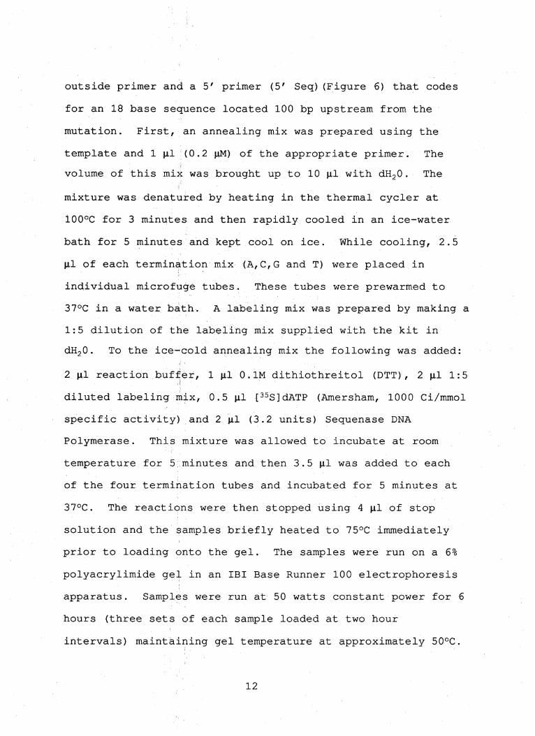
outside primer and a 5' primer (5' Seq)(Figure 6) that codes
for an 18 base sequence located 100 bp upstream from the
mutation. First, an annealing mix was prepared using the
template and 1 |il (0.2 jiM) of the appropriate primer. The
volume of this mix was brought up to 10 p-l with dHjO. The
mixture was denatured by heating in the thermal cycler at
100°C for 3 minutes and then rapidly cooled in an ice-water
bath for 5 minutes and kept cool on ice. While cooling, 2.5
|j.l of each termina.tion mix (A,C,G and T) were placed in
individual microfuge tubes. These tubes were prewarmed to
37°C in a water bath. A labeling mix was prepared by making a
1:5 dilution of the labeling mix supplied with the kit in
dH20. To the ice-cold annealing mix the following was added:
2 |il reaction buffer, 1 (j,l 0.IM dithiothreitol (DTT), 2 |a.l 1:5
diluted labeling mix, 0.5 nl [^ssjdATP (Amersham, 1000 Gi/mmol
specific activity) and 2jil (3.2 units) Sequenase DNA
Polymerase. This mixture was allowed to incubate at room
temperature for 5 minutes and then 3.5 |j.l was added to each
of the four termination tubes and incubated for 5 minutes at
37°C. The reactions were then stopped using 4 |j.l of stop
solution and the samples briefly heated to 75°C immediately
prior to loading onto the gel. The samples were run on a 6%
polyacrylimide gel in an IBI Base Runner 100 electrophoresis
apparatus. Samples were run at 50 watts constant power for 6
hours (three sets of each sample loaded at two hour
intervals) maintaining gel temperature at approximately 50°C.
12
Following the run, the gel was rinsed in a 5% Acetic Acid/5%
Methanol bath for 15 minutes and then transferred to Whatman
3MM filter paper and dried on a Labconco gel drier for 1 hour
at 75°C, exposed to Fuji X-Ray film for 72 hours then
developed and the sequence analyzed.
Cloning of mutant DNA in pcDNAl-amp plasmid. Once the
mutant sequence was confirmed, the DNA was ligated into a
pcDNAl-amp plasmid (Invitrogen) for future amplification and
analysis. The pcDNAl-amp plasmid (Figure 7) is an
approximately 4.8 kb high copy number shuttle vector
expressed in both prokaryotic and eukaryotic systems that
confers ampicillin resistance in E. Coli. First, 500 ng of
the desired DNA was digested with the restriction enzymes
Not-1 (recognition sequence GCGGCCGC) and Hind III
(recognition sequence A'^AGCTT)(7 Units each, GIBCO) at 37°C
for 6 hours and purified using the Spin Bind PGR Purification
System to remove the end fragments and unwanted enzymes. One
microgram of pcDNAl^amp was also digested in this manner and
gel purified to remove the polylinker and to prepare cohesive
ends for ligation. Seyeral ligation reactions were then set
up as follows. As a control, a 0:1 insert to vector (vector
only) ligation was performed using lOO ng cut pcDNAl-amp, 2
10.1 lOX ligase buffer and 0.5 |ol (2.5 Units) T4 DNA ligase
(GIBCO). The reaction volume was brought up to 20 ul using
dHjO. 2:1 and 3:1 insert to vector ligations were also
13

prepared as above. The reactions were allowed to continue
for 16 hours at 16°C and then were placed on ice. Twenty
microliters of GIBCO JM 109 frozen supercompetent cells were
placed in prechilled Falcon 2059 tubes and 1 (il of a 1:5
dilution of each ligation mix in dHjO was placed into the
appropriate tubes; This mixture was kept on ice for 30
minutes, heat shocked at 42°C for 45 seconds and placed back
on ice for 2 minutes, then 980 jxl of SOC medium prewarmed to
37°C was added to each tube. The tubes were incubated at 37°C
with shaking at 225 RPM for 1 hour, then 200 |xl of each mix
was plated on Luria Broth (LB) plates supplemented with
ampicillin at a concentration of 100 ji.g/ml (amp 100) and
incubated overnight at 37°C. Single colonies were selected
and grown in 2 ml LB/amp 100 at 37°C overnight. Minipreps
were performed on 1.5 ml of each culture using the Promega
Magic Miniprep kit. First the cultures were pelleted in a
microfuge at 14,000 rpm for 2 minutes and the cells
resuspended in 100 |j,l of Promega resuspension buffer, then 1
ml of Promega binding resin was added and the solution mixed
gently. This solution was then placed in a 3 ml syringe and
gently pushed through a Promega Magic Miniprep column,
followed by a wash with 2 ml of 80% isopropanol wash buffer
and elution with 50 jil TE prewarmed to 65°C. The remaining
0.5 ml of culture was saved as stock for future cultures by
adding 150 |il of 80% glycerol and storing at -20°C. The DNA
obtained was digested with Not-^l and Hind III and run on a 1%
14

agarose gel to confirm the presence of the desired 760 bp
fragment.
Maxiprep of pcDNAl-ainp/LT--p Mutant DNA. A culture was
prepared using 100 |a.l of the stored 0.5 ml Coli culture
which had tested positive for the mutant insert previously in
the miniprep and 200 ml of LB/amp 100. This culture was
grown overnight at 37°C. To obtain the desired plasmid DNA
the culture was Centrifuged at 12,O0Oxg for 15 minutes at 4°C
and the supernatant discarded. The pellet was dissolved in
10 ml of resuspension buffer containing 100 jig/ml RNase A,
0.1 mM Tris/Hei and 10 mM EDTA, pH 8.0. Once dissolved, 10
ml of lysis buffer containing 200 mM NaOH and 1% SDS was
added and then the tube was inverted 5 times and incubated at
room temperature for 5 minutes. After incubation, 10 ml of
lysis neutralization buffer containing 3.0 M potassium
acetate (KAc), pHT5.5 was then added, the tube inverted
several times and then put on ice for 20 minutes. The tube
was mixed again and centrifuged at 30,000xg for 30 minutes at
4°C. While the mixture was centrifuging, 10 ml of column
equilibration buffer containing 750 mM NaCl, 50 mM MOPS
(3-[N-Morpholino]propanesulfonic acid), 15% ethanol and 0.15%
Triton X-100 at a pH of 7.0 was added to a QIAGEN tip-500
column and allowed to enter by gravity flow. This was
followed by the supernatant obtained from the centrifuged
cell lysate. Once the lysate had flowed through the column.
15

2 washes were performed using 30 ml each of wash buffer
containing 1.0 M NaCl, 50 mM MOPS and 15% ethanol at a pH of
7.0. The column bound DNA was then eluted using 15 ml of
elution buffer containing 1.25 M NaCl, 50 mM Tris/HCl and 15%
ethanol at a pH of 8.5. To the eluate, 0.7 volumes of room
temperature isopropanol was added and the mixture centrifuged
for 30 minutes at 15,OO0xg at 4°C. The pellet was then washed
with 15 ml ice-cold 70% ethanol and centrifuged again. The
DNA pellet was allowed to dry for 10 minutes and was then
redissolved in 0,5 ml TE pH 8.0. The dNA obtained was ■ 'v' ' ■ ^ ^
analyzed on a 1% agarose gel.
COS-7 cell culture. GOS-7 mammalian (monkey kidney) cells
were maintained ip.Dulbecco's Modified Eagles Medium (DMEM)
supplemented with 10% fetal calf serum,, 2 mM glutamine,
penicillin and streptomycin (100 ug/ml each) in 10% CO2/90%
ambient atmosphere at 37°C. The cells were passaged every 3-4
days upon reaching approximately 80-90% confluence.
GOS-7 cell trahsfection. 2.5 x 10^ cells were plated into
60 mm Culture dishes the day prior to transfection and grown
in DMEM as above.: The cells were rinsed twice with 5 ml warm
phosphate buffered saline (PBS) and then with 5 ml of serum
free DMEM. A DNA/Liposome solution was prepared containing 1
|j,g DNA, 2 ml serum free DMEM and 10 |il LipofectACE (GIBCO) in
a 5 ml polystyrene tube. Tubes were prepared containing
16

LT-a, LT-p, LT-p mutant and control vector (Cav.Not) DNA. The
solution from each tube was added to a plate and the plates
incubated at 37°C in 10% CO2/90% ambient atmosphere for 5
hours. 2 ml of DMEM supplemented with 20% fetal calf serum
was then added and the plates incubated at 37°C overnight.
The media was then replaced with 3 ml of DMEM containing 10%
fetal calf serum and incubated an additional 48 hours.
Biosyn'bhe'biG labeling of COS-7 calls. The monolayer of
transfected cells was washed twice with warm PBS. To each
dish, 1 ml of cys/met-free DMEM containing 10% dialyzed fetal
calf serum and glutamine was added and the dish incubated 5
minutes at room temperature. A 150 |a.Ci/ml ^^S^cys/met label
(ICN Trans label ~10 |j,Ci/ml) was then added and the plates
incubated at 37°C for 3 hours with occasional rocking. The
supernatant was then removed and transferred to a microfuge
tube. The monolayers were lysed with 1 ml/dish ice-cold
lysis buffer (20 mM Tris-HCl, pH 8.0, 150 mM NaCl, 2% Nonidet
P-40 (NP-40), 1 mM EDTA, 40 mM iodoacetamide and 2mM
phenylmethylsulfonyl flouride) for 10 minutes at 4°C. The
lysates were then transferred to a microfuge tube and the
tubes centrifuges at 13,000 rpm for 10 minutes at 4°C. Each
sample was then ttansferred to a fresh microfuge tube for
immunoprecipitation. Each tube was precleared by adding 1 \ig
of mouse IgG and 40 (xl protein G-Sepharose (GammaBind G-
Sepharose, Pharmacia). The tubes were capped tightly and
17

rocked 1 hour at 4°G, then centrifuged briefly to pellet the
protein Q beads and the supernatant was then removed and
placed in a new tube. A mixture of 1 m-S each of three
monoclonal anti-LT-p antibodies (B27, B9 and C37)(BIOGEN) was
added along with 40 |il/tube of protein G-Sepharose to the
tubes containing the Cav.Not control, LT-p and the LT—p
mutant. One microgram of an anti-LT-a monoclonal antibody
(NC2)(Biogen) along with the protein G beads was added to the
LT-a transfection control tube. The tubes were capped
tightly and rocked 1 hour at 4°C, then centrifuged briefly to
pellet the protein G beads. The samples were aspirated and
the beads washed three times with wash buffer containing 20
mM Tris (Ph 8,0), 0.5% NP-40, 150 mM NaCl, 0.5% deoxycholate
and 0.05% sodium dodecyl sulfate (SDS) and once with PBS
azide (N3) then resuspended in 50 jil 2x SDS-PAGE sample buffer
containing 100 mM Tris-Cl (pH 6.8), 200 mM dithiothreitol, 4%
SDS, 0.2% bromophenol blue, 20% glycerol plus 4% 2-mercapto
ethanol and heated for 5 minutes at 100°C.
SDS^Polyacrylimide gel electrophoresis (SDS-PAGE).
The samples were then loaded on a 12% SDS-polyacrylamide gel
and run overnight at 10 mA constant current to separate
proteins. The gel was then soaked for 30 minutes in 1 M
sodium salicylate (fluor) and transferred to Whatman 3MM
filter paper and dried at 75°C for 1 hour. The dried gel was
18

exposed to X-Omat AR film (Eastman Kodak) and a Cronex
intensifying screen, (Du Pont) for 24 hours at -70°C and
developed.
19

RESULTS
Prepara'tion of 5' and 3' PGR fragments containing the
mutation. Through the use of the two sets of primers
discussed previously, the expected fragments were obtained.
Agarose gel eletrophoresis of the PGR products showed bands
at approximately 710 bp and 107 bp respectively (Figure 9).
The optimization of reagent concentrations and cycling
parameters resulted in the amplification of only the desired
fragments while avoiding the generation of non-specific and
unwanted fragments.
joining of fragi^en'ts by PGR Overlap Extension yields
full-length mutant. Application of the overlap extension
technique using the two fragments generated above yielded a
760 bp fragment (Figure 9) which represents the full length
mutated LT-p cDNA. The PGR reaction yielded approximately 1
|ig of DNA. Optimization of reagent and cycling parameters
eliminated hon-specific fragments.
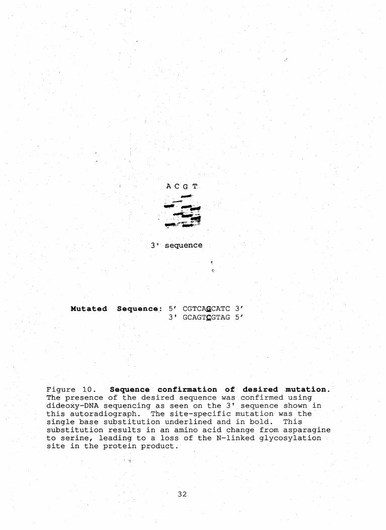
Sequence confirmation of mutation. The use of the
Sequenase PGR Product Sequencing kit allowed for rapid
sequence analysis of the PGR product. Both the 5' and 3' DNA
strands contained the desired mutation (figure 10) and no
Other random mutations were observed within 100 bp either
side of the site-directed mutation on either strand.
20

Cloning of mutant DNA in pcDNAl-amp plasmid.
Restriction digests of the vector and the PCR product
resulted in the creation of compatible cohesive ends that
allowed for specific ligation of the insert in the proper
orientation into the vector while avoiding religation of
vector alone. No transformants were noted in the vector only
control plate, while the 2:1 and 3:1 plates both contained 20
transformed colonies. Miniprep DNA from these colonies were
analyzed by restriction digests and all analyzed colonies
contained the expected 760 bp insert (figure 11).
Maxiprep of pcDNAl-amp/LT-p mutant DNA. A culture that
tested positive for the insert in the miniprep was used to
grow a large culture for the maxiprep. Approximately 250|j.g
of pcDNAl-amp/LT-p mutant DNA was obtained from a 200 ml
culture.
COS-7 cell transfection, biosynthetic labeling and
SDS-'PAGE. Immunoprecipitation of cell lysates using
monoclonal antibodies specific for LT-a or LT-p confirmed that
COS-7 cells were successfully transfected. The
autoradiograph of the labeled precipitates showed a band at
26 kDa representing the LT-a sample and indicates a
successful transfection. However, no bands were evident in
samples transfected with either LT-p or mutant LT-p.
Precipitation of these samples was attempted using three
21

different monoclonal antibodies to ensure the epitope had not
been lost during the mutagenesis, however none of the
monoclonal antibodies precipitated confirming proteins in
either LT-p sample.
22

C#nt«m«r« T«l#onwf »' 2ttb
B144 LT-P tnF UT-a
-|Hf|III HI
Ml 11 m CTIiiB mil i uiil
* .*! ? * * n« n X R RR R
' I > 1 _i "'' I " '
J L
!
f f ■ X NH HK <Mbp J- -J 1— 1 I I M
Figure 1. Schematic diagram of the region of chromosome 6 containing the LT/TNF locus. TNF, LT-a and LT-p are linked within 10 kb in the MHC on human chromosome 6. The exon^intron arrangements for each gene are similar. LT-p is coded for in an opposite direction from TNF and hT-tx. Restriction map shows sites for EcoRI (E), Xhol (X), Hindlll (H), Bglll (B), Kpnl (k), PstI (P) and Ncol (N). (From Browning et al., 1993)
23

SurfaceSecreted LT-a LT-p/LT-a Complex
Plasma OUT
Membrane
Secretory vesicle N
Golgi
LT-p
LT-a
Figure 2. A working model for LT biosynthesis and secretion. Newly formed LT-a and LT-p monomers can associate within the lumen of the endoplasmic reticulum to form trimers composed of a3, a2pi or alp3 subunits. These subunits are then processed within the Golgi and transported to the cell surface for expression as mLT (a2pi or aip2) or secreted LT (a3). (Adapted from Androlewicz et ai., 1992)
24

TNF Ugand Family
TKf LT. LTf LT*/t CCWOL CD30L C027L
148 150151157 154 156
ffiTmijffiTmr ^|m.
. 'Trimeric
i TNF/NGF RECEPTOR FAMILY
[] (] CO40 I3
TtfFfics , 4ieaU {] [] 13 (] 1] {]
C3 U3 (3" I Li! [3
Figure 3. Members of the TNF ligand and receptor family. For the ligands, an open box indicates the region of homology in the extracellular domain where the receptor binding sites are located, while the filled boxes indicate the cytoplasmic, transmembrane and extracellular stalk regions. The number of residues in each region are indicated. The receptor cys-rich repeat homology regions are shown as open boxes. Stripped boxes in the cytoplasmic region indicate homology. N or O indicate likely sites of glycosylation and P represents sites of phosphorylation (Ware, unpublished data).
25

LT-aP Complexes
a a
a
a a
P
a
15
P
(x3 a2pi alP2 P3
ReceptorBinding Interfaces
b/a b/a
b/x
z/a
2^
2/a
b/x
z/x
Figure 4. Proposed model of LT-aP complexes. The four potential trimers that are proposed for LT and the possible receptor binding interfaces for each. The a3 trimer could bind up to three TNFR which are specific for the a-a cleft. The aZpi trimer could potentially bind only one TNFR and one LT-pR, which are specific for the P-P cleft. The aip2 could not bind a TNFR but could bind up to two LT-pR. The P3, which has not been observed to occur in nature, could bind three LT—PR. The cleft formed at a-p subunit interfaces is thought to bind an as yet unidentified receptor (Ware, unpublished data).
26

%■
LT-P LT-a.
Figure 5. Molecular model of LT-a/LT-p interface. The LT-p subunit contains transmembrane and cytoplasmic domains not present on the LT-a subunit. LT-p also contains an N-linked glycosylation site very near the proposed binding region for the TNFR in the interface cleft. It is possible that this N-linked glycosylation site positions a carbohydrate that stearically blocks TNFR binding to mLT. The dark blue and yellow areas indicate regions of homology and the green area is a conserved proline thought to function in subunit association (Ware, unpublished data) .
27

Ouliside Primers:
5'pCDM8/LT-p
5'CGACTCACTATAGGGAGACC3' Melting Temp. 54°C
3'LT-p/Not-1
5'CGCGGCCGCACTCGGACCACGC3' MeIting Temp. 80°C
Inside Primers:
5'LT-pN-SOl
5'GTGACTGATGCTGACGTAGAC3' Melting Temp. 64°C
3'LT-pN-SOl
5'GTGTACGTCAGCATCAGTCAC3' Melting Temp. 64°C
5'Sequencing Primer:
5'LT-p/Seq
5'GAGACGGTGACTCCAGTG3' Melting Temp. 58°C
Figure 6. Primers utilized in PGR and sequencing reactions. Primers used in the PGR reactions include the
outside primers, 5'pGDM8/LT-p and 3'LT-p/Not-1. These primers allow for amplification of the entire LT-p gene and the 3'LTp/Not-1 also inserts a JSIot-l site in the 3' untranslated region. Inside primers include 5'LT-pN-SOl and 3'LT-pN-S02. These primers were used to insert the desired single base substitution (underlined) into the two fragments generated for use in the overlap extension procedure, changing the sequence to code for serine instead of asparagine. 5'LT-p/Seq was used to generate the 5' strand used in sequencing, this primer annealed approximately 100 bp upstream from the mutation site. A 3' strand was also generated for sequencing using the 3'LT-p/Not^l primer.
28

Creation ofLT-p Mutant Overlap Extension PGR
TargetedpCDMS/
SequenceLT-P
Denature
" Wutated sequence r
I tCombine
Denature y
t
Figure 7. Exsunple of sife-dlrected mufagenesis using overlap extension PGR. Specific base changes can be made using the overlap extension technique. Two separate PGR fragments are prepared, utilizing 5' and 3' non-mutated outside primers and complimentary internal primers that contain the desired base changes. These two fragments are amplified in separate PGR procedures, then the purified products combined, denatured and annealed and extended to form a full length template containing the mutation. The 5' and 3' outside primers are then added and another full PGR cycle run to amplify a full length mutant sequence.
29

fl* caojoo y) ox:q.03xi
XCQCQUJIUCQZXCOZX
c^^oo^ osz ca
Hpal
Pvu 1 cD
48Kb
Sfil
Nhe!
Figure 8. Map of the pcDNAl-amp plasmid used as a shuttle vector. The pcDNAl/Amp plasmid contains a strong promoter for eukaryotic expression, a ColEl sequence for high copy replication in E. Coll and ampicillin resistance in E, Coli for positive selection (Map supplied by Invitrogen).
30

-•««!rrr ;-2e>iu-»-.. ^S(r«_T-- -^as:
rSii S/? r'wv-T'iiiSS^C
Ee5iaassaSE«4HS3^-<^^
ii5« S5?:
»C£*
Figure 9. Agarose gel showing fragments made during PGR site directed mutagenesis procedure. Lane 1 contains molecular weight markers (BioMarker Low). Bands are at 1000, 700^ 525, 500, 400, 300, 200, 100 and 50 bp. Lane 2 shows the 5' fragment at approximately 710 bp. Lane 3 contains the 3' fragment of approximately 107 bp. Lane 4 shows the overlap extension product obtained by combining the fragments from lanes 2 and 3. This fragment represents the full length mutated LT-p gene of 760 bp and contains restriction enzyme sites for Hind III at the 5' end and Not-1 at the 3' end.
31
A C G T
3' sequence
Mutated Sequence: 5' GGTGAfiCATC 3' 3' GGAGTfiGTAG 5'
10. Sequence confirmation of desired mutation. The presence of the desired sequence was confirmed using dideoxy-DNA sequencing as seen on the 3' sequence shown in this autoradiograph. The site^specific mutation was the single base substitution underlined and in bold. This substitution results in an amino acid change from asparagine to serine, leading to a loss of the N-linked glycosylation site in the protein product.
32

Figure 11. Restriction digest of clone from pcDNA-amp. Following transfection, DNAminipreps were performed to confirm insertion of the cloned DNA. Restriction digests of the miniprep DNA with Not-1 and Hind III yielded a 760 bp fragment, indicating the mutated gene has been successfully inserted into the pcDNAl-amp plasmid. The left lane contains known molecular weight markers of Lambda DNA cut with the restriction enzyme Bst EII,
33

XJ
C
kDai
en. CO.
200
97
?^3t;
6.9
46
e3KS3;.u >S2«»:a%c«
Lcf'snt
30
*r»i4-s. .
*j -tV*.
SS§y
sS? '©S
14
Figure 12. Autoradiograph of protein immunoprecipitation. Transfection of COS-7 cells with LT-a, LT-p and LT-p mutant followed by immunoprecipitation with monoclonal antibodies yielded a band at 26 kD for LT-a indicating a successful transfection. Known weight markers are at 200, 97, 69, 46, 30 and 14 kDa. No bands are present for either LT-p sample. The lack of bands may indicate LT-p is being rapidly degraded when no LT-a is present.
34
CoatrciOKA CcntroiONA
♦ ir-a. OHA
'■! 1' ■ 10 100 1000 4000 10 100 1000 4000
LT-OONA ^ LJ^OHk*
3 LT^ONA
19 100 1000 4000 10 100 1000 4000
RtlotivoFluoresceneo
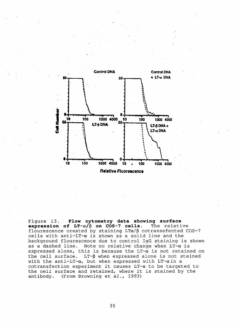
Figure 13. Flow cytometry data showing surface expression of LT—a/p on COS-7 cells. The relative flourescence created by staining LTa/p cotransefected COS-7 cells with anti'-LT-a is shown as a solid line and the background flourescence due to control IgG staining is shown as a dashed line. Note no relative change when LT-a is expressed alone, this is because the LT-a is not retained on the cell surface. LT-p when expressed alone is not stained with the anti-LT-a, but when expressed with LT-ain a cotransfection experiment it causes LT-a to be targeted to the cell surface and retained, where it is stained by the antibody. (From Browning et ai., 1993)
35
DISCUSSION
The preparation of a site-specific DNA mutant of LT-p
was shown to be possible using the PGR overlap extension
technique. Although this technique was first demonstrated to
be a practical and efficient method of site-specific
mutagenesis several years ago, when utilized with the LT-p
gene several modifications were required. The LT—p gene is
very C-G rich in its 5' end, resulting in excessively high
melting temperatures and rapid reannealing of the two DNA
strands. This made the generation of a 5' mutant DNA
fragment very difficult and impractical using standard PGR
protocols. After trying a wide variety of reaction
conditions without success, the reactions were attempted in a
vessel containing 10% DMSO. Although not normally used in
PGR protocols utilizing Taq polymerase, DMSO was used
previously in Klenow-mediated PGR reactions (Scharf at al.,
1986). Although DMSO is known to reduce the effectiveness of
Taq polymerase by approximately 50%, its use in these
reactions allowed for the generation of mutant 5' fragments
at relatively good levels, yielding approximately 1 ng of
mutant DNA per 100 |j.l reaction. It is uncertain which
parameters are affected by inclusion of 10% DMSO, but DMSO
may affect the melting temperature of the primers, the
thermal activity profile of the Taq polymerase and/or the
degree of product strand separation during denaturation.
Even at denaturation temperatures of 99°G, reactions involving
36

the 5' end of this gene attempted without DMSO were not
successful/ while addition of 10% DMSO allowed reactions to
take place with denaturation temperatures of 95°C. Inclusion
of 10% DMSO was also required in the subsequent overlap
extension protocols to create full length mutant LT-p cDNA.
Once the full length cDNA was produced, it was sequenced
in the region surrounding the mutation site to confirm the
presence of the mutation and the absence of other random
mutations. Full length sequencing will be necessary
eventually since random mutations can be created while using
Taq polymerase at a rate approaching 1/4000 base pairs (Ho et
al., 1989). Since sequencing is time consuming and tedious,
full length sequericing will be performed only if changes in
binding characteristics, cytotoxicity or trimer formation
using the mutant DNA are observed in future assays. The use
of the Sequenase PGR sequencing kit with the 5'Seq and
3'LT-p7Not-l primers allowed for direct sequencing of both
strands of the PGR product a distance of 100 bp either side
of the desired mutation. No mutations other than the desired
mutation were observed and the sample was then digested with
Not 1 and Hind III arid cloned into the pcDNAl-amp plasmid.
E. Coli cells were subsequently transformed to generate high
yields of the desired mutant DNA (approximately 250 |j.g per
maxi-prep).
Following isolation of the amplified plasmid from E.
Coll, the mutant DNA was transfected into GOS-7 cells to
37
obtain protein expression in a mammalian expression system
for immunoprecipitation analysis. A sampling of three
different monoclonal antibodies were used to ensure the
desired epitope had not been inadvertently removed by the
mutation. Results of the immunoprecipitation were
inconclusive. LT-p was not expressed at high enough levels to
observe any protein in the autoradiograph (Figure 12). If
the protein had precipitated as expected, a band would have
been seen at approximately 33 kDa in the wild type LT-p sample
and a band of slightly greater mobility would have been
expected for the LT-p lacking the N-linked glycosylation site
due to lack of the sugar residue. A control transfection of
LT-a precipitated with an anti-LT-a monoclonal antibody
yielded the expected band at 26 kDa. Several other attempts
to precipitate wild type LT-p from COS-7 cells have also
failed to detect protein. Flow cytometry analysis of cells
transfected with LT-p has shown that very small quantities of
the protein are present when expressed in conjunction with
LT-a (Figure 13). The promoter on the pcDNAl-amp plasmid is
a strong promoter, so fairly high levels of protein
expression should be expected. This would seem to indicate
that rapid protein degradation may be taking place.
Co-transfection of GOS-7 cells using both LT-a and LT-p has
been attempted with no LT-p evident on the autoradiograph (not
shown). It is not known whether a sufficient number of cells
are receiving both plasmids to yield distinguishable bands of
38

a-Pco-expression on the autoradiograph. An interesting
feature of these qotransfections is that the relative level
of LT-a expression drops as the amount of LT-p transfected .
increases. This would seem to indicate that the presence of
LT-pis having some effect on LT-a expression, but the nature
of this effect is unclear.
In baculovirus—infected insect cells, LT-p is expressed
alone or with LT-a in cotransfected cells at very high levels
(Crowe, unpublished data). This data may indicate that LT-p
alone (which has never been observed naturally) contains a
signal sequence that directs the LT-p proteins down a
degradative pathway when expressed in mammalian cells. In
insect cells, this pathway may be overwhelmed, so the protein
is expressed. However, in these cells, the protein is seen
in forms not normally observed, from monomers to aggregates,
indicating that even though it is expressed, it is expressed
in states not normally seen. When LT-a is coexpressed with
LT-p, heterotrimers are seen similar to those found in normal
mammalian cells. Together, this may indicate a change in the
3-dimensidnal conformation is occurring upon subunit
interaction which signals a secretory pathway. Another
possibility is that the LT-a subunit contains a secretory
signal and this subunit blocks the degradative signal on LT-p
when the SubunitS are together. To examine if some signal in
the transmembrane or cytoplasmic domains are signaling for
protein degradation, a soluble chinieric LT-p/myc construct was
39
prepared (Biogen) in which the transmembrane and cytoplasmic
regions of LT-p had been removed and replaced with the myc
protein start sequence. This chimeric protein was expressed
by COS-7 cells at levels similar to those seen for LT-a
(Growe et al., 1994). This would indicate that the signal
for the degradative pathway is located somewhere in the
transmembrane or cytoplasmic domains of LT-|3.
Further analysis pf liT through the use of the mutant DNA
created in this research will have to await the insertion of
the mutant gene into a baculovirus vector. Although the data
generated by transfection of COS-7 cells was interesting and
valuable/ it has not answered any questions concerning the
role of the N-linked glycosylation site in receptor binding
interactions or in trimer association. Further studies are
in progress to attempt expression of the mutant LT-p in the
baculovirus expression system. Cotransfections using LT-a
and LT-p in the baculovirus system should yield adequate
protein to address these questions.
The importance of working out the protocols required to
prepare LT-p mutants using the overlap extension technique
cannot be underestimated and will allow many important
questions about LT-p and the immune response to be answered in
the future. This; project has led to the preparation of one
LT-p mutant and has also worked out easy and rapid protocols
that should allow'for future research using LT-p mutants to be
done quickly and efficiently.
40

References
Abbas, A.K., Lichtmann, A.H., and Pober, J,S., "Cellular and Molecular Immunology", W.B. Sanders Co., Philadelphia, Pa., 1991.
Allen, R.C., Armitage, R.J., Conely, M.E., Rosenblatt, H., Jenkins, N,J., Copeland, N.G», Bedell, M.A., Edelhoff, S., Disteche/ C.M., Simoneaux, D.K., Fanslow, W.C., Belmont, Ji, and Spriggs, M.K., CD40 ligand gene defects responsible for X-linked hyper Ig-M syndrome. Science 259:990-993, 1993.
Andrews, J.S., Berger, A.E., and Ware, C.F., Characterization of the receptor for tumor necrosis factor (TNF) and lymphotoxin (LT) on human T lymphocytes: TNF and LT differ in their receptor binding properties and the induction of. MHC class I proteins on a human CD4+ T cell hybridoma. J. Iimvnol. 144:2582-2591, 1990.
Androlewicz, M.J., Browning, J.L., and Ware, C.F., Lymphotoxin is expressed in a heterotrimeric complex with a distinct 33 kDa glycoprotein on the surface of an activated human T cell hybridoma. J. Biol. Chem. 276:2542-2547, 1992.
Banner, D.W,, D'Arcy, A., James, W., Gentz, R., Schoenfeld, H-J., Brogeir, C., Loetscher, H., and Lesslauer, W., Crystal structure of the soluble human 55 kd TNF receptor—human TNF-p complex: implications for TNF receptor activation; Cell 71:765-776, 1993.
Browning, J.L., and Ribolini, A., Studies on the differing effects of tumor necrosis factor and lymphotoxin in the growth of several human tumor lines. J. Immunol. 143:1859-1867, 1989.
Browning, J.L., Androlewicz, M.J. and Ware, C.F., Lymphotoxin and an associated 33-kDa glycoprotein are expressed on the surface of an activated human T cell hybridoma. J. Immunol. 147:1230-1237, 1991.
Browning, J.L., Ngam-ek, A., Lawton, P., DeMarinis, J., Tizard, R., Chow, E.P., Hession, C., Greco, B., Foley, S., and Ware, C.F., Lymphotoxin-p: A new member of the TNF family that forms a heteromeric complex with lymphotoxin on the cell surface. Cell 72:847-856, 1993.
41

Growe, P.D., VanArsdale, T.L., Walter, B.N., Ware, C.F., Hession, C,,iEhrenfels, B., Browning, J.L., Din, W.S., Goodwin, R.G,, and Smith, C.A., A lymphotoxin-p-specific receptor. Science 264:707-709, 1994.
Digel, W., Porzolt, F., Schmid, M., Herrmann, F., Lesslauer, W. and Brockiiaus, M., High levels of circulating soluble receptors for tumor necrosis factor in Hairy cell leukemia and|type B chronic lymphocytic leukemia. J. Clin. Jnvlst. 89:1690-1693, 1992.
DiSanto, J.P., Bonnefoy, J,Y,, Gauchat, J.F., Fischer, A., and Saint BaSile, G.de, Ci)40 ligand mutations in X-*-linked immunpdeficiency with hyper-IgM. Nature 361:541-543.
■■■
Engelmann, H., Noyick, D., and Wallach, D., Two tumor necrosis factor binding proteins purified from human urine. <7. B'iol. Chem. 265:1531-1536, 1990.
Gooding, L.R., Virus proteins that counteract host immune defenses. Cell 71:5-^7, 1992.
Goodwin, R.G./ Anderson, D., Jerzy, R., Davis, T., Brannan, C.I., Copelahd, N.G., Jenkins, N.A., and Smith, C.A., Molecular clbning and expression of the type 1 and type 2 murine receptors for tuimor necrosis factor. Mol. Cell. Biol. 11:3020-3026, 1991.
Ho, S.N., Hunt, H.D., Horton, R.M., Pullen, J.K., and Pease, L.R., Site-directed mutagenesis by overlap extension using the polymerase chain reaction. Gene 77:51-59^^ 1989.
Hohmann/ H., Remy> R., Brockhaus, M., and Van Loon, A.P.G.M., Two different cell types have different major receptors for human tuinor necrosis factor (TNF a). J. Biol, Chem. 264:14927-14934, 1989.
Hollenbaugh, D., Grosmaire, L.S., Kullas, O.K., Chalupny, N.J., Braesch-Anderson, S., Noelle, F.J., Stamenkovic, I., Ledbetter, J.A., and Aruffo, A., The human T cell
antigen gp39/ a member of the TNF gene family, is a ligand for the CD40 receptor: Expression of a soluble form of gp39iwith B cell costimulatory activity. EMBO J. 11:4313-4321, 1992.
42

Horiuchi, A., Abe, i.Y,, Miyaki, M., Kimura, K., Hitsumoto, Y., Takeuchi, N.,'anci Kimura, S., Role of membrane-associated lymphotoxin (mLT) in the killing activity of lymphokine-associated killer (LAK) cells towards various tumor cell lines. Clin, Exp. Immunol. 96:152-157, 1994.
Horton, T.M., Ranheim, T.S., Aquino, L., Kusher, D.I., Saha, S.K., Ware, C.F., Wold, W.S.M., and Gooding, L.R., Adenovirus E3 14.7K protein functions in absence of other adenovirus proteins to protect transfected cells from tumor necrosis factor cytolysis. J. Virology 65:2629-2639, 1991.
Jones, E.Y., Stuart, D.I., and Walker, N.P.C., Structure of tumor necrosis factor. Nature 338:225-228, 1989.
Korthauer, U., Graf, D., Mages, H.W., Briere, F., Padayachee, M., Malcolm; S., Ugazio/ A.G., Notarangelo, L.D., Levinsky, R.J., and Kroczek, R.A., Defective expression of T-cell Cb40 causes X-linked immunodeficiency with hyper-IgM. Nature 3261:539-541, 1993.
Kriegler, M., Perez, C., DeFay, K., Albert, I., and Lu, S.U., A novel form;,of TNF/Cachectin is a cell surface
Cytotoxic transmembrane protein: Ramifications for the complex physiology of TNF. Cell 53:45-53, 1988.
Nedwin, G.E., Naylor, S.L., Sakaguchi, A.Y., Smith, D., Jarrett-Nedwin, J., Pennica, D., Goeddel, D.V., and Gray, P.W., Human lymphotoxin and tumor necrosis factor genes: Structure, horiiology and chromosomal location. Nucl. Acids Res. 13:6361-6373, 1985.
Old, L.J., Tumor necrosis factor (TNF). Science 260:630-632,; 1985.
Paul, N.L,, and Ruddle, N.H., Lymphotoxin. Ann. Rev. Immunol. 6:407-438, 1988.
Pennica, D., Kohr, W.J., Fendly, B.M,, Shire, S.J., Raab, H.E., Borchardt, P.E., Lewis, M., and Goeddel, D.V., Characterization of a recombinant extracellular domain of the type i tumor necrosis factor receptor: evidence for tumor nedroSis factor-a induced aggregation. Biochemistry 31:1134-1141, 1992.
43

Ruddle, N.H., Bergman, C.M,, McGrath, K.M., Ligenheld, E.G., Grunnet, M.Li, Padula, S.J., and Clark, R.B., An antibody to lymphotoxin and tumor necrosis factor prevents transfer of experimental allergic encephalomyelitis. J. Exp. Med. 172:1193-1200, 1990.
Scharf, S.J., Horn, G.T., and Erlich, H.A., Direct cloning and sequence analysis of enzymatically amplified genomic sequences. Science, 233:1076-1078, 1986.
Schultze, S., Potthof, K., Machleidt, T., Berkovic, D., Wiegmann, K., and Kronke, M., TNF activates NF-kB by phosphatidylcholine-specific phospholipase C-induced "acidic" sphingomyelin breakdown. Cell 71:765-776, 1992.
Smith, C.A., Davis, T., Wignall, J.M., Din, W.S., Farrah, T., Upton, C., McFadden, G., and Goodwin, R.G., T2 open reading frame from the shope fibroma virus encodes a soluble form of the TNF receptor. Biochem. Biophys. Res. commun.'176:35-342, 1991.
Smith, C.A., Williams, D., Armitage, R.J., Gliniak, B., Grabstein, K;, Fanslow, W., Farrah, T., Falk, B., Din, W.S., Davis, T., and Goodwin, R.G., The TNF/NGF superfamily of cytokines and receptors. J. Cell. Biochem. 17B:51. Keystone Symposium Feb. 1-8, 1993.
Smith, R.A., and Baglioni, C., The active form of tumor necrosis factor is a trimer. J. Bid. Chem. 262:6951
6954, 1987. ;
Spies, T., Morton,' C.C., Nedospasov, S.A., Fiers, W., Pious, D., and Strominger, J.L., Genes for tumor necrosis factor a and P are linked to the major histocompatability complex. Proc. Natl. Acad. Sci., USA, 83:8699-8702, 1986.
Togni, P.D., Goeliner, J., Ruddle, N.H., Streeter, P.R., Fick, A,, Mariathasan, S., Smith, S.C., Carlson, R., Shornick, L.P., Strauss-Schoenberger, J., Russell, J.H., Karr, R. and\Chapin, D.D., Abnormal development of peripheral lymphoid organs in mice deficient in lymphotoxin. Science 264:703-707, 1994.
Van Ostade, X., Vandenabeele, P., Everaerdt, B., Loetscher, H., Gentz, R,, Brockhaus, M., Lesslauer, W., Travernier, J., Brouckaert, P., and Fiers, W., Human TNF mutants with selective activity on the p55 receptor. Nature 361:266-269, 1993.
44

Vandenabeele, P., Declercq, W., Vercammen, D., Van de Craen, M,, Grooten, J., Loetscher, H., Brockhaus, M., Lasslauer, W., and Fiers, W., Functional characterization of the human tumor necrosis factor receptor p75 in transfected rat/mouse T cell hybridoma. J. Exp. Med. 176:1015-1024, 1992.
Vilcek, J., and .Lee, T.H,, Tumor necrosis factor: New insights into the molecular mechanisms of its multiple actions. J". Biol. Chem. 266:7313-7316, 1991.
Ware, C.F., Andrews, J.S., Shamansky, L.M., and Grayson, M.H.j Regulation of the CTL lytic pathway by tumor necrosis factor. in /Cellular Immunity and the Immunotherapy of Cancer". Lotze, M.T. and Finn, O.J., eds. Wiley—Liss, New York, New York. pp 121—128, 1990.
Ware, C.F., Crowe, p.D., Grayson, M.H., Androlewicz, M.J., and Browning, J.L., Expression of surface lymphotoxin and tumor ndcrosis factor on activated T, B and NK cells. J. Immunol, 149:3881-3888, 1992.
Zhang, X-M., Webber, I., and Chen, M-J., Site directed mutational analysis of human necrosis factor-a receptor binding site and structural-functional relationship. J. Biol. Chem. 267:24069-24074, 1992.
45


