Download - PREVALENCE AND MORPHOLOGICAL STUDIES OF …

PREVALENCE AND MORPHOLOGICAL STUDIES OF
GASTROINTESTINAL HELMINTHS OF BACKYARD CHICKEN IN
BANGLADESH
S. M. ABDULLAH
DEPARTMENT OF MICROBIOLOGY AND PARASITOLOGY
SHER-E-BANGLA AGRICULTURAL UNIVERSITY
SHER-E-BANGLA NAGAR, DHAKA-1207
JUNE, 2019

PREVALENCE AND MORPHOLOGICAL STUDIES OF
GASTROINTESTINAL HELMINTHS OF BACKYARD CHICKEN IN
BANGLADESH
BY
S. M. ABDULLAH
REGISTRATION NUMBER: 12-04875
A Thesis
Submitted to the Department of Microbiology and Parasitology
Sher-e-Bangla Agricultural University, Dhaka-1207, In Partial Fulfillment of the
Requirements for the degree of
MASTER OF SCIENCE (MS) IN PARASITOLOGY
DEPARTMENT OF MICROBIOLOGY AND PARASITOLOGY
SEMESTER: JAN-JUN/2019
APPROVED BY
Dr. Uday Kumar Mohanta Supervisor
Chairman & Associate Professor
Department of Microbiology and Parasitology
Sher-e-Bangla Agricultural University
Sher-e-Bangla Nagar, Dhaka-1207
Dr. Mohammad Saiful Islam (Rasel) Co-Supervisor
Chairman & Associate Professor
Department of Anatomy, Histology and Physiology
Sher-e-Bangla Agricultural University
Sher-e-Bangla Nagar, Dhaka-1207
Dr. Uday Kumar Mohanta Chairman of Examination Committee
Department of Microbiology and Parasitology
Sher-e-Bangla Agricultural University
Sher-e-Bangla Nagar, Dhaka-1207

MEMO NO: SAU/
CERTIFICATE
This is To cerTify ThaT The Thesis enTiTled “PREVALENCE AND
MORPHOLOGICAL STUDIES OF GASTROINTESTINAL HELMINTHS OF
BACKYARD CHICKEN IN BANGLADESH” submiTTed To The Department of
microbiology and parasitology, Sher-e-Bangla Agricultural
University, Dhaka, in partial fulfillment of the requirements for
the degree of Master of Science in Parasitology, embodies the
result of a piece of bona fide research work carried out by S. M.
ABDULLAH, Registration No. 12-04875 under my supervision and
guidance. No part of the thesis has been submitted for any other
degree or diploma.
I further certify that any help or source of information,
received during the course of this investigation has been duly
acknowledged.
Date: May, 2019
Dr. Uday Kumar Mohanta Supervisor
Place: Dhaka, Bangladesh Chairman & Associate Professor
Department of Microbiology and Parasitology
Sher-e-Bangla Agricultural University
Sher-e-Bangla Nagar, Dhaka-1207
Department of Microbiology and Parasitology
Sher-e-Bangla Agricultural University
Sher-e-Bangla Nagar, Dhaka-1207

i
ACKNOWLEDGEMENTS
All praises are due to the Almighty God, who enables the author to complete the present research work and to submit the thesis for the degree of Masters of Science (M.S.) in Parasitology under the Department of Microbiology and Parasitology.
The author deems it as a great privilege to express his gratitude, sincere appreciation, indebtedness and best regards to his respected teacher and research supervisor Dr. Uday Kumar Mohanta, Associate Professor, Department of Microbiology and Parasitology, Sher-e-Bangla Agricultural University, Dhaka for his enthusiastic vision, encouragement, guidance, co-operation and constructive criticism in making this research successful and preparing the thesis.
It gives the author a great pleasure in expressing his sincere gratitude and deepest sense of appreciation to his respectable teacher and co-supervisor Dr. Mohammad Saiful Islam (Rasel), Associate Professor, Department of Anatomy, Histology and Physiology, Sher-e-Bangla Agricultural University, Dhaka for his sincere encouragement, valuable advices, kind co-operation, helpful suggestions and instructions to conduct research work and improve the thesis.
The author also desires to express his sincere gratitude and appreciation to his honorable teacher Md. Hazzaz Bin Kabir, Assistant Professor, Department of Microbiology and Parasitology, Sher-e-Bangla Agricultural University, Dhaka for his encouragement, invaluable suggestions and sympathetic co-operation throughout the MS course and research works.
The author feels proud to express gratefulness all the staffs of this Department for their co-operation. The author likes to pay special thanks to Amrito Barman, Md. Yakub Ali, Shah Jungy Ibna Karim for their immense help. The author also expresses his cordial gratitude to all MS students, Department of Microbiology and Parasitology for their kind cooperation throughout the research period.
The author gratefully acknowledges the authority of Ministry of Science and technology, Govt. of the People's Republic of Bangladesh, for their financial support through NST fellowship to conduct the research works.
Deepest sense of gratitude and indebtedness are due to his beloved parents who sacrificed their happiness not only during the study period but also for the whole period of the author’s life.
The Author

ii
LIST OF CONTENTS
CHAPTER TITLE PAGE NO.
ACKNOWLEDGEMENT i
LIST OF CONTENTS ii-iii
LIST OF FIGURES iv-v
LIST OF TABLES vi
ACRONYMS AND ABBREVIATIONS vi
ABSTRACT vii
CHAPTER 1 INTRODUCTION 1-3
CHAPTER 2
2.1
2.2
2.3
2.4
2.5
2.6
REVIEW OF LITERATURE
Previous work on Ascaridia galli
Previous work on Heterakis gallinarum
Previous work on Raillietina tetragona
Previous work on Echinostoma revolutum
Previous work on Catatropis verrucosa
Prevalence of gastrointestinal helminths of
poultry
4-19
4
6
9
12
14
15
CHAPTER 3 MATERIALS AND METHODS 20-21
3.1 Sampling area 20
3.2 Collection of samples 20
3.3
3.4
Processing of cestodes and trematodes
Processing of nematodes
21
21
CHAPTER 4 RESULTS AND DISCUSSION 22-43
RESULTS 22-33
4.1 Morphological observation 22
4.1.1 Morphology of A. galli 22-24
4.1.2 Morphology of H. gallinarum 25-26
4.1.3 Morphology of R. tetragona 27-28
4.1.4 Morphology of C. verrucosa 29-30
4.1.5 Morphology of E. revolutum 31-33

iii
LIST OF CONTENTS (CONT’D)
CHAPTER TITLE PAGE NO.
4.2 Prevalence 34-38
4.2.1 Overall prevalence of helminths in
backyard chicken
34
4.2.2 Prevalence of helminths in different
geography
35
4.2.3 Organ Preferences of
gastrointestinal helminths
36
4.2.4 Single and mixed type of infection 38
DISCUSSION
39-43
CHAPTER 5 SUMMARY AND CONCLUSION 44
REFERENCES
45-53

iv
LIST OF FIGURES
FIGURE NO. TITLE PAGE NO.
1 Different body parts of A. galli, A. Gross sample of A.
galli. B. Anterior part of the parasite; the black arrow
indicates the lips and the blue lines indicate the papillae
on the lips. C. Cuticular striation. D. Posterior part of
the female; the black line indicates anal opening. E.
Posterior part of the male (4X). F. Posterior part of the
male (10X); the black lines indicate the unequal spicules
and the block arrow indicates the precloacal or preanal
sucker. G. Uterus of the female.
23-24
2 Different body parts of H. gallinarum, A. Slightly
curved head end. B. Anterior part of the parasite; the
black arrow indicates the lips and the blue arrow
indicates the bulb shaped esophagus. C. Bulb shaped
esophagus in high magnification (40X). D. Vulvar
opening of the female (the black line). E. Posterior part
of the male (10X); the black arrow shows the preanal
sucker and the black line shows the unequal spicules. F.
Multiple eggs in the uterus of female.
26
3 Different body parts of R. tetragona. A. Gross sample of
parasite after collection. B. Anterior part of the parasite
(10X); the blue arrow indicates rostellum and the “*”
indicate the suckers. C. Scolex in high magnification
(40X); D. The broad segment having common genital
pores on each (black arrows); Each mature proglottid
has a set of reproductive organ in the middle part.
28
4 Different body parts of C. verrucosa. A. Whole fluke
under 4X magnification; the black arrow indicates the
oral sucker, the blue arrows indicate caecal bifurcation
and the broken line indicates the cirrus sac. B. Uterine
loops (18 in numbers) of the parasite (10X); both side of
the loops covered by vitellaria. C. Cup shape oral sucker
(10X); D. The reproductive organs of parasites; the blue
arrows indicate testes and the block arrows indicates the
ovary.
30

v
LIST OF FIGURES (CONT’D)
FIGURE NO. TITLE PAGE NO.
5
6
7
Different body parts of E. revolutum. A. Gross sample of
the fluke occurring c-shaped. B. Anterior part of the fluke;
the black arrow indicates the oral sucker and the broken
line indicates the ventral sucker. C. Posterior part of the;
the black arrow indicates the ovary and the broken lines
indicate the testes. D. Oral sucker with short esophagus
(white block arrow) and caecal bifurcation. E. The 37
spines are distinct around the oral sucker. F. Numerous
eggs in the uterus.
Site preferences of different gastrointestinal helminths
Percentages of Single and Multiple type of infection
32-33
38
39

vi
LIST OF TABLES
TABLE NO. NAME PAGE NO.
1 Location and overall prevalence of helminths in
backyard poultry
34
2 Prevalence of gastrointestinal helminths in Dhaka
(N=36)
35
3 Prevalence of gastrointestinal helminths in
Mymensingh (N=7)
35
4
5
6
7
Prevalence of gastrointestinal helminths in Pabna
(N=10)
Prevalence of gastrointestinal helminths in
Bandarban (N=10)
Site preferences of different gastrointestinal
helminths (N=63)
Percentages of Single and Multiple type of
infection (N=63)
36
36
37
38
ACRONYMS AND ABBREVIATIONS
ABBREVIATION FULL MEANING
et al. = And others/Associates
HCl = Hydrochloric acid
M.S.
>
=
=
Master of Science
More than

vii
PREVALENCE AND MORPHOLOGICAL STUDIES OF
GASTROINTESTINAL HELMINTHS OF BACKYARD CHICKEN
IN BANGLADESH
ABSTRACT
Poultry is a promising sector in Bangladesh which is expanding day by day. It is also the
most appropriate income generating sector for rural women as well as landless and
marginal farmers. But the indigenous chickens are affected by various parasites.
Therefore, the present study was aimed to study the prevalence and morphological
identification of gastrointestinal helminths of backyard chicken in Dhaka, Bandarban,
Mymensingh and Pabna. A total of 63 intestines were examined for helminth parasites
from September 2017 to November 2017. This experiment was performed at the
Microbiology and Parasitology Laboratory under the Department of Microbiology and
Parasitology, Sher-e- Bangla Agricultural University, Dhaka-1207. The collected
helminths were identified according to the keys and description given by Soulsby (1982)
and Yamaguti (1958). A high rate of helminth infection (100%) was observed in
backyard chicken in Bangladesh. One cestode, Raillietina tetragona (73.01%); two
nematodes, Ascaridia galli (47.61%) and Heterakis gallinarum (38.09%); and two
trematodes, Catatropis verrucosa (23.80%) and Echinostoma revolutum (7.93%) were
encountered during the study. Ascaridia galli was mostly found in small intestine
whereas Heterakis gallinarum was found in the caecum. Echinostoma revolutum and
Raillietina tetragona were collected from both small and large intestines. Catatropis
verrucosa were recovered only from caecum. Out of 63 samples, 25.39% were infected
with single infection while the rest 74.61% were mixed infections. This work strongly
suggests that helminthosis is a very serious problem in backyard chicken and therefore,
appropriate control strategies are needed to design for better production.
Keywords: Helminths, Backyard chicken, Morphological identification, Prevalence,
Bangladesh

1
CHAPTER 1
INTRODUCTION
Poultry, specially chicken is one of the most intensively reared domesticated species, and
is the most developed and profitable animal production businesses (FAO, 1987). Its
importance in developing countries and its role in improving the nutritional status and
income of many small farmers have been recognized by various scholars and rural
development agencies in the last two decades (Eyinnaya, 1992). Animal production in
general and chickens in particular play important socioeconomic roles in developing
countries (Alders, 2004). The purposes of backyard chicken production are for income,
egg hatching for replacement, consumption, for cultural and/or religious ceremonies and
egg production (Moges et al., 2010). Poultry productivity is enhanced by application of
sound principles of health protection and management (Shane, 2005). The economic
contribution of the sector is not still proportional to the large chicken numbers, attributed
to the presence of many productions, reproduction and infrastructural constraints (Aberra,
2000). Free-range chickens from rural areas supply all of the chicken meat and egg
requirements for rural people, and about 12–13% of urban requirements (Melewas, 1989).
Like other country, poultry production is a promising sector in Bangladesh which is
increasing day by day. Poultry meat and eggs contribute approximately 37% of total
animal protein in Bangladesh (Ahmed and Islam, 1990). In Bangladesh, Poultry rearing
is the most appropriate income generating activities for rural women, especially for
landless and marginal farmers. The production of backyard poultry under semi scavenging
system is found suitable to the villagers as additional source of income and nutrient
supplement (Latif, 2001).

2
Poultry diseases is a major hindrance which interfering with poultry productivity, by
decreasing economic returns, and may therefore, negatively affect the development of the
industry (Abebe et al., 1997). Among poultry diseases, helminthosis is considered to be
the most important problem of local chickens, and major causes of ill-health and loss of
productivity in different parts of the world (Yimer et al., 2001). Parasitic diseases are
problem wherever poultry are raised, whether in large commercial operations or in small
backyard flocks and economic losses caused by parasites on poultry can be significant
(Fatihu et al., 1991).
In fact, the indigenous chickens of Bangladesh are parasitized by various parasites
(Sarkar, 1976). The domestic chicken has a wide range of feeding habits from grains,
fruits and insects which may carry stages of parasites, thus predisposing them to parasitic
infections (Adang, 1999; Oniye, 2000). The chickens pick up the parasite eggs directly
by ingesting contaminated feed, water, or litter or by eating snails, earthworms, or other
insects. Clinical signs of parasitism are unthriftyness, poor growth and feed conversion,
decreased egg production and even death in severe infections. Furthermore, parasites can
make the flock less resistant to other diseases and exacerbate existing disease conditions.
Parasitic infection or their concurrent infections result in immunosuppression, especially
in response to vaccines against some poultry diseases. Of all the intestinal worms, large
roundworms (Ascaridia galli) probably inflict the most damage in young birds being more
severely affected. A mild infection is often unnoticed but large numbers of worms,
however, interfere with feed absorption causing poor growth and production. In severe
infections, there can be actual intestinal blockage by the worms, causing death (Yousuf et
al., 2009).
However, large numbers can have a devastating effect on growth, egg production, and
over-all health. Parasitism has been attributed to cause reduced growth, low egg
production, emaciation, anaemia as well as mortality (Belonwu, 1993; Hassouini and
Belghyti, 2006; Heyradin et al., 2012). These authors reported that mortality due to
parasitic diseases was higher than those attributed to Newcastle disease and mortality
causing viral infection of poultry. The concentration of parasite eggs in the poultry
environment is an important factor which plays a major role in determining the severity

3
of the infection (Pinkney et al., 2008). Viral, bacterial and protozoan diseases may appear
to be more economically important to the farmer because they cause obvious losses in the
form of deaths of many birds at a time. However, the less obvious, but ubiquitous, losses
due to reduced productivity caused by helminthosis are economically very important to
the poultry industry (Ssenyonga, 1982).
Gastrointestinal parasites which invade the host possess morphological and physiological
features such as small thread like cylindrical body, hooks and hard body cuticle enhance
their adaptation to long living and existence in their hosts. These parasites constitute a
major factor limiting productivity of the poultry industry (Soulsby, 1982).
Of the helminth parasites of poultry birds, nematodes constitute the most important group
of helminth parasites of poultry both in number of species and the extent of damage they
cause: the main genera include Ascaridia, Heterakis and Capillaria. There are a few
species of cestodes in poultry. Like them Choanotaenia infundibulum, Davainea
proglottina, Raillietina cesticillus and Raillietina tetragona are also found in the intestine
of poultry (Matur, 2010).
Therefore, the aim of this study was:
1. To carry out the detailed morphological identification of different species of
gastrointestinal helminths affecting backyard poultry.
2. To determine the prevalence of those endoparasites in different districts of
Bangladesh.

4
CHAPTER 2
REVIEW OF LITERATURE
2.1 Morphological studies on Ascaridia galli
Lalchhandama (2010) conducted a detailed morphological study of Ascaridia galli by
scanning electron microscopy and revealed that the extreme anterior cephalic region was
a triangular mouth consisting of three prominent lips. Each of the lip was lined with fine
teeth on the internal rim and studded with eye-like sensory papillae. The body cuticle
constituted series of striations called annulations. Annuli were transverse concentric rings
and were divided into parallel subannuli. Female had a simple straight tail with a ventrally
located anal opening. The male posterior was curved and pointed, and relatively elaborate
having a precloacal sucker in addition to the anus. These posterior openings were
surrounded on both sides by a row of minute protrusions called caudal papillae and the
lateral caudal alae. The precloacal sucker was surrounded by a sclerotized ring. Light
microscopy showed that the cuticle was multilayered and continuous with the
hypodermis, which in turn was supported with a thick musculature composed of fibrillar
contractile and granular noncontractile protoplasmic portions. The body space,
pseudocoel, contained digestive tract and reproductive organs such as testis, vas deferens
and seminal vesicle in male, and ovaries, oviducts and uteri in female. The seminal vesicle
housed spermatozoa, and the uteri, fertilized eggs. The eggs were elliptical, covered with
chitinous shell that enclosed the embryo.
Katakam (2010) analysed A. galli larval recovery from the chicken intestine. The number
of larvae recovered from the intestinal wall of chickens infected with 1000
embryonated A. galli eggs and killed 15 days post infection (p.i.) by three methods
(ethylenediamine tetraacetic acid [EDTA], pepsin digestion and scraping) were
compared. The EDTA and pepsin digestion were found to be the most efficient methods
with no significant difference (P > 0.05) in the number of recovered larvae between the

5
two. Subsequently, three different A. galli cohorts were established using the polymerase
chain reaction-linked restriction fragment length polymorphism (PCR-RFLP) technique.
A 533-bp long region of the cytochrome c oxidase subunit 1 gene of the mitochondrial
DNA was targeted and 22 A. galli females were allocated to three different haplotypes.
The four females with the highest embryonation rate from each haplotype group (total 12
females) were selected and used to inoculate each of 12 chickens with a dose of 1000
embryonated eggs. The chickens were killed 15 days p.i. and A. galli larvae were
recovered from the small intestinal wall by the EDTA method and by sieving the lumen
content on a 90 µm sieve. DNA of 40 larvae from each of the three different haplotypes
was extracted using a worm lysis buffer, and PCR-RFLP analysis of these larvae revealed
the same haplotype as that of their maternal parent. The identification of distinguishable
cohorts may be a powerful tool in population studies of parasite turnover within the animal
host.
Hafiz et al. (2015) performed a study to carry out the prevalence and severity of A. galli
in White Leghorn layers (housing type: battery cage and deep litter, 50 each) and
Fayoumi-Rhode wasland Red crossbred (male and female: 50 each) flock rearing at
Government Poultry Farm, Dina, Punjab, Pakistan. Two hundred faecal samples were
examined by using standard parasitological and McMaster egg counting technique. The
overall prevalence was 24.5% at farm, 13% in White leghorn layer (battery cage=2%,
deep litter=24%) and 36% in Fayoumi-Rhode wasland Red (male=34%, female=38%). It
was also observed that White leghorn layer rearing in deep litter had more severe infection
(EPG=1920) of A. galli compare with battery cages birds (EPG=500). Parasite prevalence
was significantly related with sex (P<0.05) in Fayoumi-Rhode wasland Red and male
birds had less number of average parasites (0.34±0.47) as compared to females
(0.38±0.490). Additionally, female birds were under serious threat of infection
(EPG=2270) compared with its counterpart (EPG=1250). Given the high infection rates,
particular attention should be paid to management and provision of feed supplement to
White leghorn layer housing in deep litter and female bird of Fayoumi-Rhode was land
Red crossbred.

6
Zhao et al. (2016) performed a research to find out the detailed morphology of A. galli
using light and scanning electron microscopy, based on specimens collected from the
endangered green peafowl Pavo muticus Linnaeus (Galliformes: Phasianidae) in China.
The results revealed some erroneous and previously unreported morphological features,
including the lips lacking real denticles, the lateral alae beginning at some distance
posterior to the base of the ventrolateral lips and the caudal papillae with 4 different
morphotypes.
Akter et al. (2016) conducted a research to determine the incidence of ascariasis in
Polashbari upazilla of Gobindhaganj district during July to November 2012. Out of 500
chickens examined for presence of A. galli infestation by faecal sample examination, 365
hens and 135 cocks. The 292 female (80%) and 119 male (88.15%) were found infected
with A. galli. The highest infection rate 95.26% was found in 60 to 90 days of age group.
Infected chickens were treated with pineapple leaves extract @ 1ml/kg body weight per
OS for 7 consecutive days. The efficacy of anthelmintic treatment was evaluated by
counting fecal egg per gram (EPG) compared with pretreatment values. Body weight and
hematological changes of each chicken was recorded in pre and post treatment. In the
untreated control chickens the average EPG increased from 300 ± 11.07 to 340 ± 13.96.
The average EPG reduced from 300 ± 11.07 to 60 ± 7.40 within 28 days of pineapple
treatment. The mean body weight gain in treated chicken was significantly (p<0.01)
higher than the control. Pineapple leaves extract increased the TEC, Hb and PCV and
decreased TLC and ESR values of chickens. But in control group TEC, Hb and PCV
decreased and TLC and ESR values increased. It may be concluded that pineapple leaves
extract treatment effectively reduced the ascariasis load in chicken and improved body
weight.
2.2 Morphological studies on Heterakis gallinarum
Everett et al. (1974) reported that the reproductive potential of Heterakis gallinarum was
substantially higher in the ring-necked pheasant than in any of the eight other species of
galliform birds used on the 67 tests. Pheasants on four tests yielded an average 19.4 times
as many eggs that embryonated as were used to infect the birds, while for those on tests

7
with a highly virulent strain of Histomonas meleagridis present the return was 21.1 eggs
per egg used. Corresponding returns for chickens were 5.2 and 2.4; for guinea fowl, 9.7
and 1.3; and for turkeys, 1.9 and 0.17. Birds of the other five species gave even poorer
returns. Previous studies had indicated that 10–30 times as many heterakid eggs must
embryonate as survive to be ingested, under natural conditions. Inasmuch as the
traditional host of Heterakis gallinarum must also have been that of the virulent strains
of Histomonas meleagridis that have become man's contemporaries, they regard the ring-
necked pheasant, or some very close relative, as being the most likely host of these
parasites in the late Cenozoic and Recent Eras.
Chalvet-Monfray et al. (2004) developed a mathematical model to describe the population
dynamics of Heterakis gallinarum in a turkey flock to study its kinetics in a number of
hosts. The model includes quantitative (parasite burden) and qualitative (number of hosts
without mature parasite) descriptions of these dynamics. To understand the role of
Heterakis as a transport host, the various elements that delay the beginning of
development of the parasite population (e.g., necessary delay of larval stage, the
probability of having a male and female in the same host) were taken into account. From
published data, the negative binomial distribution parameter k = 0.24, which described
the aggregated distribution of the Heterakis among the hosts, was calculated. The
sensibility study showed that when the k parameter decreased (i.e., when the population
was more aggregated), infestation increased quantitatively (mean parasite burden
increased) but not qualitatively (the number of host without mature parasite increased).
The model demonstrated that the population dynamics of Heterakis takes time; for
instance, with an aggregated population of Heterakis at day 90, the host was mainly free
of adult parasite. These results may be used in the future to test the role of Heterakis in
the spread of Histomonas.
Papini & Cacciuttolo (2008) performed a study to conduct Heterakis gallinarum infection
in a flock of Rhode was land Red laying hens. These hens were entirely kept in houses on
a farm for commercial egg production, where a deep litter production system was adopted.
Faecal samples from 120 hens selected at random were examined by common flotation
technique and modified McMaster’s technique. H. gallinarum eggs were detected in 50%

8
of the examined samples with very low faecal egg counts. There was no evidence of
clinical signs, gross pathological lesions, and consequences on production level linkable
to heterakiasis. H. gallinarum was transmitted by direct ingestion of infective eggs from
the soil and was one of the most important intestinal helminths of poultry due to the role
it plays as vector of histomoniasis. In accordance with European legislation on the welfare
of laying hens, a progressively increasing number of farmers can adopt breeding programs
on soil.
Schwarz et al. (2011) performed two experiments. In two experiments 3-week-old
chickens were inoculated with embryonated H. gallinarum eggs, which were positive for
H. meleagridis. While birds of the first experiment were left untreated, those of the second
experiment were treated with dimetridazol to prevent H. meleagridis co-infection. Mild
to moderate histological lesions and local immune reactions with a significant increase in
CD4(+), CD8α(+), TCRαβ(+) and TCRδγ(+) cells in the lamina propria and induction of
the T-helper type 2 (Th2) cytokine interleukin-13 dominated the H. gallinarum immune
response at 2 weeks post infection. Co-infection with H. gallinarum and H. meleagridis
induced an increase in mRNA expression of the T-helper type 1 (Th1) cytokine interferon-
γ, a decrease in splenic CD4(+) cells and severe destruction of the caecal mucosa in
association with strong T-cell infiltration in the caecal lamina propria. There was no
obvious effect on the chloride secretion of the caecal epithelium, which was investigated
once the mucosa had almost recovered from the infection, in either experiment.
Das et al. (2014) investigated egg production dynamics and fecundity of H. gallinarum
residing in different caecal environments induced through different types of dietary fibre.
Growing layers were fed a standard (CON) or an insoluble- (I-) or soluble- (S-) non-starch
polysaccharides-(NSP) supplemented diet for the first 11 weeks (wk) of life in a twice-
replicated experiment. At 3wk of age, the birds were infected with 200 embryonated eggs
of H. gallinarum. Starting from 3wk post-infection (p.i.), individual daily total excreta
were collected. The number of eggs per gram of faeces (EPG) was determined (N=2240),
and the number of eggs per day (EPD) were estimated. The birds were necropsied 8wk
p.i. and the worm burdens were quantified. The nematode began to lay eggs as early as
23 d.p.i. and thereafter laid on average 436 eggs/d. I-NSP- and S-SNP-supplemented diets

9
expedited the onset of patency by approximately 5 days, and increased total egg excretion
by 110% and 185%, respectively, due to higher worm counts. The latter diet (S-SNP)
additionally increased total egg excretion by 94% due to enhanced fecundity.
Stehr et al. (2018) quantified the extent and duration of worm expulsion by chickens
experimentally infected with both Ascaridia galli and Heterakis gallinarum, and
investigated the accompanying humoral and cell-mediated host immune responses in
association with population dynamics of the worms. Results demonstrated the strong co-
expulsion of the two ascarid species in three phases. The expulsion patterns were
characterized by non-linear alterations separated by species-specific time thresholds.
Ascaridia galli burden decreased at a daily expulsion rate (e) of 4.3 worms up to a
threshold of 30.5 days p.i., followed by a much lower second expulsion rate (e = 0.46),
which resulted in almost, but not entirely, complete expulsion. Heterakis gallinarum was
able to induce reinfection within the experimental period (9 weeks). First generation H.
gallinarum worms were expelled at a daily rate of e = 0.8 worms until 36.4 days p.i., and
thereafter almost no expulsion occurred. Data on both humoral and tissue-specific cellular
immune responses collectively indicated that antibody production in chickens with
multispecies ascarid infections was triggered by Th2 polarisation. Local Th2 immune
responses and mucin-regulating genes were associated with the regulation of worm
expulsion.
2.3 Morphological studies on Raillietina tetragona
Elowni et al. (1989) examined the effect of niclosamide on Raillietina tetragona as it has
poor or variable activity against some cestodes. Since the niclosamide had clearly not
removed the worms from the birds at either 7 days of age or 17 days of age, it can only
have caused destrobilation, leaving intact scoleces in the gut. These small scoleces were
so deeply embedded and firmly attached in the intestinal mucosa that very few were found
at necropsy 24 h after treatment. Presumably they began to grow again when treatment
ceased, so that the worms from birds exposed to niclosamide treatment were consistently
smaller than worms of the same age from the untreated controls. Niclosamide was poorly
absorbed from the gastrointestinal tract and so may not affect deeply embedded scoleces.

10
The destrobilation may also have been a manifestation of a drug-induced impairment of
the parasite carbohydrate metabolism. The anticestodal activity of niclosamide has been
attributed to inhibition of glucose absorption by the tapeworm and uncoupling of the
oxidative phosphorylation process in mitochondria and destrobilation of cestodes was
known to occur as a result of reduced carbohydrate in the host’s diet.
Mu et al. (2009) compared the morphology and development of two species of Raillietina
from chicken. The body of the two species consists of scolex, neck and strobilae. Each
mature proglottid showed a set of male and female reproductive system and genital
openings on one side. Testes located on both sides of the ovary and behind vitellarium. A
complete worm of R. echinobothrida was shorter than R. tetragona, with a round scolex
and suckers and short neck. The ovary looked like leaf and vitellarium was in kidney-
shape. There were many acid particles and calcareous corpuscles in gravid proglottids.
Egg capsule showed no clear boundary and contained only one egg. However, the scolex
and suckers of R. tetragonal were oval in shape, and the neck was long and thin. The ovary
was flower-like. Each egg capsule contained 4-12 eggs and many calcareous corpuscles,
each of which was surrounded by a membrane. The male reproductive system matures
first in both species. As the two reproductive systems matured, the proglottids became
gravid after fertilization. The formation of egg capsule in the two species was similar.
Salam et al. (2010) undertook a research into Raillietina cesticillus infection in
scavenging indigenous chicken in the Kashmir valley from January 2005 to December
2006. A total of 478 birds of different age groups and both sexes were randomly selected
from 10 villages and screened through clinical, parasitological and pathoanatomical
examinations. The study indicated that 23.22% (111/478) of the chicken were infected
with R. cesticillus either singly or in association with other parasites - Amoebotaenia
sphenoides, Raillietina tetragona and Choanotaenia infundibulum. Annual occurrence of
the infection was found to be 24.03% (56/233) and 22.44% (55/245). There was a marked
seasonal difference in load and mean intensity of infection. A histologically variable
degree of degenerative changes was observed with more severe changes in heavy
infestation. The inflammatory reaction was characterized by predominant infiltration of
heterophils and lymphocytes.

11
Waghmare et al. (2014) Described on Raillietina echinobothrida (Pasquale, 1890)
(Cestoda: Davaineidae) and Studied Conserved Domain across Divergent Phylogenetic
Lineages of Class Cestoda. Raillietina (Fuhrmannetaa) echinobothrida, (Magnin, 1881)
cestode parasite of Gallus gallus domesticus was redescribed on the basis of type material
from Aurangabad, Marathawada, Maharashtra, India. The worms resembled with R.
echinobothrida, (Magnin, 1881) in having all essential morphological characters. having
scolex oval, rostellum elongated/rounded, presence of four suckers, short neck, mature
proglottids were broader than long, testes rounded and excretory canal long tube. But the
same differed due to number of testes.Butboonchoo et al. (2016) performed a research
work on Raillietina species in domestic chickens (Gallus gallus domesticus) in Phayao
province, northern Thailand. The identification of Raillietina has been based on
morphology and molecular analysis. In that study, morphological observations using light
(LM) and scanning electron microscopies (SEM) coupled with molecular analysis of the
internal transcribed spacer 2 (ITS2) region and the nicotinamide adenine dinucleotide
dehydrogenase subunit 1 (ND1) gene were employed for precise identification and
phylogenetic relationship studies of Raillietina spp. Four Raillietina species, including R.
echinobothrida, R. tetragona, R. cesticillus, and Raillietina sp., were recovered in
domestic chickens from 4 districts in Phayao province, Thailand. LM and SEM
observations revealed differences in the morphology of the scolex, position of the genital
pore, number of eggs per egg capsule, and rostellar opening surface structures in all 4
species. Phylogenetic relationships were found among the phylogenetic trees obtained by
the maximum likelihood and distance-based neighbor-joining methods. ITS2 and ND1
sequence data recorded from Raillietina sp. appeared to be monophyletic. The query
sequences of R. echinobothrida, R. tetragona, R. cesticillus, and Raillietina sp. were
separated according to the different morphological characters. This study confirmed that
morphological studies combined with molecular analyses can differentiate related species
within the genus Raillietina in Thailand.
Simões et al. (2017) conducted a detail the morphology and morphometry of R. celebenis
specimens collected in the municipality of São Gonçalo, Rio de Janeiro state, Brazil. They
examined by light and confocal scanning laser microscopy and also report the results of
molecular phylogenetic analyses to determine its relationships within the family

12
Davaineidae. Analysis of the number and size of testes, number and shape of rostellar
hooks; cirrus sac length; capsules and eggs per capsule and morphology of the mature
proglote allowed concluding that the present specimens constitute a new record of R.
celebensis in South America. Our genetic and phylogenetic analyses, based on the partial
small subunit (SSU) 18S rRNA gene, revealed R. celebensis to be in the Davaineidae
family within the Raillietina genus, in agreement with the morphological taxonomy.
Phylogenetic trees obtained by neighbor-joining and maximum likelihood methods
demonstrated R. celebensis as a unique taxonomic unit, but also some taxonomic
inconsistences. The incorporation of Brazilian R. celebensis sequences derived from
mammals in the phylogeny of davaineids was consistent with the assertion that neither
Raillietina nor Fuhrmannetta can be supported as a distinct genus.
2.4 Morphological studies on Echinostoma revolutum
Kanev (1994) completed an experiment with infected snails collected at the type-locality,
near Erlangen, Germany. Based on the specimens obtained, each stage of the life-cycle
had been redescribed. Important taxonomic features were discussed and hitherto unknown
characteristics were described. Based on extensive experimental life-cycle studies
beginning with infected snails from type-localities, it was shown that the first intermediate
host was a lymnaeid snail; the second intermediate hosts were various pulmonate and
prosobranch snails, mussels, frogs and freshwater turtles; the final hosts were birds. E.
revolutum adults had 37 collar spines and specific characteristics were expressed only in
the larvae and the host-parasite relationships. The adults of E. revolutum could not be
identified using morphological criteria and it was proposed that worms with 37 collar
spines belonging to the genus Echinostoma and occurring in naturally infected birds in
Europe and Asia be referred to an E. revolutum group.
Chantima el al. (2013) investigated the occurrence of 37-collar spined echinostome
metacercariae in freshwater snails in 6 districts of Chiang Mai Province, Thailand, from
October 2011 to April 2012. A total of 2,914 snails that belong to 12 species were
examined, and 7 snail species (Clea helena, Eyriesia eyriesi, Bithynia funiculata, Bithynia
siamensis siamensis, Filopaludina doliaris, Filopaludina sumatrensis polygramma, and

13
Filopaludina martensi martensi) were found infected with echinostome metacercariae.
The prevalence of metacercariae was the highest in Filopaludina spp. (38.5-58.7%)
followed by B. funiculata (44.0%), E. eyriesi (12.5%), B. siamensis siamensis (8.2%), and
C. helena (5.1%). Metacercariae were experimentally fed to hamsters and domestic
chicks, and adult flukes were recovered from both hosts at days 15 and 20 post-infection.
The adult flukes were identified based on morphological features, morphometrics, host-
parasite relationships, and geographical distribution. They were compatible to
Echinostoma revolutum or Echinostoma jurini, with only minor differences. As the adults
were recovered from both hamsters and chicks, our specimens were more compatible to
E. revolutum rather than E. jurini (reported only from mammals). This was the first report
for metacercariae of E. revolutum in the snail host, C. helena, and also confirmed that
Filopaludina spp., E. eryresi, and Bithynia spp. act as the second intermediate hosts of E.
revolutum under natural conditions, which were indigenously distributed in Chiang Mai
province.
Georgieva et al. (2014) conducted a study through an integration of morphological and
molecular approaches in the investigation of a dataset with larger taxonomic and
geographical coverage. More than 20,000 freshwater snails belonging to 16 species were
collected during 1998 to 2012 from various localities in eight countries in Europe. Snail
screening provided representative larval wasolates for five species of the revolutum
group, identified by their morphology. Adult wasolates for four species recovered from
natural and experimental infections were also identified. Partial fragments of the
mitochondrial nad1 and 28S rRNA genes were amplified for 74 and 16 wasolates,
respectively; these were analysed together with the sequences of Echinostoma spp.
available on GenBank. Delineation of the European Echinostoma spp. was carried out
based on molecular, morphological and ecological data. The large-scale screening
revealed infections with five Echinostoma spp., including one new species: E. revolutum
(sensu stricto), E. miyagawai, E. paraulum, E. bolschewense and Echinostoma n. sp. The
newly-generated nad1 sequences from Europe fall into six distinct, well-supported,
reciprocally monophyletic lineages corresponding to the species identifications based on
morphology; this was corroborated by the 28S rDNA sequences. The analyses of the total
nad1 dataset provided evidence for 12 monophyletic groups and five singletons, which

14
represent seven described/named species and ten cryptic species-level lineages of
Echinostoma. They concluded that nad1 should be the first choice for large-scale barcode-
based identification of the species of the revolutum group.
Mohanta et al. (2018) Precised a discrimination of Echinostoma species within the
'revolutum' group was quite difficult because of their morphological similarities. The
study was to precisely characterize the echinostomes of ducks from Bangladesh based on
both morphological and molecular characteristics. Two Echinostoma species were
identified: E. revolutum and E. robustum. In the phylogenetic trees (ITS2 and nad1), E.
revolutum and E. robustum belonged to their respective Eurasian clade, which was distinct
from the American clade. These results suggested that both species have two distinct and
geographically separated lineages, Eurasian and American. Their molecular and
morphological data combined with previously published data supports the synonymy of
E. robustum, E. miyagawai, and E. friedi previously based on either molecular or
morphological evidence.
2.5 Morphological studies on Catatropis verrucosa
Kanev et al. (1994) had been redescribed the life-cycle of C. verrucosa from infected
snails collected along the River Danube in Europe. Taxonomic problems were discussed
and the main features of the species were listed. Based on experimental life-cycle studies,
the following facts were demonstrated. The first intermediate hosts were the prosobranch
freshwater snails Bithynia tentaculata. The same snails were also first intermediate hosts
for Notocotylus imbricatus. In all these species, the species characteristics were expressed
by the adult morphology only, and the larvae could not be identified by morphological
criteria. It was proposed that tri-oculate monostome cercariae found in naturally infected
B. tentaculata and B. leachi be referred to as Cercaria imbricata group. There was no
second intermediate snail host in the life-cycle of C. verrucosa. The final hosts were birds.
The adult worms possess, on the ventral body surface, a median ridge and two lateral rows
of 12 (range 11–14) papillae per row.

15
Birmani et al. (2011) identified two trematodes of the genus Catatropis recovered from
intestine of host bird. During that study on the helminth parasites of Black Coot in Sindh
Province of Pakistan, the detailed study of the worms resulted the lack of some diagnostic
characteristics for the identification up to the species level. Therefore, these worms were
identified up to the generic level.
Rolf and Gudrun (2012) identified five out of 15 notocotylid trematodes free-ranging
Northern shovelers (Anas clypeata Linneus) in Pakistan. Out of the 31 flukes, 10
specimens were used morphological studies, 4 others were also examined by scanning
electron microscopy and one remaining trematode was cut in serial semi-thin sections for
histological evaluation in order to describe a new species. Like all species of this genus,
Catatropis pakistanensis n. sp has a median ridge starting posterior to the basis of the
cirrus sac and extends posterior to the ovary. Bilateral to this ridge there were two rows
of 9–10 ventral papillae each. Metraterm and cirrus sac were equally in length. In contrast
to most other Catatropis spp. the genital opening in C. pakistanensis was situated between
the oral sucker and bifurcation of the caeca.
2.6 Prevalence of Gastrointestinal Helminths of Poultry
Phiri et al. (2007) examined the helminths from gastrointestinal tracts of 125 free-range
chickens in Zambia and revealed a 95.2% prevalence rate. The species and their
prevalences were: Allodapa suctoria (85.6%), Tetrameres americana (80.8%), Ascaridia
galli (28.8%), Gonglonema ingluvicola (50.4%), Raillietina spp. (81.6%) and Heterakis
gallinarum (32.8%). No trematodes or Syngamus trachea were found. Mixed infections
accounted for 88.2% as compared to 7.2% of single infections. Effects of helminthoses
on weight gain were investigated in 100 growing chickens randomly assigned to treatment
and untreated control groups. There was a significant mean (+/- SEM) weight gain (gms)
of 812.8 +/- 51.4 in the treatment group and 623 +/- 57.4 in the control group (p < 0.01).
The mean (+/- SEM) worm burdens from the control group and the treatment group were
96.3 +/- 5.61 and 22.05 +/- 2.61, respectively.
16
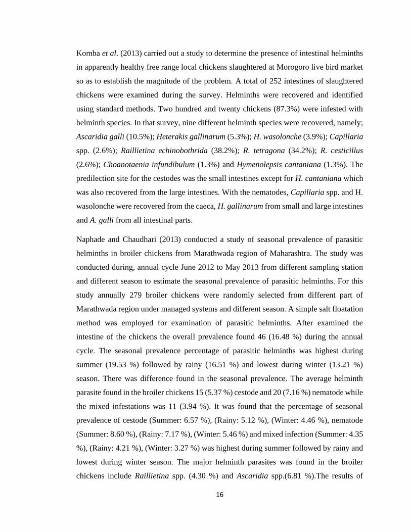
Komba et al. (2013) carried out a study to determine the presence of intestinal helminths
in apparently healthy free range local chickens slaughtered at Morogoro live bird market
so as to establish the magnitude of the problem. A total of 252 intestines of slaughtered
chickens were examined during the survey. Helminths were recovered and identified
using standard methods. Two hundred and twenty chickens (87.3%) were infested with
helminth species. In that survey, nine different helminth species were recovered, namely;
Ascaridia galli (10.5%); Heterakis gallinarum (5.3%); H. wasolonche (3.9%); Capillaria
spp. (2.6%); Raillietina echinobothrida (38.2%); R. tetragona (34.2%); R. cesticillus
(2.6%); Choanotaenia infundibulum (1.3%) and Hymenolepsis cantaniana (1.3%). The
predilection site for the cestodes was the small intestines except for H. cantaniana which
was also recovered from the large intestines. With the nematodes, Capillaria spp. and H.
wasolonche were recovered from the caeca, H. gallinarum from small and large intestines
and A. galli from all intestinal parts.
Naphade and Chaudhari (2013) conducted a study of seasonal prevalence of parasitic
helminths in broiler chickens from Marathwada region of Maharashtra. The study was
conducted during, annual cycle June 2012 to May 2013 from different sampling station
and different season to estimate the seasonal prevalence of parasitic helminths. For this
study annually 279 broiler chickens were randomly selected from different part of
Marathwada region under managed systems and different season. A simple salt floatation
method was employed for examination of parasitic helminths. After examined the
intestine of the chickens the overall prevalence found 46 (16.48 %) during the annual
cycle. The seasonal prevalence percentage of parasitic helminths was highest during
summer (19.53 %) followed by rainy (16.51 %) and lowest during winter (13.21 %)
season. There was difference found in the seasonal prevalence. The average helminth
parasite found in the broiler chickens 15 (5.37 %) cestode and 20 (7.16 %) nematode while
the mixed infestations was 11 (3.94 %). It was found that the percentage of seasonal
prevalence of cestode (Summer: 6.57 %), (Rainy: 5.12 %), (Winter: 4.46 %), nematode
(Summer: 8.60 %), (Rainy: 7.17 %), (Winter: 5.46 %) and mixed infection (Summer: 4.35
%), (Rainy: 4.21 %), (Winter: 3.27 %) was highest during summer followed by rainy and
lowest during winter season. The major helminth parasites was found in the broiler
chickens include Raillietina spp. (4.30 %) and Ascaridia spp.(6.81 %).The results of

17
parasitic helminths were discussed in relation to seasonal variation and found highest
during summer followed by rainy and lowest during winter season.
Mekibib (2014) investigated the prevalence of gastrointestinal helminth of scavenging
chicken in villages around Hawassa, Southern Ethiopia, from October 2010 to April 2011.
A total of 360 faecal samples and 122 postmortem examination were conducted. The
overall postmortem and coproscopic prevalence of scavenging chicken gastrointestinal
helminthes (GIT) were 88.5% and 77.8%, respectively. In the examined scavenging
chicken about 67.5% and 29.2% of the chickens were positive for nematodes and cestodes
species, respectively the postmortem examination revealed 51.6% infection with
Heterakis gallinarum followed by Ascarida galli (45.9%), Raillietina tetragona (20.5%),
Raillietina echinobothrida (17.2%), Capillaria species (13.1%), Raillietina cesticillus
(8.2%) and Hymenolepis cantanian (3.3%). There was a significant difference in the
overall prevalence of GI helminth parasites observed between male and female and
between age groups of chickens (P <0.05 and P < 0.01, respectively).
Alam et al. (2014) observed the prevalence of gastrointestinal helminth infections and the
gross pathological lesions produced by them from February 2012 to January 2013 in the
Department of Pharmacology of Bangladesh Agricultural University, Mymensingh. In
that study, a total of 320 indigenous chickens aged ranging from 2 to 4 months were
examined to identify the different types of gastrointestinal helminth infections in
indigenous chickens. During routine examination, six species of helminth parasites were
recorded, of which five species of nematodes such as Ascaridia galli, Heterakis
gallinarum, Capillaria spp, Acuaria hamulosa and Dispharynx spiralis; and one species
was cestode called Raillietina tetragona. The highest prevalence was observed for
Ascaridia galli (41.56%) followed in descending order by Raillietina tetragona (19.68%),
Heterakis gallinarum (15.62%), Acuaria hamulosa (8.75%), Capillaria spp. (4.68%) and
Disopharinx spiralis (1.56%). The gross pathological lesions were observed in case of
Acuaria hamulosa and Heterakis gallinarum infection. In case of Acuaria hamulosa
infection keratinization of gizzard mucosa and cross section of adult Acuaria hamulosa
were seen along with marked infiltration of neutrophils. The results of this study

18
suggested that both nematodes and cestodes were highly prevalent in indigenous chickens
in the studied werea.
Lucas (2014) comprehend the possible role of helminth infections of poultry and the
prevalence of helminth infections in slaughtered chickens was investigated. A total of 305
gastrointestinal tracts were collected from slaughtered chickens, 177 and 128 of which
were from broilers and layers respectively. The intestines were longitudinally incised and
the contents washed into cups for the recovery of worms and worm eggs using standard
parasitological methods. Results showed that helminth infections were common in the
grown birds. Ascaridia galli, Heterakis spp. and Raillietina spp. were common with the
prevalence of 22.3, 0.6 and 6.2% respectively. However, there was a breed discrepancy
in prevalence particularly in A. galli in which layers had a higher prevalence (34.4%) than
broilers (14.1%). It was concluded that the risk of helminths infections was high in grown
birds intensively managed in deep litter in the study werea and that it could be the same
in similar environments where poultry were managed on deep litter and could compound
diagnosis of other health problems of chickens.
Amaral and Alberto (2016) conducted a servey on nematodes of the genus Ascaridia,
important endoparasite of poultry. It was known Ascaridia galli was one of the most
common nematode worldwide that (round worm) affecting chickens, however there had
never been a study conducted to measure the prevalence of this round worm in Timor
Leste. To measure the prevalence of this parasite a survey was conducted in 9 villages
across three regions of Timor Leste. The survey revealed that A. galli were 5.9% (2.2-
12.5%) positive in Covalima municipality, 3.1% (0.6 - 8.9%) in Manatuto Municipality
and 15.4% (8.1 - 23.0%) in Lospalos Municipality. The overall prevalence for Timor
Leste was 7.8% (5.0 - 11.5%).
Afolabi et al. (2016) conducted a survey of intestinal parasites of chickens, carried out in
Akure, Ondo State, Nigeria from January to December, 2015. A total of 327 chickens of
different breeds were examined for gastrointestinal infections. Fecal samples obtained
from these chickens were prepared for microscopy using flotation technique. The results
showed that 67 (20.5%) of the 327 chickens examined were infected with various
gastrointestinal parasites. Among the infected chickens, the layers were the most

19
susceptible to gastrointestinal parasites with a prevalence of 88.4%, while broilers were
the least susceptible with a prevalence of 7.2%. It was further observed that the highest
prevalence of gastrointestinal infection (37.6%) was recorded among the chickens that
were kept in an extensive management system, while the lowest prevalence (9.6%) was
recorded among the chickens kept in an intensive management system.
Ogbaje et al. (2016) conducted a survey to determine the prevalence of gastrointestinal
helminthes in local chickens, broilers and layers slaughtered in Makurdi metropolis
between September 2007 and April 2008. A total of 440 samples were collected from
male and female chickens. Of the total samples examined, 200(45.5%) were from
domestic chicken, 140(31.8%) from broilers and 100(22.7%) from layers. Of the total
sample examined, 280(63.6%) were infected with one or more species of helminthes. Of
the number positive for infections, 103(23.4%) had single infection, 105(23.9%) double
infections and 60(13.6%) triple infections. Overall, 165(37.5%) of the samples had
Ascaridia galli, 122(27.7%) had Heterakis gallinarum and 214(48.6%) had various
tapeworm species. Out of the 200 samples from domestic chickens, 110(55%) were found
infected with Ascaridia galli, 80(40%) with Heterakis gallinarum and 145(72.5%) with
different tapeworm species. Of the 140 gastrointestinal tracts from broilers, 50(35.7%)
were infected with Ascaridia galli, 40(28.6%) with Heterakis gallinarum and 60(42.9%)
with various tapeworm species. Out of the 100 gastrointestinal tracts from layers, 5(5%)
were infected with Ascaridia galli, 2(2%) with Heterakis gallinarum and 9(9%) with
various tapeworm species. The species of tapeworm encountered were Raillietina species,
Choanotaenia species and Hymenolepis species. These respective species constituted
30.9%, 5.2% and 3.6% of the tapeworm burdens.

20
CHAPTER 3
MATERIALS AND METHODS
3.1 Sampling Area
The intestines of the slaughtered chickens were collected from the rural areas of four
districts of Bangladesh, namely Dhaka (36), Bandarban (10), Mymensingh (7) and Pabna
(10). This experiment was performed in the Microbiology & Parasitology Laboratory at
the Sher-e- Bangla Agricultural University, Dhaka.
3.2 Collection of samples
Around noon on each sampling day, a batch of 6 intestines were randomly picked from a
group of intestines of chickens slaughtered between early morning and at the time of
collection. On sampling days, the gastrointestinal tracts (GIT) were placed in separate,
labelled polythene bags and transported to the laboratory maintaining a cool chain
protocol.
The samples were used for the isolation of helminthes. The gastrointestinal tracts were
separated into small intestine, caecum and large intestine. The entire length of each
intestine was incised longitudinally and the contents were emptied into sieves placed in
large clean plastic cups with labelling. Contents were washed in normal saline and
examined under a light microscope. Larger helminths were collected directly by curved
needle or forceps, and smaller ones were isolated under the microscope. Worms were
grouped and counted with morphometrically before being stored in plastic bottles
containing 70% alcohol according to the method described by Permin and Hansen (1999).

21
3.3 Processing of cestodes and trematodes
In case of trematodes and cestodes, stained permanent slides were made. For this purpose,
trematodes and cestodes were collected from plastic bottles containing 70% alcohol. Then
the specimens were flattened between two glass slides with slight pressure and fixed in
70% alcohol until future works.
After flattening for a week, the specimens were dipped in 50% alcohol for one hour and
then into distilled water for another one hour. Then the specimens were transferred in
Haematoxylin-Carmine solution and kept overnight for staining. The excessive stain was
removed by 3% HCl-Alcohol. The stained specimens were washed with ascending grades
of alcohol for hardening, cleared in xylene and mounted in canada balsam. Finally, the
slides were kept until the canada balsam dried, and observed under microscope.
3.4 Processing of nematodes
In case of nematodes, the specimens were not stained because the nematodes were very
thin and transparent. Before observation, the nematodes were washed well in water to
remove the preservatives, dehydrated in 70 - 90 % alcohol as per the thickness of the
worm and cleared by submerged them in lactophenol. Then the nematodes were examined
under microscope and photographs were taken from different body parts as an aid for
identification.

22
CHAPTER 4
RESULTS AND DISCUSSION
RESULTS
Through the examination of all 63 samples, five helminths were confirmed by observing
them under light microscope. These include 2 species of nematode (A. galli & H.
gallinarum), 2 species of trematodes (E. revolutum & C. verrucosa) and only 1 species of
cestode (R. tetragona).
4.1 Morphological observation
4.1.1. Morphology of A. galli
The specimens were recovered from both small and large intestines. Adult worms were
yellowish white in color and semitransparent (Figure 1A). The oral opening is surrounded
by three prominent lips which are trilobed (Figure 1B). These features match with the
Order Ascaridida. The esophagus was filariform and the intestine is simple, which are the
special feature of Family Ascarididae. The whole body was enclosed in a tough
proteinaceous covering called cuticle. The cuticle was distinctly striated and the cuticular
alae were feebly developed (Figure 1C). Two conspicuous papillae occured on the dorsal
lip and one on each of the subventral lips (Figure 1B). A pair of so-called neck papillae
occured on the sides of the body near the anterior end. Morphologically sexual
dimorphism in ascarids was characterized by ventrally coiled tail with precloacal sucker
in males, and a blunt and rounded posterior end in females. The posterior portion of female
also possessed a single large anal opening just before the extremity and possessing one
pair of papillae just near to its tip. The tail end was rather straight and blunted (Figure
1D). The posterior end of male was comparatively elaborate and more complex (Figure
1E). There were two prominent apertures, anus towards the posterior end and precloacal

23
or preanal sucker immediately anterior to the spicules. The precloacal sucker was
supported by a sclerotized ring which serves the functions as an aid to attach during
copulation. The worms had two well-developed unequal spicules at the posterior end
(Figure 1F). The uteri of females were packed with eggs which were oval to ellipsoidal,
with a thick, smooth shell, containing a single cell. All of these morphological
characteristics are corresponded to the Genus Ascaridia, and the Species A. galli.
A B
C D

24
Figure 1: Different body parts of A. galli, A. Gross sample of A. galli. B. Anterior part of
the parasite; the black arrow indicates the lips and the blue lines indicate the papillae on
the lips. C. Cuticular striation. D. Posterior part of the female; the black line indicates anal
opening. E. Posterior part of the male (4X). F. Posterior part of the male (10X); the black
lines indicate the unequal spicules and the block arrow indicates the precloacal or preanal
sucker. G. Uterus of the female.
E
G
F

25
4.1.2 Morphology of H. gallinarum
The adult worms were collected from large intestine, specially from the caecum. Adult
worms were small and white in colour and had three well-defined lips, which are the
general characters of the Order Ascaridida. The head end was slightly curved (Figure 2A).
The esophagus was engaged with a short narrow anterior portion (pharynx) and ended in
a well-developed bulb containing a valvular apparatus (Figure 2B, 2C). These are the
common features of Family Heterakidae. The cuticle was usually with lateral flanges.
Alae, which ran almost the entire length of the body, were ridges formed by the thickening
of the cuticle. Adult female and male caecal worms differed in length, with the female (10
to 15 mm) generally being larger than that of the male (7 to 13 mm). The tail end of female
was elongated, gradually tapered. The anal opening was at the posterior part of body. The
vulva of the female was located at the middle of the body (Figure 2D). There were three
bends in the vagina after the vulva, angled posteriorly, anteriorly and once again
posteriorly. Male worms had stylet-like tail end that smoothly taper. The worms had two
well-developed unequal spicules at the posterior end (Figure 2E). Gubernaculum is
absent. The preanal sucker was easily seen, round, well-developed, surrounded by a
chitinized ring. Eggs in the uterus were ellipsoidal, with a thick, smooth shell, containing
a single cell (Figure 2F). Each of every morphological characteristic are almost identical
to the Genus Heterakis, and Species H. gallinarum.

26
Figure 2: Different body parts of H. gallinarum, A. Slightly curved head end. B. Anterior
part of the parasite; the black arrow indicates the lips and the blue arrow indicates the bulb
shaped esophagus. C. Bulb shaped esophagus in high magnification (40X). D. Vulvar
opening of the female (the black line). E. Posterior part of the male (10X); the black arrow
shows the preanal sucker and the black line shows the unequal spicules. F. Multiple eggs
in the uterus of female.
A B
C D
E F

27
4.1.3 Morphology of R. tetragona
Multiple mature cestodes, measuring 12-30 cm long were isolated from both small and
large intestines. These cestodes were whitish in color, highly elongated, dorso-ventrally
flattened (Figure 3A). They had multiple segments, bearing four suckers on their scolices.
These are the identical features of the Order Cyclophyllidea. The body is divided into the
head region called 'scolex', followed by an unsegmented 'neck', and then by highly
segmented body proper called strobila. The strobila is composed of a series of ribbon-like
body segments called proglottids, gradually enlarging from the anterior end towards the
posterior. The scolex bears an apical rounded rostellum, which is medium and armed with
many minute hooks, arranged in single row. The hooks are hammer-shaped. This is
surrounded by four ovoid suckers which are lined with several rows of spines (Figure 3B,
3C). These are the morphological features of Family Davaineidae. The mature segment is
longer than broad and the common genital pores are single and being in front of the
anterior 1/3 of the lateral margin of the mature segment. Each mature proglottid has a set
of male and female reproductive organ (Figure 3D). Testes are located on both sides of
the ovary and behind vitellarium. Each egg capsule contained 4-12 eggs and many
calcareous corpuscles, each of which is surrounded by a membrane. These features are
matched with the identical morphological characteristics of the Genus Reillietina, and
Species R. tetragona.

28
Figure 3: Different body parts of R. tetragona. A. Gross sample of parasite after
collection. B. Anterior part of the parasite (10X); the blue arrow indicates rostellum and
the “*” indicate the suckers. C. Scolex in high magnification (40X); D. The broad segment
having common genital pores on each (black arrows); Each mature proglottid has a set of
reproductive organ in the middle part.
A B
C D
* *

29
4.1.4 Morphology of C. verrucosa
A large number of minute flukes were recovered from the caecum of the poultry intestine.
Small muscular body was dorsoventrally flattened, attenuated anteriorly and broadly
rounded posteriorly. Pharynx was absent and the esophagus was very short (Figure 4A).
These are the identifying feature of Order Plagiorchiida. Cup-shaped oral sucker was
terminal (Figure 4C). There was no ventral sucker which are the morphological feature of
Family Notocotylidae. Adult flukes were 1.5-2.0 cm long and 0.5-1.0 cm wide. Long
caeca were bifurcated, smooth, extending posteriorly between the uterine loops and
vitelline follicles, then passed through the testes and ovary, and finally terminated blindly
at the level of excretory pore. Cirrus sac was elongated, containing prostatic cells and
coiled seminal vesicle. Genital pore was median, closely posterior to the caecal
bifurcation (Figure 4A). There were two testes which are irregularly lobed, located in
extracaecal field in posterior third of the body. Ovary was trilobed, situated at the
testicular level (Figure 4D). Uterus had a number of closely packed loops, overlapping
cirrus sac, reaching up to the level of Mehlis gland. Uterine loops were 18 in number
(Figure 4B). Vitellaria was fairly composed of large follicles arranged extracaecally but
at some places it overlaps the ceca, extending from the anterior third of the testes up to
the anterior third uterine loop, which are the special morphological characteristics of the
Genus Catatropis, and Species C. verrucosa.
B

30
Figure 4: Different body parts of C. verrucosa. A. Whole fluke under 4X magnification;
the black arrow indicates the oral sucker, the blue arrows indicate caecal bifurcation and
the broken line indicates the cirrus sac. B. Uterine loops (18 in numbers) of the parasite
(10X); both side of the loops covered by vitellaria. C. Cup shape oral sucker (10X); D.
The reproductive organs of parasites; the blue arrows indicate testes and the block arrows
indicates the ovary.
A
C D
B

31
4.1.5 Morphology of E. revulotum
The echinostomes were recovered from the large intestine of poultry. The Body was
muscular, dorsoventrally flattened and c shaped in appearance (Figure 5A). Pharynx was
absent and the esophagus was very short All flukes were 6-8 mm long and 1.5-2 mm wide
and had a well-developed head collar bearing 37 spines (5 angle spines and 6 lateral spines
on each side and 15 dorsal spines) (Figure 5D, 5E). The esophagus was shorter than the
diameter of the ventral sucker (Figure 5B). These morphological properties matched those
of the members of 37 collar spined echinostomes (E. revolutum group). Adult flukes had
a short forebody and did not have constriction at the level of the ventral sucker. There are
two testes, arranged in a tandem position, located at the posterior part of the body. Testes
were elongated with smooth margin and slightly separated from each other. The anterior
testis was shorter and wider than the posterior testis. The ovary was oval, median, and
transversely located between the posterior end of the uterus and cranial margin of the
anterior testis (Figure 5C). The cirrus sac was oval and located transversely between the
level of intestinal bifurcation and anterior border of ventral sucker. Multiple eggs are
located in the uterus. The eggs are oval, large, thin shelled, operculated and contain
unsegmented ovum (Figure 5F) which are the special morphological characteristics of the
Genus Echinostoma, and Species E. revolutum.

32
D
A B
C

33
Figure 5: Different body parts of E. revolutum. A. Gross sample of the fluke occurring c-
shaped. B. Anterior part of the fluke; the black arrow indicates the oral sucker and the
broken line indicates the ventral sucker. C. Posterior part of the; the black arrow indicates
the ovary and the broken lines indicate the testes. D. Oral sucker with short esophagus
(white block arrow) and caecal bifurcation. E. The 37 spines are distinct around the oral
sucker. F. Numerous eggs in the uterus.
E F

34
4.2 Prevalence
4.2.1 Overall prevalence of helminths in backyard chicken
The study was carried out in a total of 63 gastrointestinal tract of backyard chicken. Out
of the 63 examined samples, all of those were infected with various species of
gastrointestinal helminths, comprising two species of nematode, two species of
trematodes and only one species of cestode. Those parasites were found in different
locations of the gastrointestinal tracts of backyard poultry.
The nematode parasites encountered were A. galli and H. gallinarum out of which A. galli
(47.61%) was the most prevalent and H. gallinarum (38.09%) was the least. The
trematode parasites recovered included C. verrucosa and E. revolutum out of which C.
verrucosa (23.80%) was most prevalent followed by E. revolutum (7.93%). The cestode
parasites encountered was R. tetragona (73.01%). The overall prevalence is shown in
Table 1.
Table 1: Location and overall prevalence of helminths in backyard poultry
Class of
helminths
Name Location No. of
infected
chicken
Prevalence
Nematode A. galli Small intestine &
Large intestine
30 47.61%
H. gallinarum Caecum & Large
intestine
24 38.09%
Trematode C. verrucosa Caecum 15 23.80%
E. revolutum Small intestine &
Large intestine
5 7.93%
Cestode R. tetragona Small intestine 46 73.01%

35
4.2.2 Prevalence of helminths in different geography
The samples were collected from Dhaka (N=36), Pabna (N=10), Bandarban (N=10),
Mymensingh (N=7). Total 5 species of helminths were recovered from Dhaka namely A.
galli (44.44%), H. gallinarum (38.88%), C. verrucosa (33.33%), E. revolutum (13.88%)
and R. tetragona (72.22%) which were given in the Table 2.
Table 2: Prevalence of gastrointestinal helminths in Dhaka (N=36)
Helminths No. of infected chicken Prevalence
Nematode
A. galli 16 44.44%
H. gallinarum 14 38.88%
Trematode
C. verrucosa 12 33.33%
E. revolutum 5 13.88%
Cestode
R. tetragona 26 72.22%
Ascaridia galli (57.14%), H. gallinarum (42.85%), C. verrucosa (28.57%) and R.
tetragona (100.00%) were collected from Mymensingh (Table 3) and A. galli (60.00%),
H. gallinarum (50.00%), C. verrucosa (30.00%) and R. tetragona (60.00%) were
collected from Pabna (Table 4). In those areas, there were no E. revolutum.
Table 3: Prevalence of gastrointestinal helminths in Mymensingh (N=7)
Helminths No. of infected chicken Prevalence
Nematode
A. galli 4 57.14%
H. gallinarum 3 42.85%
Trematode
C. verrucosa 2 28.57%
Cestode
R. tetragona 7 100.00%

36
Table 4: Prevalence of gastrointestinal helminths in Pabna (N=10)
Helminths No. of infected chicken Prevalence
Nematode
A. galli 6 60%
H. gallinarum 5 50%
Trematode
C. verrucosa 3 30%
Cestode
R. tetragona 6 60%
Ascaridia galli (40.00%), H. gallinarum (20.00%) R. tetragona (80.00%) were collected
from Bandarban (Table 5). In those areas, no trematode was found.
Table 5: Prevalence of gastrointestinal helminths in Bandarban (N=10)
Helminths No. of infected Prevalence
Nematode
A. galli 4 40%
H. gallinarum 2 20%
Cestode
R. tetragona 8 80%
4.2.3 Organ Preferences of gastrointestinal helminths
The results showed that most of the parasites prefer to colonize the small intestine than
the large intestine. Some of the parasites were found in the caecum. No parasite was
recovered in the crop and gizzard. A. galli, the largest nematode of poultry, was mostly
found in small intestine and H. gallinarum was found mostly in the caecum. Only cestode,
R. tetragona was collected from small intestine.

37
Table 6: Site preferences of different gastrointestinal helminths (N=63)
Preferred
sites
Parasites No. infected Percentage
Small intestine A. galli 28 44.44%
E. revolutum 3 4.76%
R. tetragona 46 73.01%
Large intestine A. galli 8 12.69%
H. gallinarum 9 14.28%
E. revolutum 2 3.17%
Caecum H. gallinarum 22 34.92%
C. verrucosa 15 23.80%
Figure 6: Site preferences of different gastrointestinal helminths
44.44%
12.69%14.28%
34.92%
4.76% 3.17%
23.80%
73.01%
0.00%
10.00%
20.00%
30.00%
40.00%
50.00%
60.00%
70.00%
80.00%
Small intestine Large intestine Caecum
Per
cen
tage
Location on the host
Site preferences of different gastrointestinal helminths
A. galli H. gallinarum E. revolutum C. verrucosa R. tetragona

38
4.2.4 Single and mixed type of infection
Examined gastrointestinal tract of poultry were infected by one or more species of
helminth parasites. Among the 63 chicken, 16 were infected with single species of
helminths (25.39%) and rest 47 were infected with multiple species of helminths. In case
of the multiple infection, 28 chicken were infected with two species of helminths
(44.44%) and 19 chicken were infected with more than two species of helminths
(30.15%).
Table 7: Percentages of single and multiple type of infection (N=63)
Type of infection No. infected chicken Percentage
Single
One species 16 25.39%
Mixed
Two species 28 44.44%
> two species 19 30.15%
Figure 7: Percentages of single and multiple type of infection
0.00%
5.00%
10.00%
15.00%
20.00%
25.00%
30.00%
35.00%
40.00%
45.00%
50.00%
One species Two species > Two species
Single infection Multiple infection
Single and mixed type of infection

39
DISCUSSION
After the extensive study of the gastrointestinal tracts of backyard chicken, Gallus
domesticus, for helminth parasitism in different areas of Bangladesh, various species of
helminth parasites were recovered with a very high prevalence (100%).
Total five species of helminth parasites were recorded of which two species were
nematodes, two species were trematode and one species belonged to cestode. Similar
studies were conducted by earlier scientists. Ferdushy et al. (2014) reported that 84.6%
gastrointestinal helminth infection in Narsingdi district of Bangladesh. However, Rabbi
et al. (2006) reported relatively higher prevalence (100%) of gastrointestinal helminth
infection in indigenous chickens in Mymensingh district. This result is similar to our study
because the results of Rabbi et al. (2006) were based on a non-random sample of 80
indigenous chicken’s viscera. The smaller size of the sample and randomness is
responsible for the higher prevalence of gastrointestinal helminths other than probable
regional variation. There are lots of report having more than 50% prevalence rate from
the different parts of the world. Mekibib et al. (2014) also reported similar prevalence of
gastrointestinal helminth infections in scavenging chickens from Ethiopia. Yadev and
Tandon (1998) revealed that 90.9% of helminth infections in subtropical high rainfall area
of India. Eshetu et al. (2001) found that 91.01% chickens infected with gastrointestinal
helminthes from Amhara region Ethiopia. Nokana et al. (1991) during their survey of
helminth parasites in backyard flocks in Michigan by litter examination also showed
relatively high contamination rates. Wakelin (1964) in Britain found 59.2%, Romanenko
et al. (1985) in Roostov recorded 100% and Guclu (1994) in Turkey found 59% birds
affected with helminth parasites. Similar studies were also performed by earlier scientists
(Ssenyonga, 1982; Samad and Rahman, 1985; Huq, 1986; Kang and Suh, 1987).
There are a lot of variation in prevalence from different parts of Bangladesh. Some
helminths are relatively higher in some areas such as R. tetragona in Mymensingh (100%)
and C. verrucosa is not found from Bandarban and Pabna. This may be due to the very
low amount of smple. Therefore, the remarkable prevalence of infection observed in
backyard poultry from different parts of Bangladesh can be attributed to a number of
factors like the type of management and production system, exposure to intermediate

40
hosts, inadequate or no use of anthelmintics, the climatic conditions which alter the
population dynamics of the parasite. Backyard poultry of Bangladesh are reared in semi-
scavenging system, in which they collect maximum of their food from the nature. Their
food enterprise includes different types of seeds, kitchen wastages, insects, slugs,
earthworm etc. They act as intermediate or paratenic hosts of many bio-parasites
(Soulsby, 1982). Besides, backyard poultry can easily ingest the infective stage of many
geo-parasites during taking food from the environment. The paratenic host of A. galli and
H. gallinarum is earth worm which is very common and a favourite feed item for poultry
in Bangladesh. Several snails like Bithynia tentaculata and Lymnaea act as intermediate
hosts of C. verrucosa and E. revolutum, respectively. In case of R. tetragona, the ant of
the genera Tetramorium, Pheidole, and house fly, Musca domestica act as intermediate
host which are also available in our country, especially in the rural areas. Indigenous
chickens are very fond of scavenging various insects from the nature which might
contribute to the transmission of various helminth infection in chicken. Therefore, the
environmental and climatic conditions of the study area seem to be very favorable for
such a high intensity of infection, and also a major ecological factor like dispersal and
migration plays a significant role in maintaining such a high prevalence of helminth
infection in these study areas.
The present study reveals that single type infections were more prevalent than multiple
type infections. A good number of backyard poultry were harboring more than one type
of helminth species which is in agreement with the observation of Edgar (1953);
Kononenko and Khaizade (1983); Mpoame and Agbede (1995); Permin et al. (1999);
Poulsen et al., (2000); Mukaratirwa et al., (2001); Magwisha et al., (2002); Phiri et al.
(2007) and Luka and Ndams (2007). Majority of the host birds harboured multiple type
of infection by helminthes may be due to the prevailing environmental conditions and
free-range management systems which are very favourable to many species of helminth
parasites described by Ramadan and Znada (1992) and Watt (1996).
Our results indicate differences in predilection sites for the different helminth species
recovered, with some species being recovered from more than one location. A similar
observation was reported in the previous studies by Hussen et al., (2012) and Molla et al.,

41
(2012). This specificity in parasite distribution in the gastrointestinal tract is thought to be
an attribute of differences in physicochemical environments in various regions of the gut.
Among other factors, availability of suitable food and attachment sites; and presence of
certain specific stimuli such as pressure differences are also known to dictate parasite site
segregation along the gastrointestinal tract (Nkwengulila and Mwita, 2004). It may be due
to the interactive competition for site and food among co-habiting species as among the
factors determining parasite distribution in the gastrointestinal tract (Holmes 1973;
William and Jones, 1994).
The most prevalent helminth parasite recorded during the present study was R. tetragona
(73.01%), followed by A. galli (47.61%), H. gallinarum (38.09%), C. verrucosa (23.80%)
and E. revolutum (7.93%). The results are in the agreement with the work of many authors
(Qureshi, 1950; Wilson et al., 1994; Mpoame and Agbede, 1995; Permin et al., 1996;
Eshetu et al., 2001). However, Schou et al. (2006), Luka and Ndams (2007) reported a
higher prevalence of A. galli. The variations in prevalence can be attributed due to the
environmental conditions in the area and unavailability of intermediate hosts. The
environmental conditions like temperature and moisture favour the larval development in
contaminated droppings and facilitate transmission.
In Bangladesh, there are very limited morphological data regarding to the helminths of
backyard chicken. Identification of the helminths are based on the size of helminths and
the location of host. In this sense, only the genus can be identified. Therefore, a detailed
morphological study has been conducted.
Ascaridia galli possesses all the salient features of the ascarid nematodes. The body is
cylindrical, highly extended, covered with a collagenous cuticle, anterior end with distinct
lips, and the posterior with anus. In addition, marked sexual differences between male and
female can be easily diagnosed by the relatively longer females with straight blunt tail
end; whereas males are comparatively shorter with an elaborate, curved tail end (Ackert,
1940). The posterior components in Ascaridia, especially those of papillae and spicules,
are the identifying characters between different species (Kung, 1949). The distinguishing
characters of A galli is the difference in well-developed spicules in length (Kajerova et

42
al., 2004). The morphological features of A. galli also reflect all the characters of the
generic diagnosis (Yamaguti 1961, Hodová et al., 2008)
There are many reports on the specificity H. gallinarum to backyard poultry (Skrjabin et
al., 1961; Fedynich et al., 2005; Sherwin et al., 2013). Morphological characteristics of
adult male and female H. gallinarum nematodes resulted in new data on their
morphometric structure that can facilitate the species identification. Morphologically,
male Heterakis nematodes are more distinct. One spicule of H. gallinarum male is
visually longer than the other one. Microscopy study of the structure and position of tail
papillae in males of those species shows significant differences. In males of H. gallinarum
there are two preanal, six adanal, and four postanal pairs of papillae. (Abou Znada, 1993;
Rahman & Manap, 2014; Sheikh et al., 2016).
Whitish, highly elongated, dorso-ventrally flattened cestodes were recovered from
intestine of poultry. The mature segment is longer than broad and the common genital
pores are single. Each mature proglottid has a set of male and female reproductive organ.
Testes are located on both sides of the ovary and behind vitellaria. The morphological
features and general measurements of recovered R. tertragona in the present investigation
were agreed with the findings of many authors (El – Azzazy, 1979; Soulsby, 1982; Mahdy
and Olfat, 1988; Ramadan and Abouzanda, 1989; El – Gayer and Amal 1992; Sayed and
Gehan, 1996 and Ahmed-Nabila, 2004).
The echinostomes were recovered from the large intestine of poultry. Morphological
identification of Echinostoma species in the 37-collar-spined ‘revolutum’ group is
difficult due to the contradictory data from different hosts and geographical locations. In
this study, the identification of E. revolutum was done by description of different authors
(Kanev, 1885; Kanev, 1994 Kanev, 1995; Faltnkova, 2015). The morphological
characteristics of E. revolutum in this study was similar with those reported previously
(Kanev, 1994 and Faltnkova, 2015).
Catatropis verrucosa was recovered from the caecum of the poultry intestine which were
small muscular body, dorsoventrally flattened, attenuated anteriorly and broadly rounded
posteriorly. Cup-shaped oral sucker was terminal with no ventral sucker. Long caeca were

43
bifurcated and cirrus sac was elongated. Uterine loops were 18 in number. The shape and
location of testes and ovary along with the other morphological characteristics of this
fluke are very similar to the authors (Joyeux, 1922; Dubois, 1951; Odening, 1966; Kanev
and Vasiliev, 1986; Dvorjadkin, 1989; Looss, 1893 and Kanev et al., 1994)

44
CHAPTER 5
CONCLUSION
The present study was aimed to study the morphology and the prevalence of
gastrointestinal helminths in backyard poultry in different areas of Bangladesh (Dhaka,
Bandarban, Mymensingh and Pabna). Different species of helminth parasites were
recovered with a moderately high prevalence (100%). The smaller size of the sample and
non-randomness is responsible for the higher prevalence of gastrointestinal helminths. In
our study multi-species infection of helminths were observed in most of the chickens
which suggest that the environmental condition and the nature of the poultry rearing
system are very favorable for the transmission and persistence of the helminth parasites
in rural areas of Bangladesh. This condition of high worm burdens can make the bird more
prone to bacterial and viral infection as well more easily available to the predators as the
bird become very much unthrifty and weak. Therefore, further large-scale studies are
needed to measure the impact of helminth infection on the health and productivity of the
rural scavenging chickens in rural areas of Bangladesh.

45
REFERENCES
Abebe, W., Asfaw, T., Genete, B., Kassa, B. and Dorchies, P.H. (1997). Comparative studies of
external parasites and gastro-intestinal helminths of chickens kept under different
management systems in and around Addis Ababa (Ethiopia). Revue de Médicine
Vétérinaire, 148: 497–500.
Aberra Melesse, S., Maak, R., and Lengerken, G. (2011). Effect of long-term heat stress on some
performance traits and plasma enzyme activities in Naked-neck chickens and their F1
crosses with commercial layer breeds. Liv Sci, 141: 227-231.
Abou Znada, N. Y. (1993). First record and prevalence of the caecal nematodes, Heterakis
gallinarum and Subulura suctoria from the Guinea Fowl, Numedia meliagris in Saudi
Arabia. J King Saud Univ Sci. 5(1): 59–65.
Ackert, J.E. (1940). The large round worm of chickens. Vet Med. 35: 106-108.
Adang, L. K. (1999). Some aspects of the Biology of Columbids in Zaria, Nigeria. M.Sc. Thesis,
Department of Biological Sciences, A.B.U. Zaria, Nigeria. Pp. 98.
Afolabi, O. Simon, A. and Opeyemi, O. (2016). Intestinal Parasites of Domestic Chickens (Gallus
Gallus Domesticus) in Akure, Nigeria. Journal of Biomedicine. In Press.
10.5812/jmb.9771.
Ahmed, S. and Islam, N. (1990). Backyard poultry development project in 100 villages.
Proceeding of the 1st Conference of Bangladesh Animal Husbandry Association. Feb. 23-
24, 1985, Bangladesh Agricultural Research Council, Dhaka, Bangladesh. pp. 13-138
Ahmed. and Nabila, S.I. (2004). Some studies on parasitic helminthes infecting domestic chicken
in Beni-Sueif Governorate. M.V.Sc. Thesis, Faculty of Vet. Med., Cairo University.
Akter, M.K., Uddin, A.B.M.J., Rashid, M., Aziz, F.B., Rashid, M.B. and Hasan, M. (2016).
Studies on prevalence of ascariasis in indigenous chickens in Gaibandha district and
treatment by pineapple leaves extract. Res Agric Livest Fish. 3(1): 157-163.
Alam, M.N., Mostofa, M., Khan, M.A.H.N.A., Alim, M.A., Rahman, A.K.M.A. and Trisha, A.A.
(2014). Prevalence of gastrointestinal helminth infections in indigenous chickens of
selected areas of Barisal district, Bangladesh. Bangl. J. Vet. Med. 12(2): 135-139.
Alders, R. (2004). Poultry for profit and pleasure, FAO Diversification Booklet 3. Rome.
Amaral, A. and Alberto, A.A.P.C.J. (2016). Prevalence of Ascaridia galli in village chickens in
Timor Leste. Revista Científica Multidiciplinar da Sociedade Timorense. 2: 59-66.
Belonwu, J.A. (1993). Prevalence of Infectious Diseases of Poultry in Anambra State for 1992.
Nsukka, Nigeria: University of Nigeria.
Berhanu, M., Haileyesus, D. and Desie, S. (2014). Gastrointestinal helminthes of scavenging
chickens in Outskirts of Hawassa, Southern Ethiopia. Glob Vet. 12(4): 557-561.

46
Birmani, N.A., & Dharejo, Ali. Khan, M.M. (2011). Catatropis sp. (Trematoda: Notocotylidae)
from the black coot, Fulica atra Linnaeus, 1758 (Gruiformes: Rallidae) in Sindh Province
of Pakistan. J. Anim. Plant Sci. 21(4): 872-873.
Butboonchoo, P., Wongsawad, C., Rojanapaibul, A. and Chai, J.C. (2016). Morphology and
Molecular Phylogeny of Raillietina spp. (Cestoda: Cyclophyllidea: Davaineidae) from
Domestic Chickens in Thailand. Korean J Parasitol. 54(6): 777-786.
Chalvet-Monfray, K., Sabatier, P., Chauve, C. and Zenner, L. (2004). A Mathematical Model of
the Population Dynamics of Heterakis gallinarum in Turkeys (Meleagridis gallopavo).
Poultry Sci. 83(10):1629-1635.
Chantima, K., Chai, J. and Wongsawad, C. (2013). Echinostoma revolutum: Freshwater Snails as
the Second Intermediate Hosts in Chiang Mai, Thailand. The Korean J Parasitol. 51(2):
183-189.
Daş, G., Abel, H., Savaş, T., Sohnrey, B. and Gauly, M. (2014). Egg production dynamics and
fecundity of Heterakis gallinarum residing in different caecal environments of chickens
induced by fibre-rich diets. Vet Parasitol. 205(3-4): 606-618.
Dvorjadkin, V.A. (1987). Morphology and life cycle of Catatropis morosovi (Trematoda,
Notocotylidae) – a parasite of rodents in Primorja and Priamurja. (In Russian.) In:
MAMAEV, Y. L. (Ed.) Gel’minty I vyzvannye imi zabolevanija. Akademia nauk SSSR,
Vladivostok. pp. 29-33.
Edgar, S.A. (1953). A preliminary check list of parasites of some domestic fowls of Alabama.
Poultry Sci. 32: 949-952.
El-Azzazy, O.M. (1979). Studies on helminth parasites of poultry in Sharkia governorate.
M.V.Sc. Thesis, Fac. Vet. Med., Zagazig University.
El-Gayar. and Amal, K. A. (1992). Some studies on enteric helminthes of chickens in Ismailia
province. M.V.Sc. Thesis, Fac. Vet. Med., Suez Canal University.
Elowni, E.E., Nurelhuda, I.E.M. and Hassan, T., (1989). The effect of niclosamide on Raillietina
tetragona. Vet Res Commun. 13(6): 451-453
Eshetu, Y., Mulualem, E., Ibrahim, H., Berhanu, A. and Aberra, K., (2001). Study of
gastrointestinal helminths of scavenging chickens in four rural districts of Amhararegion,
Ethiopia. Rev Sci Tech. 20(3): 791-796.
Everett, E. Lund, A. and Chute M. (1974) The reproductive potential of Heterakis gallinarum in
various species of galliform birds: Implications for survival of H.
gallinarum and Histomonas meleagridis to recent times. Animal Parasitology Institute,
Agricultural Research Service, Beltsville, Maryland 20705, U.S.A.
Eyinnaya, U.E. (1992). A Survey of the Constraints in the Poultry Industry in Enugu State,
Nigeria. Nsukka, Nigeria: University of Nigeria.

47
FAO (Food and Agricultural Organization of the United Nations) Tech. Rep. 274415.
Rome, Italy: FAO; March 1987. Report on the expert consultation on rural poultry
development in Asia, Dhaka, Bangladesh.
Fatihu, M.Y., Ogbogu, V.C., Njoku, C.O. and Saror, D.I. (1991). Comparative studies of
gastrointestinal helminths of poultry in Zaria, Nigeria. Rev Elev Med Vet Pays Trop.
44(2):175–177.
Fedynich, A. M., Finger, R. S., Ballard, B. M., Garvon, J. M. and Mayfield, M. J. (2005).
Helminths of ross' and greater white-fronted geese wintering in South Texas, U.S.A.
Comp. Parasitol. 72(1): 33–38.
Ferdushy, T., Hasan, M.T. and Kadir, A.K.M. (2014). Cross sectional epidemiological
invesigation on the prevalence of gastrointestinal helminths in free range chickens in
Narsingdi district, Bangladesh. J Parasit Dis. 40(3): 818-22.
Georgieva, S., Faltýnková, A., Brown, R., Blasco-Costa, I., Soldánová, M., Sitko, J., Scholz, T.
and Kostadinova, A. (2014). Echinostoma 'revolutum' (Digenea: Echinostomatidae)
species complex revisited: species delimitation based on novel molecular and
morphological data gathered in Europe. Parasit Vectors. 27(7): 520-524.
Guclu, F. (1994). Helminth fauna of fowls, Turkeys, ducks, and geese in the Ankara area. Doa-
Turk Veterinarlik Vc-Hyavncilik Dorgisi. 18: 79-86.
Hafiz, A.B., Muhammad, A.R., Muhammad, A.A., Imran, A.K., Abdul, A., Zahid, M. and
Shaukat, H.M. (2015). Prevalence of Ascaridia galli in white leghorn layers and Fayoumi-
Rhode Island red crossbred flock at government poultry farm Dina, Punjab, Pakistan. Trop
Biomed. 32: 11-16.
Hafiz, A.B., Muhammad, A.R., Muhammad, A.A., Imran, A.K., Abdul, A., Zahid, M. and
Shaukat, H.M. (2015). Prevalence of Ascaridia galli in white leghorn layers and Fayoumi-
Rhode Island red crossbred flock at government poultry farm Dina, Punjab, Pakistan. Trop
Biomed. 32(1):11-16.
Hassouini, T. and Belghyti, D. (2006). Distribution gastrointestinal helminths in chicken farms
in the Gharb region-Morocco. Parasitol Res. 1: 1-5.
Heyradin, H., Hassen, C., Yosef, D. and Molalegne, B. (2012). Gastrointestinal helminthes are
highly prevalent in scavenging chickens of selected districts of Eastern Shewa zone,
Ethiopia. Pak J. Bio Sci. 15: 284-289.
Hodová, I., Baruš, V. and Tukač, V. (2008). Note on morphology of two nematode species
Ascaridia hermaphrodita and Ascaridia platyceri (Nematoda): scanning electron
microscope study. Helminthologia. 45: 109-113.
Holmes, J.C. (1973). Site selection by parasitic helminthes: interspecific interactions, site
segregation and their importance to the development of helminth communities. Can J
Zool. 51: 333-347.

48
Huq, M.S. (1986). Studies on the helminth infections of poultry under rural conditions of
Bangladesh. Bangladesh Vet J. 20: 55-60.
Hussen, H., Chaka, H., Deneke, Y. and Bitew, M. (2012). Gastrointestinal helminths are highly
prevalent in scavenging chickens of selected districts of Eastern Shewa zone, Ethiopia.
Pak J Biol Sci. 15(6): 284-289.
Joyeux, C.H. (1922). Recherches sur les notocotyles. Bull. Soc. Path. Exot. 15: 331-343.
Kajerova, V., Baruš, V. and Literak, I. (2004). Nematodes from the genus Ascaridia parasitizing
psittaciform birds: a review and determination key. Vet Med – Czech. 49: 217-223.
Kanev I. and Vasiliev I. (1986). Identification of Catatropis verrucosa (Frölich, 1789).
Proceedings of the 5th International Helminthological Symposium Organised by
Helminthological Institute of the Czechoslovak Academy of Sciences in Kosice, Oct. 22-
24, 1986, High Tatra. pp. 5-6.
Kanev, I. (1994). Life-cycle, delimitation and redescription of Echinostoma revolutum (Froelich,
1802) (Trematoda: Echinostomatidae). Syst Parasitol. 28: 125-144.
Kanev, I., Dezfuli, B.S., Nestorov, M. and Fried, B. (1999). Scanning electron microscopy of the
collar region of Deropristis inflata and Echinostoma revolutum. J Helminthol. 73: 51-57.
Kanev, I., Vassilev, I., Dimitrov, V. and Radev, V. (1994). Life-cycle, delimitation and
redescription of Catatropis verrucosa (Frolich, 1789) Odhner, 1905 (Trematoda:
Notocotylidae). Syst Parasitol. 29(2): 133-148.
Kang, Y.B. and Suh, M.S. (1987). Efficacy of febantel against Ascaridia galli and other important
nematodes in laying hens in relation to their productivity. Kor. J. Vet. Publ. Hlth. 11: 33-
38.
Katakam, K.K., Nejsum, P., Kyvsgaard, N.C., Jørgensen, C.B. and Thamsborg, S.M. (2010).
Molecular and parasitological tools for the study of Ascaridia galli population dynamics
in chickens. Avian Pathol. 39(2): 81-85.
Komba, E.V.G., Mkupasi, E.M., Mwesiga, G.K., Mbyuzi, A.O., Busagwe, Z., Mzula, A.,
Lupindu, A.M., and Nzalawahe, J. (2013). Occurrence of helminths and coccidia in
apparently healthy free-range local chickens slaughtered at Morogoro live bird market.
Tanz Vet J. 28: 55-61.
Kononenko, A.F. and Khaizade, N. (1983). The helminth fauna of some birds, aquatic and living
near water, on the south-eastern shore of the Caspian Sea. Biologicheskie resursy
Kaspiiskogo Morya. 186-199.
Kung, C.C. (1949). Notes on some avian species of Ascaridia. J Helminthol. 23: 95-106.
49
Lalchhandama, K. (2010). On the structure of Ascaridia galli, the roundworm of domestic fowl.
Science Vision. 10: 20-30.
Lalchhandama, K. (2010). On the structure of Ascaridia galli, the roundworm of domestic fowl.
Sci Vis. 10(1): 20-30
Latif, M.A. (2001). Microcredits and Savings of Rural Household of Bangladesh. Bangladesh
Development
Studies 27: 51-71.
Looss, A. (1899). Weitere Beiträge zur Kenntnis der Trematodenfauna Aegyptens. Zugleich
Versuch der natürlichen Gliederung des Genus Distomum Retzius. Zool. Jahrb. 12: 521-
784.
Lucas, N. (2014). Prevalence of gastrointestinal helminth infections in slaughtered chickens
reared in the Nsukka area of Enugu State, Nigeria. Sch. J. Agric. Vet. Sci. 7: 51-54.
Luka, S.A., Ndams, I.S. (2007). Gastrointestinal parasites of domestic chickens Gallus gallus
domesticus Linnaeus 1758 in Samaru, Zaria Nigeria. Sci World J. 2(1): 27-28.
Lund, E.E. and Chute, A.M. (1974). The reproductive potential of Heterakis gallinarum in
various species of galliform birds: Implications for survival of H. gallinarum and
Histomonas meleagridis to recent times. Int J. Parasitol. 4(5): 455-461.
Magwisha, H., Kassuku, A., Kyvsgaard, N. and Permin, A., (2002). A comparison of the
prevalence and burdens of helminth infections in growers and adult free range chickens.
Trop Anim Health Prod. 34(3): 205-214.
Mahdy. and Olfat, A. (1988). Studies on parasitic worms infesting chickens and ducks in Giza
governorate, Egypt. M.V.Sc. Thesis, Fac. Vet. Med., Cairo University.
Matur, B.M., Dawam, N.N. and Malann, Y.D. (2010). Gastrointestinal helminth parasites of local
and exotic chickens slaughtered in Gwagwalada, Abuja (FCT), Nigeria. New York Sci. J.
3: 96-99.
Mekibib, B., Dejene, H. and Sheferaw, D. (2014). Gastrontestinal helminthes of scavenging
chickens in outskirts of Hawassa, Southern Ethiopia. Glob Vet. 12(4): 557-561.
Melesse, A. (2000). Comparative studies on performance and physiological responses of
Ethiopian indigenous (”Angete-melata”) chicken and their F1 crosses to long term heat
stress. PhD Thesis, Martin-Luther University, Halle-Wittenberg, Berlin.
Melewas, J.N. (1989). The contribution of poultry to the national economy. Proceedings of the
7th Tanzania Veterinary Association Scientific Conference, December, Arusha, 13–16.
Moges. F., Abera, M. and Tadelle, D. (2010). Assessment of village chicken production system
and evaluation of the productive and reproductive performance of local chicken ecotype
in Bure district, North West Ethiopia. Afr. J. Agric. Res. 5(13):1739-1748.

50
Mohanta, U.K., Watanabe, T., Anisuzzaman, Ohari, Y., Itagaki, T. (2018). Characterization of
Echinostoma revolutum and Echinostoma robustum from ducks in Bangladesh based on
morphology, nuclear ribosomal ITS2 and mitochondrial nad1 sequences. Parasitol Int.
13:1-7.
Molla, W., Haile, H., Almaw, G. and Temesgen, W. (2012). Gastrointestinal helminths of local
backyard chickens in North Gondar Administrative Zone, Ethiopia. Revue Méd Vét.
163(7): 362-367.
Mpoame, M. and Agbede, G. (1995). The gastrointestinal helminth infections of domestic fowl
in Dschang, Western Cameroon. Revue d’ Elevage et de Med Vet des Pays Trop. 48: 147-
151.
Mu, L., Li, H.Y. and Yan B.Z. (2009). Comparative study on morphology and development of
two species of Raillietina from chicken. Chinese journal of Parasitology and parasitic
disease. 27(3): 232-236.
Mukaratirwa, S., Hove, T., Esmann, J.B., Hoj, C.H., Permin, A. and Nansen, P., (2001). A survey
of parasitic nematode infections of chickens in rural Zimbabwe. Onderstepoort J Vet Res.
68(3): 183-186.
Naphade, S.T. and Chaudhari, K.V. (2013). Prevalence of parasitic helminths in broiler chickens
from Marathwada region of Maharashtra. Weekly Science Research Journal. 1(18): 1-5
Nkwengulila, G. and Mwita, C. (2004). Spatial distribution of the parasites along the gut of the
catfish Clarias gariepinus (Burchel, 1822) (Clariidae) from the Mwanza Gulf, Lake
Victoria. Tanz J Sci. 30(1): 63-70.
Nonaka, N., Donoghue, A.R., Manzoni, A.M. and Van, V.T.W.S. (1991). A survey of helminth
parasites in backyard flocks in Michigan by litter examination. Avian Dis. 35: 554-558.
Odening, K. (1966). Physidae und Planorbidae als Wirte in den Lebenszyklen einheimischer
Notocotylidae (Trematoda: Paramphistomida). Z. Parasitenk. 27: 210-239.
Ogbaje, C.I., Agbo, E.O. and Ajanusi, O.J. (2012). Prevalence of Ascaridia galli, Heterakis
gallinarum and Tapeworm Infections in Birds Slaughtered in Makurdi Township. Int. J.
Poult. Sci. 11: 103-107.
Olajibe, A., Simon-Oke, A. and Olasunkanmi, O.A. (2016). Intestinal Parasites of Domestic
Chickens (Gallus Gallus Domesticus) in Akure, Nigeria. J Biomed Biotechnol. 1(4): 1-4.
Oniye, S.J., Audu, P.A., Adebote, D.A., Kwaghe, B.B. Ajanusi, O.J. and Nfor, M.B. (2000).
Survey of helminth parasites of laughing dove (Streptopelia senegalensis) in Zaria
Nigeria. Afr J. Natural Sci. 4:65-66.
Papini, R and Cacciuttolo, E. (2008). Observations on the occurrence of Heterakis gallinarum in
laying hens kept on soil. Ital J Anim Sci. 7(4): 487-493.

51
Permin, A. and Nansen, P. (1996). Parasitological Problems in organic poultry production.
Beretning Fra Statens Husdyrbrugsforsog. 729: 91-96.
Permin, A., Bisgaard, M., Frandsen, F., Pearman, M., Kold, J. and Nansen, P. (1999). Prevalence
of gastrointestinal helminths in different poultry production systems. Br Poult Sci. 40:
439-443.
Phiri, I.K., Phiri, A.M., Ziela, M., Chota, A., Masuku, M. and Monrad, J. (2007). Prevalence and
distribution of gastrointestinal helminths and their effects on weight gain in free-range
chickens in Central Zambia. Trop Anim Health Prod. 39(4): 309-315.
Pinckney, R.D., Coomansingh, C., Bhaiyat, M.I. and Sharma, R. (2008). Prevalence of
gastrointestinal parasites in free-range poultry in Grenada, West Indies. West Indian
Veterinary Journal. 8(1): 23-36.
Poulsen, J., Permin, A., Hindsbo, O., Yelifari, L., Nansen, P. and Bloch, P. (2000). Prevalence
and distribution of gastrointestinal helminthes and haemoparasites in young scavenging
chickens in upper eastern region of Ghana, West Africa. Prev Vet Med. 45: 237–245.
Qureshi, S.H. (1950). Incidence of helminthic infection in fowls in the Uttar Pradesh (UP). Indian
J. Helminthol. 2: 57-62.
Rabbi, A.K., Islam, A., Majumder, S., Anisuzzaman, A. and Rahman, M. (2006). Gastrointestinal
helminths infection in different types of poultry. Bangladesh J. Vet. Med. 4: 13-18.
Rahman, W. A. and Manap, N. H. (2014). Descriptions on the morphology of some nematodes
of the Malaysian domestic chicken (Gallus domesticus) using scanning electron
microscopy. Mal. J. Anim. Sci. 5(1): 35–42.
Ramadan, H.H. and Abou Znada, N.Y. (1989). Different Raillietina species (Cestoda:
Davainieda) infecting domestic fowl in Saudi Arabia. Alex. J.Vet. Sci. 5(2): 291-309.
Ramadan, H.H. and Znada, N.Y. (1991). Morphology and life history of Ascaridia galli in the
domestic fowl that are raised in Jeddah. J.K.A.U. Sci. 4: 87-99
Rolf, K. and Gudrun, W. (2012). Catatropis pakistanensis n. sp. (Trematoda: Notocotylidae) from
Northern shovelers, Anas clypeata (Anatidae: Aves) from Pakistan with some remarks on
the history of Catatropis species. Helminthologia. 49.
Romanenko, P.T., Troenko, Y.A.I.A. and Kuzyakin, A.V. (1985). Age variation in helminth
infection on chickens farm and factors farms in the Roostov region. Nahrung. 29: 304-
310.
Salam, S.T., Khan, A.R. and Mir, M.S. (2010). Prevalence and pathology of Raillietina cesticillus
in the indigenous chicken (Gallus gallus domesticus) in temperate Himalayan region of
Kashmir. Veterinarski Arhiv. 80(2): 323-328.

52
Samad, M.A. and Rahman, A. (1985). Incidence of gastrointestinal parasitic infection in domestic
fowls of Bangladesh. Poultry Adviser. 18: 33-38.
Sarker, A.J. (1976). The prevalence of avian diseases in Bangladesh Agricultural University farm.
Bangladesh
Veterinary journal 10: 61-66.
Sayed and Gehan, M. (1996). Some parasitological and immunological studies on parasites of
chickens. M.V.Sc. Thesis, Fac. Vet. Med., Assiut University.
Schuster, R.K. and Wibbelt, G. (2012). Catatropis pakistanensis n. sp. (Trematoda:
Notocotylidae) from Northern shovelers, Anas clypeata (Anatidae: Aves) from Pakistan
with some remarks on the history of Catatropis species. Helminthologia. 49(1): 43-48.
Schwarz, A., Gauly, M., Abel, H., Daş, G., Humburg, J., Weiss, A.T., Breves, G. and
Rautenschlein, S. (2011). Pathobiology of Heterakis gallinarum mono-infection and co-
infection with Histomonas meleagridis in layer chickens. Avian pathol. 40(3). 277-287.
Shane, M.S. (2005). ASA Handbook on poultry diseases, 2nd edition. American Soyabean
Association, USA., pp: 210.
Sheikh, B.A., Ahmad, F. and Sof, T.A. (2016). Morphology and prevalence of some helminth
parasites in Gallus domesticus from Gurez Valley of Jammu and Kashmir, India. Journal
of Fisheries and Livestock Production. 4(1): 159.
Sherwin, C.M., Nasr, M.A., Gale, E., Petek, M., Stafford, K., Turp, M., and Coles, G.C. (2013).
Prevalence of nematode infection and faecal egg counts in freerange laying hens:
Relations to housing and husbandry. Br. Poult. Sci. 54(1): 12–23.
Simões, O.R., Simões, S.B.E., Luque, J.L., Iniguez, A.M. and Júnior, A.M. (2017). First Record
of Raillietina celebensis (Cestoda: Cyclophyllidea) in South America: Redescription and
Phylogeny. J. Parasitol. 103(4): 359-365.
Skrjabin, K.I., Shikhobalova, N.P. and Lagodovskaya, E.A. (1961). Osnovy nematodologii:
Oksiuratozy zhivotnyh i cheloveka [Essentials of nematodology. Oxyurata of animals and
man]. Russian Academy of Sciences, Moscow (in Russian).
Soulsby, E.J.L. (1968). Helminths, Arthropods, & Protozoa of Domesticated Animals. Sixth
Edition. Wilkins Company, Baltimore. pp. 142-351.
Ssenyonga, G.S.Z. (1982). Prevalence of helminthes parasites of domestic fowl (Gallua gallus)
in Uganda. Trop Anim Health and Prod. 14: 201-204.
Stehr, M., Sciascia, Q., Metges, C.C., Gauly, M. and Daş, G. (2018). Co-expulsion of Ascaridia
galli and Heterakis gallinarum by chickens. Int J. Parasitol. 48(13): 1003-1006.
Waghmare, S., Sherkhane, A.S., Chavan R. and Gomase, V. (2014). Redescription on Raillietina
echinobothrida (Pasquale, 1890) (Cestoda: Davaineidae) and Study of Conserved

53
Domain across Divergent Phylogenetic Lineages of Class Cestoda. J Veterinar Sci
Technol. 5(3):187-192.
Wakelin, D. (1964). A survey of the intestinal helminth parasites in British domestic fowls. J
Helminthol. 38: 191-200.
Watt. (1996). Poultry Statistical Yearbook. Poult Int. 35: 8.
William, H. and Jones. A. (1994). Parasitic worms of fish. Taylor and Francis Publishers Ltd,
London.
Yadav, A.K. and Tandon, V. (1991). Helminth parasitism of domestic fowl (Gallus domesticus
L.) in a subtropical high-rainfall area of India. Beitr Trop Landwirtsch Vet Med. 29: 97-
104.
Yamaguti, S. (1961). Systema Helminthum. In: The nematodes of vertebrates. Interscience
Publishers, New York and London. p. 1261.
Yimer, E., Mulualem, E., Ibrahim, H., Berhanu A. and Aberra, K. (2001). Study of
gastrointestinal helminths of scavenging chickens in four rural districts of Amhara
Region, Ethiopia. Revue scientifique et technique de I’OIE, 20(3): 791-796.
Zhao, W.T., Guo, Y.N., Zhang, L.P. and Li, L. (2016). Ultrastructure of Ascaridia galli
(Schrank, 1788) (Nematoda: Ascaridida) from the endangered green peafowl Pavo
muticus Linnaeus (Galliformes: Phasianidae). Acta Parasitol. 61(1): 66-73.


