

Realization of radiobiological in vitro cell
experiments at conventional X-ray tubes and
unconventional radiation sources
Der Fakultät für Chemie und Physik
der Technischen Universität Bergakademie Freiberg
genehmigte
Dissertation
zur Erlangung des akademischen Grades
doctor rerum naturalium
Dr. rer. nat.,
vorgelegt
von Dipl. -Nat. Elke Beyreuther
geboren am 24.06.1981 in Meissen
Gutachter: Prof. Dr. Michael Schlömann, Freiberg
Prof. Dr. Sepp Unterricker, Freiberg
Prof. Dr. Wolfgang Enghardt, Dresden
Tag der Verleihung: 10.09.2010


In der Wissenschaft gleichen wir alle nur denKindern, die am Rande des Wissens hie und da einenKiesel aufheben, während sich der weite Ozean desUnbekannten vor unseren Augen erstreckt.
Sir Isaac Newton (1643-1727)

CONTENTS
Contents
Abbreviations iii
List of figures v
List of tables vi
Abstract vii
1 Introduction 1
2 Photon energy dependence of the relative biological effectiveness of X-rays 3
2.1 Background . . . . . . . . . . . . . . . . . . . . . . . . . . . . . . . . . . . . . . . . . 3
2.2 Sources of X-radiation . . . . . . . . . . . . . . . . . . . . . . . . . . . . . . . . . . . 5
Radiation source ELBE . . . . . . . . . . . . . . . . . . . . . . . . . . . . . . . . . . 5
Generation of bremsstrahlung . . . . . . . . . . . . . . . . . . . . . . . . . . . . . . . 5
Quasi-monochromatic channeling X-rays . . . . . . . . . . . . . . . . . . . . . . . . . 7
2.3 Dosimetric characterization . . . . . . . . . . . . . . . . . . . . . . . . . . . . . . . . 8
Terms and conditions for in vitro cell irradiations . . . . . . . . . . . . . . . . . . . . 9
Ionization chambers . . . . . . . . . . . . . . . . . . . . . . . . . . . . . . . . . . . . 10
Thermally stimulated exoelectron emission dosimeters . . . . . . . . . . . . . . . . . 11
Radiochromic EBT films . . . . . . . . . . . . . . . . . . . . . . . . . . . . . . . . . . 12
2.4 Radiobiological characterization . . . . . . . . . . . . . . . . . . . . . . . . . . . . . . 13
Human mammary epithelial cells . . . . . . . . . . . . . . . . . . . . . . . . . . . . . 13
Micronucleus assay . . . . . . . . . . . . . . . . . . . . . . . . . . . . . . . . . . . . . 14
Determination of chromosomal aberrations . . . . . . . . . . . . . . . . . . . . . . . . 15
Immunofluorescence detection of DNA double-strand breaks . . . . . . . . . . . . . . 18
2.5 Realization and results . . . . . . . . . . . . . . . . . . . . . . . . . . . . . . . . . . . 21
Investigation of a TSEE dosimetry system for determination of dose in a cell monolayer 23
Cell irradiation setup and dosimetry for radiobiological studies at ELBE . . . . . . . 37
Relative biological effectiveness of 25 and 10 kV X-rays for the induction of chromo-
somal aberrations in two human mammary epithelial cell lines . . . . . . . . . 53
DNA double-strand break signaling: X-ray energy dependence of residual co-localized
foci of γ-H2AX and 53BP1 . . . . . . . . . . . . . . . . . . . . . . . . . . . . 65
3 In vitro cell irradiation experiments with laser-accelerated
electrons at JETI 79
3.1 Background . . . . . . . . . . . . . . . . . . . . . . . . . . . . . . . . . . . . . . . . . 79
3.2 Laser wake field acceleration of electrons (LWFA) . . . . . . . . . . . . . . . . . . . . 81
3.3 Dosimetric and radiobiological characterization . . . . . . . . . . . . . . . . . . . . . 82
Dosimetry with the Faraday Cup . . . . . . . . . . . . . . . . . . . . . . . . . . . . . 83
i
CONTENTS
First systematic radiobiological experiments . . . . . . . . . . . . . . . . . . . . . . . 84
3.4 Application and first results . . . . . . . . . . . . . . . . . . . . . . . . . . . . . . . . 87
Establishment of technical prerequisites for cell irradiation experiments with laser-
accelerated electrons . . . . . . . . . . . . . . . . . . . . . . . . . . . . . . . . 89
Laser particle acceleration for radiotherapy: A first radiobiological characterization
of laser accelerated electrons . . . . . . . . . . . . . . . . . . . . . . . . . . . . 103
4 Discussion 109
5 Literature 119
Acknowledgments 129
ii

Abbreviations
Abbreviations
γ-H2AX . . . . . Phosphorylated form of the histone H2AX
53BP1 . . . . . . . Tumor protein p53 binding protein 1
ATCC . . . . . . . American Type Culture Collection
ATM . . . . . . . . . Ataxia telangiectasia mutated
BeO . . . . . . . . . Beryllium oxide
BESSY . . . . . . Berliner Elektronen-Speicherring Gesellschaft für Synchrotronstrahlung
BNC . . . . . . . . . Binucleated cell
BrdU . . . . . . . . 5-Bromo-2´-deoxyuridine
CA . . . . . . . . . . Chromosomal aberration
CB . . . . . . . . . . . Conduction band
CIS . . . . . . . . . . Cell irradiation system
CPA . . . . . . . . . Chirped pulse amplification
CR . . . . . . . . . . Channeling radiation
DAPI . . . . . . . . 4’,6-Diamidino-2-phenylindol, fluorescence dye
DMEM . . . . . . Dulbecco’s Modified Eagle Medium
DNA . . . . . . . . . Deoxyribonucleic acid
DSB . . . . . . . . . Double-strand break
EDTA . . . . . . . Ethylenediaminetetraacetic acid
ELBE . . . . . . . . Electron Linac for beams with high Brilliance and low Emittance
ESRF . . . . . . . . European Synchrotron Radiation Facility
FACS . . . . . . . . Fluorescence activated cell sorting
FITC . . . . . . . . Fluorescein, fluorescence dye
FPG . . . . . . . . . Fluorescence plus Giemsa staining
FWHM . . . . . . Full width at half maximum
FZD . . . . . . . . . Forschungszentrum Dresden-Rossendorf
IAEA . . . . . . . . International Atomic Energy Agency
IC . . . . . . . . . . . Ionization chamber
ICRP . . . . . . . . International Commission on Radiological Protection
ICRU . . . . . . . . International Commission on Radiation Units and Measurements
JETI . . . . . . . . . Jena Titanium:Sapphire laser system
LET . . . . . . . . . Linear energy transfer
LINAC . . . . . . . Linear electron accelerator
LWFA . . . . . . . Laser wake field acceleration
MEBM . . . . . . Mammary epithelial basal medium
MN . . . . . . . . . . Micronucleus / Micronuclei
p53 . . . . . . . . . . Tumor protein 53
PE . . . . . . . . . . . Plating efficiency
R2 . . . . . . . . . . . Coefficient of determination
iii

Abbreviations
RBE . . . . . . . . . Relative biological effectiveness
RBEM . . . . . . . Maximum low-dose RBE
SD . . . . . . . . . . . Standard deviation
SE/SEM . . . . . Standard error/of the mean
SF . . . . . . . . . . . Surviving fraction
SM-LWFA . . . Self-modulated laser wake field acceleration
SSK . . . . . . . . . German Commission on Radiological Protection (Strahlenschutzkommission)
TL . . . . . . . . . . . Thermoluminescence
TSEE . . . . . . . . Thermally stimulated exoelectron emission
TxRed . . . . . . . Texas Red, fluorescence dye
VB . . . . . . . . . . Valence band
iv
List of Figures
List of figures
2.1 Floor plan of the ELBE facility . . . . . . . . . . . . . . . . . . . . . . . . . . . . . . 6
2.2 Bremsstrahlung and channeling X-ray production . . . . . . . . . . . . . . . . . . . . 7
2.3 Thermally stimulated exoelectron emission . . . . . . . . . . . . . . . . . . . . . . . . 11
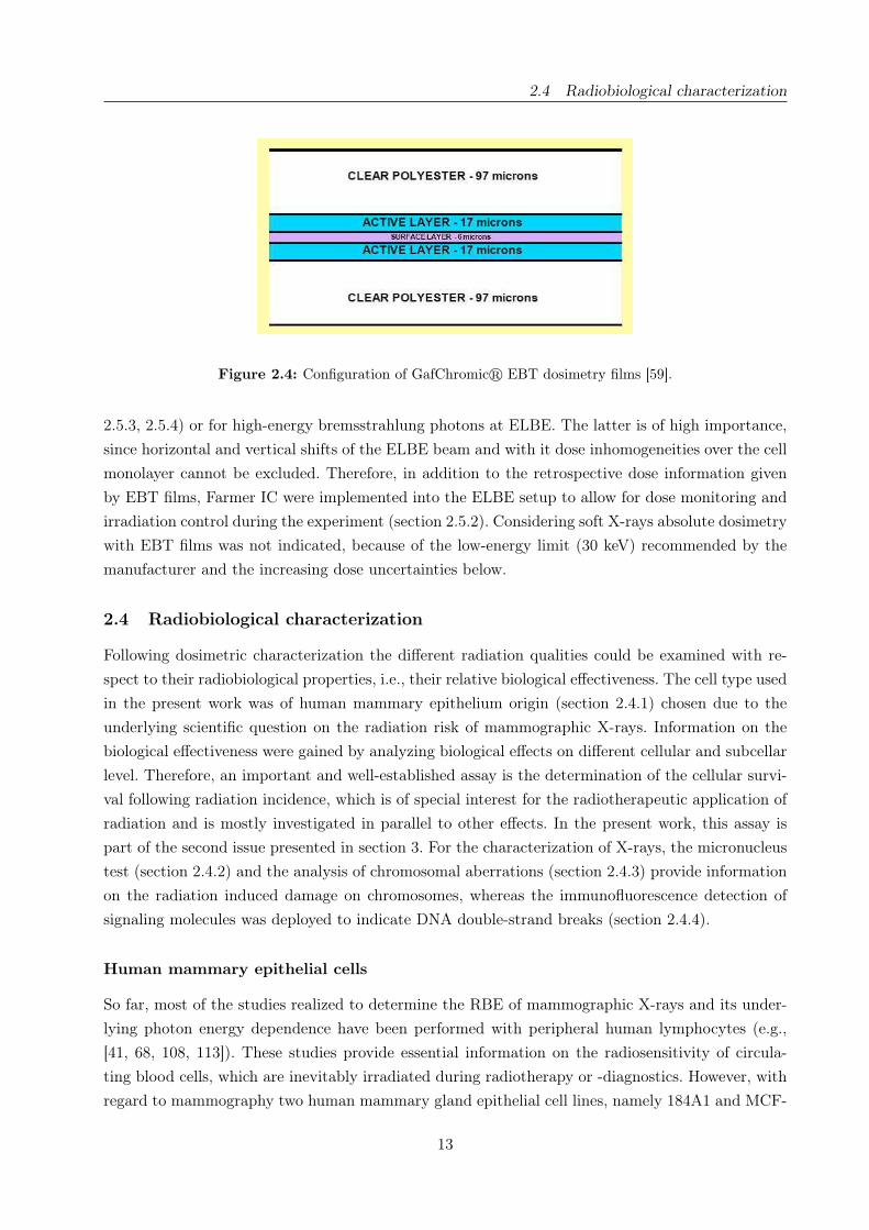
2.4 Configuration of GafChromicr EBT dosimetry films . . . . . . . . . . . . . . . . . . 13
2.5 Formation of micronuclei . . . . . . . . . . . . . . . . . . . . . . . . . . . . . . . . . . 14
2.6 Cell cycle of eukaryotic cells . . . . . . . . . . . . . . . . . . . . . . . . . . . . . . . . 15
2.7 Formation of dicentric chromosomes . . . . . . . . . . . . . . . . . . . . . . . . . . . 16
2.8 Formation of centric rings . . . . . . . . . . . . . . . . . . . . . . . . . . . . . . . . . 17
2.9 Chromosomal deletions: formation and classification . . . . . . . . . . . . . . . . . . 17
2.10 Harlekin chromosomes observed in 184A1 . . . . . . . . . . . . . . . . . . . . . . . . 18
2.11 Organization of eukaryotic chromosomes . . . . . . . . . . . . . . . . . . . . . . . . . 19
2.12 Cellular signaling cascade following DSB incidence . . . . . . . . . . . . . . . . . . . 20
2.13 Glow curves of the tested TSEE detectors . . . . . . . . . . . . . . . . . . . . . . . . 28
2.14 Time of irradiation response of TSEE detector type I, system I . . . . . . . . . . . . 29
2.15 Saturation and dose response of TSEE detector type I, system I . . . . . . . . . . . . 30
2.16 Fading effect for TSEE detector type IV . . . . . . . . . . . . . . . . . . . . . . . . . 31
2.17 Gas flow dependence of the TSEE system response . . . . . . . . . . . . . . . . . . . 32
2.18 Schematic drawing of the radiation physics cave at ELBE . . . . . . . . . . . . . . . 41
2.19 Cell irradiation system at ELBE . . . . . . . . . . . . . . . . . . . . . . . . . . . . . 42
2.20 Dose beam profile and the meander shaped track for dose homogeneity . . . . . . . . 43
2.21 Positioning of cell samples at the ELBE photon beam . . . . . . . . . . . . . . . . . 44
2.22 Calibration curve for GafChromicr EBT films . . . . . . . . . . . . . . . . . . . . . 45
2.23 Dose homogenization with 34 MV bremsstrahlung . . . . . . . . . . . . . . . . . . . . 47
2.24 Dose response curves for 34 MV bremsstrahlung and 200 kV X-rays . . . . . . . . . . 49
2.25 Energy dependent yields of excess fragments induced in 184A1 and MCF-12A . . . . 60
2.26 Dicentric chromosomes and centric rings in dependence on X-ray energy . . . . . . . 61
2.27 Co-localized foci of γ-H2AX and 53BP1 detected in cell line 184A1 . . . . . . . . . . 70
2.28 Time courses of co-localized foci after irradiation . . . . . . . . . . . . . . . . . . . . 72
2.29 X-ray energy dependent dose-effect curves for residual co-localized foci . . . . . . . . 74
3.1 Depth dose profiles of different charged particles . . . . . . . . . . . . . . . . . . . . . 80
3.2 Laser wake field acceleration of electrons . . . . . . . . . . . . . . . . . . . . . . . . . 82
3.3 Schematic illustration of the colony formation assay . . . . . . . . . . . . . . . . . . . 85
3.4 Schematic drawing of the experimental setup at JETI . . . . . . . . . . . . . . . . . 96
3.5 Exemplary dose distribution over the beam diameter and the cell area . . . . . . . . 97
3.6 Comparison of the actual doses and number of laser pulses applied . . . . . . . . . . 98
3.7 Online parameters vs. absolute doses for JETI electrons . . . . . . . . . . . . . . . . 99
3.8 Online vs. absolute dose delivered by laser-accelerated electrons . . . . . . . . . . . . 107
4.1 Illustration of the LET and its resultant consequences on DNA level . . . . . . . . . 112
v
List of Tables
List of tables
2.1 Sensitivity of the studied TSEE detectors . . . . . . . . . . . . . . . . . . . . . . . . 31
2.2 Reproducibility of the TSEE detectors compared to a reference . . . . . . . . . . . . 33
2.3 Influence of the irradiation medium on TSEE detectors response . . . . . . . . . . . 34
2.4 Effective dose rate and dose homogeneity in the region of interest . . . . . . . . . . . 48
2.5 Yield of chromosomal aberrations observed in cell line 184A1 . . . . . . . . . . . . . 58
2.6 Yield of chromosomal aberrations observed in cell line MCF-12A . . . . . . . . . . . 59
2.7 Regression coefficients and low dose RBE for excess fragments . . . . . . . . . . . . . 62
2.8 Dose response of dicentric chromosomes and centric rings . . . . . . . . . . . . . . . 63
2.9 Parameters of the photon spectra transmitted through different slide materials . . . 71
2.10 Residual co-localized foci in dependence on dose and time post irradiation . . . . . . 73
2.11 Fit parameters of the 24 hours dose response of residual co-localized foci . . . . . . . 74
2.12 RBE determined on basis of the 24 hour dose responses . . . . . . . . . . . . . . . . 75
3.1 Beam parameters of conventional and laser based particle accelerators . . . . . . . . 81
3.2 Actual parameters of the laser system and the electron beam at cell position . . . . . 97
4.1 RBE values determined for the various biological effects and X-ray energies . . . . . 109
vi
Abstract
More than hundred years after the discovery of X-rays different kinds of ionizing radiation are ubi-
quitous in medicine, applied to clinical diagnostics and cancer treatment as well. Irrespective of their
nature, the widespread application of radiation implies its precise dosimetric characterization and
detailed knowledge of the radiobiological effects induced in cancerous and normal tissue. Starting
with in vitro cell irradiation experiments, which define basic parameters for the subsequent tissue
and animal studies, the whole multi-stage process is completed by clinical trials that translate the
results of fundamental research into clinical application. In this context, the present dissertation
focuses on the establishment of radiobiological in vitro cell experiments at unconventional, but cli-
nical relevant radiation qualities.
In the first part of the present work the energy dependent biological effectiveness of photons was stu-
died examining low-energy X-rays (≤ 50 keV), as used for mammography, and high-energy photons
(≥ 20 MeV) as proposed for future radiotherapy. Cell irradiation experiments have been performed
at conventional X-ray tubes providing low-energy photons and 200 kV reference radiation as well.
In parallel, unconventional quasi-monochromatic channeling X-rays and high-energy bremsstrah-
lung available at the radiation source ELBE of the Forschungszentrum Dresden-Rossendorf were
considered for radiobiological experimentation. For their precise dosimetric characterization dosi-
meters based on the thermally stimulated emission of exoelectrons and on radiochromic films were
evaluated, whereas just the latter was found to be suitable for the determination of absolute doses
and spatial dose distributions at cell position. Standard ionization chambers were deployed for the
online control of cell irradiation experiments. Radiobiological effects were analyzed in human mam-
mary epithelial cells on different subcellular levels revealing an increasing amount of damage for
decreasing photon energy. For this reason, the assumed photon energy dependence was reconfirmed
for a cell line other than human lymphocytes, an important finding that was discussed on the 2007
Retreat of the German Commission on Radiological Protection.
After successful finalization of the photon experiments the focus of the present dissertation was
directed to the realization of in vitro cell irradiation experiments with laser-accelerated electrons.
This research was carried out in the frame of the project onCOOPtics that aims on the development
of laser-based particle accelerators, which promise accelerators of potentially compact size and more
cost-effectiveness suitable for a widespread medical application, especially for high precision hadron
therapy. The unique properties, i.e., the ultrashort bunch length and resultant ultrahigh pulse dose
rate, of these unconventional particle accelerators demand for extensive investigations with respect
to potential effects on the dosimetric and radiobiological characterization. Based on the experiences
gained at ELBE first experiments on the radiobiological characterization of laser-accelerated elec-
trons have been performed at the Jena Titanium:Sapphire laser system. After beam optimization, a
sophisticated dosimetry system was established that allow for the online control of the beam para-
meters and for the controlled delivery of dose to the cell sample. Finally, worldwide first systematic
in vitro cell irradiation experiments were carried out resulting in a reduced biological effectiveness
for laser-accelerated electrons relative to the 200 kV X-ray reference, irrespectively on the biological
effect and cell lines examined. These successful results are the basis for future in vivo studies and
experiments with laser-accelerated protons.
vii


1 Introduction
“On a new kind of rays“ - the famous report published in 1896 by Wilhelm Conrad Röntgen does not
just announce the discovery of the “X-radiation“, but also introduce the first medical application by
showing a radiography of his wife’s hand on a photographic plate [103]. More than one hundred years
later X-rays and other kinds of ionizing radiation are omnipresent in medicine, used for diagnostic
purposes and radiotherapeutic treatment as well. However, improvements and further technological
developments are still conceivable, for example by introducing new radiation qualities that promise
the reduction of radiation side-effects or the enhancement of the tumor control rate. Before a new
radiation quality can be applied in medicine their radiobiological effects on cancer and normal
tissue have to be investigated in detail. Starting with in vitro cell irradiation experiments, which
are followed by tissue and animal studies and completed by clinical trials that translate the results
of the fundamental research into medical application. Therefore, the radiation qualities of interest
have to met several interdependent requirements in order to allow for radiobiological studies:
• (i) the development, supply and adjustment of an adequate radiation source,
• (ii) their precise physical and dosimetric characterization and
• (iii) the determination of its radiobiological effectiveness by means of an appropriate biological
object and effect.
A characteristic feature of radiobiological studies is the examination of one radiation quality (X )
in comparison to a reference (R) in order to determine the Relative Biological Effectiveness (RBE),
which is defined as the inverse ratio of the absorbed doses (D) producing the same effect (E ) [57]:
(1) RBE =DR
DX|EX=ER
.
Main reasons for this approach are the elimination of influences arising from the diversity of biolo-
gical samples, fluctuations of the biological response and varying experimental, i.e., environmental,
conditions. On the other side, there exists a large amount of experimental and epidemiological data
about the radiogenic cancer risk of high-energy γ-radiation, which is recommended as reference by
the International Commission on Radiological Protection (ICRP) [57]. In consequence, photons in
this energy range, either provided by 60Co γ-emitter (1.17 and 1.33 MeV) or ordinary ∼= 200 kV
X-ray tubes, are used as reference radiation. Keeping in mind that 200 kV X-rays are twice as effec-
tive as high-energy γ-rays, it is essential for every declaration of the RBE to specify the appropriate
reference radiation source.
In practice, radiobiological studies are performed as two-armed studies investigating the same biolo-
gical effect for the examined and the reference radiation source in parallel. Hence, the requirements
(i-iii) have to be fulfilled for both radiation qualities. Moreover, the concept of RBE requires a
sufficient number of targets irradiated under similar conditions (e.g., beam properties, irradiation

Introduction
geometry, environmental conditions etc.) at both radiation qualities in order to overcome the vary-
ing radiosensitivity of biological objects. Whereas this requirement might not be the crucial factor
for in vitro cell studies, it may be a problem for animal experiments or clinical trials. Consequently,
in vitro studies are not only the first step for the establishment of a new radiation quality, but also
an important step for the definition of experimental conditions of the subsequent ones.
Taken into account these aspects, two issues, both dealing with the realization of in vitro cell irra-
diation experiments at conventional X-ray tubes and unconventional radiation sources of potential
medical interest, were studied in the present dissertation:
• Photon energy dependence of the relative biological effectiveness of X-rays.
• Establishment of in vitro cell irradiation experiments with laser-accelerated electrons.
The research work of the present dissertation was carried out in the context of the project “Radia-
tion induced cell damage“ at the Institute of Radiation Physics of the Forschungszentrum Dresden-
Rossendorf (FZD) in a close cooperation with the OncoRay - Center for Radiation Research in
Oncology. OncoRay is a joint institution of the FZD, the Dresden University of Technology and the
university hospital Carl Gustav Carus combining their physical, biological and medical knowhow
for radiation research in oncology, for example for tumor diagnostics and treatment.
The examination of the photon energy dependence of the RBE was a continuation of a research
project that was initiated several years ago aiming primarily on the investigation of the RBE of
low-energy X-rays. In the framework of the present dissertation the research on this particular topic
was continued, but extended with regard to the applied energy range and radiation sources, to
the selective improvement of the dosimetry and to the establishment and investigation of a second
human cell line as well as the analysis of further biological effects. The background, methods and
results associated with the realization of this multidisciplinary work are described in section 2.
After the successful finalization of the first issue, the research of the present dissertation was concen-
trated on a completely new scientific question: the development of laser-based ion accelerators for
radiotherapeutic application. For this reason, the joined project onCOOPtics was started in 2007
aiming on both the technological development of the new laser technology and on the physical and
biological characterization of the generated particle beams. The multidisciplinary research linked
with the project onCOOPtics is carried out by two research centers - OncoRay in Dresden, respon-
sible for the comprehensive characterization and future clinical implementation of this new radiation
quality, and ultra optics in Jena, responsible for the technological development of the laser system
and the auxiliary equipment. In this context, section 3 of the present dissertation comprises the
completion of the requirements (i-iii) mentioned above for the realization of radiobiological expe-
riments with laser-accelerated electrons. The results obtained for the investigation of the photon
energy dependence of the RBE of X-rays and during the establishment of in vitro cell irradiation
experiments with laser-accelerated electrons are discussed in section 4.
2
2 Photon energy dependence of the relative biological effectiveness
of X-rays
2.1 Background
In 2002 the German Parliament opted for the introduction of a nationwide mammography screening
program in Germany and started a continuing cost-benefit discussion on the general application of
mammography. At the same time, the group of Frankenberg et al. [32] published their controversial
study on the biological effectiveness of mammographic X-rays. Analyzing neoplastic transforma-
tions of CGL1 human hybrid cells as one type of DNA damage, this group found an RBE of about
4 comparing 29 kV X-rays and conventional 200 kV photons as reference. Taken into account the
implications for the radiation risk estimation, especially for mammography, this high RBE value
was discussed critically. Moreover, the whole experiment of Frankenberg et al. was repeated and
reevaluated [40, 45] in order to validate their finding. And indeed, values significantly higher than
one were confirmed for 29 kV X-rays compared to 220 kV X-rays [40], a 90Sr/90Y radioactive source
and a simulated atomic bomb spectrum [45], respectively. Furthermore, the analysis of several in
vitro studies [41, 68, 75, 86, 93, 108, 112, 113, 119] lead also to the conclusion that soft X-rays are
more effective per unit dose than higher energy photons. Contrary to these findings is the photon
energy independent radiation weighting factor (wR) of 1 as specified by the ICRP [56], which presu-
mes that photons of all energies will cause similar radiation effects in the considered tissue or organ.
Systematic investigations on the photon energy dependence of the RBE were performed so far by
analyzing chromosomal aberrations induced in human lymphocytes by monochromatic synchrotron
X-rays in the energy range of 1.83 - 40 keV [41, 68, 108, 112]. The obtained chromosomal aberrations
were considered relative to those induced by 200 kV X-rays or MeV photons as reference revealing
RBE values inversely proportional to the photon energy, i.e., increasing for decreasing energy [48].
The maximum RBE was found at 6.9 keV, whereas the further reduction of the photon energy
results in a minimum at 2.1 keV [108] and a significant rise for energies below [48]. The most likely
explanations for this finding are the range and local energy deposition of the generated secondary
electrons in irradiated matter (section 4).
In the framework of the ongoing discussion on the radiation risk of mammographic X-rays a close
cooperation between the research project “Radiation induced cell damage“ at the Institute and
Division of Radiation Physics at the Forschungszentrum Dresden-Rossendorf (FZD) and the On-
coRay group “Radiobiology of normal tissues and radiation protection“ of the Medical Faculty Carl
Gustav Carus at the Dresden University of Technology was initiated. Combining the physical and
dosimetric knowhow of the FZD project with the radiobiological expertise of the hospital group,
the primary aim of this cooperation was the investigation of the relative biological effectiveness of
low-energy X-rays utilizing the soft X-ray (10 and 25 kV) as well as the 200 kV X-ray reference
tube of the Medical Faculty. At first, the radiation effects of 25 kV X-rays were investigated in

2.1 Background
human fibroblasts and human keratinocytes showing a slightly enhanced RBE relative to 200 kV
X-rays. After the successful implementation of a precise dosimetry for the challenging ultra-soft
X-rays radiobiological studies have been performed with 10 kV X-rays. Although this X-ray energy
is not relevant for clinical purposes, due to their strong attenuation in air and even more in tis-
sue, 10 kV X-rays were chosen for systematic reasons and due to their mean energy of ≈ 7 keV
close to the energy of the maximum RBE. Furthermore, the human mammary breast epithelial
cell line MCF-12A was established in accordance to the underlying scientific question. Based on
these requirements radiobiological studies aiming on the determination of the relative biological
effectiveness for 10 kV and 25 kV X-rays relative to 200 kV X-rays were performed analyzing the
cellular survival and the induction of micronuclei in MCF-12A [74, 75]. Parallel to the investigation
of low-energy X-rays the project “Radiation induced cell damage“ focused also on the feasibility of
radiobiological in vitro experiments at the radiation source ELBE (Electron Linac for beams with
high Brilliance and low Emittance) of the FZD (section 2.2.1). ELBE delivers electron beams of up
to 40 MeV, which can be deployed to generate secondary radiation of radiobiological interest, like
neutrons, high-energy bremsstrahlung and quasi-monochromatic channeling X-rays [34]. Mainly the
development of the latter was of special interest for radiobiological experimentation, since tunable
quasi-monochromatic X-rays in the energy range of 10 - 100 keV [3, 73] will allow for a detailed and
systematic investigation of the relative biological effectiveness of X-rays.
Within the scope of the present dissertation, the investigation of the relative biological effectiveness
of low-energy X-rays with conventional X-ray tubes was pursued but extended by the establish-
ment of a second human mammary epithelial cell line (section 2.4.1). Furthermore, two additional
radiation effects, chromosomal aberrations (sections 2.4.3, 2.5.3) and DNA double-strand breaks
(DSB) (sections 2.4.4, 2.5.4), were analyzed providing information on the radiation damage on sub-
cellular level. In addition to these radiobiological experiments the technological development of the
channeling X-ray source (section 2.2.3) was also advanced. Moreover, selective improvements of the
challenging dosimetry of soft X-rays have been achieved by evaluating different types of dosimeters
for the precise determination of the absolute dose administered to a cell monolayer: the detection
of thermally stimulated exoelectron emission (TSEE) (sections 2.3.3, 2.5.1) and the application of
radiochromic EBT films (sections 2.3.4, 2.5.2). Beside low-energy X-rays, the research on the photon
energy dependence of the RBE of X-rays was also focused on high-energy photons of up to 34 MV.
Photons of this and even higher energies (≈ 50 MV) are better focusable to narrow pencil beams of
a few centimeter in diameter [123] allowing for the idea of a scanned photon beam, which may be
of benefit for high-precision radiotherapy. In the present work the ELBE electron beam was used to
generate high-energy photons of up to 34 MV (section 2.2.2) by means of bremsstrahlung produc-
tion. Optimized with respect to the physical and dosimetric properties required for radiobiological
experiments, e.g., sufficient beam spot size and adequate dose rate, high-energy photons of 20 MV
and 34 MV were deployed later on for radiobiological in vitro cell irradiation experiments analyzing
the induction of micronuclei (section 2.4.2, 2.5.2).
The following sections outline the basic principles and background information required to under-
4

2.2 Sources of X-radiation
stand but not mentioned in the publications (section 2.5) that comprises the experimental studies
performed in the context of the present dissertation. According to the consecutive requirements for
radiobiological experimentation given above section 2.2 includes the different radiation sources, 2.3
their dosimetric and 2.4 their biological characterization. Finally, in section 2.5, the realization and
the results of the different studies are presented in the corresponding publications.
2.2 Sources of X-radiation
In dependence on the underlying scientific question three different radiation sources were applied
in the present work to study the relative biological effectiveness of X-rays in dependence on photon
energy. Irradiations with 10 kV and 25 kV soft X-rays as well as with the 200 kV X-ray reference were
performed with conventional X-ray tubes provided at the “Klinik und Poliklinik für Strahlentherapie
und Radioonkologie“ of the university hospital Carl Gustav Carus. By contrast, the generation of
unconventional high-energy bremsstrahlung and quasi-monochromatic channeling X-rays demand
for high-energy electrons available at the radiation source ELBE.
Radiation source ELBE
The radiation source ELBE of the Forschungszentrum Dresden-Rossendorf [30, 34] provides mo-
noenergetic continuous wave electron beams with energies up to 40 MeV and average currents up to
1 mA at a micropulse repetition rate of 13 MHz [30]. The beam may be applied either directly for
radiation experiments or for the production of secondary radiation such as bremsstrahlung (conti-
nuous photon spectrum), quasi-monochromatic channeling X-rays, neutrons or positrons [30]. The
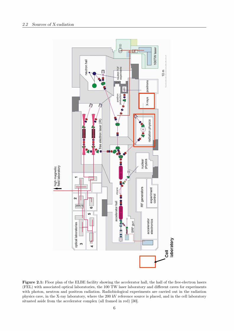
floor plan of the ELBE accelerator hall with associated experimental rooms is shown in Figure 2.1.
Concerning radiobiological experiments, two types of secondary radiation, both are produced in the
radiation physics cave of ELBE (Layout, cf. section 2.5.2), have to be considered:
• High-energy bremsstrahlung where the available photon energies extend from the keV region
up to the electron beam energy.
• Quasi-monochromatic X-rays in form of channeling radiation which offers the possibility for
more detailed investigations of the photon energy dependence of the RBE.
Generation of bremsstrahlung
Generally, bremsstrahlung is produced by focusing accelerated electrons on a (usually metallic) tar-
get. During the stopping process, the incoming electrons successively lose small portions of their
kinetic energy due to ionization of the medium or large portions of energy due to inelastic scatte-
ring from target atoms what results in the emission of bremsstrahlung photons. Ionization losses are
converted to heat (99 %), while bremsstrahlung photons of sufficient energy may partially escape
the target and form a forward directed cone of polychromatic radiation. Whereas the energy of a
single bremsstrahlung photon depends on the momentum transfer to the target atoms, i.e., on the
interaction distance between electron and atomic nucleus (Fig. 2.2a).
5
2.2 Sources of X-radiation
Figure 2.1: Floor plan of the ELBE facility showing the accelerator hall, the hall of the free-electron lasers(FEL) with associated optical laboratories, the 100 TW laser laboratory and different caves for experimentswith photon, neutron and positron radiation. Radiobiological experiments are carried out in the radiationphysics cave, in the X-ray laboratory, where the 200 kV reference source is placed, and in the cell laboratorysituated aside from the accelerator complex (all framed in red) [30].
6

2.2 Sources of X-radiation
Collisional ionization of medium atoms due to expelling of electrons from inner atomic shells leads,
additionally to bremsstrahlung, to the emission of characteristic X-rays because the vacancies are
filled up by electron transitions from outer atomic shells to inner ones. The excess of binding energy
is radiated into full solid angle in form of characteristic X-rays (Fig. 2.2a). Therefore, the continuous
bremsstrahlung spectrum is superimposed by characteristic X-ray lines of the target material.
Figure 2.2: a) Generation of bremsstrahlung and characteristic X-rays by interaction of the incomingelectrons and the target material atoms. b) Production of quasi-monochromatic X-rays by electron channelingthrough a diamond crystal.
Comparing the bremsstrahlung production in conventional X-ray tubes and at ELBE differences
occur just in the target material, the filtration and the electron energy applied. For example, tungs-
ten targets are used in the X-ray tubes and a thin aluminum foil was deployed at ELBE to generate
bremsstrahlung, respectively. Furthermore, the production of bremsstrahlung at ELBE was accom-
panied by the transmission of high-energy electrons (≤ 34 MeV) through the aluminum target.
Although separated from the photon beam by means of a deflecting dipole magnet and absorption
in the beam dump, these electrons might contribute to radiation background, e.g., by bremsstrah-
lung production in the beam line walls. In contrast, electrons in the keV range are stopped in the
thick target material of an X-ray tube. The realization and results of the experiments with high-
energy bremsstrahlung at ELBE are presented in section 2.5.2, detailed information on the X-ray
tubes are given in sections 2.5.3 and 2.5.4 together with the associated radiobiological findings.
Quasi-monochromatic channeling X-rays
Channeling radiation (CR) is generated by relativistic charged particles at traveling through a
single crystal along a periodic structure, i.e., a crystallographic axis or plane. At ELBE, relati-
vistic electrons transmit a diamond single crystal of thickness 40 - 200 µm along an appropriate
crystallographic plane, e.g., the (110) plane (Fig. 2.2b). At sufficiently small entrance angles of the
beam electrons with respect to the crystallographic planes considered, they are trapped into the
attractive average planar potential and forced to an oscillatory motion along this plane through the
crystal. During this so-called channeling process, which is similar to an accelerated sine-like motion,
the trapped beam electrons emit CR photons. Since the velocity of the electrons is approximately
7
2.3 Dosimetric characterization
that of light, the energy of CR is shifted according to the Lorentz transformation into the X-ray
domain, and the rather intense quasi-monochromatic photon flux is also directed into a forward
cone. Furthermore, the photon energy of CR can easily be tuned within an energy range of about
10 - 100 keV by only changing the energy of the electron beam (range: ≈ 8 - 40 MeV).
For medium electron energies available at ELBE the description of the channeling process given
above is not complete to deduce all characteristic features of CR, because their quantum nature
has been neglected so far. In reality, the mentioned transverse oscillations are only allowed at given
frequencies. This means that the trapped electrons occupy only discrete bound channeling states.
Consequently, the emission of quasi-monochromatic CR lines has to be understood as resulting from
transitions between these bound states. At defined beam properties, even a selective occupation of
energy levels is feasible, which leads to an increase of the intensity of dedicated CR lines [3, 129].
The present work aims at the development and adjustment of the CR source to facilitate in vitro
cell irradiations with monochromatic instead of polychromatic X-rays in order to investigate the
dependence of the RBE on the photon energy in detail (section 2.5.2). Such experiments might of
course also be performed at synchrotron radiation facilities, e.g., at the ESRF (European Synchro-
tron Radiation Facility) in Grenoble or at BESSY (Berliner Elektronen-Speicherring Gesellschaft
für Synchrotronstrahlung) in Berlin. The compact size and availability at reasonable costs promise,
however, several advantages of CR sources compared with large synchrotrons. The complete equip-
ment required for a CR source, an electron injector, an acceleration cavity and a goniometer chamber
for crystal positioning and adjustment, may fit into a 10 - 15 m long laboratory, whilst an electron
storage ring, for example at the ESRF, has a circumference of 844 m.
2.3 Dosimetric characterization
According to the requirements (i-iii) listed above for radiobiological experimentation the next step
includes the precise physical and dosimetric characterization of the different radiation qualities.
In the context of the present dissertation, the term dosimetry includes the very precise determi-
nation of the absolute dose delivered to a few micrometer thick adherent cell monolayer inside a cell
culture vessel. Otherwise, the experiment conditions at the radiation source ELBE (section 2.3.1)
demand for an online dose monitoring and the measurement of the spatial dose distribution over
the cell covered area. Whereas the former allows for the control of the irradiation, i.e. the delivery
of prescribed doses, the latter is necessary to reveal potential dose deviations. Moreover, the high
attenuation of soft X-rays through the cell culture vessel bottom has also to be considered.
Three types of dosimeters were evaluated for the different dosimetric requirements in the present
work. Standard ionization chambers (IC) (section 2.3.2) offer the unique feature of an online dose
readout and are calibrated for absolute photon dosimetry. However, IC provide the integrated dose
over a sensitive volume that usually exceed the volume of a cell. In consequence, ionization chambers
were deployed in the present work for the online control of the ELBE experiments, measuring the
direct beam exposure as well as the radiation background in the radiation physics cave. An excep-
tion was the precise dosimetric characterization of soft X-rays, where the soft X-ray chamber was
used to determine the absolute dose at cell position under consideration of the varying cell sample
8
2.3 Dosimetric characterization
geometries (sections 2.5.3 and 2.5.4). In addition, TSEE detectors and radiochromic films were con-
sidered for absolute dosimetry at ELBE and for the measurement of spatial dose distributions in
different radiation fields. TSEE detectors offer the main advantage of a just few nanometer thick
sensitive volume at the surface enabling the sampling of spatial dose distributions not only laterally
but also as depth dose distributions within the ≈ 10 µm thick cell monolayer (section 2.3.3). Fur-
thermore, the TSEE detectors allow for the measurement of absolute doses at interfaces and of dose
gradients, especially for ultra-soft X-rays after transmission through the cell culture vessel bottom.
The radiochromic films (section 2.3.4) in turn are easy to handle and available in flexible and large
sizes, suitable for the measurement of spatial dose distributions of extended and variable beam
spots. Both, the TSEE detectors as well as the radiochromic films are feasible for the retrospective
determination of absolute doses administered to a cell monolayer.
Terms and conditions for in vitro cell irradiations
The precise dosimetric characterization of the varying radiation qualities used in the present work
demands for the consideration of their specific physical and technical properties, like photon beam
attenuation and intensity variations of the ELBE beam, respectively.
Regarding the soft X-rays, the beam attenuation in different materials play an important role for the
determination of absolute doses administered to a cell monolayer adherent on a cell culture vessel
bottom. That means, especially for 10 kV X-rays, that both, the influence of the vessel bottom as
well as dose rate deviations arising from small variations of the bottom thickness were taken into
account [73]. For all X-ray energies GEANT4 simulations [35] and dosimetric measurements were
performed in parallel in order to understand the influence of different materials, namely glass and
plastic, on the photon spectra (cf. section 2.5.4). Moreover, the cell samples were irradiated upside
down at the vertical beam (from above) to minimize the material in and the influence on the X-ray
spectra (sections 2.5.3, 2.5.4).
At ELBE, the photon beam attenuation might also be a challenge for the experimentation with
quasi-monochromatic channeling X-rays, whereas for high-energy bremsstrahlung photons this in-
fluence can be neglected. However, some practical limitations for radiobiological experiments at
ELBE arise from the radiation protection directives and the reproducibility of the beam parame-
ters. Concerning the first, the directives demand for a waiting period of 20 minutes after switching
off the beam before entering the radiation physics cave and exchanging the cell samples. In conse-
quence, the irradiation procedure as practiced at conventional X-ray tubes, where cell samples that
were currently not irradiated are stored outside the experimental room and exchanged individually,
was not reasonable. To overcome this limitation a cell irradiation system (see section 2.5.2) was con-
structed that allows the storage and remote-controlled irradiation of up to 27 cell samples of different
sample geometry. The second limitation for radiobiological experimentation at ELBE is related to
the intensity of the ELBE electron beam that might shift during and between the experiments and
cause intensity or dose rate variations of the secondary radiation (CR and bremsstrahlung). By con-
trast, photon beams with stable and reliable properties are delivered at conventional X-ray tubes
allowing for the precise delivery of prescribed doses to the cell target by control of the irradiation
9
2.3 Dosimetric characterization
time. In order to compensate for potential dose rate variations at ELBE, the dose delivery has to be
monitored during irradiation and the administered doses have to be determined for each cell sam-
ple individually. The first requirement was fulfilled with the help of a Farmer ionization chamber
(section 2.3.2), that was positioned on beam axis behind the cell samples providing an online dose
signal relative to the absolute dose delivered to the cells (section 2.5.2). However, since the Farmer
IC is not applicable for the determination of absolute doses at cell position (see above), a second
dosimeter has to be implemented in order to fulfill this necessary requirement for dose-effect cur-
ves and RBE determinations. Therefore, two dosimeters based either on the thermally stimulated
emission of exoelectrons (section 2.3.3) or on the coloring of radiochromic films (section 2.3.4) were
evaluated for the measurement of absolute doses under the given experimental conditions.
Another challenge at ELBE is the varying and sometimes quite high radiation background in the
radiation physics cave. Main reasons are beam loss during tuning and optimization, bremsstrahlung
necessarily produced whenever an electron interacts with matter and tertiary radiation produced
by photo nuclear reactions, e.g., (γ,n), (γ,p) of higher energy photons with matter. During the cell
irradiation, the background was monitored with a Farmer IC and a neutron dosimeter positioned
next to the cell samples (section 2.5.2). Supplementary cell samples (controls) were prepared, but
not irradiated, in order to determine the impact of the radiation background, the ambient conditions
and the whole procedure on the cell and the examined biological effect.
Ionization chambers
Ionization chambers are in principle two electrodes of opposite polarity, which surround an ionizable
medium, mostly gas. Subsequent to radiation incidence the gas molecules are ionized and the ge-
nerated charged particles (ions and electrons) move to the appropriate electrodes and an ionization
charge proportional to the exposed radiation dose is measurable. The detection sensitivity and wor-
king energy range of an IC depend substantially on the applied gas (e.g., He, H2, N2, O2, air) and
its pressure, on the size of the sensitive volume and the sensitivity of the electronic. Furthermore,
the chambers have to be constructed in such a way that either the secondary electron equilibrium
or the Bragg-Gray principle is fulfilled. The former presumes that the same number of electrons
with identical energy distribution enter and exit the sensitive volume. The latter, most frequently
applied for higher energy particles, demand for ionization chambers being small enough to avoid
influences on the secondary particle flux and its energy distribution.
Generally, the various types of ionization chambers are widely-used as standard dosimeters for me-
dical purposes providing absolute doses and the possibility of dose monitoring during radiotherapy.
In the present work, three different types of IC were applied for the dosimetric characterization of
differing radiation qualities. A soft X-ray IC with a small sensitive volume of 0.02 cm3, a very thin
flat beryllium entrance window and an applicable photon energy range of 10 to 100 kV was applied
for 25 kV and 10 kV X-ray dosimetry. Whereas the 200 kV reference radiation and bremsstrahlung
photons of higher energy were characterized by a semi flex (sensitive volume: 0.3 cm3, photon energy
range: 30 kV - 50 MV) and a Farmer ionization chamber (sensitive volume: 0.6 cm3, photon energy
range: 30 kV - 50 MV). Corrected for ambient air temperature and pressure as well as the different
10

2.3 Dosimetric characterization
cell sample and experiment geometries, the IC provide the absolute dose or dose rate in water or
air. However, the integration over a sensitive volume that usually exceeds the size of a cell provides
rather a relative dose signal than the measurement of the absolute dose administered at cell po-
sition. The practical implementation of the different types of IC in the framework of the present
dissertation as well as the necessary corrections due to various sample and experiment geometries
are described in detail in sections 2.5.2 to 2.5.4.
Thermally stimulated exoelectron emission dosimeters
The method of exoelectron emission dosimetry is based upon the electronic band structure of semi-
conductors and the dose dependent emission of electrons from the near surface region. Concerning
a solid with lattice structure, the potentials of the periodical arranged atoms and of the quasi-free
electrons form spatio-periodical potentials whose energy differences are so small, that continuous
bands arise. Those bands emerging from overlapping orbitals of the bonded valence electrons are
abstracted to the valence band (VB); whereas the conduction band (CB) comprise those states with
free electron movement and conduction. Both bands are separated by the band gap, which is in
principle not allowed for electrons. The width of the band gap and the occupation of the two bands
classify a solid material as isolator, semiconductor or electric conductor.
Figure 2.3: Processes that result in the emission of exoelectrons: VB electrons were excited by radiationincidence and trapped into deep acceptor levels (1) either directly from the VB or from an intermediate CBlevel. Subsequently, thermal stimulation that exceed the activation energy EA lead to electron release fromthe traps (2) and increase the electron conduction (3) in the CB. Some of these electrons overcome the workfunction (ϕ) and were emitted as exoelectrons from the crystal surface (4).
Concentrating on semiconductors, their band gap, i.e., the gap energy, is low enough that electrons
from the VB can enter the CB for example by thermal excitation. As result, the excess electrons
in the CB will contribute to intrinsic electron (n-) conduction, whereas the remaining holes in the
VB cause hole or p-type conduction. Both types of conduction can be amplified by appropriate
doping of the semiconductor material. Additional electron acceptor or donor levels in the band gap
will increase the conductivity of the semiconductor. For the emission of exoelectrons, a necessary
requirement is the existence of deep electron acceptor levels positioned in the band gap near the
11
2.3 Dosimetric characterization
lowest energy level of the CB (Fig. 2.3).
Electrons excited from the valence to the conduction band, e.g., by means of ionizing radiation,
are trapped into these deep acceptors with a certain probability. The further supply of energy by
thermal or optical stimulation will result in the release of trapped electrons, i.e., stored energy,
back into the CB. Here, they contribute to electron conduction, but they also have a certain tem-
perature dependent probability to overcome the electron work function (ϕ) and to escape from the
crystal surface [50]. As the temperature is increased the density of the released electrons changes
proportional to the distribution of electron traps in the band gap. Flat traps are exhausted by lower
temperatures than deeper ones, whereas optical stimulation will simply release the electrons from
flat traps.
In the present work a Geiger-Müller counter was utilized to detect the released electrons and glow
curves, i.e., the electron number in dependence on temperature, determined for five types of be-
ryllium oxide TSEE detectors (section 2.5.1). These detectors promises several advantages for the
dosimetry of in vitro cell irradiation experiments. Since the sensitive volume is at the detector sur-
face and just a few nanometers thick TSEE detectors rather provide the spatial dose distribution at
cell position than other dosimeters with sensitive volumes on a micrometer to millimeter scale, like
IC. Moreover, TSEE detectors are suitable for the measurement of dose gradients, e.g., caused by
the strong beam attenuation of ultra-soft X-rays in the cell monolayer, and for dose determination
at interfaces. For these reasons, different types of TSEE detectors were evaluated with respect to
their basic physical properties, like dose and time response and saturation, and to their applicability
for in vitro cell irradiation experiments (section 2.5.1).
Radiochromic EBT films
The third type of dosimeter established and tested for the dosimetric characterization of the various
radiation qualities examined in the present work are GafChromicr EBT films, which were developed
for radiotherapy. The dose determination with radiochromic films is based on a polymerization
process in the active film layer (Fig. 2.4) following radiation incidence. This polymerization results
in a color change, from light to dark blue in the case of EBT films, the darker the higher the
absorbed radiation dose. Irradiated films as well as the unirradiated controls are digitized (cf.
section 2.5.2) and the shade of blue is converted to dose using a predetermined calibration curve.
The scanning procedure, the calibration and the software analysis were adopted for our requirements
from previously published protocols (e.g., [21]), that is, the calibration was performed with reference
radiation sources appropriate for our interests (sections 2.5.2, 3.4.1).
EBT films are self-developing after irradiation and easy to cut to any size and shape required,
a feature that enables the substitution of the cell monolayer in the cell culture vessel in order
to determine the dose delivered to the cells after transmission, for example of the ELBE photon
beam, through the plastic bottom. Moreover, as stated by the manufacturer, EBT films are energy
independent in the range of 30 keV to several MeV for electrons and photons and applicable for
doses of up to 8 Gy [59]. Beside absolute dosimetry, EBT films were also deployed to monitor the
dose homogeneity over different radiation fields, for example at conventional X-ray tubes (sections
12
2.4 Radiobiological characterization
Figure 2.4: Configuration of GafChromicr EBT dosimetry films [59].
2.5.3, 2.5.4) or for high-energy bremsstrahlung photons at ELBE. The latter is of high importance,
since horizontal and vertical shifts of the ELBE beam and with it dose inhomogeneities over the cell
monolayer cannot be excluded. Therefore, in addition to the retrospective dose information given
by EBT films, Farmer IC were implemented into the ELBE setup to allow for dose monitoring and
irradiation control during the experiment (section 2.5.2). Considering soft X-rays absolute dosimetry
with EBT films was not indicated, because of the low-energy limit (30 keV) recommended by the
manufacturer and the increasing dose uncertainties below.
2.4 Radiobiological characterization
Following dosimetric characterization the different radiation qualities could be examined with re-
spect to their radiobiological properties, i.e., their relative biological effectiveness. The cell type used
in the present work was of human mammary epithelium origin (section 2.4.1) chosen due to the
underlying scientific question on the radiation risk of mammographic X-rays. Information on the
biological effectiveness were gained by analyzing biological effects on different cellular and subcellar
level. Therefore, an important and well-established assay is the determination of the cellular survi-
val following radiation incidence, which is of special interest for the radiotherapeutic application of
radiation and is mostly investigated in parallel to other effects. In the present work, this assay is
part of the second issue presented in section 3. For the characterization of X-rays, the micronucleus
test (section 2.4.2) and the analysis of chromosomal aberrations (section 2.4.3) provide information
on the radiation induced damage on chromosomes, whereas the immunofluorescence detection of
signaling molecules was deployed to indicate DNA double-strand breaks (section 2.4.4).
Human mammary epithelial cells
So far, most of the studies realized to determine the RBE of mammographic X-rays and its under-
lying photon energy dependence have been performed with peripheral human lymphocytes (e.g.,
[41, 68, 108, 113]). These studies provide essential information on the radiosensitivity of circula-
ting blood cells, which are inevitably irradiated during radiotherapy or -diagnostics. However, with
regard to mammography two human mammary gland epithelial cell lines, namely 184A1 and MCF-
13

2.4 Radiobiological characterization
12A (section 2.5.3), have been chosen in the present work in order to investigate the biological
effects of low energy X-rays.
Micronucleus assay
For the first in vitro cell irradiation experiments at the high-energy photon beam at ELBE the
induction of micronuclei (MN) as an easy to handle and economic method was chosen to analyze the
biological effectiveness (section 2.5.2). Micronuclei (Fig. 2.5a) originate from two basic phenomena
- chromosome breakage, i.e., the formation of acentric fragments by means of ionizing radiation, or
the dysfunctioning of the mitotic apparatus, which results in the lagging of whole chromosomes or
chromatides during mitosis [92]. However, parts of the DNA are not incorporated in the daughter
cell nuclei and remain as micronuclei in the cytoplasm of undivided cells. For the analysis of MN,
the cells have to be blocked after the first nucleus division but before a subsequent cell division
(Fig. 2.5a) to prevent the exclusion of micronuclei [92]. Hence, binucleated cells (BNC) are formed
that contain the MN.
Figure 2.5: a) Process of micronuclei formation following ionizing radiation incidence. b) Binucleated cellof the human mammary epithelial cell line 184A1 exhibiting one micronucleus induced by 3 Gy irradiationwith 34 MV bremsstrahlung.
Regarding the ELBE experiments with high-energy bremsstrahlung, the cell preparation starts with
the plating one day before irradiation in order to assure cell adherence. Shortly before the experiment
the culture vessels were completely filled with medium and sealed with Parafilm to avoid medium
depletion as consequence of the upright positioning of the cell samples during the experiment. The
samples, each equipped with an EBT dosimetry film, were positioned in special designed sample
holders (section 2.5.2) and irradiated consecutively, at which some remain unirradiated to control
the influence arising from the radiation background and the ambient conditions. After irradiation,
the cells were treated in accordance to the protocol published by Fenech [77] and adopted for the
human mammary epithelial cells used in the present work. The cell medium was exchanged to one
containing 2 µg/ml cytochalasin B, that prevent cell but not nucleus division, and the cells were
14
2.4 Radiobiological characterization
incubated for 72 hours at normal culture conditions (section 2.5.3). Subsequently, they were washed
in 0.9 % NaCl, fixed with ice cold methanol and stained with 5 % Giemsa solution in water. One
exemplary BNC possessing one micronuclei is shown in Figure 2.5b.
The analysis of the MN was performed under a light microscope applying the identification and
scoring criteria defined by Fenech [77]. Briefly, a BNC has to possess two nuclei of approximately
equal size, with not more than six micronuclei, whereas fine nucleoplasmic bridges between and
a slight overlap or touching of MN are allowed. In addition, a micronucleus is defined, when its
diameter is between 1/16 and 1/3 of that of the cell nucleus, it is non-refractile and not linked to
the cell nucleus via a nucleoplasmic bridge. At least, the MN may overlap the boundaries of the cell
nucleus. Taken into account these definitions, the MN were analyzed in dependence on radiation
dose. The corresponding RBE value was obtained by comparing the dose response curves of 34 MV
bremsstrahlung and the 200 kV X-ray reference (section 2.5.2).
Determination of chromosomal aberrations
Irreparable damages and structural changes induced in the DNA, e.g., by ionizing radiation, might
cause a loss of replication capability and consequently cell death. However, since the radiation doses
delivered during mammography are too low to cause cell death, the investigation of inheritable DNA
damages is of capital importance. Mutations and a reduced genetic stability in daughter generations
are potential consequences. Therefore, the induction and heredity transmission of these damages
have to be quantified, especially for mammographic X-rays, in order to allow for risk estimation of
acute and late radiation effects. Such early radiation effects were determined in the present work by
analyzing chromosomal aberrations within the first cell cycle after irradiation (section 2.5.3). Chro-
mosomal aberrations (CA) are chromatin damages induced by ionizing radiation or other mutagens
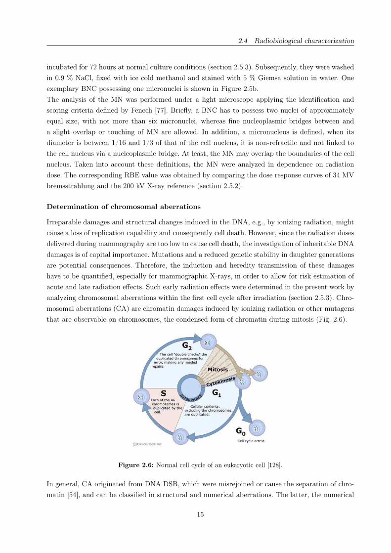
that are observable on chromosomes, the condensed form of chromatin during mitosis (Fig. 2.6).
Figure 2.6: Normal cell cycle of an eukaryotic cell [128].
In general, CA originated from DNA DSB, which were misrejoined or cause the separation of chro-
matin [54], and can be classified in structural and numerical aberrations. The latter, the numerical
15
2.4 Radiobiological characterization
aberrations or the deviation from the normal chromosome number, were not analyzed in detail in the
present work but were used to exclude cells with an abnormal chromosome set. Structural CA are
distinguished in chromosome- and chromatid-type aberrations according to the number of chroma-
tids affected. Chromosome-type aberrations are subdivided into deletions and exchange aberrations,
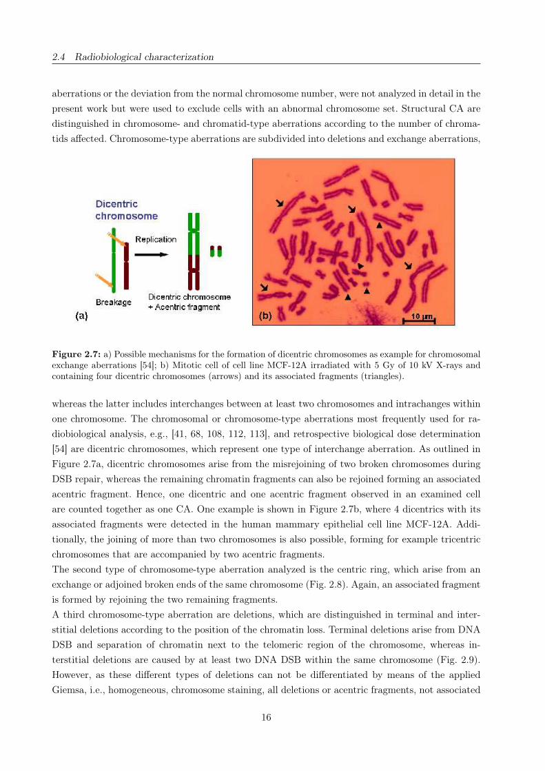
Figure 2.7: a) Possible mechanisms for the formation of dicentric chromosomes as example for chromosomalexchange aberrations [54]; b) Mitotic cell of cell line MCF-12A irradiated with 5 Gy of 10 kV X-rays andcontaining four dicentric chromosomes (arrows) and its associated fragments (triangles).
whereas the latter includes interchanges between at least two chromosomes and intrachanges within
one chromosome. The chromosomal or chromosome-type aberrations most frequently used for ra-
diobiological analysis, e.g., [41, 68, 108, 112, 113], and retrospective biological dose determination
[54] are dicentric chromosomes, which represent one type of interchange aberration. As outlined in
Figure 2.7a, dicentric chromosomes arise from the misrejoining of two broken chromosomes during
DSB repair, whereas the remaining chromatin fragments can also be rejoined forming an associated
acentric fragment. Hence, one dicentric and one acentric fragment observed in an examined cell
are counted together as one CA. One example is shown in Figure 2.7b, where 4 dicentrics with its
associated fragments were detected in the human mammary epithelial cell line MCF-12A. Addi-
tionally, the joining of more than two chromosomes is also possible, forming for example tricentric
chromosomes that are accompanied by two acentric fragments.
The second type of chromosome-type aberration analyzed is the centric ring, which arise from an
exchange or adjoined broken ends of the same chromosome (Fig. 2.8). Again, an associated fragment
is formed by rejoining the two remaining fragments.
A third chromosome-type aberration are deletions, which are distinguished in terminal and inter-
stitial deletions according to the position of the chromatin loss. Terminal deletions arise from DNA
DSB and separation of chromatin next to the telomeric region of the chromosome, whereas in-
terstitial deletions are caused by at least two DNA DSB within the same chromosome (Fig. 2.9).
However, as these different types of deletions can not be differentiated by means of the applied
Giemsa, i.e., homogeneous, chromosome staining, all deletions or acentric fragments, not associated
16

2.4 Radiobiological characterization
Figure 2.8: a) Formation of a centric ring chromosome after irradiation [54]; b) Mitotic cell of 184A1exhibiting one centric ring (arrow) but without the appendant acentric fragment. The cell was irradiatedwith 5 Gy of 10 kV soft X-rays.
with an exchange aberration, were summarized as excess fragments in the present work.
The analysis of chromosomal aberration was performed with human mammary epithelial cells arres-
ted in the first mitosis after irradiation in order to obtain the primary radiation damage unaffected
by subsequent cell divisions. This requirement is fulfilled by cell synchronization and irradiation in
G0-phase, an appropriate incubation time and the arrest of mitotic cells with colcemid. At first, the
Figure 2.9: Chromosomal deletions a) Classification [54]; b) Excess fragments (arrows) observed in cell line184A1 irradiated with a dose of 3 Gy of 10 kV soft X-rays.
cells were synchronized by means of confluent growing, that is, about one hundred percent coverage
of the cell culture vessel bottom with cells. Due to this unfavorable condition the contact inhibition
of the epithelial cells is activated and the cells were arrested in G0-phase (Fig. 2.6). After irradia-
17
2.4 Radiobiological characterization
tion the cells were released from G0-phase by subcultivation and incubated under normal growth
conditions until they enter the first mitosis. At least, the cellular toxin colcemid, which permits
the formation of spindle fibers during metaphase, was added for the last two to three hours of the
incubation time. Consequently, the sister chromatids cannot be divided and the cells were arrested
in mitosis (metaphase) enabling the analysis of CA.
The efficiency of cell arrest in the first mitosis was controlled by determining the number of cell
cycles that the mitotic cells have passed after irradiation. For this, the method of fluorescent plus
Giemsa staining, as described by Perry and Wolff [95], was applied. Cell samples, prepared and ir-
radiated simultaneously to the examined probes, were incubated with bromodeoxyuridine (BrdU) a
thymidin analogue which is taken up preferentially in the DNA during replication. After irradiation
and incubation, the cells were harvested, stained and exposed to 254 nm UV light, that causes the
photolytic degradation of DNA with embedded BrdU (section 2.5.3). Following Giemsa staining, the
degraded DNA appears in light and intact DNA in dark purple, whereas one sister chromatid ap-
pears in light purple if both DNA strands were degraded. Taken into account the semi-conservative
replication of human DNA, chromosomes with light purple chromatids have necessarily passed at
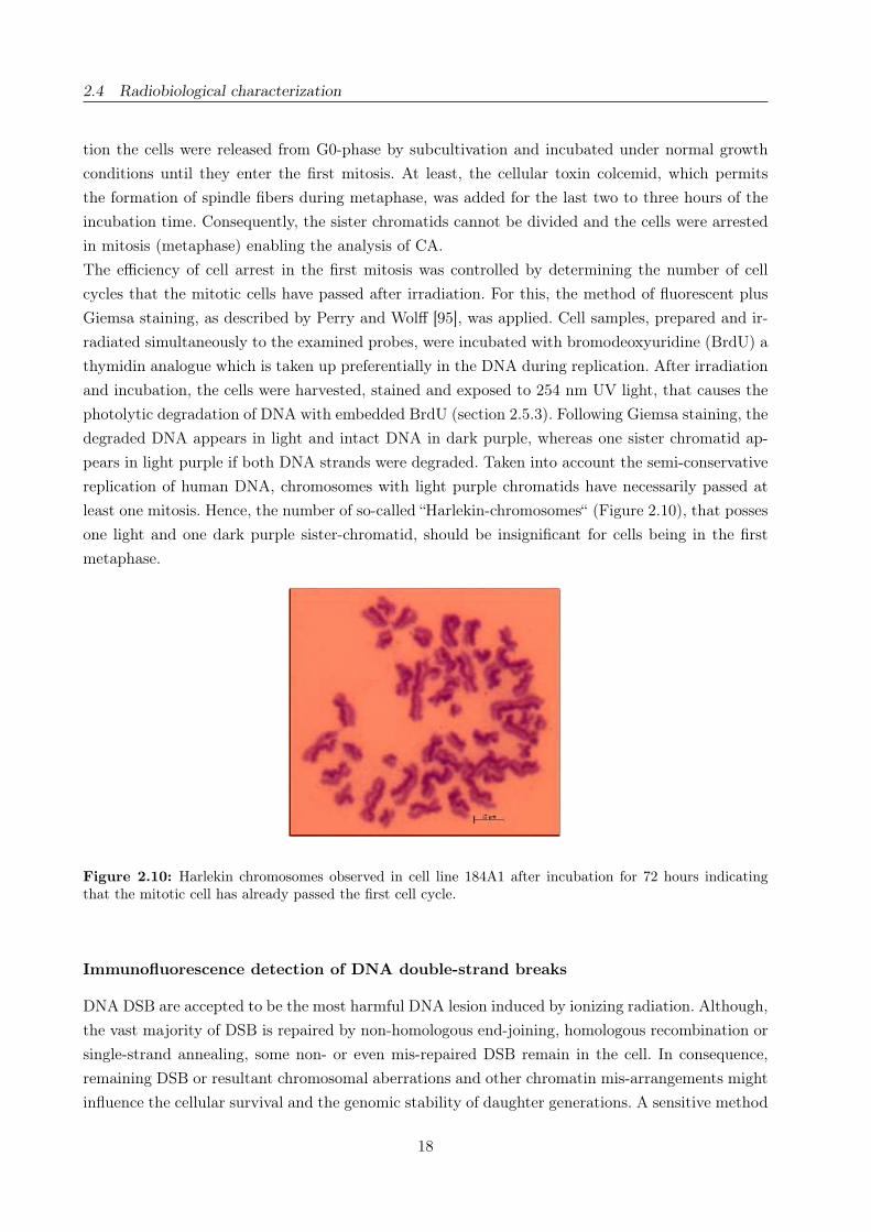
least one mitosis. Hence, the number of so-called “Harlekin-chromosomes“ (Figure 2.10), that posses
one light and one dark purple sister-chromatid, should be insignificant for cells being in the first
metaphase.
Figure 2.10: Harlekin chromosomes observed in cell line 184A1 after incubation for 72 hours indicatingthat the mitotic cell has already passed the first cell cycle.
Immunofluorescence detection of DNA double-strand breaks
DNA DSB are accepted to be the most harmful DNA lesion induced by ionizing radiation. Although,
the vast majority of DSB is repaired by non-homologous end-joining, homologous recombination or
single-strand annealing, some non- or even mis-repaired DSB remain in the cell. In consequence,
remaining DSB or resultant chromosomal aberrations and other chromatin mis-arrangements might
influence the cellular survival and the genomic stability of daughter generations. A sensitive method
18

2.4 Radiobiological characterization
Figure 2.11: Organization of the chromosomes - starting from the DNA double-helix, which is wrappedaround nucleosomes each consisting of eight histone molecules and linked by histone H1. The arrangement ofnucleosomes as well as the DNA around forms the so-called chromatin, which becomes visible as chromosomesduring mitosis (Schematic drawing adopted from [88]).
to quantify DNA double-strand breaks is based on the cellular DSB signaling pathway (Fig. 2.12)
and the proteins involved, respectively. Following DSB induction, several proteins are activated,
recruited and accumulated at the DNA DSB, whereas two of them, the phosphorylated histone
H2AX and the protein 53BP1, are often used for the quantification of DSB (e.g., [2, 104, 106]).
Regarding the eukaryotic chromatin in detail its organization in several levels is evident (Fig. 2.11).
The double-stranded DNA is wrapped around a histone core, which consists of eight histones, two
of each histone family H2A, H2B, H3 and H4 [104], forming the nucleosomes the basic units of
chromatin. Sharing the highly conservative histone folding motif [62], but differing in the N- and
C-terminal tails, each histone family is subclassified, e.g., the family H2A is divided in H2AX,
H2A1-H2A2 and H2AZ [104]. The most famous member of this family is the histone H2AX, which
represents approximately 2 - 25 % of the whole family H2A in mammals [104]. As demonstrated
by Rogakou et al. [104] the histone H2AX becomes rapidly phosphorylated at the C-terminal of
the serine 139 residue in consequence of radiation induced DNA DSB. The phospho-form of H2AX,
also referred to as γ-H2AX, appears on either side of a DNA double-strand break and ranges over
a total of 2× 10⁶ base pairs, thus involving 0.03 % of the chromatin per DSB [104]. The maximum
amount of phosphorylated H2AX is achieved in less than 10 minutes after irradiation providing a
sensitive and rapid method to detect DNA DSB. Therefore, the accumulation of hundreds of phos-
19
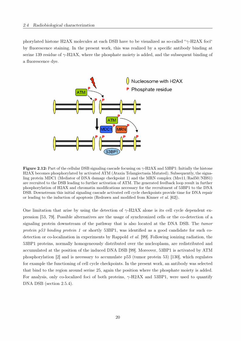
2.4 Radiobiological characterization
phorylated histone H2AX molecules at each DSB have to be visualized as so-called “γ-H2AX foci“
by fluorescence staining. In the present work, this was realized by a specific antibody binding at
serine 139 residue of γ-H2AX, where the phosphate moiety is added, and the subsequent binding of
a fluorescence dye.
Figure 2.12: Part of the cellular DSB signaling cascade focusing on γ-H2AX and 53BP1: Initially the histoneH2AX becomes phosphorylated by activated ATM (Ataxia Telangiectasia Mutated). Subsequently, the signa-ling protein MDC1 (Mediator of DNA damage checkpoint 1) and the MRN complex (Mre11/Rad50/NBS1)are recruited to the DSB leading to further activation of ATM. The generated feedback loop result in furtherphosphorylation of H2AX and chromatin modifications necessary for the recruitment of 53BP1 to the DNADSB. Downstream this initial signaling cascade activated cell cycle checkpoints provide time for DNA repairor leading to the induction of apoptosis (Redrawn and modified from Kinner et al. [62]).
One limitation that arise by using the detection of γ-H2AX alone is its cell cycle dependent ex-
pression [53, 79]. Possible alternatives are the usage of synchronized cells or the co-detection of a
signaling protein downstream of the pathway that is also located at the DNA DSB. The tumor
protein p53 binding protein 1 or shortly 53BP1, was identified as a good candidate for such co-
detection or co-localization in experiments by Rappold et al. [99]. Following ionizing radiation, the
53BP1 proteins, normally homogeneously distributed over the nucleoplasm, are redistributed and
accumulated at the position of the induced DNA DSB [99]. Moreover, 53BP1 is activated by ATM
phosphorylation [2] and is necessary to accumulate p53 (tumor protein 53) [130], which regulates
for example the functioning of cell cycle checkpoints. In the present work, an antibody was selected
that bind to the region around serine 25, again the position where the phosphate moiety is added.
For analysis, only co-localized foci of both proteins, γ-H2AX and 53BP1, were used to quantify
DNA DSB (section 2.5.4).
20

2.5 Realization and results
2.5 Realization and results
1. Investigation of a TSEE dosimetry system for determination of dose in a cell monolayer
2. Cell irradiation setup and dosimetry for radiobiological studies at ELBE
3. Relative biological effectiveness of 25 and 10 kV X-rays for the induction of chromosomal
aberrations in two human mammary epithelial cell lines
4. DNA double-strand break signaling: X-ray energy dependence of residual co-localized foci of
γ-H2AX and 53BP1
21


2.5 Realization and results
Investigation of a TSEE dosimetry system for determination of dose in a cell
monolayer
Anna Lehnert, Elke Beyreuther, Elisabeth Lessmann and Jörg Pawelke
Institute of Radiation Physics, Forschungszentrum Dresden-Rossendorf, P.O. Box 510119, D-01314
Dresden, Germany
Corresponding author:
Anna Lehnert
Institute of Radiation Physics
Forschungszentrum Dresden-Rossendorf
P.O. Box 510119
D-01314 Dresden, Germany
Fon: +49 351 260 3657
Fax: +49 351 260 3700
E-mail: [email protected]
Radiation Measurements 42:1530-37(2007)
DOI: 10.1016/j.radmeas.2007.03.007
www.elsevier.com/locate/radmeas
23

2.5 Realization and results
Abstract
A prototype system for radiobiological studies has been investigated. It is based on thermally sti-
mulated exoelectron emission (TSEE) detectors and can be used for precise determination of the
absorbed dose in a live cell monolayer of several µm thickness. In the present study, five types of
BeO detectors, different in structure and method of production, were tested in combination with
a Geiger-Müller counter. The dose response and dose range, reproducibility and long-time stability
of response, as well as the applicability in a simulated cell culture environment have been studied.
The dose response was found to be linear over two orders of magnitude and limited by the coun-
ter resolution. However, by a variation of detector sensitivity, the whole dose range of interest for
radiobiological experiments can be covered. The irradiation in a simulated cell environment was
successful only for one detector type. The system performance was found to be limited by the va-
riation in the system response for time periods longer than several hours, therefore, it is suitable
for absolute dose measurement with calibrated detectors if reproducible laboratory conditions are
provided.
Keywords: TSEE, Exoelectron emission, BeO; Geiger-Müller counter, Dosimetry, Cell monolayer
24

2.5 Realization and results
I. Introduction
Thermally stimulated exoelectron emission (TSEE) is a well-known phenomenon. Its theoretical
description is based on the band model of solids. The excitation by external irradiation results in
trapping of electrons near the bottom of the conduction band. As a result of a thermal stimulation,
the electrons may overcome the work function and leave the crystal surface (exoelectron emission).
Although optical stimulation of exoelectron emission is possible, the thermal stimulation is much
more efficient [76]. The exoelectrons are in both cases emitted from the detector surface, however,
the thermal stimulation can bring in motion also electrons from the depth of the material. At increa-
sing temperature, it results in maxima of the electron emission at distinct temperatures, which are
characteristic for the investigated material (glow curve). The use of TSEE detectors in dosimetry
is based on the relation between radiation dose and the glow curve. The mathematical description
of the glow curve in the frame of the Randall and Wilkins model [50] is similar to the theoretical
description of the thermoluminiscent (TL) glow curves and allows to calculate the depth of the
energy levels. Although a TSEE detector has several advantages over the widely used TL detectors,
no commercially available systems exist and its application in dosimetry is seldom. One of its main
advantages is the surface-based origin of the phenomenon, since the exoelectrons have an escape
depth of less than 10 nm [66]. This makes such a dosimeter very attractive in the cases where a
small sensitive volume is desired, such as detection of low-penetrating β-radiation and low-energy
X-rays as well as for studying highly inhomogeneous radiation fields at the interface of different
materials [19, 100] or in the dose build-up region of high-energy photon beams.
The most common materials used for TSEE dosimetry are ionic crystals such as alkali halides or
oxides. One widely used detector material is BeO, because of its physical stability, thermal conduc-
tivity and insolubility in water. Its low atomic number of about 7.1 approximates the mean atomic
number of tissue, making it especially suitable for application in personal dosimetry. A method in-
troduced by Kramer, based on mixing of BeO with sufficient amount of graphite powder to improve
conductivity, results in a linear dose response of the detector over 8 orders of magnitude [64]. This
method has been further developed and improved to obtain optimal detector properties [50, 63]. The
experimental investigations with BeO have proved its applicability for γ- and β-dosimetry, showing
a low dependence on photon [67] or electron energy [66] and linearity of dose response over a large
dose range [24]. These are additional advantages when compared with the widespread TL detectors.
A superconducting electron linear accelerator of high brilliance and low emittance (ELBE) is under
operation at Forschungszentrum Dresden-Rossendorf since January 2003. The relativistic electron
beam with variable time structure and energy up to 40 MeV is used to drive various kinds of secon-
dary radiation sources. Among all, in vitro radiobiological studies with high-energy bremsstrahlung
as well as with channeling X-rays, tunable in the range 10 - 100 keV, are being performed. The goal
of the present study was to investigate a prototype system for precise determination of the absorbed
dose in a live cell monolayer of several µm thickness. The advantage of a small sensitive volume of the
TSEE detector based on BeO could be useful in soft X-rays dosimetry, as well as for studying dose
build-up effects of high-energy bremsstrahlung radiation. In addition, for the determination of the
absolute dose and dose inhomogeneity in the cell, a tissue-equivalent dosimeter which could be irra-
25
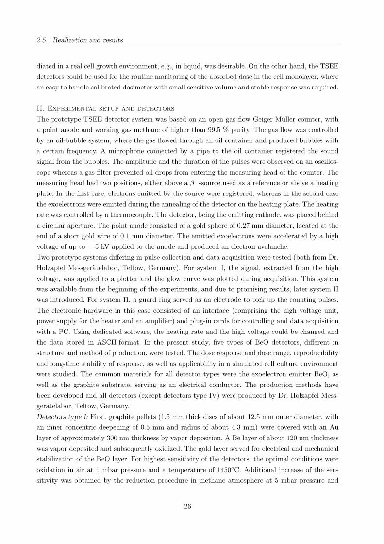
2.5 Realization and results
diated in a real cell growth environment, e.g., in liquid, was desirable. On the other hand, the TSEE
detectors could be used for the routine monitoring of the absorbed dose in the cell monolayer, where
an easy to handle calibrated dosimeter with small sensitive volume and stable response was required.
II. Experimental setup and detectors
The prototype TSEE detector system was based on an open gas flow Geiger-Müller counter, with
a point anode and working gas methane of higher than 99.5 % purity. The gas flow was controlled
by an oil-bubble system, where the gas flowed through an oil container and produced bubbles with
a certain frequency. A microphone connected by a pipe to the oil container registered the sound
signal from the bubbles. The amplitude and the duration of the pulses were observed on an oscillos-
cope whereas a gas filter prevented oil drops from entering the measuring head of the counter. The
measuring head had two positions, either above a β−-source used as a reference or above a heating
plate. In the first case, electrons emitted by the source were registered, whereas in the second case
the exoelectrons were emitted during the annealing of the detector on the heating plate. The heating
rate was controlled by a thermocouple. The detector, being the emitting cathode, was placed behind
a circular aperture. The point anode consisted of a gold sphere of 0.27 mm diameter, located at the
end of a short gold wire of 0.1 mm diameter. The emitted exoelectrons were accelerated by a high
voltage of up to + 5 kV applied to the anode and produced an electron avalanche.
Two prototype systems differing in pulse collection and data acquisition were tested (both from Dr.
Holzapfel Messgerätelabor, Teltow, Germany). For system I, the signal, extracted from the high
voltage, was applied to a plotter and the glow curve was plotted during acquisition. This system
was available from the beginning of the experiments, and due to promising results, later system II
was introduced. For system II, a guard ring served as an electrode to pick up the counting pulses.
The electronic hardware in this case consisted of an interface (comprising the high voltage unit,
power supply for the heater and an amplifier) and plug-in cards for controlling and data acquisition
with a PC. Using dedicated software, the heating rate and the high voltage could be changed and
the data stored in ASCII-format. In the present study, five types of BeO detectors, different in
structure and method of production, were tested. The dose response and dose range, reproducibility
and long-time stability of response, as well as applicability in a simulated cell culture environment
were studied. The common materials for all detector types were the exoelectron emitter BeO, as
well as the graphite substrate, serving as an electrical conductor. The production methods have
been developed and all detectors (except detectors type IV) were produced by Dr. Holzapfel Mess-
gerätelabor, Teltow, Germany.
Detectors type I: First, graphite pellets (1.5 mm thick discs of about 12.5 mm outer diameter, with
an inner concentric deepening of 0.5 mm and radius of about 4.3 mm) were covered with an Au
layer of approximately 300 nm thickness by vapor deposition. A Be layer of about 120 nm thickness
was vapor deposited and subsequently oxidized. The gold layer served for electrical and mechanical
stabilization of the BeO layer. For highest sensitivity of the detectors, the optimal conditions were
oxidation in air at 1 mbar pressure and a temperature of 1450◦C. Additional increase of the sen-
sitivity was obtained by the reduction procedure in methane atmosphere at 5 mbar pressure and
26

2.5 Realization and results
temperature of 800◦C (Holzapfel, private communication). The effective area of the detector was
43 mm2.
Detectors type II: The dimensions of the graphite substrate were 12.0× 12.0× 0.5 mm3. The detec-
tors were produced by the upper procedure, but without the Au layer. For mechanical stability of
the detectors, they were mounted in a round Al holder. The effective detector area was 144 mm2.
Detectors type III: They were basically identical with the detectors type II, but without the Al
holder, therefore, special care for handling was necessary. The effective area in this case was the
same as for detectors type II.
Detectors type IV: The graphite pellets were 1.0 mm thick discs of 17 mm outer diameter, with
an inner concentric deepening of 0.5 mm and a radius of 5.0 mm. The BeO was brought on the
surface in the form of water suspension with a defined concentration and the water was evaporated
by heating at about 50◦C without any further treatment. The effective area was 79 mm2.
Detectors type V: The graphite substrate (about 11× 11 mm2, thickness 0.5 or 1 mm) was covered
with a Be suspension of defined concentration in alcohol. After evaporation, the layer was oxidized
at 1400 - 1500◦C. The effective area was 140 mm2.
The properties of the detectors were studied after irradiation with γ- and β−-sources: 90Sr (activity
410 kBq or 20 MBq), 55Fe (nominal activity 130 kBq or 37 MBq), 22Na (activity 58 kBq), 137Cs
(dose rate in water 0.1 mGy/min at 53.7 cm source-detector distance). The reference sources were14C (activity about 3 kBq) and 63Ni (activity about 2.5 kBq).
III. Results
Working point of the Geiger-Müller counter
For both counters, operated at the same methane flow rate, the working point was set to be 4.0 kV.
For system II, the increase of the high voltage was connected with a change in the pulse height
distribution. A noise peak in the distribution was observed at channel number about 224, corres-
ponding to about 4.4 V (the pulses in the range 0.4 - 5 V were registered), regardless of the electron
source (a reference source or a TSEE detector). Its height was found to increase with increasing
high voltage and to deteriorate the counter performance at voltage higher than about 4.1 kV. Since
it was found to be at the same position even after exchange of the anode, the reason for it was
probably in the electronics of the data acquisition system. However, as presented further, the noise
peak was only one of the factors influencing the whole system performance and its contribution
could be neglected. It was not excluded in any of the further evaluations.
Glow curves
The glow curves of all detector types are presented in Figure 2.13. The irradiations were performed
with the same 90Sr source, and the heating rate was 3◦C/s. The source-detector distance of about
7 mm was optimized by the reader count rate and a reasonable irradiation time and was varying
by less than 0.5 mm for the different detector types. A difference in the glow curves was observed
as a result of the difference in the production method, but most often there was one main peak in
the range 200 - 400◦C, which makes dosimetric application convenient. Since both the number of
27

2.5 Realization and results
Figure 2.13: Glow curves of all tested detector types. Detector type V of high sensitivity is presented.Different scales of the ordinates are used for better presentation.
28

2.5 Realization and results
counts in the peak range of the glow curve and the total number of counts are proportional to the
dose, they both can be used as a dosimetric signal. In the frame of the Randall and Wilkins model
[50], a numerical evaluation of the sample glow curves, presented in Figure 2.13, was performed to
calculate the energetic distance from the trap levels to the bottom of the conductivity band. The
obtained values for all the detectors are in the realistic range 0.5 - 1.2 eV.
Dose range of the TSEE system
The dependence of the number of TSEE counts on the time of irradiation, or, respectively, dose,
has been studied for both systems and different sources. In Figure 2.14, the detector response in
dependence on irradiation time for two different sources is shown.
Figure 2.14: Time of irradiation response of detector type I, system I. The solid squares represent the peakrange of the glow curve (20 - 370◦C) with the corresponding linear fit (solid line). The open squares representthe whole range (20 - 520◦C) with the corresponding linear fit (dash line). Left panel: 55Fe irradiation. Rightpanel: 90Sr irradiation.
As seen from the figure, the linearity of response is held both for soft X-rays and for β-rays. It
was observed also after irradiation with the 137Cs source and other types of detectors. This makes
calibrated detectors useful for dose determination. However, after longer irradiation times, a plateau
in the count number was observed for detectors type I, II and III for both systems, which could
result either from saturation of the detector response, or from the limited pulse rate resolution of the
counter. This was proved by comparing the response of two different detectors after irradiation with
the same source at the same irradiation geometry. The results of irradiation with a 90Sr source for
system I, detectors type I, are presented in Figure 2.15 (left). Although both samples have different
sensitivity, reproduced by the different slopes of the linear ranges of the curves, the saturation of
response was achieved at the same count number, about 5× 10⁵ total number of counts. Saturation
at a similar number of counts was also observed for system II. Therefore, saturation due to limited
counter resolution was concluded. In order to determine the dose range, corresponding to the count
rate range of the TSEE systems, a 137Cs source, calibrated to dose rate in water of 0.1 mGy/min at
a 53.7 cm distance from the source, was used. The response for detector type I and system I, shown
in Figure 2.15 (right), after irradiation with the 90Sr source was scaled to fit the response curve
29

2.5 Realization and results
Figure 2.15: Left: saturation of system I response, measured for two samples of detector type I after irra-diation with 90Sr. Right: dose response of detector type I, system I. The measured response after irradiationwith 90Sr is shown by the solid line. The solid circles represent the response after irradiation of the samedetector with a calibrated 137Cs source.
after 137Cs irradiation for the same number of counts. This procedure is not equal to a “calibration“
of the 90Sr source, since the difference in detector response to both radiation qualities is unknown.
The dose range of system I was obtained to be 2 orders of magnitude. The upper limit, defined by
the counter saturation, was about 10 mGy, and the lower limit was defined by the statistical error
of the count number. This upper value is far from the reported saturation limit of the detectors
of up to 10 Gy [66], and the entire sensitivity range is far from the dose range necessary for cell
irradiation experiments, which is usually 0.1 - 10 Gy. This is a serious problem for dose monitoring
during irradiation. A shift in the TSEE system sensitivity could be achieved by manipulation of the
read-out system (counter geometry, gas flow), which however, would result in a rather small increase
of the upper response limit. A decrease of the detector sensitivity by orders of magnitude can be
reached by decreasing detector size, but since a 10-fold decrease of the detector radius, resulting in
100 times sensitivity decrease, would result in a detector size less than 1 mm2, it would lead to dif-
ficulties in the production and handling of such detectors. Another possibility would be to decrease
the size of the detector read-out area. This method was tested using a grid over the detector, but
it resulted in a decreased stability of response. Therefore, another method of detector production
was used and detector types IV and V, produced by sedimentation, were tested. Although detectors
type IV can be produced in very simple laboratory conditions, their testing proved that no big
variation in sensitivity could be achieved. A fading effect was observed, expressed in several orders
of magnitude drop of the response to irradiation before reaching a plateau over the first days after
production (Fig. 2.16). The detector response was evaluated immediately after irradiation with a90Sr source, and the procedure was repeated on several consecutive days. Such changes in response
were not observed for any other detector type. For detectors type V, which were produced in a
similar way, several orders of magnitude difference in sensitivity was achieved by variation of the
BeO amount during production, and the sensitivity was proved to be stable with time. The dose
30
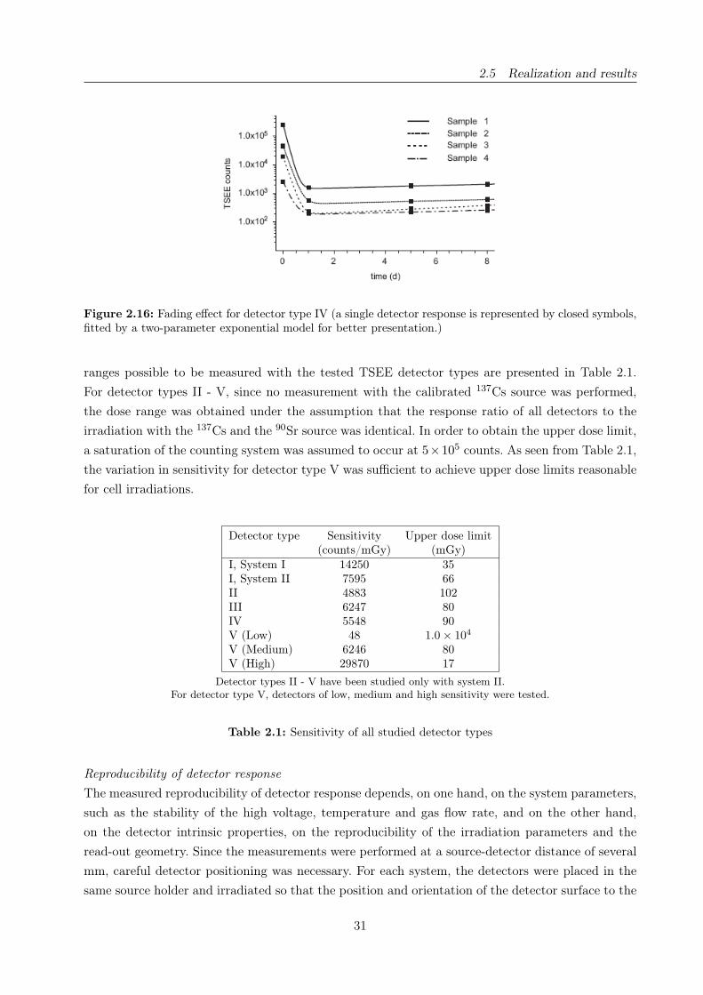
2.5 Realization and results
Figure 2.16: Fading effect for detector type IV (a single detector response is represented by closed symbols,fitted by a two-parameter exponential model for better presentation.)
ranges possible to be measured with the tested TSEE detector types are presented in Table 2.1.
For detector types II - V, since no measurement with the calibrated 137Cs source was performed,
the dose range was obtained under the assumption that the response ratio of all detectors to the
irradiation with the 137Cs and the 90Sr source was identical. In order to obtain the upper dose limit,
a saturation of the counting system was assumed to occur at 5×10⁵ counts. As seen from Table 2.1,
the variation in sensitivity for detector type V was sufficient to achieve upper dose limits reasonable
for cell irradiations.
Detector type Sensitivity Upper dose limit(counts/mGy) (mGy)
I, System I 14250 35I, System II 7595 66II 4883 102III 6247 80IV 5548 90V (Low) 48 1.0 × 10⁴
V (Medium) 6246 80V (High) 29870 17
Detector types II - V have been studied only with system II.For detector type V, detectors of low, medium and high sensitivity were tested.
Table 2.1: Sensitivity of all studied detector types
Reproducibility of detector response
The measured reproducibility of detector response depends, on one hand, on the system parameters,
such as the stability of the high voltage, temperature and gas flow rate, and on the other hand,
on the detector intrinsic properties, on the reproducibility of the irradiation parameters and the
read-out geometry. Since the measurements were performed at a source-detector distance of several
mm, careful detector positioning was necessary. For each system, the detectors were placed in the
same source holder and irradiated so that the position and orientation of the detector surface to the
31

2.5 Realization and results
source was always the same to exclude the influence of the inhomogeneity of the BeO layer. The
uncertainty of the irradiation time measurement was ± 1 s.
A parameter which could influence the stability of the system response is the gas flow rate. A li-
near increase in the counting efficiency with increasing gas flow rate was observed for the TSEE
count rate, whereas for irradiation with the reference source, an opposite dependence was observed
(Fig. 2.17). The reason for this could be the difference in the energy of the registered electrons.
Whereas the maximal energy of the electrons emitted by the reference source 63Ni was 67 keV, the
exoelectrons have energies less than 5 eV [26], so it is possible that an increase of the gas flow results
in an opposite effect on the effectiveness of electron counting. However, due to insufficient informa-
tion about the geometry and electronics of this prototype system, the effect cannot be studied in
detail. Due to the strong gas flow dependence of the count rate, the gas flow was kept constant and
controlled regularly during experiment series. The intrinsic reproducibility of the readout system at
Figure 2.17: Gas flow dependence of the TSEE system response. Left panel: reference source. Right panel:TSEE detector type I, where the solid squares represent the peak range of the glow curve (192 - 360◦C) withthe corresponding linear fit (solid line) and the open squares represent the whole range (20 - 520◦C) withthe corresponding linear fit (dash line).
constant temperature and gas flow rate can be derived from the reproducibility of the count rate
of the reference source, where the standard deviation (SD) was found to be better than 1 %. It is
presented in Table 2.2 together with the reproducibility of all detector types for irradiation with
the 90Sr source. The measurement and subsequent cooling of the detectors was repeated at least 10
times in a row, and the total numbers of exoelectron counts as well as in the region of the main
glow curve peak were evaluated. The best reproducibility of response was observed for detector
type I, which follows the expected behavior due to the presence of a mechanically and electrically
stabilizing Au layer. The reproducibility of response for detectors type IV was measured after sta-
bilization to be about 10 % (SD), which can be attributed to the method of production by simple
sedimentation of BeO. The comparison of both used systems, performed for detector type I, shows
the better stability of system II properties, due to the improved high voltage stability and tempe-
rature control. However, these measurements do not reflect the fluctuation of the system response
during routine work over weeks or months. Variations of up to 20 % were found within one week
by reference source measurements, without any change of the setup or environmental conditions.
32

2.5 Realization and results
Detector type System Number of Reproducibility (SD)experiments Peak counts % Total counts %
None (reference source) II 19 ND 0.64I I 25 1.29 1.23
II 12 0.95 0.97II II 13 2.45 2.37III II 12 2.33 2.16IV II 10 10.50 13.34V II 10 6.91 6.55
Table 2.2: Reproducibility of response for reference source and TSEE detector irradiation, presented as theSD relative to the mean value
Dose measurement in a simulated cell environment
The irradiation of living cells is complicated by the requirement of optimal environmental condi-
tions, and especially by the presence of appropriate liquid (e.g., cell culture medium) covering the
cells. Therefore, the application of TSEE detectors for dose sampling in the conditions of real cell
environment and the study of interface effects would require irradiation in the presence of liquid.
All studied detector types were tested for applicability for this purpose. The tested liquids were
water, propanol and cell medium, whereas water and propanol were chosen for their simplicity of
chemical composition. The irradiation in water resulted in big and inconsistent variations of the
detector response. The irradiation in cell culture medium resulted in a complete change of the glow
curve and inadequate detector response, caused probably by the influence of the organic consti-
tuents on the electric properties of the irradiated surface, however, no irreversible change of the
detector response was observed. Finally, propanol was chosen as a working medium, its fast evapo-
ration allowing immediate evaluation of the glow curve. The summary of the detector performance
is presented in Table 2.3. First, a well-known problem for detectors based on surface excitations is
the tribo effect, expressed in detection of a signal after a contact of the surface with any material
without irradiation. The induction of the tribo effect was checked by wetting the detectors with
propanol. An irradiation was performed before wetting as well as after evaporation of the propanol.
No change of the detector response, which would point at a tribo effect, was observed (see Table 2.3,
column 2). As a second step, the study of the influence of the liquid during irradiation on detector
response was performed. The source was placed directly on the detector if it was not coming in
contact with the sensitive layer of the detector, otherwise a holder was used, and this setup was
placed in a vessel with liquid. The reproducibility of detector response for irradiation in propanol
was checked with a 22Na source.
The thickness of the liquid layer enclosed between the source and the detector was 1 - 3 mm. The
reproducibility of response of all detector types was proved to be worse compared to irradiation in
air (cf. Table 2.2). In addition, the plausibility of detector response after irradiation in propanol
was checked with the 22Na source using the same geometry. The positron decay of 22Na leads to
annihilation radiation at 511 keV in addition to the decay γ-line at 1275 keV. From the photon mass
33
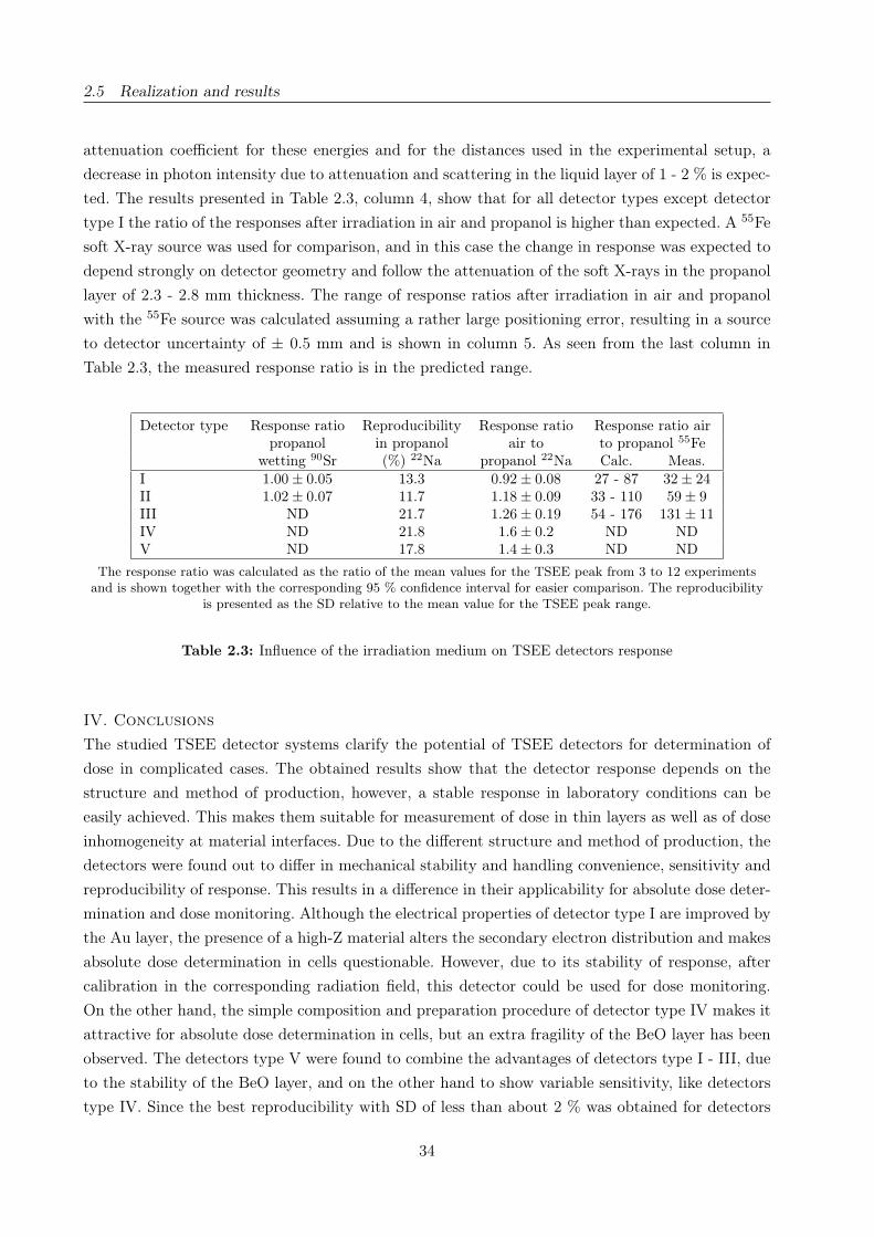
2.5 Realization and results
attenuation coefficient for these energies and for the distances used in the experimental setup, a
decrease in photon intensity due to attenuation and scattering in the liquid layer of 1 - 2 % is expec-
ted. The results presented in Table 2.3, column 4, show that for all detector types except detector
type I the ratio of the responses after irradiation in air and propanol is higher than expected. A 55Fe
soft X-ray source was used for comparison, and in this case the change in response was expected to
depend strongly on detector geometry and follow the attenuation of the soft X-rays in the propanol
layer of 2.3 - 2.8 mm thickness. The range of response ratios after irradiation in air and propanol
with the 55Fe source was calculated assuming a rather large positioning error, resulting in a source
to detector uncertainty of ± 0.5 mm and is shown in column 5. As seen from the last column in
Table 2.3, the measured response ratio is in the predicted range.
Detector type Response ratio Reproducibility Response ratio Response ratio airpropanol in propanol air to to propanol 55Fe
wetting 90Sr (%) 22Na propanol 22Na Calc. Meas.I 1.00 ± 0.05 13.3 0.92 ± 0.08 27 - 87 32 ± 24II 1.02 ± 0.07 11.7 1.18 ± 0.09 33 - 110 59 ± 9III ND 21.7 1.26 ± 0.19 54 - 176 131 ± 11IV ND 21.8 1.6 ± 0.2 ND NDV ND 17.8 1.4 ± 0.3 ND ND
The response ratio was calculated as the ratio of the mean values for the TSEE peak from 3 to 12 experimentsand is shown together with the corresponding 95 % confidence interval for easier comparison. The reproducibility
is presented as the SD relative to the mean value for the TSEE peak range.
Table 2.3: Influence of the irradiation medium on TSEE detectors response
IV. Conclusions
The studied TSEE detector systems clarify the potential of TSEE detectors for determination of
dose in complicated cases. The obtained results show that the detector response depends on the
structure and method of production, however, a stable response in laboratory conditions can be
easily achieved. This makes them suitable for measurement of dose in thin layers as well as of dose
inhomogeneity at material interfaces. Due to the different structure and method of production, the
detectors were found out to differ in mechanical stability and handling convenience, sensitivity and
reproducibility of response. This results in a difference in their applicability for absolute dose deter-
mination and dose monitoring. Although the electrical properties of detector type I are improved by
the Au layer, the presence of a high-Z material alters the secondary electron distribution and makes
absolute dose determination in cells questionable. However, due to its stability of response, after
calibration in the corresponding radiation field, this detector could be used for dose monitoring.
On the other hand, the simple composition and preparation procedure of detector type IV makes it
attractive for absolute dose determination in cells, but an extra fragility of the BeO layer has been
observed. The detectors type V were found to combine the advantages of detectors type I - III, due
to the stability of the BeO layer, and on the other hand to show variable sensitivity, like detectors
type IV. Since the best reproducibility with SD of less than about 2 % was obtained for detectors
34
2.5 Realization and results
type I - III, they can be used in cases, when high precision is necessary. The dose range, in which
the studied TSEE system can be used, was found to be limited by the counter resolution, but by a
variation of detector sensitivity, the whole dose range of interest for radiobiological experiments can
be covered. This makes the system suitable for dose measurement with calibrated detectors during
routine irradiation. However, its performance is limited by the variation in the system response for
time periods longer than several hours. The irradiation in a simulated cell environment was suc-
cessful only for detectors type I, however, this type is least tissue-equivalent. Although the TSEE
detectors could be of big interest for dosimetry at the ELBE X-ray source, it requires improvement
to become suitable for precise dose determination. Furthermore, independence of the response on
dose rate has to be proved, since the time structure of the ELBE radiation source allows variability
of dose rate by several orders of magnitude.
Acknowledgments
We express our gratitude to Dr. G. Holzapfel for the helpful discussions and the response to the
specific requirements of our task in the detectors production. We thank Dr. J. Lesz for the technical
support and M. Sommer for the assistance in the experimental work at the Technische Universität
Dresden.
Author contributions
Study concept and design A.L., J.P.; Experimental studies A.L., E.B., E.L.; Data acquisition A.L.,
E.B.; Data analysis A.L.; Literature research A.L.; Manuscript preparation A.L.; Manuscript editing
J.P., E.B.; Guarantor of integrity of the entire study J.P.; Manuscript review was carried out by all
authors.
35


2.5 Realization and results
Cell irradiation setup and dosimetry for radiobiological studies at ELBE
Karl Zeila, Elke Beyreuthera, Elisabeth Lessmanna, Wolfgang Wagnera and Jörg Pawelkea,b
a Institute of Radiation Physics, Forschungszentrum Dresden-Rossendorf, Dresden, Germanyb OncoRay, Center for Radiation Research in Oncology, Medical Faculty Carl Gustav Carus, Uni-
versity of Technology Dresden, Dresden, Germany
Corresponding author:
Karl Zeil
Institute of Radiation Physics
Forschungszentrum Dresden-Rossendorf
P.O. Box 510119
D-01314 Dresden, Germany
Fon: +49 351 260 2614
Fax: +49 351 260 3700
E-mail: [email protected]
Nuclear Instruments and Methods in Physics Research Section B:
Beam Interactions with Materials and Atoms 267(14):2403-10(2009)
DOI: 10.1016/j.nimb.2009.04.15
www.elsevier.com/locate/nimb
37
2.5 Realization and results
Abstract
The radiation source ELBE delivers different types of secondary radiation, which is used for cell
irradiation studies in radiobiological research. Thereby an important issue is the determination
of the biological effectiveness of photon radiation as a function of photon energy by using low-
energetic, monochromatic channeling radiation (10 - 100 keV) and high-energetic bremsstrahlung
(up to 40 MV). Radiobiological studies at the research facility ELBE demand special technical and
dosimetric prerequisites. Therefore, a cell irradiation system (CIS) has been designed, constructed
and installed at the beam line. The CIS allows automatic irradiation of a larger cell sample number
and the compensation of spatial inhomogeneity of the dose distribution within the beam spot. The
recently introduced GafChromicr EBT radiochromic film model has been used to verify the cell
irradiation dose deposition achieving a dose uncertainty of < 5 %. Both, the installed cell irradia-
tion system and the developed dosimetric procedure based on the use of the EBT film have been
experimentally tested at ELBE. The biological effectiveness of 34 MV bremsstrahlung with respect
to 200 kV X-rays from a conventional X-ray tube has been determined. An RBE value of 0.75 has
been measured in good agreement with literature.
Keywords: Radiobiology, Cell irradiation, Dosimetry, Radiochromic film
PACS: 87.53.-j; 87.53.Bn; 87.55.-N; 07.85.Fv; 29.20.Ej; 41.60.-m
38
2.5 Realization and results
I.Introduction
The basic device of the radiation source ELBE [30, 34] is a superconducting electron linear accele-
rator which provides an electron beam of high brilliance and low emittance with maximum electron
energy of 40 MeV and a high average beam current of 1 mA. The electron beam is used for the
production of different types of secondary radiation which are applied for several research topics.
The primary electrons can also be used for experiments.
The different types of radiation are applied for radiobiological research as one important issue.
There the determination of the biological effectiveness of ionizing photon radiation as a function
of photon energy [48, 74, 75] represents a major scientific objective. Very intense, low-energetic,
quasi-monochromatic, and energy tunable (10 - 100 keV) channeling radiation (CR) is generated by
channeling of relativistic electrons in diamond crystals [129] at the radiation physics beam line at
ELBE. Additionally, high-energetic bremsstrahlung of up to 40 MV photon energy can be delivered.
The presented work is part of radiobiological studies which are related to photon radiation applied
in medical application such as mammography screening and radiotherapy. In order to compare ra-
diation qualities, the relative biological effectiveness (RBE) has to be introduced. It is defined by
the fraction of the dose delivered by a reference radiation quality to the dose delivered by a given
radiation quality, whereas both radiation qualities are responsible for the same biological effect.
The general basis of radiobiological experiments is RBE determination by measuring of dose-effect
curves for in vitro cell systems.
Usually radiobiological studies are performed on conventional high-voltage X-ray tubes or medical
acceleration facilities. Both sources deliver broad polychromatic bremsstrahlung with a high photon
flux. Thus, therapeutic dose values (few Gy per daily fraction) can be delivered in a sufficiently
small irradiation duration (dose rate ≈ 1 Gy/min) to be independent from repairing processes in
human cells. Due to the high reproducibility of beam parameters of conventional radiation sources,
a large number of samples can be irradiated in stable conditions in order to cope with the biological
diversity. Considering the dosimetry, a standardized radiation field is used. All changes in the ra-
diation geometry resulting in differences of beam absorption, scattering or dose build up effect are
taken into consideration by applying tabled correction factors. In practical irradiation experiments,
cell samples are irradiated at a vertical beam and the delivered dose is controlled by presetting a
certain irradiation duration.
Cell irradiation experiments at the research facility ELBE demand special technical prerequisites
which mainly arises from the use of the intense CR. The intensity and the pointing of the ELBE
beam show temporal variations which have to be monitored and taken into account. But also the
complex tuning and optimization of the flexible electron beam line by the operators has to be men-
tioned. Both influence the reproducibility of irradiation experiments. Moreover, the ELBE beam
provides a very small divergence, which leads to an increased spatial inhomogeneity of the dose
distribution within the beam spot. For this reason the dose has to be measured for each irradiated
sample. As a basic and cost efficient instrument self-developing radiochromic film dosimeters (mo-
del GafChromicr EBT Prototype A, henceforth referred to as EBT) have been used. Their high
spatial resolution, low energy dependence and near tissue-equivalence make them highly suitable
39

2.5 Realization and results
for measurements of dose distributions in a wide photon energy range and in radiation fields with
high dose gradients [13, 59, 91]. The EBT films were applied to quantitatively measure the dose
delivered to each individual cell sample and online dose rate measurement (ionization chambers)
was applied for experimental control.
For the reason of radiation protection regulations after switching off the beam, the cell samples can
be taken out of the experiment room only after a waiting time of 20 min. Thus, the irradiation
of a larger sample number requires an automatized irradiation procedure for which a cell irradia-
tion system (CIS) has been designed, constructed and installed at ELBE. Spatial inhomogeneity
of the dose distribution within the beam spot can be compensated by a special motion feature of
the CIS in the case where simple beam scattering methods are not possible (limited intensity and
strong attenuation of low-energetic CR). Supplementary, the CIS allows the use of various sample
sizes with different geometries according to the biological endpoint to be investigated. For testing
a procedure to spatially homogenize the delivered dose over the area of a single cell sample, the
EBT films have been applied. In the following the installed cell irradiation setup at ELBE and the
developed dosimetric procedure based on the use of the EBT film will be presented. Finally, first ex-
perimental results demonstrating the feasibility of the developed methods will be discussed in detail.
II. Irradiation setup at ELBE
The elements of the radiation physics beam line at ELBE and the design of the CR source have
already been described in detail in [89, 129]. A schematic drawing of the beam line parts essen-
tial for the radiobiological studies is shown in Figure 2.18. The electron beam is focused into the
target chamber where different target positions are available. The generation of highly intense CR
with photon energies in the range of 10 to 100 keV is realized by introducing diamond crystals
(40 . . . 200 µm thick) [129]. High-energetic, polychromatic bremsstrahlung up to 40 MV is produced
by insertion of foils made of aluminum for practical reasons. After passing the target chamber,
the photon beam is separated from the electrons by a bending magnet. The photon beam leaves
the beam line through a vacuum window of beryllium (19 mm in diameter, 100 µm thick) and is
available for experiments at the irradiation site where the cell irradiation system (CIS) is situated.
Along the beam line, two electron beam position monitors (BPM) are installed in front of the tar-
get area and in the beam dump for beam setting and electron beam current determination. Further
beam monitoring devices especially dedicated to electron beam transmission measurements in CR
experiments are discussed in [89].
It should be mentioned that incomplete electron beam transmission to the beam dump due to scatte-
ring of electrons to the walls of the beam line [90] results in an uncontrolled source of bremsstrahlung
and neutron background. Due to variations in the beam setting in conjunction with unavoidable
electron scattering in the target and, correspondingly, with production of background radiation, the
measurement of the electron beam current is not sufficiently sensitive for estimation of photon flux
at the irradiation site. Therefore, ionization chambers or alternatively photodiodes placed down the
cell sample are used to measure the primary photon flux and to control cell irradiation experiments.
40
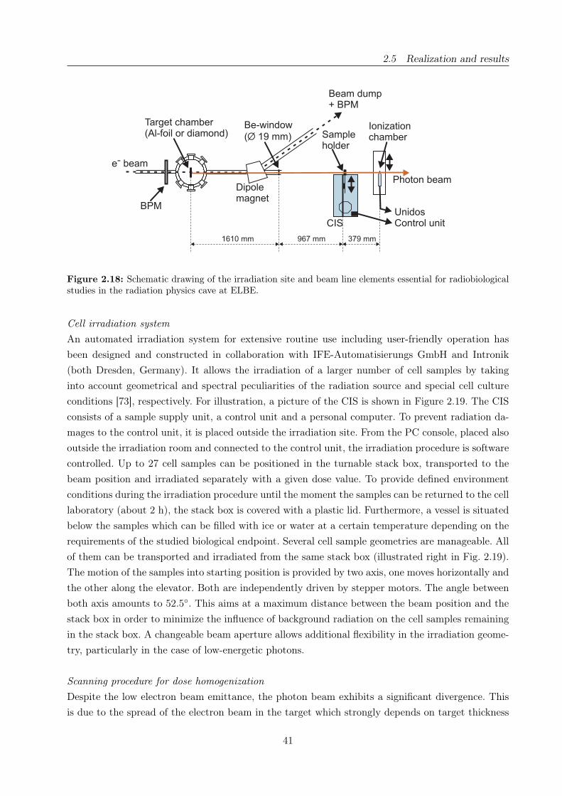
2.5 Realization and results
Beam dump+ BPM
Dipolemagnet
UnidosControl unit
Be-window
( 19 mm)Ø
CIS
Ionizationchamber
Photon beam
e beam-
BPM
Sampleholder
Target chamber(Al-foil or diamond)
379 mm967 mm1610 mm
Figure 2.18: Schematic drawing of the irradiation site and beam line elements essential for radiobiologicalstudies in the radiation physics cave at ELBE.
Cell irradiation system
An automated irradiation system for extensive routine use including user-friendly operation has
been designed and constructed in collaboration with IFE-Automatisierungs GmbH and Intronik
(both Dresden, Germany). It allows the irradiation of a larger number of cell samples by taking
into account geometrical and spectral peculiarities of the radiation source and special cell culture
conditions [73], respectively. For illustration, a picture of the CIS is shown in Figure 2.19. The CIS
consists of a sample supply unit, a control unit and a personal computer. To prevent radiation da-
mages to the control unit, it is placed outside the irradiation site. From the PC console, placed also
outside the irradiation room and connected to the control unit, the irradiation procedure is software
controlled. Up to 27 cell samples can be positioned in the turnable stack box, transported to the
beam position and irradiated separately with a given dose value. To provide defined environment
conditions during the irradiation procedure until the moment the samples can be returned to the cell
laboratory (about 2 h), the stack box is covered with a plastic lid. Furthermore, a vessel is situated
below the samples which can be filled with ice or water at a certain temperature depending on the
requirements of the studied biological endpoint. Several cell sample geometries are manageable. All
of them can be transported and irradiated from the same stack box (illustrated right in Fig. 2.19).
The motion of the samples into starting position is provided by two axis, one moves horizontally and
the other along the elevator. Both are independently driven by stepper motors. The angle between
both axis amounts to 52.5◦. This aims at a maximum distance between the beam position and the
stack box in order to minimize the influence of background radiation on the cell samples remaining
in the stack box. A changeable beam aperture allows additional flexibility in the irradiation geome-
try, particularly in the case of low-energetic photons.
Scanning procedure for dose homogenization
Despite the low electron beam emittance, the photon beam exhibits a significant divergence. This
is due to the spread of the electron beam in the target which strongly depends on target thickness
41

2.5 Realization and results
Elevator
Turnable
ELBE photon Pb−Aperture
Stack box
Beam
Horizontal carriage
Power supply
Ice container
Plastic cover
Figure 2.19: View of the cell irradiation system (left). Presentation of the cell sample holders with differentgeometry (top-right) and operation of the horizontal carriage (bottom-right).
and electron energy. Additionally, photon scattering on parts of the beam line and associated beam
attenuation leads to a confined beam spot in the plane perpendicular to the beam axis. The spot
size corresponds to the diameter of the beryllium vacuum window (see Fig. 2.18). At the cell sample
position, which is situated in a distance of 96.7 cm to the vacuum window and 257.6 cm to the
production target, the spot has a diameter of about 30 mm. Inside this spot the dose rate is not
uniformly distributed.
In order to compensate for beam non-uniformities, in radiation therapy flattening filters are used.
There, a uniform dose distribution is ensured by energy dependent absorption in the filter medium
which is not suitable for radiobiological studies with low-energetic photons of several keV due to
strong intensity attenuation.
Since a scanning option of the ELBE photon beam is not supported, the sample has to be moved.
This is realized by a scanning feature of the CIS. The samples are moved in the plane perpendi-
cular to the primary beam direction providing equal dose delivery to each cell of the sample. In
the present CIS design, the scanning movement is controlled by a list of user-defined coordinates
(scanning program) in order to take into account any cell sample geometry. A schematic drawing
of a scanning trajectory used in this study is shown right in Figure 2.20. The trajectories mainly
consist of a multiple of horizontal lines with a certain gap in between and a certain length according
to the geometry of the cell sample, but any trajectory is conceivable. By the use of spiral scanning
tracks, the homogenization effect can be increased [5]. For the reason of simplification, this kind of
trajectory was not used in our study so far. The choice of the coordinates and hence the maximum
field size is only limited by the outer frame size of the CIS and the stroke of the linear axis. A field
size up to 15 × 15 cm2 is accessible which is sufficient for the most applications in radiobiologi-
cal investigations. In this study, an in-house computer routine has been developed to simulate the
motion-sequence of the CIS taking into account the technical specifications of the stepping motors.
42

2.5 Realization and results
With the aid of a dose distribution determined with EBT films during static irradiation at ELBE,
the resulting distribution after the scanning movement of the sample can be calculated. For this,
the stationary distribution is propagated numerically along the given coordinates and summarized.
The resulting dose within the area of the cell sample and the duration of the irradiation process
can be predicted.
Furthermore, the quality of the dose uniformity, expressed as the fraction of the standard deviation
of the average dose in the region of interest σD and the average dose D, can be optimized. This is
done by varying the coordinates and, correspondingly, the gap between the lines and the line length.
The total irradiation duration is controlled by the scan speed as well as the number of repetitions
of the scanning program. It has to be practicable for radiobiological studies, and consequently a
maximum dose rate per unit of area is desirable. Another important parameter is the dose fraction
coming from background radiation onto the cell samples while they remain in the stack box. Apart
from shielding, this fraction can only be minimized by reduction of the total irradiation duration.
The dose delivered to the cell samples depends furthermore on the scan speed and on the fluctua-
tions of the electron and, correspondingly, the photon beam intensity. These fluctuations can be
compensated by an external feedback speed control signal. Therefore, the control unit of the irra-
diation system provides an interface for a voltage signal of ± 10 V. For calibration, the user adjusts
a certain beam intensity and relates the corresponding feedback signal of an external detector to the
initial scan speed as reference point for which further changes of the primary photon beam intensity
are computer-controlled. Both feedback signal as well as velocity and position of the linear guides
are logged with the PC for experimental control and further evaluation.
y
x
Track of thesample centre
x
D
y
D
Sample shapeat centralbeam axisposition
Figure 2.20: Schematic view of a dose beam profile and the meander-shaped track of the sample center fordose homogeneity over a circular sample.
Irradiation geometry
In radiobiological studies, mainly X-ray tubes are used for irradiation of biological objects. Attached
cells are commonly irradiated in culture flasks or petri dishes with a vertical beam. Contrarily, in
vitro cell experiments at the horizontal ELBE photon beam require a vertical position of the cell
layer. Drying of the cell culture has to be prevented by placing the cells in a dish filled with culture
medium. Moreover, sterility of the cell sample and the culture medium has to be assured during the
whole process of irradiation and preparation until the final step of cell fixation. Another limiting
condition comes from the energy range of the studied radiation field. In the case of low-energetic
photons the cross section for photo effect increases strongly with decreasing photon energy. In order
43
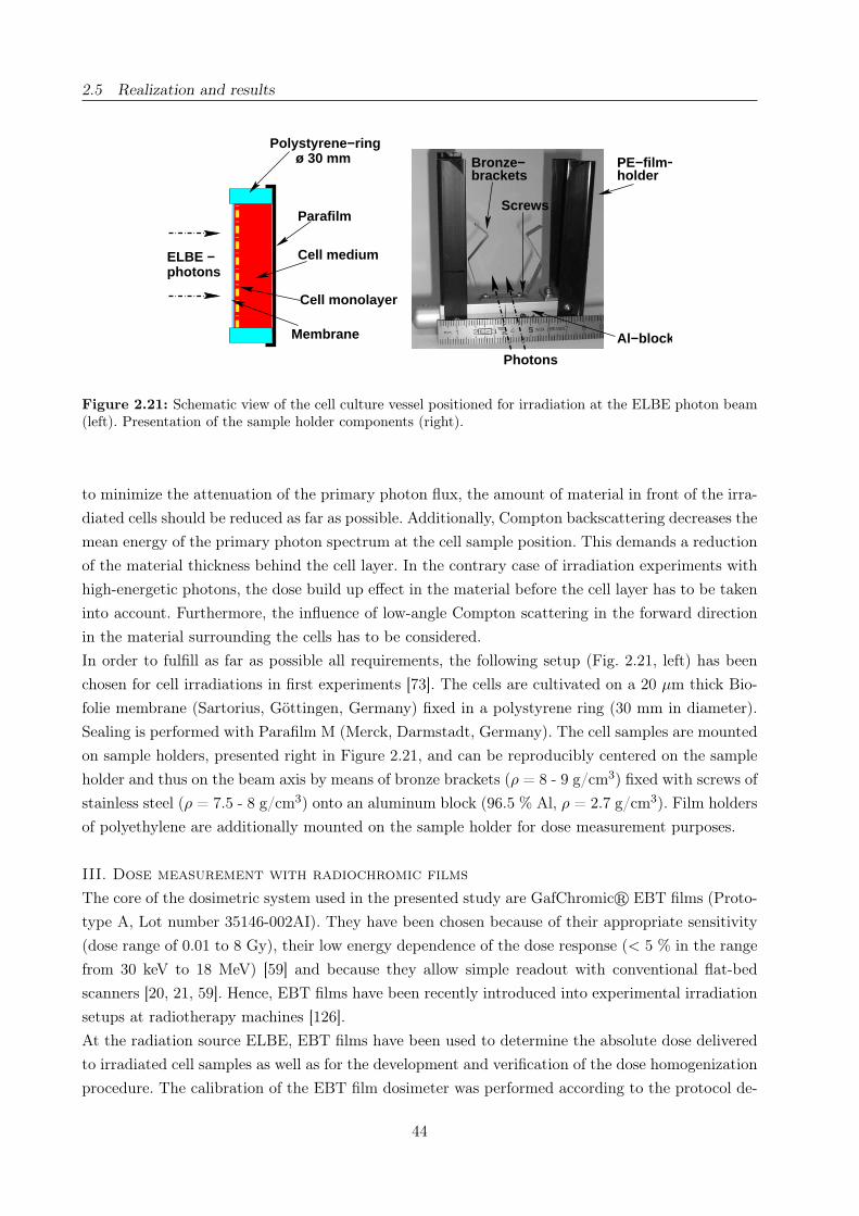
2.5 Realization and results
Photons
holderPE−film−
Al−block
photonsELBE −
Parafilm
ø 30 mm
Cell medium
Cell monolayer
Polystyrene−ring
Membrane
bracketsBronze−
Screws
Figure 2.21: Schematic view of the cell culture vessel positioned for irradiation at the ELBE photon beam(left). Presentation of the sample holder components (right).
to minimize the attenuation of the primary photon flux, the amount of material in front of the irra-
diated cells should be reduced as far as possible. Additionally, Compton backscattering decreases the
mean energy of the primary photon spectrum at the cell sample position. This demands a reduction
of the material thickness behind the cell layer. In the contrary case of irradiation experiments with
high-energetic photons, the dose build up effect in the material before the cell layer has to be taken
into account. Furthermore, the influence of low-angle Compton scattering in the forward direction
in the material surrounding the cells has to be considered.
In order to fulfill as far as possible all requirements, the following setup (Fig. 2.21, left) has been
chosen for cell irradiations in first experiments [73]. The cells are cultivated on a 20 µm thick Bio-
folie membrane (Sartorius, Göttingen, Germany) fixed in a polystyrene ring (30 mm in diameter).
Sealing is performed with Parafilm M (Merck, Darmstadt, Germany). The cell samples are mounted
on sample holders, presented right in Figure 2.21, and can be reproducibly centered on the sample
holder and thus on the beam axis by means of bronze brackets (ρ = 8 - 9 g/cm3) fixed with screws of
stainless steel (ρ = 7.5 - 8 g/cm3) onto an aluminum block (96.5 % Al, ρ = 2.7 g/cm3). Film holders
of polyethylene are additionally mounted on the sample holder for dose measurement purposes.
III. Dose measurement with radiochromic films
The core of the dosimetric system used in the presented study are GafChromicr EBT films (Proto-
type A, Lot number 35146-002AI). They have been chosen because of their appropriate sensitivity
(dose range of 0.01 to 8 Gy), their low energy dependence of the dose response (< 5 % in the range
from 30 keV to 18 MeV) [59] and because they allow simple readout with conventional flat-bed
scanners [20, 21, 59]. Hence, EBT films have been recently introduced into experimental irradiation
setups at radiotherapy machines [126].
At the radiation source ELBE, EBT films have been used to determine the absolute dose delivered
to irradiated cell samples as well as for the development and verification of the dose homogenization
procedure. The calibration of the EBT film dosimeter was performed according to the protocol de-
44
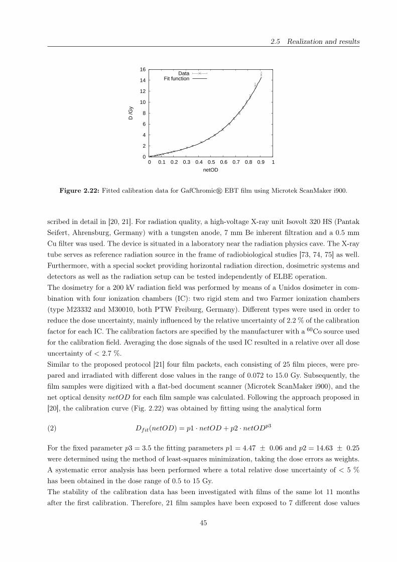
2.5 Realization and results
0
2
4
6
8
10
12
14
16
0 0.1 0.2 0.3 0.4 0.5 0.6 0.7 0.8 0.9 1
D /G
y
netOD
DataFit function
Figure 2.22: Fitted calibration data for GafChromicr EBT film using Microtek ScanMaker i900.
scribed in detail in [20, 21]. For radiation quality, a high-voltage X-ray unit Isovolt 320 HS (Pantak
Seifert, Ahrensburg, Germany) with a tungsten anode, 7 mm Be inherent filtration and a 0.5 mm
Cu filter was used. The device is situated in a laboratory near the radiation physics cave. The X-ray
tube serves as reference radiation source in the frame of radiobiological studies [73, 74, 75] as well.
Furthermore, with a special socket providing horizontal radiation direction, dosimetric systems and
detectors as well as the radiation setup can be tested independently of ELBE operation.
The dosimetry for a 200 kV radiation field was performed by means of a Unidos dosimeter in com-
bination with four ionization chambers (IC): two rigid stem and two Farmer ionization chambers
(type M23332 and M30010, both PTW Freiburg, Germany). Different types were used in order to
reduce the dose uncertainty, mainly influenced by the relative uncertainty of 2.2 % of the calibration
factor for each IC. The calibration factors are specified by the manufacturer with a 60Co source used
for the calibration field. Averaging the dose signals of the used IC resulted in a relative over all dose
uncertainty of < 2.7 %.
Similar to the proposed protocol [21] four film packets, each consisting of 25 film pieces, were pre-
pared and irradiated with different dose values in the range of 0.072 to 15.0 Gy. Subsequently, the
film samples were digitized with a flat-bed document scanner (Microtek ScanMaker i900), and the
net optical density netOD for each film sample was calculated. Following the approach proposed in
[20], the calibration curve (Fig. 2.22) was obtained by fitting using the analytical form
(2) Dfit(netOD) = p1 · netOD + p2 · netODp3
For the fixed parameter p3 = 3.5 the fitting parameters p1 = 4.47 ± 0.06 and p2 = 14.63 ± 0.25
were determined using the method of least-squares minimization, taking the dose errors as weights.
A systematic error analysis has been performed where a total relative dose uncertainty of < 5 %
has been obtained in the dose range of 0.5 to 15 Gy.
The stability of the calibration data has been investigated with films of the same lot 11 months
after the first calibration. Therefore, 21 film samples have been exposed to 7 different dose values
45

2.5 Realization and results
in the range of 0.7 to 14.6 Gy using the 200 kV filtered X-ray source. Subsequently, the netOD
values of the 3 samples with same dose have been averaged and inserted in Eq. 2. By comparing the
calculated dose with the actually delivered dose, a relative deviation of . 4 % has been determined.
This is within the total relative dose uncertainty of 5 % confirming the good stability of the EBT
film over a long period of time.
Dosimetric procedure
An important technical prerequisite for irradiation experiments is the presetting of a certain dose
value as precise as required. Direct measurement of the electron beam current is not efficiently
sensitive. This is due to variations in the beam setting in conjunction with electron scattering and,
correspondingly, with production of bremsstrahlung background radiation in parts of the beam line.
For this reason, the photon flux has been measured directly using an ionization chamber positioned
2956 mm behind the target (see Fig. 2.18). A dose rate of 1 Gy/min is adjusted by variation of
the electron beam current at the accelerator injector. By presetting a certain irradiation duration,
several dose values are provided, whereas on/off-switching of the electron beam is realized with a
precision of ± 2 s at the injector gate. Due to their poor spatial resolution, ionization chambers
are not suitable for the determination of the spatial dose distribution at the cell position. This fact
becomes even more important when the photon beam is not homogeneous. Also the influence of
the sample geometry cannot be taken into account. Therefore, the EBT films have been introduced.
The film samples were placed in special holders 5 mm in front of the cell samples (see right in
Fig. 2.21). For the dose value of a certain cell sample, the film dose signal was averaged over the
sample area. Subsequently, a correction of the obtained dose value is necessary which takes into
account the beam induced interactions in the material surrounding the cell samples as well as the
film itself, in accordance with the used photon energy range. Therefore, a piece of film is cut to fit
in the Biofolie vessel. It is irradiated instead of the cell layer at exactly the same position. With the
measured dose value in front of the sample Dfilm and the dose measured in the vessel DBiofolie, a
calibration factor k can be calculated by following expression
(3) k =DBiofolie − Dbckg
Dfilm − Dbckg.
The background dose Dbckg describes the dose fraction delivered to the cell samples in the stack
box during the whole irradiation process (about 2 h). It is due to background radiation coming
from photons as well as electrons mainly scattered in the region of the bending magnet and the
beam dump (see Fig. 2.18). The background dose was measured using several film samples well
distributed in the stack box. Vice versa, by applying Eq. 3, the corrected dose can be calculated
as function of k, Dfilm and Dbckg. Due to temporal and spatial variations of the photon beam
intensity and background radiation, the absolute dose was retrospectively measured with films for
each single cell sample. It should also be mentioned, that according to the radiobiological objectives
the dose fraction coming from background radiation has to be sufficiently small. For this reason an
appropriate over all duration of the irradiation procedure needs to be chosen.
46

2.5 Realization and results
− Horizontal lines: 7− Repetitions: 8
b) Program:
− Diameter: 68 mm
2 cm 2 cm
2 cm
a) Stationary irradiation c) Scanned experimental
d) Scanned simulated
3.00
2.70
2.40
2.10
1.80
Dos
e /G
y
3.00
2.70
2.40
2.10
1.80
Dos
e /G
y
2.10
1.79
1.48
1.18
0.87
Dos
e /G
y
Figure 2.23: Dose homogenization with 34 MV bremsstrahlung in the area indicated by the dashed circles(petri dish); a) Measured dose distribution after stationary irradiation, b) Scanning program, c) Resultingdose distribution measured with a moved film sample and d) Predicted dose distribution.
IV. Application
Homogenization procedure in experiments
The feasibility of the developed dose homogenization method as well as the described dosimetry
procedure have been demonstrated in independent experiments by reason of simplification. The
verification of the dose homogenization procedure has been performed with 34 MV bremsstrahlung.
Here, an aluminum foil of 18 µm thickness served as a production target. For experimental prepa-
ration, a stationary dose distribution measured on a previous experiment was taken to set up the
scanning program. The program was optimized for homogeneously distributed dose within the area
of a petri dish with a diameter of 45 mm. During the experiment, another stationary dose distri-
bution has been determined to reconfirm simulated and measured distribution after the scanning
process. As can be observed in Figure 2.23, the obtained distributions match very well. Since the
beam spot is not symmetric in the stationary distribution (Fig. 2.23) the actually homogenized area
is slightly displaced relative to its expected position (dashed circles). This effect has to be taken into
account in following experiments, or better yet, the symmetry of the beam spot and the position of
the dose maximum have to be online monitored.
47

2.5 Realization and results
Dose distribution D (Gy) σD/D (%) Deff (mGy/min)Stationary 1.53 14.1 365Scanned experimental 2.87 3.5 280Scanned simulated 2.91 2.3 −
Table 2.4: Comparison of the averaged effective dose rate Deff and the homogeneity of the different dosedistributions σD/D inside the region of the dashed circles in Fig. 2.23
For further illustration of the homogenization effect, the homogeneity parameter σD/D of the diffe-
rent dose distributions is listed in Table 2.4. As can be seen, the inhomogeneity has been successfully
reduced. The averaged effective dose rate Deff , where effective means after subtraction of the back-
ground dose fraction measured in the stack box, is listed in Table 2.4 as well. As expected, the
sample movement leads to a reduction of Deff in comparison to the static irradiation which reveals
another limitation. Since dose rate reduction correlates with an increase in irradiation duration, the
dose delivered to the cell samples in the stack box due to isotropic and polychromatic background
radiation is also augmented. Hence, the homogenization effect is decreased. In order to minimize the
background dose, the film samples were removed from the irradiation site directly after two samp-
les have been irradiated, with and without movement (14 min). But due to the large expenditure
of time for opening and closing the radiation protection doors, this is not suitable in routine use.
Therefore, in the following experiments the fraction of the background dose has been reduced by
one order of magnitude with a 2.5 mm thick Pb foil for additional shielding of the stack box.
For simplification this experimental run was performed without the correction of the relative inten-
sity fluctuations amounting only to 1 - 2 %. Contrarily, in the more time-consuming cell irradiation
experiments (about 2 h), significant fluctuations (about 10 %) can occur which then have to be
compensated.
RBE of 34 MV bremsstrahlung
The feasibility of the dosimetry method and the CIS has been demonstrated in the first cell irradia-
tion series at ELBE. This study was aimed at the determination of the RBE of 34 MV brems-
strahlung relative to 200 kV X-rays. On four experiment days, cell monolayers in Biofolie vessels
(about 40000 cells per sample) were irradiated. The 200 kV reference irradiation was performed
with the mentioned Isovolt X-ray tube in petri dishes (about 50000 cells per sample). The used
cell line 184A1 (purchased from ATCC, USA) consisted of the human mammary gland epithelial
cells originating from mammoplasty tissue of a 21 year old woman showing no detectable breast
epithelial cell pathology [120]. For the evaluation of radiation induced effects, the micronuclei (MN)
induction was used [74, 75]. After irradiation, the cells were observed under a light microscope and
the fraction of binucleated cells (BNC) with MN as an expression of the frequency of radiation
damage was determined.
As mentioned the absolute dose delivered to the cell layer was measured with film dosimeters in
front of each single cell sample. Since the beam spot on the film dosimeter was of the same size
48
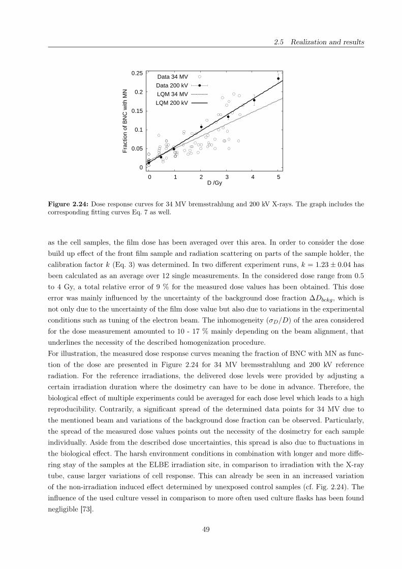
2.5 Realization and results
0
0.05
0.1
0.15
0.2
0.25
0 1 2 3 4 5
Fra
ctio
n of
BN
C w
ith M
N
D /Gy
Data 34 MV
Data 200 kV
LQM 34 MV
LQM 200 kV
Figure 2.24: Dose response curves for 34 MV bremsstrahlung and 200 kV X-rays. The graph includes thecorresponding fitting curves Eq. 7 as well.
as the cell samples, the film dose has been averaged over this area. In order to consider the dose
build up effect of the front film sample and radiation scattering on parts of the sample holder, the
calibration factor k (Eq. 3) was determined. In two different experiment runs, k = 1.23 ± 0.04 has
been calculated as an average over 12 single measurements. In the considered dose range from 0.5
to 4 Gy, a total relative error of 9 % for the measured dose values has been obtained. This dose
error was mainly influenced by the uncertainty of the background dose fraction ∆Dbckg, which is
not only due to the uncertainty of the film dose value but also due to variations in the experimental
conditions such as tuning of the electron beam. The inhomogeneity (σD/D) of the area considered
for the dose measurement amounted to 10 - 17 % mainly depending on the beam alignment, that
underlines the necessity of the described homogenization procedure.
For illustration, the measured dose response curves meaning the fraction of BNC with MN as func-
tion of the dose are presented in Figure 2.24 for 34 MV bremsstrahlung and 200 kV reference
radiation. For the reference irradiations, the delivered dose levels were provided by adjusting a
certain irradiation duration where the dosimetry can have to be done in advance. Therefore, the
biological effect of multiple experiments could be averaged for each dose level which leads to a high
reproducibility. Contrarily, a significant spread of the determined data points for 34 MV due to
the mentioned beam and variations of the background dose fraction can be observed. Particularly,
the spread of the measured dose values points out the necessity of the dosimetry for each sample
individually. Aside from the described dose uncertainties, this spread is also due to fluctuations in
the biological effect. The harsh environment conditions in combination with longer and more diffe-
ring stay of the samples at the ELBE irradiation site, in comparison to irradiation with the X-ray
tube, cause larger variations of cell response. This can already be seen in an increased variation
of the non-irradiation induced effect determined by unexposed control samples (cf. Fig. 2.24). The
influence of the used culture vessel in comparison to more often used culture flasks has been found
negligible [73].
49
2.5 Realization and results
However, a statistically significant radiobiological result has been obtained. The data was fitted by
means of the linear-quadratic model
(4) y(D) = c + αD + βD2,
with y(D) as corresponding fraction of BNC with MN for dose values D. The parameter c stands for
the background level given by non-irradiated control samples that were kept in the incubator in our
laboratory. The fitting parameters α and β were obtained by applying the method of least-square
minimization using the reciprocal standard deviations as weights. Finally, an RBE of 0.75 ± 0.05
was calculated as a fraction of the α-coefficients for 34 MV and 200 kV as reference. The given
uncertainty represents exclusively the statistic accuracy. The relative dose error of 9 % has to be
considered additionally.
In the literature no data is reported that can be directly compared to the here measured RBE of MV
bremsstrahlung. In several experiments [48] monoenergetic 60Co radiation is compared to X-rays in
the range of 200 to 350 kV. The results show an RBE ranging from 0.25 to 1, strongly dependent
on the applied high voltage and filtration of the used X-ray tube. Furthermore the RBE is strongly
influenced by the used cell line and the investigated biological endpoint. The best matching RBE of
0.8 with an important uncertainty of 38 % has been determined for human lymphocytes using 60Co
radiation versus 350 kV X-rays filtered with 1.12 mm Cu and 1.02 mm Al for induction of micro-
nuclei. However, the RBE obtained in our experiments is in good agreement with the systematics
of the RBE values reported in [48].
It should also be mentioned that for the application of 34 MV bremsstrahlung the influence of photo
nuclear particles, e.g., (γ,n), (γ,p) created by high-energetic photons hitting parts of the beam line
or the concrete wall of the cave has to be considered. Investigations of 50 MV bremsstrahlung com-
pared with 60Co radiation for several cell lines (V79-379A, GSH+/+ and U-1690) using clonogenic
assay show an RBE in the range of 0.988 to 1.009 [96]. Thus, an influence of photo nuclear reactions
is only marginal, if one accepts an error within a fraction of a per cent. In our experiments the neu-
tron background dose rate was monitored by means of a neutron detector (purchased from Berthold,
Bad Wildbad, Germany) consisting of a proportional counter filled with 3He gas and embedded in
a Bonner sphere of polyethylene with a diameter of 25 cm. It was situated in a distance of about
1 m to the stack box of the irradiation system. An equivalent dose rate in the range from 10 to
50 mSv/h has been measured. In comparison with the primary photon dose rate of ≈ 1 Gy/min,
the influence of photo neutrons has been consequently found to be negligible.
V. Conclusion
In the present work, physical and technical prerequisites for in vitro cell irradiation experiments in
the frame of radiobiological studies have been established and their feasibility has been successfully
tested with 34 MV bremsstrahlung. Thereby, one major topic was the setup of the automatic cell
irradiation system and its installation at the ELBE beam line for routine cell irradiation studies.
An appropriate procedure to compensate spatial inhomogeneity of the dose distribution within the
50
2.5 Realization and results
beam spot has been demonstrated. The second essential part was the dosimetry with GafChromicr
EBT film dosimeter. The films were calibrated with a 200 kV radiation field, where an accuracy of
5 % in the dose range of 0.5 to 15 Gy has been accomplished. Several specifications quoted by the
manufacturer or determined in [21] have been reconfirmed. As practical, reliable and low-budget
instruments, the EBT films were found to be highly suitable dosimetric instruments in experiments
involving unconventional radiation sources. Based on the EBT film dosimeter, two dedicated me-
thods have been separately developed. The film based measurement of the absolute dose delivered
to the cell sample and the homogenization of the applied dose over the area of the cell samples.
Both methods have already been introduced in extensive radiobiological studies to investigate the
RBE of photon radiation as a function of the photon energy. Although a minor energy dependence
of the dose response in the range from 30 keV up to 18 MeV has been quoted by the manufacturer
[59] and validated in several works [12, 16, 102], further investigations according to the experimental
requirements are of advantage. Especially for low-energetic photons (< 30 keV) the dose response
has to be verified either by means of our X-ray tube in combination with a soft X-ray ionization
chamber or in collaboration with the manufacturer.
The described methods can be easily transferred to experiments at ELBE with low-energetic CR
or even with the primary electron beam after verification of the film calibration. Certainly, in the
case of changed experimental conditions the contributions of dose build up effect and occurring
scattering effects to the dose distribution have to be verified. For 34 MV bremsstrahlung, this has
been exemplary demonstrated.
However, extensive radiobiological studies have been started. This includes RBE investigations for
different cell lines and various endpoints, such as clonogenic cell survival, induction of micronuclei,
chromosomal aberrations and DNA double strand break induction [6].
Acknowledgements
We are obliged to the ELBE crew for their interest and support of this work.
Author contributions
Study concept and design K.Z., E.B., J.P., W.W.; Experimental studies K.Z., E.B., J.P., E.L.,
W.W.; Data acquisition K.Z., E.L., E.B.; Literature research E.B., K.Z.; Data analysis K.Z., E.B.;
Manuscript preparation K.Z.; Manuscript editing J.P., E.B., W.W., K.Z.; Guarantor of integrity of
the entire study J.P.; Manuscript review was carried out by all authors.
51

2.5 Realization and results
Relative biological effectiveness of 25 and 10 kV X-rays for the induction of
chromosomal aberrations in two human mammary epithelial cell lines
Elke Beyreuthera, Wolfgang Dörrb, Anna Lehnerta, Elisabeth Lessmanna and Jörg Pawelkec,a
a Forschungszentrum Dresden-Rossendorf, Dresden, Germanyb Department of Radiotherapy and Radiation Oncology, Medical Faculty Carl Gustav Carus, Tech-
nische Universität Dresden, Dresden, Germanyc OncoRay, Center for Radiation Research in Oncology, Technische Universität Dresden, Dresden,
Germany
Corresponding author:
Elke Beyreuther
Institute of Radiation Physics
Forschungszentrum Dresden-Rossendorf
P.O. Box 510119
D-01314 Dresden, Germany
Fon: +49 351 260 3552
Fax: +49 351 260 3700
E-mail: [email protected]
Radiation and Environmental Biophysics 48(3):333-40(2009)
DOI: 10.1007/s00411-009-0221-4
www.springerlink.com
53

2.5 Realization and results
Abstract
Administration of ionizing radiation for diagnostic purposes can be associated with a risk for the
induction of tumors. Therefore, particularly with regard to general screening programs, e.g., with
mammography, cost-benefit considerations must be discussed including risk estimation depending
upon the radiation quality administered. The present study was initiated to investigate the in
vitro X-ray energy dependence for the induction of chromosomal aberrations in the two mammary
epithelial cell lines, 184A1 and MCF-12A. The induced excess fragments, dicentric chromosomes
and centric rings were analyzed and the relative biological effectiveness (RBE) was determined
for 10 and 25 kV X-rays relative to 200 kV X-rays. The assumed energy dependence with higher
values for 10 kV X-rays was confirmed for the excess fragments, with RBEM values of 1.92 ± 0.26
and 1.40 ± 0.12 for 10 kV X-rays and 1.17 ± 0.12 and 0.97 ± 0.10 for 25 kV photons determined
for cell lines 184A1 and MCF-12A, respectively. Meaningful results for the induction of dicentric
chromosomes and centric rings were obtained only for higher doses with RBE values of 1.31 ± 0.21
and 1.70 ± 0.29 for 184A1 and 1.08 ± 0.08 and 1.43 ± 0.12 for MCF-12A irradiated with 25 and
10 kV X-rays, respectively.
54

2.5 Realization and results
I. Introduction
During the last years the relative biological effectiveness (RBE) of low-energy X-rays, particularly
in the range below 50 keV, has been a topic of continuous discussions. One reason for this is a po-
tential risk of radiation-induced cancer by mammography, especially in the light of the introduction
of mammography screening programs in some countries, e.g., USA, The Netherlands and Germany
[122]. Based on in vitro data [32, 40, 45, 68, 74, 75, 86, 108, 112, 113, 119], it must be assumed that
X-rays in the low energy range are more effective per unit dose than X-rays of higher energy. In
contrast, a weighting factor wR of 1 is specified in the ICRP Recommendations [57], independent
of the photon energy.
Radiobiological investigations focusing on the RBE of soft X-rays, using the induction of micronuclei
as an endpoint, have been performed with human fibroblasts and keratinocytes [119] irradiated with
25 kV, and on human mammary epithelial cells irradiated with 25 kV [75] and 10 kV X-rays [74].
The maximum low-dose RBE values obtained for the number of micronuclei per binucleated cell
and relative to photons from a 200 kV X-ray tube, range from 1.4 to 4.1 for 25 and 10 kV X-rays,
respectively. Furthermore, enhanced RBE values have been found by experimental studies of neo-
plastic transformations of human hybrid cell line CGL1 irradiated with 29 kV X-rays compared to
different reference radiation sources [32, 40, 45]. Frankenberg et al. [32] found an RBE of about 4
by using 200 kV X-rays filtered with 0.6 mm Cu as reference. Similar results have been obtained
using 220 kV X-rays filtered with 4.05 mm Al and 0.5 mm Cu [40] as well as a 90Sr-90Y electron
source or a simulated atomic bomb spectrum [45].
Relative biological effectiveness values for the induction of chromosomal aberrations were deter-
mined depending upon the photon energy, especially for dicentric chromosomes in human lympho-
cytes [68, 108, 112, 113]. The photons applied were either generated by synchrotrons and were
monoenergetic within an energy range of 1.83 - 40 keV. Or, they were produced by X-ray tubes
with various filtrations and show a broad spectral distribution. Hill [48] reviewed these data and
concluded an increasing RBE with decreasing photon energy to a maximum at 6.9 keV, followed
by a decrease at lower energies. At the maximum of 6.9 keV Sasaki et al. [108] found an RBE of
4.6 ± 2.6 for monoenergetic synchrotron X-rays compared to photons from a 200 kV X-ray tube.
Accordingly, a maximum RBEM of 7.7 ± 3.0 was obtained for 6.9 keV monoenergetic synchrotron
X-rays relative to 60Co γ-rays [68].
Most of the experimental studies on the determination of RBE values for the induction of chromoso-
mal aberrations have been performed on human lymphocytes and rarely on other human cell lines.
However, mammary epithelial cells are of particular interest for the radiation induction of breast
cancer. Therefore, two human mammary epithelial cell lines, MCF-12A and 184A1, were chosen in
the present study to determine the relative biological effectiveness of soft X-rays for the induction
of chromosomal aberrations, especially for excess fragments. The irradiations were performed with
photons of 10 and 25 kV, and compared to 200 kV X-rays as reference.
55

2.5 Realization and results
II. Material and Methods
Cell culture
In the present experiments two human mammary gland epithelial cell lines, MCF-12A and 184A1
(both purchased from ATCC, USA), were applied.
The primary culture of 184A1 was established from the mammoplasty tissue of a 21-year-old wo-
man and shows no detectable breast epithelial cell pathology. Applying the carcinogen benzo[a]pyren
[120], a chemical transformation was induced resulting in the continuous cell line 184A1. This cell
line is adherent and immortal, but not malignant, with a chromosome modal number of 45, which
was determined in our laboratory using passage 7 cells. Monolayered 184A1 cells were maintai-
ned in serum-free mammary epithelial basal medium (MEBM, Cambrex, USA) supplemented with
5 µg/ml human epidermal growth factor, 0.5 µg/ml hydrocortisone, 5 µg/ml insulin, 52 µg/ml bo-
vine pituitary extract, 5 µg/ml gentamicin sulfate/amphotericin-B (all as MEGM SingleQuotsr,
Cambrex), 5 µg/ml human apo-transferrin (Sigma-Aldrich, USA) and 50 µg/ml prostaglandin E1
(Calbiochem, Germany). The medium and supplements were optimized for fast growing and high
plating efficiencies of epithelial cell lines [42], whereas the addition of prostaglandin E1 resulted in
a maximum plating efficiency of 35.2 ± 2.9 %.
Cell line MCF-12A, also originated from mammoplasty tissue, was derived by applying elevated
temperatures to the primary culture [94]. However, the mammoplasty tissue exhibited a kind of
benign breast disease (intraductal hyperplasia) and the derived cell line, although non-tumorigenic,
possesses several consistent numerical and structural aberrations, correlated with a variable chro-
mosome number between 65 and 71 [94]. A chromosome modal number of 64 was determined for the
present experiments. For the culture of cell line MCF-12A, described more detailed in Lehnert et al.
[75], a 1:1 mixture of DMEM and Ham´s F12 medium was supplemented with 5 % horse serum (all
from Biochrom, Germany), 0.5 µg/ml hydrocortisone, 10 µg/ml insulin (both from Sigma-Aldrich)
and 20 ng/ml epidermal growth factor (Becton Dickinson, USA). The cholera toxin (100 ng/ml,
Calbiochem, USA) originally added in the formula was substituted by 50 µg/ml Prostaglandin E1,
leading to a plating efficiency of 61.5 ± 4.7 %.
For detachment, a solution of 0.25 % trypsin and 0.03 % EDTA in PBS (all from Biochrom) was
applied for 10 min at 37◦C. Both cell lines were grown in 25 cm2 culture flasks (Nunc or Greiner,
Germany), with a seeding density of 6,000 cells per cm2, and incubated at 37◦C in humidified at-
mosphere containing 5 % CO2. The media, normally 5 ml per flask, were exchanged every third day
and subculturing was performed before reaching 70 % confluence.
In order to obtain synchronized cells, they were plated at densities between 6,000 and 12,000 cells
per cm2 and allowed to grow for 11 - 12 days to confluence arrest. Synchronization was verified by
the BrdU staining (see below), and for 184A1 additionally a FACS analysis was performed. Using
the BD FACSCaliburTM system (Becton Dickinson, USA) and cells stained with 50 µg/ml propi-
dium iodide, a fraction of 97 % of the cells was found in G0/G1-phase.
Chromosomal aberrations and differential chromatid labeling
For the determination of chromosomal aberrations (CA) and the number of cell cycles passed after
56
2.5 Realization and results
irradiation, we applied the method of Fluorescent plus Giemsa staining (FPG), as described by
Perry and Wolff [95], optimized for the cell lines used in this study. After irradiation, the cells were
subcultured at a density of 8,000 cells per cm2 and incubated for 45 h (184A1) or 36 h (MCF-12A),
respectively. In order to arrest mitotic cells in the first post irradiation division, colcemid (Biochrom)
was added to the culture medium for the last 2 h at a final concentration of 0.74 µg/ml for 184A1
and for 3 h at 0.29 µg/ml for MCF-12A. After harvesting, a hypotonic treatment with a solution
of 3 mg/ml trisodium citrate and 2.35 mg/ml potassium chloride (both from Merck, Germany) was
performed for 15 min and finally, the cells were fixed in a 3:1 methanol/acetic acid fixative (both
from VWR, Germany). The metaphase suspension was dropped onto grease-free, cold, wet slides
and stained with 2.1 % Giemsa (Merck) in PBS after one week.
In order to determine the number of cell cycles passed after irradiation, cells from the control group
as well as from the group irradiated with the highest dose of 5 Gy were incubated with 5-Bromo-
2´-deoxyuridine (BrdU, Sigma-Aldrich) at final concentrations of 3 µg/ml for 184A1 and 5 µg/ml
for MCF-12A, simultaneously to the probes where chromosomal aberrations were determined. The
cells incubated with BrdU were harvested and the slides were stained for 10 min in a solution of
0.1 mg/ml Hoechst H33258 (Bisbenzimide, VWR) in Weise buffer (25.1 mg/ml disodium hydrogen-
phosphate dodecahydrate, Merck and 9.1 mg/ml potassium dihydrogen phosphate, Sigma-Aldrich).
Subsequently, photolysis with 254 nm UV light was performed; the slides were incubated for 30 min
at 61◦C in a twofold SSC solution, made up of 13.2 mg/ml sodium chloride and 2.2 mg/ml trisodium
citrate (both from Merck), and eventually stained with Giemsa as described above.
Irradiation and dosimetry
The irradiation setups, dosimetry and the determination of the spectral dose distribution were des-
cribed previously in detail [74, 75]. In brief, the reference irradiation was performed with an Isovolt
320/20 X-ray tube (Roentgen Seifert, Germany) operated at 200 kV with a tungsten anode, 7 mm
Be inherent filtration and a 0.5 mm Cu filter. The culture flasks were placed horizontally at a focus-
to-cell distance of 45 cm and irradiated with a vertical X-ray beam through the top of the dish
and the culture medium. Three flasks were irradiated simultaneously at a dose rate of 0.3 Gy/min
for 0.5 Gy and 1.2 Gy/min for higher doses; those which were not currently irradiated were kept
on ice. Dosimetry was performed with a Unidos dosimeter and an ionization chamber type 31003
(semi-flex tube chamber 0.3 cm3, both PTW, Germany).
Irradiations with soft X-rays were performed using a tungsten target Darpac 150-MC X-ray tube
(Forward Raytech, UK) at 25 or 10 kV. The inherent filtration was 2 mm Be, the focus-to-cell
distance 25 cm. Dose rates were measured with a small soft X-ray ionization chamber type 23342
(sensitive volume 0.02 cm3) and a Unidos dosimeter (both from PTW). The application of a 0.3 mm
Al filter resulted in a dose rate of 2 Gy/min for cell irradiations with 25 kV. For 10 kV no additional
filtration was applied and the dose rate was 0.5 Gy/min. In order to minimize the beam attenuation
through the medium in case of the soft X-rays, the culture flasks were placed upside down in a
foamed polystyrene holder, and hence the cells were irradiated through the plastic bottom of the
flasks. Irradiation times did not exceed 10 min and no consequences from medium depletion were
57

2.5 Realization and results
expected. The irradiations were performed at room temperature and the irradiated flasks were kept
on ice until the experiment was finished. Afterwards all the flasks were treated simultaneously as
described before.
Dose distribution and homogeneity within the irradiated field of both X-ray tubes were determined
using GafChromic R© EBT dosimetry films (ISP, USA), which are self-developing, consist of tissue
equivalent materials and show high spatial resolution. The dose variation within the irradiated field
was lower than 3.0 % for the 200 kV X-rays, 4.0 % for 25 kV X-rays and 2.5 % for 10 kV X-rays.
In order to calculate the correct dose at the position of the cells for the 10 kV irradiations, the
thickness of the bottom was measured for all used culture flasks at five homogeneously distributed
positions. A previously defined correlation between thickness and dose was applied to calculate the
exact doses and their corresponding standard errors. In each experiment, two flasks per dose point
were irradiated, and two additional flasks for the BrdU staining were irradiated with 5 Gy.
Data evaluation
Chromosomal analyses were performed with a Zeiss Axiostar microscope at a magnification of 1,000.
Three independent experiments were analyzed for each dose point, except for the 25 kV irradiations
of 184A1 with two successful experiments. Acentric fragments, dicentric chromosomes and centric
Dose (Gy) Cells Dic. Rings Excess Distribution of exfrag. σ2/λ u valuescored fragments 0 1 2 3
200 kV X-rays
0 1,235 0 0 30 1,205 30 0.98 -0.590.5 668 0 0 31 637 31 0.96 -0.841 554 1 0 44 510 44 0.92 -1.312 571 11 2 84 495 68 8 1.04 0.773 300 12 2 48 254 44 2 0.93 -0.915 300 15 12 86 226 63 10 1 1.02 0.23
25 kV X-rays
0 1,120 0 0 22 1,098 22 0.98 -0.450.5 636 0 0 31 605 31 0.95 -0.861 450 4 0 41 410 39 1 0.96 -0.612 361 7 6 64 306 46 9 1.11 1.453 200 13 5 40 163 34 3 0.96 -0.465 200 19 11 63 143 51 6 0.88 -1.21
10 kV X-rays
0 1,035 0 0 30 1,006 28 1 1.04 0.900.492 ± 0.002 762 1 0 48 717 42 3 1.06 1.250.980 ± 0.008 728 11 1 88 643 82 3 0.95 -0.991.966 ± 0.016 663 41 6 138 545 100 16 2 1.11 2.052.953 ± 0.021 278 23 11 78 214 52 10 2 1.13 1.584.922 ± 0.035 300 55 22 103 220 59 19 2 1.15 1.79
The dose values for 10 kV are the mean from all experiments ± SD
Table 2.5: Yields of excess fragments (exfrag.), dicentric chromosomes (dic.) and centric rings observed inmetaphase cells of cell line 184A1 after irradiation with 200, 25 and 10 kV X-rays.
58
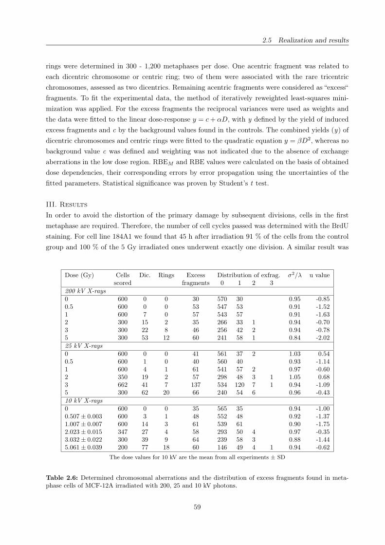
2.5 Realization and results
rings were determined in 300 - 1,200 metaphases per dose. One acentric fragment was related to
each dicentric chromosome or centric ring; two of them were associated with the rare tricentric
chromosomes, assessed as two dicentrics. Remaining acentric fragments were considered as “excess“
fragments. To fit the experimental data, the method of iteratively reweighted least-squares mini-
mization was applied. For the excess fragments the reciprocal variances were used as weights and
the data were fitted to the linear dose-response y = c + αD, with y defined by the yield of induced
excess fragments and c by the background values found in the controls. The combined yields (y) of
dicentric chromosomes and centric rings were fitted to the quadratic equation y = βD2, whereas no
background value c was defined and weighting was not indicated due to the absence of exchange
aberrations in the low dose region. RBEM and RBE values were calculated on the basis of obtained
dose dependencies, their corresponding errors by error propagation using the uncertainties of the
fitted parameters. Statistical significance was proven by Student’s t test.
III. Results
In order to avoid the distortion of the primary damage by subsequent divisions, cells in the first
metaphase are required. Therefore, the number of cell cycles passed was determined with the BrdU
staining. For cell line 184A1 we found that 45 h after irradiation 91 % of the cells from the control
group and 100 % of the 5 Gy irradiated ones underwent exactly one division. A similar result was
Dose (Gy) Cells Dic. Rings Excess Distribution of exfrag. σ2/λ u valuescored fragments 0 1 2 3
200 kV X-rays
0 600 0 0 30 570 30 0.95 -0.850.5 600 0 0 53 547 53 0.91 -1.521 600 7 0 57 543 57 0.91 -1.632 300 15 2 35 266 33 1 0.94 -0.703 300 22 8 46 256 42 2 0.94 -0.785 300 53 12 60 241 58 1 0.84 -2.02
25 kV X-rays
0 600 0 0 41 561 37 2 1.03 0.540.5 600 1 0 40 560 40 0.93 -1.141 600 4 1 61 541 57 2 0.97 -0.602 350 19 2 57 298 48 3 1 1.05 0.683 662 41 7 137 534 120 7 1 0.94 -1.095 300 62 20 66 240 54 6 0.96 -0.43
10 kV X-rays
0 600 0 0 35 565 35 0.94 -1.000.507 ± 0.003 600 3 1 48 552 48 0.92 -1.371.007 ± 0.007 600 14 3 61 539 61 0.90 -1.752.023 ± 0.015 347 27 4 58 293 50 4 0.97 -0.353.032 ± 0.022 300 39 9 64 239 58 3 0.88 -1.445.061 ± 0.039 200 77 18 60 146 49 4 1 0.94 -0.62
The dose values for 10 kV are the mean from all experiments ± SD
Table 2.6: Determined chromosomal aberrations and the distribution of excess fragments found in meta-phase cells of MCF-12A irradiated with 200, 25 and 10 kV photons.
59

2.5 Realization and results
obtained for MCF-12A 36 h post irradiation, with 95 % for the controls and 100 % for the irradiated
cells. In addition, the mitotic indices were determined depending upon the dose and energy app-
lied. For 184A1 the fraction of mitotic cells was reduced from 3.0 ± 0.4 % in the control groups to
0.26± 0.08 % in cell samples irradiated with the highest dose of 5 Gy. A reduction from 7.7± 1.5 %
for the controls to 4.0 ± 0.3 % for the 5 Gy irradiations was obtained for MCF-12A cell line.
The chromosomal aberrations depending upon X-ray energy and dose are presented in Tables 2.5
and 2.6, with pooled data from two to three independent experiments for each energy and dose
point. For the 10 kV irradiations the calculated dose values are given as the mean from all pooled
data for the same dose point together with their corresponding standard deviations.
To verify, whether the excess fragments and dicentric chromosomes were Poisson distributed, the
relative variance (σ2/λ) and the unit normal deviate (u) were calculated as test quantities. If the
relative variance approximated unity, the intercellular distributions of these aberrations followed a
Poisson distribution, and if the absolute value of the test quantity u did not exceed 1.96, no signifi-
cant over- or under-dispersion was present at the 5 % level of significance [25]. The attained relative
variances were about one in all instances. Moreover, no systematic change with dose or energy was
found for the test quantity u, and, for the excess fragments, a significant over- or under-dispersion
occurred only at one of ten doses of 10 and 200 kV X-rays, respectively. Additionally, by analyzing
the distribution of dicentric chromosomes (data not shown), significant over- or under-dispersion
was found only for one dose of 25 kV and two doses of 10 kV photons. Hence, the induced excess
fragments and dicentric chromosomes follow an underlying Poisson distribution. The derived distri-
butions of excess fragments for the cell lines 184A1 and MCF-12A are also included in Tables 2.5
and 2.6, together with their corresponding test quantities.
Figure 2.25: Yields of excess fragments per metaphase cell (± SEM ) induced in cell lines 184A1 (a) andMCF-12A (b) by 200 kV (box, solid line), 25 kV (triangle, dashed-dotted line) and 10 kV (circle, dashedline) X-rays in dependence on dose.
Both types of chromosomal aberrations, excess fragments and exchange aberrations, represented by
dicentric and centric ring chromosomes, were analyzed with regard to their underlying dose depen-
dencies. In the case of excess fragments, no significant β-values were obtained by the fit, so that
60
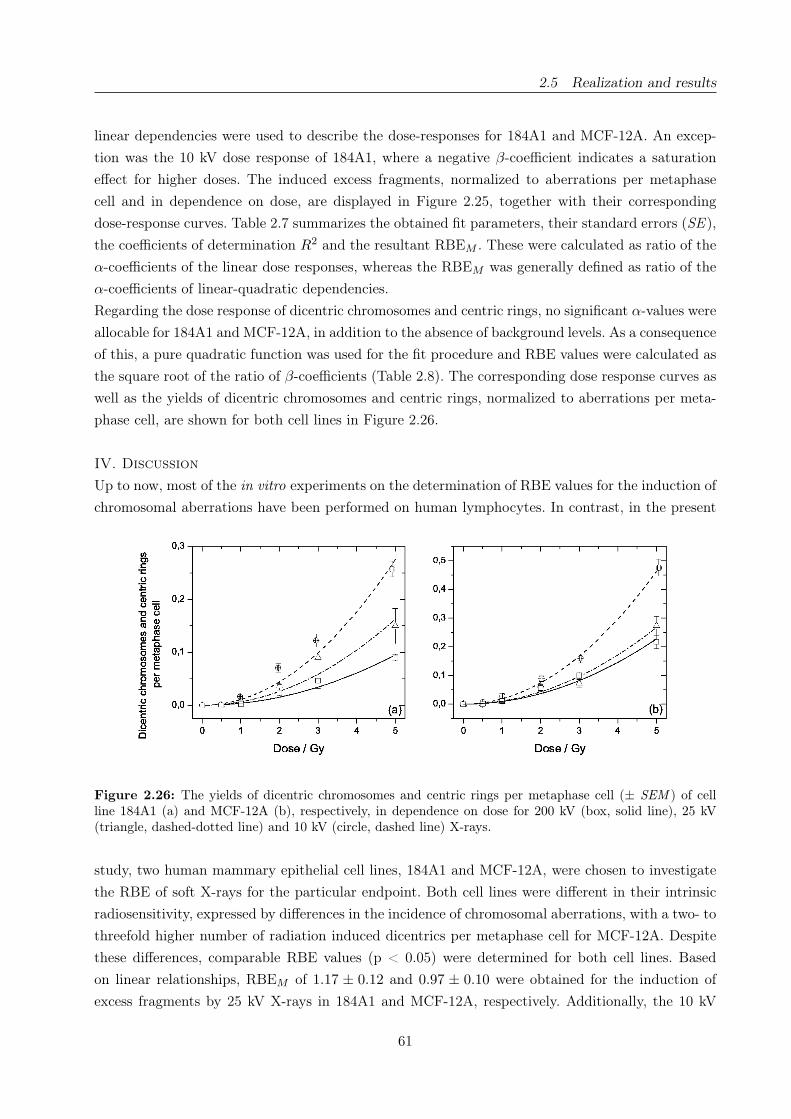
2.5 Realization and results
linear dependencies were used to describe the dose-responses for 184A1 and MCF-12A. An excep-
tion was the 10 kV dose response of 184A1, where a negative β-coefficient indicates a saturation
effect for higher doses. The induced excess fragments, normalized to aberrations per metaphase
cell and in dependence on dose, are displayed in Figure 2.25, together with their corresponding
dose-response curves. Table 2.7 summarizes the obtained fit parameters, their standard errors (SE ),
the coefficients of determination R2 and the resultant RBEM . These were calculated as ratio of the
α-coefficients of the linear dose responses, whereas the RBEM was generally defined as ratio of the
α-coefficients of linear-quadratic dependencies.
Regarding the dose response of dicentric chromosomes and centric rings, no significant α-values were
allocable for 184A1 and MCF-12A, in addition to the absence of background levels. As a consequence
of this, a pure quadratic function was used for the fit procedure and RBE values were calculated as
the square root of the ratio of β-coefficients (Table 2.8). The corresponding dose response curves as
well as the yields of dicentric chromosomes and centric rings, normalized to aberrations per meta-
phase cell, are shown for both cell lines in Figure 2.26.
IV. Discussion
Up to now, most of the in vitro experiments on the determination of RBE values for the induction of
chromosomal aberrations have been performed on human lymphocytes. In contrast, in the present
Figure 2.26: The yields of dicentric chromosomes and centric rings per metaphase cell (± SEM ) of cellline 184A1 (a) and MCF-12A (b), respectively, in dependence on dose for 200 kV (box, solid line), 25 kV(triangle, dashed-dotted line) and 10 kV (circle, dashed line) X-rays.
study, two human mammary epithelial cell lines, 184A1 and MCF-12A, were chosen to investigate
the RBE of soft X-rays for the particular endpoint. Both cell lines were different in their intrinsic
radiosensitivity, expressed by differences in the incidence of chromosomal aberrations, with a two- to
threefold higher number of radiation induced dicentrics per metaphase cell for MCF-12A. Despite
these differences, comparable RBE values (p < 0.05) were determined for both cell lines. Based
on linear relationships, RBEM of 1.17 ± 0.12 and 0.97 ± 0.10 were obtained for the induction of
excess fragments by 25 kV X-rays in 184A1 and MCF-12A, respectively. Additionally, the 10 kV
61

2.5 Realization and results
Radiation quality c ± SE (α ± SE)/Gy−1 (β ± SE)/Gy−2 R2 RBEM ± SE
184A1
200 kV 0.024 ± 0.004 0.052 ± 0.004 0a 0.99425 kV 0.020 ± 0.004 0.061 ± 0.004 0a 0.995 1.17 ± 0.1210 kV 0.029 ± 0.005 0.100 ± 0.011 -0.007 ± 0.002 0.999 1.92 ± 0.26
MCF-12A
200 kV 0.050 ± 0.009 0.035 ± 0.002 0a 0.94525 kV 0.068 ± 0.011 0.034 ± 0.003 0a 0.894 0.97 ± 0.1010 kV 0.058 ± 0.010 0.049 ± 0.003 0a 0.985 1.40 ± 0.12
Coefficient c was given by the corresponding background of excess fragments.a The β-coefficient was set to zero
Table 2.7: Regression coefficients and the derived low-dose RBE obtained for the linear dose-effect relation-ships of excess fragments induced in 184A1 and MCF-12A by 200, 25 and 10 kV X-rays.
irradiations of 184A1 and MCF-12A revealed RBEM of 1.92± 0.26 and 1.40± 0.12. Similar results
of 1.4±0.2 and 1.4±0.3 were determined in human lymphocytes for the same endpoint by applying
29 kV (filtered with 30 µm Mo) [113] and 10 kV X-rays (0.02 mm Cr, 5.4 keV mean energy) [105],
in comparison to 220 kV photons (0.5 mm Cu). And in good accordance with the present results,
RBE values of 1.13±0.03 [75] and 1.21±0.03 [74] were achieved in our laboratory for the clonogenic
survival using cell line MCF-12A and the same 25 and 10 kV X-ray tubes.
Despite these consensuses, the RBEM calculated here for excess fragments are based on linear rela-
tionships instead of linear-quadratic relations generally assumed for that endpoint (e.g.,[107]). The
existence of composite fragments generated by exchange processes of two single primary fragments
cannot be excluded, especially for higher doses. These composite fragments, which are not distin-
guishable by Giemsa staining, lead to an underestimation of the real amount of excess fragments,
and therefore, contribute to saturation effects, as observed in the present work for the 10 kV dose
response of 184A1. Boei et al. [9] partly solved this problem by applying fluorescence in situ hy-
bridization in combination with telomeric DNA. In addition, the amount of detected chromosomal
aberrations may be influenced by the sampling time. Again, Boei et al. [10] investigated this pheno-
menon for human lymphocytes and found increasing amounts of dicentrics and acentric fragments
for later sampling times within the same post irradiation mitosis. Regarding the epithelial cells in
the present work, the sampling time was optimized for harvesting a maximum number of cells in the
first post irradiation mitosis. By accident, the chosen incubation times seem to be ideal for lower
doses whereas for higher doses the cell cycle delay lead to reduced amounts of excess fragments.
Additionally, as indicated by the mitotic indices, a remarkable number of cells were not able to meet
the first mitosis after irradiation with the highest dose of 5 Gy.
Systematic investigations on the dependence of the RBE on photon energy have been performed by
several groups [68, 108, 112, 113] for the induction of dicentric chromosomes in human lymphocytes
by applying monoenergetic X-rays in the energy range of 1.83 - 40 keV as well as X-ray tubes with
various filtrations and a broad spectral distribution. The results were reviewed by Hill [48], who
concluded increasing RBE values with decreasing energy in the range of about 1.2 MeV to some keV
62
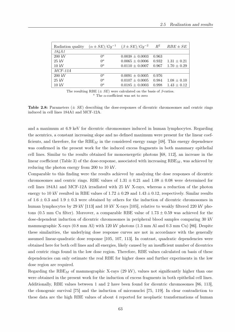
2.5 Realization and results
Radiation quality (α ± SE)/Gy−1 (β ± SE)/Gy−2 R2 RBE ± SE
184A1
200 kV 0a 0.0038 ± 0.0003 0.96325 kV 0a 0.0065 ± 0.0006 0.932 1.31 ± 0.2110 kV 0a 0.0110 ± 0.0007 0.967 1.70 ± 0.29
MCF-12A
200 kV 0a 0.0091 ± 0.0005 0.97625 kV 0a 0.0107 ± 0.0005 0.984 1.08 ± 0.1010 kV 0a 0.0185 ± 0.0003 0.998 1.43 ± 0.12
The resulting RBE (± SE) were calculated on the basis of β-ratios.a The α-coefficient was set to zero
Table 2.8: Parameters (± SE ) describing the dose-responses of dicentric chromosomes and centric ringsinduced in cell lines 184A1 and MCF-12A.
and a maximum at 6.9 keV for dicentric chromosomes induced in human lymphocytes. Regarding
the acentrics, a constant increasing slope and no defined maximum were present for the linear coef-
ficients, and therefore, for the RBEM in the considered energy range [48]. This energy dependence
was confirmed in the present work for the induced excess fragments in both mammary epithelial
cell lines. Similar to the results obtained for monoenergetic photons [68, 112], an increase in the
linear coefficient (Table 3) of the dose-response, associated with increasing RBEM , was achieved by
reducing the photon energy from 200 to 10 kV.
Comparable to this finding were the results achieved by analyzing the dose responses of dicentric
chromosomes and centric rings. RBE values of 1.31 ± 0.21 and 1.08 ± 0.08 were determined for
cell lines 184A1 and MCF-12A irradiated with 25 kV X-rays, whereas a reduction of the photon
energy to 10 kV resulted in RBE values of 1.72 ± 0.29 and 1.43 ± 0.12, respectively. Similar results
of 1.6 ± 0.3 and 1.9 ± 0.3 were obtained by others for the induction of dicentric chromosomes in
human lymphocytes by 29 kV [113] and 10 kV X-rays [105], relative to weakly filtered 220 kV pho-
tons (0.5 mm Cu filter). Moreover, a comparable RBE value of 1.73 ± 0.59 was achieved for the
dose-dependent induction of dicentric chromosomes in peripheral blood samples comparing 30 kV
mammographic X-rays (0.8 mm Al) with 120 kV photons (1.3 mm Al and 0.3 mm Cu) [86]. Despite
these similarities, the underlying dose response curves are not in accordance with the generally
assumed linear-quadratic dose response [105, 107, 113]. In contrast, quadratic dependencies were
obtained here for both cell lines and all energies, likely caused by an insufficient number of dicentrics
and centric rings found in the low dose region. Therefore, RBE values calculated on basis of these
dependencies can only estimate the real RBE for higher doses and further experiments in the low
dose region are required.
Regarding the RBEM of mammographic X-rays (29 kV), values not significantly higher than one
were obtained in the present work for the induction of excess fragments in both epithelial cell lines.
Additionally, RBE values between 1 and 2 have been found for dicentric chromosomes [86, 113],
the clonogenic survival [75] and the induction of micronuclei [75, 119]. In clear contradiction to
these data are the high RBE values of about 4 reported for neoplastic transformations of human
63
2.5 Realization and results
CGL1-hybrid cells irradiated with 29 kV X-rays, compared to higher energy photons of a 200 kV
(0.6 mm Cu) [32] or 220 kV (0.5 mm Cu) [40] X-ray tube. These results clarify that the assessment
of the biological effectiveness cannot be performed on the basis of one individual biological endpoint
and cell type. Cell line and type-specific differences in the radiosensitivity and repair capability
should be noted for a realistic estimation of the radiation risk. Considering the two human mam-
mary epithelial cell lines used in the present study, a higher radiosensitivity, expressed by a higher
frequency of dicentrics, was obtained for MCF-12A. Inherent genomic disorders of this cell line [94]
and their influence on the repair capability are reasonable explanations.
Author contributions
Study concept and design A.L., E.L., J.P., E.B.; Literature research E.B., A.L.; Experimental stu-
dies E.L., A.L.; Data acquisition E.L.; Data analysis and Manuscript preparation E.B.; Manuscript
editing A.L., J.P., W.D., E.B.; Guarantors of integrity of the entire study J.P., W.D.; Manuscript
review was carried out by all authors.
64
DNA double-strand break signaling: X-ray energy dependence of residual co-localized
foci of γ-H2AX and 53BP1
Elke Beyreuthera, Elisabeth Lessmanna, Jörg Pawelkeb,a and Stefan Pieckb
a Institute of Radiation Physics, Forschungszentrum Dresden-Rossendorf, Dresden, Germanyb OncoRay, Center for Radiation Research in Oncology, Medical Faculty Carl Gustav Carus, Uni-
versity of Technology Dresden, Dresden, Germany
Running header: X-ray energy dependent DNA double-strand break signaling
Corresponding author:
Elke Beyreuther
Institute of Radiation Physics
Forschungszentrum Dresden-Rossendorf
P.O. Box 510119
D-01314 Dresden, Germany
Fon: +49 351 260 3552
Fax: +49 351 260 3700
E-mail: [email protected]
International Journal of Radiation Biology 85(11):1042-50(2009)
DOI: 10.1080/09553000903232884
www.informahealthcare.com/rab
2.5 Realization and results
Abstract
Purpose: The application of ionizing radiation for medical purposes requires the investigation of in-
duced and persistent DNA damages, especially for soft X-rays that are assumed to be more effective
than higher energy photons. Therefore, we examine the energy dependent time and dose response
of residual DNA damage foci for soft X-rays in comparison to 200 kV photons.
Materials and methods: DNA damage present in cell line 184A1 within 48 h after irradiations
with 10 kV, 25 kV and 200 kV photons was analyzed by immunochemical detection of co-localized
γ-H2AX (phosphorylated histone H2AX) and 53BP1 (tumor protein 53 binding protein) foci.
Results: The dose dependencies of the co-located foci revealed significant energy dependent dif-
ferences with increasing amounts of residual foci at decreasing X-ray energy independent on post
irradiation time. Dose dependent RBE (relative biological effectiveness) values ranging from 4 to
7 were determined for 10 kV relative to 200 kV X-rays based on the 24 hour dose responses. For
25 kV photons, ratios considerably higher than one were obtained only for doses above 2 Gy.
Conclusions: The expected energy dependence with increasing DNA damage at decreasing pho-
ton energy was confirmed for the residual co-localized foci measured at different time points after
irradiation.
Keywords: γ-H2AX, 53BP1, 184A1, soft X-ray, DNA DSB
66

2.5 Realization and results
I. Introduction
DNA double-strand breaks (DSB) represent one of the most dangerous lesions induced by ionizing
radiation. Unrepaired DSB can lead to reduced clonogenic survival, while incorrect repair can re-
sult in chromosomal damages, genetic instabilities and transformations. Several methods have been
established to detect DSB, whereas one of the most sensitive ones is based on intracellular DNA
DSB signaling pathways that are switched on rapidly after the induction of radiation damage. As
an early step, the histone H2AX is phosphorylated at the sites of DNA DSB [104]. Subsequently,
the phosphorylated histone (γ-H2AX) is accumulated by distinct nuclear foci that are detectable
with appropriate antibodies. Due to its rapid appearance and good correlation to the number of
DSB, the determination of γ-H2AX foci have been used as a sensitive method to detect DSB at
a broad dose range, starting with doses of some cGy [80, 106]. However, the cell cycle dependent
expression of γ-H2AX with high amounts at late S- and G2/M-phase could reduce the detection
sensitivity [53, 79]. Confluent non-proliferating cells are then typically used for such experiments
to avoid the cycle-dependent influence. Another alternative is the detection of a second signaling
molecule downstream of the pathway in overlapping with the γ-H2AX signal.
The tumor protein 53 binding protein 1 (TP53BP1/53BP1) has been proposed as a good candidate
for double staining experiments by Rappold et al. [99]. This group demonstrated rapid radiation-
induced relocation of 53BP1 from a homogeneous distribution to nuclear foci that were stable and
co-localized with γ-H2AX foci for long times. Additionally, a clear dose dependence of 53BP1 foci
formation was apparent, but no clear dependence on cell cycle stage [2, 85, 127]. Accordingly, co-
localized 53BP1 and γ-H2AX foci have been used to detect DNA double-strand breaks.
Medical application of ionizing radiation requires the quantification of induced and even more per-
sistent DNA damages to estimate the risk of late effects. In this context, photon energies below
50 keV are of particular interest, due to their wide usage for therapeutic and diagnostic purposes
and their assumed enhanced relative biological effectiveness (RBE). Continuous discussions arise
especially with the introduction of mammography screening programs and the potential risk of
radiation-induced breast cancer in consequence of repeated screening (e.g., [37, 39]). Taking into
account these considerations, human mammary gland epithelial cells have been chosen by our group
to investigate the RBE of soft X-rays [6, 74, 75].
Nevertheless, systematic investigations on the photon energy dependence of the RBE have only
been performed for the induction of chromosomal aberrations in human lymphocytes. Summarizing
these studies, Hill [48] concluded an increasing RBE with decreasing photon energy and a maxi-
mum at 6.9 keV. At this particular energy, RBE values of 4.6 ± 2.6 and 7.7 ± 3.0 were obtained
comparing 6.9 keV monochromatic synchrotron X-rays and 200 kV photons [108] or 60Co γ-rays
[68], respectively.
In the present work, the amount of co-localized γ-H2AX and 53BP1 foci was determined in the hu-
man mammary gland epithelial cell line 184A1 with respect to post irradiation time and radiation
energy. Photons of 25 kV and 200 kV were applied to measure the kinetics within 48 hours. Dose
responses were examined 2 hours, 24 hours and 48 hours after irradiation using both X-ray tubes
as well as 10 kV photons. RBE values were calculated on basis of the 24 hour dose responses.
67

2.5 Realization and results
II. Material and Methods
Cell line
The human mammary gland epithelial cell line 184A1, described more detailed in Beyreuther et al.
[6], was derived by applying the carcinogen benzo[a]pyren to epithelial cells established from the
mammoplasty tissue of a 21-year-old healthy woman [120]. The cells are adherent and immortal, but
not malignant, and show low genomic instability and the absence of estrogen receptors [121]. The
cells were obtained from the American Type Culture Collection (ATCC, LGC Promochem, Wesel,
Germany) and maintained in serum-free mammary epithelial basal medium (MEBM, Lonza, Ver-
viers, Belgium) supplemented with 5 µg/ml human epidermal growth factor, 0.5 µg/ml hydrocortiso-
ne, 5 µg/ml insulin, 52 µg/ml bovine pituitary extract, 5 µg/ml gentamicin sulfate/amphotericin-B
(all as MEGM SingleQuotsr, Lonza), 5 µg/ml human apo-transferrin (Sigma-Aldrich, Taufkir-
chen, Germany) and 50 µg/ml prostaglandin E1 (VWR, Darmstadt, Germany). Cells were grown
in 25 cm2 culture flasks (Nunc, Wiesbaden or Greiner, Frickenhausen, Germany) and incubated at
37◦C in a humidified atmosphere containing 5 % CO2. The growth medium (5 ml per flask) was
exchanged every third day and subculturing was performed before reaching 70 % confluence. For
detachment, a solution of 0.25 % trypsin and 0.03 % ethylenediaminetetraacetic acid (EDTA) in
phosphate buffered saline (PBS) (all from Biochrom, Berlin, Germany) was applied for 10 min
at 37◦C.
To assure adherence, proliferating cells were plated in 8-well-chamber slides one day prior the irra-
diation experiment. Two different slide materials, soda-lime glass (VWR) and PermanoxTM plastic
(VWR), were utilized to assess the kinetics and the X-ray energy dependence, respectively. Cham-
ber slides enclosed with Parafilm (Brand, Wertheim, Germany) were positioned upside down in
the irradiation field of the X-ray tubes. The experiment and the storage of probes that were not
currently treated take place at room temperature. Afterwards, the cells were transferred back to
the incubator as soon as possible.
Irradiation and dosimetry
Irradiations with 10 kV and 25 kV soft X-rays were performed with a tungsten target Darpac
150-MC X-Ray tube (Forward Raytech, Swindon, UK) possessing an inherent filtration of 2 mm
Be. Due to beam attenuation, the chamber slides were placed upside down in a specially designed
foamed polystyrene holder with a resulting focus-to-cell distance of about 25 cm. Considering the
beam attenuation, the dose rate was measured with a small soft X-ray ionization chamber (Type
23342) with a sensitive volume of 0.02 cm3 and a Unidos dosimeter (both from PTW, Freiburg,
Germany). The measured dose rate at the cell monolayer position for 25 kV X-rays (0.3 mm Al
filter) was 2 Gy/min for the plastic and 0.25 Gy/min for the glass slides. For 10 kV X-rays (no
additional filtration) a dose rate of 0.65 Gy/min was attained for plastic slides. Due to their strong
attenuation, the glass slides were not used for this X-ray energy.
The reference irradiations were performed with two 200 kV X-ray tubes (Yxlon International A/S,
Taastrup, Denmark or Isovolt 320/20, Roentgen Seifert, Ahrensburg, Germany), both equipped with
a tungsten anode and filtered with 0.5 mm Cu. They were however differing in inherent filtration
68

2.5 Realization and results
with 7 mm Be for the Isovolt 320/20 and 3 mm Be + 3 mm Al for the Yxlon tube, respectively. To
achieve comparable conditions the chamber slides were also placed upside down with a focus-to-cell
distance of 44 cm. The dose rates, measured with a Unidos dosimeter and two Farmer ionization
chambers (Type 30010, 0.6 cm3 sensitive volume, PTW) were 1.34 Gy/min and 1.24 Gy/min for
the PermanoxTM (Yxlon) and glass (Isovolt) slides, respectively. A current reduction from 20 mA
to 5 mA, necessary for the 0.25 Gy irradiations, resulted in a dose rate of 0.27 Gy/min (on glass).
Dose distributions and homogeneities within the irradiation fields were measured with GafChromicr
EBT films (ISP Corp., Wayne, NJ, USA), which consist of tissue equivalent materials, are self-
developing and show high spatial resolution. The determined dose variation within the fields of 10,
25 and 200 kV X-rays was better than 4.0 %. Because of this high homogeneity, chamber slides
that were assembled for the same radiation dose but analyzed after different periods of time were
irradiated simultaneously for each experiment.
Influence of the different slide materials on the photon spectra
Since the densities of the different slide materials varied between 2.5 g/cm3 for glass and 1.19 g/cm3
for plastic, the material influence on the primary photon spectra have to be considered carefully.
Spectra of the three radiation qualities applied were provided by Lehnert et al. [74, 75]. Together
with the simulation tool GEANT4 (version 8.1 [35]) these spectra were utilized to simulate the
transition of the original photon spectra through glass or plastic. Moreover, the number and energy
of secondary electrons generated in the slide material and escaping into the cell monolayer have
been determined by approximating the cell monolayer with a 10 micrometer thick water layer in
the GEANT4 simulation.
Immunofluorescence staining of γ-H2AX and 53BP1
The applied method of immunofluorescence staining of the two DSB signaling molecules (γ-H2AX
and 53BP1) was adopted from Eke et al. [27] and customized for our cell line.
Following irradiation, the cells were incubated at 37◦C for times between 5 minutes and 48 hours.
Subsequently, they were fixed in 1 % formaldehyde/PBS (5 min, Sigma-Aldrich/Biochrom), was-
hed two times in PBS/glycine (VWR), permeabilized for 10 min in ice cold 0.25 % Triton X/PBS
(VWR), and washed in PBGT, which is a solution of 0.5 % Gelatine (neolab, Heidelberg, Germany)
and 0.05 % Tween 20 (Sigma-Aldrich) in PBS. Cells were then incubated with an anti-phospho-
Histone H2AX monoclonal antibody (1:1000, 1 h, Chemicon via Millipore, Schwalbach, Germany),
Alexa Fluor 594 goat anti-mouse secondary antibody (1:400, 30 min, Invitrogen, Karlsruhe, Germa-
ny), 53BP1 rabbit polyclonal antibody cocktail (1:3000, 1 h, Acris, Hiddenhausen, Germany), and
Alexa Fluor 488 goat anti-mouse secondary antibody (1:1000, 30 min, Invitrogen). The antibodies
and fluorescent dyes were diluted in PBGT, which is also used for washing between the antibody
incubations in a wet chamber at 37◦C. Lastly, the samples were washed two times in PBS and moun-
ted under coverslips with 4 µl 4’-6-Diamidino-2-phenylindole (DAPI)/Vectashield (1200) mounting
medium (Axxora, Lörrach, Germany) per well. Kinetic investigations were performed with longer
incubation times of 90 minutes for the primary and 45 minutes for the secondary antibodies.
69
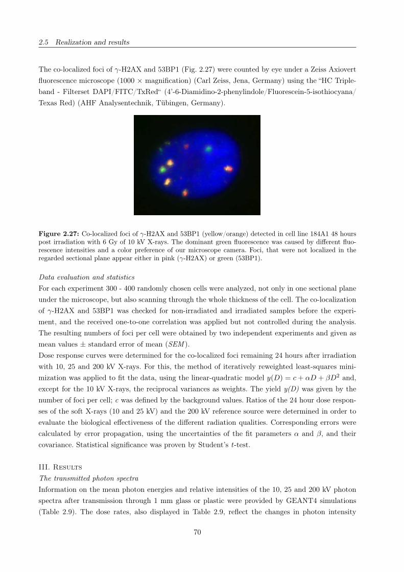
2.5 Realization and results
The co-localized foci of γ-H2AX and 53BP1 (Fig. 2.27) were counted by eye under a Zeiss Axiovert
fluorescence microscope (1000 × magnification) (Carl Zeiss, Jena, Germany) using the “HC Triple-
band - Filterset DAPI/FITC/TxRed“ (4’-6-Diamidino-2-phenylindole/Fluorescein-5-isothiocyana/
Texas Red) (AHF Analysentechnik, Tübingen, Germany).
Figure 2.27: Co-localized foci of γ-H2AX and 53BP1 (yellow/orange) detected in cell line 184A1 48 hourspost irradiation with 6 Gy of 10 kV X-rays. The dominant green fluorescence was caused by different fluo-rescence intensities and a color preference of our microscope camera. Foci, that were not localized in theregarded sectional plane appear either in pink (γ-H2AX) or green (53BP1).
Data evaluation and statistics
For each experiment 300 - 400 randomly chosen cells were analyzed, not only in one sectional plane
under the microscope, but also scanning through the whole thickness of the cell. The co-localization
of γ-H2AX and 53BP1 was checked for non-irradiated and irradiated samples before the experi-
ment, and the received one-to-one correlation was applied but not controlled during the analysis.
The resulting numbers of foci per cell were obtained by two independent experiments and given as
mean values ± standard error of mean (SEM ).
Dose response curves were determined for the co-localized foci remaining 24 hours after irradiation
with 10, 25 and 200 kV X-rays. For this, the method of iteratively reweighted least-squares mini-
mization was applied to fit the data, using the linear-quadratic model y(D) = c + αD + βD2 and,
except for the 10 kV X-rays, the reciprocal variances as weights. The yield y(D) was given by the
number of foci per cell; c was defined by the background values. Ratios of the 24 hour dose respon-
ses of the soft X-rays (10 and 25 kV) and the 200 kV reference source were determined in order to
evaluate the biological effectiveness of the different radiation qualities. Corresponding errors were
calculated by error propagation, using the uncertainties of the fit parameters α and β, and their
covariance. Statistical significance was proven by Student’s t-test.
III. Results
The transmitted photon spectra
Information on the mean photon energies and relative intensities of the 10, 25 and 200 kV photon
spectra after transmission through 1 mm glass or plastic were provided by GEANT4 simulations
(Table 2.9). The dose rates, also displayed in Table 2.9, reflect the changes in photon intensity
70

2.5 Realization and results
and energy, taking into account the corresponding photon energy dependence of the mass energy-
absorption coefficients [51].
Regarding the 200 kV reference source, only small changes of the mean photon energy, beam inten-
sity and dose rates of the transmitted photon spectra were achieved for the different slide materials.
By contrast, the usage of glass slides result in clearly shifted mean photon energies and considerably
reduced intensities for the 25 and 10 kV photons. Moreover, no dose signal was detectable after at-
tenuation of the 10 kV X-rays by 1 mm soda-lime glass. Slightly shifted mean photon energies and
reduced photon intensities and dose rates were also obtained for both soft X-rays passing through
a 1 mm plastic slide. The reduced dose rates were compensated by longer irradiation times but not
Influence on Mean photon Relative No. of sec. Mean energy of Dose ratethe spectra energy photon electrons per sec. electrons /Gy/min
/keV intensity 106 photons /keV
200 kV photons
Undisturbed2 89.78 1 - - 1.361/1.302
Plastic2 89.76 0.990 50 21.4 1.341
Glass2 90.29 0.962 207 43.7 1.242
25 kV photons
Undisturbed 15.42 1 - - 2.24Plastic 15.74 0.909 81 7.0 2.04Glass 19.26 0.180 148 10.3 0.25
10 kV photons
Undisturbed 6.93 1 - - 1.47Plastic 7.46 0.490 99 3.7 0.65Glass 9.15 1e-05 0 - not detectable
200 kV X-ray tubes: 1Yxlon International A/S; 2Isovolt 320/20, Roentgen Seifert
Table 2.9: Parameters of the applied photon spectra transmitted through 1 mm soda-lime glass orPermanoxr plastic. Mean photon energies of the undisturbed spectra were provided by Lehnert et al.
[74, 75]. The mean energies and relative intensities of the transmitted photons as well as the fraction andmean energy of secondary (sec.) electrons produced in the slides and escaping into the cell monolayer weresimulated by GEANT4. For the 200 kV reference source the parameters were simulated on basis of the spec-trum of the Isovolt tube, whereas only small deviations were expected for the Yxlon tube. Dose rates weremeasured in consideration of the irradiation geometry.
exceeding 10 minutes to avoid effects of medium depletion. Furthermore, the 10 kV X-ray tube was
not applied for cell irradiations on glass slides.
Concerning the production of secondary electrons, additional simulations were performed to ascer-
tain the spectra and number of those electrons that were transferred from the slides to the cell
monolayer. For the glass slides, both the number and the mean energy of the produced secondary
electrons were increased with X-ray energy. Consequently, the electron range and with it the proba-
bility that these electrons penetrate the cell nucleus and contribute to DNA damage was increased.
By comparison, fewer secondary electrons with a lower mean energy and range were produced in
the plastic slides, associated with a lower probability of penetrating the cell nucleus.
71

2.5 Realization and results
Kinetics
In a first series of experiments, the time course of foci appearance and loss was studied after 25 kV
and 200 kV X-ray irradiations of the human mammary epithelial cell line 184A1. Despite the higher
yields obtained for 25 kV photons, similar biphasic and energy independent kinetics were found
(Fig. 2.28). Additionally, a constant maximum was attained 0.5 and 2 hours after irradiation with
0.25 and 2 Gy, respectively. The repair, or better loss, half times measured for the higher dose were
4.5 h and approximately 15 h for the steep and the flat sloping curve progression, respectively. A
dose reduction to 0.25 Gy lead to a change in foci loss half times to 3.5 h and 25 h for the faster
and slower decreasing slopes.
0 1 2 3 4 5 6 25 30 35 40 45 500
1
2
3
4
5
6
7
8
9
Co-
loca
lized
foci
per
cel
l
Time after irradiation / h
200 kV X-rays: 2 Gy 0.25 Gy
25 kV X-rays: 2 Gy 0.25 Gy
Figure 2.28: Time courses of co-localized foci measured between 5 minutes and up to 48 hours postirradiation. Cells were irradiated with doses of 0.25 Gy (squares) and 2 Gy (circles) of 25 kV (black) or200 kV (open) X-rays. All values are displayed as average ± SEM based on two independent experiments.
X-ray energy and dose dependence of the number of co-localized foci of γ-H2AX and 53BP1
In addition to the kinetic considerations, the X-ray energy dependence and dose-effects for low and
higher energy X-rays were ascertained in more detail. For this, visible co-localized foci were analyzed
at three different times following irradiation with doses of up to 6 Gy (Table 2.10). The earliest
time (2 h) was chosen because of the maximum obtained at 2 Gy; remaining foci were determined
at 24 and 48 hours following irradiation.
In order to evaluate the possible influence of the slide material on the induction of γ-H2AX and
53BP1 foci, the results obtained at 2, 24 and 48 hours after irradiations with 25 kV and 200 kV
photons were compared (not shown). No significant differences (p ≤ 0.05) were found between the
number of foci determined on glass or plastic slides for both X-ray energies. For this, it was con-
cluded that dose rate compensation was done quite well and that the shifted mean energy and
produced secondary electrons have in this case no great influence on DNA damage. The dose re-
sponses (Table 2.10) measured 2 hours after irradiation revealed a significant increase in the number
72

2.5 Realization and results
Dose/Gy Average number of foci per cell
2 h 24 h 48 h
200 kV X-rays
0 0.40 ± 0.05 0.31 ± 0.04 0.31 ± 0.010.49 ± 0.02 1.03 ± 0.09 0.43 ± 0.01 0.26 ± 0.031.01 ± 0.02 2.72 ± 0.25 0.50 ± 0.03 0.28 ± 0.012.01 ± 0.02 4.84 ± 0.21 0.87 ± 0.06 0.37 ± 0.064.00 ± 0.03 8.42 ± 0.04 1.51 ± 0.01 0.60 ± 0.066.01 ± 0.03 - 3.75 ± 0.18 0.94 ± 0.06
25 kV X-rays
0 0.27 ± 0.02 0.29 ± 0.09 0.26 ± 0.040.51 ± 0.04 2.84 ± 0.22 0.62 ± 0.03 0.31 ± 0.041.00 ± 0.04 6.02 ± 0.03 0.75 ± 0.14 0.39 ± 0.102.00 ± 0.04 8.26 ± 0.02 0.98 ± 0.02 0.51 ± 0.123.99 ± 0.04 11.39 ± 0.02 3.01 ± 0.12 1.29 ± 0.056.01 ± 0.05 - 5.87 ± 0.12 2.22 ± 0.12
10 kV X-rays
0 0.40 ± 0.03 0.32 ± 0.09 0.26 ± 0.030.50 ± 0.02 5.57 ± 0.08 1.72 ± 0.04 0.66 ± 0.081.00 ± 0.03 7.59 ± 0.06 2.07 ± 0.04 0.95 ± 0.032.00 ± 0.05 12.30 ± 0.36 3.59 ± 0.29 1.22 ± 0.154.00 ± 0.08 17.22 ± 0.76 4.13 ± 0.02 1.47 ± 0.086.00 ± 0.12 - 6.40 ± 0.08 2.70 ± 0.38
Table 2.10: Dose response of cell line 184A1, measured on PermanoxTM at different time points afterirradiation with 10 kV, 25 kV and 200 kV X-rays. The dose of 6 Gy was excluded at the first time (2 h) dueto analytical limits. Yields of two independent experiments were pooled (± SEM ) and the dose values weregiven with their uncertainties.
of foci with decreasing X-ray energy. These X-ray energy dependent differences partly disappeared
later on with no significant different results (p ≤ 0.05) for doses up to 2 Gy of 200 and 25 kV X-rays
(Fig. 2.29, Table 2.10). Nevertheless, the yields measured for 10 kV X-rays remained elevated and
comparable results were obtained for higher doses (4 Gy and more) and both soft X-ray energies
24 and 48 hours post irradiation. The number of co-localized foci per cell, determined 24 hours
after irradiation, was used to calculate the dose response curves displayed in Figure 2.29 for the
three X-ray energies investigated. Table 2.11 summarizes the corresponding regression parameters,
their standard errors and covariances as well as the coefficients of determination (R2). A negative
β-coefficient was obtained for 10 kV X-rays likely caused by foci overlap or an occlusion effect [98]
at higher doses. The biological effectiveness of the soft X-rays was assessed regarding their 24 hour
dose responses in relation to the 200 kV reference source. The results (Table 2.12) reveal a ratio or
relative biological effectiveness (RBE) of about one considering doses of up to 2 Gy of 25 and 200 kV
X-rays. Significantly enhanced values (p ≤ 0.05) were obtained for all doses of 10 kV relative to
200 kV photons, whereas comparable results were ascertained for both soft X-rays and doses above
3 Gy. For the data achieved at 48 hours, although elevated for 10 kV X-rays, regression was not
realized as the cell cycle duration is generally 30 hours.
73
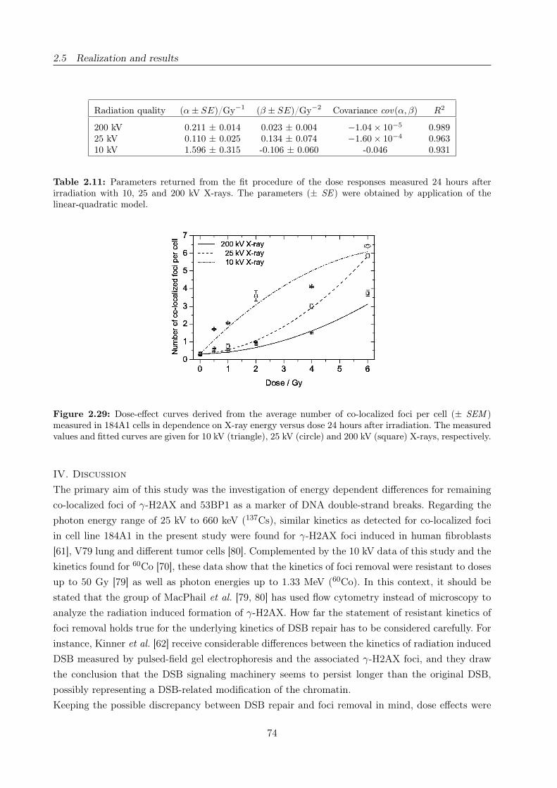
2.5 Realization and results
Radiation quality (α ± SE)/Gy−1 (β ± SE)/Gy−2 Covariance cov(α, β) R2
200 kV 0.211 ± 0.014 0.023 ± 0.004 −1.04 × 10⁻⁵ 0.98925 kV 0.110 ± 0.025 0.134 ± 0.074 −1.60 × 10⁻⁴ 0.96310 kV 1.596 ± 0.315 -0.106 ± 0.060 -0.046 0.931
Table 2.11: Parameters returned from the fit procedure of the dose responses measured 24 hours afterirradiation with 10, 25 and 200 kV X-rays. The parameters (± SE ) were obtained by application of thelinear-quadratic model.
Figure 2.29: Dose-effect curves derived from the average number of co-localized foci per cell (± SEM )measured in 184A1 cells in dependence on X-ray energy versus dose 24 hours after irradiation. The measuredvalues and fitted curves are given for 10 kV (triangle), 25 kV (circle) and 200 kV (square) X-rays, respectively.
IV. Discussion
The primary aim of this study was the investigation of energy dependent differences for remaining
co-localized foci of γ-H2AX and 53BP1 as a marker of DNA double-strand breaks. Regarding the
photon energy range of 25 kV to 660 keV (137Cs), similar kinetics as detected for co-localized foci
in cell line 184A1 in the present study were found for γ-H2AX foci induced in human fibroblasts
[61], V79 lung and different tumor cells [80]. Complemented by the 10 kV data of this study and the
kinetics found for 60Co [70], these data show that the kinetics of foci removal were resistant to doses
up to 50 Gy [79] as well as photon energies up to 1.33 MeV (60Co). In this context, it should be
stated that the group of MacPhail et al. [79, 80] has used flow cytometry instead of microscopy to
analyze the radiation induced formation of γ-H2AX. How far the statement of resistant kinetics of
foci removal holds true for the underlying kinetics of DSB repair has to be considered carefully. For
instance, Kinner et al. [62] receive considerable differences between the kinetics of radiation induced
DSB measured by pulsed-field gel electrophoresis and the associated γ-H2AX foci, and they draw
the conclusion that the DSB signaling machinery seems to persist longer than the original DSB,
possibly representing a DSB-related modification of the chromatin.
Keeping the possible discrepancy between DSB repair and foci removal in mind, dose effects were
74
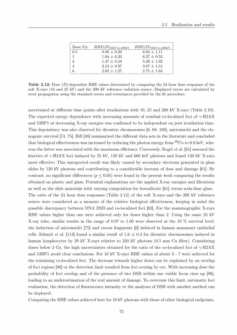
2.5 Realization and results
Dose/Gy RBE(D)25kVvs.200kV RBE(D)10kVvs.200kV
0.5 0.80 ± 0.20 6.94 ± 1.111 1.04 ± 0.33 6.37 ± 0.532 1.47 ± 0.58 5.39 ± 1.024 2.13 ± 0.97 3.87 ± 1.516 2.62 ± 1.27 2.75 ± 1.63
Table 2.12: Dose (D)-dependent RBE values determined by comparing the 24 hour dose responses of thesoft X-rays (10 and 25 kV) and the 200 kV reference radiation source. Displayed errors are calculated byerror propagation using the standard errors and covariances provided by the fit procedure.
ascertained at different time points after irradiations with 10, 25 and 200 kV X-rays (Table 2.10).
The expected energy dependence with increasing amounts of residual co-localized foci of γ-H2AX
and 53BP1 at decreasing X-ray energies was confirmed to be independent on post irradiation time.
This dependency was also observed for dicentric chromosomes [6, 68, 108], micronuclei and the clo-
nogenic survival [74, 75]. Hill [48] summarized the different data sets in the literature and concluded
that biological effectiveness was increased by reducing the photon energy from 60Co to 6.9 keV, whe-
reas the latter was associated with the maximum efficiency. Conversely, Kegel et al. [61] assessed the
kinetics of γ-H2AX foci induced by 25 kV, 120 kV and 660 keV photons and found 120 kV X-rays
most effective. This unexpected result was likely caused by secondary electrons generated in glass
slides by 120 kV photons and contributing to a considerable increase of dose and damage [61]. By
contrast, no significant differences (p ≤ 0.05) were found in the present work comparing the results
obtained on plastic and glass. Potential explanations are the applied X-ray energies and filtrations
as well as the slide materials with varying composition for borosilicate [61] versus soda-lime glass.
The ratio of the 24 hour dose responses (Table 2.12) of the soft X-rays and the 200 kV reference
source were considered as a measure of the relative biological effectiveness, keeping in mind the
possible discrepancy between DNA DSB and co-localized foci [62]. For the mammographic X-rays
RBE values higher than one were achieved only for doses higher than 2. Using the same 25 kV
X-ray tube, similar results in the range of 0.97 to 1.60 were observed at the 10 % survival level,
the induction of micronuclei [75] and excess fragments [6] induced in human mammary epithelial
cells. Schmid et al. [113] found a similar result of 1.6 ± 0.3 for dicentric chromosomes induced in
human lymphocytes by 29 kV X-rays relative to 220 kV photons (0.5 mm Cu filter). Considering
doses below 2 Gy, the high uncertainties obtained for the ratio of the co-localized foci of γ-H2AX
and 53BP1 avoid clear conclusions. For 10 kV X-rays RBE values of about 3 - 7 were achieved for
the remaining co-localized foci. The decrease towards higher doses can be explained by an overlap
of foci regions [80] or the detection limit resulted from foci scoring by eye. With increasing dose the
probability of foci overlap and of the presence of two DSB within one visible focus rises up [98],
leading to an underestimation of the real amount of damage. To overcome this limit, automatic foci
evaluation, the detection of fluorescence intensity or the analyses of DSB with another method can
be deployed.
Comparing the RBE values achieved here for 10 kV photons with those of other biological endpoints,
75
2.5 Realization and results
no consistent conclusion can be drawn. Irradiating the epithelial cells with the same 10 kV X-ray
tube values of 1.21 ± 0.03 and in the range of 1.4 - 1.9 were obtained at the 10 % survival level
[74] and for the induction of chromosomal aberrations [6], respectively. By contrast, the application
of this tube and cell type results in an RBE of 4.1 ± 1.0 analyzing the number of micronuclei per
binucleated cell. Both findings were affirmed by 1.40 ± 0.34 and 4.6 ± 2.6 for dicentric chromosomes
induced in human lymphocytes by 10 kV photons (0.02 mm Cr filter) [105] or 6.9 keV monoenergetic
synchrotron X-rays [108], respectively.
This result can be ascribed to the selected biological endpoint, varying radiosensitivities of the cell
lines and the delivered photon spectra. It is quite evident that for 6.9 keV, the energy associated
with the maximum biological effectiveness, monoenergetic photons [108] are more effective than
polychromatic photons delivered by X-ray tubes. In addition, utilized filters and slide materials also
affect the photon spectra and the RBE. Beside this physical contribution the choice of biological
endpoint and with it the cellular processes that take place within the time range after irradiation
are crucial. For instance, Blöcher [8] investigated the number of DNA double-strand breaks occur-
ring in Ehrlich ascites tumor cells after irradiations with 3.4 MeV α-particles (241Am) and 140 kV
X-rays. As result, increasing RBE values with post irradiation time were obtained for DNA DSB
analyzed 24 hours and shortly after irradiation [8]. In the present work, this finding was confirmed
just for the dose responses obtained 2 and 24 hours post irradiation. Longer incubation times that
exceed the cell cycle duration of 30 hours result in stable or reduced RBE values (not shown). And,
despite different initial yields and repair rates, this result is accompanied by comparable numbers
of residual co-localized foci of γ-H2AX and 53BP1 for doses up to 2 Gy of 25 and 200 kV X-rays
(p ≤ 0.05) (Fig. 2.28) and doses above 2 Gy of 25 kV and 10 kV X-rays (p ≤ 0.01/0.05 for 4/6 Gy)
(Table 2.10), respectively. The elevated levels, found for doses above 2 Gy and for the whole dose
range delivered by 10 kV photons, can be interpreted as unrepaired DSB persisting in cells main-
tained in cell cycle arrest. Otherwise, Löbrich and Jeggo [69] have pointed out that cells with DSB
levels below a certain G2/M checkpoint threshold were released from checkpoint and can enter mito-
sis. In the following the reproductive capacity might be reduced by unstable chromosomal exchange
aberrations that disrupt mitosis or stable chromosomal rearrangements like translocations that lead
to genetic instabilities [69].
It is an open question as to how far the in vitro data presented here for human mammary epithelial
cells as well as those of other cell lines might contribute to the cost-benefit discussion of mam-
mography screening. Unquestionably, these investigations provide useful hints about the biological
effectiveness of mammographic X-rays and reveal the underlying photon energy dependence of the
RBE. But, for realistic estimations and a possible implementation in radiation risk calculations,
biological objects, endpoints and doses more closely to real clinical conditions should be chosen.
Moreover, radiobiological differences that arise from differing spectra of clinically applied mammo-
graphic X-ray tubes should be verified using the hints derived from investigations of lower energy
X-rays, like the 10 kV photons in the present work. Taking into account all these considerations the
concerns pertaining to repeated mammography screening and the potential accumulated radiation
damage were neither approved nor dispelled with the present results.
76
2.5 Realization and results
V. Conclusion
The expected energy dependence with increasing amounts of damage for decreasing X-ray energy
was confirmed for residual co-localized γ-H2AX and 53BP1 foci. Accordingly, RBE values calculated
on the basis of the 24 hour dose effects reveal that soft X-rays induce more DSB than higher energy
photons. For 25 kV this was only proven for doses above 2 Gy.
Acknowledgements
The authors are grateful to Dr. N. Cordes for providing protocols and fruitful discussions, K. Storch
for her introduction to the work, and F. Leonhard for her assistance during the experiments. We
also thank Dr. L. Karsch for the performance of the GEANT4 simulations. The work was supported
by the BMBF, Grant No. 03ZIK042.
Author contributions
Study concept and design E.B., E.L., J.P., S.P; Experimental studies E.L., E.B.; Data acquisition
E.L.; Literature research and Data analysis E.B.; Manuscript preparation E.B., S.P.; Manuscript
editing J.P., S.P., E.B.; Guarantor of integrity of the entire study J.P.; Manuscript review was
carried out by all authors.
77


3 In vitro cell irradiation experiments with laser-accelerated
electrons at JETI
3.1 Background
Over the last two decades, the laser technology has rapidly progressed and high intensity lasers
are now available enabling the laser-based acceleration of charged particles. Ultrashort laser pulses
(fs ... ps) can be focused to intensities of more than 1019 W/cm2 being equivalent to the intensity
achieved by focusing the whole sunlight arriving on earth on the tip of a pencil. The high-intensity
laser pulses are focused in a gaseous or solid state target leading to charge separation and conse-
quently to the formation of plasma. Subsequently, the interaction of the intensive laser light and
the plasma results in electric fields in the order of 100 GV/m [29] or even 1 TV/m [43, 117] excee-
ding those achieved in conventional particle accelerators (some tens of MV/m) by several orders of
magnitude. Hence, electrons [29, 36, 84], protons [97, 117] and ions [43] can be accelerated within
distances of a few mm or cm to the high energies required, for example for physical or radiobiological
experimentation, and the construction of more compact particle accelerators becomes conceivable.
Moreover, the perspective of compact and potentially more cost-effective particle accelerators gives
rise to the expectation that medical application, especially for radiotherapy, might become feasible
in the upcoming decade.
Today, the majority of tumors treated in radiotherapy are irradiated with high-energy photons and
electrons (≤ 20 MeV). These particles are delivered by medical linear electron accelerators (LINAC),
that fit well in a hospital treatment room and are sophisticated with respect to dose delivery, quality
assurance and treatment planning. By contrast, the radiotherapeutic application of protons and ions
is provided in just a fistful therapy centers worldwide. Reasons are the investment costs for a proton
and ion accelerator facility, i.e., accelerator, beam line, hospital etc., being more than one order of
magnitude higher than for a conventional LINAC, and the huge dimension of the corresponding
accelerator technique (synchrotron, cyclotron) as well as of the auxiliary “equipment“, that prevent
the implementation in an existing hospital.
Disregarding the costs, the application of protons and ions offers several advantages for the radio-
therapy of specific tumors: their inverse dose profile (Fig. 3.1) and, in the case of ions, a higher RBE
value in the dose maximum (Bragg peak) at the end of their trajectory. For electrons and photons,
the maximum dose deposition is achieved shortly beneath the entrance surface, for example the
patient skin, followed by a successive fall off. By contrast, for protons, and likewise for ions, where
the dose deposition is maximized in the so-called Bragg-peak, accompanied with a steep fall off
behind. This characteristic in combination with a low lateral scattering enables the treatment of
deep-seated tumors in the vicinity of organs at risk under sparing of the surrounding tissue. Consi-
dering the RBE, those for ions follow in principle the dose distribution, that is the RBE maximum
is also obtained in a finite range, whereas almost constant RBE values are found along the whole
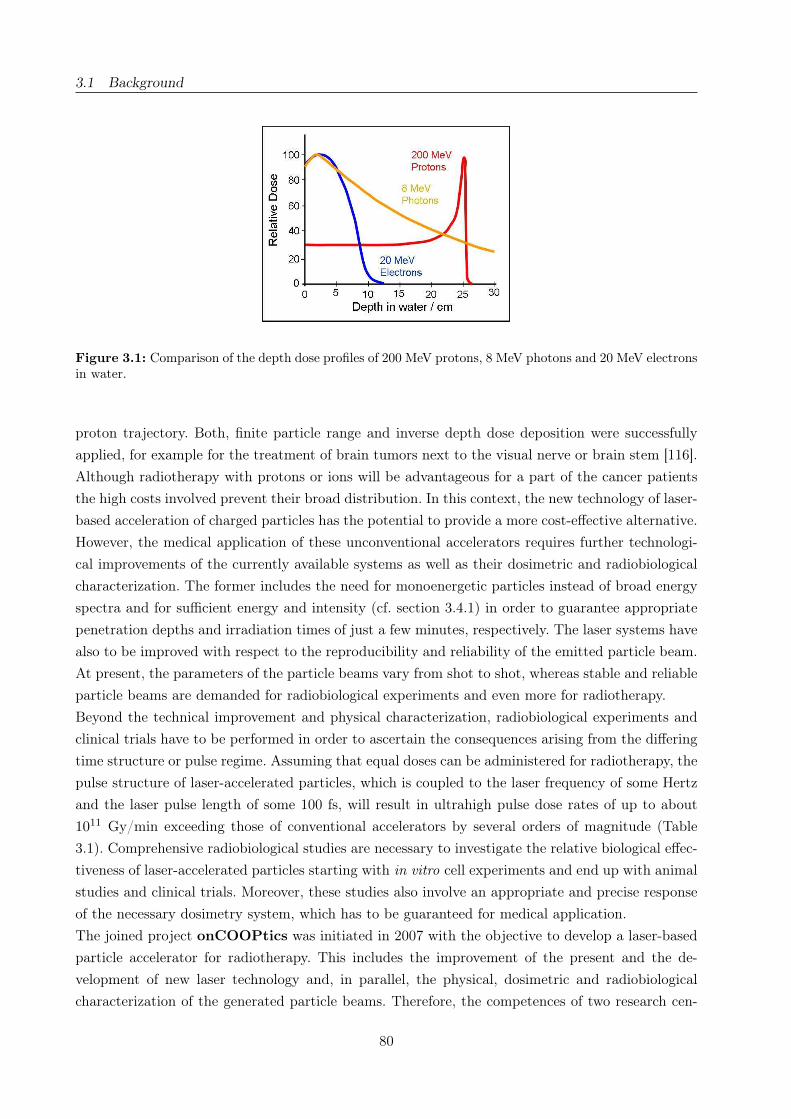
3.1 Background
Figure 3.1: Comparison of the depth dose profiles of 200 MeV protons, 8 MeV photons and 20 MeV electronsin water.
proton trajectory. Both, finite particle range and inverse depth dose deposition were successfully
applied, for example for the treatment of brain tumors next to the visual nerve or brain stem [116].
Although radiotherapy with protons or ions will be advantageous for a part of the cancer patients
the high costs involved prevent their broad distribution. In this context, the new technology of laser-
based acceleration of charged particles has the potential to provide a more cost-effective alternative.
However, the medical application of these unconventional accelerators requires further technologi-
cal improvements of the currently available systems as well as their dosimetric and radiobiological
characterization. The former includes the need for monoenergetic particles instead of broad energy
spectra and for sufficient energy and intensity (cf. section 3.4.1) in order to guarantee appropriate
penetration depths and irradiation times of just a few minutes, respectively. The laser systems have
also to be improved with respect to the reproducibility and reliability of the emitted particle beam.
At present, the parameters of the particle beams vary from shot to shot, whereas stable and reliable
particle beams are demanded for radiobiological experiments and even more for radiotherapy.
Beyond the technical improvement and physical characterization, radiobiological experiments and
clinical trials have to be performed in order to ascertain the consequences arising from the differing
time structure or pulse regime. Assuming that equal doses can be administered for radiotherapy, the
pulse structure of laser-accelerated particles, which is coupled to the laser frequency of some Hertz
and the laser pulse length of some 100 fs, will result in ultrahigh pulse dose rates of up to about
1011 Gy/min exceeding those of conventional accelerators by several orders of magnitude (Table
3.1). Comprehensive radiobiological studies are necessary to investigate the relative biological effec-
tiveness of laser-accelerated particles starting with in vitro cell experiments and end up with animal
studies and clinical trials. Moreover, these studies also involve an appropriate and precise response
of the necessary dosimetry system, which has to be guaranteed for medical application.
The joined project onCOOPtics was initiated in 2007 with the objective to develop a laser-based
particle accelerator for radiotherapy. This includes the improvement of the present and the de-
velopment of new laser technology and, in parallel, the physical, dosimetric and radiobiological
characterization of the generated particle beams. Therefore, the competences of two research cen-
80
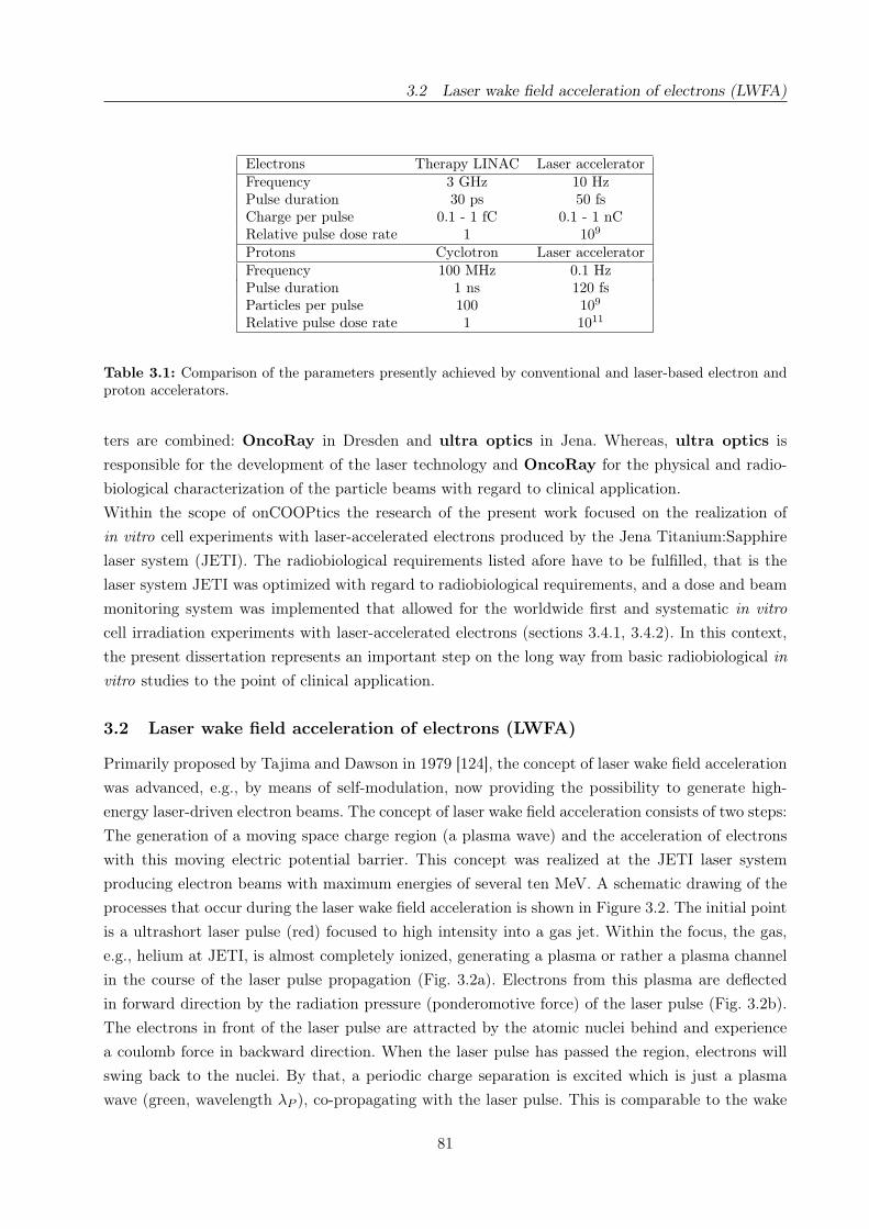
3.2 Laser wake field acceleration of electrons (LWFA)
Electrons Therapy LINAC Laser acceleratorFrequency 3 GHz 10 HzPulse duration 30 ps 50 fsCharge per pulse 0.1 - 1 fC 0.1 - 1 nCRelative pulse dose rate 1 109
Protons Cyclotron Laser acceleratorFrequency 100 MHz 0.1 HzPulse duration 1 ns 120 fsParticles per pulse 100 109
Relative pulse dose rate 1 1011
Table 3.1: Comparison of the parameters presently achieved by conventional and laser-based electron andproton accelerators.
ters are combined: OncoRay in Dresden and ultra optics in Jena. Whereas, ultra optics is
responsible for the development of the laser technology and OncoRay for the physical and radio-
biological characterization of the particle beams with regard to clinical application.
Within the scope of onCOOPtics the research of the present work focused on the realization of
in vitro cell experiments with laser-accelerated electrons produced by the Jena Titanium:Sapphire
laser system (JETI). The radiobiological requirements listed afore have to be fulfilled, that is the
laser system JETI was optimized with regard to radiobiological requirements, and a dose and beam
monitoring system was implemented that allowed for the worldwide first and systematic in vitro
cell irradiation experiments with laser-accelerated electrons (sections 3.4.1, 3.4.2). In this context,
the present dissertation represents an important step on the long way from basic radiobiological in
vitro studies to the point of clinical application.
3.2 Laser wake field acceleration of electrons (LWFA)
Primarily proposed by Tajima and Dawson in 1979 [124], the concept of laser wake field acceleration
was advanced, e.g., by means of self-modulation, now providing the possibility to generate high-
energy laser-driven electron beams. The concept of laser wake field acceleration consists of two steps:
The generation of a moving space charge region (a plasma wave) and the acceleration of electrons
with this moving electric potential barrier. This concept was realized at the JETI laser system
producing electron beams with maximum energies of several ten MeV. A schematic drawing of the
processes that occur during the laser wake field acceleration is shown in Figure 3.2. The initial point
is a ultrashort laser pulse (red) focused to high intensity into a gas jet. Within the focus, the gas,
e.g., helium at JETI, is almost completely ionized, generating a plasma or rather a plasma channel
in the course of the laser pulse propagation (Fig. 3.2a). Electrons from this plasma are deflected
in forward direction by the radiation pressure (ponderomotive force) of the laser pulse (Fig. 3.2b).
The electrons in front of the laser pulse are attracted by the atomic nuclei behind and experience
a coulomb force in backward direction. When the laser pulse has passed the region, electrons will
swing back to the nuclei. By that, a periodic charge separation is excited which is just a plasma
wave (green, wavelength λP ), co-propagating with the laser pulse. This is comparable to the wake
81

3.3 Dosimetric and radiobiological characterization
of a moving boat. In principle, the plasma wave describes the electron density distribution behind
the laser pulse and propagates in the same direction but under a small phase shift (Fig. 3.2b). In
the second step, electrons that oscillate in phase and being fast enough are trapped in the wave
and start to “surf“ on the plasma wave (Fig. 3.2c) running with the laser pulse. These electrons
surf down the wave, gain velocity and thereby energy from the potential gradient formed by the
plasma wave (Fig. 3.2d). With ongoing propagation of both the laser pulse and the plasma wave,
the electrons are accelerated in accordance to their gradient position, where the broad distribution
of occupied positions causes a broadband electron spectrum. Maximum energies of several tens MeV
can be achieved during this process along less than 1 mm of acceleration length, since ultrahigh
electric fields arise from the charge separation in wake of the high intensity laser pulse.
Figure 3.2: a) Schematic diagram of the incident laser, the plasma channel and the emitted electron beam[83]. b-d) Illustration of the process of laser wake field acceleration of electrons by means of a high intensitylaser (modified after [14]). The laser pulse is represented by a red curve, electrons are given in blue, the greencurve describes the electron density.
3.3 Dosimetric and radiobiological characterization
Usually the JETI laser system is applied for physical experiments, where a few single electron shots
with poor reproducibility are sufficient for further evaluation. Contrary, a stable and reliable beam
delivery is required for in vitro cell irradiations and even more for future radiotherapeutic appli-
82

3.3 Dosimetric and radiobiological characterization
cation. Therefore, in a first step, the parameters of the JETI laser system were adjusted to meet
radiobiological requirements and to allow for cell irradiation experiments (section 3.4.1).
Based on the dosimetric experiences gained at the ELBE accelerator various types of dosimeters
were implemented at the JETI system in order to characterize the laser-accelerated electrons with
regard to their general beam parameters and dosimetric properties. External beam parameters,
like beam spot size and dose homogeneity over the irradiated field, were determined by means of
GafChromicr EBT films (section 2.3.4), which were also used for the retrospective determination
of the absolute dose administered to the cell samples during irradiation. Comparable with the ex-
periments at ELBE, the pulse to pulse fluctuations of the JETI electron beam parameters enforce
the implementation of an online dose and beam monitoring system. However, the Farmer ionization
chamber (cf. section 2.3.2) used at ELBE is calibrated in accordance to the measurement of photons
and not suitable for electron dosimetry. In consequence, a Roos ionization chamber, developed and
calibrated for the dose measurement of therapeutic MeV electron beams, was introduced providing
online dose information during the experiment. Moreover, a Faraday cup was implemented in the
JETI setup also allowing for the online control of beam parameters, especially the charge delivery
for each single pulse. The general functioning of a Faraday cup and the specification applied in the
present work are described in sections 3.3.1 and 3.4.1, respectively. The experimental set-up at JETI
inclusive EBT films, Roos electron chamber and Faraday cup as well as the practical limits arising
from the set-up and the spectral energy distribution of the laser-accelerated electrons are discussed
in section 3.4.1.
For the radiobiological characterization of laser-accelerated electrons both human normal (fibro-
blasts, mammary epithelium) and human tumor (squamous carcinoma) cell lines were deployed
analyzing the influence on the cellular survival and the number of residual DNA DSB (cf. section
2.4.4) following irradiation.
Dosimetry with the Faraday Cup
In principle, a Faraday cup is deployed to measure the current of a charged particle beam, e.g.,
electrons, protons or heavier ions. It consists of a solid metallic body, heavy and long enough to
stop the particles under investigation, at which an electric potential is applied. Incoming charged
particles lead to a potential change, that is discharged by a resistor or amplifier. The resultant
current (I ) is being processed by means of an electrometer or oscilloscope and can be used to
estimate the number of charged particles:
(5) I =N · e−
t
with N the number of electrons e− (elementary charge), either incident or needed to compensate a
positive charge, detected in a time t.
The Faraday cup used in the present work (section 3.4.1) consists of a 20 cm long aluminum cylinder
and was constructed in accordance to the measurement of electron bunches with energies of up to
50 MeV. Moreover, the bunch (i.e., pulse) charge was readout as voltage per bunch, because the
83
3.3 Dosimetric and radiobiological characterization
ultrashort pulse duration of ≈ 5 ps prevented the readout of currents. The voltage, recorded for
every single pulse, allowed for the recalculation of the number of electrons per bunch by means of a
known conversion factor. In order to apply the Faraday cup for absolute charge measurement it has
to be operated in vacuum. This requirement cannot be met for the experimentation with living cell
samples, for which reason the Faraday cup has to be operated on air in the present work. Therefore,
it was used for pulse monitoring during the experiment, whereas the absolute doses administered
to the cell samples were determined retrospectively by means of EBT films (section 2.3.4).
First systematic radiobiological experiments
Two biological endpoints were analyzed in order to determine the biological effectiveness of laser-
accelerated electrons relative to conventional 200 kV X-rays. The analysis of residual DNA double-
strand breaks (cf. section 2.4.4), was chosen for practical reasons, e.g., a short handling time after
irradiation. As a second method, the influence of laser-accelerated electrons on the cellular survival
was investigated by means of the clonogenic survival assay, representing the “gold standard“ in
radiotherapy related research. The measured variable of this method is the reproduction capability,
which is inherent in normal tissue stem cells and tumor stem cells, but also in immortalized cell lines
of originally non dividing tissues, like the mammary gland epithelium. Moreover, the reproduction
capability is of great importance for such tumor cells, that tend to recover and repopulate after
radiotherapy. In consequence, new radiation qualities have to be characterized with respect to
their influence on the clonogenic survival, especially of tumor cells but also of normal tissue cell
lines. Following irradiation, three cellular mechanisms are known that lead to the reduction of the
reproduction capability of a cell line [44]:
• Most frequently in human normal and tumor cells is the mitotic death, describing the
accumulation of DNA damages by subsequent cell divisions up to a certain degree. Beyond
this limit the cell is not capable of completing the cell cycle or cell division.
• The interphase death affect those cells that die before entering mitosis, for instance by
apoptosis.
• The differentiation of clonogenic cells will result in specialized cells, for example skin cells,
neurons, etc., without cell dividing capability.
In order to quantify the clonogenic survival of irradiated cells, their colony forming ability is determi-
ned and compared to those of non-irradiated controls. Therefore, cell solutions with defined concen-
trations are prepared enabling for the plating of a defined number of cells (Fig. 3.3). Subsequently,
the cells are incubated under normal growth conditions, long enough to allow for the formation of
colonies, fixed and stained. For analysis, the number of colonies, consisting of at least 50 cells, are
counted and the plating efficiencies (PE ) and surviving fractions (SF ) are calculated. The former,
the PE, is achieved by dividing the number of plated cells and colonies received:
(6) PE =Number of colonies
Number of plated cells
84
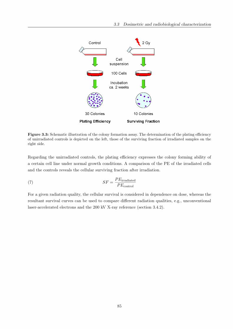
3.3 Dosimetric and radiobiological characterization
Figure 3.3: Schematic illustration of the colony formation assay. The determination of the plating efficiencyof unirradiated controls is depicted on the left, those of the surviving fraction of irradiated samples on theright side.
Regarding the unirradiated controls, the plating efficiency expresses the colony forming ability of
a certain cell line under normal growth conditions. A comparison of the PE of the irradiated cells
and the controls reveals the cellular surviving fraction after irradiation.
(7) SF =PEirradiated
PEcontrol
For a given radiation quality, the cellular survival is considered in dependence on dose, whereas the
resultant survival curves can be used to compare different radiation qualities, e.g., unconventional
laser-accelerated electrons and the 200 kV X-ray reference (section 3.4.2).
85

3.4 Application and first results
3.4 Application and first results
1. Establishment of technical prerequisites for cell irradiation experiments with laser-accelerated
electrons
2. Laser particle acceleration for radiotherapy: A first radiobiological characterization of laser-
accelerated electrons
87


3.4 Application and first results
Establishment of technical prerequisites for cell irradiation experiments with
laser-accelerated electrons
Elke Beyreuthera, Wolfgang Enghardtb,a, Malte Kaluzac, Leonhard Karschb, Lydia Laschinskyb,
Elisabeth Lessmanna, Maria Nicolaic, Jörg Pawelkeb,a, Christian Richterb,a, Roland Sauerbreya,
Hans-Peter Schlenvoigtc, Michael Baumannb
a Forschungszentrum Dresden-Rossendorf, Bautzner Landstraße 400, 01314 Dresden, Germanyb OncoRay, Center for Radiation Research in Oncology, Technische Universität Dresden, Fetscher-
str. 74, 01307 Dresden, Germanyc Institut für Optik und Quantenelektronik, Friedrich-Schiller-Universität Jena, Max-Wien-Platz 1,
07743 Jena, Germany
Short running title: Laser accelerated electrons for cell irradiation experiments.
Corresponding author:
Elke Beyreuther
Institute of Radiation Physics
Forschungszentrum Dresden-Rossendorf
P.O. Box 510119
D-01314 Dresden, Germany
Fon: +49 351 260 3552
Fax: +49 351 260 3700
E-mail: [email protected]
Medical Physics 37(4):1392-1400(2010)
DOI: 10.1118/1.3301598
www.medphys.org
89

3.4 Application and first results
Abstract
Purpose: In recent years, laser-based acceleration of charged particles has rapidly progressed and
medical applications, e.g., in radiotherapy, might become feasible in the coming decade. Require-
ments are monoenergetic particle beams with long-term stable and reproducible properties as well
as sufficient particle intensities and a controlled delivery of prescribed doses at the treatment site.
Although conventional and laser-based particle accelerators will administer the same dose to the
patient, their different time structures could result in different radiobiological properties. Therefore,
the biological response to the ultrashort pulse durations and the resulting high peak dose rates of
these particle beams have to be investigated. The technical prerequisites, i.e., a suitable cell irra-
diation setup and the precise dosimetric characterization of a laser-based particle accelerator, have
to be realized in order to prepare systematic cell irradiation experiments.
Methods: The Jena Titanium:Sapphire laser system (JETI) was customized in preparation for cell
irradiation experiments with laser-accelerated electrons. The delivered electron beam was optimized
with regard to its spectrum, diameter, dose rate and dose homogeneity. A custom-designed beam
and dose monitoring system, consisting of a Roos ionization chamber, a Faraday cup and EBT-1
dosimetry films, enables real-time monitoring of irradiation experiments and precise determination
of the dose delivered to the cells. Finally, as proof-of-principle experiment cell samples were irradia-
ted using this setup.
Results: Laser-accelerated electron beams, appropriate for in vitro radiobiological experiments,
were generated with a laser shot frequency of 2.5 Hz and a pulse length of 80 fs. After laser-
acceleration in the helium gas jet, the electrons were filtered by a magnet, released from the vacu-
um target chamber and propagated in air for a distance of 220 mm. Within this distance a lead
collimator (aperture 35 mm) was introduced leading, along with the optimized setup, to a beam
diameter of 35 mm, sufficient for the irradiation of common cell culture vessels. The corresponding
maximum dose inhomogeneity over the beam spot was less than 10 % for all irradiated samples.
At cell position, the electrons posses a mean kinetic energy of 13.6 MeV, a bunch length of about
5 ps (FWHM) and a mean pulse dose of 1.6 mGy per bunch. Cross correlations show clear linear
dependencies for the online recorded accumulated bunch charges, pulse doses and pulse numbers on
absolute doses determined with EBT-1 films. Hence, the established monitoring system is suitable
for beam control and a dedicated dose delivery. Additionally, reasonable day-to-day stable and re-
producible properties of the electron beam were achieved.
Conclusions: Basic technical prerequisites for future cell irradiation experiments with ultrashort,
pulsed laser-accelerated electrons were established at the JETI laser system. The implemented onli-
ne control system is suitable to compensate beam intensity fluctuations and the achieved accuracy
of dose delivery to the cells is sufficient for radiobiological cell experiments. Hence, systematic in
vitro cell irradiation experiments can be performed, being the first step towards clinical application
of laser-accelerated particles. Further steps, including the transfer of the established methods to ex-
periments on higher biological systems or to other laser-based particle accelerators, will be prepared.
Key words: laser electron acceleration (SM-LWFA), laser radiotherapy, cell irradiations
90

3.4 Application and first results
I. Introduction
Starting with the invention of the technique of chirped pulse amplification (CPA) [81], a tremendous
progress in high-power laser technology was made over the past two decades. The associated increase
in peak power and intensity available with such systems together with substantial reductions in sys-
tem size has enabled the realization of table-top laser-based particle accelerators easily fitting into
a university-scale laboratory. Furthermore, a careful control of the experimental parameters allowed
for the first time the observation of electron [29, 36, 84], proton [97, 117] and ion pulses [43]. Narrow
or even quasi-monoenergetic spectra of the accelerated particles make such laser systems candidates
for next generation particle accelerators. During the interaction of the laser pulse with the plasma,
electric fields in the order of 100 GV/m [29] or even 1 TV/m [43, 117] can be achieved, exceeding
the field strengths available in conventional electromagnetic accelerators [109] by more than four
orders of magnitude. This makes the laser-technology attractive for the realization of compact par-
ticle accelerators. Another important property of laser-driven particle pulses is the ultrashort pulse
duration, which initially can be as short as the laser pulse length itself, i.e., of the order of 100 fs.
The perspective of compact and potentially more cost-effective accelerators initiated discussions on
their application in radiotherapy [31, 71]. Therefore, the particle beams have to meet several requi-
rements. The particle spectra should be monoenergetic with energies of . 50 MeV for electrons,
. 230 MeV for protons and . 430 MeV/amu for carbon ions. Intensities should amount up to
≈ 1010 particles per second. Additionally, stable and long time reproducible properties of the beams
as well as a homogeneous exposure of the irradiation field are needed [109]. The delivered dose rates
should be high enough to guarantee treatment times of a few minutes [109]. Further demands on
electron and proton acceleration and required dose properties were discussed in several feasibility
studies [15, 28, 31, 60, 78, 83].
Regarding the electrons, energies of ≤ 1 GeV [72] and maximum intensities of ≤ 3×10⁹ electrons per
bunch [29, 36] were achieved by laser-based particle acceleration. However, these parameters were
measured in single shot experiments, and the available repetition rates are currently not sufficient
to obtain the intensities and dose rates required for therapy. For protons and ions, both the energies
and the intensities presently achieved are insufficient for radiotherapy.
In addition to technological requirements, the radiobiological characteristics of these new quality
of particle beams have to be investigated. Compared to conventional radiotherapy, the ultrashort
pulse duration of laser-accelerated particles lead to 109−1011 times higher peak dose rates and peak
currents of some thousand amperes [36]. Although conventional and laser-based accelerators will
administer the same dose to the patient, the different time structures and peak currents could re-
sult in different radiobiological properties. Therefore, the biological effectiveness of laser-accelerated
particles has to be characterized in appropriate experiments. Previous studies have been performed
with ultrashort pulsed electron [18, 87, 115] and soft X-ray [49, 118, 125] beams delivered with ma-
ximum pulse dose rates of 109 Gy/s [87] for electrons and 1013 Gy/s [118] for photons, respectively.
In all of these studies the cellular survival was investigated revealing either no influence [18, 87, 125]
or a reduction [49, 115, 118] of the cytotoxicity by means of the ultrahigh dose rate.
So far, no systematic radiobiological studies are published for laser-accelerated particles. The first at-
91

3.4 Application and first results
tempts in this context were recently made for protons by Yogo et al. [131], those for laser-accelerated
electrons in the present work. Several requirements, depending on each other, have to be fulfilled in-
cluding the improvement of the laser particle acceleration technology for potential radiotherapeutic
application and, simultaneously, their biological characterization starting with in vitro cell irradia-
tion experiments. An important step will be the transition from basic physical experiments, where
few single shots with poor reproducibility are considered for further evaluation, to routine particle
beam delivery with long-term stable and reliable properties. Moreover, these properties have to be
controlled to such an extent, that prescribed doses can be applied to any biological object under
observation.
The aim of the present work was the establishment of the technical prerequisites for cell irradiation
experiments at the Jena Titanium:Sapphire laser system (JETI) [110]. This includes the optimiza-
tion of the laser system and the electron beam in accordance with radiobiological requirements, like
the transition to stable and reproducible irradiation of biological samples. A custom-designed beam
and dose monitoring system was implemented that enables a real-time monitoring of the irradiation
and a controlled dose delivery to biological samples. Moreover, a first test of cell irradiation experi-
ment was performed demonstrating the feasibility of radiobiological studies at the JETI accelerator.
II. Material and methods
The Jena Titanium:Sapphire laser system
The electron pulses were generated in a vacuum chamber, hereafter referred to as target chamber,
using the 10 TW laser system JETI. Pulses of 80 fs pulse duration (FWHM) [97, 117], containing
800 mJ of energy at a central wave length of 800 nm, were delivered with a maximum repetition
frequency of 10 Hz. Every fourth pulse was focused to an intensity of 5× 10¹⁸ W/cm2 into a pulsed
subsonic helium gas jet which was generated by a cylindrical nozzle of 1 mm diameter. The gas
density profile was approximately Gaussian, and the peak gas density could be varied in the range
of 0.5− 3× 10¹⁹ molecules/cm3 by changing the backing pressure of the gas jet. In this regime, the
laser pulse undergoes relativistic self-focusing and self-modulation and produces pulses of highly
relativistic electrons by self-modulated laser wake field acceleration (SM-LWFA) [47, 110]. The need
of a sufficient high average dose, i.e., electron number, per shot made it necessary to tune the acce-
leration conditions by carefully changing the gas jet parameters and its position. The electron beam
propagates over a length of about 400 mm in vacuum and was released from the target chamber
through a 1 mm thick aluminum vacuum window. Subsequently, the electron beam and adjacent
components have to be optimized to enable radiobiological experiments.
Cell irradiation geometry
With regard to radiotherapy, the dose response of both, normal tissue and tumor cells, has to be
investigated for several biological endpoints, e.g., the number of DNA double-strand breaks and the
cellular survival, to get a comprehensive picture of the biological effectiveness of laser-accelerated
electrons. For this purpose, the whole setup has to be customized in such a way that the irradiation
of adherent cell monolayers in standard cell culture vessels becomes feasible. Moreover, an online
92

3.4 Application and first results
dose monitoring system has to be chosen suitable for the usage of different sample geometries. In
the present work, the electron beam diameter and homogeneity as well as the experimental setup
were adjusted to the application of standard petri dishes (Greiner, Frickenhausen, Germany) and 8-
well-chamber slides (VWR, Darmstadt, Germany). The former exhibit an inner diameter of 32 mm,
whereas the latter ones consist of 9 × 9 mm2 plastic wells arranged in two lines on a plastic slide.
Single wells are separated by 1 mm plastic and a little gap of air. Both, the petri dishes and the
8-well-chamber slides, possess an 1 mm plastic bottom, where the cell monolayers are grown. In
general, petri dishes are applied to investigate the cellular survival [75], whereas the 8-well-chamber
slides are used for the analysis of DNA double-strand breaks [7].
For in vitro cell experiments, the irradiation time and culture medium supply have to be considered
carefully in order to avoid effects of medium depletion to the cells. At JETI, the delivery of an
horizontal electron beam demand for the upright positioning of the cell samples and for the enclo-
sing of medium filled culture vessels with Parafilm M (Merck, Darmstadt, Germany). For practical
reasons, e.g., handling procedure and the risk of medium loss, half-filled vessels were used for short
irradiation times (≤ 10 min), whereas longer irradiations were performed with completely filled ves-
sels. In consequence, the upright positioning of the cell samples resulted in a 10 mm thick medium
layer of differing filling height and limited by the plastic bottom and the height of the culture vessel
enclosed with Parafilm. During irradiation the plastic bottoms of the culture vessels were turned in
beam direction minimizing the material in front of the cell monolayer.
Beam monitoring and dosimetry system
Because the electron beam delivered by the JETI system fluctuates in both, beam intensity and
dose rate, the control of dose delivery during irradiation and a precise determination of dose for
each individual cell sample are required.
Absolute dosimetry was realized with GafChromic R© EBT-1 dosimetry films (ISP Corp., Wayne,
NJ, USA), which are self-developing after irradiation, offer a high spatial resolution and feature a
low thickness of 234 µm. However, their main advantages are an almost dose rate [33] and, for the
relevant energy range (MeV), energy [101] independent dose response. Irradiated films were digitized
with an Epson Perfection V750 flat-bed document scanner (Seiko Epson Corp., Nagano, Japan) and
their optical density (film darkness) was converted to dose within an IDL code (Interactive Data
Language, ITT Visual Information Solutions, Boulder, CO, USA). The EBT-1 dosimetry films were
calibrated in accordance to a previously described protocol [132], that was adopted from Devic et
al. [21]. For this purpose, electrons of 6 MeV as delivered by a clinical electron accelerator and
characterized by in-house dosimetry in compliance to DIN6800-2 [1] were applied.
For cell irradiation experiments, two EBT-1 films have been positioned both in front and behind
of each cell sample. The front film was applied for precise retrospective dosimetry and the rear one
for additional control. By preliminary tests with both standard cell culture vessels chosen, a dose
correction factor, defined as the ratio of the dose at the cell location to the dose in front of the cell
vessel, of 1.03± 0.05 has been determined considering the dose build up effect in the culture vessel
bottom. Hence, the dose delivered to the cell monolayer inside the culture vessel can be deduced
93
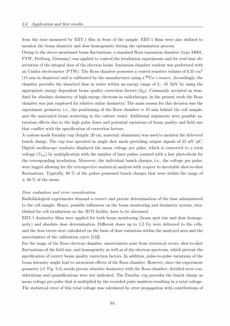
3.4 Application and first results
from the dose measured by EBT-1 film in front of the sample. EBT-1 films were also utilized to
monitor the beam diameter and dose homogeneity during the optimization process.
Owing to the above mentioned beam fluctuations, a standard Roos ionization chamber (type 34001,
PTW, Freiburg, Germany) was applied to control the irradiation experiments and for real-time ob-
servation of the integral dose of the electron beam. Ionization chamber readout was performed with
an Unidos electrometer (PTW). The Roos chamber possesses a vented sensitive volume of 0.35 cm3
(15 mm in diameter) and is calibrated by the manufacturer using a 60Co γ-source. Accordingly, the
chamber provides the absorbed dose in water within an energy range of 2 - 45 MeV by using the
appropriate energy dependent beam quality correction factors (kQ). Commonly accepted as stan-
dard for absolute dosimetry of high-energy electrons in radiotherapy, in the present work the Roos
chamber was just employed for relative online dosimetry. The main reason for this decision was the
experiment geometry, i.e., the positioning of the Roos chamber ≈ 45 mm behind the cell sample,
and the associated beam scattering in the culture vessel. Additional arguments were possible sa-
turation effects due to the high pulse doses and potential variations of beam quality and field size
that conflict with the specification of correction factors.
A custom-made Faraday cup (length: 20 cm, material: aluminum) was used to monitor the delivered
bunch charge. The cup was operated in single shot mode providing output signals of 25 mV/pC.
Digital oscilloscope readouts displayed the mean voltage per pulse, which is converted to a total
voltage (Utot) by multiplication with the number of laser pulses counted with a fast photodiode for
the corresponding irradiation. Moreover, the individual bunch charges, i.e., the voltage per pulse,
were logged allowing for the retrospective statistical analysis with respect to inevitable shot-to-shot
fluctuations. Typically, 80 % of the pulses possessed bunch charges that were within the range of
± 40 % of the mean.
Dose evaluation and error consideration
Radiobiological experiments demand a correct and precise determination of the dose administered
to the cell sample. Hence, possible influences on the beam monitoring and dosimetry system, esta-
blished for cell irradiations at the JETI facility, have to be discussed.
EBT-1 dosimetry films were applied for both beam monitoring (beam spot size and dose homoge-
neity) and absolute dose determination. Different doses up to 5.2 Gy were delivered to the cells,
and the dose errors were calculated on the basis of dose variations within the analyzed area and the
uncertainties of the calibration curve [132].
For the usage of the Roos electron chamber, uncertainties arise from statistical errors, shot-to-shot
fluctuations of the field size, and homogeneity as well as of the electron spectrum, which prevent the
specification of correct beam quality correction factors. In addition, pulse-to-pulse variations of the
beam intensity might lead to saturation effects of the Roos chamber. However, since the experiment
geometry (cf. Fig. 3.4) avoids precise absolute dosimetry with the Roos chamber, detailed error con-
siderations and quantifications were not indicated. The Faraday cup provides the bunch charge as
mean voltage per pulse that is multiplied by the recorded pulse numbers resulting in a total voltage.
The statistical error of this total voltage was calculated by error propagation with contributions of
94

3.4 Application and first results
the standard deviation of the average voltage per pulse and an intrinsic statistical uncertainty of
0.1 % of the cup. Additionally, a systematic error of 10 mV was assumed for the conversion of the
total voltage in applied electron bunch charge, due to a possible offset of the measurement system.
The number of laser pulses (≤ 3000) was accurately counted with a fast photodiode. Only reading
errors (± 1 pulse) by the operator might contribute to an uncertainty. All errors given in the figures
and the text were calculated on the basis of the afore described statistical uncertainties. Systematic
errors were not included in calculations and fit procedures.
Proof-of-principle test irradiation
Subsequent to beam optimization and setup adjustment, a test cell irradiation experiment was per-
formed to evaluate the practical procedure and cell handling at JETI. Mimicking the measurement
of dose response curves, 47 cell samples were prepared in 8-well-chamber slides and irradiated with
doses between 0.3 and 5.2 Gy. The experiment was performed at two consecutive days, which also
allows to test the long-term stability and day-to-day reproducibility of the laser system and the
electron beam, respectively. Moreover, with this test experiment the interplay of all components,
which means the tuning and operation of the laser system, the electron beam delivery, the dose
monitoring system and the cell irradiation, was investigated.
Besides practical evaluation, the test cell irradiation experiment was also used to evaluate how far
the JETI system met basic requirements for radiobiological experimentation. Therefore, the electron
beam should be well defined with respect to its spectral intensity distribution and to the character
and spectral intensity distribution of the associated secondary radiation. The beam cross-sectional
area should cover at least 0.5 cm2, whereas the dose deviation within this area should be less than
10 %. Furthermore, the irradiation of living cells demand for a quantifiable dose delivery within an
uncertainty of typically less than 10 % and irradiation times of a few minutes, that is, the dose rate
or beam intensity should be high enough.
III. Results
Optimized setup for radiobiological experiments
The final experimental setup for cell irradiations at the JETI facility (Fig. 3.4) was the result of an
iterative optimization process. The setup was adjusted according to radiobiological requirements,
like an appropriate beam spot size, and to minimize the number of necessary setup components.
Moreover, low-energy electrons (≤ 2 MeV) have to be separated from the beam since only energies
above this limit are used for radiotherapy and because of the energy dependence of the biological
effectiveness [52]. For this, a pair of permanent dipole magnets [material: Neodymium Iron Boron,
NdFeB] with opposing polarity and a magnetic flux density of 0.04 T were used for energy filtra-
tion. Each magnet consisted of eight smaller ones (40× 20× 2 mm3) and the two dipole magnets of
the energy filter were separated by 145 mm along the electron beam trajectory (see Fig. 3.4). The
electrons were forced on a S-shaped trajectory, and those with energies below 2 MeV were deflected
from the beam and could not pass the lead collimator. Subsequently, the slowest electrons were
decelerated down to approximately 1 MeV at cell position by 1 mm aluminum (vacuum window),
95

3.4 Application and first results
Figure 3.4: Schematic drawing of the experimental setup at the JETI system including the electron beamgeneration, the beam line and the online beam monitoring system (not to scale). The electrons propagate inair at a distance of about 220 mm.
220 mm air and 1 mm plastic (culture vessel bottom). This energy limit is high enough to minimize
possible interference to the examined radiobiological effect.
The lead collimator (10 × 10 × 10 cm3, aperture of 35 mm) was introduced in order to protect
the cell samples from deflected electrons and inevitably emitted secondary radiation. As shown by
an irradiation with one dipole magnet deflecting all electrons, the measured dose contribution of
bremsstrahlung, produced by wake field acceleration (betatron radiation) and deflection, was ne-
gligible. Moreover, the aperture of the lead collimator and the optimized distance between vacuum
chamber and cell sample (220 mm) resulted in a beam diameter of 35 mm, sufficient to irradiate a
petri dish or the four middle chambers (21 × 21 mm2) of the 8-well-chamber slides. Special sample
holders were designed enabling a practical and fast alignment of one petri dish or one chamber slide
with the electron beam axis. At the end of the beam line (cf. Fig. 3.4), the Roos electron chamber
and the Faraday cup were installed in a distance of 45 mm from the cell sample. Due to practical
reasons, the smaller Roos chamber was mounted at the entrance of the Faraday cup (diameter of
the sensitive volume: ≈ 10 cm), minimizing disturbances of the Faraday cup signal. Adjusting the
Roos chamber centric to the electron beam, the entire sensitive volume of the chamber (0.35 cm3)
was irradiated.
Characterization of the electron beam
Since both cell irradiation experiments and future radiotherapeutic application require irradiation
times of a few minutes [109], the mean dose rate of the electron beam had to be maximized. For
this the beam pulse intensity, i.e., electron yield, was increased gradually with laser power. More-
over, the repetition rate of the laser-accelerated electron pulses was maximized by improving the
vacuum in the target chamber and therefore allowing an high helium gas flow per time. The actual
achieved parameters of the laser system and a summary of the electron beam parameters at cell
position are shown in Table 3.2. According to the propagation of the electron beam in the vacuum
window and in air, the electron beam was broadened to a bunch length of 5 ps (FWHM) as esti-
mated by GEANT4 simulations (version 8.1 [35]). Moreover, the corresponding electron spectrum
96
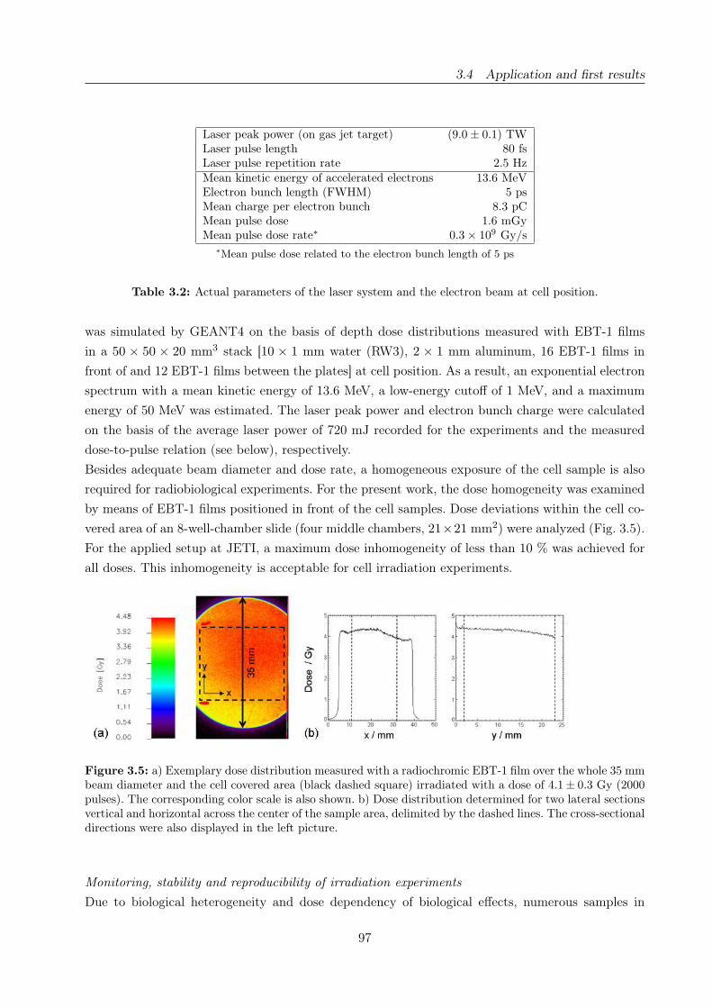
3.4 Application and first results
Laser peak power (on gas jet target) (9.0 ± 0.1) TWLaser pulse length 80 fsLaser pulse repetition rate 2.5 HzMean kinetic energy of accelerated electrons 13.6 MeVElectron bunch length (FWHM) 5 psMean charge per electron bunch 8.3 pCMean pulse dose 1.6 mGyMean pulse dose rate∗ 0.3 × 10⁹ Gy/s
∗Mean pulse dose related to the electron bunch length of 5 ps
Table 3.2: Actual parameters of the laser system and the electron beam at cell position.
was simulated by GEANT4 on the basis of depth dose distributions measured with EBT-1 films
in a 50 × 50 × 20 mm3 stack [10 × 1 mm water (RW3), 2 × 1 mm aluminum, 16 EBT-1 films in
front of and 12 EBT-1 films between the plates] at cell position. As a result, an exponential electron
spectrum with a mean kinetic energy of 13.6 MeV, a low-energy cutoff of 1 MeV, and a maximum
energy of 50 MeV was estimated. The laser peak power and electron bunch charge were calculated
on the basis of the average laser power of 720 mJ recorded for the experiments and the measured
dose-to-pulse relation (see below), respectively.
Besides adequate beam diameter and dose rate, a homogeneous exposure of the cell sample is also
required for radiobiological experiments. For the present work, the dose homogeneity was examined
by means of EBT-1 films positioned in front of the cell samples. Dose deviations within the cell co-
vered area of an 8-well-chamber slide (four middle chambers, 21×21 mm2) were analyzed (Fig. 3.5).
For the applied setup at JETI, a maximum dose inhomogeneity of less than 10 % was achieved for
all doses. This inhomogeneity is acceptable for cell irradiation experiments.
Figure 3.5: a) Exemplary dose distribution measured with a radiochromic EBT-1 film over the whole 35 mmbeam diameter and the cell covered area (black dashed square) irradiated with a dose of 4.1± 0.3 Gy (2000pulses). The corresponding color scale is also shown. b) Dose distribution determined for two lateral sectionsvertical and horizontal across the center of the sample area, delimited by the dashed lines. The cross-sectionaldirections were also displayed in the left picture.
Monitoring, stability and reproducibility of irradiation experiments
Due to biological heterogeneity and dose dependency of biological effects, numerous samples in
97

3.4 Application and first results
several independent repetitions have to be investigated in radiobiological experiments. Therefore,
a stable beam with long-term reproducible properties as well as a beam monitoring system that
ensures a controlled dose delivery to the sample is required. To meet these demands, the stability
and reproducibility of the laser-accelerated electron beam were determined and the custom-designed
beam and dose monitoring system was tested at two experimental days.
At the beginning of each day, the system was tuned to a stable dose-to-pulse relation of about
2 mGy/pulse. Subsequently, this dose-to-pulse relation was used to estimate the number of laser
pulses necessary to deliver the prescribed doses, whereas the actual absolute doses were determined
retrospectively by means of EBT-1 films. Correlating the absolute doses and the number of laser
pulses (Fig. 3.6) the preset dose-to-pulse relation was confirmed by 1.90 ± 0.04 mGy/pulse for the
second day. For the first day, the linear fit of the data reveals a dose-to-pulse relation of 1.30± 0.02
mGy/pulse. The two outliers (Fig. 3.6, black arrows) were caused by a 90 min interruption of the
experiment at day one. During this interruption, the vacuum pumps cooled down, which resulted
in an increased pumping rate and hence a higher electron yield correlated with an increased dose
per pulse. The effect vanished 45 min after the break. In response to these outliers, which were also
detected by the online monitoring system, the vacuum and temperature of the JETI system were
carefully controlled at the second day in order to ensure constant dose-to-pulse relations.
Figure 3.6: Actual doses at cell position compared to the number of applied laser pulses individually forboth experimental days. The arrows mark two outliers due to different pumping rates and correspondingvacuum conditions. Dose errors were calculated by error propagation; fit parameters are given in the text.
In addition to the number of laser pulses, the relative accumulated dose and the bunch charge
measured with the Roos electron chamber and the Faraday cup were correlated to the EBT-1 film-
measured absolute dose in order to reveal the beam stability and their capability for online dose
monitoring. For the Roos electron chamber, this comparison (Fig. 3.7a) results in a linear dose-
to-dose dependence of 1.61 ± 0.03 GyEBT/GyRoos for the film-measured relative to the chamber-
measured dose at both days. Furthermore, a clear linear correlation with a slope or sensitivity of
0.010±0.001 GyEBT/V (Fig. 3.7b) was obtained for the total voltage (Utot) provided by the Faraday
98

3.4 Application and first results
cup. In consequence, the pulse voltage, which is a measure of the delivered bunch charge, can be
applied to estimate the number of laser pulses required to deliver a particular dose. The apparent
wider distribution of accumulated doses (Roos chamber) and total voltages (Faraday cup) at higher
film-measured doses is explainable with the filling level of the cell samples. As mentioned before, the
samples were completely filled with medium if the irradiation time exceeds 10 minutes in order to
avoid effects of medium depletion. Hence, volume deviations and small air bubbles in the medium
might lead to differences in beam scattering and electron stopping-power, resulting in measurable
effects. In contrast, samples irradiated with lower doses (≤ 2 Gy) were filled with an accurately
pipetted volume providing constant conditions for electron beam transmission.
Figure 3.7: Accumulated relative dose (a) and total voltage (b) measured during the irradiation andcompared to the absolute doses determined retrospectively by means of EBT-1 dosimetry films. Completely(grey) and half-filled samples (black) were distinguished in the graph, whereas the linear fits were performedwith all data. Displayed errors are statistical errors, calculated by error propagation.
The good linearity of the relative dose measurements (Roos chamber and Faraday cup) over the
whole dose range also indicates a negligible contribution of low-energy electrons (≤ 2 MeV) to the
electron dose delivered to the cells. For longer irradiation times, a 1 cm thick medium layer was
necessary. Furthermore, the electron stopping-power in water is ≈ 2 MeV/cm [51]. Hence, low-
energy electrons would contribute to the dose measured with EBT-1 films in front of the sample,
but not to the relative dose detected behind with the Roos electron chamber or Faraday cup. As
a consequence, a deviation from the linear fit curve, especially in the high dose region, would be
observable, if electrons below 1 MeV would significantly contribute to the dose.
To sum up, the observed parameters (number of laser pulses, accumulated dose measured with the
Roos electron chamber and pulse voltage obtained with the Faraday cup) can be applied to monitor
the electron beam and to provide relative dose information during cell irradiation. Moreover, the
good linear correlations between the film-measured absolute doses and these parameters indicate a
stable beam with reproducible properties during the whole experiment. Overall, the precision and
stability achieved during these irradiations as well as the dosimetry and beam monitoring system
are sufficient for radiobiological cell irradiation experiments.
99

3.4 Application and first results
IV. Discussion
To date, the application of laser-based particle acceleration for radiotherapy has been described
only theoretically (e.g., [15, 28, 31, 60, 78, 83]). The proposed properties and parameters of such
beams are currently not experimentally validated. A first step in this direction are the experiments
reported in this work, demonstrating the technological improvement of the JETI laser system and
its feasibility for cell irradiation experiments with laser-accelerated electrons. Although not yet ful-
filling the requirements needed for radiotherapeutic applications with respect to beam energy and
stability, field size and dose rate, the parameters achieved at JETI are sufficient for radiobiological
cell experiments.
For clinical radiotherapy, particle beams have to be monoenergetic with sufficient charge and ne-
gligible fluctuations of the physical parameters [109]. By contrast, only a configuration of the JETI
laser accelerator producing broad exponential electron spectra led to beams with sufficient charge
and reproducibility. However, this broad spectrum can be accepted for cell irradiation experiments
because the underlying setting allows for a constant beam delivery. Moreover, the energy was high
enough to achieve the required penetration depth. Low-energy electrons of ≤ 2 MeV are separated
from the beam due to their elevated linear energy transfer that could influence the radiation in-
duced effect in the cell sample. The remaining electrons were released from the target chamber in
an intensity of up to 3 × 10⁸ electrons/bunch that is sufficient for the necessary dose delivery in
cell irradiation experiments. For radiotherapy, one order of magnitude more electrons is required to
achieve the desired ≈ 1010 particles per second and the typical dose rates of some Gy/min [109],
respectively. Nevertheless, the beam diameter of 35 mm and a dose homogeneity better than 90 %
allow for a homogeneous exposure of the whole cell sample.
In parallel to the adjustment of JETI, a dose and beam monitoring system, consisting of EBT-1
dosimetry films, a Roos electron chamber and a Faraday cup, was implemented at the experimental
setup. Initially applied for the optimization and fine-tuning of the beam, the devices were deployed
later on for monitoring the dose and beam delivery during the experiment. The EBT-1 films were
used for retrospective determination of the absolute dose delivered to the cells. A comparison of the
doses determined by EBT-1 films and measured online with the Roos electron chamber revealed a
correlation factor of 1.61 ± 0.03 GyEBT/GyRoos. Thus, the Roos electron chamber underestimates
the actual applied doses, whereas the relative dose information is sufficient to monitor the beam and
control the irradiation experiment. The beam scattering in the ≈ 1 cm thick cell sample and the
distance of ≈ 4.5 cm between the sample and the Roos electron chamber are most likely responsible
for the dose deviation. A clear linear dependence was also found for the comparison of the Faraday
cup signal and the dose delivered to the cells determined by EBT-1 films.
In spite of these encouraging results for dose monitoring, more detailed considerations on the influ-
ence of the ultrahigh dose rates on the measured signal are desirable if the Roos electron chamber
should be applied for absolute dosimetry. A strong pulse dose dependency of the dose measurement
was found by Di Martino et al. [22] for pulse doses above 1 cGy. Regarding the mean pulse dose of
2 mGy achieved in the present work, a dose correction factor of 1.03 has to be applied in order to
correct the displayed dose for the induced saturation effect [22]. However, since the Roos chamber
100
3.4 Application and first results
was deployed for relative and not for absolute dosimetry, this factor is of little importance. In ad-
dition, the application of the Roos chamber for absolute dosimetry would also demand a constant
electron spectrum allowing for the specification of a correct beam quality correction factor kQ.
Future cell irradiation experiments will require a stable particle beam with reproducible properties.
For the experiments presented here, these requirements were investigated by comparing the number
of laser pulses, the accumulated dose and the bunch charge with the actual dose delivered to the
cells during two experimental days. A stable dose-to-pulse relation was obtained within each day,
but there were differences of approximately 45 % between both days. However, these day-to-day
fluctuations of the dose rate can be corrected with the information from the online beam monitoring
system. Already, an improvement was achieved by carefully controlling the vacuum conditions and
the temperature of the JETI system. Further developments and advancements of the laser accelera-
tion technology and the auxiliary equipment are expected to lead to more stable and reproducible
particle beams in future.
Although various outstanding improvements were identified, the main goal of the present work was
achieved. The transition from experiments with single electron shots of poor reproducibility to rela-
tive stable and repeatable irradiations was accomplished and a test cell irradiation experiment was
performed demonstrating that the JETI system mets key requirements for radiobiological in vitro
experimentation. The achieved electron energy and intensity were high enough for the irradiation
of living cells; the beam diameter of 35 mm and a dose homogeneity better than 90 % over the
corresponding cross-sectional area guaranteed the homogeneous exposure of cell monolayers in dif-
ferent sample geometries. Moreover, the setup and handling procedure were found to be practical
for comprehensive radiobiological studies, the dose delivery to the cells was controllable and quanti-
fiable with sufficient accuracy, and the emitted secondary radiation were quantified and found to be
negligible at the cell position. For these reasons, the JETI laser system fulfills basic radiobiological
requirements and was successfully prepared for systematic in vitro cell irradiation studies.
IV. Conclusion
The aim of the presented work was the advancement of the laser-driven accelerator technology in
order to establish the basic technical prerequisites for future in vitro cell irradiation experiments
with ultrashort pulsed laser-accelerated particles as one step toward radiotherapeutic application.
Therefore, the JETI laser system was optimized now allowing for the delivery of stable electron
beams sufficient for radiobiological experiments, instead of single shots with low reproducibility.
Moreover, an online control system was implemented that enables the compensation of beam and
dose fluctuations and the delivery of prescribed doses with sufficient accuracy for radiobiological cell
experiments. For this reason, systematic in vitro cell irradiation experiments are prepared, being a
step further toward clinical application of laser-accelerated particles. The performance of such sys-
tematic cell experiments will result in information on the biological effectiveness of laser-accelerated
particle beams. With this work, major technological progress has been achieved on the way towards
establishing a laser-particle accelerator for radiotherapeutic application. In further steps, the trans-
ferability of the established methods to experiments on higher biological systems (e.g., tissue or
animals) or other laser-based particle accelerators has to be proven.
101
3.4 Application and first results
Acknowledgments
This work has been supported by the German Federal Ministry of Education and Research (BMBF)
under contract 03ZIK445.
Author contributions
Study concept and design E.B., J.P., L.K., E.L., H.-P.S.; Experimental studies E.B., L.K., E.L.,
J.P., H.-P.S., L.L., C.R., M.N.; Data acquisition E.L., L.K., E.B., H.-P.S., L.L., C.R.; Literature
research E.B., L.K.; Data analysis E.B., L.K., C.R.; Definition of intellectual content J.P., M.B.,
W.E., R.S.; Manuscript preparation E.B., H.-P.S.; Manuscript editing R.S., M.K., H.-P.S., W.E.,
L.K., J.P., M.B., C.R., L.L.; Guarantors of integrity of the entire study J.P., M.B., W.E., M.K.,
R.S.; Manuscript review was carried out by all authors.
102

Laser particle acceleration for radiotherapy: A first radiobiological characterization of
laser accelerated electrons
Jörg Pawelkea,b, Elke Beyreutherb, Wolfgang Enghardta,b, Malte Kaluzac, Leonhard Karscha,
Lydia Laschinskya, Elisabeth Lessmannb, Doreen Naumburgera, Maria Nicolaic, Christian
Richtera,b, Roland Sauerbreyb, Hans-Peter Schlenvoigtc, Michael Baumanna
a OncoRay - Center for Radiation Research in Oncology, Technische Universität Dresden, Medical
Faculty, Dresden, Germanyb Forschungszentrum Dresden-Rossendorf, Institute of Radiation Physics, Dresden, Germanyc Friedrich-Schiller-Universität Jena, Institute of Optics and Quantumelectronics, Jena, Germany
Corresponding author:
Jörg Pawelke
OncoRay-Center for Radiation Research in Oncology
Medical Faculty Carl Gustav Carus
Technische Universität Dresden
Fetscherstraße 74
D-01307 Dresden, Germany
Fon: +49 351 458 7430
Fax: +49 351 458 7311
E-mail: [email protected]
O. Dössel and W.C. Schlegel (Eds.): WC 2009, IFMBE Proceedings 25/III, pp. 502-04, 2009.
www.springerlink.com
3.4 Application and first results
Abstract
In recent years, the technology of laser-based particle acceleration has developed at such a rate
that compact and potentially more cost-effective accelerators are promised for medical application,
e.g., for high precision hadron radiotherapy. Necessary requirements are the supply of stable and
reliable particle beams with reproducible properties, sufficient particle intensities and monoener-
getic spectra. Additionally, a precise dose delivery in an appropriate time and the exposure of a
desired irradiation field are needed. Beside these physical demands, the consequences on detection
and dosimetry as well as the radiobiological effect on living cells have to be investigated for the
ultrashort pulsed laser-based particle beams.
As a first step, the laser accelerator facility at the Jena Titanium:Sapphire system was customized
for in vitro cell irradiation experiments and the delivered electron beam was improved with regard
to its spectrum, diameter, dose rate and dose homogeneity. Furthermore, a custom-designed beam
and dose monitoring system was established that enables real-time monitoring of the irradiation
experiments and a precise determination of the dose delivered to the cells. Moreover, stable and
reproducible beam properties were achieved during the whole three month experiment campaign.
Dose-effect-curves were obtained for four cell lines and two endpoints, generally displaying a lower
biological effectiveness for short-pulsed laser-accelerated electrons relative to the continuous 200 kV
X-ray reference irradiation. Possible reasons will be discussed.
Keywords: laser-based particle acceleration, radiotherapy, in vitro cell experiments, biological ef-
fectiveness
104

3.4 Application and first results
I. Introduction
Over the past years, the technological progress in high-power laser technology results in increasing
peak powers and intensities associated with substantial reductions in system size, which enables the
realization of table-top laser-based particle accelerators. This novel technology of particle accelera-
tion promises accelerators of compact size and reasonable costs that may contribute to a widespread
use of high precision hadron radiotherapy.
Some basic properties of laser-acceleration are reasonably well known from theory, simulations and
fundamental physical experiments [29, 36, 43, 84, 117], but considering medical application several
further requirements have to be fulfilled. The particle beam should be stable and reliable with re-
producible properties; the intensity and with it the dose rate should be high enough to guarantee
appropriate irradiation times. Likewise, a precise and controlled dose delivery and the exposure of
a desired irradiation field are required.
In addition to the physical demands, the radiobiological properties of these particle beams have
to be characterized. Compared to conventional electromagnetic accelerators, the ultrashort pulse
durations (in the order of 100 fs) of laser-accelerated particle beams result in 109 − 1011 times
higher peak dose rates and peak currents of some thousand Ampere [29]. These differences in time
structure and dose delivery could lead to different radiobiological effects, although both kinds of
accelerators will administer the same dose to the patient. Consequently, the biological effectiveness
of laser-accelerated particle beams have to be investigated starting from human cells and end up
with clinical studies.
As a first step in this chain, laser-accelerated electrons were applied for in vitro cell irradiations in
order to investigate the dose dependent induction of radiation damage. For that reason, the expe-
rimental setup at the Jena Titanium:Sapphire (JETI) laser system [110] as well as the generated
electron beam were customized with regard to radiobiological requirements. In addition, a custom-
designed beam and dose monitoring system was established. Subsequently, dose-effect-curves for four
cell lines and two endpoints were obtained and compared to the results of a conventional 200 kV
X-ray tube.
II. Material and Methods
The Jena Titanium:Sapphire laser system (JETI)
Electron pulses were generated using the 10 TW laser system JETI that delivers 80 fs pulses (800 mJ
energy, 800 nm central wave length) [110] at a repetition rate of 2.5 Hz. Focused into a subsonic
helium gas jet the laser pulses produce plasma and accelerate electrons in the forward direction.
The generated electron beam left the vacuum system through a 1 mm thin aluminum window and
propagated in air by reason of the living cell samples that demand for atmospheric pressure. In the
following, the laser system, the beamline and the electron beam itself had to be optimized in order
to perform radiobiological experiments.
Setup of the beam monitoring and dosimetry system
A beam monitoring and dosimetry system, consisting of a Faraday cup, a Roos ionization cham-
105
3.4 Application and first results
ber (sensitive volume of 0.35 cm2, PTW, Germany) and GafChromicr EBT dosimetry films (ISP,
USA) were established at the JETI system. Firstly used for beam optimization, the system was
employed later on for a careful control of the cell irradiation experiments. Here, the Faraday cup
and the ionization chamber provide an online dose information, whereas the radiochromic films were
applied for retrospective precise dosimetry.
Cell irradiation experiments
Samples of two squamous cell carcinoma (FaDu, SKX) and two normal tissue (mammary gland
epithelial cells 184A1, human skin fibroblasts HSF2) cell lines were irradiated with prescribed doses
in the range of 0.3 to 10 Gy at several experiment days over a period of three months. During
irradiation each sample was equipped with two EBT radiochromic films, one in front of and one
behind the cell monolayer, providing a retrospective precise dose determination.
Following irradiation the dose dependent cellular survival was measured using the clonogenic survi-
val assay. Additionally, the immunochemical detection of co-localized γ-H2AX and 53BP1 molecules
[27] was applied to analyze DNA double-strand breaks which remain in the cells 24 hours post ir-
radiation. Parallel to the experiments at the JETI electron accelerator reference irradiations were
performed with a conventional 200 kV X-ray tube.
III. Results
Adjusted setup for radiobiological experiments
The measured exponential energy spectrum of the JETI electron beam was limited to a minimum
energy of 2 MeV using a pair of permanent magnets for energetic filtration. Additionally, the beam
spot size was adjusted by means of a lead collimator (10×10×10 cm3) with an aperture of 35 mm.
As a result, a MeV electron beam of 35 mm diameter was achieved sufficient to irradiate common
cell sample vessels like petri dishes (32 mm inner diameter). The dose homogeneity was improved
resulting in less than 10 % inhomogeneity over the cell sample as proven by radiochromic films.
The electron yield and with it the mean dose rate of the pulsed electron beam was increased gra-
dually with laser power using the Faraday cup to monitor the delivered bunch charge. In the end,
a mean dose rate of 0.36 Gy/min or 2.4 mGy per pulse was achieved for cell irradiation.
Beam monitoring, stability and reproducibility
Basic requirements for radiobiological experiments are a stable beam with reproducible properties
as well as a beam monitoring system that ensures a controlled dose delivery to the cells. The beam
stability and reproducibility were checked by comparing the actual dose, determined retrospectively
with EBT radiochromic films, to the parameters monitored during the experimental period of ten
weeks. As result, clear linear dependencies were achieved for all three parameters - the number of
laser pulses, the accumulated dose (Roos ionization chamber) and the bunch charge (Faraday cup)
- recorded. Hence, the beam properties were stable and reproducible over the time and the para-
meters can be deployed to control the dose delivery to the cells. Exemplary, the correlation of the
doses measured with the Roos ionization chamber and the EBT radiochromic films were displayed
106
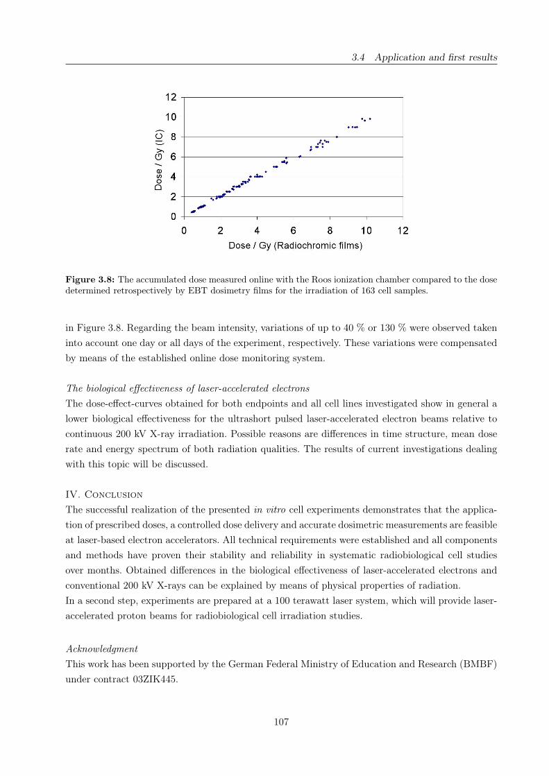
3.4 Application and first results
Figure 3.8: The accumulated dose measured online with the Roos ionization chamber compared to the dosedetermined retrospectively by EBT dosimetry films for the irradiation of 163 cell samples.
in Figure 3.8. Regarding the beam intensity, variations of up to 40 % or 130 % were observed taken
into account one day or all days of the experiment, respectively. These variations were compensated
by means of the established online dose monitoring system.
The biological effectiveness of laser-accelerated electrons
The dose-effect-curves obtained for both endpoints and all cell lines investigated show in general a
lower biological effectiveness for the ultrashort pulsed laser-accelerated electron beams relative to
continuous 200 kV X-ray irradiation. Possible reasons are differences in time structure, mean dose
rate and energy spectrum of both radiation qualities. The results of current investigations dealing
with this topic will be discussed.
IV. Conclusion
The successful realization of the presented in vitro cell experiments demonstrates that the applica-
tion of prescribed doses, a controlled dose delivery and accurate dosimetric measurements are feasible
at laser-based electron accelerators. All technical requirements were established and all components
and methods have proven their stability and reliability in systematic radiobiological cell studies
over months. Obtained differences in the biological effectiveness of laser-accelerated electrons and
conventional 200 kV X-rays can be explained by means of physical properties of radiation.
In a second step, experiments are prepared at a 100 terawatt laser system, which will provide laser-
accelerated proton beams for radiobiological cell irradiation studies.
Acknowledgment
This work has been supported by the German Federal Ministry of Education and Research (BMBF)
under contract 03ZIK445.
107

3.4 Application and first results
Author contributions
Study concept and design J.P., L.L., L.K., C.R., E.L., H.-P.S., E.B.; Experimental studies L.L.,
E.L., L.K., C.R., J.P., H.-P.S., M.N., E.B., D.N.; Data acquisition E.L., L.L., L.K., C.R.; Literature
research E.B., Data analysis L.L., D.N., C.R., L.K.; Manuscript preparation E.B., J.P.; Manuscript
editing J.P., M.B., E.B.; Guarantors of integrity of the entire study J.P., M.K., R.S., W.E., M.B.;
Manuscript review was carried out by all authors.
108

4 Discussion
The overarching issue of the present dissertation was the realization of in vitro cell irradiation
experiments at conventional and unconventional radiation sources, bearing in mind the potential
application of their emitted radiation for medical purposes. Two specific topics were addressed
within this general context - the photon energy dependence of the relative biological effectiveness of
X-rays and the performance of first in vitro cell experiments at a laser-driven electron accelerator.
Therefore, several consecutive requirements, like the establishment of appropriate radiation sources
and their dosimetric and biological characterization, have to be fulfilled independent on the applied
radiation quality.
The first issue was motivated by the ongoing cost-benefit discussion that begun in 2002 with the
introduction of a nationwide mammography screening program in Germany and the simultaneous
publication of an alarming high RBE value for mammographic X-rays by Frankenberg et al. [32].
Accordingly, several in vitro studies were initiated aiming on the photon energy dependence of the
RBE [40, 41, 45, 68, 105, 111, 112, 113], especially in the energy range below 50 keV. Likewise in our
laboratory, where the biological response to low-energy X-rays was reviewed for human mammary
gland epithelial cells. Previous experiments performed with cell line MCF-12A [74, 75] were not only
pursued in the present work, but also extended by the establishment and examination of a second
human mammary epithelial cell line (184A1). Moreover, two additional endpoints, the investiga-
tion of chromosomal aberrations and the detection of DNA double-strand breaks, were established
and successfully applied (sections 2.4.3 and 2.4.4) to analyze the relative biological effectiveness of
25 kV and 10 kV X-rays (Table 4.1). Regarding the resulting RBE values (Table 4.1), the biological
Biological endpoint RBE ± ∆ RBECell line 184A1 Cell line MCF-12A
10 kV X-rays
Excess fragments: 1.92 ± 0.26 1.40 ± 0.10Dicentric chromosomesand centric rings: 1.70 ± 0.29 1.43 ± 0.12Residual DNA DSB(24 hours post irradiation): 2.8 − 6.9∗ -25 kV X-rays
Excess fragments: 1.17 ± 0.12 0.97 ± 0.10Dicentric chromosomesand centric rings: 1.31 ± 0.21 1.08 ± 0.08Residual DNA DSB(24 hours post irradiation): 0.8 − 2.6∗ -34 MV bremsstrahlung
MN pro BNC 0.75 ± 0.05 -∗ Dose dependent RBE values
Table 4.1: RBE values determined in the framework of the present dissertation on basis of dose-effect-curvesfor various biological endpoints and photon energies using conventional 200 kV X-rays as reference.
effectiveness of 25 kV mammographic X-rays seems to be equal or just slightly enhanced compared
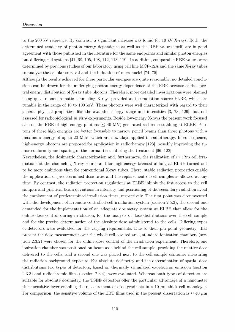
Discussion
to the 200 kV reference. By contrast, a significant increase was found for 10 kV X-rays. Both, the
determined tendency of photon energy dependence as well as the RBE values itself, are in good
agreement with those published in the literature for the same endpoints and similar photon energies
but differing cell systems [41, 68, 105, 108, 112, 113, 119]. In addition, comparable RBE values were
determined by previous studies of our laboratory using cell line MCF-12A and the same X-ray tubes
to analyze the cellular survival and the induction of micronuclei [74, 75].
Although the results achieved for these particular energies are quite reasonable, no detailed conclu-
sions can be drawn for the underlying photon energy dependence of the RBE because of the spec-
tral energy distribution of X-ray tube photons. Therefore, more detailed investigations were planned
using quasi-monochromatic channeling X-rays provided at the radiation source ELBE, which are
tunable in the range of 10 to 100 keV. These photons were well characterized with regard to their
general physical properties, like the available energy range and intensities [3, 73, 129], but not
assessed for radiobiological in vitro experiments. Beside low-energy X-rays the present work focused
also on the RBE of high-energy photons (≤ 40 MV) generated as bremsstrahlung at ELBE. Pho-
tons of these high energies are better focusable to narrow pencil beams than those photons with a
maximum energy of up to 20 MeV, which are nowadays applied in radiotherapy. In consequence,
high-energy photons are proposed for application in radiotherapy [123], possibly improving the tu-
mor conformity and sparing of the normal tissue during the treatment [96, 123].
Nevertheless, the dosimetric characterization and, furthermore, the realization of in vitro cell irra-
diations at the channeling X-ray source and for high-energy bremsstrahlung at ELBE turned out
to be more ambitious than for conventional X-ray tubes. There, stable radiation properties enable
the application of predetermined dose rates and the replacement of cell samples is allowed at any
time. By contrast, the radiation protection regulations at ELBE inhibit the fast access to the cell
samples and practical beam deviations in intensity and positioning of the secondary radiation avoid
the employment of predetermined irradiation times, respectively. The first point was circumvented
with the development of a remote-controlled cell irradiation system (section 2.5.2); the second one
demanded for the implementation of an adequate dosimetry system at ELBE that allow for the
online dose control during irradiation, for the analysis of dose distributions over the cell sample
and for the precise determination of the absolute dose administered to the cells. Differing types
of detectors were evaluated for the varying requirements. Due to their pin point geometry, that
prevent the dose measurement over the whole cell covered area, standard ionization chambers (sec-
tion 2.3.2) were chosen for the online dose control of the irradiation experiment. Therefore, one
ionization chamber was positioned on beam axis behind the cell sample, providing the relative dose
delivered to the cells, and a second one was placed next to the cell sample container measuring
the radiation background exposure. For absolute dosimetry and the determination of spatial dose
distributions two types of detectors, based on thermally stimulated exoelectron emission (section
2.3.3) and radiochromic films (section 2.3.4), were evaluated. Whereas both types of detectors are
suitable for absolute dosimetry, the TSEE detectors offer the particular advantage of a nanometer
thick sensitive layer enabling the measurement of dose gradients in a 10 µm thick cell monolayer.
For comparison, the sensitive volume of the EBT films used in the present dissertation is ≈ 40 µm
110

Discussion
thick. However, the TSEE detectors were found to be not practicable for routine cell irradiations,
since their mechanical stability and dose response reproducibility under real experiment conditions
were inadequate. Consequently, radiochromic EBT films were deployed for the retrospective deter-
mination of the absolute dose administered to the cell sample, of dose inhomogeneities over the
cell covered area and of deviations of the electron beam positioning. Moreover, the implementation
of EBT films in front of each sample allows for the retrospective analysis of beam deviations for
individual cell samples and, hence, for the tracking of beam deviations during the experiment.
Based on the observation of the radiation background the channeling X-ray source was found to
be not applicable for radiobiological experiments in the framework of the present dissertation, by
reason that the background dose contribution is as high as the planned dose delivery to the cells.
The main part of the background radiation is produced by electrons, that were scattered in the
diamond crystal, loss some energy and are subsequently not fully deflected to the beam dump
(cf. section 2.5.2, e.g., Fig. 2.18, for setup). In the following, these electrons might hit the beam
line components and generate bremsstrahlung. Moreover, a significant contribution to the radiation
background arise from bremsstrahlung produced during the electron stopping in the beam dump. A
minor, but unavoidable, source of radiation background are non-channeling electrons that produce
bremsstrahlung directly in the diamond crystal. Although, this bremsstrahlung is also emitted in
forward direction, like the quasi-monochromatic channeling X-rays, their contribution to the entire
background is very small. Practicable solutions for background reduction are a better shielding of
the beam line and the beam dump or the guidance of channeling X-rays in the adjacent experimen-
tal hall using the lead-containing concrete wall as additional shielding. However, this is connected
with high investment costs and new extensive practical regulations in order to fulfill the radiation
protection directives.
Contrary to the channeling source, applicable beam parameters and an acceptable radiation back-
ground level were obtained for the high-energy bremsstrahlung source (section 2.5.2). Following
dosimetric characterization, first in vitro cell irradiation experiments were performed aiming on the
analysis of micronuclei induced by 34 MV bremsstrahlung in cell line 184A1. The resulting RBE
value, relative to the 200 kV X-ray reference, is shown in Table 4.1. Presently, very few data on
the RBE of high-energy photons can be found in the literature; indeed their exist no possibility
of direct comparison for the RBE value of 34 MV bremsstrahlung obtained in the present disser-
tation. Similar RBE values of about 1 were revealed by Persson et al. [96], which investigated the
clonogenic survival of 50 MV bremsstrahlung relative to those after 60Co γ-radiation. Furthermore,
RBE values ranging from 0.25 to 1 were ascertained comparing the biological effectiveness of several
reference radiation sources, like 60Co γ-rays and 200 - 350 kV X-rays [48]. Summarizing these data,
the RBE value obtained in the present work is quite reasonable demonstrating that radiobiological
experimentation under the harsh conditions at ELBE is possible. However, taken into account the
medical application of high-energy photons (≥ 20 MeV), neutrons arising from high-energy brems-
strahlung via photo nuclear reactions with parts of the beam line have to be considered. At ELBE
the neutron background dose rate was monitored by means of a neutron detector (section 2.5.2),
whereas Persson et al. [96], who obtained a similar RBE, have identified just a marginal neutron
111
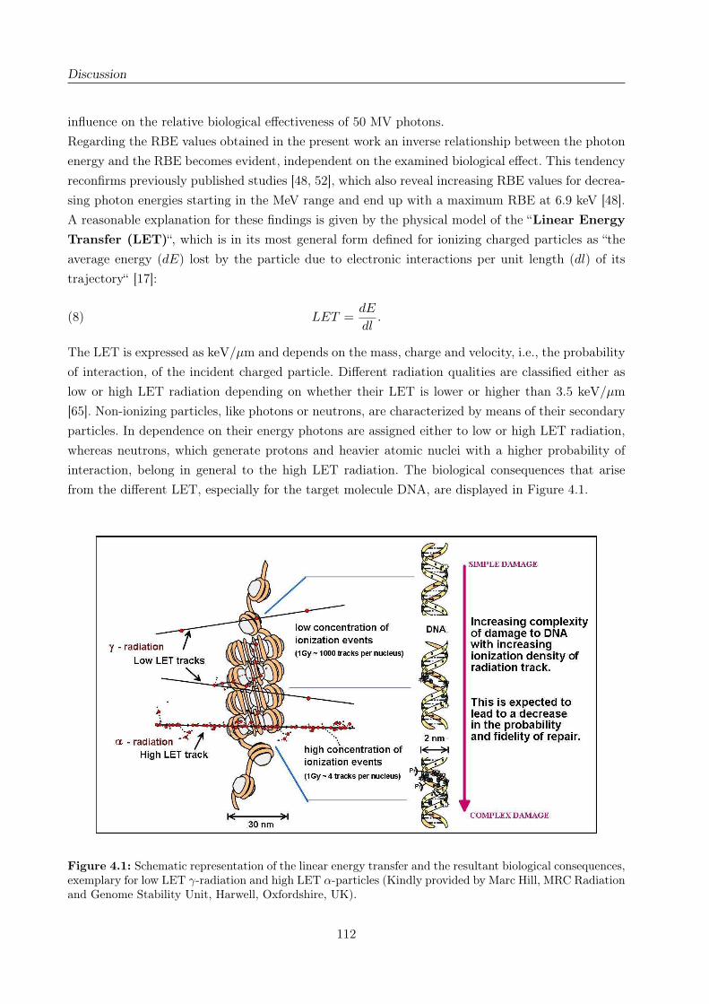
Discussion
influence on the relative biological effectiveness of 50 MV photons.
Regarding the RBE values obtained in the present work an inverse relationship between the photon
energy and the RBE becomes evident, independent on the examined biological effect. This tendency
reconfirms previously published studies [48, 52], which also reveal increasing RBE values for decrea-
sing photon energies starting in the MeV range and end up with a maximum RBE at 6.9 keV [48].
A reasonable explanation for these findings is given by the physical model of the “Linear Energy
Transfer (LET)“, which is in its most general form defined for ionizing charged particles as “the
average energy (dE) lost by the particle due to electronic interactions per unit length (dl) of its
trajectory“ [17]:
(8) LET =dE
dl.
The LET is expressed as keV/µm and depends on the mass, charge and velocity, i.e., the probability
of interaction, of the incident charged particle. Different radiation qualities are classified either as
low or high LET radiation depending on whether their LET is lower or higher than 3.5 keV/µm
[65]. Non-ionizing particles, like photons or neutrons, are characterized by means of their secondary
particles. In dependence on their energy photons are assigned either to low or high LET radiation,
whereas neutrons, which generate protons and heavier atomic nuclei with a higher probability of
interaction, belong in general to the high LET radiation. The biological consequences that arise
from the different LET, especially for the target molecule DNA, are displayed in Figure 4.1.
Figure 4.1: Schematic representation of the linear energy transfer and the resultant biological consequences,exemplary for low LET γ-radiation and high LET α-particles (Kindly provided by Marc Hill, MRC Radiationand Genome Stability Unit, Harwell, Oxfordshire, UK).
112

Discussion
Considering the photon energy dependence of the RBE, the range and accompanied LET distribu-
tion of the generated secondary electrons are the most likely explanation for the obtained tendencies.
For high-energy photons or γ-rays the secondary electrons released in matter are of high-energy and
long ranging. Consequently, their probability of interaction and therefore the linear energy transfer
are quite low, e.g., 0.22 keV/µm [55] for 60Co γ-rays. With decreasing photon energy both energy
and range of the secondary electrons also decrease resulting in an increasing probability of interac-
tion and in LET values reaching those of high-LET radiation. Moreover, the range of the secondary
electrons arrives at distances that are comparable and even lower than the size of a cell (≈ 30 µm).
Exemplary for 6.9 keV, the LET amounts to 4.1 keV/µm and the maximum range of the secondary
electrons is in the order of 1.2 µm [4] associated with a higher probability of consecutive interactions
within the same cell, i.e., with a higher risk of DNA DSB induction. Even higher LET values might
be proposed for photon energies below the maximum at 6.9 keV, but the range of their secondary
electrons (down to nm) is too short to induce complex DNA damages, like DSB. In addition, the
very low range of these secondary electrons is contrary to the LET model, where the energy distri-
bution is regarded on a trajectory.
Although the underlying photon energy dependence of the RBE is commonly accepted and explai-
nable with the LET model, no revision or energy dependent specification of the radiation weighting
factor for photons was realized within the radiation protection directives of the ICRP [57]. The
results obtained by the various in vitro studies were of course discussed with respect to the under-
estimation of the radiation risk, especially for low-energy photons (e.g., [11, 37, 57]), but there are
several reasonable arguments against an adjustment. The absolute numbers and energy dependent
variations of the RBE values achieved for photons are quite low compared to those found for other
radiation qualities, like heavy ions or neutrons. For example, RBE values ranging from 16.6 (36 keV)
to 23.4 (385 keV) have been determined for the neutron based induction of dicentric chromosomes
in human lymphocytes using 220 kV X-rays as reference [114]. In this context, the twofold to sixfold
higher biological effectiveness of low-energy X-rays (≤ 50 keV) relative to 60Co γ-rays and 200 kV X-
rays as reference do not justify an energy dependent radiation weighting factor, respectively. Even
more, if one considers the modification of the photon energy spectra by the transition from flat
cell samples to large human bodies. Low-energy photons will be absorbed in the outer skin layers,
whereas high-energy photons will interact with tissue atoms and produce secondary electrons that
might contribute to the organ doses. However, the maximum photon energy difference and with it
the RBE variations averaged over the whole human body will be much smaller than for cell culture
samples used in radiobiological experiments [23].
Another important aspects are the absence of significant epidemiological data and the question, to
what extent in vitro data can be applied to estimate the radiation risk for large and complex hu-
man bodies. So far, the high RBE values revealed by in vitro cell experiments are not supported by
animal studies or epidemiological surveys [52, 58], due to the fact that the existing epidemiological
data cannot be analyzed with respect to particular energies [52] or that the underlying statistic is
insufficient [58]. Moreover, the revision of the weighting factor has to be considered carefully, since it
will have practical consequences on radiation protection regulations [23]. Taking into account these
113
Discussion
arguments and the absence of more comprehensive epidemiological studies, the ICRP still specifies
a radiation weighting factor of 1 for all photon energies in their 2007 recommendations [58].
An analogue line of arguments has to be regarded to judge the in vitro results determined for mam-
mographic X-rays. The most important question in this context is again the validity or adaptability
of in vitro data for the radiation risk estimation for healthy women which attend the mammo-
graphy screening program. Less relevant for these considerations is the cellular survival following
irradiation, because the average glandular dose administered to the female breast lies in the range
of 4.5 to 21.4 mGy per screening [46]. By contrast, doses of ≥ 500 mGy are typically required for
radiobiological examination in order to detect any effect on the cellular survival with reasonable
statistic. In consequence, the induction of radiation late effects, especially the induction of cancer,
whereto the analysis of micronuclei [74, 75, 119], chromosomal aberrations [6, 41, 68, 112, 113] or
remaining DNA double-strand breaks [7] may provide useful hints, is more relevant to estimate the
risk of mammography. In doing so, RBE values between 1 and 2, not confirming the alarming RBE
of about 4 published by Frankenberg et al. [32], were obtained by several groups comparing mam-
mographic X-rays to higher energy photons. These results, including the new data obtained within
this dissertation, were presented on the 2007 Retreat of the German Commission on Radiological
Protection (SSK) [38]. There and in other committees, the different in vitro results were discussed,
since RBE values higher than one still indicate an additional risk for woman that participate on a
nationwide mammography screening program. Again, the validity of the in vitro data, even though
consistent for the varying groups, remains unclear for the risk estimation of mammography scree-
ning. Controversial points are the complexity of the human body, the time frame of cancer induction
(≈ 20 a), which is incompatible with the methodology of in vitro investigations, and the absence
of significant epidemiological or clinical studies [46, 52]. Although the need of more detailed and
target-oriented research was identified, reasonable precautions were taken anyway. Mammography
screening is just indicated for women between 50 and 69 years by reason of the mean cancer in-
cidence time, which would be an additional risk for younger women. The examination of younger
or familial predisposed women is considered carefully [46] taken into account their higher risk for
tumor induction. Moreover, the SSK recommends the independent evaluation of the German mam-
mography screening program with respect to quality-assurance and risk-benefit analysis [39].
To sum up, the experiments and investigations realized within this particular issue of the present
dissertation are a valuable contribution to the ongoing cost-benefit discussion on the mammogra-
phy screening program. Our analysis of DSB related biological endpoints result in photon energy
dependent RBE values that were similar to those published for other cell lines and to known RBE
trends. For this reason, the in vitro data base is extended by an additional cell system, being more
relevant for mammography; whereas the collected data provide useful hints on the underlying me-
chanism that lead to reduced survival or the incidence of cancer. Beside these meaningful results,
necessary and important knowledge and experiences were acquired in the course of the realization
of in vitro cell experiments at unconventional secondary radiation sources. The established dosime-
tric methods, sample setups and handling procedures can be transferred to other (unconventional)
radiation qualities allowing for an efficient radiobiological characterization.
114
Discussion
Exactly these experiences were required for the second issue of the present dissertation - the realiza-
tion of radiobiological in vitro cell experiments with laser-accelerated electrons. These experiments
were carried out at the Jena Titanium:Sapphire laser system representing so far an unconventio-
nal class of particle accelerators that are considered to replace the huge and cost-intensive proton
and ion acceleration technique nowadays applied in radiotherapy. In general, these unconventional
particle accelerators are based on high intensity lasers that allow for the acceleration of charged
particles (electrons, protons, ions) in a very short distance, i.e., by means of ultrahigh acceleration
gradients (section 3.2). The generated particle beams are delivered in the same time regime as the
laser pulse (≈ 100 fs), a feature that result in ultrashort particle beam pulses with ultrahigh dose
rates during the pulse exceeding those of conventional particle accelerators by several orders of
magnitude. In consequence, one or just a couple of bunches (i.e. pulses) will be sufficient to deliver
the therapeutic dose of about 2 Gy to the patient. Before such a new type of particle accelerator
can be applied for medical purposes, the radiobiological consequences that arise from the unusual
irradiation regime have to be investigated and compared to those of a continuous reference radiati-
on. Therefore, the laser technology has to be adapted for radiobiological requirements, e.g., for the
delivery of stable particle beams. Furthermore, the dosimetric properties have to be characterized
very precisely in order to allow for radiobiological in vitro studies that aim on the determination of
the RBE of laser-accelerated particles.
Worldwide first experiments in this direction were performed with JETI electrons already avai-
lable with sufficient intensity and energy for cell irradiation, whereas available laser-driven proton
[97, 117] and ion [43, 82] beams are still insufficient on both parameters. The electron beam was
characterized and optimized with respect to radiobiological properties using the dosimetric methods
implemented at ELBE and an in-house made Faraday cup. That is, the electron energy spectrum
was limited by a low-energy cutoff (≈ 3 MeV) and the beam spot as well as the beam intensity were
adjusted to sample size and the performance of cell irradiations in a reasonable time, respectively.
However, the intensity and vice versa the pulse dose rate used for the cell experiments are not ma-
ximized, since the JETI system was not capable of providing single electron pulses with maximum
intensity and a controlled pulse dose at the same time. In regard to patient treatment, especially
the last point will be hazardous and consequently the pulse dose rate was adjusted to ≈ 109 Gy/s
anyhow exceeding those of conventional electron accelerators by several orders of magnitude.
Beside electron beam characterization, the readouts of the established beam and dose monitoring
system were found to be linear correlated to the absolute dose administered at cell position. Hence,
the system was applicable for a careful online control of the irradiation and enables the delivery of
prescribed doses to the cells, a feature so far unique for laser-based particle accelerators. Further-
more, the linear dose correlations were confirmed for repeated experiment days indicating long-term
stable and reproducible beam properties as demanded for radiobiological studies and being stringent
for radiotherapy. Accordingly, the performance of systematic in vitro cell irradiations was feasible
and a substantial progress from physical single-shot experiments with poor reproducibility towards
medical application was achieved. Further advancements of the laser technology and the auxiliary
equipment are expected to lead to even more stable and reproducible particle beams, necessary for
115

Discussion
patient irradiation in future.
In parallel to the technological improvement, a test cell irradiation was performed at the JETI la-
ser system followed by the worldwide first systematic radiobiological in vitro cell experiments with
laser-accelerated electrons. The results obtained at the JETI system reveal a reduced biological
effectiveness for laser-accelerated and therefore ultrashort pulsed electron beams in comparison to
continuous 200 kV X-ray irradiation. This finding was consistent for the examined human tumor
and human normal cell lines and for both analyzed biological effects, that is the cellular survival and
the residual DNA double-strand breaks. Some decades afore similar experiments were accomplished
addressing the influence of high pulse dose rates on the RBE of electrons [18, 87, 115]. For this
purpose, conventional electron sources were adjusted to shorter electron pulses with maximum dose
rates of up to 109 Gy/s during the pulse delivering the required dose to the cells in one single or a
few electron pulses. As result, a comparable or even higher survival was achieved by comparing the
pulsed radiation quality with a continuous reference source. Similarly, laser-driven bremsstrahlung
possessing a maximum dose rate of 1013 Gy/s [49, 118] result also in a comparable or reduced bio-
logical effectiveness.
One hypothesis that might explain these findings is the influence of the ultrahigh pulse dose rate
on the oxygen effect, which generally describes the influence of the oxygen content and supply on
the radiation effect. Following radiation incidence, cellular oxygen becomes ionized and hence being
able to ionize DNA components and to induce DNA damages. Moreover, the interaction of oxygen
and DNA “radicals“, e.g., at DNA breaks, prevent the binding of repair proteins; open DNA ends are
not connected and the DNA damage is “fixed“. For continuous cell irradiation the dose delivery is
in such an extent that oxygen consumption and replacement by means of diffusion are balanced on
a certain level. By contrast, for (ultra-)short pulsed particle beams with high dose rates during the
pulse, the cellular oxygen is considerably reduced during the pulse and the pulse frequency might
be shorter than the time needed to replace the cellular oxygen. In consequence, less cellular oxygen
is available for subsequent particle pulses, which result in a reduced amount of induced and fixed,
i.e., not repaired, DNA damages. This in turn might lead to a higher cellular survival and lower
biological effectiveness, respectively.
Nevertheless, the other physical parameters of the laser-accelerated electron beam can also be hy-
pothesized as potential reasons for the reduced biological effectiveness obtained in the framework of
the present work. Compared to the 200 kV X-ray reference, the JETI electrons exhibit a lower mean
dose rate, averaged over the irradiation time, but a several orders of magnitude higher pulse dose
rate within the electron pulse. In addition, the energy spectra of 200 kV X-rays (≈ 40 - 200 keV)
and JETI electrons (3 - 20 MeV) are significantly different resulting in varying LET influences. The
individual contributions of these beam parameters on the biological effectiveness of laser-accelerated
electrons are investigated in running experiments initiated by the findings of this dissertation.
In summary, this particular issue of the present dissertation was also carried out successfully. The
technological improvement of the JETI laser system towards stable and reproducible cell irradia-
tions was performed in such an extent that the delivery of prescribed doses is possible. Applying
this unique feature first systematic in vitro studies were realized providing not only useful radio-
116
Discussion
biological results, but also a feedback and starting points for further improvements of the laser
and auxiliary technique. Outstanding questions on the potential reasons for the reduced biological
effectiveness of laser-driven electrons will be answered in future. In the end, it can be concluded
that laser-accelerated electrons will not provide any disadvantage for radiotherapy, that means tu-
mor cells are killed under sparing of the normal tissue. Based on the successful results obtained
with laser-accelerated electrons in the present work, future experiments will be concentrated on
laser-accelerated protons and their potential role in radiotherapy.
117

5 Literature
[1] DIN 6800-2, Procedures of dosimetry with probe type detectors for photon and electron ra-
diation - Part 2: Ionization chamber dosimetry of high energy photon and electron radiation.
2008.
[2] L. Anderson, C. Henderson, and Y. Adachi. Phosphorylation and rapid relocalization of 53BP1
to nuclear foci upon DNA damage. Mol. Cell. Biol., 21(5):1719–29, 2001.
[3] B. Azadegan. Investigation of planar channeling radiation on diamond and quartz crystals
at electron energies between 14 and 34 MeV and probing the influence of ultrasonic waves on
Channeling Radiation. Dissertation, Dresden University of Technology, 2007.
[4] M. J. Berger, J. S. Coursey, M. A. Zucker, and J. Chang. ESTAR, PSTAR and ASTAR:
Computer Programs for Calculating Stopping-Power and Range Tables for Electrons, Protons
and Helium Ions (Version 1.2.3). http://physics.nist.gov/star, 2009.
[5] E. H. Berkowitz. Dose compensation by differential pattern scanning. US Patent, 4449051,
1984.
[6] E. Beyreuther, W. Dörr, A. Lehnert, E. Lessmann, and J. Pawelke. Relative biological effec-
tiveness of 25 and 10 kV X-rays for the induction of chromosomal aberrations in two human
mammary epithelial cell lines. Radiat. Environ. Biophys., 48(3):333–40, 2009.
[7] E. Beyreuther, E. Lessmann, J. Pawelke, and S. Pieck. DNA double-strand break signalling:
X-ray energy dependence of residual co-localised foci of γ-H2AX and 53BP1. Int. J. Radiat.
Biol., 85(11):1042–50, 2009.
[8] D. Blöcher. DNA double-strand break repair determines the RBE of alpha-particles. Int. J.
Radiat. Biol., 54(5):761–71, 1988.
[9] J. J. Boei, S. Vermeulen, J. Fomina, and A. T. Natarajan. Detection of incomplete exchanges
and interstitial fragments in X-irradiated human lymphocytes using a telomeric PNA probe.
Int. J. Radiat. Biol., 73(6):599–603, 1998.
[10] J. J. Boei, S. Vermeulen, and A. T. Natarajan. Detection of chromosomal aberrations by
fluorescence in situ hybridization in the first three postirradiation divisions of human lympho-
cytes. Mutat. Res., 349(1):127–35, 1996.
[11] D. J. Brenner, S. G. Sawant, M. P. Hande, et al. Routine screening mammography: how
important is the radiation-risk side of the benefit-risk equation? Int. J. Radiat. Biol.,
78(12):1065–67, 2002.
[12] M. J. Butson, T. Cheung, and P. K. N. Yu. Weak energy dependence of EBT Gafchromic
film dose response in the 50 kVp-10 MVp X-ray range. Appl. Radiat. Isot., 64(1):60–62, 2006.
[13] M. J. Butson, P. K. N. Yu, T. Cheung, and P. Metcalfe. Radiochromic film for medical
radiation dosimetry. Mater. Sci. Eng. R, 41(3-5):61–120, 2003.
[14] Center for ultrafast optical science, College of Engineering, University of Michigan, USA.
Wakefield electron acceleration.
www.engin.umich.edu/research/cuos/ResearchGroups/HSF/Research/, 23.03.2010.
[15] C. Chiu, M. Fomytskyi, F. Grigsby, et al. Laser electron accelerators for radiation medicine:
A feasibility study. Med. Phys., 31(7):2042–52, 2004.
[16] S. T. Chiu-Tsao, Y. Ho, R. Shankar, L. Wang, and L. B. Harrison. Energy dependence of
response of new high sensitivity radiochromic films for megavoltage and kilovoltage radiation
energies. Med. Phys., 32(11):3350–54, 2005.
[17] Commitee to Assess Health Risks from Exposure to low levels of Ionizing Radiation. Health
risks from exposure to low levels of ionizing radiation. BEIR VII Phase 2, 2006.
[18] J. Cygler, N. V. Klassen, C. K. Ross, T. J. Bichay, and G. P. Raaphorst. The survival of aerobic
and anoxic human glioma and melanoma cells after irradiation at ultrahigh and clinical dose
rates. Radiat. Res., 140(1):79–84, 1994.
[19] L. A. R. da Rosa, M. Seidenbusch, and D. F. Regulla. Dose profile assessment at gold-tissue
interfaces by using TSEE. Radiat. Prot. Dosimetry, 85(1-4):433–36, 1999.
[20] S. Devic, J. Seuntjens, G. Hegyi, et al. Dosimetric properties of improved GafChromic films
for seven different digitizers. Med. Phys., 31(9):2392–2401, 2004.
[21] S. Devic, J. Seuntjens, E. Sham, et al. Precise radiochromic film dosimetry using a flat-bed
document scanner. Med. Phys., 32(7):2245–53, 2005.
[22] F. Di Martino, M. Giannelli, A. C. Traino, and M. Lazzeri. Ion recombination correction for
very high dose-per-pulse high-energy electron beams. Med. Phys., 32(7):2204–10, 2005.
[23] G. Dietze and W. G. Alberts. Why it is advisable to keep wR = 1 and Q = 1 for photons and
electrons. Radiat. Prot. Dosimetry, 109(4):297–302, 2004.
[24] J. S. Durham, S. E. Merwin, and K. L. Swinth. Skin dose evaluations using exoelectron
dosemeters. Radiat. Prot. Dosimetry, 39(1-3):67–70, 1991.
[25] A. A. Edwards, D. C. Lloyd, and R. J. Purrott. Radiation induced chromosome aberrations
and the poisson distribution. Radiat. Environ. Biophys., 16(2):89–100, 1979.
[26] H. Eichenmüller. Thermally stimulated exoelectron emission (TSEE) from metal and semi-
conductor surfaces. Radiat. Prot. Dosimetry, 4(3-4):281–85, 1983.
120

[27] I. Eke, V. Sandfort, K. Storch, et al. Pharmacological inhibition of EGFR tyrosine kinase
affects ILK-mediated cellular radiosensitization in vitro. Int. J. Radiat. Biol., 83(11-12):793–
802, 2007.
[28] J. Fan, W. Luo, E. Fourkal, et al. Shielding design for a laser-accelerated proton therapy
system. Phys. Med. Biol., 52:3913–30, 2007.
[29] J. Faure, Y. Glinec, A. Pukhov, et al. A laser-plasma accelerator producing monoenergetic
electron beams. Nature, 431:541–44, 2004.
[30] Forschungszentrum Dresden-Rossendorf. Radiation source ELBE.
http://www.fzd.de, 24.03.2010.
[31] E. Fourkal, B. Shahine, M. Ding, et al. Particle in cell simulation of laser-accelerated proton
beams for radiation therapy. Med. Phys., 29(12):2788–98, 2002.
[32] D. Frankenberg, K. Kelnhofer, K. Bär, and M. Frankenberg-Schwager. Enhanced neoplastic
transformations by mammography X rays relative to 200 kVp X rays: indication for a strong
dependence on photon energy of the RBE(M) for various end points. Radiat. Res., 157(1):99–
105, 2002. Erratum in: Radiat. Res. 158(1), 126 (2002).
[33] M. Fuss, E. Sturtewagen, C. De Wagter, and D. Georg. Dosimetric characterization of Gaf-
Chromic EBT film and its implication on film dosimetry quality assurance. Phys. Med. Biol.,
52:4211–25, 2007.
[34] F. Gabriel, P. Gippner, E. Grosse, et al. The Rossendorf radiation source ELBE and its FEL
projects. Nucl. Instrum. Methods Phys. Res. B, 161-63:1143–47, 2000.
[35] GEANT4 collaboration. GEANT4 a simulation toolkit. Nucl. Instrum. Methods Phys. Res.
A, 506(3):250–303, 2003.
[36] C. G. R. Geddes, C. Toth, J. v. Tilborg, et al. High-quality electron beams from a laser
wakefield accelerator using plasma-channel guiding. Nature, 431:538–41, 2004.
[37] German Commission on Radiological Protection. Evaluation of the relative biological effec-
tiveness of different types of ionizing radiation. Statement of the German Commission on
Radiological Protection. Bonn: Urban & Fischer, Band 53, 2005.
[38] German Commission on Radiological Protection. Summary and Assessment of the 2007 Re-
treat of the German Commission on Radiological Protection: Biological Effectiveness of low
doses of ionizing radiation [in German]. www.ssk.de, 2007.
[39] German Commission on Radiological Protection. Evaluation of the effectiveness of quality-
assured mammography screening in Germany. Statement of the German Commission on Ra-
diological Protection. [in German]. www.ssk.de, 2008.
121
[40] W. Göggelmann, C. Jacobsen, W. Panzer, et al. Re-evaluation of the RBE of 29 kV x-rays
(mammography x-rays) relative to 220 kV x-rays using neoplastic transformation of human
hybrid CGL1-hybrid cells. Radiat. Environ. Biophys., 42(3):175–82, 2003.
[41] C. Guerrero-Carbajal, A. A. Edwards, and D. C. Lloyd. Induction of chromosome aberration in
human lymphocytes and its dependence on X ray energy. Radiat. Prot. Dosimetry, 106(2):131–
35, 2003.
[42] S. L. Hammond, R. G. Ham, and M. R. Stampfer. Serum-free growth of human mamma-
ry epithelial cells: rapid clonal growth in defined medium and extended serial passage with
pituitary extract. Proc. Natl. Acad. Sci. U.S.A., 81(17):5435–39, 1984.
[43] B. M. Hegelich, B. J. Albright, J. Cobble, et al. Laser acceleration of quasi-monoenergetic
MeV ion beams. Nature, 439:441–44, 2006.
[44] T. Herrmann and M. Baumann. Klinische Strahlenbiologie. 3. Aufl. Gustav Fischer Verlag,
1997.
[45] G. J. Heyes and A. J. Mill. The neoplastic transformation potential of mammography x rays
and atomic bomb spectrum radiation. Radiat. Res., 162(2):120–27, 2004.
[46] G. J. Heyes, A. J. Mill, and M. W. Charles. Enhanced biological effectiveness of low energy
X-rays and implications for the UK breast screening program. Br. J. Radiol., 79(939):195–200,
2006.
[47] B. Hidding, K.-U. Amthor, B. Liesfeld, et al. Generation of quasimonoenergetic electron
bunches with 80-fs laser pulses. Phys. Rev. Lett., 96(10):105004, 2006.
[48] M. A. Hill. The variation in biological effectiveness of X-rays and gamma rays with energy.
Radiat. Prot. Dosimetry, 112(4):471–81, 2004.
[49] M. A. Hill, D. L. Stevens, S. J. Marsden, et al. Is the increase in relative biological effectiveness
of high LET particles due to spatial or temporal effects? Characterization and OER in V79-4
cells. Phys. Med. Biol., 47(19):3543–55, 2002.
[50] G. Holzapfel. Zur Exoelektronen-Emission (Kramer-Effekt) von Berylliumoxid. Dissertation,
Technische Universität Berlin, 1968.
[51] J. H. Hubbell and S. M. Seltzer. Tables of X-Ray Mass Attenuation Coefficients and Mass
Energy-Absorption Coefficients (version 1.4). http://physics.nist.gov/xaamdi, 2004.
[52] N. Hunter and C. R. Muirhead. Review of the relative biological effectiveness dependence on
linear energy transfer for low-LET radiations. J. Radiol. Prot., 29(1):5–21, 2009.
[53] Y. Ichijima, R. Sakasai, N. Okita, et al. Phosphorylation of histone H2AX at M phase in
human cells without DNA damage response. Biochem. Biophys. Res. Commun., 336(3):807–
12, 2005.
122
[54] International Atomic Energy Agency. Biological Dosimetry: Chromosomal aberration analysis
for dose assessment. Technical Report Series No. 260, 1986.
[55] International Commission on Radiation Units and Measurements. Linear Energy Transfer.
ICRU Report, 16, 1970.
[56] International Commission on Radiological Protection. ICRP Publication 60: 1990 Recom-
mendations of the International Commission on Radiological Protection. Annals of the ICRP,
21(1-3), 1991.
[57] International Commission on Radiological Protection. ICRP Publication 92: Relative Biolo-
gical Effectiveness (RBE), Quality Factor (Q), and Radiation Weighting Factor (wR). Annals
of the ICRP, 33(4), 2003.
[58] International Commission on Radiological Protection. ICRP Publication 103: The 2007 Re-
commendations of the International Commission on Radiological Protection. Annals of the
ICRP, 37(2-4), 2007.
[59] International Specialty Products (ISP). Gafchromic R© EBT: self-developing film for radio-
therapy dosimetry. International Specialty Products Inc., Wayne, NJ, USA, 2007.
[60] K. K. Kainz, K. R. Hogstrom, J. A. Antolak, et al. Dose properties of a laser accelerated
electron beam and prospects for clinical application. Med. Phys., 31(7):2053–67, 2004.
[61] P. Kegel, E. Riballo, M. Kühne, P. A. Jeggo, and M. Löbrich. X-irradiation of cells on glass
slides has a dose doubling impact. DNA repair (Amst.), 6(11):1692–97, 2007.
[62] A. Kinner, W. Wu, C. Staudt, and G. Iliakis. γ-H2AX in recognition and signaling of DNA
double-strand breaks in the context of chromatin. Nucleic Acids Res., 36(17):5678–94, 2008.
[63] W. Kottler, D. Lerch, W. Kriegseis, A. Scharmann, and B. Wörner. Evaporated beryllium
oxide films for TSEE dosimetry. Nucl. Instrum. Methods Phys. Res., 175(1):101–03, 1980.
[64] J. Kramer. Exoelektronen-Dosimeter für Röntgen- und Gammastrahlen. Z. Angew. Phys.,
20:411–17, 1966.
[65] H. Krieger. Grundlagen der Strahlungsphysik und des Strahlenschutzes. [in German]. B.G.
Teubner, Wiesbaden., 1 edition, 2004.
[66] W. Kriegseis, M. Petel, A. Scharmann, and C. U. Wieters. Potentials of TSEE for beta ray
dosimetry. Radiat. Prot. Dosimetry, 14(2):151–55, 1986.
[67] W. Kriegseis, K. Rauber, A. Scharmann, et al. Dependence of the TSEE response of BeO
thin films on photon energy and composition of cover materials. Radiat. Prot. Dosimetry,
47(1-4):143–46, 1993.
123
[68] M. Krumrey, G. Ulm, and E. Schmid. Dicentric chromosomes in monolayers of human lym-
phocytes produced by monochromatized synchrotron radiation with photon energies from
1.83 keV to 17.4 keV. Radiat. Environ. Biophys., 43(1):1–6, 2004.
[69] M. Löbrich and P. A. Jeggo. The impact of negligent G2/M checkpoint on genomic instability
and cancer induction. Nat. Rev. Cancer, 7(11):861–69, 2007.
[70] E. L. Leatherbarrow, J. V. Harper, F. A. Cucinotta, and P. O’Neill. Induction and quanti-
fication of gamma-H2AX foci following low and high LET-irradiation. Int. J. Radiat. Biol.,
82(2):111–18, 2006.
[71] K. W. D. Ledingham, W. Galster, and R. Sauerbrey. Laser-driven proton oncology - a unique
new cancer therapy? Br. J. Radiol., 80(959):855–58, 2007.
[72] W. P. Leemans, B. Nagler, A. J. Gonsalves, et al. GeV electron beams from a centimetre-scale
accelerator. Nat. Phys., 2:696–99, 2006.
[73] A. Lehnert. Establishment of the physical and technical prerequisites for the determination of
the relative biological effectiveness of low-energy monochromatic X-rays. Wiss.-Tech. Berichte,
FZR-441, 2005.
[74] A. Lehnert, W. Dörr, E. Lessmann, and J. Pawelke. RBE of 10 kV X rays determined for the
human mammary epithelial cell line MCF-12A. Radiat. Res., 169(3):330–36, 2008.
[75] A. Lehnert, E. Lessmann, J. Pawelke, and W. Dörr. RBE of 25 kV X-rays for the survival
and induction of micronuclei in the human mammary epithelial cell line MCF-12A. Radiat.
Environ. Biophys., 45(4):253–60, 2006.
[76] J. Lesz, A. Scharmann, and G. Holzapfel. Connected TSEE/OSEE readouts of BeO thin-film
dosemeter. Jpn. J. Appl. Phys., 24(Suppl. 24-4):259–61, 1985.
[77] M. Fenech. The cytokinesis-block micronucleus technique: a detailed description of the method
and its application to genotoxicity studies in human populations. Mutat. Res., 285(1):35–44,
1993.
[78] C.-M. Ma, I. Velchev, T. Lin, et al. Laser-proton acceleration for radiation therapy. Medical
Physics, 2007 AAPM Annual Meeting Program, 34(6):2550, 2007.
[79] S. H. MacPhail, J. P. Banáth, Y. Yu, E. Chu, and P. L. Olive. Cell cycle-dependent expression
of phosphorylated histone H2AX: reduced expression in unirradiated but not X-irradiated G1-
phase cells. Radiat. Res., 159(6):759–67, 2003.
[80] S. H. MacPhail, J. P. Banáth, Y. Yu, et al. Expression of phosphorylated histone H2AX in
cultured cell lines following exposure to X-rays. Int. J. Radiat. Biol., 79(5):351–58, 2003.
[81] P. Maine, D. Strickland, P. Bado, M. Pessot, and G. Mourou. Generation of ultrahigh peak
power pulses by chirped pulse amplification. IEEE J. Quantum Electron., 24(2):398–403, 1988.
124
[82] A. Maksimchuk, S. Gu, K. Flippo, D. Umstädter, and V. Bychenkov. Forward ion acceleration
in thin films driven by a high-intensity laser. Phys. Rev. Lett., 84(18):4108–11, 2000.
[83] V. Malka, S. Fritzler, E. Lefebvre, E. d’Humières, and R. Ferrand. Practicability of proton-
therapy using compact laser systems. Med. Phys., 31(6):1587–92, 2004.
[84] S. P. D. Mangles, C. D. Murphy, Z. Najmudin, et al. Monoenergetic beams of relativistic
electrons from intense laser-plasma interactions. Nature, 431:535–38, 2004.
[85] E. Marková, N. Schultz, and I. Y. Belyaev. Kinetics and dose-response of residual
53BP1/gamma-H2AX foci: co-localization, relationship with DSB repair and clonogenic sur-
vival. Int. J. Radiat. Biol., 83(5):319–29, 2007.
[86] M. Mestres, M. R. Caballín, L. Barrios, M. Ribas, and J. F. Barquinero. RBE of X rays of
different energies: a cytogenetic evaluation by FISH. Radiat. Res., 170(1):93–100, 2008.
[87] H. B. Michaels, E. R. Epp, C. C. Ling, and E. C. Peterson. Oxygen sensitization of CHO cells
at ultrahigh dose rates: Prelude to oxygen diffusion studies. Radiat. Res., 76(3):510–21, 1978.
[88] Nature Education. Chromatin-has-highly-complex-structure-with-several-levels of organiza-
tion. http://www.nature.com/scitable/content/Chromatin-has-highly-complex-structure-with-
several-18847, 06.04.2010.
[89] W. Neubert, B. Azadegan, W. Enghardt, et al. Electron beam monitoring for channeling
radiation measurements. Nucl. Instrum. Methods Phys. Res. B, 254(2):319–28, 2007.
[90] W. Neubert, W. Enghardt, U. Lehnert, et al. Advanced Monte Carlo for Radiation
Physics, Particle Transport Simulation and Applications: Optimization of a tunable quasi-
monochromatic x-ray source for cell irradiations. Monte Carlo 2000 Conference, Lisbon, Por-
tugal. Springer, Berlin - Heidelberg - New York, 2001.
[91] A. Niroomand-Rad, C. R. Blackwell, B. M. Coursey, et al. Radiochromic film dosimetry:
Recommendations of AAPM Radiation Therapy Committee Task Group 55. Med. Phys.,
25(11):2093–2115, 1998.
[92] H. Norppa and G. C.-M. Falck. What do human micronuclei contain? Mutagenesis, 18(3):221–
33, 2003.
[93] A. Panteleeva, D. Slonina, K. Brankovic, et al. Clonogenic survival of human keratinocytes and
rodent fibroblasts after irradiation with 25 kV x-rays. Radiat. Environ. Biophys., 42(2):95–100,
2003.
[94] R. J. Pauley, T. J. Paine, and H. D. Soule. Immortal human mammary epithelial cell sublines.
US Patent, 5206165, 1993.
[95] P. Perry and S. Wolff. New Giemsa method for the differential staining of sister chromatids.
Nature, 251(5471):156–58, 1974.
125
[96] L. M. Persson, B. K. Lind, M. R. Edgren, I. Hedlöf, and A. Brahme. RBE of 50 MV scanned
bremsstrahlung beams determined using clonogenic assay. Int. J. Radiat. Biol., 78(4):275–84,
2002.
[97] S. M. Pfotenhauer, O. Jäckel, A. Sachtleben, et al. Spectral shaping of laser generated proton
beams. New J. Phys., 10:033034, 2008.
[98] A. L. Ponomarev, S. V. Costes, and F. A. Cucinotta. Stochastic properties of radiation-
induced DSB: DSB distributions in large scale chromatin loops, the HPRT gene and within
the visible volumes of DNA repair foci. Int. J. Radiat. Biol., 84(11):916–29, 2008.
[99] I. Rappold, K. Iwabuchi, T. Date, and J. Chen. Tumor suppressor p53 binding protein 1
(53BP1) is involved in DNA damage-signaling pathways. J. Cell Biol., 153(3):613–20, 2001.
Erratum in: J. Cell Biol. 154(2), 469 (2001).
[100] D. F. Regulla and U. Leischner. Comparing interface dosimetry with conventional methods
and TSEE. Radiat. Prot. Dosimetry, 4(3-4):174–76, 1983.
[101] C. Richter, J. Woithe, L. Karsch, and J. Pawelke. Energy dependence of EBT-1 radiochromic
film response for photon (10 kVp-15 MVp) and electron beams (6-18 MeV) readout by a flatbed
scanner. Med. Phys., 36(12):5506–14, 2009.
[102] A. Rink, I. A. Vitkin, and D. A. Jaffray. Energy dependence (75 kVp to 18 MV) of radiochromic
films assessed using a real-time optical densitometer. Med. Phys., 34(2):458–63, 2007.
[103] W. C. Röntgen. On a new kind of rays. Nature, 53(1369):274–76, 1896.
[104] E. P. Rogakou, D. R. Pilch, A. H. Orr, V. S. Ivanova, and W. M. Bonner. DNA double-stranded
breaks induce histone H2AX phosphorylation on serine 139. J. Biol. Chem., 273(10):5858–68,
1998.
[105] H. Roos and E. Schmid. Analysis of chromosome aberrations in human peripheral lymphocytes
induced by 5.4 keV x-rays. Radiat. Environ. Biophys., 36(4):251–54, 1998.
[106] K. Rothkamm and M. Löbrich. Evidence for a lack of DNA double-strand break repair in
human cells exposed to very low x-ray doses. Proc. Natl. Acad. Sci. U.S.A., 100(9):5057–62,
2003.
[107] M. S. Sasaki, S. Endo, Y. Ejima, et al. Effective dose of A-bomb radiation in Hiroshima and
Nagasaki as assessed by chromosomal effectiveness of spectrum energy photons and neutrons.
Radiat. Environ. Biophys., 45(2):79–91, 2006.
[108] M. S. Sasaki, K. Kobayashi, K. Hieda, et al. Induction of chromosome aberrations in human
lymphocytes by monochromatic X-rays of quantum energy between 4.8 and 14.6 keV. Int. J.
Radiat. Biol., 56(6):975–88, 1989.
[109] W. H. Scharf. Biomedical Particle Accelerators. AIP Press, New York, 1994.
126
[110] H.-P. Schlenvoigt, K. Haupt, A. Debus, et al. A compact synchrotron radiation source driven
by a laser-plasma wakefield accelerator. Nat. Phys., 4(2):130–33, 2008.
[111] E. Schmid. Verifizierung der unterschiedlichen biologischen Wirksamkeit von Photonen
im Energiebereich von 10 keV - 6 MeV mittels onkogener Transformationsraten an Maus-
Embryofibroblasten und in der menschlichen CGL 1 - Hybridzelllinie. Schriftenreihe Reaktor-
sicherheit und Strahlenschutz, BMU-2005-657, 2005.
[112] E. Schmid, M. Krumrey, G. Ulm, H. Roos, and D. Regulla. The maximum low-dose RBE of
17.4 and 40 keV monochromatic X rays for the induction of dicentric chromosomes in human
peripheral lymphocytes. Radiat. Res., 160(5):499–504, 2003.
[113] E. Schmid, D. Regulla, H.-M. Kramer, and D. Harder. The effect of 29 kV X rays on the dose
response of chromosome aberrations in human lymphocytes. Radiat. Res., 158(6):771–77,
2002.
[114] E. Schmid, D. Schlegel, S. Guldbakke, R.-P. Kapsch, and D. Regulla. RBE of nearly mo-
noenergetic neutrons at energies of 36 keV-14.6 MeV for induction of dicentrics in human
lymphocytes. Radiat. Environ. Biophys., 42(2):87–94, 2003.
[115] R. J. Schulz, R. Nath, and J. R. Testa. The effects of ultra-high dose rates on survival and
sublethal repair in Chinese-hamster cells. Int. J. Radiat. Biol. Relat. Stud. Phys. Chem. Med.,
33(1):81–88, 1978.
[116] D. Schulz-Ertner, T. Haberer, O. Jäkel, et al. Radiotherapy for chordomas and low-grade
chondrosarcomas of the skull base with carbon ions. Int. J. Radiat. Oncol. Biol. Phys.,
53(1):36–42, 2002.
[117] H. Schwoerer, S. Pfotenhauer, O. Jäckel, et al. Laser-plasma acceleration of quasi-
monoenergetic protons from microstructured targets. Nature, 439:445–48, 2006.
[118] K. Shinohara, H. Nakano, N. Miyazaki, M. Tago, and R. Kodama. Effects of single-pulse
(≤ 1 ps) X-rays from laser-produced plasmas on mammalian cells. J. Radiat. Res. (Tokio),
45(4):509–14, 2004.
[119] D. Slonina, K. Spekl, A. Panteleeva, et al. Induction of micronuclei in human fibroblasts and
keratinocytes by 25 kV x-rays. Radiat. Environ. Biophys., 42(1):55–61, 2003.
[120] M. R. Stampfer and J. C. Bartley. Induction of transformation and continuous cell lines from
normal human mammary epithelial cells after exposure to benzo[a]pyrene. Proc. Natl. Acad.
Sci. U.S.A., 82(8):2394–98, 1985.
[121] M. R. Stampfer and P. Yaswen. Culture models of human mammary epithelial cell transfor-
mation. J. Mammary Gland Biol. Neoplasia, 5(4):365–78, 2000.
127
[122] Strahlenschutzkommission des BMU. Brustkrebs- und Zervixkarzinom-Screening. Berichte
der Strahlenschutzkommission, Heft 42, 2004.
[123] R. Svensson, B. Lind, and A. Brahme. Beam characteristics and clinical possibilities of a
new compact treatment unit design combining narrow pencil beam scanning and segmental
multileaf collimation. Med. Phys., 25(12):2358–69, 1998.
[124] T. Tajima and J. M. Dawson. Laser electron-accelerator. Phys. Rev. Lett., 43(4):267–70, 1979.
[125] C. Tillman, G. Grafström, A.-C. Jonsson, et al. Survival of mammalian cells exposed to
ultrahigh dose rates from a laser-produced plasma x-ray source. Radiology, 213(3):860–65,
1999.
[126] N. Tomic, M. Gosselin, J. F. Wan, et al. Verification of cell irradiation dose deposition using
a radiochromic film. Phys. Med. Biol., 52(11):3121–31, 2007.
[127] J. Torudd, M. Protopopova, R. Sarimov, et al. Dose-response for radiation-induced apoptosis,
residual 53BP1 foci and DNA-loop relaxation in human lymphocytes. Int. J. Radiat. Biol.,
81(2):125–38, 2005.
[128] University of Leicester. Genetics Education Networking for Innovation and Excellence (GE-
NIE). http://www.le.ac.uk/ge/genie/vgec/introduction/index.html, 23.03.2010.
[129] W. Wagner, B. Azadegan, M. Sobiella, et al. An intense channeling radiation source. Nucl.
Instrum. Methods Phys. Res. B, 266(2):327–34, 2008.
[130] B. Wang, S. Matsuoka, P. B. Carpenter, and S. J. Elledge. 53BP1, a mediator of the DNA
damage checkpoint. Science, 298(5597):1435–38, 2002.
[131] A. Yogo, K. Sato, M. Nishikino, et al. Application of laser-accelerated protons to the demons-
tration of DNA double-strand breaks in human cancer cells. Appl. Phys. Lett., 94:181502,
2009.
[132] K. Zeil, E. Beyreuther, E. Lessmann, W. Wagner, and J. Pawelke. Cell irradiation setup
and dosimetry for radiobiological studies at ELBE. Nucl. Instrum. Methods Phys. Res. B,
267(14):2403–10, 2009.
128

Acknowledgments
Acknowledgments
The present dissertation is based on the research I have carried out at the Institute of Radia-
tion Physics at the Forschungszentrum Dresden-Rossendorf in close cooperations with the other
OncoRay partners, especially the University hospital Carl Gustav Carus at the Technische Uni-
versität Dresden. I am very grateful to all people that have contributed to the realization of this
work. It is not possible to enumerate all of them, but I would like to thank some people in particular.
First of all I would like to thank Prof. Michael Schlömann, who gave me the possibility to carry
out my Ph.D. graduation at the Technische Universität Bergakademie Freiberg. Thanks also to
Prof. Sepp Unterricker for his efforts and his ongoing interest on my Ph.D. work. I acknowledge
Prof. Wolfgang Enghardt for the possibility to carry out my doctoral studies in Rossendorf, for the
scientific incentives and his support during the last years.
My most sincere thanks are addressed to my supervisor Dr. Jörg Pawelke, without whom the present
dissertation would not have been possible. Dr. Pawelke not only introduced me in radiation physics,
he also provided valuable scientific guidance, time for fruitful discussions and continuous motivation.
I am very grateful to Elisabeth Lessmann, whose assistance during the radiobiological studies was
essential and who exercised great patience in introducing me to cellular work. Moreover, I thank
Dr. Anna Lehnert for her guidance at the beginning of my Ph.D. time and her help and motivation
during my research.
My thank goes also to the former and the present heads of the Institute of Radiation Physics at
the Forschungszentrum Dresden-Rossendorf, Prof. Eckart Grosse and Prof. Thomas Cowan, which
gave me the possibility to carry out my research. My colleagues Dr. Behnam Azadegan, Dr. Fine
Fiedler, Dr. Daniela Kunath, Kristin Laube, Marlen Priegnitz, Jenny Philipp, Sebastian Schöne,
Dr. Georgy Shakirin and Dr. Wolfgang Wagner from the Division of Radiation Physics I thank for
the pleasant atmosphere and helpful suggestions. From the Laser Particle Acceleration group of the
Forschungszentrum Dresden-Rossendorf I want to mention Karl Zeil, who has done much of the
work associated with the EBT films. I thank Manfred Sobiella and the Technical Department for
their assistance, and the ELBE crew for their interest and support.
I am also grateful to my colleagues of the onCOOPtics group, namely Dr. Leonhard Karsch, Lydia
Laschinsky, Dr. Doreen Naumburger, Christian Richter, Michael Schürer and, again, Elisabeth Less-
mann and Dr. Jörg Pawelke, for the successful laser experiments. Moreover I thank Dr. Hans-Peter
Schlenvoigt, Prof. Malte Kaluza and Maria Nicolai from the Institut für Optik und Quantenelektro-
nik at the Friedrich-Schiller-Universität in Jena for their operation and assistance at JETI. From the
OncoRay groups I want to mention Katja Storch for introducing me in immunochemical detection
and Prof. Michael Baumann for his motivation. I am also thankful to Prof. Wolfgang Dörr and his
129
Acknowledgments
group for the good cooperation.
My special thanks go to my family for their support and to my friends for their assistance and
motivation during my Ph.D. time. Finally, I want to express my gratitude to my husband Markus
for his lovingly attention and steady encouragement during the ups and downs of my Ph.D. time.
Mathilda, thank you for giving me unlimited happiness and adding a new dimension to our life.
130

Declaration
Hiermit versichere ich, dass ich die vorliegende Arbeit ohne unzulässige Hilfe Dritter und ohne Be-
nutzung anderer als der angegebenen Hilfsmittel angefertigt habe; die aus fremden Quellen direkt
oder indirekt übernommenen Gedanken sind als solche kenntlich gemacht.
Bei der Auswahl und Auswertung des Materials sowie bei der Herstellung des Manuskripts habe ich
Unterstützungsleistungen von folgenden Personen erhalten: siehe Danksagung
Weitere Personen waren an der Abfassung der vorliegenden Arbeit nicht beteiligt. Die Hilfe eines
Promotionsberaters habe ich nicht in Anspruch genommen. Weitere Personen haben von mir keine
geldwerten Leistungen für Arbeiten erhalten, die nicht als solche kenntlich gemacht worden sind.
Die Arbeit wurde bisher weder im Inland noch im Ausland in gleicher oder ähnlicher Form einer
anderen Prüfungsbehörde vorgelegt.
Dresden,


