117
JRRDJRRD Volume 47, Number 2, 2010
Pages 117–132
Journal of Rehabil itation Research & Development
Reflex responses to combined hip and knee motion in human chronic spinal cord injury
Ming Wu, PhD;1* Brian D. Schmit, PhD21Sensory Motor Performance Program, Rehabilitation Institute of Chicago, Chicago, IL, and Department of Physical Medi-cine and Rehabilitation, Northwestern University Medical School, Chicago, IL; 2Department of Biomedical Engineer-ing, Marquette University, Milwaukee, WI
Abstract—The relative contributions of hip and knee proprio-ceptors to the origination of extensor spasms were examined in11 subjects with chronic spinal cord injury (SCI). Ramp andhold extension and combined hip and knee oscillation move-ments were imposed to the right leg while the ankle was held ina static position by a custom-designed robot. Isometric jointtorques of the hip, knee, and ankle and surface electromyo-grams (EMGs) from seven leg muscles were recorded follow-ing controlled hip and knee extension. A stereotypical torqueresponse consisting of hip flexion, knee extension, and ankleplantar flexion was observed following hip and knee perturba-tions. Further, the hip or knee joint posture modulated the spas-tic reflexes triggered by the extension movement of the otherjoint, with larger responses observed with the hip and kneeextended. In addition, combined hip and knee oscillationmovements were imposed to one leg with four different phas-ing conditions. The phasing between the hip and knee modu-lated the reflex activity triggered by hip and knee oscillations.The EMG patterns of the spastic reflexes were generally con-sistent with muscle timing during locomotion in human SCI.This knowledge may help identify rehabilitation strategies thatproduce functional movements in human SCI.
Key words: EMG, extensor spasms, hip and knee extension,locomotion, reflex, rehabilitation, sartorius, sensory afferents,spasticity, spinal cord injury.
INTRODUCTION
Extensor spasms differ from classically described spas-ticity, which is defined in the context of hyperexcitablestretch reflexes [1], although newer definitions have been
proposed [2]. Extensor spasms consist of muscle activityacross multiple joints, including muscles that are non-stretched or even shortened [3–5]. Despite the fact thatextensor spasms are a common complication in humanswith spinal cord injury (SCI), with the prevalence estimatedat approximately 82 percent [6–8], the precise triggers formultijoint extensor spasms are not well understood. Clini-cal observations suggest that proprioceptive stimuli fromthe hip and knee are the most likely triggers for extensorspasms. Specifically, individuals with SCI experienceextensor spasms when shifting from a sitting to a supineposition (i.e., bilateral hip extension) [6,9]. In addition,manual manipulation of one leg using an imposed hip andknee extension movement acts as a relatively potent trigger
Abbreviations: ADD = hip adductors, ANOVA = analysis ofvariance, ASIA = American Spinal Injury Association, CHKE =combined hip and knee extension movement, EMG = elec-tromyogram, HEKE = hip extension movement with kneeextended, HEKF = hip extension movement with knee flexed,KEHE = knee extension movement with hip extended, KEHF =knee extension movement with hip flexed, MG = medial gastroc-nemius, MH = medial hamstrings, RF = rectus femoris, SCI =spinal cord injury, SD = standard deviation, SOL = soleus, ST =semitendinosus, TA = tibialis anterior, VM = vastus medialis.*Address all correspondence to Ming Wu, PhD; SensoryMotor Performance Program, Rehabilitation Institute ofChicago, 345 East Superior Street, Room 1406, Chicago, IL60611; 312-238-0700; fax: 312-238-2208.Email: [email protected]:10.1682/JRRD.2009.07.0093
118
JRRD, Volume 47, Number 2, 2010
for extensor spasms [5]. Since spasms may hinder activitiesof daily living, interfere with transfers and sleep, and causepain to patients, especially those with incomplete SCI [10],an improved understanding of the neural mechanismswould be helpful in the prevention and treatment of spasms.
In controlled laboratory tests, imposed hip or kneeextension movements, applied separately, trigger multi-joint spastic reflexes that are consistent with clinicalobservations of extensor spasms in human SCI [3–4]. Asa result, we postulate that the interaction of both the hipand knee proprioceptors plays a significant role in trig-gering extensor spasms. Hip proprioceptive stimuli havebeen shown to be a dominant trigger for spastic reflexes.For instance, imposed ramp and hold hip extensionmovement produces multijoint spastic reflexes in humanSCI [3]. Furthermore, coordinated muscle activities areproduced by imposed hip oscillations [11–12]. Althoughthese previous studies highlight the contribution of hipproprioceptors to the spastic reflexes, the knee was main-tained in an extended posture during the hip movementsto produce the spastic responses. In a similar manner,imposed knee extension movement alone produces acomparable multijoint reflex response in human SCI [4].These observations suggest that extensor spasms aremediated by polysynaptic pathways involving activationof organized interneuronal circuits located within the iso-lated spinal cord that are modulated similarly by both hipand knee proprioceptive inputs. However, the interactionbetween these two joint afferents in triggering spasticreflexes and the potential link between these spasticreflexes and locomotor patterns remain unclear.
The objective of this study was to characterize the rela-tive role of the hip and knee proprioceptors in triggeringextensor spasms in human SCI. We hypothesized that reflexresponses triggered by hip and knee oscillation movementswould depend on the relative phasing of the hip and knee.Specifically, we postulated that hip/knee phasing that issimilar to nondisabled walking would produce a muscleactivity pattern that resembles the pattern during gait.
MATERIALS AND METHODS
SubjectsEleven subjects with the clinical features described in
Table 1 were recruited to this study. Inclusion criteriaincluded a history of SCI (>12 months) with associatedspasticity. Participants (mean age: 40, range: 25–68)
included both complete (American Spinal Injury Associa-tion [ASIA] classification A) and incomplete (ASIA B, C,and D) individuals with SCI. At the time of the study,4 of the 11 subjects were prescribed antispastic medica-tions (baclofen) to reduce the intensity and frequency ofspastic reflexes. Exclusion criteria included multiple cen-tral nervous system lesion sites, urinary tract infection,other secondary infections, heterotopic ossification, res-piratory insufficiency, significant osteoporosis, or inabilityto give informed consent.
Test ApparatusThe experimental apparatus for this study was an instru-
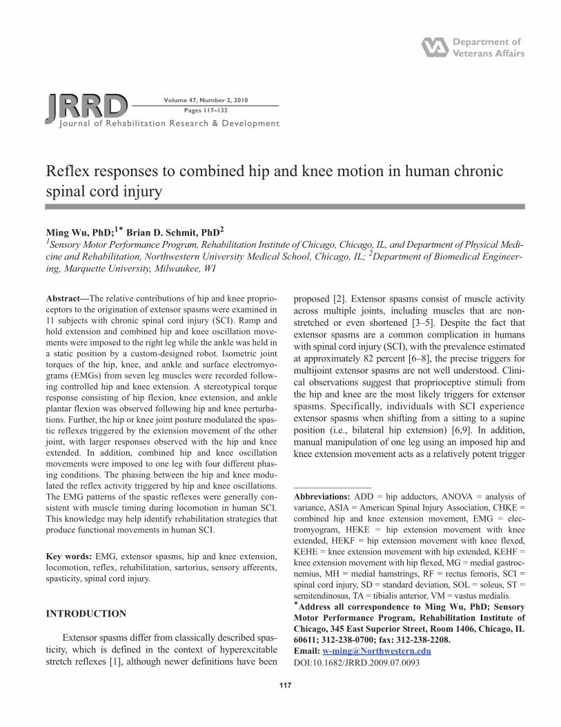
mented hip/knee actuator modified from a device used in aprevious study [11]. Briefly, the apparatus (Figure 1) wasconstructed to measure the multijoint torque response to pre-cisely controlled hip and knee movement with the subjectsupine on a therapy table. The hip and knee joints of therobotic system were rotated by two servomotors through aplanetary gearbox and a ball-screw mechanism, respec-tively, while the ankle joint was secured in a static position.The system was programmed to shut down instantly if itwas outside specific preset ranges. Safety screws were usedas mechanical stops to restrict the motion ranges of theactuators. The foot was placed in a footplate with a clampplaced on the dorsum of the foot and a strap to secure theheel. The hip-knee and knee-ankle links were adjusted to fitindividual leg size. The hip portion of the leg brace wasaffixed to a servomotor drive system (MT 704A1-R1C1,Kollmorgen Electro-Optical; Northampton, Massachu-setts). A newly constructed knee actuator, which included a
Table 1.Subject clinical characteristics.
Subject Level of Injury*
ASIAClass
Age(yr)
Time Since Injury (yr) Medication
A T10–11 A 26 8 Baclofen(intrathecal pump)
B C5 C 25 4 Baclofen (80 mg)C C7–C8 A 32 6 NoneD C6 C 43 16 Baclofen
(40 mg 3�/day)E C5–C6 C 45 18 NoneF T6 A 24 7 NoneG T1 D 68 5 Baclofen (100 mg)H T8 A 44 28 NoneI C5–C6 C 41 24 NoneJ C5 B 44 27 NoneK C6–C7 D 45 22 None
*Neurological injury levels: C = cervical, T = thoracic.ASIA = American Spinal Injury Association.
119
WU and SCHMIT. Spastic reflexes triggered by hip and knee motion in human SCI
servomotor (BE343JR-KMSN, Parker Hannifin Corpora-tion; Rohnert Park, California), a ball-screw mechanism,and a four-bar linkage mechanism, was affixed to the knee-ankle brace to impose a controlled motion to the knee. Hipand ankle torques were measured with use of hollow-flanged transducers (S. Himmelstein and Company; Hoff-man Estates, Illinois), while the knee torque was calculatedfrom the force recorded by a tension and compression loadcell (Honeywell Sensotec; Columbus, Ohio) mounted at thebottom of the motor. Hip and knee joint positions were mea-sured with use of potentiometers coupled to the servomotorand rotation shaft of the linkage mechanism, respectively.
Surface electromyograms (EMGs) were recorded fromthe tibialis anterior (TA), soleus (SOL), medial gastrocne-mius (MG), vastus medialis (VM), rectus femoris (RF),medial hamstrings (MH), and hip adductors (ADD) of theright leg in all subjects. Ag-AgCl (silver/silver chloride)
2.5 cm square pregelled electrodes (Vermed Inc; BellowsFalls, Vermont) were applied with 2.5 cm center-to-centerspacing over the respective muscle bellies on lightlyabraded skin. Active preamplifiers with shielded leads wereattached to the electrodes and connected to an OctopusAMT-8 EMG unit (Bortec Biomedical Ltd; Calgary,Canada). All channels were amplified (total gain = 500),low-pass filtered (450 Hz), and sampled (1,000 Hz) withuse of a data acquisition card (National Instruments Corpo-ration; Austin, Texas) on a personal computer. CustomLabVIEW software (National Instruments Corporation)acquired the data and output the velocity command signalsto the servomotor systems.
Imposed Hip And Knee MovementHip and knee movements were imposed to the right
leg of all 11 subjects. Each subject was transferred to atrisection therapy table and placed in a supine position.The center of rotation of the right hip joint was alignedwith the axis of rotation of the servomotor system, theknee joint was aligned with one rotation axis of a four-bar linkage mechanism, and the brace was adjusted toalign the ankle with the appropriate torque transducer.Alignment of the hip and knee joints was confirmed byan absence of leg translation during manually imposedflexion and extension of the joint, assessed visually. Thepelvis was secured to the table with a strap across theiliac crest to inhibit pelvic rotation. The leg was placed inthe brace with the ankle at 10° to 30° plantar flexion andwas held in the same position for the duration of the test.The contralateral limb was supported in a slightly flexed(10° to 30°) position at the hip, with the knee at approxi-mately 90° and the ankle allowed to rest in a positiondetermined by gravity (slightly plantar flexed).
Controlled hip movements were imposed by the Koll-morgen servomotor system. The start position was set at30° to 40° of hip flexion and the end position at between10° of hip flexion and 10° of hip extension, a position thatcorresponded approximately to maximum hip extension,depending on each subject’s range of motion. Similarly, thecontrolled knee movements were imposed with use of theball-screw mechanism and Kollmorgen servomotor system.The start position was set at 70° to 75° of knee flexion andthe end position at 5° to 40° of knee flexion, a position thatcorresponded approximately to full knee extension for eachsubject. Because range of motion differed among subjects,a slightly different hip and knee range of movement wasused for each subject (Table 2). Figure 1 shows the motionof the hip and knee relative to the sagittal coordinate
Figure 1.Servomotor systems with leg brace attachment. Servomotor drive systemactuates leg from hip and knee joints, and ankle joint is held statically byleg brace. Hip joint is driven directly by rotation motor, while knee jointis driven by motor through ball-screw mechanism and linkage. Reactiontorque transducers at hip and ankle measure joint torque response toimposed movement. Reaction force transducer is attached at bottomof ball-screw mechanism to measure force from which knee joint torqueis calculated.
120
JRRD, Volume 47, Number 2, 2010
system. A ramp and hold movement was imposed to the hipin which the motor rotated the hip at a constant velocity of60°/s, from the hip flexed to the hip extended and back (atotal range of 20° to 50°, depending on the subject), with a10-second hold in extension. A similar perturbation wasimposed to the knee, with its velocity set at about 67°/s (atotal range of 35° to 65°, depending on the subject). Thisperturbation, consisting of controlled hip and/or kneeextension movement, produces a stereotypical reflexresponse that resembles an extensor spasm [3–4].
Five different types of perturbation were applied tothe hip and knee joints: combined hip and knee extensionmovement (CHKE), hip extension movement with kneeflexed (HEKF) or extended (HEKE), and knee extensionmovement with hip flexed (KEHF) or extended (KEHE).Each set of conditions was repeated three times, with themean of the three measurements in each condition usedfor statistical analyses. The five conditions were appliedin random order for each set of trials.
Following the tests of ramp and hold movements,combined hip and knee oscillation movements wereimposed to the leg for 10 cycles with four different phas-ing conditions: (1) phase 0: the hip and knee were movedin the same phase, i.e., the hip and knee reached flexedand extended positions simultaneously; (2) phase 0.50:the hip and knee were moved 180° out of phase, i.e., thehip reached a flexed position while the knee reached anextended position and vice versa; (3) phase 0.25: the hipwas moved 90° (1/4 of the cycle) earlier than the knee;and (4) phase 0.75: the hip was moved 270° (3/4 of thetotal cycle) earlier than the knee. A time interval of about3 minutes was allowed between trials. Each set of condi-
tions was repeated three times, with the four phasing con-ditions randomized for each set of trials.
Analysis
Ramp Stretch MovementsThe reflex joint torque data were obtained for each
movement trial during the 10-second hold period with thehip (or knee) extended and the other joint, i.e., knee (or hip)flexed or extended. The torque measurements recordedimmediately at the end of the movements were excludedfrom the analysis because of large inertial artifacts. Duringthe hold periods, the gravitational/passive torque offset wasremoved by subtraction of a torque measurement in the endposition, when there was no muscle activity (i.e., at the endof the hold period). The torque signals were then low-passfiltered at 5 Hz with use of a fourth-order Butterworth filter(butter/filtfilt; MATLAB Command, The MathWorks; Nat-tick, Massachusetts).
The peak torque of the hip, knee, and ankle during thehold period were the primary measurements used to com-pare the reflex response across conditions. The peaktorques of the hip, knee, and ankle joints were identifiedfor each movement trial in each condition. An outlier analy-sis was conducted on the data, and two subjects wereexcluded from further analysis. One subject producedtorque responses >3 standard deviation (SD) above themean, and the other subject produced no response. Thepeak torques of the hip, knee, and ankle were comparedacross five movement conditions with use of a one-wayrepeated measures analysis of variance (ANOVA) (n = 9).Significance was tested at � = 0.05.
The EMG signals were also used to compare the spas-tic reflex across different conditions. The surface EMG sig-nals were rectified and enveloped with use of an 8 Hz, low-pass, fourth-order Butterworth filter (butter/filtfilt; MAT-LAB Command, The MathWorks). The area of the rectifiedand smoothed EMG signals was calculated during the holdperiod (from the end of the movement, for 5 seconds) forthe five different test conditions. A one-way ANOVA wasused to statistically compare the EMG activities across thefive different conditions (� = 0.05).
Sinusoidal MovementsFor the sinusoidal movement tests, smoothed recti-
fied EMG signals were analyzed to identify the effect ofthe modulation of the phasing between the hip and kneeon the muscle activity pattern. The surface EMGs were
Table 2.Subject test parameters indicating starting position and range ofmotion of hip and knee joints and static ankle angle during movement.
SubjectAnkle PlantarFlexion Angle
(°)
Knee FlexionAngle (°)
KneeRange (°)
Hip FlexionAngle (°)
HipRange
(°)A 20 70 52 40 50B 20 70 65 40 50C 15 75 62 40 40D 20 75 50 30 40E 25 75 61 30 30F 20 75 61 30 30G 25 75 62 40 50H 15 75 60 40 30I 10 75 35 40 40J 30 75 48 30 40K 15 70 48 30 20
121
WU and SCHMIT. Spastic reflexes triggered by hip and knee motion in human SCI
rectified and enveloped with use of an 8 Hz, low-pass,fourth-order Butterworth filter (butter/filtfilt; MATLABCommand, The MathWorks). The area of the rectifiedand smoothed EMG signals was calculated for the middlesix cycles across the four different phasing conditions,i.e., phases 0, 0.25, 0.50, and 0.75. (Note that the EMGsignals from the very beginning and the ending of themovement were excluded because of the transitions atthose points.) The EMG areas were averaged across sixcycles for each phase condition and then normalized tothe average of the four phase conditions. A one-wayrepeated measures ANOVA was used to statisticallycompare the EMG activities across four different phasingconditions, with the significance set at � = 0.05.
Muscle timing patterns during each sinusoidal cyclewere examined with use of circular statistics according tomethods outlined by Batschelet [13] and previouslyapplied to imposed hip movements [11–12]. The EMGphase analysis was pursued only if the EMG data demon-strated sufficient muscle activity. The threshold valueswere determined with use of the area of rectified andsmoothed EMG data; the data were excluded if the area ofeach cycle was less than 15 percent of the averaged areaacross all six cycles and three trials. Phase analysis con-sisted of the use of rectified and smoothed EMG signalsthat were normalized from 0° to 360°, where 180° repre-sents full hip and knee extension and 0° represents full hipand knee flexion. For each movement cycle, the rectangu-lar coordinates (x, y) of the resultant vector were calcu-lated, respectively, as shown in Equations (1) and (2):
and
where were the observed angles of the correspond-ing unit vectors.
For these rectangular coordinates, the phase angle foreach cycle was then calculated with use of Equation (3).To characterize the phasing across cycles, we normalizedthe vector length r to a unit vector and converted thepolar coordinates to rectangular coordinates to find themean x and y coordinates. After normalization, the meanphase angles and vector lengths were then calculated
(Equations (3) and (4), respectively) across all six cyclesand three trials for each subject.
and
Only the middle six cycles of each trial were used foranalysis, and the first and last two cycles were discardedbecause of the unsynchronized onset of hip and kneemovement for different phase-shift conditions. The meanphase angles and vector lengths were used to detect sig-nificant phasic muscle activity during the imposed hipand knee oscillation.
To determine whether significant phasing in EMGexisted, we performed Raleigh’s test for one-sidedness (� =0.05) [13]. All data sets that showed a significant trendwere plotted in polar coordinates. To identify the possiblecontribution of the stretch reflex to the muscle activitiestriggered by hip and knee motion, we estimated the timeintervals in which muscles were being stretched. The lengthchanges of the biarticular hip/knee muscles were obtainedwith the model described by Frigo and Pedotti [14]. Themodel was applied to the RF and to the semitendinosus(ST), the latter chosen to represent the MH. We comparedtimes of muscle lengthening with timing of muscle activitymeasured experimentally to aid interpretation of the resultsby identifying the possible contribution of stretch reflexesto the overall observed responses.
RESULTS
Ramp and Hold Movement ResponsesA typical reflex response, from subject I, is shown in
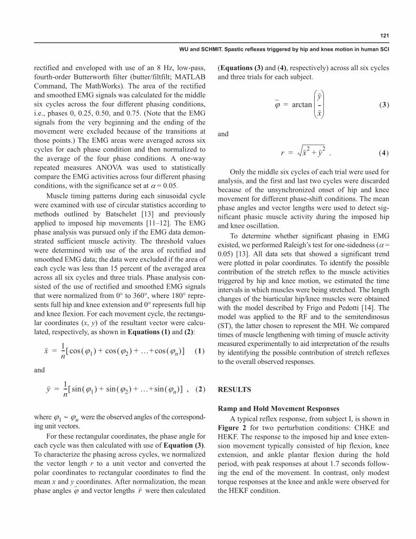
Figure 2 for two perturbation conditions: CHKE andHEKF. The response to the imposed hip and knee exten-sion movement typically consisted of hip flexion, kneeextension, and ankle plantar flexion during the holdperiod, with peak responses at about 1.7 seconds follow-ing the end of the movement. In contrast, only modesttorque responses at the knee and ankle were observed forthe HEKF condition.
x 1n--- �1� � �2� � �+ �n� �cos+cos+cos� = 1� �
y 1n--- �1� � �2� � �+ �n� �sin+sin+sin� ,= 2� �
�1 �n
� r
� arctany-x� � � �� �
= 3� �
r x2 y2+ .= 4� �
122
JRRD, Volume 47, Number 2, 2010
EMG data were consistent with the recorded torqueresponses, with similar patterns of activity across two con-ditions but larger magnitude of EMG signals for the CHKEcondition. A typical surface EMG response, from subject I,to imposed hip and knee movement and hip movementwith knee flexed is shown in Figure 2(b). During the holdperiod, the muscle activities of the knee and ankle exten-sors, represented by the VM, MG, and SOL, were activatedby the CHKE or HEKF conditions, although the muscleactivities were much smaller for the HEKF condition. TheRF (a hip flexor/knee extensor) was also activated follow-ing the end of the movement, coinciding with the measuredhip flexion and knee extension torques, with larger
responses for the CHKE condition. The knee flexors, repre-sented by MH, and the ankle dorsiflexors, represented byTA, were also activated following the movement, againwith larger responses for the CHKE condition.
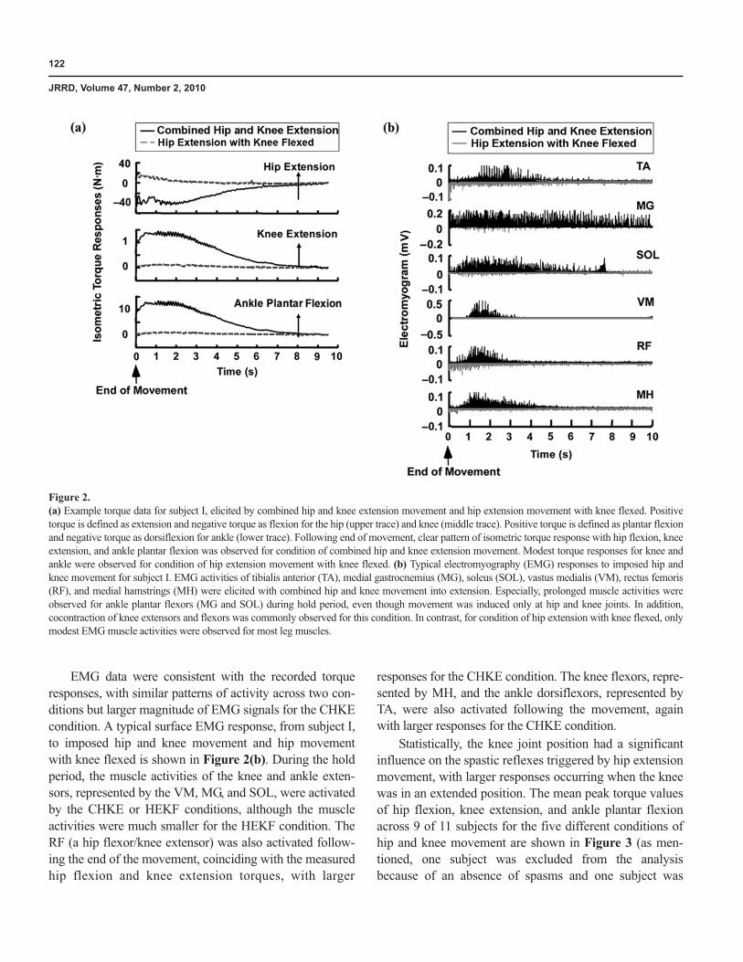
Statistically, the knee joint position had a significantinfluence on the spastic reflexes triggered by hip extensionmovement, with larger responses occurring when the kneewas in an extended position. The mean peak torque valuesof hip flexion, knee extension, and ankle plantar flexionacross 9 of 11 subjects for the five different conditions ofhip and knee movement are shown in Figure 3 (as men-tioned, one subject was excluded from the analysisbecause of an absence of spasms and one subject was
Figure 2.(a) Example torque data for subject I, elicited by combined hip and knee extension movement and hip extension movement with knee flexed. Positivetorque is defined as extension and negative torque as flexion for the hip (upper trace) and knee (middle trace). Positive torque is defined as plantar flexionand negative torque as dorsiflexion for ankle (lower trace). Following end of movement, clear pattern of isometric torque response with hip flexion, kneeextension, and ankle plantar flexion was observed for condition of combined hip and knee extension movement. Modest torque responses for knee andankle were observed for condition of hip extension movement with knee flexed. (b) Typical electromyography (EMG) responses to imposed hip andknee movement for subject I. EMG activities of tibialis anterior (TA), medial gastrocnemius (MG), soleus (SOL), vastus medialis (VM), rectus femoris(RF), and medial hamstrings (MH) were elicited with combined hip and knee movement into extension. Especially, prolonged muscle activities wereobserved for ankle plantar flexors (MG and SOL) during hold period, even though movement was induced only at hip and knee joints. In addition,cocontraction of knee extensors and flexors was commonly observed for this condition. In contrast, for condition of hip extension with knee flexed, onlymodest EMG muscle activities were observed for most leg muscles.
123
WU and SCHMIT. Spastic reflexes triggered by hip and knee motion in human SCI
excluded because the datum was classified as an outlier,defined as a magnitude >3 SD above the populationmean). A one-way ANOVA (different conditions of hipand knee movement) indicated that the reflex responses tohip extension movement were significantly affected by theknee joint position (p < 0.05) (Figure 3). Post hoc pairscomparisons indicated that the peak torque of hip flexionwas significantly greater for HEKE than for HEKF andKEHF (Tukey test, p < 0.05) (Figure 3(a)). The peak hipflexion torque responses were also significantly greater forCHKE than for KEHF (Tukey test, p < 0.05). No signifi-cant difference was found in peak hip flexion torquebetween the KEHE condition and the conditions with onejoint in a flexed posture (KEHF or HEKF; p = 0.66 and p =0.24, respectively). The peak knee extension torque wasalso significantly greater with HEKE and CHKE thanwhen one joint was in a flexed posture (HEKF or KEHF;Tukey test, p < 0.05) (Figure 3(b)), despite the potentialmechanical advantage associated with the more flexedknee posture [15]. The ankle plantar flexion torque wassignificantly larger for perturbations with biarticular exten-sion (CHKE and HEKE) than HEKF (Tukey test, p <0.05). No significant difference was found between CHKEand KEHF or HEKE and KEHF (p = 0.10 and p = 0.11,respectively) for the ankle plantar flexion torque. In sum-mary, the torque responses were generally larger when theleg posture at the end of the imposed movement consistedof both hip and knee extension, although the magnitude ofthe torque responses were relatively smaller followingthe knee extension movement than the hip extensionmovement.
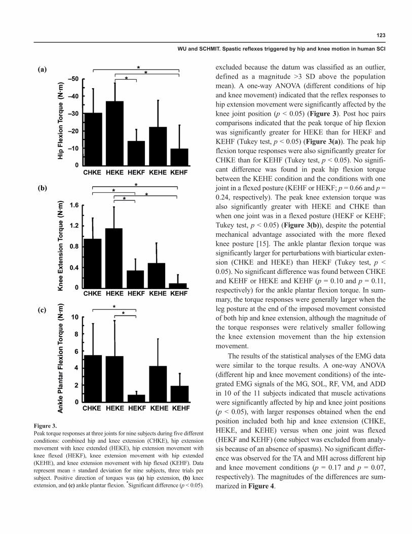
The results of the statistical analyses of the EMG datawere similar to the torque results. A one-way ANOVA(different hip and knee movement conditions) of the inte-grated EMG signals of the MG, SOL, RF, VM, and ADDin 10 of the 11 subjects indicated that muscle activationswere significantly affected by hip and knee joint positions(p < 0.05), with larger responses obtained when the endposition included both hip and knee extension (CHKE,HEKE, and KEHE) versus when one joint was flexed(HEKF and KEHF) (one subject was excluded from analy-sis because of an absence of spasms). No significant differ-ence was observed for the TA and MH across different hipand knee movement conditions (p = 0.17 and p = 0.07,respectively). The magnitudes of the differences are sum-marized in Figure 4.
Figure 3.Peak torque responses at three joints for nine subjects during five differentconditions: combined hip and knee extension (CHKE), hip extensionmovement with knee extended (HEKE), hip extension movement withknee flexed (HEKF), knee extension movement with hip extended(KEHE), and knee extension movement with hip flexed (KEHF). Datarepresent mean ± standard deviation for nine subjects, three trials persubject. Positive direction of torques was (a) hip extension, (b) kneeextension, and (c) ankle plantar flexion. *Significant difference (p < 0.05).
124
JRRD, Volume 47, Number 2, 2010
EMG During Sinusoidal OscillationsThe EMG responses to imposed hip and knee sinusoi-
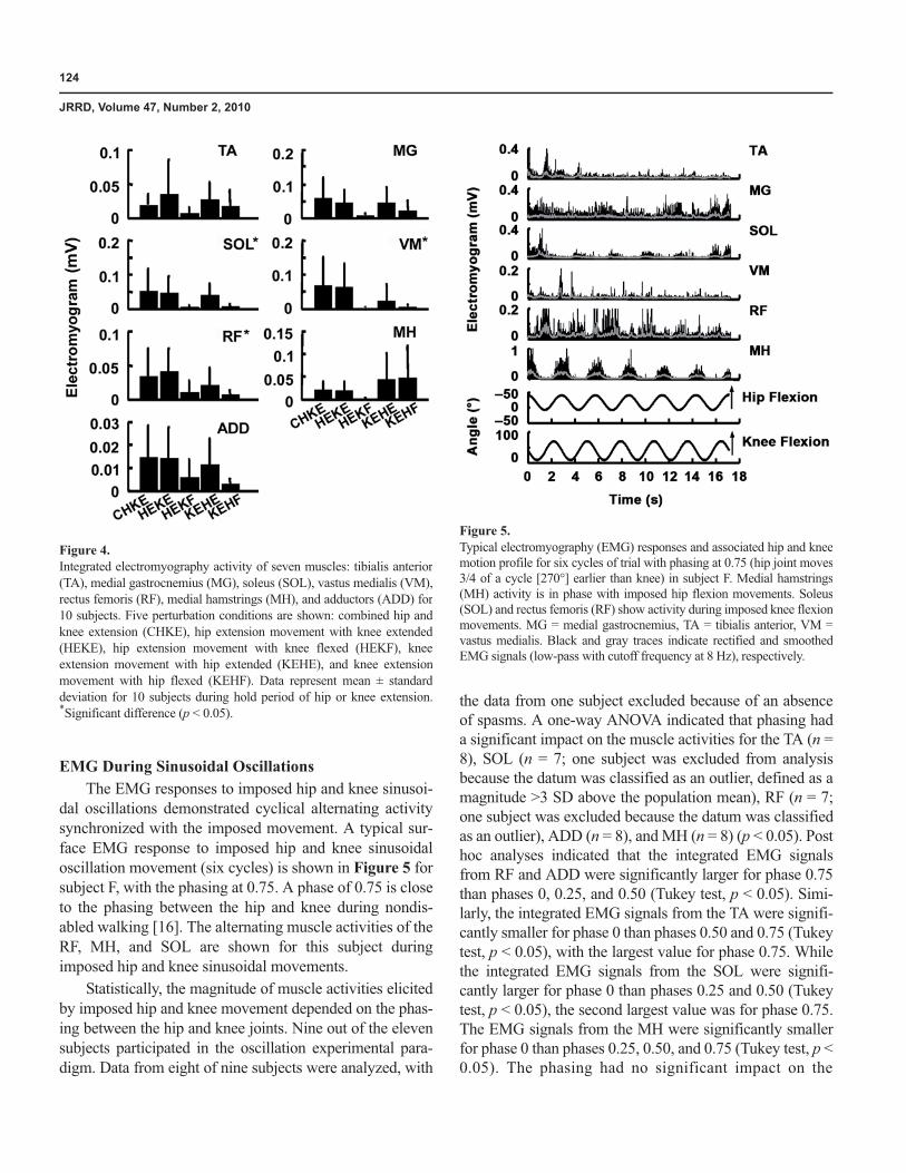
dal oscillations demonstrated cyclical alternating activitysynchronized with the imposed movement. A typical sur-face EMG response to imposed hip and knee sinusoidaloscillation movement (six cycles) is shown in Figure 5 forsubject F, with the phasing at 0.75. A phase of 0.75 is closeto the phasing between the hip and knee during nondis-abled walking [16]. The alternating muscle activities of theRF, MH, and SOL are shown for this subject duringimposed hip and knee sinusoidal movements.
Statistically, the magnitude of muscle activities elicitedby imposed hip and knee movement depended on the phas-ing between the hip and knee joints. Nine out of the elevensubjects participated in the oscillation experimental para-digm. Data from eight of nine subjects were analyzed, with
the data from one subject excluded because of an absenceof spasms. A one-way ANOVA indicated that phasing hada significant impact on the muscle activities for the TA (n =8), SOL (n = 7; one subject was excluded from analysisbecause the datum was classified as an outlier, defined as amagnitude >3 SD above the population mean), RF (n = 7;one subject was excluded because the datum was classifiedas an outlier), ADD (n = 8), and MH (n = 8) (p < 0.05). Posthoc analyses indicated that the integrated EMG signalsfrom RF and ADD were significantly larger for phase 0.75than phases 0, 0.25, and 0.50 (Tukey test, p < 0.05). Simi-larly, the integrated EMG signals from the TA were signifi-cantly smaller for phase 0 than phases 0.50 and 0.75 (Tukeytest, p < 0.05), with the largest value for phase 0.75. Whilethe integrated EMG signals from the SOL were signifi-cantly larger for phase 0 than phases 0.25 and 0.50 (Tukeytest, p < 0.05), the second largest value was for phase 0.75.The EMG signals from the MH were significantly smallerfor phase 0 than phases 0.25, 0.50, and 0.75 (Tukey test, p <0.05). The phasing had no significant impact on the
Figure 4.Integrated electromyography activity of seven muscles: tibialis anterior(TA), medial gastrocnemius (MG), soleus (SOL), vastus medialis (VM),rectus femoris (RF), medial hamstrings (MH), and adductors (ADD) for10 subjects. Five perturbation conditions are shown: combined hip andknee extension (CHKE), hip extension movement with knee extended(HEKE), hip extension movement with knee flexed (HEKF), kneeextension movement with hip extended (KEHE), and knee extensionmovement with hip flexed (KEHF). Data represent mean ± standarddeviation for 10 subjects during hold period of hip or knee extension.*Significant difference (p < 0.05).
Figure 5.Typical electromyography (EMG) responses and associated hip and kneemotion profile for six cycles of trial with phasing at 0.75 (hip joint moves3/4 of a cycle [270°] earlier than knee) in subject F. Medial hamstrings(MH) activity is in phase with imposed hip flexion movements. Soleus(SOL) and rectus femoris (RF) show activity during imposed knee flexionmovements. MG = medial gastrocnemius, TA = tibialis anterior, VM =vastus medialis. Black and gray traces indicate rectified and smoothedEMG signals (low-pass with cutoff frequency at 8 Hz), respectively.
125
WU and SCHMIT. Spastic reflexes triggered by hip and knee motion in human SCI
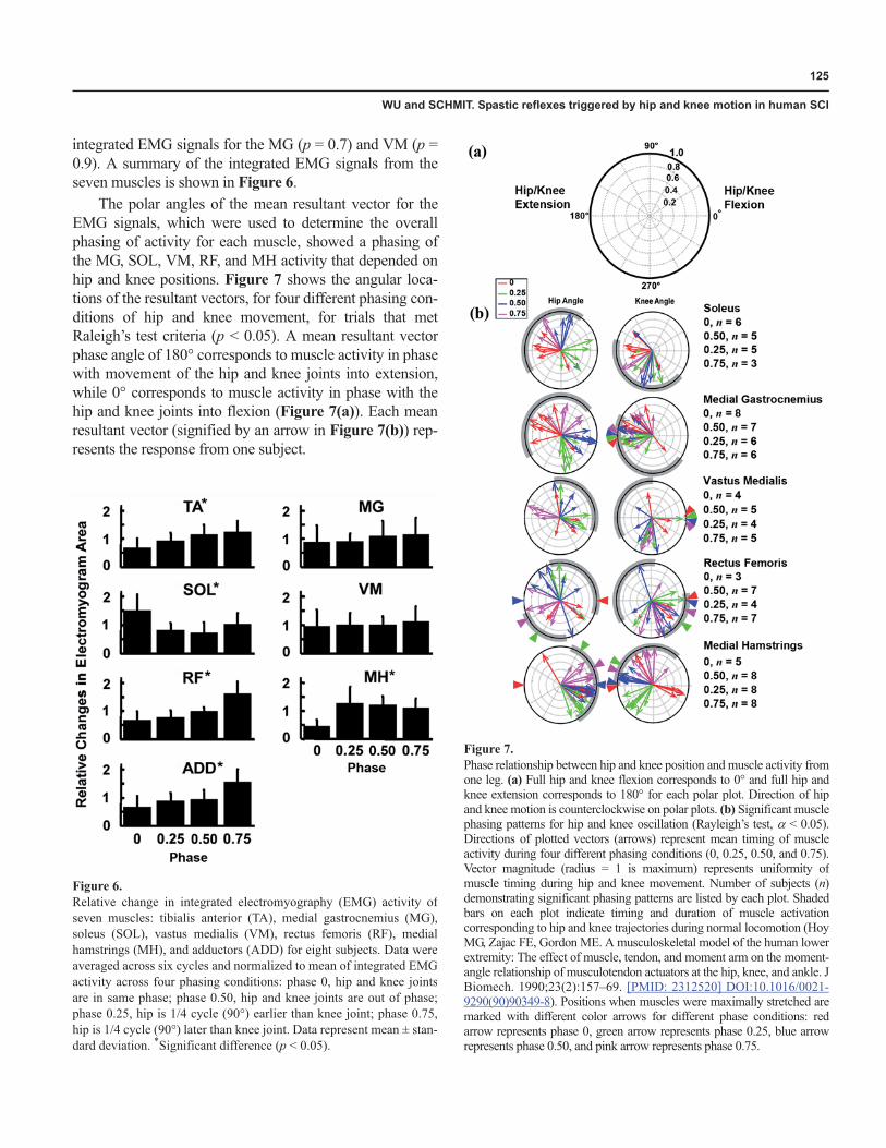
integrated EMG signals for the MG (p = 0.7) and VM (p =0.9). A summary of the integrated EMG signals from theseven muscles is shown in Figure 6.
The polar angles of the mean resultant vector for theEMG signals, which were used to determine the overallphasing of activity for each muscle, showed a phasing ofthe MG, SOL, VM, RF, and MH activity that depended onhip and knee positions. Figure 7 shows the angular loca-tions of the resultant vectors, for four different phasing con-ditions of hip and knee movement, for trials that metRaleigh’s test criteria (p < 0.05). A mean resultant vectorphase angle of 180° corresponds to muscle activity in phasewith movement of the hip and knee joints into extension,while 0° corresponds to muscle activity in phase with thehip and knee joints into flexion (Figure 7(a)). Each meanresultant vector (signified by an arrow in Figure 7(b)) rep-resents the response from one subject.
Figure 6.Relative change in integrated electromyography (EMG) activity ofseven muscles: tibialis anterior (TA), medial gastrocnemius (MG),soleus (SOL), vastus medialis (VM), rectus femoris (RF), medialhamstrings (MH), and adductors (ADD) for eight subjects. Data wereaveraged across six cycles and normalized to mean of integrated EMGactivity across four phasing conditions: phase 0, hip and knee jointsare in same phase; phase 0.50, hip and knee joints are out of phase;phase 0.25, hip is 1/4 cycle (90°) earlier than knee joint; phase 0.75,hip is 1/4 cycle (90°) later than knee joint. Data represent mean ± stan-dard deviation. *Significant difference (p < 0.05).
Figure 7.Phase relationship between hip and knee position and muscle activity fromone leg. (a) Full hip and knee flexion corresponds to 0° and full hip andknee extension corresponds to 180° for each polar plot. Direction of hipand knee motion is counterclockwise on polar plots. (b) Significant musclephasing patterns for hip and knee oscillation (Rayleigh’s test, � < 0.05).Directions of plotted vectors (arrows) represent mean timing of muscleactivity during four different phasing conditions (0, 0.25, 0.50, and 0.75).Vector magnitude (radius = 1 is maximum) represents uniformity ofmuscle timing during hip and knee movement. Number of subjects (n)demonstrating significant phasing patterns are listed by each plot. Shadedbars on each plot indicate timing and duration of muscle activationcorresponding to hip and knee trajectories during normal locomotion (HoyMG, Zajac FE, Gordon ME. A musculoskeletal model of the human lowerextremity: The effect of muscle, tendon, and moment arm on the moment-angle relationship of musculotendon actuators at the hip, knee, and ankle. JBiomech. 1990;23(2):157–69. [PMID: 2312520] DOI:10.1016/0021-9290(90)90349-8). Positions when muscles were maximally stretched aremarked with different color arrows for different phase conditions: redarrow represents phase 0, green arrow represents phase 0.25, blue arrowrepresents phase 0.50, and pink arrow represents phase 0.75.
126
JRRD, Volume 47, Number 2, 2010
The phasing arrows show that for the majority of thetrials, the SOL and MG were phased with knee extension(with the exception of one subject for the MG), while theRF and VM were phased with knee flexion (with the excep-tion of one subject for the RF and two subjects for the VM).This phasing pattern is reflected by a tighter grouping ofvectors in the plots of SOL, MG versus knee angle, and RF,VM plots versus hip angle, respectively. In the majority ofthe trials, the MH phased with hip flexion (except for onesubject). No apparent phasing trend was observed for TAversus the hip and knee position (not shown in Figure 7). Inaddition, the phasing of MH versus the hip position wasaffected slightly by knee phasing, and MG, SOL versusknee angle was influenced by the hip phasing. These lesserinfluences were observed as shifts in the plots of the phas-ing of muscle activity of MG, SOL versus knee position,and MH versus hip position, depending on the phase of theopposite joint. A summary of the phasing of six muscles isshown in Figure 7(b). For comparison, the timing of mus-cle activation versus hip and knee position during nondis-abled locomotion is represented by the gray bars in Figure7(b) [16]. The activity in most muscles was appropriatelyphased with the position of the hip and knee correspondingto nondisabled walking, when the phasing of hip and kneemovement was similar to the nondisabled walking pattern,i.e., phase 0.75. However, the muscle activity of the VM forphase 0.75 was antiphased with the position of the hip andknee corresponding to nondisabled walking.
DISCUSSION
Following imposed hip and knee combination exten-sion movements, a reflex pattern of hip flexion, kneeextension, and ankle plantar flexion was observed in 10 of11 subjects with chronic SCI. These responses were con-sistent with clinical descriptions of the extensor spasmsthat are observed following movement from a sitting to asupine position [6]. The reflex response to hip or kneeextension movement depended on the position of the otherjoint, with greater reflex responses occurring when theother joint was extended. Furthermore, the largest muscleactivity occurred when the phasing of hip and knee move-ment was closest to the nondisabled walking pattern.These results suggested that proprioceptive informationfrom both the hip and knee joints contributes to a commonmultijoint reflex that may be, at least in part, involved inthe neural pathway of locomotion in human SCI.
Role of Interneuronal Excitability in SCI SpasticityResults from the current study emphasize that spastic
reflexes in chronic human SCI include multijoint responses,suggesting the involvement of interneuronal pathways. Forinstance, hip or knee extension alone or simultaneous hipand knee extension triggered a multijoint response thatincluded prominent activity in the SOL, which is notstretched or shortened by the imposed knee and hip move-ments. These results are consistent with previous studiesindicating that subjects with chronic SCI produce multijointreflex responses to single joint movements at other joints,such as flexor reflexes triggered by movement of the ankle[17] and knee joints [18] and extensor reflexes triggered bymovement of the hip or knee [3–4]. The multijoint nature ofthese reflex responses supports the notion that spasticreflexes in chronic SCI can be attributed, at least in part, toan increased excitability in the coupling between specificmodules within the spinal cord.
The reflex response to imposed hip and knee exten-sion movement might involve the excitation of commoninterneuronal pathways originating from the propriocep-tive afferents of a single biarticular hip/knee extensor.The torque pattern of ankle plantar flexion, knee exten-sion, and hip flexion was triggered by hip or knee exten-sion alone or simultaneous hip and knee extension. Themultijoint nature of the response, which included activa-tion of nonstretched muscles, suggests that the movementactivated spinal centers that control multiple musclegroups. The position of the hip or knee joint influencedthe magnitude of extensor spasms triggered by move-ment of the other joint. As a result, we postulate that acoupling between hip and knee afferents exists when trig-gering extensor spasms, possibly involving sensory sig-nals from muscles that cross both hip and knee joints.The sartorius, a combined hip and knee flexor, may playan important role as the sensor in triggering the response.
Another potential trigger for the extensor spasmsobserved in the current study is a reflex chain in whichimposed hip and knee extension produce stretch reflexactivation of the hip and knee flexors during the move-ment [19–20], which, in turn, produces a flexor reflexresponse [21–22] that then activates the neural circuitryassociated with extensor spasms. However, extensorspasms are considerably reduced when the hip or knee isflexed, suggesting stretch-related afferents from hip orknee flexors may not readily account for the observedmultijoint reflexes.
127
WU and SCHMIT. Spastic reflexes triggered by hip and knee motion in human SCI
Spasticity Management in Human SCIThe findings from this study emphasize the pervasive
nature of extensor spasm activity in human SCI and havepotential implications for the management and clinicalassessment of spastic motor behaviors. Extensor spasmsare manifested more frequently than other types of spas-tic reflexes [6] and are often considered more disablingthan other spastic motor behaviors. For instance, extensorspasms have been known to force a patient out of awheelchair and to interfere with transfers [7,23]. Notethat the responses observed in the current study includehip flexion torque and thus resemble “extensor spasms”observed clinically when patients with SCI move from asitting to a supine posture. Other types of extensorspasms are observed clinically that can involve strong hipextension. The mechanism for these extensor spasmslikely differs from the responses observed in the currentstudy, likely involving a different sensory trigger.
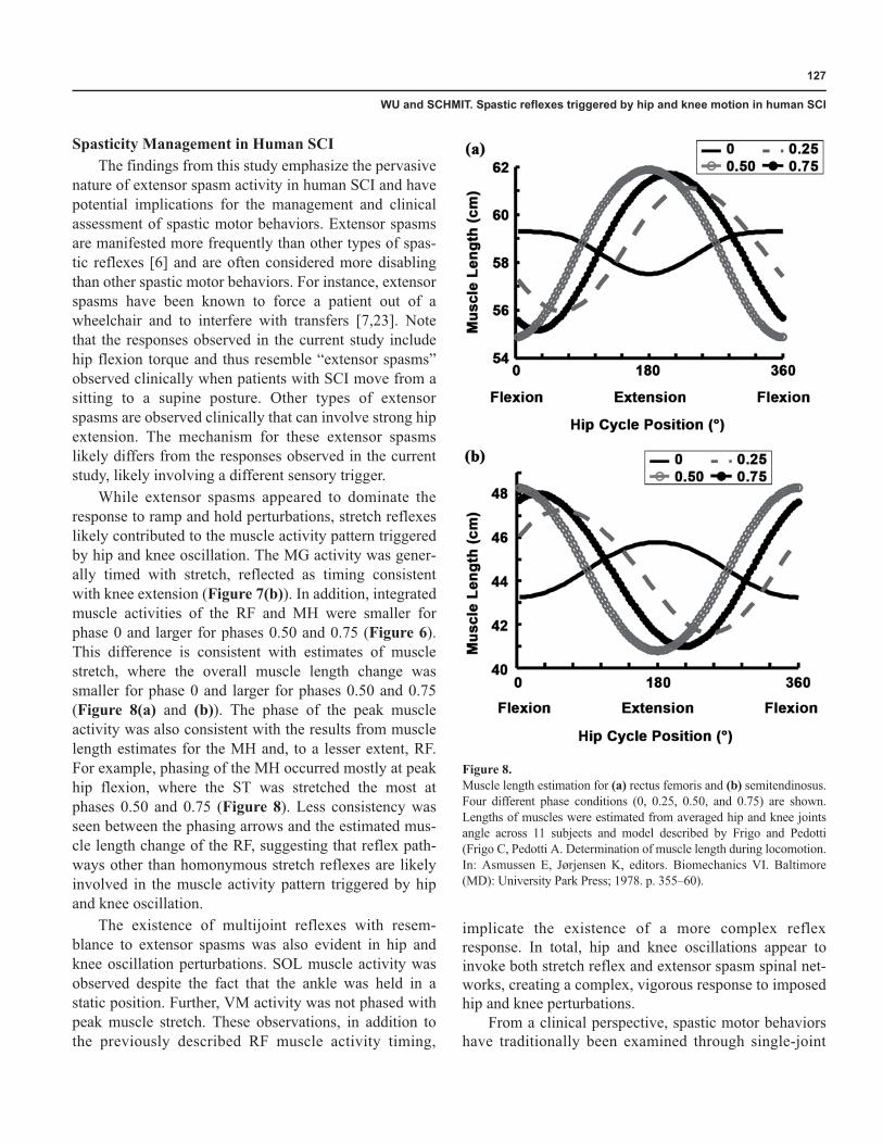
While extensor spasms appeared to dominate theresponse to ramp and hold perturbations, stretch reflexeslikely contributed to the muscle activity pattern triggeredby hip and knee oscillation. The MG activity was gener-ally timed with stretch, reflected as timing consistentwith knee extension (Figure 7(b)). In addition, integratedmuscle activities of the RF and MH were smaller forphase 0 and larger for phases 0.50 and 0.75 (Figure 6).This difference is consistent with estimates of musclestretch, where the overall muscle length change wassmaller for phase 0 and larger for phases 0.50 and 0.75(Figure 8(a) and (b)). The phase of the peak muscleactivity was also consistent with the results from musclelength estimates for the MH and, to a lesser extent, RF.For example, phasing of the MH occurred mostly at peakhip flexion, where the ST was stretched the most atphases 0.50 and 0.75 (Figure 8). Less consistency wasseen between the phasing arrows and the estimated mus-cle length change of the RF, suggesting that reflex path-ways other than homonymous stretch reflexes are likelyinvolved in the muscle activity pattern triggered by hipand knee oscillation.
The existence of multijoint reflexes with resem-blance to extensor spasms was also evident in hip andknee oscillation perturbations. SOL muscle activity wasobserved despite the fact that the ankle was held in astatic position. Further, VM activity was not phased withpeak muscle stretch. These observations, in addition tothe previously described RF muscle activity timing,
implicate the existence of a more complex reflexresponse. In total, hip and knee oscillations appear toinvoke both stretch reflex and extensor spasm spinal net-works, creating a complex, vigorous response to imposedhip and knee perturbations.
From a clinical perspective, spastic motor behaviorshave traditionally been examined through single-joint
Figure 8.Muscle length estimation for (a) rectus femoris and (b) semitendinosus.Four different phase conditions (0, 0.25, 0.50, and 0.75) are shown.Lengths of muscles were estimated from averaged hip and knee jointsangle across 11 subjects and model described by Frigo and Pedotti(Frigo C, Pedotti A. Determination of muscle length during locomotion.In: Asmussen E, Jørjensen K, editors. Biomechanics VI. Baltimore(MD): University Park Press; 1978. p. 355–60).
128
JRRD, Volume 47, Number 2, 2010
movements, assuming they result from velocity-dependentstretch reflexes [24]. Results from the current study indicatethat spastic reflexes also involve multisegmental interneu-ronal pathways, and thus, the management of spastic phe-nomena should include assessments of multijoint reflexes.More accurate assessments of spastic reflexes in SCI mightbe obtained through comprehensive evaluations that mea-sure multiple reflex behaviors [5,25].
Involvement of Locomotor Pathways in the Reflexes Triggered by Hip and Knee Oscillation
Hip proprioceptors have been shown to be a primarycontributor to the reflex generation and modulation ofmuscle activity in spinalized animal models of locomo-tion. Alternating flexor and extensor activity is elicitedby hip oscillation in spinalized and decerebrate cats [26–27]. In particular, hip afferents are important for initiatingthe transition from stance to swing in spinalized cats dur-ing walking [28–30]. Further, the activation of flexormuscles during walking in decerebrate cats has beenassociated with afferent drive from the sartorius muscle[30–31]. Stretch of the sartorius muscle could contributeto the effects of hip/knee motion in the current study.
Recent studies in human SCI also indicate that multi-joint reflexes triggered by unilateral hip oscillationentrain to the frequency of the imposed oscillation in amanner similar to spinalized animals [11–12]. The abilityto modulate reflex activity in an organized pattern similarto locomotion suggests that reflex activity triggered byhip afferents may involve similar pathways for the spinalcontrol of walking in human SCI [32]. In addition, hipkinematics modulate muscle activity in human SCI dur-ing treadmill stepping [33–34] such that swing isenhanced when hip extension is augmented during thelate stance phase of gait [33,35]. In addition, while “hipwalking” (knee joints fixed in an extended position), amuscle activity pattern similar to nondisabled walking isproduced [33]; whether this would occur if the kneejoints were fixed in a flexed position remains unclear.The muscle activity patterns produced by combined hipand knee extension movements observed in the currentstudy are generally consistent with these previous reports.
Although not often emphasized, knee proprioceptorsmay also be involved in locomotor reflexes in humanSCI. Imposed knee extension triggers a reflex responsesimilar to imposed hip extension [4] or the combined hipand knee extension movement observed in the currentstudy. Furthermore, the effects of modulation of phasing
between the hip and knee joints on the reflex muscleactivity suggest that both hip and knee afferents convergeonto a common pathway at the spinal level. Specifically,the phasing between hip and knee affected the size of thespastic reflex muscle activity elicited by the hip and kneeoscillation. The spastic reflex muscle activity pattern wasmore consistent with a locomotion pattern when thephasing between the hip and knee was close to the legkinematics of nondisabled walking. These results suggestthat the spastic reflexes triggered by the hip and kneeoscillation may be partially involved in locomotor mus-cle activity and highlight the potential contribution ofsensory information from hip and knee afferents to loco-motor reflexes in human SCI. While this analogy withlocomotion is potentially interesting, some inconsisten-cies existed between the current experimental setup andnondisabled walking conditions. For example, subjectswere lying semireclined on a test bed and the ankle jointwas secured in a stationary position with no loadinginput, which is quite different from walking.
Results from the current study are not completelyconsistent with the observations from animal studies. Forinstance, afferent information from the knee has not beenshown to contribute significantly to the modulation oflocomotor rhythms in the spinalized cat preparation [28].However, activation of flexor muscles during walking indecerebrate cats has been associated with afferents drivefrom the sartorius muscle [30–31]. In addition, it remainsunclear whether a muscle pattern similar to nondisabledwalking would be produced by hip walking with kneejoints locked in a flexed position in human SCI. On thebasis of the results of the current study, we postulate thatthe magnitude of the muscle activity would be dramati-cally reduced.
The muscle activity produced by hip and knee oscilla-tion, with the exception of the VM, was most appropri-ately phased for nondisabled walking when the phasingbetween hip and knee movement was closest to nondis-abled walking. The muscle activity of the VM wasantiphased with the hip position, even when the phasingbetween the hip and knee movement was similar to non-disabled walking. This dissimilarity of the reflex responsewith the gait pattern was also observed in the reflexes trig-gered by hip unilateral or bilateral oscillation in humanSCI [11–12]. The activation of the VM during hip andknee extension might be associated with a permanentchange in spinal reflex organization, since the VM activityalso increases during a flexion withdrawal reflex [36].
129
WU and SCHMIT. Spastic reflexes triggered by hip and knee motion in human SCI
Similarly, ankle load release, which produces a similarreflex response as hip stretch in animals models [37–38],also produces extensor spasms with increased VM activityin human SCI [18].
Intersubject VariabilityAntispastic medications may have affected the magni-
tude of reflex responses for some subjects in the currentstudy. At the time of the study, 4 of 11 subjects were pre-scribed baclofen to manage their spasms. Notably, one sub-ject with an intrathecal baclofen pump was excluded fromanalysis because of the absence of extensor spasms follow-ing the hip and knee movement perturbations. In addition,level of injury has been suggested to affect locomotor EMGactivity such that higher (cervical) lesion levels produceEMG activity more closely resembling nondisabled loco-motor EMG patterns during treadmill stepping [39]. Weobserved no trends in the magnitude of the response withlesion level in the current study; however, our sample sizemay not have been sufficient to detect an effect of lesionlocation. No conclusions regarding antispastic medications,injury level, injury completeness (ASIA classification), ortime postinjury could be made in the current study becauseof the high number of potential covariates, which limitedthe sample size for each subject type.
LimitationsOur conclusions are subject to several experimental
limitations. For instance, although we validated the load-cell calibration using a standard weight and we have pre-viously demonstrated the reliability of similar load-cellbased techniques for measuring spastic reflexes [40], wedid not systemically quantify the reliability of jointtorque measurements with the apparatus used in the cur-rent study. Note that the hip torque measurements and theartifacts produced by the apparatus have previously beenexamined in detail [11–12]. The robotic arm moved theleg at a speed of 60°/s to elicit leg extensor spasms in thecurrent study, although other faster speeds could also beused. The rationale for use of this speed was that spasmscould be reliably elicited at 60°/s, faster speeds producelarger inertial torque, and a previous study indicated thatmagnitude of extensor spasms in human SCI is notstrongly velocity-dependent [4]. In addition, the repeatedmovements across all conditions might have impactedthe magnitude of elicited spasms because of musclefatigue or habituation of the response. To circumventpotential bias in the results from changes in the response
over time, we randomized the order of the differentexperimental conditions, although we conducted theramp and hold experiments first and the hip and kneeoscillation experiments second.
CONCLUSIONS
Results from this study demonstrate that the positionof the hip or knee modulates the spastic reflexes triggeredby extension movement of the other joint (knee or hip),with larger responses observed with the opposite joint inthe extended position. Such behaviors support the role ofknee and hip proprioceptors, especially the biarticular sar-torius muscle afferents, in the initiation of extensor spasmsin human SCI. In addition, the reflex responses triggeredby hip and knee oscillation are likely due to organizedpathways located within the lumbosacral region of the spi-nal cord, possibly associated with locomotor reflexes.
ACKNOWLEDGMENTS
Author Contributions:Study concept and design: M. Wu, B. Schmit.Acquisition of data: M. Wu, B. Schmit.Analysis and interpretation of data: M. Wu, B. Schmit.Drafting of manuscript: M. Wu.Critical revision of manuscript for important intellectual content: B. Schmit.Statistical analysis: M. Wu.Obtained funding: M. Wu, B. Schmit.Administrative, technical, or material support: M. Wu, B. Schmit.Study supervision: B. Schmit.Financial Disclosures: The authors have declared that no competing interests exist.Funding/Support: This material was based on work supported by the National Institute on Disability and Rehabilitation Research Switzer Distinguished Research Fellowship (grant H133F050031), Paralyzed Veterans of America Research Foundation (grant 2447), and the Falk Medical Research Trust.Additional Contributions: We thank Ms. Tanya Onushko for help in the experimental setup.Institutional Review: Informed consent was obtained and all proce-dures were conducted in accordance with the Helsinki Declaration of 1975 and approved by the institutional review boards of Northwestern University, Chicago, Illinois, and Marquette University, Milwaukee, Wisconsin.Participant Follow-Up: The authors do not plan to inform partici-pants of the publication of this study. However, participants have been encouraged to check the study Web site for updated publications.
130
JRRD, Volume 47, Number 2, 2010
REFERENCES
1. Katz RT, Rymer WZ. Spastic hypertonia: Mechanisms andmeasurement. Arch Phys Med Rehabil. 1989;70(2):144–55.[PMID: 2644919]
2. Pandyan AD, Gregoric M, Barnes MP, Wood D, Van Wijck F,Burridge J, Hermens H, Johnson GR. Spasticity: Clinical per-ceptions, neurological realities and meaningful measurement.Disabil Rehabil. 2005;27(1–2):2–6. [PMID: 15799140]DOI:10.1080/09638280400014576
3. Schmit BD, Benz EN. Extensor reflexes in human spinalcord injury: Activation by hip proprioceptors. Exp BrainRes. 2002;145(4):520–27. [PMID: 12172664]DOI:10.1007/s00221-002-1134-5
4. Wu M, Hornby TG, Hilb J, Schmit BD. Extensor spasmstriggered by imposed knee extension in chronic human spi-nal cord injury. Exp Brain Res. 2005;162(2):239–49.[PMID: 15586272]DOI:10.1007/s00221-004-2173-x
5. Benz EN, Hornby TG, Bode RK, Scheidt RA, Schmit BD.A physiologically based clinical measure for spasticreflexes in spinal cord injury. Arch Phys Med Rehabil.2005;86(1):52–59. [PMID: 15640989]DOI:10.1016/j.apmr.2004.01.033
6. Little JW, Micklesen P, Umlauf R, Britell C. Lower extremitymanifestations of spasticity in chronic spinal injury. Am JPhys Med Rehabil. 1989;68(1):32–36. [PMID: 2917056]DOI:10.1097/00002060-198902000-00009
7. Barolat G, Maiman DJ. Spasms in spinal cord injury: A studyof 72 subjects. J Am Paraplegia Soc. 1987;10(2):35–39.[PMID: 3450779]
8. Dimitrijevic MR, Lissens MA, Mckay WB. Characteristicsand extent of motor activity recovery after spinal cordinjury. In: Seil FJ, editor. Advances in neural regenerationresearch. New York (NY): Wiley-Liss; 1990. p. 381–405.
9. Kuhn RA. Functional capacity of the isolated human spinalcord. Brain. 1950;73(1):1–51. [PMID: 15420313]DOI:10.1093/brain/73.1.1
10. Sjölund BH. Pain and rehabilitation after spinal cordinjury: The case of sensory spasticity? Brain Res Rev.2002;40(1–3):250–56. [PMID: 12589923]DOI:10.1016/S0165-0173(02)00207-2
11. Steldt RE, Schmit BD. Modulation of coordinated muscleactivity during imposed sinusoidal hip movements in humanspinal cord injury. J Neurophysiol. 2004;92(2):673–85.[PMID: 15044520]DOI:10.1152/jn.00677.2003
12. Onushko T, Schmit BD. Reflex response to imposed bilat-eral hip oscillations in human spinal cord injury. J Neuro-physiol. 2007;98(4):1849–61. [PMID: 17652410]DOI:10.1152/jn.00461.2007
13. Batschelet E. Circular statistics in biology. New York (NY):Academic Press; 1981.
14. Frigo C, Pedotti A. Determination of muscle length duringlocomotion. In: Asmussen E, Jørjensen K, editors. Biome-chanics VI. Baltimore (MD): University Park Press; 1978.p. 355–60.
15. Hoy MG, Zajac FE, Gordon ME. A musculoskeletal modelof the human lower extremity: The effect of muscle, ten-don, and moment arm on the moment-angle relationship ofmusculotendon actuators at the hip, knee, and ankle. J Bio-mech. 1990;23(2):157–69. [PMID: 2312520]DOI:10.1016/0021-9290(90)90349-8
16. Perry J. Gait analysis: Normal and pathological function.Thorofare (NJ): SLACK; 1992.
17. Schmit BD, McKenna-Cole A, Rymer WZ. Flexor reflexes inchronic spinal cord injury triggered by imposed ankle rotation.Muscle Nerve. 2000;23(5):793–803. [PMID: 10797404]DOI:10.1002/(SICI)1097-4598(200005)23:5<793::AID-MUS18>3.0.CO;2-T
18. Wu M, Schmit BD. Spastic reflexes triggered by ankle loadrelease in human spinal cord injury. J Neurophysiol. 2006;96(6):2941–50. [PMID: 16855114]DOI:10.1152/jn.00186.2006
19. Burke D, Gillies JD, Lance JW. The quadriceps stretchreflex in human spasticity. J Neurol Neurosurg Psychiatry.1970;33(2):216–23. [PMID: 4245691]DOI:10.1136/jnnp.33.2.216
20. Burke D, Gillies JD, Lance JW. Hamstrings stretch reflexin human spasticity. J Neurol Neurosurg Psychiatry. 1971;34(3):231–35. [PMID: 4255176]DOI:10.1136/jnnp.34.3.231
21. Schmit BD, Benz EN, Rymer WZ. Afferent mechanisms forthe reflex response to imposed ankle movement in chronicspinal cord injury. Exp Brain Res. 2002;145(1):40–49.[PMID: 12070743]DOI:10.1007/s00221-002-1080-2
22. Wu M, Hornby TG, Kahn JH, Schmit BD. Flexor reflexresponses triggered by imposed knee extension in chronichuman spinal cord injury. Exp Brain Res. 2006;168(4):566–76. [PMID: 16151779]DOI:10.1007/s00221-005-0113-z
23. Sköld C, Levi R, Seiger A. Spasticity after traumatic spinalcord injury: Nature, severity, and location. Arch Phys MedRehabil. 1999;80(12):1548–57. [PMID: 10597805]DOI:10.1016/S0003-9993(99)90329-5
24. Lance JW. Pathophysiology of spasticity and clinical expe-rience with baclofen. In: Feldman RG, Young RR, KoellaWP, editors. Spasticity: Disordered motor control. Chicago(IL): Year Book Medical Publishers; 1980. p.185–204.
25. Priebe MM, Sherwood AM, Thornby JI, Kharas NF,Markowski J. Clinical assessment of spasticity in spinalcord injury: A multidimensional problem. Arch Phys Med
131
WU and SCHMIT. Spastic reflexes triggered by hip and knee motion in human SCI
Rehabil. 1996;77(7):713–16. [PMID: 8670001]DOI:10.1016/S0003-9993(96)90014-3
26. Andersson O, Grillner S. Peripheral control of the cat’s stepcycle. II. Entrainment of the central pattern generators forlocomotion by sinusoidal hip movements during “fictivelocomotion.” Acta Physiol Scand. 1983;118(3):229–39.[PMID: 6312752]DOI:10.1111/j.1748-1716.1983.tb07267.x
27. Kriellaars DJ, Brownstone RM, Noga BR, Jordan LM.Mechanical entrainment of fictive locomotion in the decere-brate cat. J Neurophysiol. 1994;71(6):2074–86.[PMID: 7931503]
28. Grillner S, Rossignol S. On the initiation of the swing phaseof locomotion in chronic spinal cats. Brain Res. 1978;146(2):269–77. [PMID: 274169]DOI:10.1016/0006-8993(78)90973-3
29. Hiebert GW, Whelan PJ, Prochazka A, Pearson KG. Con-tribution of hind limb flexor muscle afferents to the timingof phase transitions in the cat step cycle. J Neurophysiol.1996;75(3):1126–37. [PMID: 8867123]
30. Lam T, Pearson KG. Proprioceptive modulation of hipflexor activity during the swing phase of locomotion indecerebrate cats. J Neurophysiol. 2001;86(3):1321–32.[PMID: 11535680]
31. Lam T, Pearson KG. Sartorius muscle afferents influencethe amplitude and timing of flexor activity in walkingdecerebrate cats. Exp Brain Res. 2002;147(2):175–85.[PMID: 12410332]DOI:10.1007/s00221-002-1236-0
32. Knikou M, Kay E, Schmit BD. Parallel facilitatory reflexpathways from the foot and hip to flexors and extensors inthe injured human spinal cord. Exp Neurol. 2007;206(1):146–58. [PMID: 17543951]DOI:10.1016/j.expneurol.2007.05.004
33. Dietz V, Müller R, Colombo G. Locomotor activity in spinalman: Significance of afferent input from joint and load recep-
tors. Brain. 2002;125(Pt 12):2626–34. [PMID: 12429590]DOI:10.1093/brain/awf273
34. Harkema SJ, Hurley SL, Patel UK, Requejo PS, DobkinBH, Edgerton VR. Human lumbosacral spinal cord inter-prets loading during stepping. J Neurophysiol. 1997;77(2):797–811. [PMID: 9065851]
35. Dobkin BH, Harkema S, Requejo P, Edgerton VR. Modula-tion of locomotor-like EMG activity in subjects with com-plete and incomplete spinal cord injury. J Neurol Rehabil.1995;9(4):183–90. [PMID: 11539274]
36. Deutsch KM, Hornby TG, Schmit BD. The intralimb coordi-nation of the flexor reflex response is altered in chronic humanspinal cord injury. Neurosci Lett. 2005;380(3):305–10.[PMID: 15862907]DOI:10.1016/j.neulet.2005.01.060
37. Conway BA, Hultborn H, Kiehn O. Proprioceptive inputresets central locomotor rhythm in the spinal cat. Exp BrainRes. 1987;68(3):643–56. [PMID: 3691733]DOI:10.1007/BF00249807
38. Duysens J, Pearson KG. Inhibition of flexor burst genera-tion by loading ankle extensor muscles in walking cats.Brain Res. 1980;187(2):321–32. [PMID: 7370733]DOI:10.1016/0006-8993(80)90206-1
39. Dietz V, Nakazawa K, Wirz M, Erni T. Level of spinal cordlesion determines locomotor activity in spinal man. ExpBrain Res. 1999;128(3):405–9. [PMID: 10501813]DOI:10.1007/s002210050861
40. Starsky AJ, Sangani SG, McGuire JR, Logan B, SchmitBD. Reliability of biomechanical spasticity measurementsat the elbow of people poststroke. Arch Phys Med Rehabil.2005;86(8):1648–54. [PMID: 16084821]DOI:10.1016/j.apmr.2005.03.015
Submitted for publication July 8, 2009. Accepted inrevised form December 8, 2009.


