Download - Reject film Analysis


In Radiology many groups of professionals are involved who share their responsibility which are clearly defined. Each group has an important part in the output of the entire process, and their overall roles, as well as their specific quality assurance roles, are interdependent, requiring close cooperation specially in large healthcare delivering system where Each staff member must have qualifications (education, training and experience) appropriate to his or her role and responsibility.

Film/Image analysis’ is the regulatory
process through which the actual
quality performance is measured,
compared with existing standards,
and the actions necessary to keep or
regain conformance with the
standards.
Film Analysis?????????

Reject Film analysis is all those planned and systematic actions necessary to provide adequate confidence that a product or service will satisfy the given requirements for quality of Image or Radiograph.
It is a well-established indicator of quality control in radiology department.
Reject Film Analysis????????

World Health Organization (WHO) has recommended a permissible reject rate of 5%.
Conference of Radiographic Control Programme Directorate (CRCPD’S) committee on Quality Assurance (QA) recommend a higher reject rate of 10%.

X-rays, one of the important diagnostic
modalities being used worldwide in the
healthcare services despite being
associated with some radiation exposure
to the patients. So the analysis of results
become important for the safe, fast and
error free delivering of services to the
patients.

The practice of radiographic imaging has undergone several changes with evolution of Digital Technology but traditional conventional film-screen radiography systems provide good image quality, high spatial resolution, generally low costs and off course living style which is particularly an important factor in health care delivery services in developing countries like India.

A number of films are discarded for one reason or another???????

OBJECTIVE OF REJECT FILM ANALYSISMinimize Patient exposure. Cost Reduction.High throughput. Better image quality.Identify the main errors and put measures
to reduce them. Support claims for more funding to replace,
modify or repair faulty equipment.Rejected film may be informative for
teaching purposes.

Medical x-ray exposures are the largest man-made source of ionizing radiation. Recent developments in medical imaging have led to rapid increases in a number of high dose x-ray examinations performed with significant consequences in individual patient doses and the collective dose of the population as a whole.
Minimize Patient exposure

• The International Commission on Radiological Protection (ICRP) recommends that such medical exposure should be kept as low as reasonably achievable (ALARA principle).
• One way of achieving this is through a quality assurance program, which includes reject film analysis.
• As it reduces the number of repeat x-ray.

COST REDUCTION
Control on repeat X-ray will reduce the cost of X-ray film, Chemicals, electricity and other cost.

High throughput
Control on repeat X-ray will reduce the waiting of patient.

Better image quality
Reject film analysis helps to identify the reason of reject film and helps in to enhance the image quality of the patients.

Support claims for more funding to replace, modify or repair faulty equipment.
In conventional radiography underexposure was the most frequent factor responsible for the retake X-rays as compared to the other factors.The differ image quality in conventional radiographs due to the process of developing the X-ray films is eliminated with the use of digital radiography.


In digital radiography, the most frequent factor responsible for re-take X-ray is
- positioning error (30%). - underexposure (28%). - overexposure (26%). - patient movements(6%). - portable procedure (1%). - grid cut-off (0.5%). - others (8.5%).

• advancement in computer technology.• expansion of storage capacities in these
devices.• Different postprocessing tools.• possibility for multimodality image
display.• use of computer-aided diagnosis
software. • tele-radiology
In the near future, digital radiography system is proposed to be more important in clinical practice because of

Rejected film may be informative for teaching purposes.
Eyes See what the mind knows

• Quality assurance in diagnostic radiology is of paramount importance to provide quality services leading to better diagnostic yield and thus accurate and timely treatment. Reject analysis study was done in conventional radiography to find out the incidence and the causative factors so that necessary steps be taken to avoid these factors resulting in less repetition of films thus reducing cost and unnecessary radiation to patients and personnel working in radiology department


Its Radiographer’s responsibility to use proper Markers which ensure the viewer about
1. The side(Left or Right) of the patient has examined.
2. Identity of the patient.3. Time interval (For contrast Study like BMFT, IVP)4. Identifying the operators.
Note:- The first two must be present on the radiograph when we are taking radiograph on conventional x-ray film or with the help of computed Radiography System.

These 8 types of markers accurately decide the body positioning and direction.
This marker projects the elapsed time after injection of contrast medium
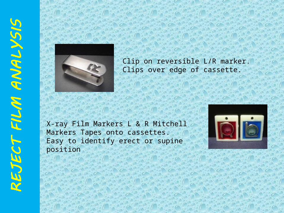
Clip on reversible L/R marker. Clips over edge of cassette.
X-ray Film Markers L & R Mitchell Markers Tapes onto cassettes.Easy to identify erect or supine position

Task Allocation Matrix
Task Responsibility Frequency
Verify Patient ID and exam info Radiographer Each exam
Verify Patient Positioning Radiographer Each view
Verify Image Quality – release or repeat Lead Radiographer Each image
Verify exam in PACS Lead Radiographer Each exam
Reconcile patient data/image counts in PACS Medical Informatics Incidental
Report substandard images Radiologist Incidental
Erase cassette-based image receptors Radiographer Start-of-shift
Test image receptor uniformity Radiographer Weekly
Clean cassette-based image receptors Radiographer Monthly
Compile and review reject analysis data Lead Radiographer Monthly
Verify display calibrations Clinical Engineer Quarterly
Review QC indicators QA Committee Quarterly
Verify receptor calibrations Medical Physicist Semi-Annual
Verify x-ray generator functions Medical Physicist Annual

28
Image plate artifacts ( In CR ).
Artifacts in Digital Radiography
Due to cracking Imaging Plate

Radiograph without markers

wrongly placed Marker


POSITIONING ERROR

POSITIONING ERROR

UNDER EXPOSED X-RAY FILM

OVER EXPOSED X-RAY FILM

PATIENT POSITONING ERROR

Film stuck to each other during processing

38
Plate Reading Artifact ( in CR )
Line caused from dirt Line caused from dirt collected in a CR Reader.collected in a CR Reader.
Damaged Laser beam head in CR reader. Appears as multiple linear white lines.

39
3. Image processing artifacts
Missing lines or pixels ( indicating digitization problems ).

40
4.operator errors4.operator errors
Digital detector is MORE sensitive
towel used to help in positioning a child.
Double exposure: Radiographs of both feet and pelvis (arrowheads) on a single film.
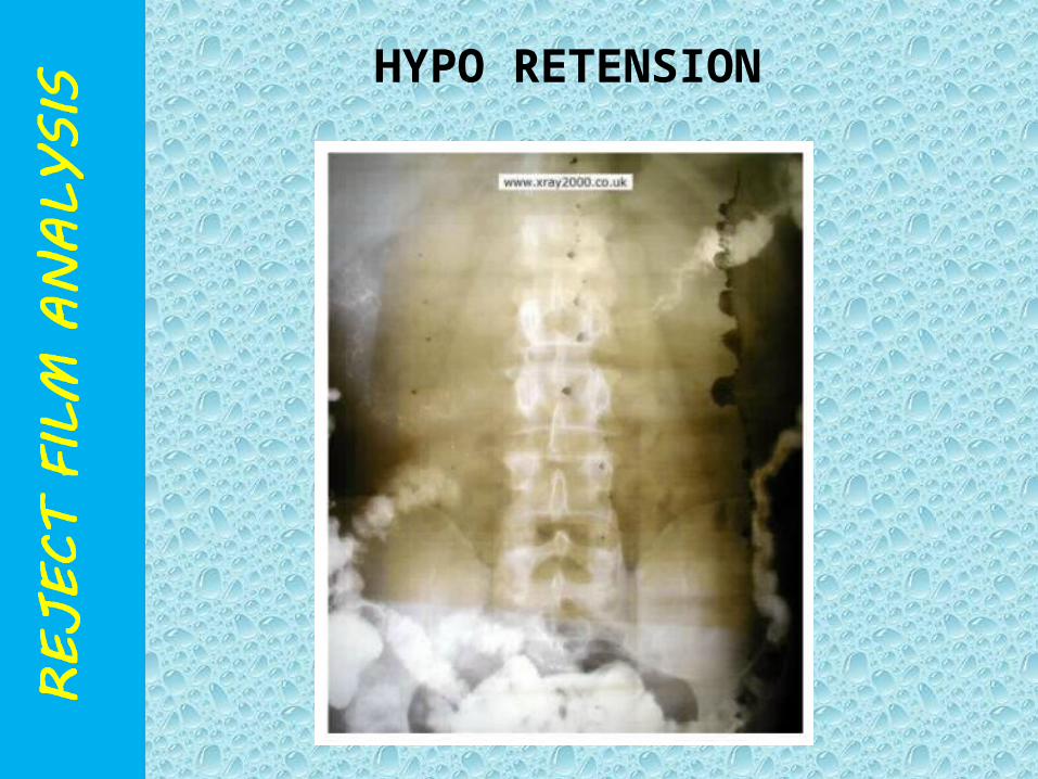
HYPO RETENSION

Pi Line Artefact


STATIC MARK

STATIC MARKS

FINGER MARK

Crimping Marks

CRIMPING MARKS

Water Stain


H.T. (High Voltage) Cable

ROTATION

DOUBLE EXPOSURE

BACK SCATTER or CASSETTE UPSIDE DOWN

CASSETTE LIGHT LEAKAGE
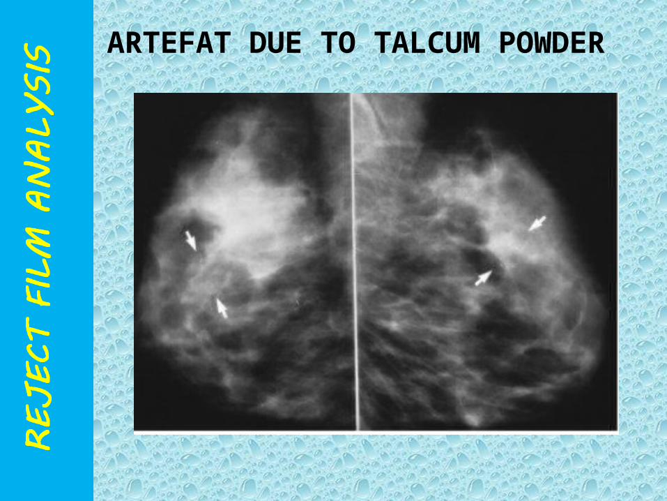
ARTEFAT DUE TO TALCUM POWDER

ARTEFACT DUE TO DEODORANT

ARTEFACT DUE TO HAIR BUN

CLOTHS RIBBINGS

GRID CUTOFF

• JEWELLERY

HEARING AID

LIGHTER IN POCKET

SCATTERED FOG
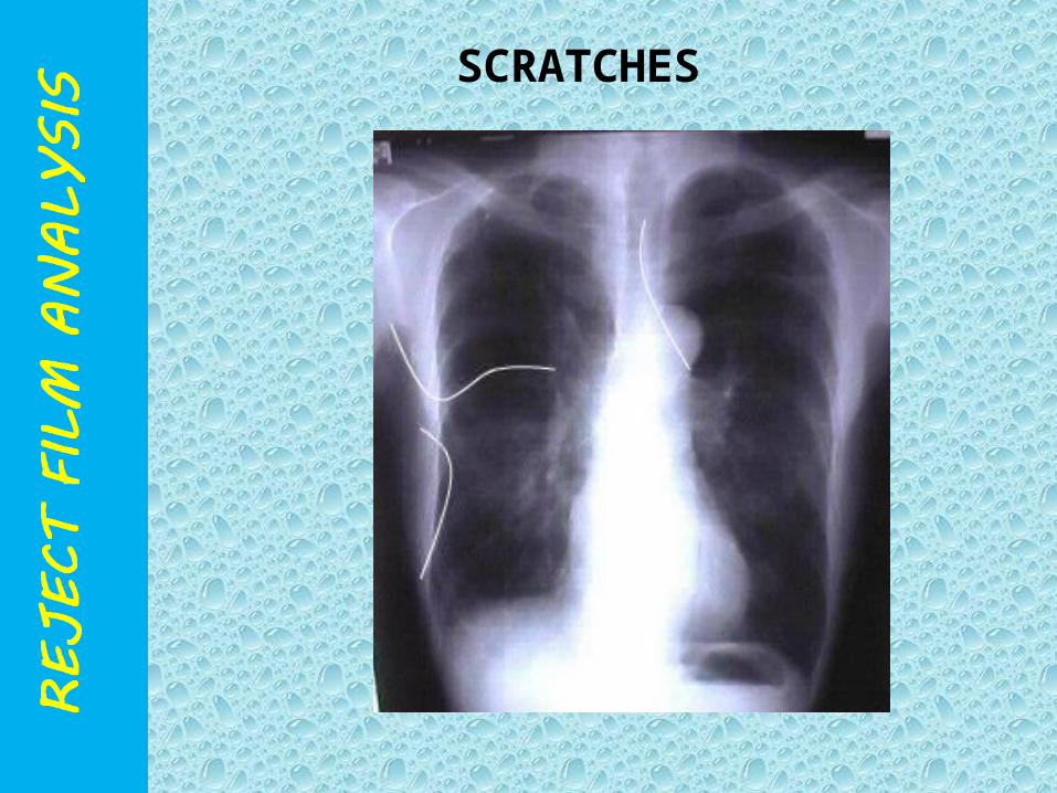
SCRATCHES
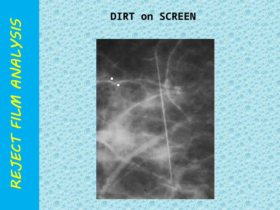
DIRT on SCREEN

SAND BAG

Earring Artefact

DUE TO CHOLE CLIP
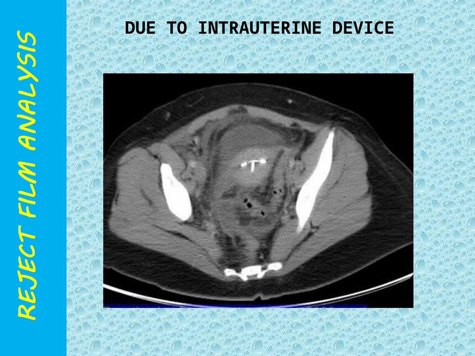
DUE TO INTRAUTERINE DEVICE

MOTION ARTEFACT
The percentage of repeats should guide the facility to focus their efforts to those areas needing the most attention. For example, films that are too light or too dark may be due to processing problems, equipment problems that require repair or calibration, or technique charts may need updating.
Corrective Action


