Download - Repression: Hypoxic Genes in Yeast Rox1p, Tup1p, Ssn6/Cyc8p and Mot3p Transcriptional regulation
Repression: Hypoxic Genes in Yeast
Rox1p, Tup1p, Ssn6/Cyc8p and Mot3p
Transcriptional regulation
Regulation of gene expression
• Almost as important as the genetic repertoire itself– The chimp and human gene sequences are almost identical –
yet gene expression leads to very distinct results
• Five (six?)regulatory levels:– (DNA copy number)– Transcription– mRNA stability– Translation– Post-translational modifications– Protein stability
A yeast model for repression of gene transcription
• The transcription of the yeast ANB1 gene is highly repressed in the presence of oxygen
• ANB1 codes for the essential eIF-5A protein involved in translation initiation or mRNA export from the nucleus
• In the presence of oxygen, ANB1 is strongly repressed, and an aerobic counterpart, TIF51A, which codes for and almost identical protein, is activated. Yeast needs the eIF-5A protein from one or the other gene to survive
• ANB1 is closely linked to the yeast oxygen-activatedCYC1 gene, which codes for the Iso-1-cytochrome that is required for respiration
Isolation of mutations affecting ANB1 repression
Inversion of regulatory region
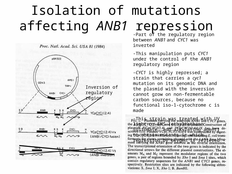
-Part of the regulatory region between ANB1 and CYC1 was inverted
-This manipulation puts CYC1 under the control of the ANB1 regulatory region
-CYC1 is highly repressed; a strain that carries a cyc1 mutation on its genomic DNA and the plasmid with the inversion cannot grow on non-fermentable carbon sources, because no functional iso-1-cytochrome c is made
-This strain was treated with UV light or EMS (ethylmethane sulfonate, and alkylating agent) to obtain mutants in which repression by oxygen is relieved
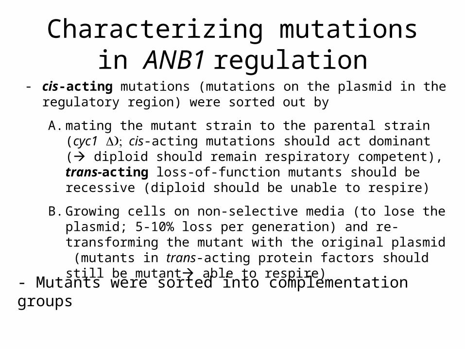
Characterizing mutations in ANB1 regulation
- cis-acting mutations (mutations on the plasmid in the regulatory region) were sorted out by
A. mating the mutant strain to the parental strain (cyc1 cis-acting mutations should act dominant ( diploid should remain respiratory competent), trans-acting loss-of-function mutants should be recessive (diploid should be unable to respire)
B. Growing cells on non-selective media (to lose the plasmid; 5-10% loss per generation) and re-transforming the mutant with the original plasmid (mutants in trans-acting protein factors should still be mutant able to respire)
- Mutants were sorted into complementation groups
Characterization of the rox1 mutation
• The initial rox1 mutant displayed de-repression of the ANB1 gene, as well as de-repression of several other oxygen repressed genes
• Genetic analysis indicated the mutation was in one gene

Cloning of the rox1 mutation• De-repression of hypoxic genes does not have a
detectable phenotypeCreation of a reporter construct, integration into the
URA3 locus of the rox1 mutant strainThe resulting strain is ura3- and expresses the lacZ gene
product (β-galactosidase) constitutively
ANB1/lacZANB1 promoter
URA3
UR A3
ANB1/lacZANB1 promoterUR A3
Restriction fragment from plasmid
(select for FOA resistance)

Cloning of rox1 mutation (2)
rox1 mutant cells with integrated ANB1-lacZ fusion on medium containing X-gal all colonies are blue (β-galactosidase expressed)
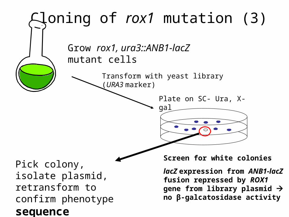
Cloning of rox1 mutation (3)
Grow rox1, ura3::ANB1-lacZ mutant cells
Transform with yeast library (URA3 marker)
Plate on SC- Ura, X-gal
Screen for white colonies
lacZ expression from ANB1-lacZ fusion repressed by ROX1 gene from library plasmid no β-galcatosidase activity
Pick colony, isolate plasmid, retransform to confirm phenotype sequence

The Rox1 protein is the repressor of hypoxic genes
• Rox1p is a DNA – binding repressor protein with an N-terminal HMG (High Mobility Group) -DNA-binding domain and a rather undefined C-terminal “repression domain”
• The DNA – binding domain has high similarity to the DNA-binding domain of the human Sry gene involved in sex-determination and to proteins conferring resistance to the drug cis-platin used in cancer therapy
• The DNA – binding domain is roughly L-shaped and introduces 90o bends into DNA
Rox1 binding site consensus:
YYYATTGTTCTC
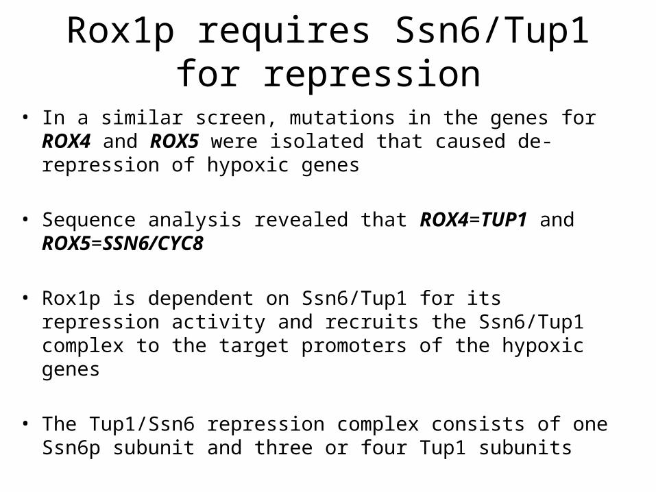
Rox1p requires Ssn6/Tup1 for repression
• In a similar screen, mutations in the genes for ROX4 and ROX5 were isolated that caused de-repression of hypoxic genes
• Sequence analysis revealed that ROX4=TUP1 and ROX5=SSN6/CYC8
• Rox1p is dependent on Ssn6/Tup1 for its repression activity and recruits the Ssn6/Tup1 complex to the target promoters of the hypoxic genes
• The Tup1/Ssn6 repression complex consists of one Ssn6p subunit and three or four Tup1 subunits

Model of protein and nucleosome interactions at the RNR3 promoter. A, a schematic map of the chromatin organization over the RNR3 promoter under the repressed and derepressed conditions. B, cooperative protein-DNA-nucleosome interactions at the URS. Arrows indicate the approximate locations of MNase hypersensitivity detected by high resolution mapping in repressed cells. The larger arrow indicates the position of the strongest hypersensitive site. The stoichiometry of Crt1 to the Ssn6-Tup1 complexes is not based upon experimental evidence.
Tup1/Ssn6 interacts with nucleosomes to form a repressive chromatine structure
B. Li and J. C. ReeseSsn6-Tup1 Regulates RNR3 by Positioning Nucleosomes and Affecting the Chromatin Structure at the Upstream Repression SequenceJ. Biol. Chem, September 7, 2001; 276(36):
33788 - 33797.
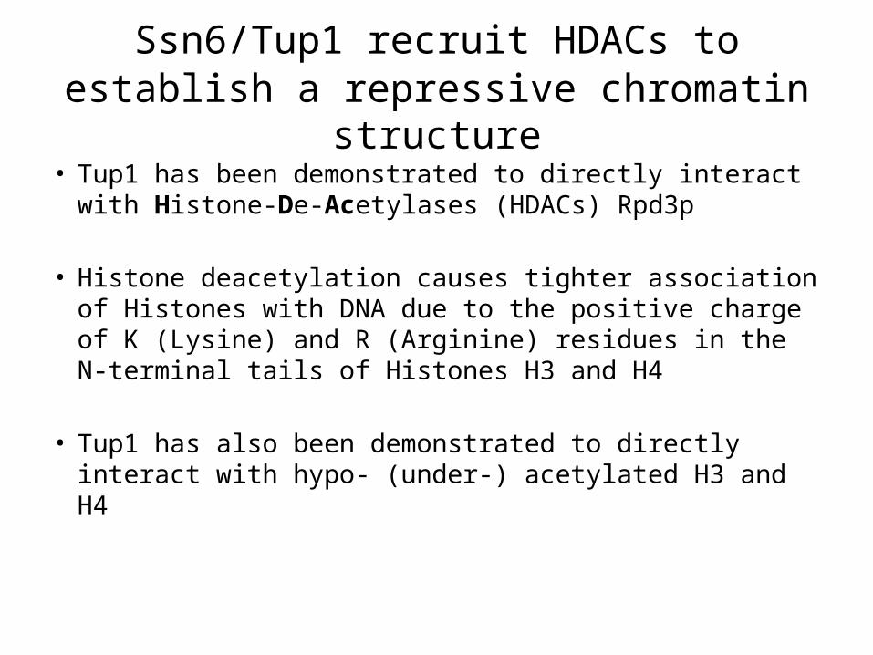
Ssn6/Tup1 recruit HDACs to establish a repressive chromatin structure
• Tup1 has been demonstrated to directly interact with Histone-De-Acetylases (HDACs) Rpd3p
• Histone deacetylation causes tighter association of Histones with DNA due to the positive charge of K (Lysine) and R (Arginine) residues in the N-terminal tails of Histones H3 and H4
• Tup1 has also been demonstrated to directly interact with hypo- (under-) acetylated H3 and H4
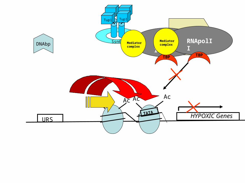
URSHYPOXIC GenesTATATATA
TATA
DNAbp Ssn6
Tup1 Tup1
AcAcAc Ac
HDAC
RNApolIIMediator complex
TBP
RNApolIIMediator complex
TBP
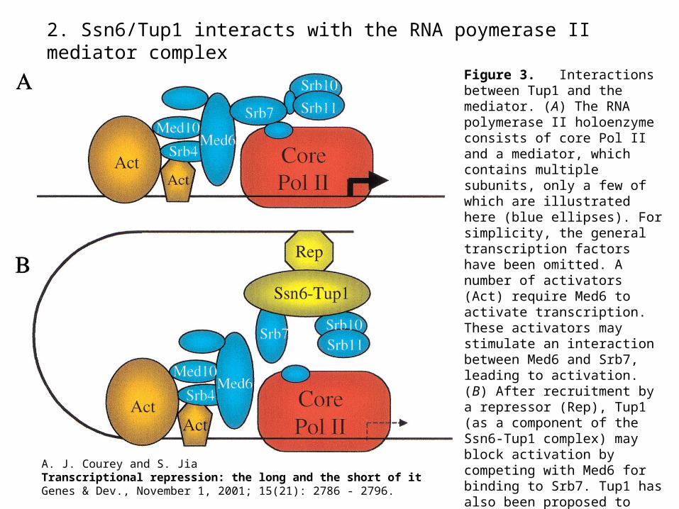
2. Ssn6/Tup1 interacts with the RNA poymerase II mediator complex
Figure 3. Interactions between Tup1 and the mediator. (A) The RNA polymerase II holoenzyme consists of core Pol II and a mediator, which contains multiple subunits, only a few of which are illustrated here (blue ellipses). For simplicity, the general transcription factors have been omitted. A number of activators (Act) require Med6 to activate transcription. These activators may stimulate an interaction between Med6 and Srb7, leading to activation. (B) After recruitment by a repressor (Rep), Tup1 (as a component of the Ssn6-Tup1 complex) may block activation by competing with Med6 for binding to Srb7. Tup1 has also been proposed to engage in an inhibitory interaction with
Srb10/Srb11.
A. J. Courey and S. JiaTranscriptional repression: the long and the short of itGenes & Dev., November 1, 2001; 15(21): 2786 - 2796.

S sn 6/Tu p 1
(A A C 3 H E M 1 3 C O X 5 B S U T 1 ...)
(C Y C 1S O D 2T IF 5 1 A ...)
Oxygen regulation in yeast
Mot3
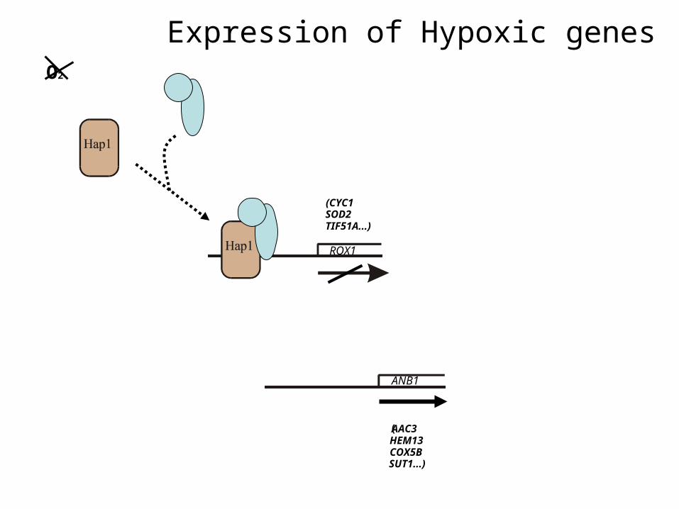
Expression of Hypoxic genes
ROX1
ANB1
(AAC3 HEM13 COX5B SUT1...)
(CYC1SOD2TIF51A...)
O2

Promoter analysis
• What determines the efficiency of repression?
• - Sequence of repressor binding sites
• - Number of operators/ repressor binding sites
• - Position?
• - Modulating factors?
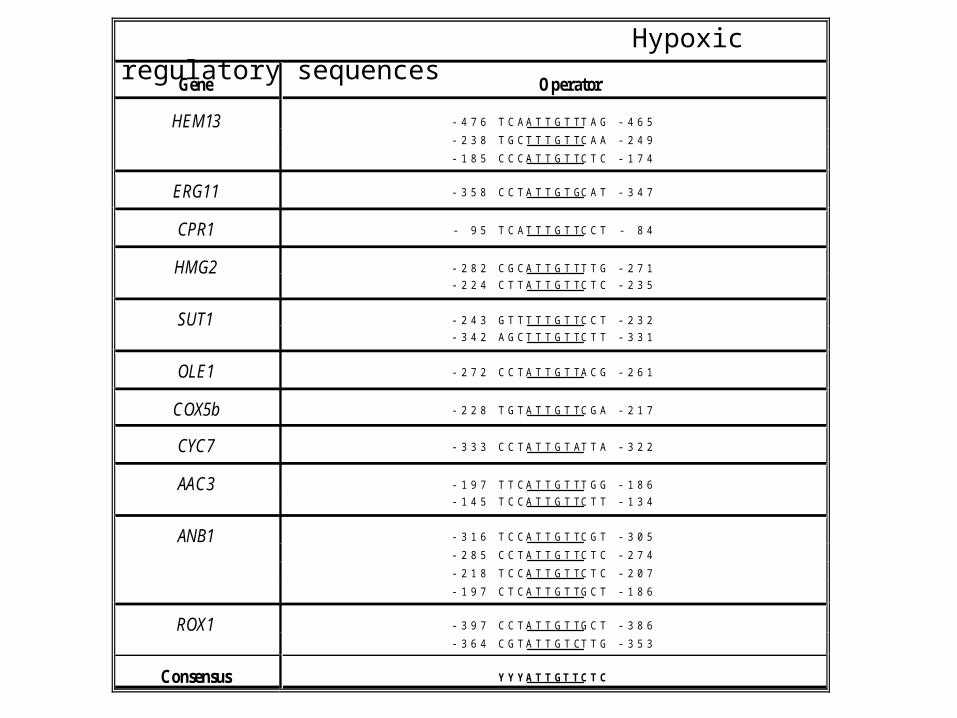
Gene Operator
HEM13 -476 TCAATTGTTTAG -465-238 TGCTTTGTTCAA -249-185 CCCATTGTTCTC -174
ERG11 -358 CCTATTGTGCAT -347
CPR1 - 95 TCATTTGTTCCT - 84
HMG2 -282 CGCATTGTTTTG -271-224 CTTATTGTTCTC -235
SUT1 -243 GTTTTTGTTCCT -232-342 AGCTTTGTTCTT -331
OLE1 -272 CCTATTGTTACG -261
COX5b -228 TGTATTGTTCGA -217
CYC7 -333 CCTATTGTATTA -322
AAC3 -197 TTCATTGTTTGG -186-145 TCCATTGTTCTT -134
ANB1 -316 TCCATTGTTCGT -305-285 CCTATTGTTCTC -274-218 TCCATTGTTCTC -207-197 CTCATTGTTGCT -186
ROX1 -397 CCTATTGTTGCT -386-364 CGTATTGTCTTG -353
Consensus YYYATTGTTCTC
Hypoxic regulatory sequences
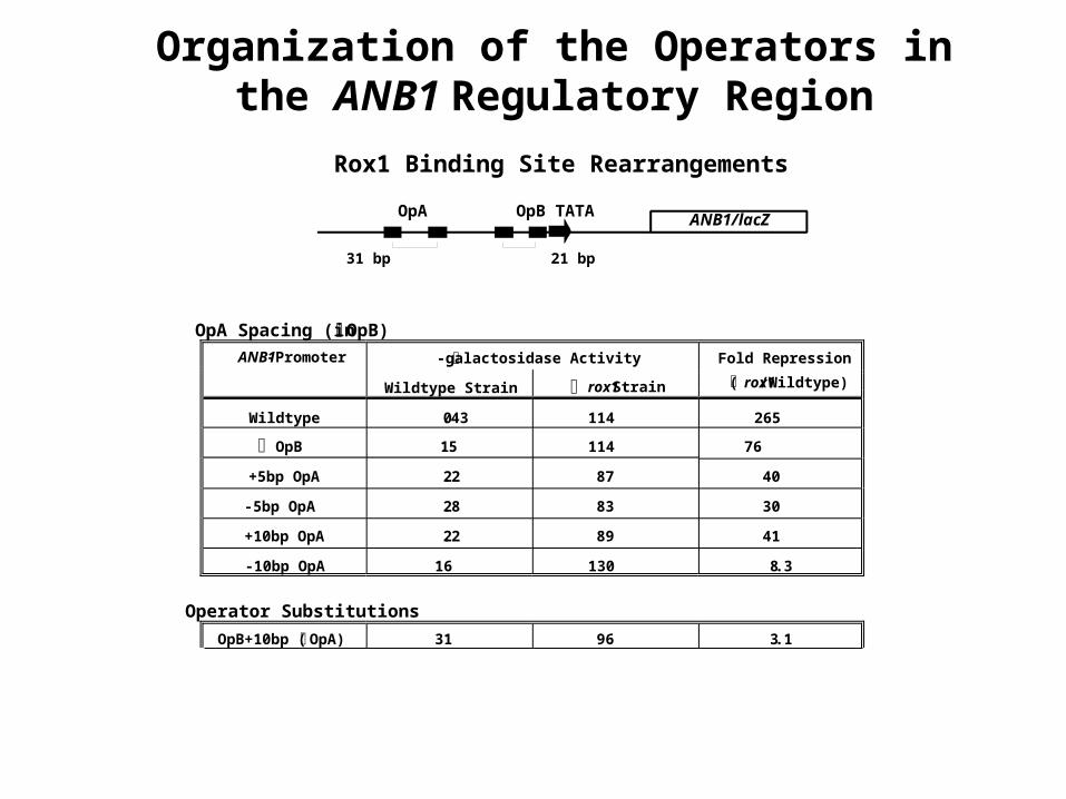
OpA Spacing (in OpB)
ANB1-Promoter -galactosidase Activity Fold Repression
( rox1/Wildtype)Wildtype Strain rox1 Strain
Wildtype 0.43 114 265
OpB 1.5 114 76
+5bp OpA 2.2 87 40
-5bp OpA 2.8 83 30
+10bp OpA 2.2 89 41
-10bp OpA 16 130 8.3
Operator Substitutions
OpB+10bp ( OpA) 31 96 3.1
OpB in OpA site 26 91 3.5
OpA in OpB site 0.86 43 50
ANB1/lacZOpA OpB TATA
31 bp 21 bp
Organization of the Operators in the ANB1 Regulatory Region
Rox1 Binding Site Rearrangements

Operatorconstruct1
β-galactosidase units2 Fold repression
Wild type rox1Δ
OpA in B position(+)
0.42 ± 0.17 65 ± 26 152
OpA in B position (-)
0.72 ± 0.18 44 ± 7.3 61
OpB in A position(+)
27.5 ± 11 102 ± 32 3.7
OpB in A position(-)
23.3 ± 10.5 80 ± 27 3.4
OpA 1.1 ± 0.6 84 ± 32 76
OpB 9.6 ± 3.8 81 ± 16 8.4
ANB1/Z UAS 3 41 2
OpA OpB1 212
3 434
Role of position for repressor efficiency
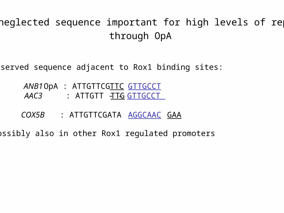
A neglected sequence important for high levels of repression
conserved sequence adjacent to Rox1 binding sites:
ANB1 OpA : ATTGTTCGTTCGTTGCCT AAC3 : ATTGTT - - TTGGTTGCCT COX5B : ATTGTTCGATA AGGCAAC GAA
possibly also in other Rox1 regulated promoters
through OpA

ANB1-Promoter11 β-galactosidase Activity2 Fold Repression(rox1Δ/Wildtype)
Wildtype rox1Δ
ΔOpB OpA-wildtype 1.3 ± 0.7 97 ± 34 75
OpA 1(-10) 22 ± 5 116 ± 14 5
OpA 2(-10) 1.6 ± 0.7 88 ± 12 55
OpA 3 1.1 ± 0.2 93 ± 25 84
OpA 4 10 ± 3 101 ± 38 10
OpA 5 15 ± 4 102 ± 19 7
OpA 6 4.4 ± 3 107 ± 31 24
ΔOpA OpB-wildtype 12 ± 8 83 ± 16 7
A N B 1/la cZU A S
O p A
T C G T T G C C T G T T T T T T T G CAAAAAAA
The sequence TGCCT is responsible for stronger repression from OpA

Insertion of the conserved sequence adjacent to the OpA 5’ Rox1 binding site improves repression from OpB
ANB1-Promoter11 β-galactosidase Activity2 Fold Repression(rox1Δ/Wildtype)
Wildtype rox1Δ
ΔOpB OpA-wildtype 1.3 ± 0.7 97 ± 34 75
ΔOpA OpB-wildtype 12 ± 8 83 ± 16 7
OpB 7(+10) 5.6 ± 2 91 ± 23 16
OpB 8(+10) 31 ± 2 96 ± 9 3
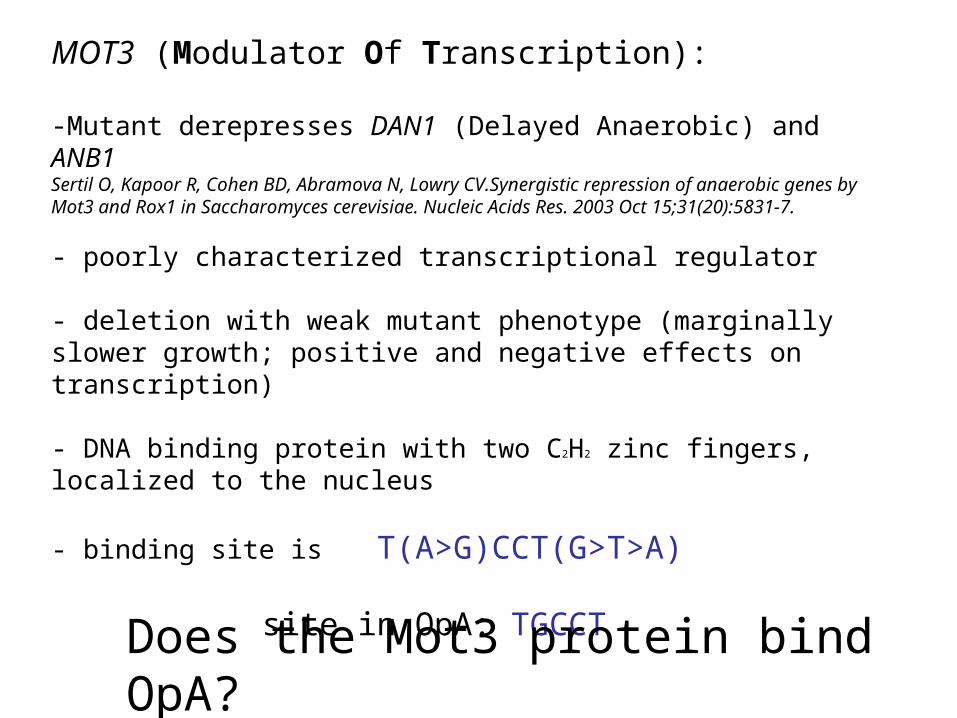
MOT3 (Modulator Of Transcription):
-Mutant derepresses DAN1 (Delayed Anaerobic) and ANB1Sertil O, Kapoor R, Cohen BD, Abramova N, Lowry CV.Synergistic repression of anaerobic genes by Mot3 and Rox1 in Saccharomyces cerevisiae. Nucleic Acids Res. 2003 Oct 15;31(20):5831-7.
- poorly characterized transcriptional regulator
- deletion with weak mutant phenotype (marginally slower growth; positive and negative effects on transcription)
- DNA binding protein with two C2H2 zinc fingers, localized to the nucleus
- binding site is T(A>G)CCT(G>T>A)
site in OpA: TGCCT
Does the Mot3 protein bind OpA?

Electrophoretic mobility shift assay (EMSA)
• Used in analysis of DNA binding properties of proteins
• Binding target (DNA or RNA, often a short oligomer containing protein binding sites) is labelled radioactively
• Binding of protein to DNA results in retardation of the migration of the labelled DNA band

EMSA - PrincipleDNA with binding site DNA – protein complex
(High molecular weight, bulky)
- -
++

Rox1
Mot3Rox1
Mot3
The Mot3 protein binds specifically to OpA in the ANB1 promoter
- 1 5 1 1 1 5 5 5
- - - 5x 20x - 5x 20x - - 20x -
- - - - - 20x - - 20x - - 20x
competitor DNA
labelled DNA
OpA
OpA (-Mot3 site)
OpA
Mot3 site
Rox1 site
Does Mot3p play a role in ANB1 repression in vivo?
Anaerobic Aerobic
WT
mot
3ro
x1tu
p1
WT
mot
3
rox1
tup1
1 2 3 4 5 6 7 8
5 6
ACT1
TIF51A
ANB1
TIF51A
ANB1
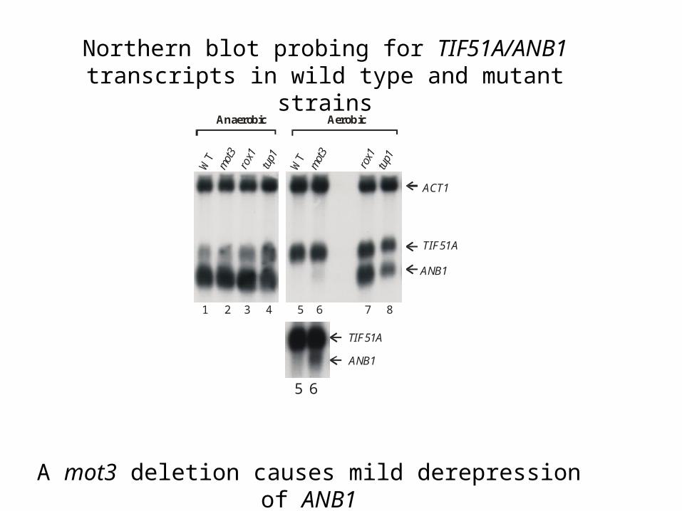
A mot3 deletion causes mild derepression of ANB1
Northern blot probing for TIF51A/ANB1 transcripts in wild type and mutant strains
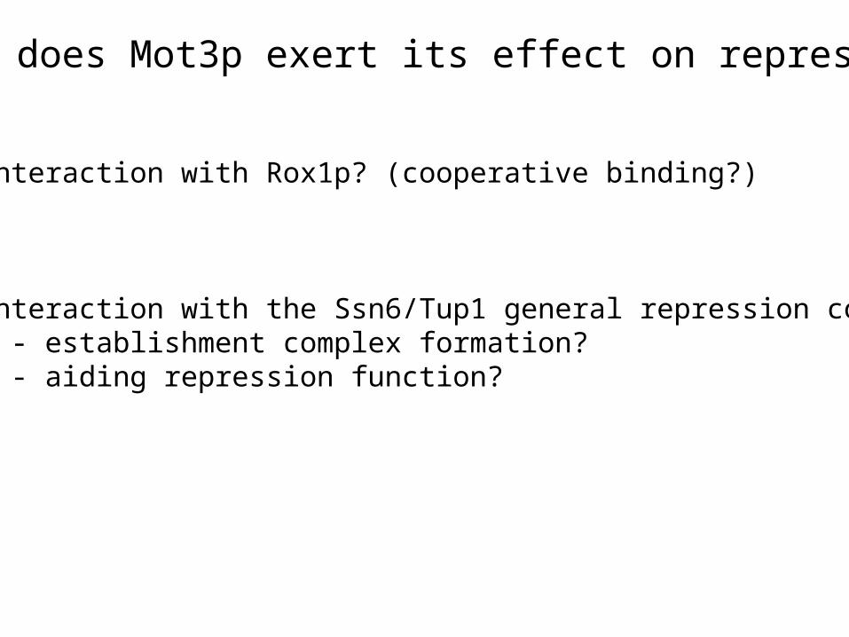
How does Mot3p exert its effect on repression?
1. Interaction with Rox1p? (cooperative binding?)
2. Interaction with the Ssn6/Tup1 general repression complex?- establishment complex formation?- aiding repression function?

+R1 -R1
20ngMot3
25ng Rox1 MBP
FreeDNA
20ngGST-Mot3
Mot3 and Rox1 do not bind DNA cooperatively in vitro
+R1
Rox1 site
Mot3 site
-R1
labelled DNA
competitor DNA
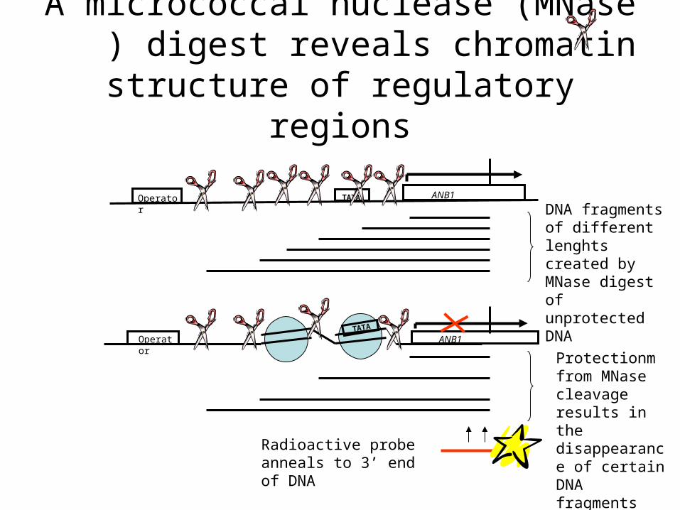
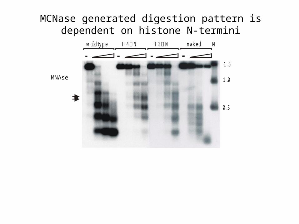
A micrococcal nuclease (MNase ) digest reveals chromatin structure of
regulatory regions
Operator ANB1TATA
Operator ANB1TATA
Radioactive probe anneals to 3’ end of DNA
DNA fragments of different lenghts created by MNase digest of unprotected DNA
Protectionm from MNase cleavage results in the disappearance of certain DNA fragments
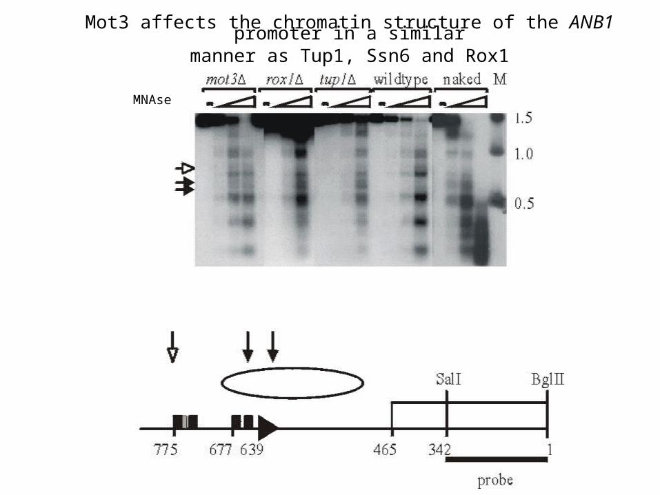
Mot3 affects the chromatin structure of the ANB1 promoter in a similarmanner as Tup1, Ssn6 and Rox1
MNAse
w ild ty p e H 4 N H 3 N n ak e d M
1 .5
1 .0
0 .5
MCNase generated digestion pattern is dependent on histone N-termini
MNAse

Summary
Operator efficiency:
- operator orientation and position relative to the TATA box do only play a minor role in operator efficiency
- the sequence TGCCT between OpA binding sites is responsible for higher repression efficiency of OpA compared to OpB
- the TGCCT sequence improves repression from OpB when inserted
- the TGCCT sequence is bound specifically by the transcription factor Mot3, a zinc finger protein protein that has been reported affect the expression of various other genes
- deletion of the MOT3 gene causes partial derepression of hypoxic genes
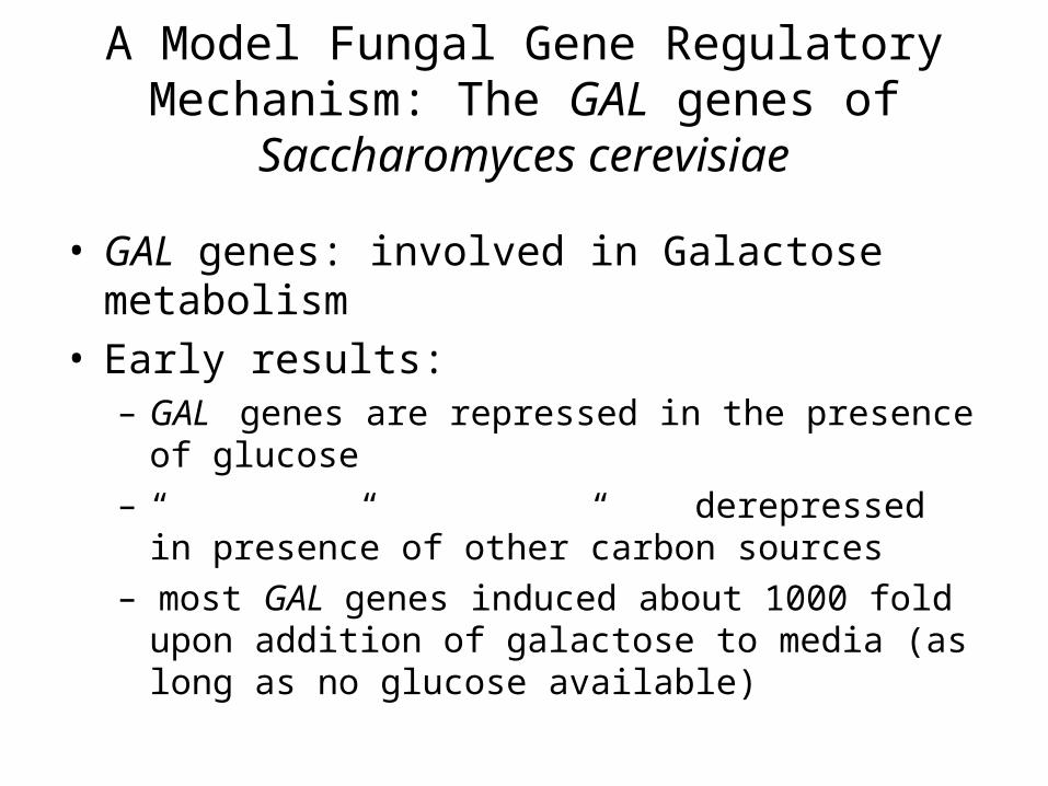
A Model Fungal Gene Regulatory Mechanism: The GAL genes of
Saccharomyces cerevisiae
• GAL genes: involved in Galactose metabolism• Early results:
– GAL genes are repressed in the presence of glucose– “ “ “ derepressed in presence of other
carbon sources– most GAL genes induced about 1000 fold upon
addition of galactose to media (as long as no glucose available)

GAL mutant phenotypes:GAL1, GAL7, GAL10, MEL1, (GAL5): If mutant, cells cannot utilize galactose; a specific enzymatic activity in galactose breakdown pathway missing
GAL2: Mutant cells cannot utilize galactose, but all enzymatic activities are present in cell extract
GAL4: Mutant cells cannot utilize galactose, none of the enzymatic activities are present in cell extract
GAL3: In combination with mutation in any one mutation in GAL1, GAL7, GAL10, MEL1 (GAL5), cells cannot utilize galactose, and all of the enzymatic activities are missing
GAL80: All enzymatic activities are constitutively expressed
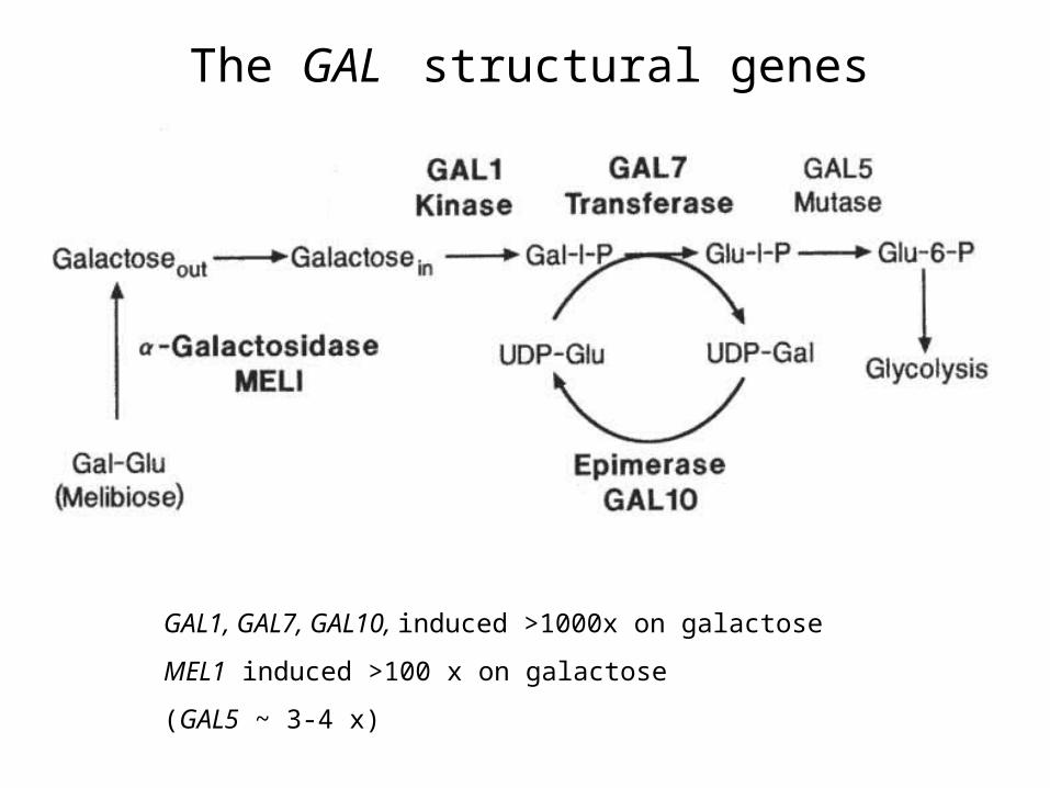
The GAL structural genes
GAL1, GAL7, GAL10, induced >1000x on galactose
MEL1 induced >100 x on galactose
(GAL5 ~ 3-4 x)
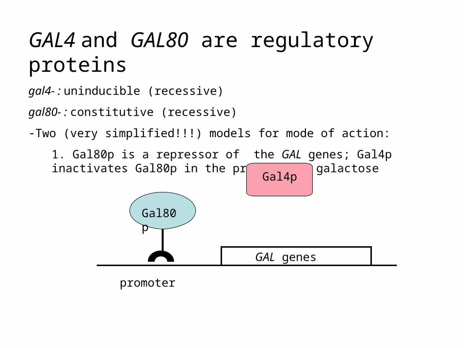
GAL4 and GAL80 are regulatory proteinsgal4- : uninducible (recessive)
gal80- : constitutive (recessive)
-Two (very simplified!!!) models for mode of action:
1. Gal80p is a repressor of the GAL genes; Gal4p inactivates Gal80p in the presence of galactose
GAL genes
promoter
Gal80p
Gal4p

GAL genes
promoter
Gal80
Gal4p
Galactose
GAL genes
promoter
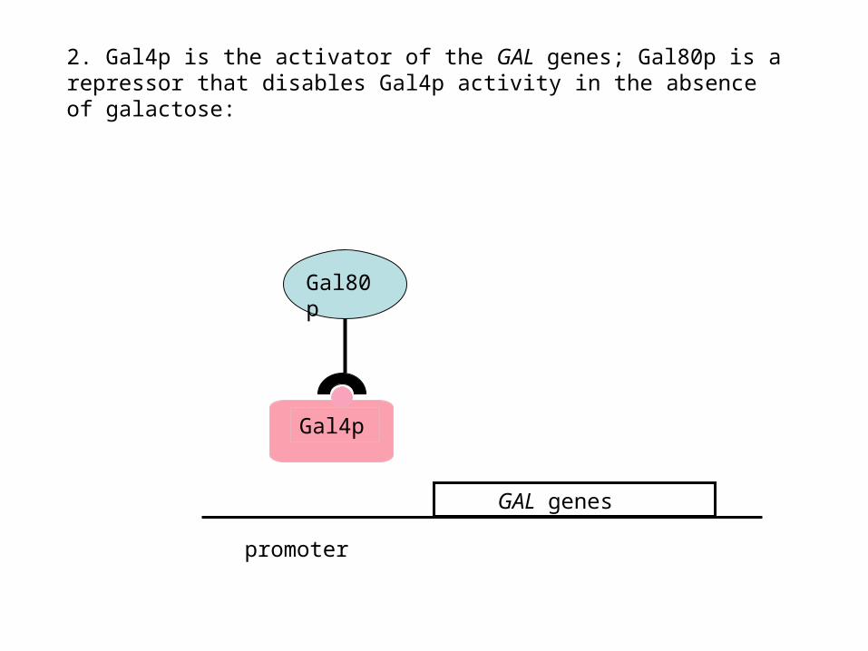
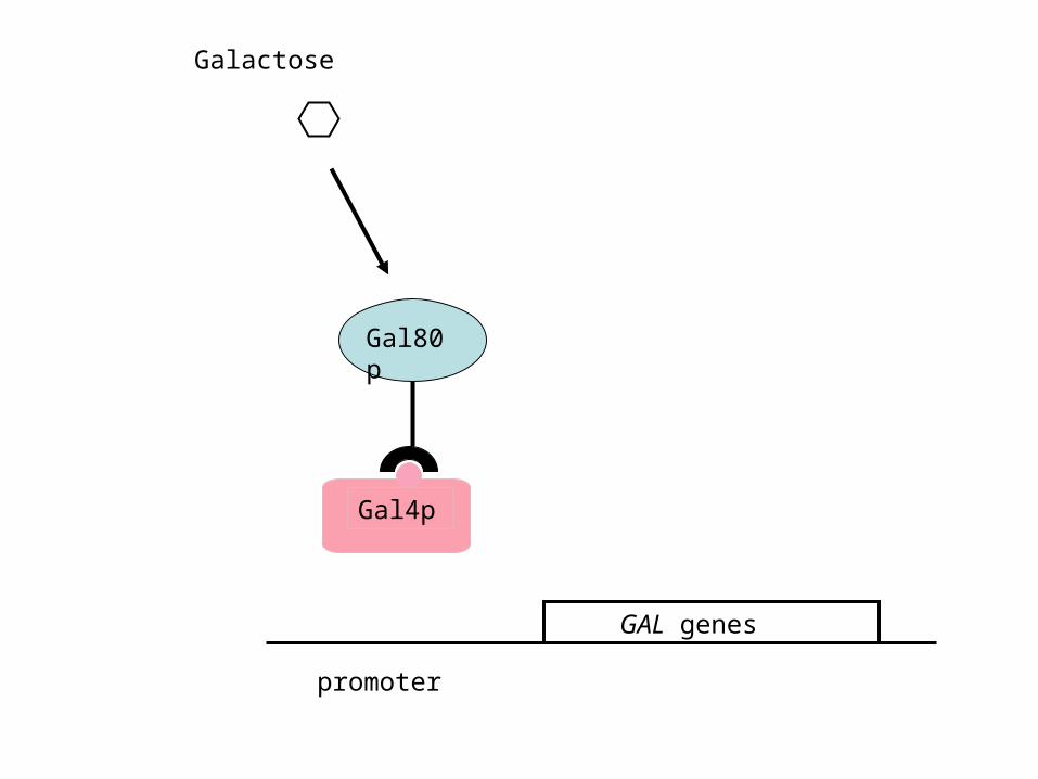
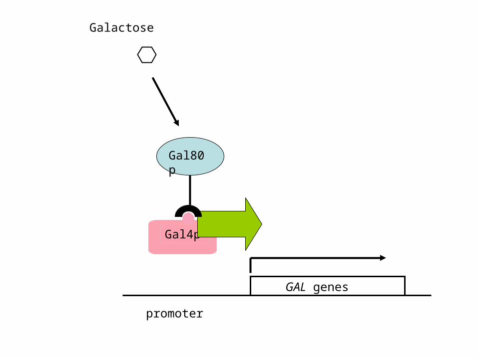
2. Gal4p is the activator of the GAL genes; Gal80p is a repressor that disables Gal4p activity in the absence of galactose:
Gal80p
Gal4p
Galactose
GAL genes
promoter
Gal80p
Gal4p
GAL genes
promoter
Gal4p
Gal80p
Galactose
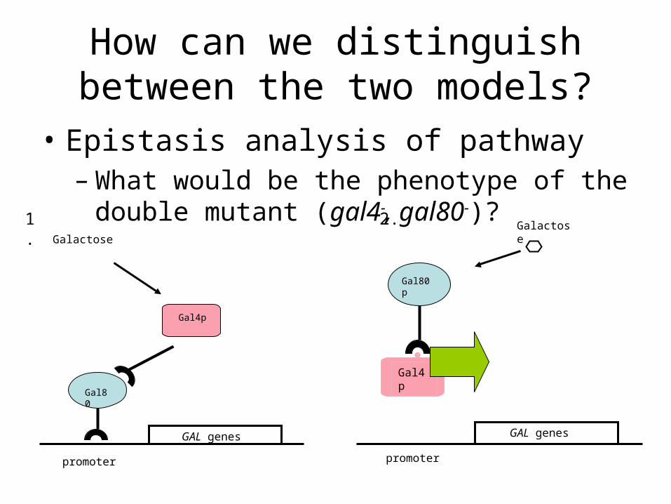
How can we distinguish between the two models?
• Epistasis analysis of pathway– What would be the phenotype of the double
mutant (gal4-, gal80-)?
promoter
GAL genes
Gal80p
Galactose
GAL genes
promoter
Gal80
Galactose
Gal4p
1. 2.
Gal4p
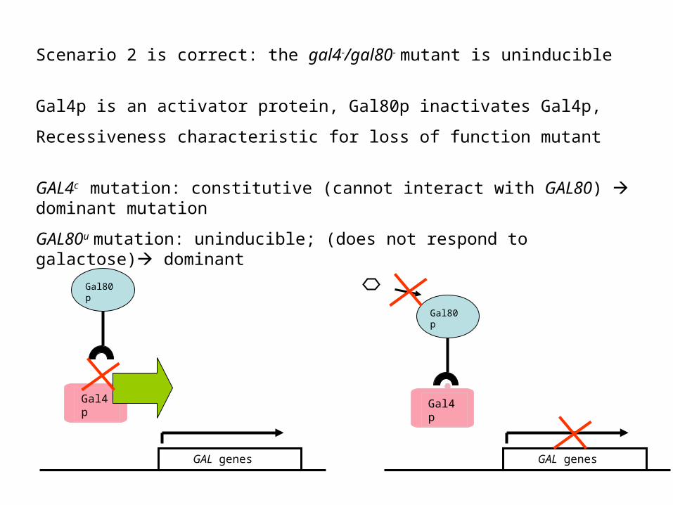
Scenario 2 is correct: the gal4-/gal80- mutant is uninducible
Gal4p is an activator protein, Gal80p inactivates Gal4p,
Recessiveness characteristic for loss of function mutant
GAL4c mutation: constitutive (cannot interact with GAL80) dominant mutation
GAL80u mutation: uninducible; (does not respond to galactose) dominant
GAL genes
Gal80p
Gal4p
GAL genes
Gal80p
Gal4p
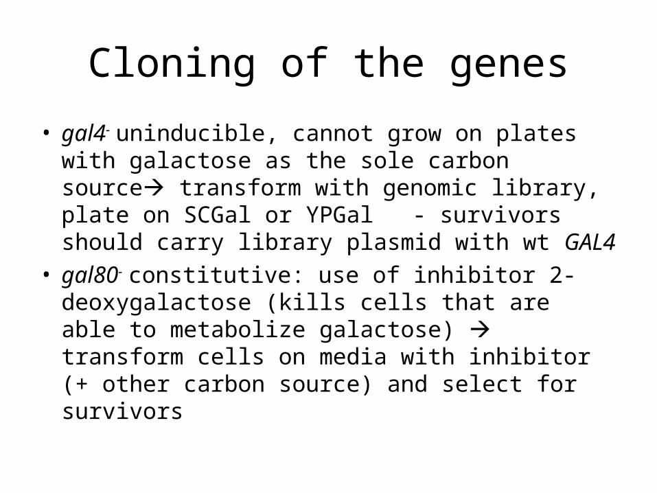
Cloning of the genes
• gal4- uninducible, cannot grow on plates with galactose as the sole carbon source transform with genomic library, plate on SCGal or YPGal
- survivors should carry library plasmid with wt GAL4
• gal80- constitutive: use of inhibitor 2-deoxygalactose (kills cells that are able to metabolize galactose) transform cells on media with inhibitor (+ other carbon source) and select for survivors
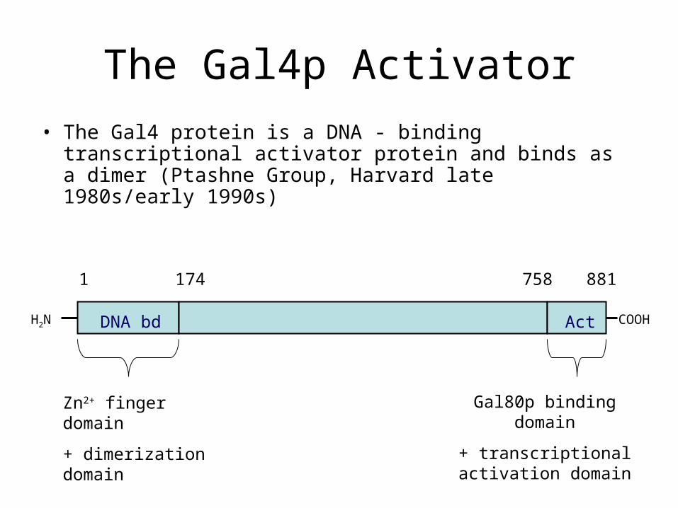
The Gal4p Activator
• The Gal4 protein is a DNA - binding transcriptional activator protein and binds as a dimer (Ptashne Group, Harvard late 1980s/early 1990s)
H2N COOH
1 174 758 881
DNA bd Act
Zn2+ finger domain
+ dimerization domain
Gal80p binding domain
+ transcriptional activation domain

Gal4p binds UAS sequences in the regulatory region of GAL structural genes
GAL genesTATAUAS
Gal4p
UAS: upstream activation sequence
TATA – box: AT-rich sequence required for transcription machinery assembly

lacZTATAUAS
Deletion analysis of promoter region identified Gal4p binding sites
Gal4p binding site:
5’ –CGGAG/CGACA-3’
3’TCAGG/CAGGC-5’
-Site is promiscuous (can function if front of many genes
-Orientation & position independent (symmetrical site, wide range of upstream region from where it can exert transactivation)
“Gal4 17-mer”

Gal4p is a modular protein
H2N
1 174 758 881
DNA bd Act
Gal4p
Activ./Gal80 ia
bd
lacZTATAUAS
VP16
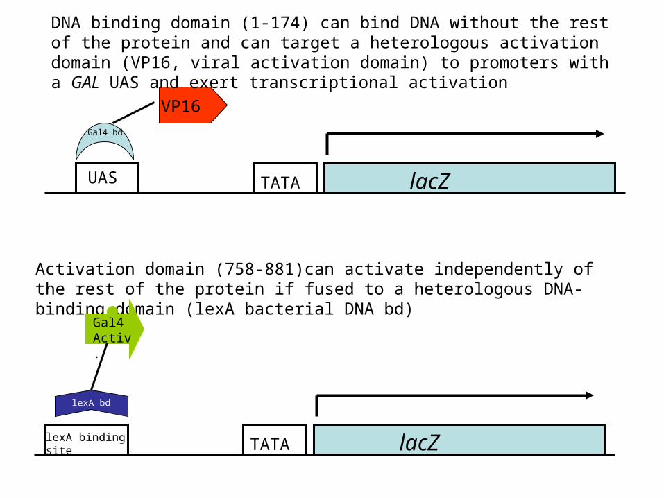
Activation domain (758-881)can activate independently of the rest of the protein if fused to a heterologous DNA-binding domain (lexA bacterial DNA bd)
lacZTATAlexA binding site
Gal4 Activ.
Gal4 bd
lexA bd
DNA binding domain (1-174) can bind DNA without the rest of the protein and can target a heterologous activation domain (VP16, viral activation domain) to promoters with a GAL UAS and exert transcriptional activation
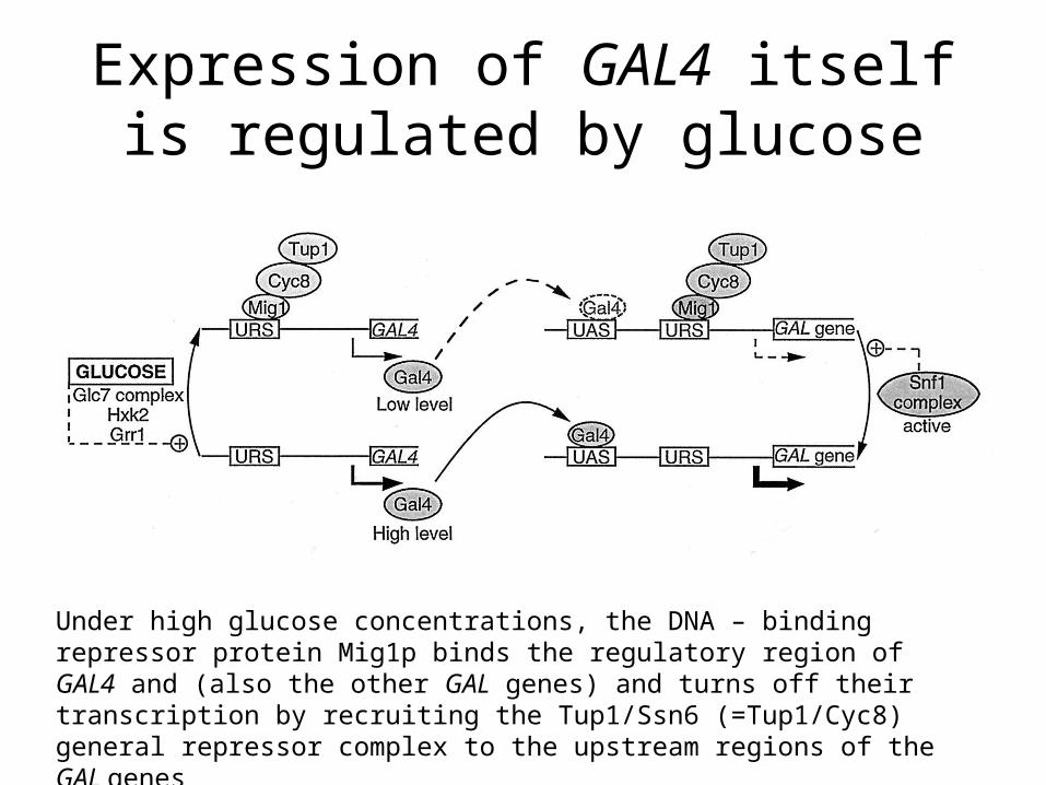
Expression of GAL4 itself is regulated by glucose
Under high glucose concentrations, the DNA – binding repressor protein Mig1p binds the regulatory region of GAL4 and (also the other GAL genes) and turns off their transcription by recruiting the Tup1/Ssn6 (=Tup1/Cyc8) general repressor complex to the upstream regions of the GAL genes

The galactose sensor: Gal3p
• Gal3p is a protein with high similarity (homology) to galactokinase
• No enzymatic activity
• In the presence of galactose, Gal3p binds the sugar and removes the Gal80p repressor from the Gal4p activator
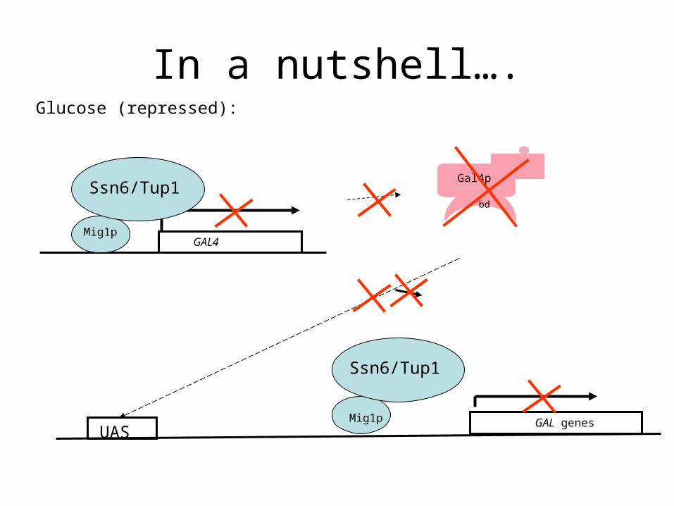
In a nutshell….Glucose (repressed):
Gal4p
bd
GAL4Mig1p
Ssn6/Tup1
GAL genesMig1p
Ssn6/Tup1
UAS

other carbon source than Glucose (derepressed):
GAL4
GAL genesUAS
Mig1p
Ssn6/Tup1
P P
Gal4p
bd
Gal80p
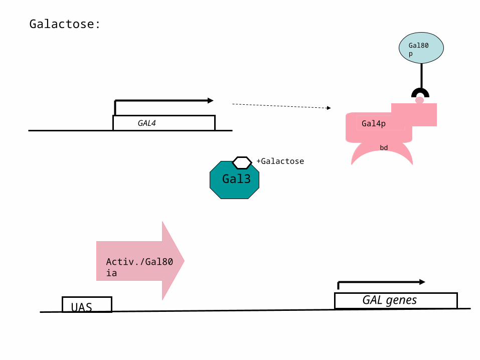
Galactose:
GAL4
UAS
Gal4p
bd
Gal80p
+Galactose
Activ./Gal80 ia
GAL genes
Gal3
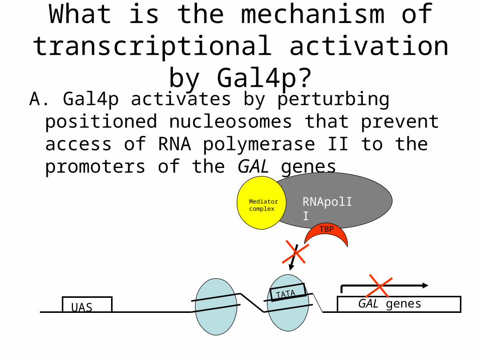
What is the mechanism of transcriptional activation by Gal4p?A. Gal4p activates by perturbing positioned
nucleosomes that prevent access of RNA polymerase II to the promoters of the GAL genes
UAS GAL genesTATA
RNApolIIMediator complex
TBP
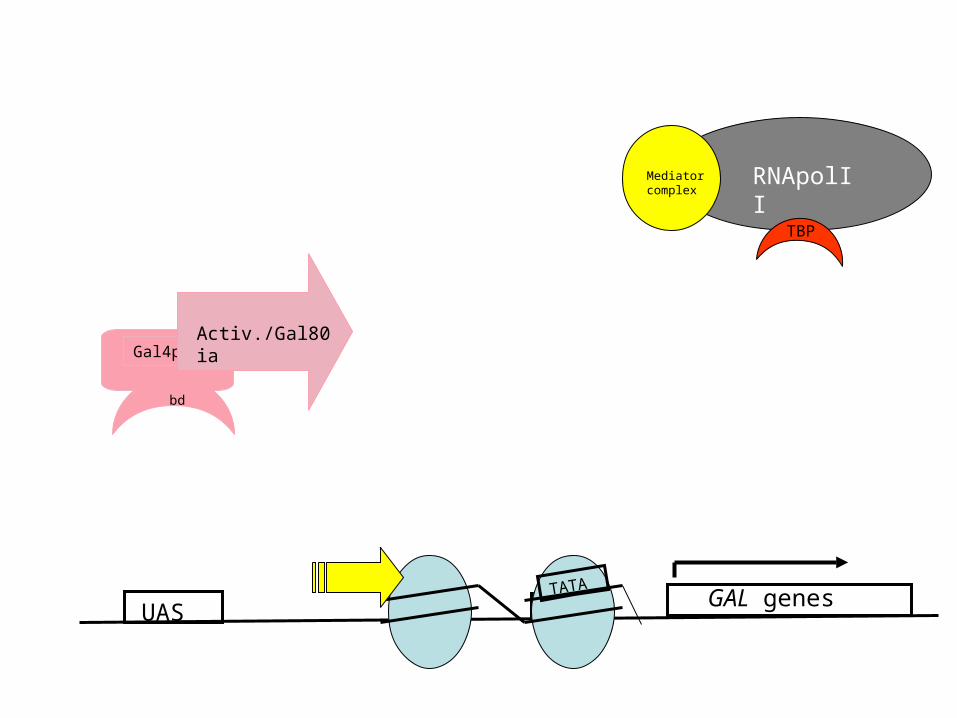
UAS
Gal4p
bd
Activ./Gal80 ia
GAL genesTATA
RNApolIIMediator complex
TBP
TATA
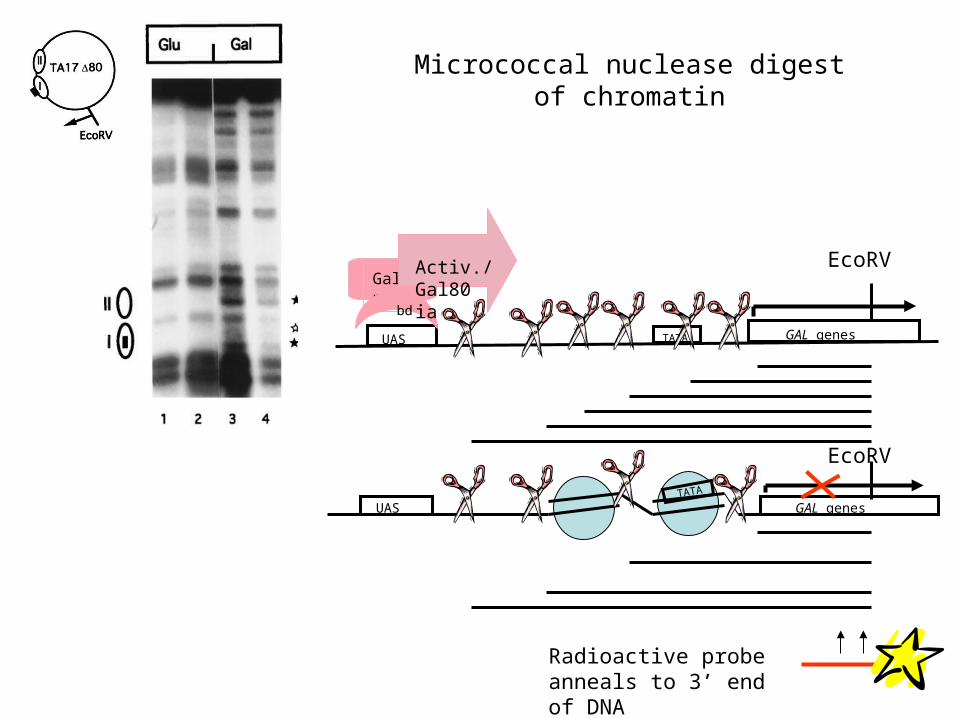
Micrococcal nuclease digest of chromatin
UAS GAL genesTATA
UAS GAL genesTATA
Radioactive probe anneals to 3’ end of DNA
Gal4p
bd
Activ./Gal80 ia
EcoRV
EcoRV
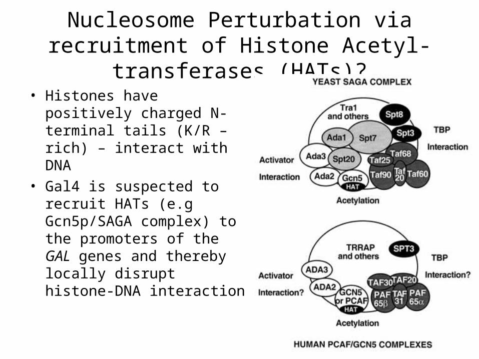
Nucleosome Perturbation via recruitment of Histone Acetyl-transferases (HATs)?
• Histones have positively charged N-terminal tails (K/R – rich) – interact with DNA
• Gal4 is suspected to recruit HATs (e.g Gcn5p/SAGA complex) to the promoters of the GAL genes and thereby locally disrupt histone-DNA interaction
TATA
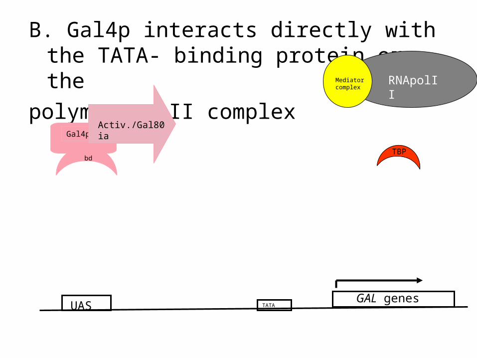
B. Gal4p interacts directly with the TATA- binding protein or the
polymerase II complex
UAS
Gal4p
bd
Activ./Gal80 ia
RNApolIIMediator complex
TBP
GAL genes

Relevance of the Gal regulation research today?
• General understanding of basic molecular principles of gene activation
• Model for the functioning of biological regulatory circuits– A general mechanism for network-dosage
compensation in gene circuits. Acar M, Pando BF, Arnold FH, Elowitz MB, van Oudenaarden A. Science. 2010 Sep 24;329(5999):1656-60
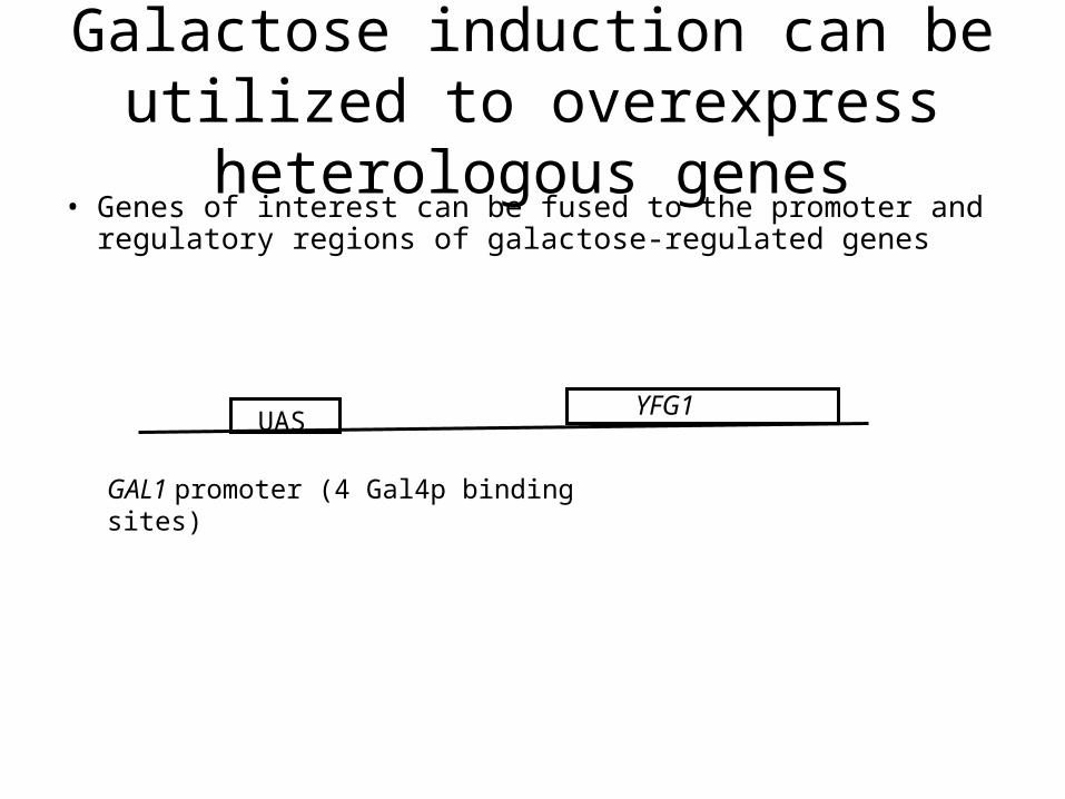
Galactose induction can be utilized to overexpress heterologous genes• Genes of interest can be fused to the promoter and
regulatory regions of galactose-regulated genes
UASYFG1
GAL1 promoter (4 Gal4p binding sites)
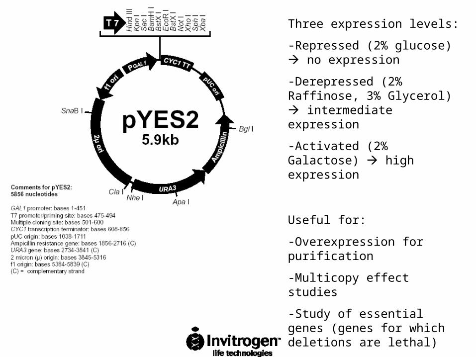
Three expression levels:
-Repressed (2% glucose) no expression
-Derepressed (2% Raffinose, 3% Glycerol) intermediate expression
-Activated (2% Galactose) high expression
Useful for:
-Overexpression for purification
-Multicopy effect studies
-Study of essential genes (genes for which deletions are lethal)

Similar: Oleate induction:
-Oleate induced genes are involved in peroxisomal proliferation and in -oxidation
-Activator is a heterodimer of the Oaf1p/Pip2p activators which bind to oleate response elements (OREs)
-The ORE consensus is currently viewed as two inverted CGG triplets spaced by 14 (formerly 15) to 18 intervening nucleotides (N), i.e. CGGN3TNAN8-12CCG
-Currently, the plasmid available has the promoter and terminator sequences of the oleate-induced CTA1 (peroxisomal catalase) gene
-CTA1 is glucose repressed similar to the GAL genes
-Three expression levels:
-Repressed (2% Glucose)
-Derepressed (2% Raffinose, 3% Glycerol)
-Activated (0.2% oleate, 0.02% Tween, 0.05% Glucose)

Expression from inducible promoters allows investigation of essential genes
• Essential genes are genes required for viability of the cell
• Deletions of these genes are inviable, deletion are only viable as heterozygous diploids, or deletion strains have to carry a plasmid with a wild type copy of the gene
• Shuffling in plasmids carrying mutant partial function alleles is one way of investigating the function
• Introduction of plasmids with the essential gene expressed from an inducible promoter allow more precise investigation


