
Research ArticleAntibiotic Resistance of Diverse Bacteria fromAquaculture in Borneo
M. M. Kathleen, L. Samuel, C. Felecia, E. L. Reagan, A. Kasing, M. Lesley, and S. C. Toh
Department of Molecular Biology, Faculty of Resource Science and Technology, Universiti Malaysia Sarawak,94300 Kota Samarahan, Sarawak, Malaysia
Correspondence should be addressed to L. Samuel; [email protected]
Received 2 May 2016; Revised 28 July 2016; Accepted 29 August 2016
Academic Editor: Marcel H. Zwietering
Copyright © 2016 M. M. Kathleen et al.This is an open access article distributed under the Creative CommonsAttribution License,which permits unrestricted use, distribution, and reproduction in any medium, provided the original work is properly cited.
The administration of antimicrobials in aquaculture provides a selective pressure creating a reservoir of multiple resistant bacteriain the cultured fish and shrimps as well as the aquaculture environment. The objective of this study was to determine the extent ofantibiotic resistance in aquaculture products and aquaculture’s surrounding environment in Sarawak, Malaysian Borneo. Ninety-four identified bacterial isolates constituted of 17 genera were isolated from sediment, water, and cultured organisms (fish andshrimp) in selected aquaculture farms. These isolates were tested for their antibiotic resistance against 22 antibiotics from severalgroups using the disk diffusion method. The results show that the highest resistance was observed towards streptomycin (85%,𝑛 = 20), while the lowest resistance was towards gentamicin (1.1%, 𝑛 = 90). The multiple antibiotic resistant (MAR) index of theisolates tested ranged between 0 and 0.63. It was suggested that isolates with MAR index > 0.2 were recovered from sources withhigh risk of antibiotic resistant contamination. This study revealed low level of antibiotic resistance in the aquaculture bacterialisolates except for streptomycin and ampicillin (>50% resistance, 𝑛 = 94) which have been used in the aquaculture industry forseveral decades. Antibiotic resistant patterns should be continuously monitored to predict the emergence and widespread of MAR.Effective action is needed to keep the new resistance from further developing and spreading.
1. Introduction
Since the discovery of penicillin in 1928 by a Scottish sci-entist Alexander Fleming followed by the release of manyother earlier drugs onto the market to treat infection, thedevelopment of drug resistance in various sectors includingaquaculture has been reported [1–3]. The misuse and abuseof the antimicrobial drugs are among the important factorsthat have contributed to the rise of resistant microbes aroundthe world. Antibiotics, which have saved millions of lives andwere also known asmiracle drug in the past, are no longer theultimate way for the treatment of infections because bacteriahave continued to develop multiple resistance towards manydifferent types or classes of the drugs [4].
Antimicrobial agents have been widely used in fish farm-ing for either therapeutic, prophylactic, or other purposes [5].The antibiotics are normally used to increase growth as wellas feed efficiency in the animals [6]. However, some of the
antibiotics have been frequently used in both veterinary andhuman medicine such as sulfonamides, chloramphenicol,tetracycline, nitrofurans [5], oxytetracycline [7], neomycin,erythromycin, streptomycin, prefuran, and enrofloxacin [8].The evolution of bacteria towards antibiotic resistance hasbeen accelerated distinctly by selective pressure due toinappropriate and overuse of the antibiotics [3, 9]. In theefforts to cope with this problem, scientists have acceleratedthe search for alternative antimicrobial agents by screeningmanypotential sources includingmedicinal plants [10, 11] andmicrobes [12, 13].
Aquaculture is an important sector in the agriculture in-dustry and is rapidly growing to meet the world’s demandsfor protein source. This sector is challenged with the diversetype of diseases and bacterial infections; and antibiotics arean excellent tool to circumvent the problem [14, 15]. Thepresence of bacteria with multiple antibiotic resistance foundin food products has become a threat to public health as
Hindawi Publishing CorporationInternational Journal of MicrobiologyVolume 2016, Article ID 2164761, 9 pageshttp://dx.doi.org/10.1155/2016/2164761

2 International Journal of Microbiology
there is potential that the carried or acquired genes are trans-ferred to other bacteria of clinical significance [16–18]. Someantibiotics which are commonly used in food-producinganimals are also used in human medicine, reducing theantibiotic’s efficiency when treating infections and increasingthe morbidity and mortality associated with diseases. Theresistance limits the choice of antibiotics for the diseasetreatment [4, 16, 19, 20].
The use of antibiotics needs to be monitored from timeto time to evaluate the emergence and spread of bacterialresistance towards antimicrobial agents [5, 21]. There islimited data on the antibiotic resistance of bacteria in fish andother cultured organisms sampled directly from fish farmsas well as the aquaculture environment. Therefore, this studyaims to determine this.
2. Materials and Methods
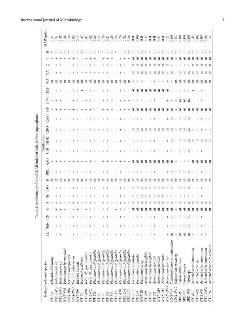
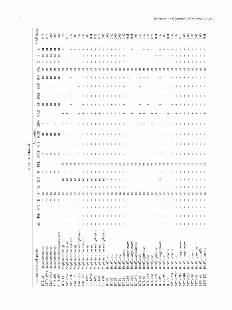
2.1. Bacterial Strains. Sampling was carried out at aquacul-ture farms located at selected districts in Sarawak, MalaysianBorneo, including Kuching, Bintulu, Limbang, Miri, andSampadi (Lundu). Three types of samples were collectedwhich were the sediment, water, and cultured species. Cul-tured species refers to fish or shrimp. InKuching, Bintulu, andMiri, the cultured organisms collected were the fish while inLimbang and Sampadi (Lundu), the cultured organisms col-lected were shrimps. Sampling and sample processing wereperformed according to standard operating protocol by [22].The isolation, designation, and identification of the isolateswere carried out as reported by Kathleen and coworkersearlier [23]. The list of bacteria and their source of origin arelisted in Table 1.
2.2. Antibiotic Susceptibility Tests. Ninety-four bacterial iso-lates from 17 different genera [23] were assessed for theirsusceptibility to different antibiotics utilizing the disk dif-fusion method according to method described by Clinicaland Laboratory Standards Institutes (CLSI) [24] on Mueller-Hinton agar (MHA).The bacterial groups and the antibioticstested are listed in Table 1. Briefly, fresh bacterial culture with0.5 McFarland turbidity was swabbed onto the MHA surfaceusing sterile cotton buds. Commercial antimicrobial discs(Oxoid, UK) were evenly embedded onto the inoculated agarincubated at 37∘C for 18 to 24 hours. Escherichia coli strainfrom American Type Culture Collection (ATCC) 25922 wasused as control.
2.3. Data Collection and Analysis. The diameter of completeinhibition zone formed around the antibiotic discs wasmeasured to the nearest whole millimeter using standardizedruler. The results obtained were analyzed as resistant orsusceptible according to standard interpretative table by CLSI[25] and Bonnet [26]. Multiple antibiotic resistance (MAR)index was then determined for each isolate by dividing thenumber of antibiotics to which an isolate is resistant withthe total number of antibiotics tested [2]. The MAR indexis an indicator to identify the risk contamination that ispotentially hazardous to human. Calculated value of more
than 0.2 indicates that the isolates were isolated from highrisk sources [27].
3. Results
In this study, commonly used antibiotics in both veteri-nary and human medicine were selected for the antibioticsusceptibility testing. The antibiotic selection also dependson the bacterial genera because different bacterial generarequire different classes of antibiotics for optimal antibacte-rial activity. Some of the antibiotics were not tested in thisstudy because some bacterial genera are naturally resistantto certain classes of antibiotics; hence the antibiotics wereexcluded from the analysis. The antibiotic profile of thebacterial isolates from the aquaculture fish and shrimps andtheir environment is shown in Table 1.
The bacterial isolates showing the top five highest per-centages of resistant were towards streptomycin (85%, 𝑛 =20), followed by ampicillin (56.8%, 𝑛 = 74), penicillin (47.1%,𝑛 = 51), erythromycin (43.1%, 𝑛 = 51), and cephalotin(42.3%, 𝑛 = 71). The bacterial isolates showing the top fivehighest percentages of susceptible were towards gentamicin(1.1%, 𝑛 = 90), followed by tobramycin (2.2%, 𝑛 = 90),chloramphenicol (4.0%, 𝑛 = 75), norfloxacin (5%, 𝑛 = 80),and amikacin (5.6%, 𝑛 = 90). The other bacterial isolates andtheir percentage of resistant are shown in Table 1.
In this study, the antibiotic resistant patterns for all iso-lates were also determined tomonitor the spread of antibioticresistance. Sixty-one different antibiotic resistance patternswere observed among the isolates through this study (datanot shown). The resistance patterns were highly variable;20.2% (𝑛 = 94) isolates have no resistance towards anyantibiotics tested, 16% (𝑛 = 94) isolates were resistant to onlyone antibiotic, and 63.8% (𝑛 = 94) isolates were resistantto multiple antibiotics. Chryseobacterium spp. isolated fromwater in Limbang aquaculture farm was resistant towards 12out of 19 antibiotics tested, which was the highest amount ofantibiotic resistance observed in this study.
Most isolates (53.2%, 𝑛 = 94) possess distinctive pattern.However, there are also patterns (11 patterns) shared by 2 ormore bacteria isolates. The pattern shared by most isolates(19 isolates, 𝑛 = 94) is 0% resistance (MAR index equal to0). There is at least an isolate with 0% resistance towards allantibiotics tested in all sampling locations except for Bintulu.There is difference in the resistance pattern for bacteriaisolated from water, sediment, and the cultured organisms.Bacteria that were isolated from the same pond and samesource of origin (sediment, water, or cultured organisms)possess different antibiotic patterns. This may be due to thegenus and species difference of the bacteria isolates. Theantibiotic resistant patterns of the bacterial genera are moreinfluenced by the location from where the isolates wereisolated rather than their genus.
Multiple antibiotic resistant (MAR) index analysis wasintroduced by Krumperman in 1983 [27]. This analysis hasbeen used to group the different sources from where thebacteria were recovered using the frequency of antibioticsresistance [28]. Isolates with MAR < 0.2 were determined
International Journal of Microbiology 3
Table1:Antibiotic
profi
leandMARindexof
isolatesfrom
aquacultu
re.
Isolates
code
andspecies
Antibiotics
MARindex
Ak
Tob
CNK
STe
DO
PPR
LAMP
CIP
NOR
CRO
CAZ
KFIPM
SXT
RDNA
CF
EBT
-F14
Edwa
rdsiella
tarda
−−−−
+−−
nt+
+−−−−
++−
nt−−
+nt
0.32
BTL-2W
3En
terobacter
sp.
−−−
++−−
nt−
+−−−−
++−
nt+−
+nt
0.37
BTL-2W
4En
terobacter
sp.
+−−
++−−
nt−
++−−−
++−
nt+−
+nt
0.47
MYY
-W9
Enterobacte
rhormaechei
−−−
++−−
nt−
+−−−−
+−−
nt−−
+nt
0.26
LBG-2P6
Serratialiquefacie
ns−−−−
+−−
nt+
+−−−−
+−−
nt−−
+nt
0.26
LBG-2W5
Serratiamarcescens
−−−−
++
+nt−
++−−−
++−
nt−−
+nt
0.42
MYY
-F9
Escherich
iacoli
−−−
++−−
nt−
+−−−−−−−
nt−−−
nt0.16
BT-F7
Escherich
iaalbertii
−−−−
++
+nt
++
+−−−
+−
+nt
+−−
nt0.47
BTL-W8
Klebsiella
pneumoniae
−−−
++−−
nt+
++−−−
++−
nt+−
+nt
0.47
BT-2W2
Klebsiella
pneumoniae
−−−−
++
+nt
++
+−−−
+−−
nt−−
+nt
0.42
BT-2S3
Plesiomonas
shige
lloides
−−−−
+−−
nt−−
++−−−−−
nt+−
+nt
0.26
BT-F1
Plesiomonas
shige
lloides
−−−−−−−
nt−−
+−
++−−−
nt+−−
nt0.21
BT-F6
Plesiomonas
shige
lloides
−−−−
+−−
nt−
+−−−−
++−
nt−−
+nt
0.26
BT-F10
Plesiomonas
shige
lloides
−−−−−−−
nt−
++−−−
+−−
nt−−
+nt
0.21
BT-F13
Plesiomonas
shige
lloides
−−−−
+−−
nt−
+−−−−
++−
nt−−
+nt
0.26
BTL-F4
Plesiomonas
shige
lloides
+−−
++−−
nt+
+−−−−−−−
nt−−−
nt0.26
BTL-F10
Plesiomonas
shige
lloides
−−−−−−−
nt+
++−
++−−−
nt−−−
nt0.26
BTL-2F4
Plesiomonas
shige
lloides
−−−−
+−−
nt+
+−−−
++−
nt−−
+nt
0.32
BTL-2F7
Plesiomonas
shige
lloides
−−−−
+−−
nt−
+−−−−
++−
nt−−
+nt
0.26
BT-2F10
Pseudomonas
putid
a−−−
ntnt
ntnt
nt−
nt−−
nt−
nt−
nt+
ntnt
ntnt
0.11
BT-2F5
Pseudomonas
putid
a−−−
ntnt
ntnt
nt−
nt−−
nt−
nt−
nt−
ntnt
ntnt
0.00
MYY
-S5
Pseudomonas
sp.
−−−
ntnt
ntnt
nt−
nt−−
nt−
nt−
nt+
ntnt
ntnt
0.11
BT-2S9
Aeromonas
hydrophila
−−−
ntnt
ntnt
nt+
nt+
nt+
nt−
nt−
ntnt
ntnt
0.33
BT-F12
Aeromonas
hydrophila
−−−
ntnt
ntnt
nt−
nt−
+nt−
nt−
nt−
ntnt
ntnt
0.11
BT-F15
Aeromonas
jand
aei
−−−
ntnt
ntnt
nt−
nt−−
nt+
nt−
nt−
ntnt
ntnt
0.11
MYY
-2F8
Aeromonas
jand
aei
−−−
ntnt
ntnt
nt+
nt+−
nt+
nt−
nt+
ntnt
ntnt
0.44
MYY
-2F4
Aeromonas
punctata
−−−
ntnt
ntnt
nt−
nt−−
nt−
nt−
nt+
ntnt
ntnt
0.11
MYY
-2W3Ae
romonas
jand
aei
−+−
ntnt
ntnt
nt−
nt−−
nt−
nt+
nt−
ntnt
ntnt
0.22
LBG-W
6Stenotrophom
onas
maltophilia
ntnt
ntnt
nt−
ntnt
ntnt−
ntnt
ntnt
+−
+nt
+nt
nt0.60
LBG-2W2
Chryseobacteriu
msp.
++
++
+−−
nt+
+−−
++
++−
nt−−
+nt
0.63
SPD-P7
Vibriorotiferianu
snt
ntnt
ntnt−
ntnt
nt−
ntnt
ntnt
ntnt−
ntnt−
ntnt
0.00
SPD-S6
Vibriofischeri
ntnt
ntnt
nt−
ntnt
nt−
ntnt
ntnt
ntnt−
ntnt−
ntnt
0.00
SPD-W
5Vibriosp.
ntnt
ntnt
nt−
ntnt
nt−
ntnt
ntnt
ntnt−
ntnt−
ntnt
0.00
BT-W
2Ac
inetobacterb
aumannii
+−−
ntnt
+−
nt+
nt−
nt+−
nt−
+nt
ntnt
ntnt
0.45
BT-2W8
Acinetobactersp.
−−−
ntnt−−
nt−
nt−
nt−−
nt−−
ntnt
ntnt
nt0.06
BT-2W9
Acinetobacterb
aumannii
−−−
ntnt−−
nt−
nt−
nt−
+nt−−
ntnt
ntnt
nt0.09
BTL-2W
2Ac
inetobacterb
aumannii
+−−
ntnt−−
nt+
nt−
nt+
+nt−−
ntnt
ntnt
nt0.36
BT-2F9
Acinetobactercalcoaceticu
s−−−
ntnt−−
nt+
nt−
nt+
+nt−−
ntnt
ntnt
nt0.27
4 International Journal of Microbiology
Table1:Con
tinued.
Isolates
code
andspecies
Antibiotics
MARindex
Ak
Tob
CNK
STe
DO
PPR
LAMP
CIP
NOR
CRO
CAZ
KFIPM
SXT
RDNA
CF
EBT
L-2F1
Acinetobactersp.
−−−
ntnt−−
nt+
nt−
nt+−
nt−−
ntnt
ntnt
nt0.18
MYY
-2W4
Acinetobactersp.
−−−
ntnt−−
nt+
nt−
nt+−
nt−−
ntnt
ntnt
nt0.18
LBG-2W3
Acinetobactersp.
−−−
ntnt−−
nt−
nt−
nt−−
nt−−
ntnt
ntnt
nt0.00
SPD-2P1
Acinetobactersp.
−−−
ntnt−−
nt−
nt−
nt+−
nt−−
ntnt
ntnt
nt0.09
SPD-2P8
Acinetobactercalcoaceticu
s−−−
ntnt−−
nt−
nt−
nt−−
nt−−
ntnt
ntnt
nt0.00
BTL-F1
Staphylococcus
sp.
−−−−
nt−
nt+
nt+−−
++−−−−
nt−−
+0.26
MYY
-W6
Staphylococcus
sciuri
−−−−
nt−
nt−
nt+−−
++−−−−
nt−−−
0.16
MYY
-2F2
Staphylococcus
xylosus
−−−−
nt−
nt+
nt+−−−−
++−
+nt−−
+0.32
LBG-P2
Staphylococcus
sp.
−−−−
nt−
nt−
nt−−−−−−−−−
nt−−
+0.05
LBG-2P3
Staphylococcus
saprophyticus−−−−
nt−
nt+
nt+−−
++−−
+−
nt−−
+0.32
LBG-2P7
Staphylococcus
xylosus
−−−−
nt−
nt+
nt+−−
++
+−−−
nt−−
+0.32
SPD-W
7Staphylococcus
sp.
−−−−
nt−
nt−
nt−−−−−−−−−
nt−−
+0.05
SPD-W
3Staphylococcus
sp.
−−−−
nt−
nt+
nt−−−−−−−−−
nt−−
+0.11
SPD-W
1Staphylococcus
saprophyticus−−−−
nt−
nt+
nt+−−
++−−−−
nt−−−
0.21
SPD-P1
Staphylococcus
sp.
−−−−
nt−
nt+
nt−−−
++−−−−
nt−−−
0.16
SPD-S9
Staphylococcus
saprophyticus−−−−
nt−
nt−
nt−−−−−−−−
+nt−−−
0.05
BT-S1
Bacillussp.
−−−−
nt−−−
nt−−−−−−−−−
nt−−−
0.00
BT-S2
Bacillussp.
−−−−
nt+
++
nt+−−−−
+−−
+nt−
++
0.42
BT-S3
Bacillussp.
−−−−
nt−−−
nt−−−−−−−−−
nt−−−
0.00
BT-S4
Bacillussp.
−−−−
nt−−−
nt−−−−−−−−−
nt−−−
0.00
BT-2S1
Bacilluscereus
−−−−
nt−−
+nt
+−−
++
+−
++
nt−
+−
0.42
BT-2S5
Bacillusm
egaterium
−−−−
nt−−−
nt+−−−−−−−
+nt−−−
0.11
BT-2S8
Bacillusm
egaterium
−−−−
nt−−−
nt−−−−
+−−−−
nt−−
+0.11
BT-2S10
Bacillusa
ryabhatta
i−−−−
nt−−−
nt+−−−−−−−−
nt−−−
0.05
BTL-S1
Bacillussp.
−−−−
nt−−
+nt
+−−
++
+−
++
nt−−−
0.37
BTL-S3
Bacillusp
umilu
s−−−−
nt−−
+nt
+−−
++−−−
+nt
+−
+0.37
BTL-2S4
Bacillussp.
−−−−
nt−−
+nt
+−−−−
+−−
+nt−−
+0.26
BTL-2S8
Bacillussp.
−−−−
nt−−
+nt
+−−
++−−−−
nt−−−
0.21
BTL-2S9
Bacillusp
umilu
s−−−−
nt−−−
nt−−−−
+−−−−
nt−
+−
0.11
BT-2W5
Bacillusm
egaterium
−−−−
nt−
++
nt+−−−−
+−−
+nt−
++
0.37
BTL-2W
7Ba
cillussp.
−−−−
nt−−−
nt−−−−−−−−−
nt−−
+0.05
BT-2F1
Bacilluscereus
−−−−
nt−−−
nt−−−−
+−−−−
nt−−
+0.11
MYY
-2S4
Bacilluscereus
−−−−
nt−−
+nt
+−−−−
+−−
+nt−
++
0.32
MYY
-2W7
Bacillussp.
−−−−
nt−−
+nt
+−−
++
+−
++
nt−−−
0.37
MYY
-2F7
Bacillusm
egaterium
−−−−
nt−−
+nt
+−−−−−−−
+nt−−−
0.16
MYY
-S10
Bacillusm
egaterium
−−−−
nt−−−
nt−−−−−−−−
+nt−−−
0.05
MYY
-2S5
Bacillussp.
−−−−
nt−−−
nt−−−−−−−−−
nt−−−
0.00
MYY
-S9
Bacilluscereus
−−−−
nt−−
+nt
+−−
++−−
++
nt−−−
0.32
MYY
-S3
Bacillusp
umilu
s−−−−
nt−−−
nt−−−−
+−−−−
nt−−−
0.05
LBG-S4
Bacillussubtilis
−−−−
nt−−−
nt−−−−−−−−−
nt−−−
0.00
LBG-2S5
Bacillusinfantis
−−−−
nt−−
+nt−−−−
+−−−−
nt−−−
0.11

International Journal of Microbiology 5
Table1:Con
tinued.
Isolates
code
andspecies
Antibiotics
MARindex
Ak
Tob
CNK
STe
DO
PPR
LAMP
CIP
NOR
CRO
CAZ
KFIPM
SXT
RDNA
CF
ELB
G-S7
Bacillusp
umilu
s−−−−
nt−−−
nt−−−−
+−−−−
nt−−−
0.05
LBG-2S10Ba
cillussp.
−−−−
nt−−−
nt−−−−−−−−−
nt−−−
0.00
LBG-P6
Bacillusp
umilu
s−−−−
nt+−
+nt−−−−−
+−−
+nt−
++
0.32
LBG-W
8Ba
cillusv
ietnamensis
−−−−
nt−−−
nt−−−−
+−−−
+nt−
+−
0.16
LBG-2S8
Bacillusjeotgali
−−−−
nt−−−
nt−−−−−−−−−
nt−−−
0.00
SPD-P10
Bacilluscereus
−−−−
nt−−
+nt
+−−
++
+−−
+nt−−−
0.32
SPD-P4
Bacilluscereus
−−−−
nt−−−
nt−−−
++−−−−
nt−−
+0.16
SPD-2S3
Bacilluscereus
−−−−
nt−−−
nt−−−
++−−−−
nt−−
+0.16
SPD-2S4
Bacilluscereus
−−−−
nt+−
+nt
+−−
++
+−
++
nt−
++
0.53
SPD-S8
Bacillussp.
−−−−
nt−−−
nt−−−−−−−−−
nt−−−
0.00
SPD-2W4Ba
cilluscereus
−−−−
nt−−−
nt+−−−
+−−−−
nt+−
+0.21
LBG-2P9
Microbacteriu
msp.
−−−−
nt−−−
nt−
++−
+−−−−
nt−
+−
0.21
SPD-S7
Exiguobacterium
profun
dum−−−−
nt−−−
nt−−−−−−−−−
nt−−−
0.00
BT-2W7
Comam
onas
teste
roni
−−−−
nt−−
+nt
+−−−−
+−−
+nt−
++
0.32
BT-2S6
Exiguobacterium
indicum−−−−
nt−−
+nt
+−−−−
+−−
+nt−
++
0.32
Totalisolatestested
9090
9071
2085
8151
3974
9180
8190
7191
8561
2075
7151
Totalresistantisolates
52
16
177
524
1542
124
2534
3013
825
63
2622
Percentage
ofresis
tant
isolates
(%)
5.6
2.2
1.18.5
85.0
8.2
6.2
47.1
38.5
56.8
13.2
5.0
30.9
37.8
42.3
16.5
9.441.0
30.0
4.0
36.6
43.1
Note:iso
latecode
BT:B
atuTu
juh,BT
L:Bintulu,LB
G:Lim
bang
,SPD
:Sam
padi,M
YY:M
iri,S:sedim
ent,W:w
ater,F:fish,and
P:shrim
p.Symbo
l“+”:resistant,“−
”:susceptib
le,“nt”:n
otteste
d,Ak:am
ikacin,Tob
:tob
ramycin,C
N:gentamicin,K
:kanam
ycin,S:stre
ptom
ycin,Te:tetracyclin
e,DO:doxycyclin
e,P:
penicillinG,P
RL:piperacillin,A
MP:
ampicillin,
CIP:
ciprofl
oxacin,N
OR:
norfloxacin,C
RO:ceft
riaxone,C
AZ:
cefta
zidime,KF
:cephalotin
,IMP:
imipenem
,SXT
:sulfametho
xazole/tr
imetho
prim
,RD:rifampin,
NA:n
alidixicacid,C
:phenicols,
F:nitro
furantoin,
andE:
erythrom
ycin.

6 International Journal of MicrobiologyPe
rcen
tage
of b
acte
ria (%
)
Sampling locations
MAR < 0.2 (%)MAR > 0.2 (%)
100
80
60
40
20
0
BT BTL LBG SPD MYY
73.3
11.7
Figure 1:MAR index analysis based on sampling locations; BT: BatuTujuh (Seven Miles), BTL: Bintulu, LBG: Limbang, SPD: Sampadi,and MYY: Miri.
as isolates recovered from low risk sources of contaminationwhile isolates with MAR > 0.2 were from high risk sources[27]. In this study, the MAR index ranged from 0 to 0.63.Analysis on overall isolates regardless of sampling locationrevealed that 63.1% (𝑛 = 94) isolates belong to group MAR< 0.2 while 36.9% (𝑛 = 94) isolates belong to group MAR >0.2. MAR index was also assessed according to the samplinglocation (Figure 1). Bintulu (BTL) has the highest percentage(73.3%, 𝑛 = 16) of bacteria isolated from high antibiotic-contaminated sources while Sampadi (SPD) has the lowestpercentage (11.7%, 𝑛 = 17).
4. Discussion
Assessment of the antibiotic resistance among aquaculturebacteria against antimicrobial agents is important for updateon the bacterial antibiotic resistance patterns. It is part of asurveillance systemaiming atmonitoring emerging antibioticresistant bacteria and their widespread. Isolation of antibioticresistant bacteria from aquaculture products and aquacultureenvironment indicates the health risk associated with theaquaculture.There had been reports ondetection of antibioticresistance genes in bacteria isolated from aquaculture prod-ucts that can be transferred to humanmicrobiota.Thismatteris becoming critical if the resistance genes are transferrableto human pathogens. Providing effective treatment towardsthis infection becomes a problem to themedical practitionersas the choice of antibiotics is limited. Thus, any sourceof antibiotic resistant bacteria must be carefully monitored[4].
In antibiotic resistance analysis, the history of antibioticapplication in particular area is reflected by the percentageof bacterial resistance to antibiotics [21]. The frequency ofantibiotics usage is related to the level of resistance amongbacteria [2, 29]. In this present study, high percentage ofsusceptibility was observed towards gentamicin, tobramycin,and chloramphenicol. Similarly, Lim and Kasing [21] andHatha et al. [17] in their respective studies observed thatalmost none of the bacteria tested were resistant to gen-tamicin and chloramphenicol. Low frequency of antibioticresistant bacteriamay indicate the less activity associatedwiththe contamination of antibiotics in the area.
The use of chloramphenicol in aquaculture has beenbanned in certain countries including Malaysia, Korea, andJapan since 1983 [30]. This is because of the adverse effect ofchloramphenicol in humans, even at very low dosage, whichcan cause other side effects like severe or fatal blood prob-lems.The problems associatedwith blood are like anemia and“grey syndrome,” a syndrome of cyanosis and cardiovascularcollapse, which occurs particularly in newborn babies. Thisposes risks to the workers handling the products containingthis antibiotic [5, 31]. Banning of antibiotics has aided inreducing the number of antibiotic resistant bacteria in anenvironment.This present study revealed very lowpercentageof resistance towards chloramphenicol (4%). Aarestrup etal. [32] reported the significant reduction in the frequencyof vancomycin-resistant Enterococci from broiler after thebanning of vancomycin in Denmark in 1995. This provesthat government involvement in preventing dissemination ofantibiotic resistant bacteria is important.
High percentage of streptomycin, ampicillin, and peni-cillin G resistance was observed in this study. Similarly, highampicillin and streptomycin resistance were also observedby Zhang et al. [33] in their study on antibiotic resistancedetection in E. coli strains isolated from two differentaquaculture systems in South China. Hatha et al. [17] alsorecorded high resistance of ampicillin. In another study,identified isolates frommangrove soil in Malaysia were 100%resistant towards ampicillin and penicillin while 77.8% of theisolates were resistant towards streptomycin [34]. Son et al.[35] in their study on Aeromonas hydrophila isolated fromTilapia mossambica recorded 100% ampicillin resistance and57% streptomycin resistance. Abdullahi et al. [36] observed100% ampicillin resistance in Pseudomonas spp. isolated fromSarawak aquaculture environment. A study carried out byAkinbowale et al. [15] recorded 54.8% ampicillin resistanceof aquaculture bacteria in Australia. In contrast with theresults in this study, low resistance of streptomycin (21.2%)and ampicillin (6.1%) was observed in a study on 33 marinebacteria isolates by You et al. [37].
In this study, farmers in the aquaculture farms wherethe sampling was carried out stated that there was nohistory of utilization of antibiotics in their farms. Despitethe absence of antibiotics as medicines or in feeds, highresistance was observed to commonly used antibiotics suchas streptomycin, ampicillin, and penicillin. High resistanceof ampicillin and streptomycin in this study and otherresearches was not surprising as these antibiotics were amongthe first antibiotics introduced since the discovery of peni-cillin [38]. Although antibiotic usage in the studied farmshas been stopped decades ago, antibiotic contamination isstill possible as there may still be residues of antibiotics leftin the environment. Bacteria isolated from the sediment ofthe aquaculture pondmay have acquired antibiotic resistancecharacteristics through unconsumed foods and the culturedorganism’s faeces that contain the remaining antibiotics [39–41]. The unconsumed foods and faeces will be deposited inthe sediment and the composition of sediment microbiotawill be altered due to selective pressure [39]. Fish feeds werea possible reservoir for antibiotic resistant genes in the farmsediments [41].

International Journal of Microbiology 7
In this present study, Bintulu (BTL) recorded the highestpercentage (73.3%, 𝑛 = 16) of bacteria isolated from highantibiotic-contaminated sources (MAR > 0.2) while Sampadi(SPD) aquaculture farm recorded the least number (11.7%,𝑛 = 17) of MAR index > 0.2 isolates. The high number ofbacteria with MAR > 0.2 was found in Bintulu. The aqua-culture farm is located at an area that has many agricul-ture activities (e.g., pig farming, duck farming, and dragonfruit cultivation) surrounding it. There is the possibilitythat antibiotics from the animal feeds or medications wereabsorbed into the sediment causing bacterial selection inthe nearby environment.Multiple antibiotic resistant bacteriamight have travelled through water from these agricultureactivities to the aquaculture ponds. Buschmann et al. [42]in their study suggested that antibiotic resistant bacteriain mariculture farm may be transported by water currentwhich flows from surrounding farms that utilize antibioticsexcessively. Antibiotic resistance patternmay vary dependingon the geographical locations and selective pressure [43, 44]and these patterns change rapidly from time to time.
The different patterns exhibited by different strains orspecies suggest how complex is the understanding of theantibiotics resistance in the study area. In this study, theresistance patterns were highly variable; 20.2% (𝑛 = 94)isolates have no resistance towards any antibiotics tested, 16%(𝑛 = 94) isolates were resistant to only one antibiotic, and63.8% (𝑛 = 94) isolates were resistant to multiple antibiotics.
Awareness on antibiotic resistance threat should beinstilled in the community regardless of age as precaution andprevention step against dissemination of antibiotic resistantbacteria. The community must be educated on antibioticsand their effects on public health. Many surveillance pro-grams had also been introduced to monitor the emergenceand spread of antibiotic resistant bacteria. It has also beensuggested by Son et al. [4] that plasmid screening shouldbe considered as an additional procedure in the monitor-ing programs to trace antibiotic resistance dissemination.Alternatives to treatment using antibiotics like probiotics,vaccines, and antimicrobials from plants should be alsoconsidered. However, most of the alternatives could notreally effectively replace antibiotics, so they act as additionalpreventive measures rather than alternatives.
5. Conclusion
The MAR indexing has revealed that 63.1% of the isolatescame from low antibiotic usage area. Although antibioticresistance in aquaculture in the Malaysian Borneo is stillin its infancy, the need for continuous monitoring of theantibiotic resistance patterns should not be overlooked andthe community should be educated on the awareness ofantibiotic resistance and its implication on human health andenvironment.
Competing Interests
The authors declare that they have no competing interests.
Acknowledgments
This research study was supported by research Grant no.FRGS/01(16)/745/2010(31) and Department of MolecularBiology, Faculty of Resource Science and Technology, Uni-versiti Malaysia Sarawak.
References
[1] K. H.Ng, L. Samuel,M.M. Kathleen, S. S. Leong, andC. Felecia,“Distribution and prevalence of chloramphenicol-resistancegene in Escherichia coli isolated from aquaculture and otherenvironment,” International Food Research Journal, vol. 21, no.4, pp. 1321–1325, 2014.
[2] L. Samuel, M. M. Marian, K. Apun, M. B. Lesley, and R. Son,“Characterization of Escherichia coli isolated from cultured cat-fish by antibiotic resistance and RAPD analysis,” InternationalFood Research Journal, vol. 18, no. 3, pp. 971–976, 2011.
[3] M. R. Dorsch, “Rapid detection of bacterial antibiotic resis-tance: preliminary evaluation of PCR assays targeting tetra-cycline resistance genes,” Tech. Rep., Human Protection andPerformance Division, 2007.
[4] R. Son, G. Rusul, and M. I. A. Karim, “Conjugal transferof plasmids and antibiotic resistance among Escherichia coliisolated from animals in a rural area in Sarawak (Malaysia),”Journal of AppliedMicrobiology, vol. 82, no. 2, pp. 240–244, 1997.
[5] M. Shariff, G. Nagaraj, F. H. C. Chua, and Y. G. Wang, “Theuse of chemicals in aquaculture in Malaysia and Singapore,”in Use of Chemicals in Aquaculture in Asia, J. R. Arthur, C.R. Lavilla Pitogo, and R. P. Subasinghe, Eds., pp. 127–140,Southeast Asian Fisheries Development Centre, AquacultureDepartment, Iloilo, Philippines, 2000.
[6] M. J. Pelczar, E. C. S. Chan, N. R. Krieg, and M. F. Pelczar, Eds.,Microbiology, McGraw-Hill, New York, NY, USA, 5th edition,1986.
[7] C. M. Benbrook, “Antibiotic drug use in U.S. aquaculture,”Institute for Agriculture and Trade Policy (IATP) Report, 2002.
[8] H. Supriyadi and A. Rukyani, “The use of chemicals in aquacul-ture in Indonesia,” in Use of Chemicals in Aquaculture in Asia:Proceedings of the Meeting on the Use of Chemicals in Aqua-culture in Asia 20-22 May 1996, Tigbauan, Iloilo, Philippines,J. R. Arthur, C. R. Lavilla-Pitogo, and R. P. Subasinghe, Eds.,pp. 113–118, AquacultureDepartment, Southeast Asian FisheriesDevelopment Center, Iloilo, Philippines, 2000.
[9] M. A. Akond, S. M. R. Hassan, S. Alam, and M. Shirin,“Antibiotic resistance of Escherichia coli isolated from poultryand poultry environment of bangladesh,” American Journal ofEnvironmental Sciences, vol. 5, no. 1, pp. 47–52, 2009.
[10] J. L. Rıos and M. C. Recio, “Medicinal plants and antimicrobialactivity,” Journal of Ethnopharmacology, vol. 100, no. 1-2, pp. 80–84, 2005.
[11] T. Rahman, M. M. R. Akanda, M. M. Rahman, and M. B. R.Chowdhury, “Evaluation of the efficacies of selected antibioticsand medicinal plants on common bacterial fish pathogens,”Journal of the Bangladesh Agricultural University, vol. 7, no. 1,pp. 163–168, 2010.
[12] C. Wong, P. Proksch, L. T. Tan, S. Lihan, A. Mujahid, and M.Muller, “Isolation, identification and screening of antimicrobialproperties of themarine-derived endophytic fungi frommarinebrown seaweed,”Microbiology Indonesia, vol. 9, no. 4, 2015.
[13] S. Lihan, C. S. Lin, I. Ahmad, F. M. Sinang, N. K. Hua, and A.A. Sallehin, “Antimicrobial producing microbes isolated from

8 International Journal of Microbiology
soil samples collected from Nanga Merit Forest in Sarawak,Malaysian Borneo,” European Journal of Experimental Biology,vol. 4, no. 1, pp. 494–501, 2014.
[14] C. J. Rodgers and M. D. Furones, “Antimicrobial agents inaquaculture: practice, needs and issues,” inTheUse of VeterinaryDrugs and Vaccines in Mediterranean Aquaculture, no. 86, pp.41–59, IRTA-Centre d’Aquicultura, Ctra. PobleNou, SantCarlesde la Rapita, Tarragona, Spain, 2009.
[15] O. L. Akinbowale, H. Peng, and M. D. Barton, “Antimicrobialresistance in bacteria isolated from aquaculture sources inAustralia,” Journal of Applied Microbiology, vol. 100, no. 5, pp.1103–1113, 2006.
[16] Y. Zulkifli, N. B.Alitheen,A. R. Raha et al., “Antibiotic resistanceand plasmid profiling of Vibrio parahaemolyticus isolated fromcockles in Padang, Indonesia,” International Food ResearchJournal, vol. 16, no. 1, pp. 53–58, 2009.
[17] M. Hatha, A. A. Vivekanandhan, G. Julie Joice, and Christol,“Antibiotic resistance pattern of motile aeromonads from farmraised fresh water fish,” International Journal of Food Microbiol-ogy, vol. 98, no. 2, pp. 131–134, 2005.
[18] J. Davies, “Inactivation of antibiotics and the dissemination ofresistance genes,” Science, vol. 264, no. 5157, pp. 375–382, 1994.
[19] S. Suzuki, “Tetracycline resistance gene in Asian aquaticenvironments,” in Interdisciplinary Studies on EnvironmentalChemistry—Biological Responses to Contaminants, N. Hama-mura, S. Suzuki, S. Mendo, C. M. Barroso, H. Iwata, and S.Tanabe, Eds., pp. 1–8, Terrapub, Tokyo, Japan, 2010.
[20] S. Graslund, K. Holmstrom, and A. Wahlstrom, “A field surveyof chemicals and biological products used in shrimp farming,”Marine Pollution Bulletin, vol. 46, no. 1, pp. 81–90, 2003.
[21] M. H. Lim and A. Kasing, “Antimicrobial susceptibilities ofVibrio parahaemolyticus isolates from tiger shrimps (Penaeusmonodon) aquaculture in Kuching, Sarawak,” Research Journalof Microbiology, vol. 8, no. 1, pp. 55–62, 2013.
[22] G. Huys, “Sampling and sample processing procedures for theisolation of aquaculture-associated bacteria,” Standard Operat-ing Procedures, 2003.
[23] M. M. Kathleen, L. Samuel, C. Felecia, K. H. Ng, M. B.Lesley, and A. Kasing, “(GTG)
5
-PCR analysis and 16s rRNAsequencing of bacteria from Sarawak aquaculture environ-ment,” International Food Research Journal, vol. 21, no. 3, pp.915–920, 2014.
[24] Clinical and Laboratory Standards Institute, Performance Stan-dards for Antimicrobial Susceptibility Testing: Nineteenth Infor-mation Supplement, CLSI, 2009.
[25] Clinical and Laboratory Standards Institute (CLSI), Perfor-mance Standards for Antimicrobial Susceptibility Testing: Nine-teenth Information Supplement, CLSI, Pennsylvania, Pa, USA,2012.
[26] R. Bonnet, Comite de I’Antibiogramme de la Societe Francaisede Microbiologie, CASFM, 2012.
[27] P. H. Krumperman, “Multiple antibiotic resistance indexing ofEscherichia coli to identify high-risk sources of fecal contami-nation of foods,” Applied and Environmental Microbiology, vol.46, no. 1, pp. 165–170, 1983.
[28] O. Osundiya, R. Oladele, and O. Oduyebo, “Multiple Antibi-otic Resistance (MAR) indices of Pseudomonas and Klebsiellaspecies isolates in Lagos University Teaching Hospital,” AfricanJournal of Clinical and Experimental Microbiology, vol. 14, no. 3,pp. 164–168, 2013.
[29] D. J. Austin, K. G. Kristinsson, and R. M. Anderson, “Therelationship between the volume of antimicrobial consumptionin human communities and the frequency of resistance,” Pro-ceedings of the National Academy of Sciences of the United Statesof America, vol. 96, no. 3, pp. 1152–1156, 1999.
[30] M. H. Yoo, M.-D. Huh, E.-H. Kim, H.-H. Lee, and H. D. Jeong,“Characterization of chloramphenicol acetyltransferase geneby multiplex polymerase chain reaction in multidrug-resistantstrains isolated from aquatic environments,” Aquaculture, vol.217, no. 1–4, pp. 11–21, 2003.
[31] K. Li, L. Liu, C. L. Xu, and X. G. Chu, “Rapid determina-tion of chloramphenicol residues in aquaculture tissues byimmunochromatographic assay,”Analytical Sciences, vol. 23, no.11, pp. 1281–1284, 2007.
[32] F. M. Aarestrup, F. Bager, N. E. Jensen, M. Madsen, A. Meyling,and H. C. Wegener, “Surveillance of antimicrobial resistancein bacteria isolated from food animals to antimicrobial growthpromoters and related therapeutic agents in Denmark,” ActaPathologica, Microbiologica, et Immunologica Scandinavica, vol.106, no. 6, pp. 606–622, 1998.
[33] R.-Q. Zhang, G.-G. Ying, H.-C. Su, L.-J. Zhou, and Y.-S. Liu,“Antibiotic resistance and genetic diversity of Escherichia coliisolates from traditional and integrated aquaculture in SouthChina,” Journal of Environmental Science and Health, Part BPesticides, Food Contaminants, and Agricultural Wastes, vol. 48,no. 11, pp. 999–1013, 2013.
[34] K. C. A. Jalal, U. T. Nur Fatin, M. A. Mardiana et al., “Antibioticresistance microbes in tropical mangrove sediments in eastcoast peninsular, Malaysia,” African Journal of MicrobiologyResearch, vol. 4, no. 8, pp. 640–645, 2010.
[35] R. Son, G. Rusul, A. M. Sahilah, A. Zainuri, A. R. Raha, and I.Salmah, “Antibiotic resistance and plasmid profile ofAeromonashydrophila isolates from cultured fish, Telapia (Telapia mossam-bica),” Letters in Applied Microbiology, vol. 24, no. 6, pp. 479–482, 1997.
[36] R. Abdullahi, S. Lihan, S. B. Carlos, L. M. Bilung, K. M. Mikal,and F. Collick, “Detection of oprL gene and antibiotic resistanceof Pseudomonas aeruginosa from aquaculture environment,”European Journal of Experimental Biology, vol. 3, pp. 148–152,2013.
[37] K. G. You, C.W. Bong, and C.W. Lee, “Antimicrobial resistancein bacteria isolated from tropical coastal waters of peninsularMalaysia,”Malaysia Journal of Science, vol. 31, no. 2, pp. 111–120,2012.
[38] G. J. Tortora, B. R. Funke, and C. L. Case, Microbiology:An Introduction, Pearson Benjamin Cummings by PearsonEducation, Inc, San Francisco, Calif, USA, 9th edition, 2007.
[39] H. T. Tu, F. Silvestre, M.-L. Scippo, J.-P. Thome, N. T. Phuong,and P. Kestemont, “Acetylcholinesterase activity as a biomarkerof exposure to antibiotics and pesticides in the black tigershrimp (Penaeus monodon),” Ecotoxicology and EnvironmentalSafety, vol. 72, no. 5, pp. 1463–1470, 2009.
[40] A. Noorlis, F. M. Ghazali, Y. K. Cheah et al., “Antibioticresistance and biosafety of Vibrio cholerae and Vibrio para-haemolyticus from freshwater fish at retail level,” InternationalFood Research Journal, vol. 18, no. 4, pp. 1523–1530, 2011.
[41] M. Tamminen, A. Karkman, A. Lohmus et al., “Tetracyclineresistance genes persist at aquaculture farms in the absence ofselection pressure,” Environmental Science and Technology, vol.45, no. 2, pp. 386–391, 2011.
International Journal of Microbiology 9
[42] A.H. Buschmann,A. Tomova, A. Lopez et al., “Salmon aquacul-ture and antimicrobial resistance in the marine environment,”PLoS ONE, vol. 7, no. 8, Article ID e42724, 2012.
[43] K. Satyanarayana, “Detection of antimicrobial resistance incommon gramnegative and grampositive bacteria encounteredin infectious disease—an update,” ICMR Research InformationBulletin, vol. 3, pp. 1–3, 2009.
[44] M. B. Lesley, L. Velnetti, Y. K. Cheah et al., “Antibiotic resistanceand plasmid profiling of vibrio parahaemolyticus isolated fromcockles (Anadara granosa) at Tanjung Karang, Kuala Selangor,”International Food Research Journal, vol. 18, no. 3, pp. 1183–1188,2011.

Submit your manuscripts athttp://www.hindawi.com
Hindawi Publishing Corporationhttp://www.hindawi.com Volume 2014
Anatomy Research International
PeptidesInternational Journal of
Hindawi Publishing Corporationhttp://www.hindawi.com Volume 2014
Hindawi Publishing Corporation http://www.hindawi.com
International Journal of
Volume 2014
Zoology
Hindawi Publishing Corporationhttp://www.hindawi.com Volume 2014
Molecular Biology International
GenomicsInternational Journal of
Hindawi Publishing Corporationhttp://www.hindawi.com Volume 2014
The Scientific World JournalHindawi Publishing Corporation http://www.hindawi.com Volume 2014
Hindawi Publishing Corporationhttp://www.hindawi.com Volume 2014
BioinformaticsAdvances in
Marine BiologyJournal of
Hindawi Publishing Corporationhttp://www.hindawi.com Volume 2014
Hindawi Publishing Corporationhttp://www.hindawi.com Volume 2014
Signal TransductionJournal of
Hindawi Publishing Corporationhttp://www.hindawi.com Volume 2014
BioMed Research International
Evolutionary BiologyInternational Journal of
Hindawi Publishing Corporationhttp://www.hindawi.com Volume 2014
Hindawi Publishing Corporationhttp://www.hindawi.com Volume 2014
Biochemistry Research International
ArchaeaHindawi Publishing Corporationhttp://www.hindawi.com Volume 2014
Hindawi Publishing Corporationhttp://www.hindawi.com Volume 2014
Genetics Research International
Hindawi Publishing Corporationhttp://www.hindawi.com Volume 2014
Advances in
Virolog y
Hindawi Publishing Corporationhttp://www.hindawi.com
Nucleic AcidsJournal of
Volume 2014
Stem CellsInternational
Hindawi Publishing Corporationhttp://www.hindawi.com Volume 2014
Hindawi Publishing Corporationhttp://www.hindawi.com Volume 2014
Enzyme Research
Hindawi Publishing Corporationhttp://www.hindawi.com Volume 2014
International Journal of
Microbiology
![White Paper Antibiotic Use and Resistance: Moving Forward ... - Symp... · White Paper: Antibiotic Use & Resistance [3] BACKGROUND The symposium Antibiotic Use and Resistance: Moving](https://cdn.vdocument.in/doc/165x107/5f0aa9c27e708231d42cb9b0/white-paper-antibiotic-use-and-resistance-moving-forward-symp-white.jpg)


