Download - Richard Reynolds Rheumatology Journal Club
Richard Reynolds
Rheumatology Journal Club
Background
• Uric acid is the end product of purine metabolism in humans (and other hominoids).
• Hyperuricemia, UA levels > 6.8mg/dL, can cause gouty arthritis.
• MSU crystals bind nod-like receptors and cause production of proinflammatory IL-1b.
• Hyperuricemia also known to be associated with a number of diseases involving an inflammatory component, e.g., atherosclerosis.
• Previous work by this group demonstrated that soluble uric acid primes PBMCs by stimulating IL-1b, Il-6 production, and lowering production of anti-inflammatory IL-1Ra.
• Here the authors are exploring these results in more detail, trying to ascertain the mechanism by which soluble UA is upregulating cytokine production.
• Appears to involve elements of the autophagy system.
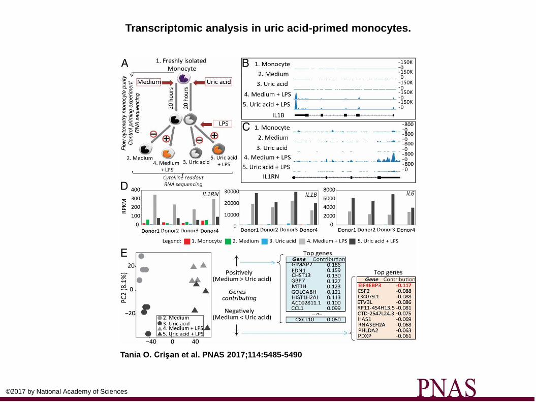
Transcriptomic analysis in uric acid-primed monocytes.
Tania O. Crişan et al. PNAS 2017;114:5485-5490
©2017 by National Academy of Sciences
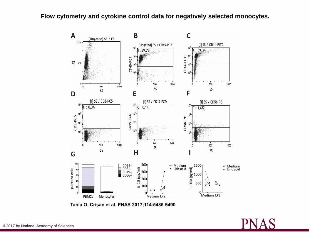
Flow cytometry and cytokine control data for negatively selected monocytes.
Tania O. Crişan et al. PNAS 2017;114:5485-5490
©2017 by National Academy of Sciences
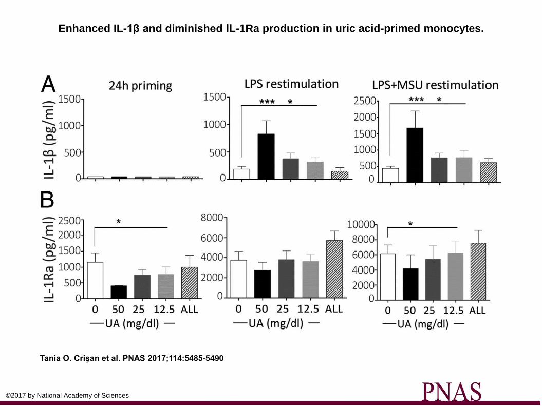
Enhanced IL-1β and diminished IL-1Ra production in uric acid-primed monocytes.
Tania O. Crişan et al. PNAS 2017;114:5485-5490
©2017 by National Academy of Sciences

Transcriptomic analysis in uric acid-primed monocytes.
Tania O. Crişan et al. PNAS 2017;114:5485-5490
©2017 by National Academy of Sciences
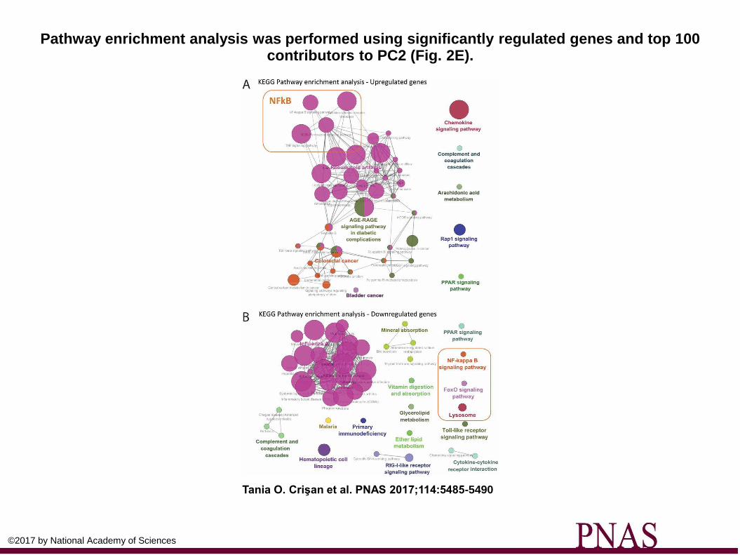
Pathway enrichment analysis was performed using significantly regulated genes and top 100 contributors to PC2 (Fig. 2E).
Tania O. Crişan et al. PNAS 2017;114:5485-5490
©2017 by National Academy of Sciences
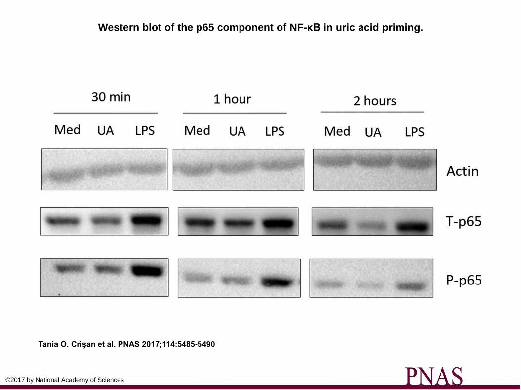
Western blot of the p65 component of NF-κB in uric acid priming.
Tania O. Crişan et al. PNAS 2017;114:5485-5490
©2017 by National Academy of Sciences

Phosphorylation of AKT and lack of ROS or AMPK induction by uric acid.
Tania O. Crişan et al. PNAS 2017;114:5485-5490
©2017 by National Academy of Sciences
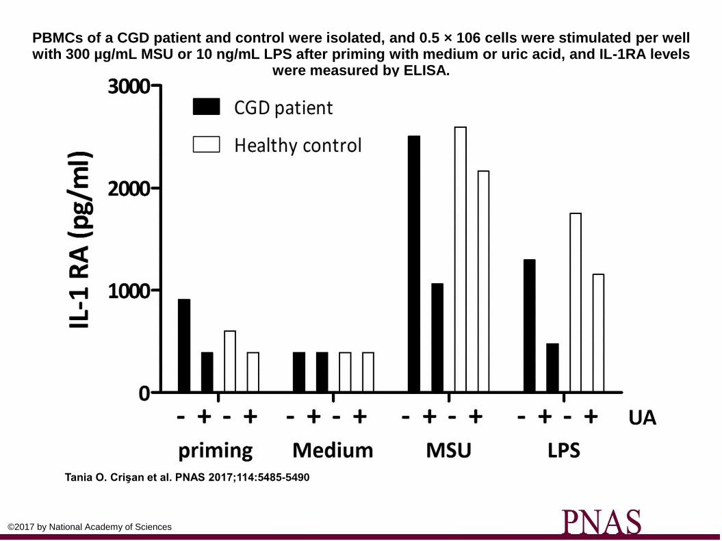
PBMCs of a CGD patient and control were isolated, and 0.5 × 10⁶ cells were stimulated per well with 300 µg/mL MSU or 10 ng/mL LPS after priming with medium or uric acid, and IL-1RA levels
were measured by ELISA.
Tania O. Crişan et al. PNAS 2017;114:5485-5490
©2017 by National Academy of Sciences
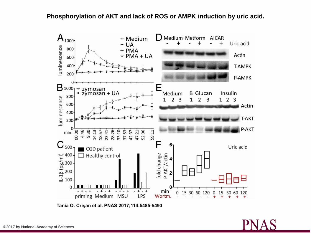
Phosphorylation of AKT and lack of ROS or AMPK induction by uric acid.
Tania O. Crişan et al. PNAS 2017;114:5485-5490
©2017 by National Academy of Sciences
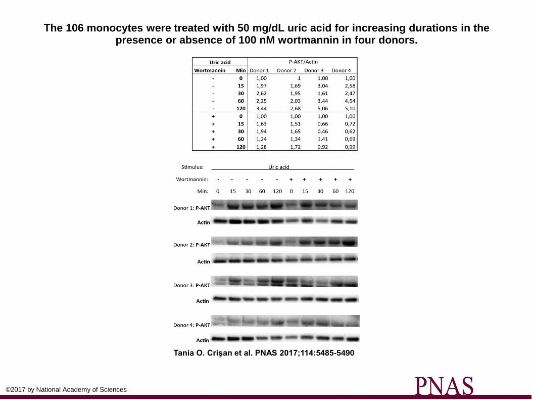
The 106 monocytes were treated with 50 mg/dL uric acid for increasing durations in the presence or absence of 100 nM wortmannin in four donors.
Tania O. Crişan et al. PNAS 2017;114:5485-5490
©2017 by National Academy of Sciences
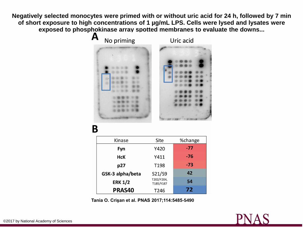
Negatively selected monocytes were primed with or without uric acid for 24 h, followed by 7 min of short exposure to high concentrations of 1 µg/mL LPS. Cells were lysed and lysates were
exposed to phosphokinase array spotted membranes to evaluate the downs...
Tania O. Crişan et al. PNAS 2017;114:5485-5490
©2017 by National Academy of Sciences
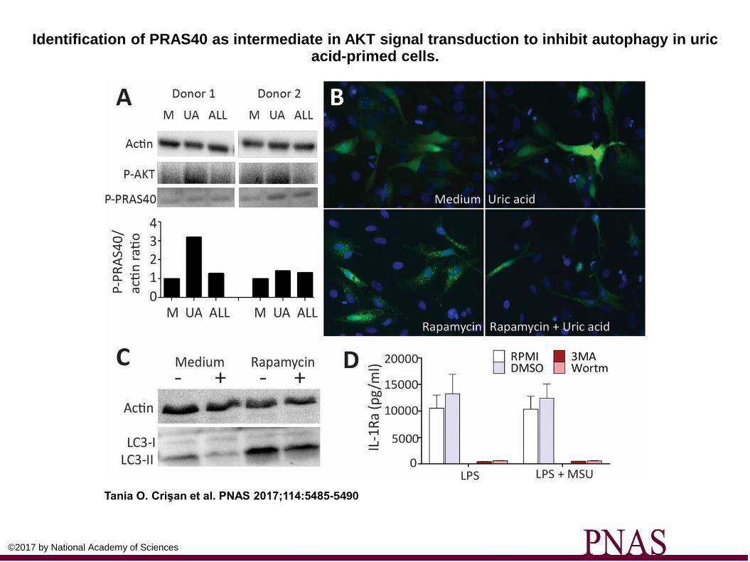
Identification of PRAS40 as intermediate in AKT signal transduction to inhibit autophagy in uric acid-primed cells.
Tania O. Crişan et al. PNAS 2017;114:5485-5490
©2017 by National Academy of Sciences
Take homes
• The authors have suggested a autophagy mechanism for the increased proinflammatory and decreased anti-inflammatory cytokine production which occurs in response to uric acid.
• Apparently the mechanism is via the AKT signaling pathway.
Pathway analysis implicated mTOR signaling (downstream of AKT).
A top hit from the RNA-seq experiment was overexpressed in uric acid treated cells and involved mTOR.
Uric acid exhibited modulatory effects on several components of the AKT pathway, i.e., AKT, PRAS40, autophagosome formation.
• Uric acid stimulated monocytes may inhibit autophagy.
This seems to be manifested by known downstream effects on increased
production of IL-1b, and decreased anti-inflammatory IL1Ra.
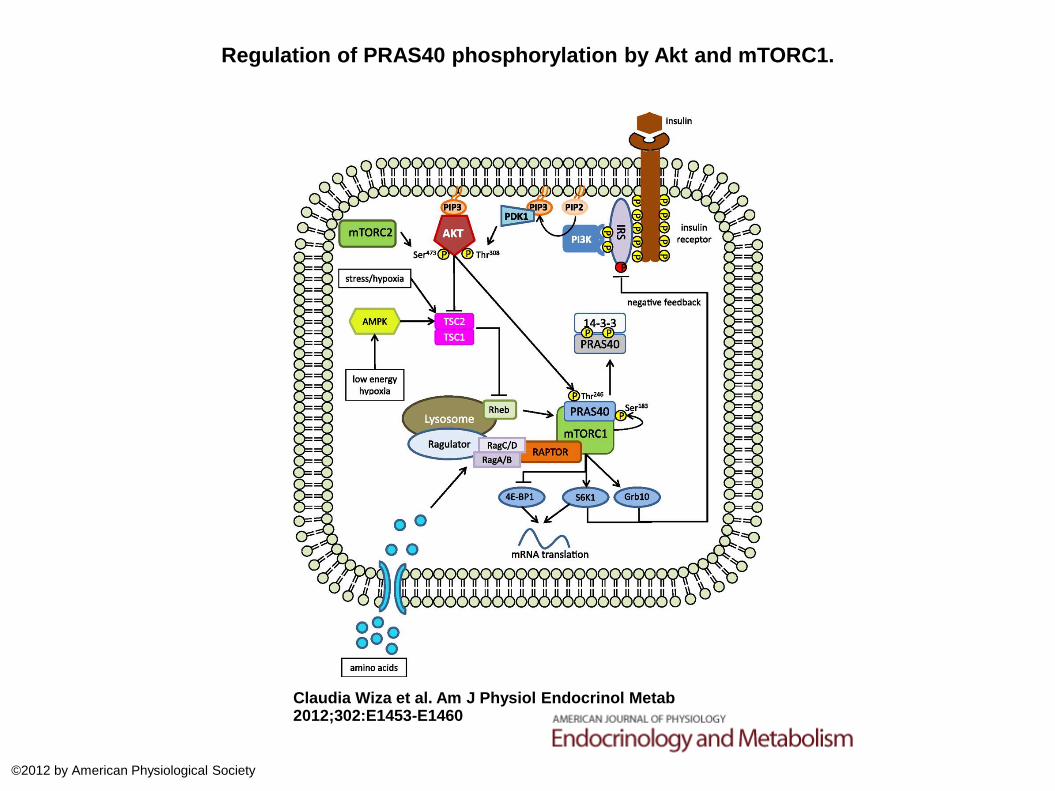
Regulation of PRAS40 phosphorylation by Akt and mTORC1.
Claudia Wiza et al. Am J Physiol Endocrinol Metab 2012;302:E1453-E1460
©2012 by American Physiological Society


