Running head: Mobile gene silencing in Arabidopsis
Name: Dacheng Liang
Address: CSIRO Plant Industry, Canberra, Australian Capital Territory 2601,
Australia
Telephone number: +61 2 6246 5363
e-mail: [email protected]; [email protected];
Plant Physiology Preview. Published on May 11, 2012, as DOI:10.1104/pp.112.197129
Copyright 2012 by the American Society of Plant Biologists
www.plantphysiol.orgon July 7, 2018 - Published by Downloaded from Copyright © 2012 American Society of Plant Biologists. All rights reserved.
2
Gene silencing in Arabidopsis spreads from the root to the
shoot, through a gating barrier, by template-dependent,
non-vascular, cell to cell movement
Dacheng Liang 1*, Rosemary G. White 1*, and Peter M. Waterhouse 1,2* 1 CSIRO Plant Industry, Canberra, Australian Capital Territory 2601, Australia
2 School of Biological Sciences, University of Sydney, Sydney, New South Wales 2006, Australia
www.plantphysiol.orgon July 7, 2018 - Published by Downloaded from Copyright © 2012 American Society of Plant Biologists. All rights reserved.

3
Footnote
Financial source: This work was supported by the Commonwealth Scientific and
Industrial Research Organisation (Federation Fellow grant to P.M.W).
Address correspondence to: [email protected]; [email protected];
www.plantphysiol.orgon July 7, 2018 - Published by Downloaded from Copyright © 2012 American Society of Plant Biologists. All rights reserved.
4
Abstract
Upward long-distance mobile silencing has been shown to be phloem-mediated
in several different Solanaceous species. We show that the Arabidopsis seedling
grafting system and a counterpart inducible system generate upwardly spreading long
distance silencing that travels not in the phloem but by template-dependent reiterated
short-distance cell-to-cell spread through the cells of the central stele. Examining the
movement of the silencing front revealed a largely unrecognized zone of tissue, below
the apical meristem, that is resistant to the silencing signal and may provide a gating
or protective barrier against sRNA signals. Using a range of auxin and actin transport
inhibitors revealed that, in this zone, alteration of vesicular transport together with
cytoskeleton dynamics prevented or retarded the spread of the silencing signal. This
suggests that sRNAs are transported from cell to cell via plasmodesmata rather than
diffusing from their source in the phloem.
Words 142
www.plantphysiol.orgon July 7, 2018 - Published by Downloaded from Copyright © 2012 American Society of Plant Biologists. All rights reserved.
5
INTRODUCTION
The coordination of growth and development in multi-cellular organisms relies
on both local and long-distance communication between cells and tissues. Plants have
specialised vascular tissue, the phloem and xylem, to transport nutrient, hormone and
signaling molecules to mediate long-distance exchange of developmental and defence
information. The phloem transports photoassimilates from source to various sink
tissues, and also transports macromolecules including mRNAs and small RNAs
(Lough and Lucas, 2006; Atkins et al., 2010).
RNA interference (RNAi) is an important viral defence system found in plants,
and in some animals. It is guided by short interfering (si)RNAs and operates by
directed RNA degradation (Cogoni and Macino, 2000; Waterhouse et al., 2001;
Novina and Sharp, 2004). Post-transcriptional gene silencing is also mediated by the
RNAi mechanism and can spread both locally (Himber et al., 2003; Dunoyer et al.,
2005) and long-distance to most parts of the plant (Palauqui et al., 1997; Voinnet and
Baulcombe, 1997; Brosnan et al., 2007).
Long-distance silencing has been extensively studied in Nicotiana species by
grafting experiments using silencing rootstocks, often generating small RNAs from
synthetic hairpin RNA (hpRNA) transgenes, and scions containing target reporter
genes (Mlotshwa et al., 2002; Kalantidis et al., 2008). However, for detailed
molecular analysis of this process, there is neither the detailed genome sequence
information nor the mutant stocks available in Nicotiana that are available in
Arabidopsis thaliana. When a graft-transmissible, rootstock-to-scion, GFP silencing
system was developed in Arabidopsis (Brosnan et al., 2007), this appeared to provide
an excellent way to further study the components and mechanisms of mobile RNAi.
Indeed, results from this system suggested that long-distance silencing required
elements of both transcriptional and post-transcriptional silencing pathways, including
DCL3, RDR2, Pol IV and RDR6, but that production of the signal did not require
DCL2, 3 or 4. More recent work using a similar Arabidopsis grafting system, but
examining shoot-to-root silencing (Dunoyer et al., 2010a; Molnar et al., 2010),
concluded that one or more of the DCLs is needed for signal production. Dunoyer et
al. (2010b) provided evidence that the mobile signal is composed of 21-nt small
RNAs and Molnar et al (2010) presented data showing that the mobile signal directing
epigenetic modification in grafting experiments is made up of 24-nt siRNAs.
www.plantphysiol.orgon July 7, 2018 - Published by Downloaded from Copyright © 2012 American Society of Plant Biologists. All rights reserved.
6
In Nicotiana, the patterns of long-distance systemic silencing, movement of
some viruses, and the spreading of phloem-translocated dye are all quite similar,
suggesting that a silencing signal moves through the phloem transport pathway
(Roberts et al., 1997; Voinnet et al., 1998; Tournier et al., 2006). However, some
virus-host combinations can show a “recovery” symptom (Wingard, 1928; Matthews,
1973), in which the lower leaves of an infected plant display virus symptoms but the
leaves at the top of the plant appear healthy. Intriguingly, leaves at the interface can
have their older, distal portions showing virus symptoms and their younger, proximal
portions appearing healthy (e.g. Fig. 7 in Wingard, 1928). This bi-zonal pattern is
quite different to the vascular pattern that might be expected from an anti-viral signal
transported through phloem.
The Arabidopsis system developed by Brosnan et al. (2007) also gave a
distinctly non-vascular pattern of silencing, including the production of bi-zonal
patterned leaves, in the newly-formed scions of grafted plants. To better understand
how this silencing pattern is produced and to help resolve the apparently conflicting
results about the components required to generate long-distance silencing, we
examined the process in greater detail. To do this we generated a system that relies on
inducible root-specific production of the silencing signal rather than incurring the
restrictions and possible complications of grafting. We refer to this transgenic system
as RtSS (Root-to-Shoot Silencing). Using this system we demonstrate that long-range
root-to-shoot silencing in Arabidopsis spreads largely by a series of cell-to-cell short
range mobile silencing events, rather than by transport through the phloem, and that
the silencing front slows down at the transition zone between hypocotyl and epicotyl.
Experiments using cellular trafficking inhibitors provided evidence suggesting that
cells of this region act as a gating barrier for the transmission of the root-to-shoot
silencing signals in Arabidopsis.
RESULTS
Graft-Transmissible mRNA Silencing in Arabidopsis
Brosnan et al. (2007) used graft-transmissible silencing of GFP in Arabidopsis
to examine the induction, transport and effector components of the silencing process.
Silencing of GFP in the scion was observed only in newly formed leaves and not in
older tissues. From this, it was concluded that long-distance signaling, as distinct from
short-distance cell-to-cell spread of transgene silencing, was causing the effect.
www.plantphysiol.orgon July 7, 2018 - Published by Downloaded from Copyright © 2012 American Society of Plant Biologists. All rights reserved.

7
Nevertheless, the silencing in the newly formed leaves was confined to proximal
tissue that was meristematic at grafting rather than spreading to distal tissues in a
vascular pattern as seen in Nicotiana grafting experiments (Palauqui et al., 1997;
Fusaro et al., 2006). Therefore, we examined the process in Arabidopsis in more
detail. The grafting procedure, 35S-GFP reporter and S1 (GF hairpin only; hpGF) and
S2 (GF hairpin plus 35S-GFP) GFP silencer lines were the same as those used in
Brosnan et al. (2007). The only difference was that in our experiments the grafted
plantlets were maintained in Petri dishes on MS medium rather than being transferred
to soil. From more than 50 grafts using hpGF rootstocks, the scions of nearly 70% of
successful grafts (Fig. 1A, B) showed conspicuous silencing in the basal portion of
some rosette leaves, termed bi-zonal silencing (Fig. 1C). The pattern was very
different from that of GFP silencing in expanding leaves of GFP-expressing N.
benthamiana plants following agroinfiltration in lower leaves with a hpGF construct
(Fig. 1D; Voinnet and Baulcombe, 1997). Of the remaining ~30% of grafts, most
showed no silencing but some displayed a somewhat vascular silencing pattern (Fig.
1E).
From leaf counts on 20 successful grafts showing silencing, the first true leaf to
display silencing ranged from leaf 6 to leaf 10 (Fig. 1G). Examination of scion apices
immediately prior to and 6 days after grafting revealed that the transferred scion had
3-5 leaf primordia which increased to 5-8 leaves, including leaf primordia, six days
later. Arabidopsis Col-0 plants grown in long-days undergo quicker transition to
flowering than plants grown under short-days and this developmental transition has
been shown to coincide with decreased movement of symplastic tracer into the shoot
apical meristem (Gisel et al., 2002). We asked whether this transition would also lead
to changes in RNA-mediated gene silencing movement. We monitored silencing in
grafts grown under either long-day or short-day conditions. All grafts showed similar
rates of silencing, and in this experiment, the 7th or 8th leaf was the first silenced organ
in both conditions (Figure 1H).
Two other features associated with long distance silencing in Nicotiana are the
ability of the silencing signal to self-perpetuate, and that the silencing is lost in the
next generation. To test whether these occur in Arabidopsis, 35S-GFP scions (10 per
time point) were removed from their hpGF rootstocks 3, 5, 7 and 9 days post grafting
(dpg) and maintained on MS+sucrose medium. Two scions from the 7 dpg, and one
from the 9 dpg time point, developed silencing in the newly emerging leaves as the
www.plantphysiol.orgon July 7, 2018 - Published by Downloaded from Copyright © 2012 American Society of Plant Biologists. All rights reserved.
8
excised scions continued to grow on the medium, thus demonstrating that once
initiated in scion tissue, the silencing can self-perpetuate.
Grafted plants showing scion silencing were transferred to soil and allowed to
flower and set seed. Although these plants showed complete GFP silencing
throughout the rosette leaves, stems, flowers and siliques, the newly formed seeds
within the siliques displayed strong GFP expression (Fig 1F). All seedlings
germinated from these seeds had strong, ubiquitous GFP expression. This shows that
the silencing had been lost and was not inherited by the next generation. Hence, apart
from the non-vascular silencing pattern, the characteristics from these grafting
experiments were consistent with the effects from a long-distance silencing signal.
However, they are in contrast to the phloem-mediated source-to-sink transport
through the hypocotyl that would be presumed to mainly flow from the scion to the
rootstock. One possible explanation could be that the silencing signal moved against
this phloem flow.
A GFP Signal Can Move from Sink to Source in Phloem
Phloem, as a component of the vascular system, generally transports
photoassimilates from source cells and tissues to sink cells, which would be from
shoot to root tissues in Arabidopsis seedling hypocotyls. It has been widely reviewed
that proteins, including GFP (Imlau et al., 1999), RNAs, and gene silencing signals,
can move through the phloem (Ghoshroy et al., 1997; Kehr and Buhtz, 2008; Turgeon
and Wolf, 2009). However, when wild type scions were grafted onto rootstocks
expressing GFP controlled by the AtSUC2 promoter, which is active only in the
phloem companion cells (Fig. 2A; Stadler et al., 2005b), free GFP was translocated
across the graft junction in the hypocotyl into scion tissue (Fig. 2B-D). This suggests
that while slow, proteins can move in the phloem against the predominantly source-
to-sink phloem flow. To further investigate the root to shoot signal transport, without
the plant stress and delay due to vascular reconnection of grafting, we developed a
new system.
A New Root-To-Shoot Silencing (RtSS) System
To establish a transgenic system that could mimic grafting experiments, we
combined the dexamethasone- (Dex-) inducible pOp/LhG4-GR system with a tissue-
specific promoter to control expression of hpRNA. Previous work has shown that the
www.plantphysiol.orgon July 7, 2018 - Published by Downloaded from Copyright © 2012 American Society of Plant Biologists. All rights reserved.

9
pOp/LhG4-GR system regulates very stringent transgene expression (Craft et al.,
2005) and that this can be used for inducible RNAi (Wielopolska et al., 2005). We
used the same GFP-expressing transgenic reporter line and S1 GF hpRNA construct
from Brosnan et al. (2007) but replaced the 35S promoter control of the hpRNA with
the pOp/LhG4-GR system and regulated that with the root-specific promoter,
TobRB7 (Yamamoto et al., 1991). With this construct the transcriptional activator
(LhG4-GR) is retained in the cell cytoplasm until Dex is added to displace
cytoplasmic heat shock protein from the GR binding site, allowing it to enter the
nucleus. This activator binds to the 6xOP sequence and induces bidirectional
transcription of GF hpRNA and the GUS gene (Fig. 3A). The hairpin RNA covering
the first 400 bp fragment of the GFP gene is under the control of the pOp promoter,
which is activated by LhG4 entering the nucleus (Fig. 3A). The construct was
transformed into 35S-GFP-expressing plants and selected on hygromycin-containing
medium.
Three independent hygromycin-resistant transformants (T1) showing no GFP
silencing without Dex induction were propagated and their seed (T2) germinated on
hygromycin selection medium. The seed from one line gave the 3:1
(resistance:sensitivity) segregation ratio indicative of a single locus T-DNA insertion.
This transgenic line was used in all subsequent experiments. The GUS expression in
T2 seedlings germinated and maintained on 10 µM Dex-containing MS medium for 7
to 28 days could be seen in the mature root and was undetectable in the stem, leaf
blade, veins, or in the shoot or root apical meristems, which was similar to the
expression pattern described in transgenic tobacco plants containing the TobRB7-
GUS construct (Yamamoto et al., 1991; Fig. 3B). No GUS activity was detected in
any sister seedlings grown on MS medium without Dex (Fig. 3B). Since GUS
expression was induced in roots, the hpGF RNA should also be produced in this tissue,
and silencing observed in the shoot must come from a mobile silencing signal
emanating from the root. In three replicate experiments, 20 RtSS seedlings were
germinated and grown on Dex-containing media for 14 days and the same number
grown on Dex-free media. In all three experiments, all of the Dex-treated seedlings
showed GFP silencing in their newly emerging leaves (Fig. 3C), whereas the
ubiquitous GFP expression in the untreated plants was unaltered.
RtSS Mimics Four Features of Grafted Plants
www.plantphysiol.orgon July 7, 2018 - Published by Downloaded from Copyright © 2012 American Society of Plant Biologists. All rights reserved.

10
In three experiments using a total of 120 RtSS plants treated with Dex at early
stages (1 to 10-day-old plants), over 90% of them showed a bi-zonal silencing pattern
in their rosette leaves (Fig. 3C, D) that was indistinguishable from the patterns
obtained after grafting (Fig. 1C). Similarly, a small proportion of plants displayed a
vascular silencing pattern. A second feature shared by graft-silenced and RtSS-
induced plants is the recovery of GFP expression in the next generation. Of seedlings
germinated from seed set by Dex-treated RtSS plants displaying silencing throughout
most of the rosette leaves and all of the floral bolt, 100% had high levels of GFP
expression. This attribute has been retained in each of the seven generations of this
RtSS line that has been tested.
At the molecular level, the graft-transmissible silencing in Arabidopsis is
similar to that of induced silencing spread in N. benthamiana. When a silencing
construct is targeted against one region of a reporter transgene, siRNAs are generated
outside the targeted region of the gene’s transcript, and most abundantly from the 3’
adjacent region (Vaistij et al., 2002). This process is termed transitivity (Sijen et al.,
2001; Vaistij et al., 2002; Himber et al., 2003). In Arabidopsis grafts using GFP
scions on hpGF rootstocks, siRNAs produced in the scion are overwhelmingly against
the “P” region, which is in the 3’ direction from the “GF” target of the silencing
hairpin (Brosnan et al., 2007). In our RtSS plants after exposure to Dex, both P- and
GF-derived siRNA were detected in the roots, however, only P-derived siRNAs were
detected in the silenced shoots (Fig. 3E), indicating this predominantly unidirectional
transitivity of silencing. Brosnan et al. (2007) also showed that there was no
detectable DNA methylation or histone modification in the promoter or coding
regions of the target gene, implying that scion silencing caused by a mobile silencing
signal occurred at the post-transcriptional level. However, two recent reports showed
that 24-nt siRNAs corresponding to the promoter region can cause graft-transmissible
transcriptional gene silencing (Bai et al., 2011; Melnyk et al., 2011). To check
whether the silencing in the RtSS system was transcriptional or post-transcriptional,
we performed a nuclear run-on assay. In both Dex-induced and control non-induced
RtSS plants, the nascent transcripts from GF and P fragments accumulated to a
comparable level (Fig. 3F), and as expected there was no transcript in the wild type
Columbia Arabidopsis. These results clearly show that silencing induced by mobile
signals operates at the post-transcriptional level, which is consistent with previous
findings (Crete et al., 2001). From this combined evidence, we conclude that our RtSS
www.plantphysiol.orgon July 7, 2018 - Published by Downloaded from Copyright © 2012 American Society of Plant Biologists. All rights reserved.
11
system very closely mimics the effects produced by grafting hpRNA-expressing
rootstocks onto hpRNA-target-expressing scions.
Root Silencing Is Correlated With Production of 21-nt Small RNAs
Since GF-hpRNA was induced exclusively in the roots, we might expect
silencing to occur first in this tissue. When the roots of RtSS plants were exposed to
Dex for 1 or 2 days, the existing root tissue continued to show GFP fluorescence,
although less brightly than in untreated plants (Fig. 4A and B). However, by the third
day of treatment, new lateral roots had emerged that were silenced for GFP expression
(arrow in Fig. 4C). This silencing became more obvious over time (see arrows in Fig.
4 D-G), until after 12 days of treatment, all of the roots were silenced (Fig. 4H). In
small RNA northern blots, the 21-nt GF-siRNA in roots gradually increased over the
first 3 days of Dex treatment (Fig. 4I) as did the secondary siRNA (P region-derived
siRNA), and after 12 days of treatment (Fig. 3E) both GF and P 21nt sRNAs were
readily detectable. This accumulation of siRNAs correlated with the initiation of
silencing.
Developmental Age Affects but Does Not Negate Mobile Silencing
The trauma of cutting and the induction of healing and vascular reconnection
that takes place in establishing grafts may influence or mask the processes being
examined in grafting experiments. In Arabidopsis this is further confounded by the
technical necessity of making grafts using very young seedlings. Our RtSS system
removes this concern and this restriction. In tobacco, the age, and developmental
stage, of the tissues used in grafting experiments has been shown to have a major
impact on the production of long-distance silencing (Crete et al., 2001). Similarly, the
induction of long-distance silencing by agroinfiltration in N. benthamiana only
operates efficiently in juvenile plants. To investigate the effect of timing on the
production of root-to-shoot silencing in Arabidopsis, RtSS plants were treated with
Dex at different ages ranging from 7 to 15 days after germination. This revealed that
Arabidopsis was capable of producing mobile silencing at later developmental stages
(Fig. 5). All of the plants in this experiment became silenced, irrespective of the time
of Dex induction, taking between 12 and 22 days from Dex induction to the first signs
of GFP silencing in the shoot. The last plant of each group showed silencing 5-8 days
after the first plant in every case. However, the spatio-temporal distribution of
www.plantphysiol.orgon July 7, 2018 - Published by Downloaded from Copyright © 2012 American Society of Plant Biologists. All rights reserved.
12
silenced tissue in the plant was strongly affected by the timing of the treatment, with
plants treated at a younger age developing more rapid and widespread silencing (Fig.
6A). Nevertheless, the first marked silencing in leaves of all plants was in the bi-zonal
pattern (Fig. 6A).
Bi-Zonal Distribution of siRNA in the RtSS Leaf
We noticed that silencing could eventually spread through the whole plant
(except for the first 2 or 3 leaves) (Fig. 6A), so the origin of the bi-zonal silencing
pattern in leaves of both Dex-induced RtSS plants and GFP-scions on hpGF
rootstocks was intriguing (Fig. 1C, 3D and 6A, B), especially if the silencing signals
were phloem-mobile. In plants induced with Dex at 5, 10 or 15 days after germination,
the bi-zonal GFP silencing pattern was always observed in the basal petiole and leaf
tissue of the first silenced leaf at 12-22 days after treatment.
This phenomenon prompted us to investigate how the silencing signal itself was
distributed in the shoot. The silenced (red autofluorescence from chlorophyll) and
non-silenced (green fluorescence from GFP) parts were harvested separately and total
RNA from these samples was analysed on an siRNA northern blot. As shown in
Figure 6B, GF-derived siRNAs could not be detected in either the silenced or non-
silenced parts of the leaves, which agrees with our previous experiment analysing
siRNAs in grafts and RtSS plants (Fig. 3E). However, the P-derived secondary
siRNAs could be detected only in the GFP-silenced tissue, not in the GFP expressing
leaf tissue (Fig. 6B). This bi-zonal distribution of P-derived siRNA coincided
precisely with the silencing pattern.
A Slow Front of Cell-to-Cell Silencing Spreads through the Hypocotyl
The time for a molecule transported in the phloem to travel from a source to a
sink tissue within a small, herbaceous plant typically ranges from a few minutes to
several hours (Kiefer and Slusarenko, 2003; Windt et al., 2006). In the Arabidopsis
grafting experiments, it generally took 7 to 10 days for the silencing initiator rootstock
to induce the appearance of silencing in the shoot. This lag could be the time needed
to reconnect phloem sieve elements, re-establish plasmodesmata between cells, load,
move, and unload the signal, then transform the signal through RNAi-associated
cellular machinery into GFP silencing in the shoot tissues. However, with the RtSS
system, which gives transcription and siRNA production within hours of Dex
www.plantphysiol.orgon July 7, 2018 - Published by Downloaded from Copyright © 2012 American Society of Plant Biologists. All rights reserved.

13
application (Fig. 4I and Fig. S1) and has no necessity for re-connection or vascular
repair, it took 12 to 22 days for silencing to appear in the shoot (Fig. 5 and 6A). This
led us to further question the route taken by the signal. If the signal were spread via
the phloem, we would expect cells adjacent to the two phloem strands in the
hypocotyl to be the first to show GFP silencing.
We examined longitudinal sections of RtSS hypocotyls at different times after
Dex treatment and observed the first GFP silencing in a region of cells at the base of
the hypocotyl 4 days after Dex treatment. This region continued to expand shootwards
at a rate of 377 μm/day within the central tissue until it had reached the top of the
hypocotyl (Fig. 7 and Fig. S2). The cortical cells were slower to silence, retaining
GFP fluorescence up to 13-14 d after Dex induction, and showing partial silencing by
16 d (Fig. 7). The epidermal cells, which are symplastically isolated from internal
tissues (Duckett et al., 1994; Stadler et al., 2005a), were not silenced and continued to
fluoresce. During the 14 d period of these experiments the hypocotyl lengths did not
increase (Gendreau et al., 1997).
As the silencing front moved through the hypocotyl-shoot or hypocotyl-epicotyl
junction (HEJ), the rate of movement slowed to 56 μm/day (Fig. 7 and Fig. S2). A
central group of larger cells below the meristem was not silenced within the 16 day
duration of this experiment (13 and 16 days in Fig. 7). Eventually, the silencing
progressed around these central unsilenced cells and was then observed in the shoot
meristem and leaf tissues.
Root-to-Shoot Silencing Is Prevented If Not Bridged by Cells Providing a
Silencing Amplification Template
We reasoned that if the apparent long-distance silencing arises from slow cell-
to-cell spread, it would require an uninterrupted symplastic path of cells expressing
GFP mRNA between the initiating cells in the rootstock and the visible receptor cells
in the shoot. The GFP mRNA transcribed within each cell would provide the
incoming siRNAs with a template to generate more siRNAs that could then invade
adjacent cells and thus produce a reiterated silencing signal. Such a relay
amplification mechanism has been proposed by Himber et al. (2003). To test this
possibility, we needed to interrupt the path with cells producing no GFP mRNA and
observe the effect on the spread of silencing. Grafting a section of wild type
www.plantphysiol.orgon July 7, 2018 - Published by Downloaded from Copyright © 2012 American Society of Plant Biologists. All rights reserved.
14
Arabidopsis stem or hypocotyl between the initiator rootstock and the recipient shoot
would accomplish this but would be technically challenging, so we took another
approach.
We made a Dex-inducible RtSS-hpGF construct controlled by the REA
promoter which gives exclusively root tip expression in Arabidopsis (Fig. 8A, B) and
transformed it into wild type Columbia to generate REA-RtSS-hpGF-expressing
plants. By crossing the REA-RtSS-hpGF line with 35S-GFP plants, we then generated
REA-RtSS-hpGF-35S-GFP plants. The REA-RtSS-hpGF and REA-RtSS-hpGF-35S-
GFP lines, together with the original TobRb7-RtSS line, were then used as rootstocks
for grafting to 35S-GFP scions. In the Dex-induced REA-RtSS-hpGF rootstock there
is no bridge of GFP mRNA expression between the rootstock and the scion and none
of 27 grafts showed any sign of silencing throughout the life of the graft (Fig. 8C).
However, all 30 grafts using the Dex-induced REA-RtSS-hpGF-35S-GFP as rootstock
gave root-to-shoot silencing (Fig. 8D), although silencing took considerably longer to
appear than in 35S-GFP/TobRb7-RtSS grafts (Fig. 8E) and was first detected at the
base of the floral stem or in new branches originating from the rosette. These results
show that the long-distance cell-to-cell silencing movement in Arabidopsis requires
the overlapping expression of the mobile silencing signal and the target mRNA.
A simple explanation for the mobile silencing requiring uninterrupted root-to-
shoot GFP transgene expression could be that GFP mRNAs are needed as a template
from which siRNA-guided RNA dependent RNA polymerase, RDR6 (Dalmay et al.,
2000; Schwach et al., 2005), could generate more dsRNA and hence facilitate the
production of more siRNAs. The newly generated siRNAs would pass to adjacent
cells to continue the process and the long distance spread of the signal would be a
reiterative amplification process. To test this, the RtSS system was transferred into an
rdr6 background. As predicted by the model, induced RtSS in this genotype produced
local silencing in the roots (Fig. S3) but was unable to generate mobile GFP silencing.
A Central, Symplastically Isolated Zone Under the Meristem.
If mobile silencing depends on slow, cell-to-cell spread, we would expect the
silencing front to move at a constant rate from root to shoot, rather than arresting at
the HEJ (Fig. 7). Closer analysis revealed that the central tissue in this zone, below
the shoot meristem, comprised large, loosely packed cells with reduced points of
contact with each other (Fig 9A). At the periphery of the zone, the cells were small
www.plantphysiol.orgon July 7, 2018 - Published by Downloaded from Copyright © 2012 American Society of Plant Biologists. All rights reserved.
15
and densely packed. The route of fewest cells across the HEJ to the meristem would
be through the large central cells (route 1 in Fig 9A). However they, and the
surrounding layer of small cells, remained unsilenced (Fig 7 day 16) suggesting that
the silencing front took a path to the meristem and leaf primordia that
circumnavigated this zone (route 2 in Fig 9A).
To investigate the symplastic connections from the hypocotyl, through this
central zone to the shoot apical meristem, we loaded the symplastic tracer dye, CFDA,
into the roots. The non-fluorescent CFDA enters living cells, and after the acetate
groups are cleaved off by endogenous esterases, the green fluorescent CF is trapped in
the cell cytoplasm and moves cell-to-cell via plasmodesmata. In 7-d old seedlings, CF
in the cytoplasm of hypocotyl cells moved upwards into cells of the central zone and
was rapidly seen in the meristematic cells above (Fig. 9B). However, in 14-d old
seedlings, CF did not enter this tissue, suggesting closed plasmodesmata there (Fig.
9C), although the dye could move in the smaller cells around the central zone. Dex-
induced RtSS plants are 13-15 d old when the silencing signal reaches the HEJ (Fig. 7
day 16, Fig. 9D), at exactly the time when this zone prevents entry of either CF or
silencing signals. Since plasmodesmata appeared to be critical for signal transmission,
we tested a number of inhibitors known to affect cell-to-cell transport.
Auxin Transport Inhibitors, Repress the Spread of Root-to-Shoot Silencing and
Identify a Gating Barrier.
Targeting of plasmodesma protein components (Sagi et al., 2005; Thomas et al.,
2008) and trafficking of some virus movement proteins through plasmodesmata
depends on both a secretory pathway (Haupt et al., 2005; Ju et al., 2005; Genovés et
al., 2010; Schoelz et al., 2011) and a component of cell cytoskeleton-directed
transport (Ding et al., 1996; Harries et al., 2009; Su et al., 2010; Radford and White,
2011; White and Barton, 2011). To test whether these processes were involved in the
spread of RNA silencing, we applied several chemical agents to Dex-induced plants at
the highest concentrations known to be inhibitory without causing morbidity. We first
tested inhibitors of cytoskeleton assembly or function, since the actin-myosin
cytoskeleton is critical for cell-to-cell transport of viruses and other macromolecules
in many cases (reviewed in White and Barton, 2011). However, neither latrunculin B,
an actin polymerisation inhibitor, cytochalasin D, an actin filament disrupter, nor
jasplakinolide, which can stabilise plant actin filaments, prevented the spread of
www.plantphysiol.orgon July 7, 2018 - Published by Downloaded from Copyright © 2012 American Society of Plant Biologists. All rights reserved.

16
silencing in Dex-treated RtSS plants (Fig. 10A). An inhibitor of myosin function, 2,3-
butanedione monoxime (BDM), which binds the myosin head to actin filaments, also
had no significant effect. In contrast, n-ethyl maleimide (NEM), which detaches
myosin from actin, caused earlier silencing such that almost 100% of plants showed
silencing by 15 days in NEM-treated plants compared to less than 40% silencing in
control or BDM-treated plants (Fig. 10A,B).
We then applied 25 µM Brefeldin A (BFA), an inhibitor of vesicle trafficking in
the secretory pathway, to 5-day old Dex-treated RtSS plants in which the silencing
front had just moved through the root-hypocotyl junction (Fig. 7). Although BFA-
treated plants showed phenotypes such as stunting and agravitropism, the spread of
cell-to-cell silencing was only slightly delayed (Fig. 10A), also suggesting that the
silencing signal may not require the secretory pathway.
Both the secretory pathway and the actin cytoskeleton are altered by auxin
transport inhibitors (Pétrašek et al., 2003; Dhonukshe et al., 2008), and one such
inhibitor can partially complement the phenotypic defects caused by mutation in a
component of the endogenous silencing pathway (Lu and Fedoroff, 2000). We,
therefore, speculated that auxin transport, or transporters, may play a role in root to
shoot movement of the silencing signal. Treating Dex-induced RtSS plants with the
auxin transport inhibitors TIBA (50 µM) or PBA (25 µM) prevented the spread of
silencing from the root to the shoot for more than 33 days (Fig. 10C), whereas the
Dex-induced control RtSS plants were fully silenced by day 27 (Fig. 10A,C).
Lowering the concentrations of TIBA and PBA to 15 µM reduced, but did not negate,
this effect (Fig. 10C). The inhibitory effect of TIBA could be alleviated by
transferring treated plants to TIBA-free medium (all of 6 plants transferred eventually
showed shoot silencing). Two other auxin transport inhibitors, NPA and HFCA (Fig.
10C) also greatly retarded the movement of root-to-shoot silencing.
To assess whether TIBA blocked the spread of silencing throughout the shoot or
only affected its movement across the HEJ, we exposed RtSS plants to Dex at
different time points then transferred them to 50 µM TIBA. If the silencing front had
not crossed the HEJ before the addition of TIBA (before day 11) silencing stopped at
this junction (Fig. 10E,F). If the front had passed the HEJ before addition of TIBA, it
continued unabated into the shoot tissues (Fig 10D). These results suggest that the
HEJ is a gating zone for RNA signals in Arabidopsis and that it operates, by an as yet
www.plantphysiol.orgon July 7, 2018 - Published by Downloaded from Copyright © 2012 American Society of Plant Biologists. All rights reserved.

17
unknown mechanism, using auxin transport machinery that facilitates the passage of
sRNAs through plasmodesmata.
DISCUSSION
Transport Route of Systemic Silencing in Arabidopsis
It has been widely accepted that a mobile silencing signal initiated in the
rootstock of grafted tobacco plants or in Agrobacterium-infiltrated N. benthamiana
leaves is transported through the phloem to induce silencing in a vascular-associated
pattern in the leaves of the scion or in newly initiated leaves, respectively (Palauqui et
al., 1997; Roberts et al., 1997; Voinnet and Baulcombe, 1997; Voinnet et al., 1998;
Citovsky and Zambryski, 2000; Tournier et al., 2006; Kehr and Buhtz, 2008).
Recently, Molnar et al. (2010) suggested that GFP silencing in Arabidopsis was more
efficient in a shoot to root direction and that its spread was via the phloem in a source
to sink direction. Silencing and GFP protein can move rapidly from shoot to root
(Ghoshroy et al., 1997; Imlau et al., 1999; Kehr and Buhtz, 2008; Turgeon and Wolf,
2009), but we also observed effective silencing and GFP movement from root to shoot
(Fig. 1A-C and Fig. 2).
In this current study and in previous work (Brosnan et al., 2007) using grafted
Arabidopsis, silencing initiated in the rootstock induced silencing in newly emerging,
but not mature, leaves of the scion. Although this appeared to provide an excellent
system with which to study long distance signal transport and subsequent silencing in
remote tissues it gave a very different silencing pattern in vegetative tissues to that
seen in N. benthamiana. In either grafted or RtSS plants, if the silencing signal from
the hairpin silencer is a small RNA or a protein-RNA complex, it should be generated
within, or have the capacity to move into, root phloem. However, the net photo-
assimilate flow in the Arabidopsis seedling is from shoot to root (especially through
the hypocotyl), and like the GFP from roots (Fig. 2), silencing signal molecules may
move only a short distance upwards through the phloem. We observed that the
silencing moved upwards from cell to cell in the vascular parenchyma and cortical
tissues, to generate the pattern previously interpreted to indicate long distance
phloem-mediated transport.
On this upward journey silencing also spreads throughout young leaf primordia
and may subsequently advance slowly as a front down the length of the leaves (Fig.
11) at a rate of about 5-7 cells per day. If a petiole and leaf are already well developed
www.plantphysiol.orgon July 7, 2018 - Published by Downloaded from Copyright © 2012 American Society of Plant Biologists. All rights reserved.
18
when the silencing reaches its stem/petiole junction, the advancing front has a
considerable distance to travel before appearing in the leaf blade and must traverse
older, less permeable plasmodesmata (Oparka et al., 1999; Burch-Smith et al., 2011).
This pattern is very similar to the upward movement of free GFP from the shoot
apical meristem (Kim et al., 2005b), which also displays a bizonal pattern, giving
green fluorescence in the basal portion of young leaves, but no fluorescence in the
older, apical portion. It thus gives the outward appearance of being unsilenced for a
long time (e.g. the oldest leaves in Fig 1B, C, Fig. 6A). However, when the front
reaches young leaves and leaf primordia it moves more rapidly, being aided by
division and expansion of the petiole and leaf cells (e.g. the bi-zonally silenced leaves
in Fig. 1C and 6A).
When the front reaches the shoot apex, all subsequent tissues produced will be
silenced (apical leaves in Fig. 1B, 6A). The architecture of 1-2 week old Arabidopsis
plants is such that the distance between the apical meristem and the hypocotyl-
epicotyl transition zone is very small. This allows the silencing, travelling cell-to-cell,
to reach the apex and produce silencing throughout newly emerging leaves sometimes
even before the bi-zonal silencing is obvious in the slightly older leaves (Fig. 11). The
timing of silencing induction, the growing conditions for the plants and where the
silencing signals are initiated will have a dramatic effect on the silencing pattern as
they alter the relationship between the position of the silencing front and the
developmental architecture of the plant (Fig. 6A, Fig. 8D and Fig. 11). Such
developmental features include the symplastically isolated outer tissues of the
hypocotyl, and the developmental junction between the hypocotyl and epicotyl.
Because the front of silencing did not enter the epidermal cells of the hypocotyl,
the silencing of its internal tissues could not be detected beneath the strong GFP
expression in the outer cell layers by observation of intact tissues using a fluorescence
dissecting microscope. This slow front of cell-to-cell silencing in the internal tissues
provides a mechanism for long-distance spread with a long time period between
induction at the base of the hypocotyl and distal silencing, and, if the silencing signal
is unable to enter and move through the phloem, provides the explanation for the non-
vascular pattern of distal silencing. It is also consistent with the slightly earlier
appearance of shoot silencing in grafting experiments as the silencing front would be
initiated from the top of the hypocotyl, where the graft junction is usually made.
www.plantphysiol.orgon July 7, 2018 - Published by Downloaded from Copyright © 2012 American Society of Plant Biologists. All rights reserved.
19
Symplastic Domains Restrict Silencing Spread
Cell-to-cell information transfer via plasmodesmata in plants is often confined
to specific tissue domains termed symplastic domains (Ding et al., 2003; Ding and
Itaya, 2007). Within a domain, symplastic signalling molecules appear to move freely,
but at domain boundaries, their symplastic transport is either blocked completely or is
only one-way, either into or out of the domain. The regulated traffic across domain
boundaries is one mechanism to define and coordinate plant development (Ding et al.,
2003; Roberts and Oparka, 2003; Ding and Itaya, 2007). One well-known domain
boundary exists between the epidermal and internal tissues of the Arabidopsis
hypocotyl, such that even a small fluorescent tracer, such as carboxyfluorescein (CF),
is unable to move from the epidermis into the underlying cortical tissues (Duckett et
al., 1994). This boundary can be seen in our RtSS plants, in which the central part of
the hypocotyl is progressively silenced while the epidermis retains GFP fluorescence
(Fig. 7). However, the epidermis is not completely or permanently isolated as the GFP
expression in this tissue is eventually silenced.
Our results reveal the existence of a second symplastic boundary at the HEJ,
where the rate of silencing spread was slowed (Fig. 7). This region is similar to the
symplastic sub-domain at the HEJ described by Kim et al. (2005a,b), and was shown
to prevent shootward spread of silencing signals in Arabidopsis embryos by
Kobayashi and Zambryski (2007). The central cells of this region are large, loosely
packed, and have reduced points of contact with each other (Fig. 9A). At the
periphery of the zone, the cells are small and densely packed. The route of fewest
cells across the HEJ to the meristem is through the large central cells (route 1 in Fig.
9A). However they, and the surrounding layer of small cells, remained unsilenced
(Fig. 7 and Fig. 9D) and the silencing front took a path to the meristem that
circumnavigated the zone (route 2 in Fig. 9A). This deviated route may partially
account for the silencing front’s apparent retardation, although progress was only 15%
of the speed through the hypocotyl. As discussed below, the spread of silencing from
cell to cell requires the expression of the target GFP mRNA to fuel the amplification
of the silencing signal. If the cells of the HEJ were transcriptionally inactive this
could prevent the silencing front from passing through the tissue, however strong GFP
expression was observed in these cells, negating this explanation. Another potential
mechanism preventing the silencing penetrating these cells is that they are isolated by
plasmodesmata closure, since in 14-d-old RtSS plants, the small tracer dye, CF, could
www.plantphysiol.orgon July 7, 2018 - Published by Downloaded from Copyright © 2012 American Society of Plant Biologists. All rights reserved.
20
not enter these cells. Although CF was able to enter this tissue zone in 7-d old plants,
silencing signals were excluded even in young embryos (Kobayashi and Zambryski,
2007), suggesting that the silencing signal is either too large, or lacks the required
signal sequence, to traverse the connecting plasmodesmata. Interestingly, there is an
additional symplastic boundary to dye transport just below the L3 layer of the shoot
apical meristem (SAM), seen especially well in inflorescence meristems (Gisel et al.,
1999), but this boundary appeared to have little effect on silencing spread. The front
of silencing migrated around the HEJ zone then silenced not only the L1-L3 layers of
the SAM but also several additional internal cell layers (Fig. 9A, D). Indeed, even
when the floral bolt stem and inflorescence meristem were clearly silenced (e.g. Fig. 7,
day 16), these HEJ tissues remained unsilenced.
Auxin and other flavonoids are known to accumulate in the upper part of the
Arabidopsis hypocotyl (Murphy et al., 2000; Peer et al., 2001), which may alter
hormone transport or other cell functions. Four inhibitors of auxin transport, TIBA,
PBA, NPA and HFCA, were assayed for their effects on the spread of silencing
through the HEJ, and all of them either arrested or retarded the spread. These
compounds are also described as inhibitors of vesicle transport (Geldner et al., 2001;
Dhonukshe et al., 2008) and it has been recently reported that miRNAs are
transported in mammals in secretory vesicles (Kosaka et al., 2010). One possibility is
that sRNAs may move from cell to cell in plants by regulated vesicular transport,
however, the well-known vesicle transport inhibitor BFA had little effect on silencing
spread. We note that NPA and HFCA were only effective when applied at the highest
concentrations, but TIBA and PBA blocked spread at moderate concentrations.
Dhonukshe et al. (2008) showed that TIBA and PBA affected actin dynamics by
stabilising actin filaments, whereas NPA appeared to have little or no effect on actin
dynamics (Geldner et al., 2001; Petrášek et al., 2003). Furthermore, TIBA and NPA
had opposite effects on the growth of hyl1 mutants, suggesting that their modes of
action are genetically separable (Lu and Fedoroff, 2000). This raises the tantalising
possibility that the combination of a functional actin cytoskeleton and localised
vesicle transport is required for cell-to-cell movement of the silencing signal. Perhaps
TIBA and PBA are the most effective inhibitors of silencing spread through the HEJ
because they act on both actin stabilisation and vesicle motility.
Bi-Zonal Silencing in Grafts and RtSS Reflects Source–Sink Transitions
www.plantphysiol.orgon July 7, 2018 - Published by Downloaded from Copyright © 2012 American Society of Plant Biologists. All rights reserved.
21
A striking feature of both the grafting and RtSS system was the production of
bi-zonal silencing in the first silenced leaves followed by silencing of all subsequent
leaf primordia. We suggest that this indicates a limit to the movement of signal
through older plasmodesmata in the leaf tips (Oparka et al., 1999; Burch-Smith et al.,
2011). Cytoplasmic GFP can move throughout young leaf primordia, but even in very
young leaves, it will move from a site of synthesis within or just below the shoot
apical meristem only into the lower part of the leaf blade (Kim et al., 2005b). This
pattern of GFP movement exactly parallels the pattern of GFP silencing we observed.
As petioles elongate and leaf blades enlarge, cytoplasmic GFP is restricted to the
veins (Kim et al., 2005b), and we very occasionally observed vascular-pattern
silencing, although its absence is further evidence that the silencing signal generally
moved cell-to-cell rather than through the phloem.
This bi-zonal pattern in rosette leaves is also very reminiscent of the pattern
seen in plants showing the “recovery” phenotype observed in some plant virus/host
combinations for close to a century (Wingard, 1928). In such situations the plant
appears ubiquitously infected by the virus but then produces new leaves with a bi-
zonal pattern of virus symptoms only in the apical portion of the leaf, followed by
leaves and tissues that are completely symptomless. A similar “recovery” from
symptoms also commonly occurs when a virus infects a transgenic plant expressing a
transgene derived from a fragment of the virus (Moore et al., 2001).
Our results and the transgene-mediated viral recovery seem closely related. In
the latter case, the transgenic plants express mRNA containing virus-derived
sequences in every cell and, once initiated by virus infection, the signal and silencing
can move around the plant by both cell-to-cell and phloem-mediated transport. We
suggest that once the anti-viral silencing signal reaches the apical meristem, it can
spread cell-to-cell to the limits of plasmodesma permeability. The spread is fuelled by
RDR-mediated secondary siRNA production from the viral transgene. The
meristematic cells are dividing to generate new leaves and because they now contain
siRNAs amplified from transgene mRNA, the new tissue is protected from invasion
by the virus. The same principles can be applied to “natural” virus recovery, but they
require critically balanced conditions. The template for secondary siRNA production
is the viral RNA, so the recovery phenotype is perpetuated by virus replication and
secondary siRNA production achieving a balance in the peripheral meristem cells so
www.plantphysiol.orgon July 7, 2018 - Published by Downloaded from Copyright © 2012 American Society of Plant Biologists. All rights reserved.

22
that new tissue is generated from cells with amplified siRNA levels sufficient to keep
the viral replication at a subliminal level.
Signal Amplification Is Essential for Transmission of Cell-to-Cell Silencing
Previous work analysing graft-transmitted silencing in Nicotiana (Palauqui et al.,
1997) and Arabidopsis (Brosnan et al., 2007) concluded that transmission of the
silencing signal did not require the hairpin RNA and the target mRNA to be expressed
in the same tissue. In Nicotiana, a 30-cm long wild-type intergraft between a silenced
rootstock and target-expressing scion did not interfere with transmission of the signal
(Palauqui et al., 1997). However, we show here that in Arabidopsis, separation of the
REA-RtSS-hpGF and the 35S-GFP target within a single plant prevented silencing in
the target scion (Fig. 8). This raises the question; how is the signal transmitted in
grafted Arabidopsis? We suggest that there is direct exchange of genes and cell
components at the graft junction, as seen in Nicotiana tabacum (Stegemann and Bock,
2009). Tissue from the graft junction between N. tabacum scions expressing nuclear
and cytoplasmic YFP and rootstocks expressing chloroplastic GFP was excised and
cultured on selection medium containing antibiotics that would have eliminated tissue
expressing only a single transgene (Stegemann and Bock, 2009). The surviving callus
tissue contained both cytoplasmic YFP and chloroplastic GFP, indicating exchange of
transgenes at the junction where the two tissues reconnected. A similar exchange, not
only of transgenes, but also of proteins and RNA, may also occur between cells at the
graft junctions between Arabidopsis rootstocks and scions. This would explain some
of the contradictory results on the identity of the silencing signal molecules. For
example, grafting experiments using a rootstock expressing a hpGF RNA, but no GFP
mRNA, in a dcl2,3,4 defective background were able to induce silencing in scions
containing the 35S-GFP transgene (Brosnan et al., 2007). This was interpreted to
mean that the siRNAs made by DCL2, 3 or 4 were unnecessary for silencing and,
therefore, were not the signal. However, by sharing cell contents at the graft junction,
DCLs from the scion cell fusion-partner have access to hpGF-RNA from the rootstock
cell partner enabling the production of siRNA. Furthermore, the siRNAs have access
to GFP mRNA from which they could amplify secondary siRNAs to fuel the cell-to-
cell spread of silencing through the hypocotyl to the apex and then generate the usual
silencing pattern. This is consistent with our demonstration that the RtSS system
cannot function in an rdr6 background. With this scenario, the results do not negate
www.plantphysiol.orgon July 7, 2018 - Published by Downloaded from Copyright © 2012 American Society of Plant Biologists. All rights reserved.
23
the suggestion that siRNAs are a long distance silencing signal (Dunoyer et al., 2010b;
Molnar et al., 2010).
In conclusion, we have shown that the Arabidopsis seedling grafting system
using a GFP reporter scion with a hpRNA silencing-initiating rootstock, and a
counterpart inducible system, generate long distance silencing that operates by
reiterated short-distance cell-to-cell movement. This contrasts with the situation in
Nicotiana species, in which long-distance silencing of transgenes, such as GFP, is
clearly phloem-mediated. Nevertheless, the Arabidopsis systems recapitulate the bi-
zonal leaf pattern seen in viral recovery symptoms and provide a mechanism for the
symptom generation. They also provide a model for the challenges faced by viruses
that infect plants via the roots, such as those vectored by nematodes and soil-borne
fungi. Examining the movement of the silencing front revealed that there is a
previously unrecognized zone of tissue, below the apical meristem, that is resistant to
the silencing signal and may play some part in providing a gating or protective barrier
against signals and/or viruses. Intriguingly, auxin transport inhibitors that also modify
cytoskeleton dynamics prevented the spread of the silencing signal around this zone
suggesting that sRNA transport from cell to cell may be actively gated by
plasmodesmata, rather than spread by unregulated diffusion.
MATERIALS AND METHODS
Plasmid Construct and Arabidopsis Transformation
We used the binary vector pH-top as the backbone for the specific expression of
RNAi (Craft et al., 2005). Briefly, the LHG4 and GR and tml terminator fragments
were amplified from pOp-off2 with primers containing restriction enzyme sites
(LhG4-2F1, aaaggtacccgggaggatccttggagaggacagacgtcgaagatc; LhG4-1F1, cagacgtcg
aagatcatgAAACCGGTAACGTTATACGACGTCGCTGAAT; LhG4R1, aaaagatctag
cttctgaataagccctcgtaatatattttcatgaag; Tml-terF1, AaagtcgacagcggcgcgccatcctgcagGAT
CTTTCCGCATAATTCCC; Tml-terR2, aaaggtacctgccgtacggtccctaggGATCGTGGT
GATATTAAAGAGAGTTA; BamHIGR-partLhF2, aaaggatccATTTCATTTGGAGA
GGACACGCTGACATCCCAATTCCGGG; GR-partLhF1, TGACATCCCAATTCC
GGGCGGAatggctagtgaagctcgaaaaacaaag; GR-partLhR1, CAAGCTCGAGGTCGCG
ACACCGATCAGCAAGCTTTGTTTACCAGCCAGC) and sequentially cloned into
pH-top to form an intermediate vector pGRLOP. A fragment of the first 400 nt of
GFP was amplified with the following primers: attB1-ASC-FhR1, GGGGACAAGTT
www.plantphysiol.orgon July 7, 2018 - Published by Downloaded from Copyright © 2012 American Society of Plant Biologists. All rights reserved.
24
TGTACAAAAAAGCAGGCTGGCGCGCCCCTCCTTGAAGT; attB2-GhF1,
GGGACCACTTTGTACAAGAAAGCTGGGTATGGTGAGCAAGGGCGAGGA.
This fragment was then introduced into pDONR201 using BP recombination,
followed by a LR reaction with pOpoff2 (Wielopolska et al., 2005), then a 1.9 kb AscI
fragment from the plasmid above containing the hairpin GF and a PDK intron was
cloned into the AscI site of pGRLOP to form the pGRLOP-hpGF plasmid. The
TobRB7 fragment was amplified using the following primer: RobTob7-proF1,
5'tgacctaggGTCCTACACAATGTGAATTTG3'; RobTob7-proR1,
5'agtcgtacgTAGTTCTCACTAGAAAAATGC3', then it was cloned into pGRLOP-
hpGF to form the final construct pTob-GRLOP-hpGF.
For the REA-hpGF construct, the REA fragment was amplified using the
following primer: 1rootspF1, 5’aaacctaggTGCAGAGGTAGATATGGGTC3’;
1rootspR1, 5’tttcgtacgACAGGTTATGGAGTTTAGGG3’. The amplified fragment
was inserted into pGRLOP-hpGF with the partial fragment of the Rubisco small
subunit promoter to form pREA-hpGF.
These constructs were then co-transformed with pSoup vector into
Agrobacterium tumefaciens GV3101. The wild-type Columbia Arabidopsis and
previously used 35S-GFP Arabidopsis (Brosnan et al., 2007) plants were transformed
with Agrobacterium containing pTob-GRLOP-hpGF and pREA-hpGF respectively
using the floral dipping method. Transformed plants were selected on medium
containing 15 mg/L Hygromycin and screened by observing GFP fluorescence. Plants
with autonomous silencing were discarded and only plants with GFP fluorescence
maintained through their entire life were selected for the further study. These plants
displayed inducible silencing.
Grafting and locally-induced systemic silencing
To investigate the mobility of GFP from rootstock into scion tissue, we grafted
Arabidopsis C24 wildtype scions onto SUC2-GFP rootstocks (Stadler et al., 2005b).
In all subsequent graft-mediated silencing experiments, plants containing the 35S-
GFP construct (Brosnan et al., 2007) were used as the scion, and RtSS, 35S-hpGF or
the S1 silencer plants described previously (Brosnan et al., 2007) were used as the
rootstock. In all cases, the grafting procedure was as described in detail by Brosnan et
al. (2007). Longitudinally sectioned grafts were examined using a Leica SP2 confocal
laser scanning microscope. Systemic GFP silencing in Nicotiana benthamiana was
www.plantphysiol.orgon July 7, 2018 - Published by Downloaded from Copyright © 2012 American Society of Plant Biologists. All rights reserved.
25
induced by agroinfiltration of the lower leaves of 21 day old GFP-expressing (16c)
plants with Agrobacterium tumefaciens, containing a 35S:GFP construct, essentially
as described by Voinnet et al. (1998).
Dex Induction, GUS Staining and Fluorescence Microscopy
For Dex treatment, seeds were germinated on 10 µM Dex-containing MS
medium, or plants growing on vertical plates were transferred to 10 µM Dex-
containing medium and grown vertically. Dex-treated RtSS plants were either
maintained on agar medium or transferred to soil (where they were drenched with
Dex solution) to observe GFP silencing. For GUS staining, 7-day old plants were
immersed in the GUS staining buffer (50 mM sodium phosphate buffer, pH 7.0, 1
mM EDTA, 0.5 mM potassium ferricyanide, 0.5 mM potassium ferrocyanide, 0.1%
Triton X-100, 1 mM X-gluc) at 37 ℃ overnight, then destained by rinsing in
phosphate buffer then stored in 70% ethanol.
For GFP fluorescence, the plants were screened using a NightSea torch
(BlueStar), and individual plants were examined using a Leica MZFLIII fluorescence
dissecting microscope equipped with an Axiocam digital camera, or photographed
with a Nikon D2 camera using UV illumination with appropriate filters. For more
detailed analysis, longitudinal or transverse sections were examined on a Zeiss
Axioimager fluorescence microscope, or on a Leica SP2 confocal laser scanning
microscope.
RNA Isolation and Northern Blots
Total RNA from shoots or roots was extracted using TRI-reagent (Sigma)
according to the manufacturer’s procedure. About 25 µg total RNA from each sample
was separated on a 17% denaturing polyacrylamide gel, then blotted onto Hybond-N+
membrane using a Bio-rad electroblotting apparatus. The blotted membrane was then
UV cross-linked and baked for 2 hours at 80 ℃ . Hybridization analyses were
essentially performed as described previously (Fusaro et al., 2006).
Nuclear Run-On Assays
DNA probe fragments including the target regions (GF and P regions of the
GFP gene), positive controls (18S, Ubiquitin, Actin), and a negative control (E. coli
www.plantphysiol.orgon July 7, 2018 - Published by Downloaded from Copyright © 2012 American Society of Plant Biologists. All rights reserved.
26
replication protein) were amplified, cleaned and fixed on Hybond-N+ membrane
using a Bio-rad blotter. Nuclear run-on analyses were carried out as described
previously (Meng and Lemaux, 2003).
Tracer analysis
Five-day old and twelve-day old Col0 plants were grown on MS medium then
transferred to fresh agar with the central part of their root system placed on sterile
Parafilm™ until they were 7- and 14-days old, respectively. Before application, a
fresh working solution of 2 µM 5,6-carboxyfluorescein diacetate (CFDA; Sigma) in
distilled water was prepared from a 1 mM stock solution in DMSO. The CFDA
solution was then applied to the part of the root sytem lying on the parafilm in the 7-
and 14-day old plants. The roots were crushed with a pair of forceps to allow CFDA
to enter into the internal tissues. The resulting carboxyfluorescein (CF) fluorescence
was monitored under a fluorescence dissecting microscope until CF was detected in
the shoot. Those plants with CF fluorescence in the shoots were dissected to examine
the fluorescence in the shoot apex and upper part of the hypocotyl under the confocal
microscope.
Inhibitor experiments
Five-day old plants germinated on Dex-containing medium or normal MS
medium were transferred to media containing the inhibitors or equal amounts of
solvents (controls). Additional controls included transfer of Dex-untreated plants to
MS medium. Brefeldin A (BFA, Sigma-Aldrich), 2,3,5-Triiodobenzoic acid (TIBA,
Sigma-Aldrich), N-1-Naphthylphthalamidic acid (NPA, Sigma-Aldrich), 9-Hydroxy-
9-fluorenecarboxylic acid (HFCA, Sigma-Aldrich) were diluted from 100 mM stocks
in DMSO. 2-(1-pyrenoyl) benzoic acid (PBA, OlChemIm Ltd) was dissolved in
DMSO at a stock concentration of 30 mM, 2 µM Cytochalasin D (Sigma-Aldrich, 10
mg/ml stock in DMSO), 1 µM Latrunculin B (Sigma-Aldrich, 2 mM stock in DMSO),
1 µM jasplakinolide (Calbiochem, 1 mM stock in DMSO), 2.5 mM 2,3-butanedione
monoxime (BDM, 500 mM stock solution, freshly dissolved in water), 50 µM N-
ethylmaleimide (NEM, 50 mM stock, freshly dissolved in water) were made to their
final dilutions in MS agar plates. The concentrations used inhibited plant growth and
were at the high end of concentrations applied to Arabidopsis for BFA (e.g Baskin
and Bivens, 1995), TIBA (e.g Dhonukshe et al., 2008), NPA (e.g. Okada et al., 1991),
www.plantphysiol.orgon July 7, 2018 - Published by Downloaded from Copyright © 2012 American Society of Plant Biologists. All rights reserved.
27
HFCA (e.g. Okada et al., 1991), PBA (e.g. Dhonukshe et al., 2008), Cytochalasin D
(e.g. Collings et al., 2006), Latrunculin B (e.g. Collings et al., 2006), jasplakinolide
(e.g. Dhonukshe et al., 2008), BDM (e.g. Baskin and Bivens, 1995; Paves and Truve,
2007), and NEM (e.g. Paves and Truve, 2007).
Acknowledgements
Thanks to Chris Helliwell and Ming-Bo Wang for many discussions, and to Carl
Davies for his help in taking photos. We would also like to thank Adriana Fusaro for
the N. benthiama experiment, and Bethany Clark, Ebony Perkins, Anna Wielopolska
and Judith Gaudron for technical support.
REFERENCES
Atkins CA, Smith PM, Rodriguez-Medina C (2010) Macromolecules in phloem exudates - a review. Protoplasma 248: 165-172
Bai S, Kasai A, Yamada K, Li T, Harada T (2011) A mobile signal transported over a long distance induces systemic transcriptional gene silencing in a grafted partner. J Exp Bot 62: 4561-4570
Baskin TI, Bivens NJ (1995) Stimulation of radial expansion in arabidopsis roots by inhibitors of actomyosin and vesicle secretion but not by various inhibitors of metabolism. Planta 197: 514-521
Brosnan CA, Mitter N, Christie M, Smith NA, Waterhouse PM, Carroll BJ (2007) Nuclear gene silencing directs reception of long-distance mRNA silencing in Arabidopsis. Proc Natl Acad Sci USA 104: 14741-14746
Burch-Smith TM, Stonebloom S, Xu M, Zambryski PC (2011) Plasmodesmata during development: re-examination of the importance of primary, secondary, and branched plasmodesmata structure versus function. Protoplasma 248: 61-74
Citovsky V, Zambryski P (2000) Systemic transport of RNA in plants. Trends Plant Sci 5: 52-54
Cogoni C, Macino G (2000) Post-transcriptional gene silencing across kingdoms. Curr Opin Genet Dev 10: 638-643
Collings DA, Lill AW, Himmelspach R, Wasteneys GO (2006) Hypersensitivity to cytoskeletal antagonists demonstrates microtubule-microfilament cross-talk in the control of root elongation in Arabidopsis thaliana. New Phytol 170: 275-290
Craft J, Samalova M, Baroux C, Townley H, Martinez A, Jepson I, Tsiantis M, Moore I (2005) New pOp/LhG4 vectors for stringent glucocorticoid-dependent transgene expression in Arabidopsis. Plant J 41: 899-918
Crete P, Leuenberger S, Iglesias VA, Suarez V, Schob H, Holtorf H, van Eeden S, Meins F (2001) Graft transmission of induced and spontaneous post-transcriptional silencing of chitinase genes. Plant J 28: 493-501
Dalmay T, Hamilton A, Rudd S, Angell S, Baulcombe DC (2000) An RNA-dependent RNA polymerase gene in Arabidopsis is required for
www.plantphysiol.orgon July 7, 2018 - Published by Downloaded from Copyright © 2012 American Society of Plant Biologists. All rights reserved.
28
posttranscriptional gene silencing mediated by a transgene but not by a virus. Cell 101: 543-553
Dhonukshe P, Grigoriev I, Fischer R, Tominaga M, Robinson DG, Hasek J, Paciorek T, Petrasek J, Seifertova D, Tejos R, Meisel LA, Zazimalova E, Gadella TW, Jr., Stierhof YD, Ueda T, Oiwa K, Akhmanova A, Brock R, Spang A, Friml J (2008) Auxin transport inhibitors impair vesicle motility and actin cytoskeleton dynamics in diverse eukaryotes. Proc Natl Acad Sci USA 105: 4489-4494
Ding B, Itaya A (2007) Control of directional macromolecular trafficking across specific cellular boundaries: a key to integrative plant biology. J Integr Plant Biol 49: 1227-1234
Ding B, Itaya A, Qi Y (2003) Symplasmic protein and RNA traffic: regulatory points and regulatory factors. Curr Opin Plant Biol 6: 596-602
Ding B, Kwon M-O, Warnberg L (1996) Evidence that actin filaments are involved in controlling the permeability of plasmodesmata in tobacco mesophyll. Plant J 10: 157-164
Duckett CM, Oparka KJ, Prior DAM, Dolan L, Roberts K (1994) Dye-coupling in the root epidermis of Arabidopsis is progressively reduced during development. Development 120: 3247-3255
Dunoyer P, Brosnan CA, Schott G, Wang Y, Jay F, Alioua A, Himber C, Voinnet O (2010a) An endogenous, systemic RNAi pathway in plants. EMBO J 29: 1699-1712
Dunoyer P, Himber C, Voinnet O (2005) DICER-LIKE 4 is required for RNA interference and produces the 21-nucleotide small interfering RNA component of the plant cell-to-cell silencing signal. Nat Genet 37: 1356-1360
Dunoyer P, Schott G, Himber C, Meyer D, Takeda A, Carrington JC, Voinnet O (2010b) Small RNA duplexes function as mobile silencing signals between plant cells. Science 328: 912-916
Fusaro AF, Matthew L, Smith NA, Curtin SJ, Dedic-Hagan J, Ellacott GA, Watson JM, Wang MB, Brosnan C, Carroll BJ, Waterhouse PM (2006) RNA interference-inducing hairpin RNAs in plants act through the viral defence pathway. EMBO Rep 7: 1168-1175
Geldner N, Friml J, Stierhof YD, Jurgens G, Palme K (2001) Auxin transport inhibitors block PIN1 cycling and vesicle trafficking. Nature 413: 425-428
Gendreau E, Traas J, Desnos T, Grandjean O, Caboche M, Höfte H (1997) Cellular basis of hypocotyl growth in Arabidopsis thaliana. Plant Physiol 114: 295-305
Genovés A, Navarro JA, Pallás V (2010) The intra- and intercellular movement of Melon necrotic spot virus (MNSV) depends on an active secretory pathway. Mol Plant Microbe Interact 23: 263-272
Ghoshroy S, Lartey R, Sheng J, Citovsky V (1997) Transport of proteins and nucleic acids through plasmodesmata. Annu Rev Plant Physiol Plant Mol Biol 48: 27-50
Gisel A, Barella S, Hempel FD, Zambryski P (1997) Temporal and spatial regulation of symplastic trafficking during development in Arabidopsis thaliana apices. Development 126: 1879-1889
Gisel A, Hempel FD, Barella S, Zambryski P (2002) Leaf-to-shoot apex movement of symplastic tracer is restricted coincident with flowering in Arabidopsis. Proc Natl Acad Sci USA 99: 1713-1717
www.plantphysiol.orgon July 7, 2018 - Published by Downloaded from Copyright © 2012 American Society of Plant Biologists. All rights reserved.

29
Harries PA, Park J-W, Sasaki N, Ballard KD, Maule AJ, Nelson RS (2009) Differing requirements for actin and myosin by plant viruses for sustained intercellular movement. Proc Natl Acad Sci USA 106: 17594-17599
Haupt S, Cowan GH, Ziegler A, Roberts AG, Oparka KJ, Torrance L (2005) Two plant viral movement proteins traffic in the endocytic recycling pathway. Plant Cell 17: 164-181
Himber C, Dunoyer P, Moissiard G, Ritzenthaler C, Voinnet O (2003) Transitivity-dependent and -independent cell-to-cell movement of RNA silencing. EMBO J 22: 4523-4533
Imlau A, Truernit E, Sauer N (1999) Cell-to-cell and long-distance trafficking of the green fluorescent protein in the phloem and symplastic unloading of the protein into sink tissues. Plant Cell 11: 309-322
Ju H-J, Samuels TD, Wang Y-S, Blancaflor E, Payton M, Mitra R, Krishnamurthy K, Nelson RS, Verchot-Lubicz J (2005) The potato virus X TGBp2 movement protein associates with endoplasmic reticulum-derived vesicles during virus infection. Plant Physiol 138: 1877-1895
Kalantidis K, Schumacher HT, Alexiadis T, Helm JM (2008) RNA silencing movement in plants. Biol Cell 100: 13-26
Kehr J, Buhtz A (2008) Long distance transport and movement of RNA through the phloem. J Exp Bot 59: 85-92
Kiefer IW, Slusarenko AJ (2003) The pattern of systemic acquired resistance induction within the Arabidopsis rosette in relation to the pattern of translocation. Plant Physiol 132: 840-847
Kim I, Cho E, Crawford K, Hempel FD, Zambryski PC (2005a) Cell-to-cell movement of GFP during embryogenesis and early seedling development in Arabidopsis. Proc Natl Acad Sci USA 102: 2227-2231
Kim I, Kobayashi K, Cho E, Zambryski PC (2005b) Subdomains for transport via plasmodesmata corresponding to the apical-basal axis are established during Arabidopsis embryogenesis. Proc Natl Acad Sci USA 102: 11945-11950
Kobayashi K, Zambryski P (2007) RNA silencing and its cell-to-cell spread during Arabidopsis embryogenesis. Plant J 50: 597-604
Kosaka N, Iguchi H, Yoshioka Y, Takeshita F, Matsuki Y, Ochiya T (2010) Secretory mechanisms and intercellular transfer of microRNAs in living cells. J Biol Chem 285: 17442-17452
Lough TJ, Lucas WJ (2006) Integrative plant biology: role of phloem long-distance macromolecular trafficking. Annu Rev Plant Biol 57: 203-232
Lu C, Fedoroff N (2000) A mutation in the Arabidopsis HYL1 gene encoding a dsRNA binding protein affects responses to abscisic acid, auxin, and cytokinin. Plant Cell 12: 2351-2366
Matthews REF (1973) Induction of disease by viruses, with special reference to turnip yellow mosaic virus. Annu Rev Phytopathol 11: 147-168
Melnyk CW, Molnar A, Bassett A, Baulcombe DC (2011) Mobile 24 nt small RNAs direct transcriptional gene silencing in the root meristems of Arabidopsis thaliana. Curr Biol 21: 1678-1683
Meng L, Lemaux P (2003) A simple and rapid method for nuclear run-on transcription assays in plants. Plant Mol Biol Rep 21: 65-71
Mlotshwa S, Voinnet O, Mette MF, Matzke M, Vaucheret H, Ding SW, Pruss G, Vance VB (2002) RNA silencing and the mobile silencing signal. Plant Cell 14 Suppl: S289-301
www.plantphysiol.orgon July 7, 2018 - Published by Downloaded from Copyright © 2012 American Society of Plant Biologists. All rights reserved.
30
Molnar A, Melnyk CW, Bassett A, Hardcastle TJ, Dunn R, Baulcombe DC (2010) Small silencing RNAs in plants are mobile and direct epigenetic modification in recipient cells. Science 328: 872 - 875
Moore CJ, Sutherland PW, Forster RL, Gardner RC, MacDiarmid RM (2001) Dark green islands in plant virus infection are the result of posttranscriptional gene silencing. Mol Plant Microbe Interact 14: 939-946
Murphy A, Peer WA, Taiz L (2000) Regulation of auxin transport by aminopeptidases and endogenous flavonoids. Planta 211: 315-324
Novina CD, Sharp PA (2004) The RNAi revolution. Nature 430: 161-164 Okada K, Ueda J, Komaki MK, Bell CJ, Shimura Y (1991) Requirement of the
auxin polar transport system in early stages of Arabidopsis floral bud formation. Plant Cell 3: 677-684
Oparka KJ, Roberts AG, Boevink P, Cruz SS, Roberts I, Pradel KS, Imlau A, Kotlizky G, Sauer N, Epel B (1999) Simple, but not branched, plasmodesmata allow the nonspecific trafficking of proteins in developing tobacco leaves. Cell 97: 743-754
Palauqui JC, Elmayan T, Pollien JM, Vaucheret H (1997) Systemic acquired silencing: transgene-specific post-transcriptional silencing is transmitted by grafting from silenced stocks to non-silenced scions. EMBO J 16: 4738-4745
Paves H, Truve E (2007) Myosin inhibitors block accumulation movement of chloroplasts in Arabidopsis thaliana leaf cells. Protoplasma 230: 165-169
Peer WA, Brown DE, Tague BW, Muday GK, Taiz L, Murphy AS (2001) Flavonoid accumulation patterns of transparent testa mutants of Arabidopsis. Plant Physiol 126: 536-548
Petrášek J, Černá A, Schwarzerová K, Elčkner M, Morris DA, Zažı�malová E (2003) Do phytotropins inhibit auxin efflux by impairing vesicle traffic? Plant Physiol 131: 254-263
Radford J, White R (2011) Inhibitors of myosin, but not actin, alter transport through Tradescantia plasmodesmata. Protoplasma 248: 205-216
Roberts AG, Cruz SS, Roberts IM, Prior D, Turgeon R, Oparka KJ (1997) Phloem unloading in sink leaves of Nicotiana benthamiana: comparison of a fluorescent solute with a fluorescent virus. Plant Cell 9: 1381-1396
Roberts AG, Oparka KJ (2003) Plasmodesmata and the control of symplastic transport. Plant Cell Environ 26: 103-124
Sagi G, Katz A, Guenoune-Gelbart D, Epel BL (2005) Class 1 reversibly glycosylated polypeptides are plasmodesmal-associated proteins delivered to plasmodesmata via the Golgi apparatus. Plant Cell 17: 1788-1800
Schoelz JE, Harries PA, Nelson RS (2011) Intracellular transport of plant viruses: finding the door out of the cell. Mol Plant 4: 813-831
Schwach F, Vaistij FE, Jones L, Baulcombe DC (2005) An RNA-dependent RNA polymerase prevents meristem invasion by potato virus X and is required for the activity but not the production of a systemic silencing signal. Plant Physiol 138: 1842-1852
Sijen T, Fleenor J, Simmer F, Thijssen KL, Parrish S, Timmons L, Plasterk RH, Fire A (2001) On the role of RNA amplification in dsRNA-triggered gene silencing. Cell 107: 465-476
Stadler R, Lauterbach C, Sauer N (2005a) Cell-to-cell movement of green fluorescent protein reveals post-phloem transport in the outer integument and identifies symplastic domains in Arabidopsis seeds and embryos. Plant Physiol 139: 701-712
www.plantphysiol.orgon July 7, 2018 - Published by Downloaded from Copyright © 2012 American Society of Plant Biologists. All rights reserved.

31
Stadler R, Wright KM, Lauterbach C, Amon G, Gahrtz M, Feuerstein A, Oparka KJ, Sauer N (2005b) Expression of GFP-fusions in Arabidopsis companion cells reveals non-specific protein trafficking into sieve elements and identifies a novel post-phloem domain in roots. Plant J 41: 319-331
Stegemann S, Bock R (2009) Exchange of genetic material between cells in plant tissue grafts. Science 324: 649-651
Su S, Liu Z, Chen C, Zhang Y, Wang X, Zhu L, Miao L, Wang X-C, Yuan M (2010) Cucumber mosaic virus movement protein severs actin filaments to increase the plasmodesmal size exclusion limit in tobacco. Plant Cell 22: 1373-1387
Thomas CL, Bayer EM, Ritzenthaler C, Fernandez-Calvino L, Maule AJ (2008) Specific targeting of a plasmodesmal protein affecting cell-to-cell communication. PLoS Biol 6(1) e7. doi:10.1371/journal.pbio.0060007
Tournier B, Tabler M, Kalantidis K (2006) Phloem flow strongly influences the systemic spread of silencing in GFP Nicotiana benthamiana plants. Plant J 47: 383-394
Turgeon R, Wolf S (2009) Phloem transport: cellular pathways and molecular trafficking. Annu Rev Plant Biol 60: 207-221
Vaistij FE, Jones L, Baulcombe DC (2002) Spreading of RNA targeting and DNA methylation in RNA silencing requires transcription of the target gene and a putative RNA-dependent RNA polymerase. Plant Cell 14: 857-867
Voinnet O, Baulcombe DC (1997) Systemic signalling in gene silencing. Nature 389: 553
Voinnet O, Vain P, Angell S, Baulcombe DC (1998) Systemic spread of sequence-specific transgene RNA degradation in plants is initiated by localized introduction of ectopic promoterless DNA. Cell 95: 177-187
Waterhouse PM, Wang M-B, Lough T (2001) Gene silencing as an adaptive defence against viruses. Nature 411: 834-842
White RG, Barton DA (2011) The cytoskeleton in plasmodesmata: a role in intercellular transport? J Exp Bot 62: 5249-5266
Wielopolska A, Townley H, Moore I, Waterhouse P, Helliwell C (2005) A high-throughput inducible RNAi vector for plants. Plant Biotechnol J 3: 583-590
Windt CW, Vergeldt FJ, de Jager PA, van As H (2006) MRI of long-distance water transport: a comparison of the phloem and xylem flow characteristics and dynamics in poplar, castor bean, tomato and tobacco. Plant Cell Environ 29: 1715-1729
Wingard SA (1928) Hosts and symptoms of ring spot, a virus disease of plants. J Agric Res 37: 127-153
Yamamoto YT, Taylor CG, Acedo GN, Cheng CL, Conkling MA (1991) Characterization of cis-acting sequences regulating root-specific gene expression in tobacco. Plant Cell 3: 371-382
FIGURE LEGENDS
Figure 1. Graft-transmissible gene silencing in Arabidopsis. A, A graft between a
35S-GFP scion and 35S-hpGF rootstock 3 d after grafting. B, Graft as in A showing
initial silencing 10 d after grafting. C, Bi-zonal silencing (arrow) in scion leaves 22 d
www.plantphysiol.orgon July 7, 2018 - Published by Downloaded from Copyright © 2012 American Society of Plant Biologists. All rights reserved.

32
after grafting. D, Vascular silencing pattern in GFP-expresssing N. benthamiana
plants infiltrated with a hpGF construct in lower leaves. E, Some Arabidopsis grafts
show initial vascular silencing followed by bi-zonal leaf silencing (arrow). F, GFP
expression is recovered in the seeds of fully silenced scions (left), on the right is a WT
Col-0 silique. G, In 20 grafts of 35S-GFP on hpGF rootstock, the majority showed
initial silencing on the 7th or 8th leaf. H, In both short-day (SD) and long-day (LD)
conditions, initial silencing was seen in the 7th leaf.
Figure 2. Grafts between Arabidopsis C24 plants with either scion or rootstock
expressing SUC2-GFP, producing fluorescence in the phloem companion cells. A,
Graft between SUC2-GFP rootstock with C24 scion (dotted outline) after 3 d showing
GFP in the two phloem strands of the rootstock hypocotyl, but no GFP in the scion
above the graft junction (dashed red line). B, After 18 d, GFP can be seen in phloem
below the graft junction but not above the graft at low magnification. Xylem (X) has
proliferated in the centre of the hypocotyl. At higher magnification, GFP can be
detected in the phloem (P) of the scion (C) and is concentrated in the phloem
companion cells (CC) (D). Sieve elements (SE) show blue fluorescence from aniline-
blue stained callose, red = chlorophyll autofluorescence.
Figure 3. Phenotypic and molecular characterisation of the root to shoot silencing
system (RtSS) Arabidopsis lines. A, Physical map of the inducible vector TobRb7-
hpGF. In this vector, a root specific promoter from tobacco, TobRb7, controls the
expression of GR-LHG4 which in turn activates the expression of a silencing hairpin,
hpGF, and a GUS reporter when Dexamethasone (Dex) is present. B, Induced GUS
expression in TobRB7-GRLHG4/Op-hpGF (RtSS) plants. There is no GUS
expression in RtSS plants without Dex (-Dex); after Dex treatment, RtSS plants show
GUS expression only in roots (+Dex). Enlarged views of shoot (black rectangle) and
root-hypocotyl junction (white rectangle) show GUS expression confined below the
junction. C, RtSS plants after 16 d treatment with Dex showing GFP silencing in the
shoots (+Dex), but no silencing without Dex (-Dex). Arrows indicate silencing in
young leaves. D, 22-d-old RtSS plants showing bi-zonal leaf silencing after Dex
induction. E, Northern blot analysis of siRNA production in the RtSS plants with and
without Dex induction. #4 and #9 are two separate RtSS lines. R: root. S: shoot. F,
Nuclear run-on assay showing that shoot silencing is post-transcriptional. I: 41 day-
www.plantphysiol.orgon July 7, 2018 - Published by Downloaded from Copyright © 2012 American Society of Plant Biologists. All rights reserved.

33
old non-induced RtSS plant. II: Completely silenced RtSS plant after 31 days of Dex-
treatment. III: Wild type Columbia Arabidopsis.; Uq: Ubiquitin; Act: Actin; Rep: E.
coli replication protein.
Figure 4. Inducible GFP silencing in the roots of Dex-induced RtSS plants and time
course of production of 21 nt siRNAs from the hpGF. A, A plant without Dex. B, 2
day after Dex treatment. C, Initial silencing in the root can be seen 3 d after treatment.
D to G, The silencing is becomes more obvious with time. H, Roots are completely
silenced by 12 d after Dex treatment. I, small RNA northern blot analysis of roots
treated with Dex. Wt: wild type Columbia Arabidopsis. Roots of T1, T2 and T3 plants
on MS medium without Dex (indicated by 0) were used as controls. Roots from the
T3 generation treated with Dex showed increasing expression of 21nt siRNAs from
the GF and P, with a peak 3 d after, and maintained at 12 d after induction. White
arrows indicate the silenced roots.
Figure 5. RtSS plants were treated with Dex at different ages (days after germination;
DAG) and the percentage showing silencing was recorded for the following 9 to 21
days.
Figure 6. Spread of silencing in RtSS shoots when induced at different times after
germination and siRNA distribution in the silenced and un-silenced leaves. A, Plants
were treated with Dex at 5, 10 or 15 days after germination. In 5-d old plants,
silencing appeared first in the petioles of the 5th, 6th and 7th leaves after 20 d with bi-
zonal silencing in the 8th leaf. All lateral organs formed after the 8th leaf, and the
floral bolt stem, were silenced. In 15-d old plants treated with Dex, initial silencing
was seen in rosette leaf petioles at 30 d, and the inflorescence meristem, floral bolt
stem and flowers escaped silencing. By 55 d, silencing had progressed through the
bolt stem and flowers but was less complete than in plants treated earlier with Dex.
Numbers 1 to 8 denote order of leaf appearance. B, Small RNA detected in the
silenced (red) and non-silenced (green) segments of leaves from two different RtSS
lines (#4 and #9), 45 days after 3-d Dex induction of 10-d old plants.
Figure 7. Silencing progression in the hypocotyl of RtSS plants treated with Dex
when 5 d old (time 0). The long arrow indicates the root-hypocotyl junction where
www.plantphysiol.orgon July 7, 2018 - Published by Downloaded from Copyright © 2012 American Society of Plant Biologists. All rights reserved.
34
expression of both GUS and the inducible silencer ends. In median longitudinal
sections (upper panels, day 0-16) GFP silencing was detected 4 d after Dex treatment
and gradually migrated upwards from the base of hypocotyl (lower horizontal line),
with the silencing front (upper horizontal line) reaching the top of the hypocotyl 4-5 d
later, at 8-9 d after Dex induction. By day 12-13, silencing had crossed the hypocotyl-
epicotyl junction (HEJ) (dotted line), then continued into higher shoot tissues.
However, even 16 d after Dex treatment when most shoot tissues were silenced, an
isolated internal domain of tissue remained unsilenced (outlined), as did hypocotyl
epidermal cells. AM: axillary meristem; C: cortex; stem: floral bolt stem; V: vascular
tissue; X: xylem; asterisk: base of cotyledon petiole. Cross-sections of hypocotyls
(lower 4 panels, day 0-16) show the central xylem strand (X, slightly less bright
central zone at day 0) within the vascular cylinder (V), surrounded by two layers of
cortical cells (C) and the outer layer of epidermal cells (E). The vascular cylinder
shows strong GFP fluorescence at day 0 (all images at the same exposure), but is
completely silenced above the root-hypocotyl junction by day 12, while the cortex
and epidermis still show GFP fluorescence. By day 16, the inner cortex shows some
silencing, and free GFP can be seen in the two phloem strands (P). In RtSS
hypocotyls without Dex (16 -Dex), the central tissue retains bright GFP fluorescence.
Figure 8. Silencing was not transmitted through Arabidopsis tissue lacking the target
gene. A, GUS expression in REA-RtSS-hpGF and GFP expression in REA-RtSS-
hpGF-35S-GFP plant before Dex treatment. B, GUS expression in REA-RtSS-hpGF
and GFP expression in REA-RtSS-hpGF-35S-GFP plant after Dex treatment. C, No
GFP silencing was seen in the grafts of 35S-GFP scions on REA-hpGF rootstocks
throughout the life of the plants. D, GFP silencing was seen first at the base of 35S-
GFP scions grafted onto REA-RtSS-hpGF-35S-GFP rootstocks treated with Dex. The
white rectangle indicates the enlarged base of the scion. E, Systemic silencing was
observed in 35S-GFP scions grafted onto Dex-induced RtSS rootstocks.
Figure 9. Silencing route at the hypocotyl-epicotyl junction (HEJ) in Arabidopsis and
CF loading experiments. A, In route 1 indicated by red arrows, the silencing moves
through small tightly packed cells and then a zone with large loosly packed cells. In
route 2 (yellow arrows), silencing moves around this zone to the meristem. The HEJ
zone is enclosed by a dashed line. B, CFDA was loaded into the root of a 7-day-old
www.plantphysiol.orgon July 7, 2018 - Published by Downloaded from Copyright © 2012 American Society of Plant Biologists. All rights reserved.

35
WT Col0 plant and green CF fluorescence could be detected in the HEJ zone (dashed
outline). C, In a 14-day-old WT Col0 plant, the CF fluorescence was mainly detected
around the edge of the HEJ. D, The internal HEJ tissue remained unsilenced in 16-
day-old RtSS plants grown on medium containing Dex.
Figure 10. Effects of cytoskeleton and hormone inhibitors on root to shoot silencing.
A, Percentage of systemic silencing 22 days after treatment with different inhibitors
(27 days after Dex induction). B, NEM can slightly accelerate the rate of systemic
silencing. C, silencing on the shoot can be fully stopped by 50 µM TIBA and 25 µM
PBA, and partially inhibited by the lower concentration of 15 µM TIBA or 15 µM
PBA. 50 µM or 200 µM NPA also slowed shoot silencing. 50 or 100 µM HFCA
severely slowed shoot silencing. D, RtSS plants that were treated with Dex for longer
times, such as 11 days after Dex (DAD) still developed systemic silencing when
treated with 50 µM TIBA. E,F, Confocal images of longitudinal sections. E, A 32-
day-old plant treated with 100 µM HFCA did not yet show shoot silencing, but
showed silencing in the meristematic HEJ zones. F, A 38-day-old RtSS plant treated
with DEX for 5 days, then with TIBA, showed silencing excluded from the HEJ.
Arrowheads indicate cut cotyledon petioles. The circle denotes the HEJ zone.
Experiments were repeated 2-3 times, with at least 20 plants per treatment. BFA:
Brefeldin A. TIBA, 2,3,5-Triiodobenzoic acid. PBA, 2-(1-pyrenoyl)benzoic acid.
NPA, N-1-Naphthylphthalamic acid. HFCA, 9-Hydroxy-9-fluorenecarboxylic acid.
CytD: Cytochalasin D. LatB: Latrunculin B. BDM: 2,3-butanedione monoxime. NEM:
N-ethylmaleimide.
Figure 11. A simplified model of root to shoot silencing. A, A silencing front moves
through the hypocotyl cell-to-cell involving a signal amplifying mechanism from root
to shoot. The red colour denotes the silencing front. The green ellipse denotes a tissue
domain within the cotyledon node zone. B, When the silencing front reaches the
hypocotyl-epicotyl junction just below this tissue domain, the rate of silencing slows.
Once the front gets through/around this barrier (C), the silencing signal moves to the
meristem and causes silencing of all subsequent lateral organs (D). The silencing front
can also move into the petioles of older existing leaves, and penetrates the lower parts
of younger leaves with primary (more open) plasmodesmata. Leaf expansion reveals
bi-zonal silencing in these leaves (D). E, If the silencing signal is generated later, the
www.plantphysiol.orgon July 7, 2018 - Published by Downloaded from Copyright © 2012 American Society of Plant Biologists. All rights reserved.
36
silencing front can also move through the barrier zone, but cannot cause silencing of
the inflorescence meristem due to the lower rate of cell-to-cell movement. F, The
silencing front can be stopped at the barrier zone by the actin-stabilizer TIBA, but
cell-to-cell movement in other cell types or tissues cannot be stopped. L1-L8, leaf
number 1 to leaf number 8; cot: cotyledon; IM: inflorescence meristem; RL: rosette
leaf; CL: cauline leaf.
Figure S1. Development of GUS expression in RtSS plants following Dex induction.
No GUS expression was detected in the first 30 min after Dex treatment. By 1 hour
after Dex induction, GUS expression was detected in the roots, and reached a
maximum by 3 h after Dex treatment.
Figure S2. Calculation of the rate of migration of the silencing front through Dex-
induced RtSS plants. The distance from the base of the hypocotyl to the silencing
front was measured in longitudinally sectioned hypocotyls at daily intervals after Dex
treatment. Silencing was first detected at the root-hypocotyl junction 4 days (n=5 )
after Dex induction, as shown in Figure 7. The average length of hypocotyls was 1432
± 299 µM (n=12), and the time for silencing front to migrate from the root-hypocotyl
junction to the site of cotyledon insertion (top of hypocotyl) was 3.8 ± 0.6 days,
giving an average rate of silencing in the hypocotyl of 377 µM per day. The distance
from the top of the hypocotyl to the hypocotyl-epicotyl junction was 254 ± 82 µM
(n=14) which the silencing front took 4.5 ± 1.4 days to cross, giving an average rate
of silencing of 56 µM per day, which is only 15% of the rate of migration up the
hypocotyl.
Figure S3. A genetic screening of RtSS (unpublished results) identified a new allele of
RDR6. Plants carrying this mutation show characteristic phenotypes displayed by rdr6
mutants (Dalmay et al., 2000), for example, downward-curled leaves (A-D) compared
to the wildtype RtSS (E, F). DNA sequencing revealed a C to T transition at
nucleotide position 1339 in the second exon of the RDR6 gene. This mutation leads to
the substitution of arginine (R) with tryptophan (W) at amino acid position 447 in a
highly conserved domain. The rdr6 mutants in the RtSS background showed reduced
GFP expression in roots after Dex induction (A, B), but no silencing in the shoot (B,
www.plantphysiol.orgon July 7, 2018 - Published by Downloaded from Copyright © 2012 American Society of Plant Biologists. All rights reserved.

37
D). For comparison, a RtSS wildtype plant is shown in which silencing has
commenced in the shoot (F).
www.plantphysiol.orgon July 7, 2018 - Published by Downloaded from Copyright © 2012 American Society of Plant Biologists. All rights reserved.
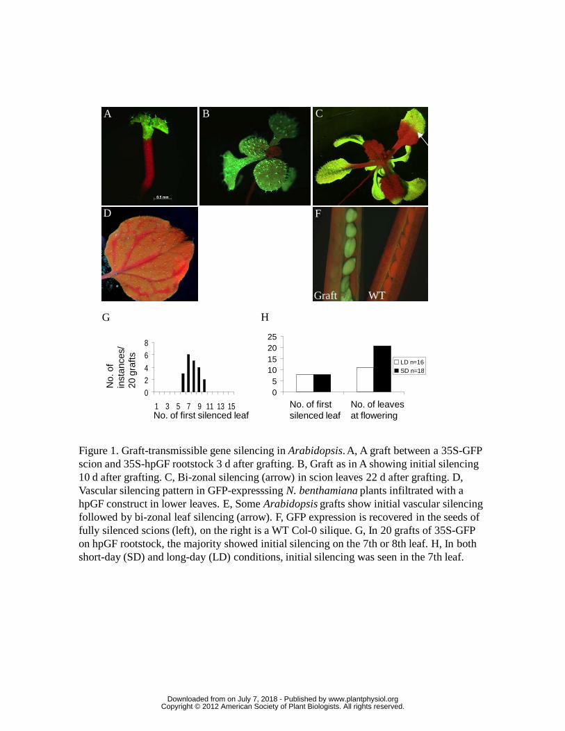
05
10152025
No. of initialsilencing leaf
No. of leaf atflowering stage
LD n=16SD n=18
A B C
D E F
Figure 1. Graft-transmissible gene silencing in Arabidopsis. A, A graft between a 35S-GFP scion and 35S-hpGF rootstock 3 d after grafting. B, Graft as in A showing initial silencing 10 d after grafting. C, Bi-zonal silencing (arrow) in scion leaves 22 d after grafting. D, Vascular silencing pattern in GFP-expresssing N. benthamiana plants infiltrated with a hpGF construct in lower leaves. E, Some Arabidopsis grafts show initial vascular silencing followed by bi-zonal leaf silencing (arrow). F, GFP expression is recovered in the seeds of fully silenced scions (left), on the right is a WT Col-0 silique. G, In 20 grafts of 35S-GFP on hpGF rootstock, the majority showed initial silencing on the 7th or 8th leaf. H, In both short-day (SD) and long-day (LD) conditions, initial silencing was seen in the 7th leaf.
G H
024
68
1 3 5 7 9 11 13 15
No.
of
inst
ance
s/20
gra
fts
No. of first silenced leaf
WTGraft
No. of first silenced leaf
No. of leaves at flowering
www.plantphysiol.orgon July 7, 2018 - Published by Downloaded from Copyright © 2012 American Society of Plant Biologists. All rights reserved.

Figure 2. Grafts between Arabidopsis C24 plants with either scion or rootstock expressing SUC2-GFP, producing fluorescence in the phloem companion cells. A, Graft between SUC2-GFP rootstock with C24 scion (dotted outline) after 3 d showing GFP in the two phloem strands of the rootstock hypocotyl, but no GFP in the scion above the graft junction(dashed red line). B, After 18 d, GFP can be seen in phloem below the graft junction but not above the graft at low magnification. Xylem (X) has proliferated in the centre of the hypocotyl. At higher magnification, GFP can be detected in the phloem (P) of the scion (C) and is concentrated in the phloem companion cells (CC) (D). Sieve elements (SE) show blue fluorescence from aniline-blue stained callose, red = chlorophyll autofluorescence.
B
C D
A
X
X
CD
X
200μm
20 μm50 μm
CC
SE
P
www.plantphysiol.orgon July 7, 2018 - Published by Downloaded from Copyright © 2012 American Society of Plant Biologists. All rights reserved.
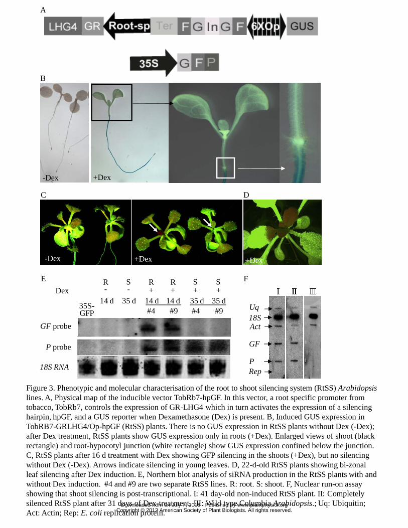
C
E
(D)
+Dex
Figure 3. Phenotypic and molecular characterisation of the root to shoot silencing system (RtSS) Arabidopsislines. A, Physical map of the inducible vector TobRb7-hpGF. In this vector, a root specific promoter from tobacco, TobRb7, controls the expression of GR-LHG4 which in turn activates the expression of a silencing hairpin, hpGF, and a GUS reporter when Dexamethasone (Dex) is present. B, Induced GUS expression in TobRB7-GRLHG4/Op-hpGF (RtSS) plants. There is no GUS expression in RtSS plants without Dex (-Dex); after Dex treatment, RtSS plants show GUS expression only in roots (+Dex). Enlarged views of shoot (black rectangle) and root-hypocotyl junction (white rectangle) show GUS expression confined below the junction. C, RtSS plants after 16 d treatment with Dex showing GFP silencing in the shoots (+Dex), but no silencing without Dex (-Dex). Arrows indicate silencing in young leaves. D, 22-d-old RtSS plants showing bi-zonal leaf silencing after Dex induction. E, Northern blot analysis of siRNA production in the RtSS plants with and without Dex induction. #4 and #9 are two separate RtSS lines. R: root. S: shoot. F, Nuclear run-on assay showing that shoot silencing is post-transcriptional. I: 41 day-old non-induced RtSS plant. II: Completely silenced RtSS plant after 31 days of Dex-treatment. III: Wild type Columbia Arabidopsis.; Uq: Ubiquitin; Act: Actin; Rep: E. coli replication protein.
+Dex-Dex
GF
Act18S
Rep
F
D
#9
P probe
GF probe
R
#4 #9 #435S-GFP
35 d+Dex + + +
R S SR S- -
35 d14 d 14 d14 d 35 d
18S RNAP
Uq
Ⅰ Ⅱ Ⅲ
B
A
-Dex +Dex
www.plantphysiol.orgon July 7, 2018 - Published by Downloaded from Copyright © 2012 American Society of Plant Biologists. All rights reserved.
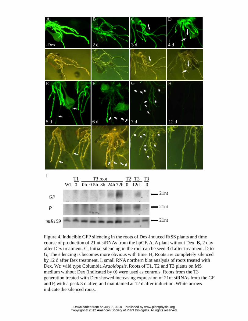
Figure 4. Inducible GFP silencing in the roots of Dex-induced RtSS plants and time course of production of 21 nt siRNAs from the hpGF. A, A plant without Dex. B, 2 day after Dex treatment. C, Initial silencing in the root can be seen 3 d after treatment. D to G, The silencing is becomes more obvious with time. H, Roots are completely silenced by 12 d after Dex treatment. I, small RNA northern blot analysis of roots treated with Dex. Wt: wild type Columbia Arabidopsis. Roots of T1, T2 and T3 plants on MS medium without Dex (indicated by 0) were used as controls. Roots from the T3 generation treated with Dex showed increasing expression of 21nt siRNAs from the GF and P, with a peak 3 d after, and maintained at 12 d after induction. White arrows indicate the silenced roots.
I
GF21nt
P
miR159
21nt
21nt
WT 0 0h 0.5h 3h 24h 72h 0 12d 0T3 rootT1 T2 T3 T3
B
3 d 4 d
5 d 6 d 7 d
-Dex 2 d
C D
E F G H
12 d
A
www.plantphysiol.orgon July 7, 2018 - Published by Downloaded from Copyright © 2012 American Society of Plant Biologists. All rights reserved.

Figure 5. RtSS plants were treated with Dex at different ages (days after germination; DAG) and the percentage showing silencing was recorded for the following 9 to 21 days.
Days after Dex induction
% S
ilenc
ed p
lant
s
9 11 13 15 17 19 21
20
40
60
80
100
0
www.plantphysiol.orgon July 7, 2018 - Published by Downloaded from Copyright © 2012 American Society of Plant Biologists. All rights reserved.

Figure 6. Spread of silencing in RtSS shoots when induced at different times after germination and siRNA distribution in the silenced and un-silenced leaves. A, Plants were treated with Dex at 5, 10 or 15 days after germination. In 5-d old plants, silencing appeared first in the petioles of the 5th, 6th and 7th leaves after 20 d with bi-zonal silencing in the 8th leaf. All lateral organs formed after the 8th leaf, and the floral bolt stem, were silenced. In 15-d old plants treated with Dex, initial silencing was seen in rosette leaf petioles at 30 d, and the inflorescence meristem, floral bolt stem and flowers escaped silencing. By 55 d, silencing had progressed through the bolt stem and flowers but was less complete than in plants treated earlier with Dex. Numbers 1 to 8 denote order of leaf appearance. B, Small RNA detected in the silenced (red) and non-silenced (green) segments of leaves from two different RtSS lines (#4 and #9), 45 days after 3-d Dex induction of 10-d old plants.
B
Red
Green
Red
#4 #9
WT
Green
35S-GFP
10 mm
A
12
3
4
5
6
78
20d5d 30d 40d 55d
25d10d 30d 40d 55d
20d15d 30d 40d 55d
GF probe
P probe
www.plantphysiol.orgon July 7, 2018 - Published by Downloaded from Copyright © 2012 American Society of Plant Biologists. All rights reserved.
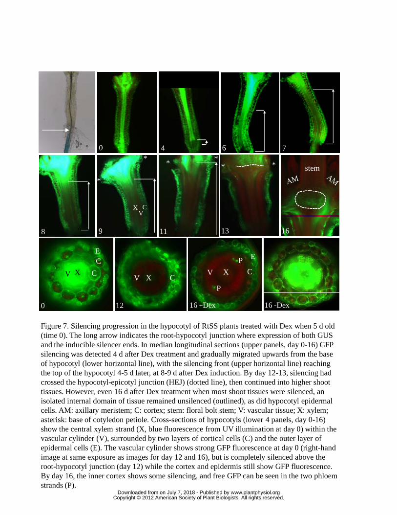
Figure 7. Silencing progression in the hypocotyl of RtSS plants treated with Dex when 5 d old (time 0). The long arrow indicates the root-hypocotyl junction where expression of both GUS and the inducible silencer ends. In median longitudinal sections (upper panels, day 0-16) GFP silencing was detected 4 d after Dex treatment and gradually migrated upwards from the base of hypocotyl (lower horizontal line), with the silencing front (upper horizontal line) reaching the top of the hypocotyl 4-5 d later, at 8-9 d after Dex induction. By day 12-13, silencing had crossed the hypocotyl-epicotyl junction (HEJ) (dotted line), then continued into higher shoot tissues. However, even 16 d after Dex treatment when most shoot tissues were silenced, an isolated internal domain of tissue remained unsilenced (outlined), as did hypocotyl epidermal cells. AM: axillary meristem; C: cortex; stem: floral bolt stem; V: vascular tissue; X: xylem; asterisk: base of cotyledon petiole. Cross-sections of hypocotyls (lower 4 panels, day 0-16) show the central xylem strand (X, blue fluorescence from UV illumination at day 0) within the vascular cylinder (V), surrounded by two layers of cortical cells (C) and the outer layer of epidermal cells (E). The vascular cylinder shows strong GFP fluorescence at day 0 (right-hand image at same exposure as images for day 12 and 16), but is completely silenced above the root-hypocotyl junction (day 12) while the cortex and epidermis still show GFP fluorescence. By day 16, the inner cortex shows some silencing, and free GFP can be seen in the two phloem strands (P).
stem
0
8 1613119
6 74
X CV
**
**
*
*
12 16 +Dex
E
X CVX CV
P
P E
0
X C
CE
16 -Dex
V
www.plantphysiol.orgon July 7, 2018 - Published by Downloaded from Copyright © 2012 American Society of Plant Biologists. All rights reserved.
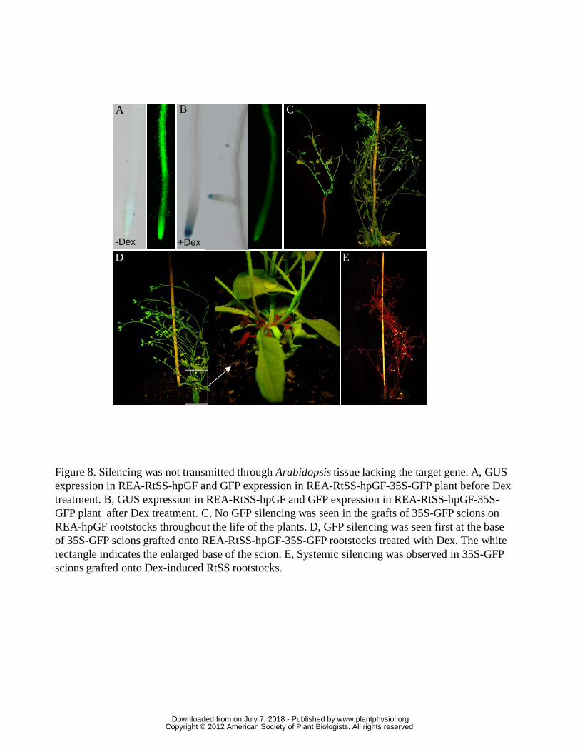
Figure 8. Silencing was not transmitted through Arabidopsis tissue lacking the target gene. A, GUS expression in REA-RtSS-hpGF and GFP expression in REA-RtSS-hpGF-35S-GFP plant before Dex treatment. B, GUS expression in REA-RtSS-hpGF and GFP expression in REA-RtSS-hpGF-35S-GFP plant after Dex treatment. C, No GFP silencing was seen in the grafts of 35S-GFP scions on REA-hpGF rootstocks throughout the life of the plants. D, GFP silencing was seen first at the base of 35S-GFP scions grafted onto REA-RtSS-hpGF-35S-GFP rootstocks treated with Dex. The white rectangle indicates the enlarged base of the scion. E, Systemic silencing was observed in 35S-GFP scions grafted onto Dex-induced RtSS rootstocks.
C
E
BA
D
-Dex +Dex
www.plantphysiol.orgon July 7, 2018 - Published by Downloaded from Copyright © 2012 American Society of Plant Biologists. All rights reserved.
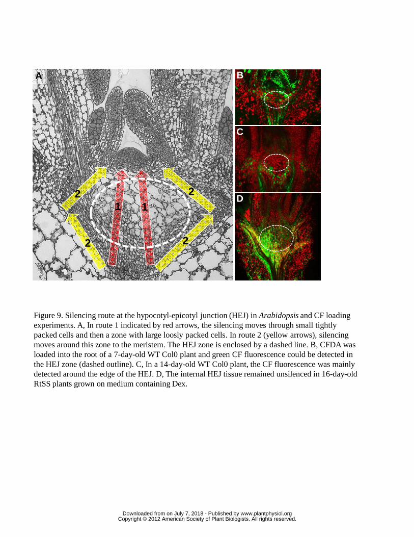
Figure 9. Silencing route at the hypocotyl-epicotyl junction (HEJ) in Arabidopsis and CF loading experiments. A, In route 1 indicated by red arrows, the silencing moves through small tightly packed cells and then a zone with large loosly packed cells. In route 2 (yellow arrows), silencing moves around this zone to the meristem. The HEJ zone is enclosed by a dashed line. B, CFDA was loaded into the root of a 7-day-old WT Col0 plant and green CF fluorescence could be detected in the HEJ zone (dashed outline). C, In a 14-day-old WT Col0 plant, the CF fluorescence was mainly detected around the edge of the HEJ. D, The internal HEJ tissue remained unsilenced in 16-day-old RtSS plants grown on medium containing Dex.
1
2
2
2
21
(C)
A B
C
D
www.plantphysiol.orgon July 7, 2018 - Published by Downloaded from Copyright © 2012 American Society of Plant Biologists. All rights reserved.
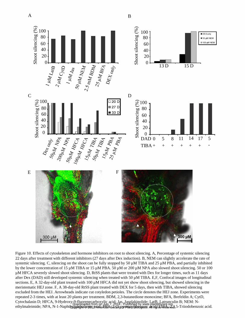
Figure 10. Effects of cytoskeleton and hormone inhibitors on root to shoot silencing. A, Percentage of systemic silencing 22 days after treatment with different inhibitors (27 days after Dex induction). B, NEM can slightly accelerate the rate of systemic silencing. C, silencing on the shoot can be fully stopped by 50 µM TIBA and 25 µM PBA, and partially inhibited by the lower concentration of 15 µM TIBA or 15 µM PBA. 50 µM or 200 µM NPA also slowed shoot silencing. 50 or 100 µM HFCA severely slowed shoot silencing. D, RtSS plants that were treated with Dex for longer times, such as 11 days after Dex (DAD) still developed systemic silencing when treated with 50 µM TIBA. E,F, Confocal images of longitudinal sections. E, A 32-day-old plant treated with 100 µM HFCA did not yet show shoot silencing, but showed silencing in the meristematic HEJ zone. F, A 38-day-old RtSS plant treated with DEX for 5 days, then with TIBA, showed silencing excluded from the HEJ. Arrowheads indicate cut cotyledon petioles. The circle denotes the HEJ zone. Experiments were repeated 2-3 times, with at least 20 plants per treatment. BDM, 2,3-butanedione monoxime; BFA, Brefeldin A; CytD, Cytochalasin D; HFCA, 9-Hydroxy-9-fluorenecarboxylic acid; Jas, Jasplakinolide; LatB, Latrunculin B; NEM: N-ethylmaleimide; NPA, N-1-Naphthylphthalamic acid; PBA, 2-(1-pyrenoyl)benzoic acid; TIBA, 2,3,5-Triiodobenzoic acid.
F
B
Shoo
t sile
ncin
g (%
)
13 D 15 D
200
406080
100
D
Shoo
t sile
ncin
g (%
)
200
406080
100
TIBA + + + + + + -DAD 0 5 8 11 14 17 5
C
Shoo
t sile
ncin
g (%
)
0
406080
100
20
A
200
406080
100Sh
oot s
ilenc
ing
(%)
E
300 µM
www.plantphysiol.orgon July 7, 2018 - Published by Downloaded from Copyright © 2012 American Society of Plant Biologists. All rights reserved.
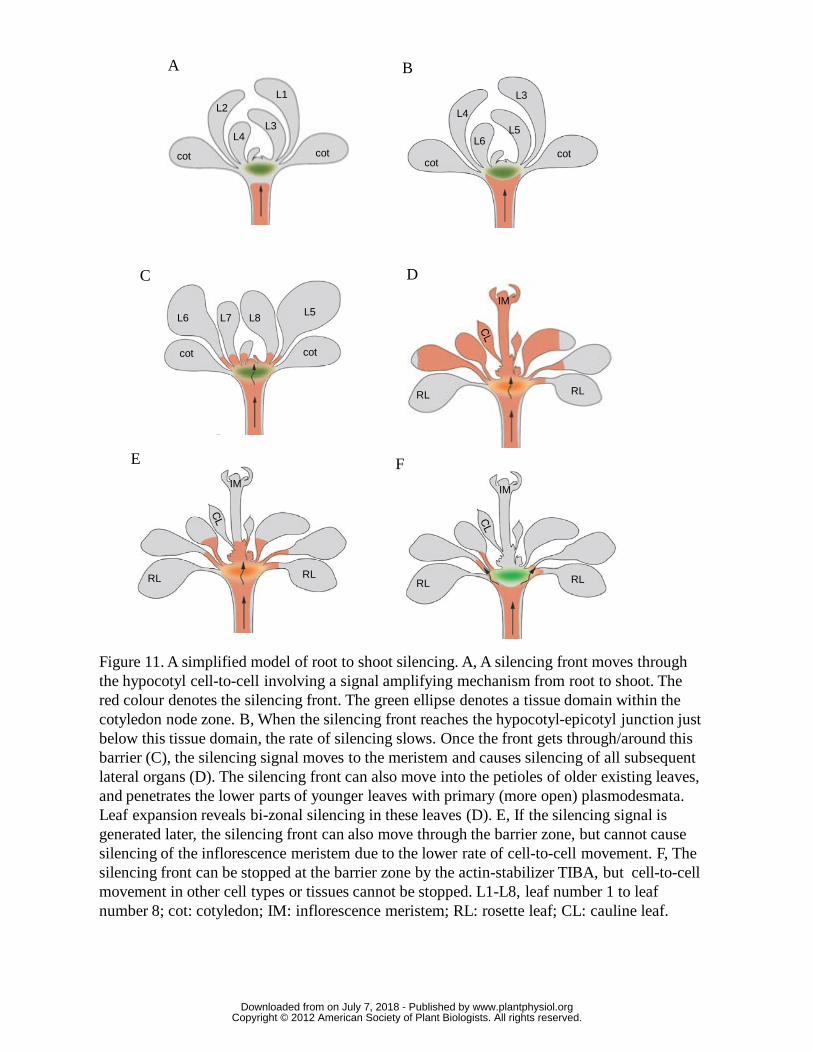
Figure 11. A simplified model of root to shoot silencing. A, A silencing front moves through the hypocotyl cell-to-cell involving a signal amplifying mechanism from root to shoot. The red colour denotes the silencing front. The green ellipse denotes a tissue domain within the cotyledon node zone. B, When the silencing front reaches the hypocotyl-epicotyl junction just below this tissue domain, the rate of silencing slows. Once the front gets through/around this barrier (C), the silencing signal moves to the meristem and causes silencing of all subsequent lateral organs (D). The silencing front can also move into the petioles of older existing leaves, and penetrates the lower parts of younger leaves with primary (more open) plasmodesmata. Leaf expansion reveals bi-zonal silencing in these leaves (D). E, If the silencing signal is generated later, the silencing front can also move through the barrier zone, but cannot cause silencing of the inflorescence meristem due to the lower rate of cell-to-cell movement. F, The silencing front can be stopped at the barrier zone by the actin-stabilizer TIBA, but cell-to-cell movement in other cell types or tissues cannot be stopped. L1-L8, leaf number 1 to leaf number 8; cot: cotyledon; IM: inflorescence meristem; RL: rosette leaf; CL: cauline leaf.
A B
C
E F
L1L2
L3L4
cot cot cotcot
L3
L4
L5L6
cotcot
L5L6 L7 L8
RL RL
RL RL
IM
IM
RL RL
IM
D
www.plantphysiol.orgon July 7, 2018 - Published by Downloaded from Copyright © 2012 American Society of Plant Biologists. All rights reserved.


