
Sanglifehrin A acts as a potent inhibitor of the mitochondrial permeability transition and
reperfusion injury of the heart by binding to cyclophilin-D at a different site from
Cyclosporin A.
Samantha J. Clarke*, Gavin P. McStay* and Andrew P. Halestrap
Department of Biochemistry, School of Medical Sciences, University of Bristol, Bristol, BS8 1TD,
UK.
Short title: Sanglifehrin inhibition of the MPTP and reperfusion injury
Correspondence to: Professor A.P. Halestrap, Department of Biochemistry, School of Medical
Sciences, University of Bristol, Bristol, BS8 1TD.
Phone 44 (0)117 9288592, FAX 44 (0)117 9288274
Email [email protected]
Abbreviations
ANT adenine nucleotide translocase, CyP-A cyclophilin A, CyP-D cyclophilin D, CsA
cyclosporin A, LDH lactate dehydrogenase, LVDP left ventricular developed pressure, LVEDP
left ventricular end diastolic pressure, MPTP mitochondrial permeability transition pore, PEG
polyethylene glycol, PPIase peptidyl-prolyl cis-trans isomerase, SfA sanglifehrin A
* These two authors contributed equally to this work
Copyright 2002 by The American Society for Biochemistry and Molecular Biology, Inc.
JBC Papers in Press. Published on July 2, 2002 as Manuscript M202191200 by guest on A
pril 7, 2018http://w
ww
.jbc.org/D
ownloaded from
Clarke et al page 2
SUMMARY
Cyclosporin A (CsA) inhibits opening of the mitochondrial permeability transition pore (MPTP), a
critical event in some forms of necrotic and apoptotic cell death, by binding to cyclophilin D (CyP-
D) and inhibiting its peptidyl-prolyl cis-trans isomerase (PPIase) activity. Sanglifehrin A (SfA)
like CsA exerts its immunosuppressive action by binding to cyclophilin A, but at a different site to
CsA, and unlike the latter does not inhibit calcineurin activity. Here we demonstrate that SfA
inhibits the PPIase activity of CyP-D (K0.5 2 nM) and acts as a potent inhibitor of MPTP opening
under both energised and de-energised conditions. However, unlike CsA, the dose response curve
for inhibition by SfA is sigmoidal rather than hyperbolic, suggesting a multimeric structure for the
MPTP with cooperativity between subunits. Furthermore, SfA does not prevent CyP-D binding to
sub-mitochondrial particles or detergent solubilised adenine nucleotide translocase (ANT),
implying that CyP-D binding to the ANT does not require PPIase activity, but pore opening does.
Once bound to the MPTP, SfA is not readily dissociated and inhibition of pore opening is
maintained following extensive washing. To investigate the potential of SfA as an inhibitor of cell
death in vivo we used the Langendorff perfused rat heart. SfA caused a time-dependent inhibition
of the MPTP that was maintained on mitochondrial isolation to a greater extent than was CsA
inhibition. We demonstrate that SfA, like CsA, improves the recovery of left ventricular developed
pressure during reperfusion after 30 min global ischemia, and greatly reduces lactate dehydrogenase
release implying inhibition of necrotic damage. Since SfA does not inhibit calcineurin activity, our
data suggest that it may be more desirable than CsA for protecting tissues recovering from ischemic
episodes and for studying the role of the MPTP in cell death.
Keywords
MPTP, ischemia, cyclophilin, apoptosis, necrosis, mitochondria, adenine nucleotide translocase
by guest on April 7, 2018
http://ww
w.jbc.org/
Dow
nloaded from

Clarke et al page 3
INTRODUCTION
A critical event in some forms of necrotic and apoptotic cell death is the opening of the
mitochondrial permeability transition pore (MPTP) (1-4), whose formation is widely thought to
involve an interaction between the adenine nucleotide translocase (ANT) and cyclophilin D (CyP-D
(1, 2). To date, the most specific inhibitor of the MPTP is cyclosporin A (CsA) (5) which acts by
inhibiting the peptidyl-prolyl cis-trans isomerase (PPIase) activity of CyP-D (6, 7). Indeed, CsA
can act a potent inhibitor of both apoptoic and necrotic cell death under some circumstances (1-3).
Nevertheless, inhibition of cell death by CsA does not provide definitive evidence for a critical role
of the MPTP in the process, since CsA exerts other effects on cell function. In particular, the
immunosuppressant action of the drug is mediated through inhibition of calcineurin, a calcium-
activated protein phosphatase involved in the regulation of gene expression and other intracellular
functions (8-10). However, there are CsA analogues such as N-Me-Ala-6-cyclosporin A and N-
Me-Val-4-cyclosporin A that do not inhibit calcineurin activity yet still inhibit the PPIase activity
of CyP-D, antagonise the opening of the MPTP and protect cells from apoptotic and necrotic cell
death (11-13). In this paper we investigate another immunosuppressant drug, Sanglifehrin A, that is
unrelated to CsA but like CsA binds tightly to cyclophilin A (CyP-A) with a K0.5 of 4-7 nM,
inhibiting its PPIase activity (K0.5 13 nM). However, unlike the CyP-A/CsA complex, the CyP-
A/SfA complex does not inhibit calcineurin (14, 15). Rather, SfA specifically blocks T cell
proliferation in response to interleukin 2 through a mechanism involving an NFkB-mediated
increase in the expression of the tumor suppressor genes p53 and p21, with the latter binding to and
inhibiting the appearance of cell cycle kinase activity cyclin E-Cdk2 (14, 16, 17).
If SfA were able to bind tightly to CyP-D and inhibit its PPIase activity and the opening of
the MPTP, its lack of effect on calcineurin might make it an alternative to N-Me-Ala-6-cyclosporin
A and N-Me-Val-4-cyclosporin A to study the role of the MPTP in cell death. Here, we
demonstrate that SfA is as potent as CsA as an inhibitor of the PPIase activity of CyP-D and of
MPTP opening. It is also able to improve functional recovery of rat hearts reperfused following an
ischemic episode, and reduce necrotic damage. However, SfA shows some important differences
by guest on April 7, 2018
http://ww
w.jbc.org/
Dow
nloaded from
Clarke et al page 4
from CsA in its mode of action on the MPTP. It demonstrates a sigmoidal rather than a hyperbolic
dose response curve for MPTP inhibition and does not prevent CyP-D binding to the ANT. These
data provide new insights into the molecular mechanism of the MPTP.
EXPERIMENTAL PROCEDURES
Preparation of mitochondria and sub-mitochondrial particles (SMPs) Percoll-purified rat liver
mitochondria and SMPs were prepared as described previously (18, 19). For measurement of
MPTP opening under de-energised conditions they were stored on ice overnight before use to
deplete them of adenine nucleotides (18).
MPTP opening The opening of the MPTP was determined under energised or de-energised
conditions by following the decrease in light scattering (monitored as A520) that accompanies
mitochondrial swelling at 25oC (6). The buffer for energised conditions was 125 mM KCl, 20 mM
Mops, 10 mM, 2mM KPi, 2mM succinate, 0.5 µM rotenone pH 7.2, and that for de-energised
conditions was 150 mM KSCN, 20 mM Mops, 10 mM Tris, 2mM nitrilotriacetic acid, 2 µM
A23187, 0.5 µM rotenone, 0.5 µM antimycin A, pH 7.2. Additions of CsA (Calbiochem) or SfA
(Novartis) were made as required. MPTP opening was initiated by addition of CaCl2 to give the
free [Ca2+] indicated. The sensitivity of the MPTP to [Ca2+] was also determined by following the
shrinkage of pre-swollen mitochondria (as an increase in A520) upon addition of polyethylene glycol
(PEG–2000) as described previously (18, 20). De-energised buffer was used and pre-swollen
mitochondria were pre-incubated for 2 min with the required [Ca2+] and other additions before
initiation of shrinkage by the addition of 7% (w/v) PEG. Rates of swelling and shrinking of
mitochondria were determined by differentiation of the A520 time course (18).
PPIase activity of CyP-D The PPIase activity of recombinant CyP-D (produced by thrombin
cleavage of GST-CyP-D (21)) was determined by following the rate of hydrolysis of N-succinyl-
Ala-Ala-Pro-Phe-p-nitroanilide by chymotrypsin as described previously(6, 7). Chymotrypsin only
by guest on April 7, 2018
http://ww
w.jbc.org/
Dow
nloaded from
Clarke et al page 5
hydrolyses the trans form of the peptide, and hydrolysis of the cis form, whose concentration is
maximised by using a stock dissolved in trifluoroethanol containing 470 mM LiCl (22), is limited
by the rate of cis-trans isomerisation.
CyP-D binding to SMPs Recombinant CyP-D (10 µg), prepared as described previously (21), was
added to 100 µg SMPs (4 mg protein / ml in isolation medium) and incubated for 15 min at 22oC
before sedimenting by centrifugation (157,000g for 5 min at 4°C). After washing once in isolation
buffer, bound CyP-D was determined by SDS-PAGE and Western blotting with anti-CyP-D
antibodies (21).
Binding of ANT to GST-CyP-D This was performed essentially as described previously using
detergent-solubilised purified inner mitochondrial membrane and recombinant GST-CyP-D bound
to glutathione-sepharose (21). When required, the bound GST-CyP-D was pre-incubated with CsA
or SfA (25 µM ) for 15 min at 22oC prior to addition of solubilised IMMs. After extensive washing
the specifically bound protein was then eluted with glutathione and analysed by SDS-PAGE and
Western blotting with anti-ANT antibodies (21).
Heart Perfusion Isolated rat hearts were perfused in Langendorff mode as described previously (23)
with continuous monitoring of left ventricular developed pressure (LVDP) and left ventricular end
diastolic pressure (LVEDP) using a pressure transducer connected to a water-filled balloon inserted
into the left ventricle. Following control perfusion for 50 min, with addition of CsA or SfA as
required, perfusion was halted to initiate global isothermic (37oC) ischemia. After 30 min ischemia
perfusion was restarted and continued for 10 min in the presence of drug and a further 20 min in its
absence. Samples of perfusate were collected prior to ischemia and every 1 min during reperfusion
for the determination of lactate dehydrogenase (LDH) activity spectrophotometrically.
by guest on April 7, 2018
http://ww
w.jbc.org/
Dow
nloaded from

Clarke et al page 6
RESULTS
SfA inhibits the PPIase activity of CyP-D and MPTP opening
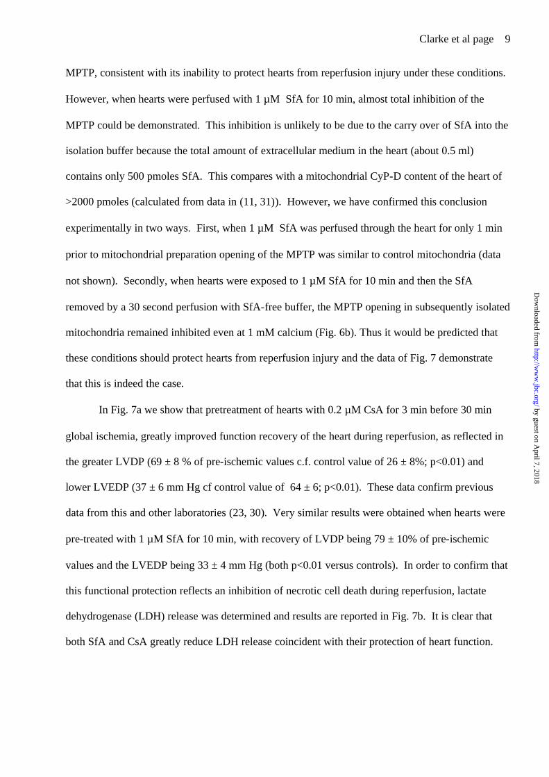
In Fig. 1 we show that SfA inhibits the PPIase activity of recombinant CyP-D with a Ki (± S.E.) of
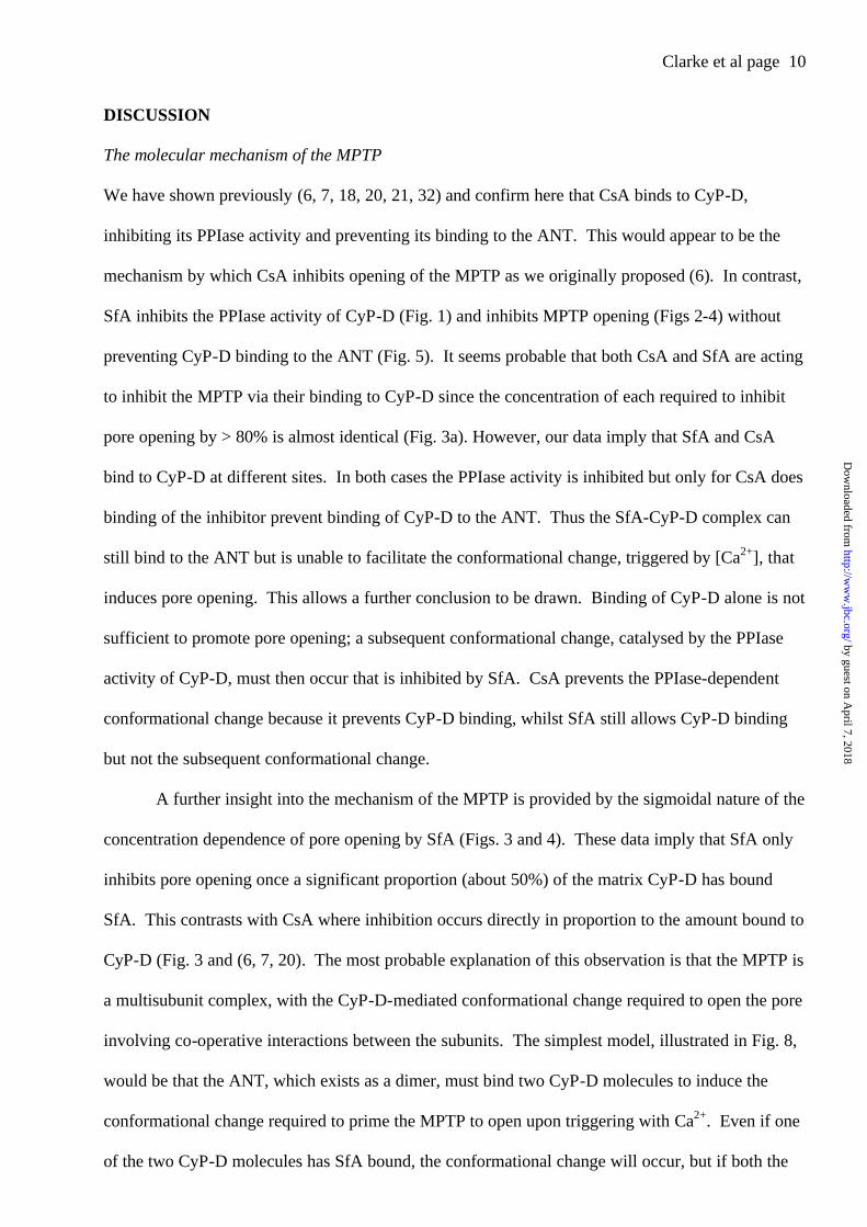
2.2 ± 0.7 nM, a similar potency to that of CsA (6, 7). In Fig. 2a we demonstrate that sub-
micromolar concentrations of SfA inhibit opening of the MPTP of energised mitochondria induced
by 100 nmoles Ca2+ per mg mitochondrial protein. However, the concentration dependence of
inhibition was complex with the lowest concentration of SfA (100 nM) failing to exhibit any
inhibition no matter how long the mitochondria were exposed to the drug. We have previously
shown that energised mitochondria are not ideal for probing the mechanism of the MPTP since
many factors can affect the sensitivity of pore opening to [Ca2+]. These include changes in
membrane potential, matrix adenine nucleotide concentrations and calcium transport (18). In
addition, the accumulation of calcium in the presence of phosphate can lead to an increase in light
scattering prior to pore opening that becomes dominant when the MPTP is inhibited (6, 24-28).
This is apparent in Fig 2a. For this reason, we routinely use de-energised conditions (iso-osmotic
KSCN buffer with respiratory chain inhibitors) in the presence of the calcium ionophore A23187 to
ensure that calcium equilibrates across the inner mitochondrial membrane. Mitochondria are also
stored overnight following their preparation, since they lose a fair proportion of their adenine
nucleotides during the first few hours of storage on ice which causes the sensitivity of the MPTP
towards calcium to change over this period. Using these conditions it is possible to obtain detailed
dose response curves for CsA analogues allowing calculation of K0.5 values that closely match the
Ki for inhibition of the PPIase activity of CyP-D (6, 7).
The data of Fig. 2b show some typical traces for the inhibition of the MPTP by CsA and
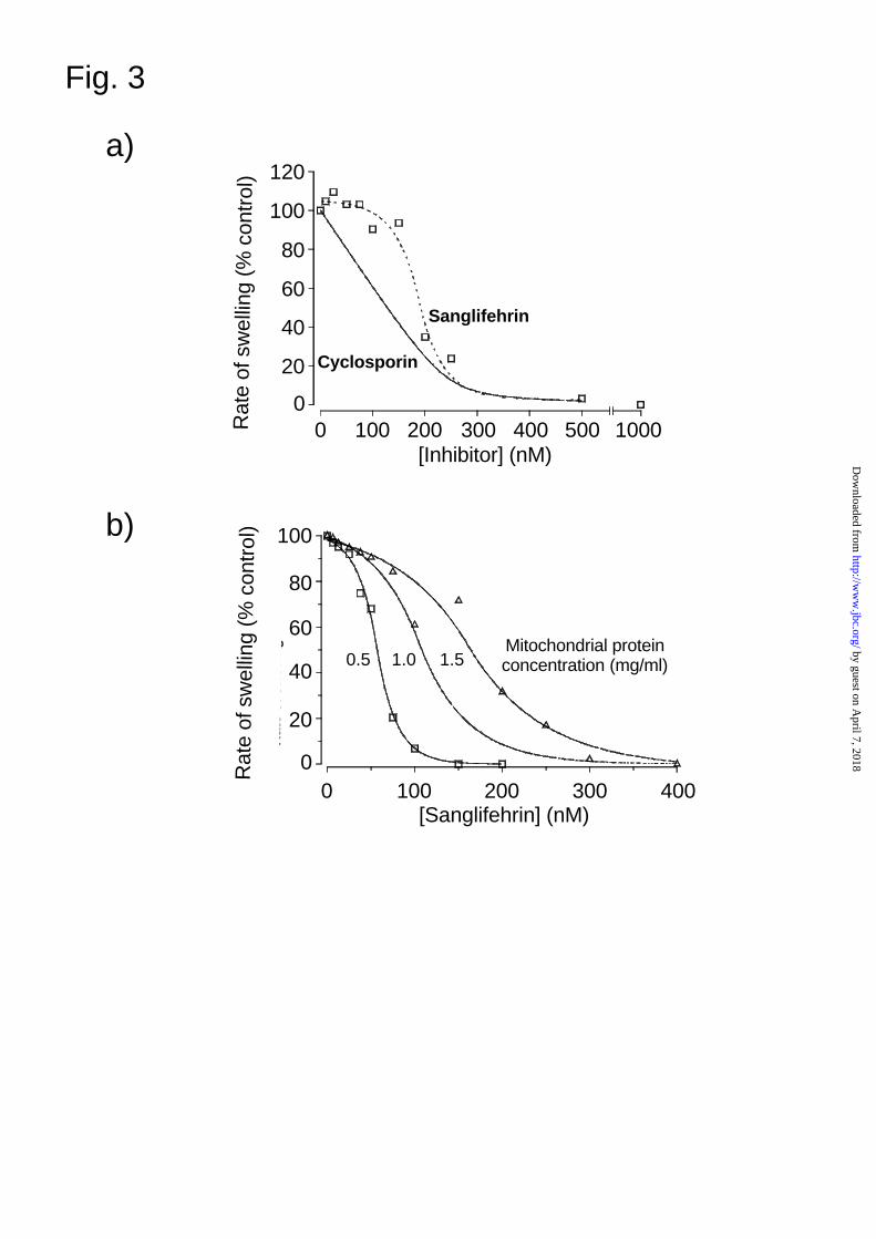
SfA under such de-energised conditions whilst in Fig. 3 we present detailed dose response curves.
These data show clearly that CsA and SfA are behaving differently. Whereas inhibition by CsA is
progressive, and can be fitted to the equation for binding to a single site as described previously (6,
7), the inhibition by SfA is sigmoidal with little or no inhibition apparent at low inhibitor
concentrations. However, a steep concentration dependence for inhibition developed as SfA
by guest on April 7, 2018
http://ww
w.jbc.org/
Dow
nloaded from

Clarke et al page 7
reached a concentration at which CsA gave about 50% inhibition, the value being dependent on the
concentration of mitochondria in the assay (Fig. 3b). The concentration of SfA required to inhibit
pore opening by >80% was almost identical to that when CsA was used (Fig 3a) consistent with
both inhibitors associating with the same target protein (CyP-D). SfA also inhibited the MPTP
when assayed using the shrinkage assay, and here too the concentration dependence for inhibition
was sigmoidal as shown in Fig. 4a. The effect of SfA, like that by CsA (18), was to decrease the
sensitivity of the MPTP to [Ca2+] as shown in Fig. 4b. Thus, even in the presence of a
concentration of SfA that gave maximal inhibition (1 µM), the pore could be opened at higher
[Ca2+].
Distinct effects of SfA and CsA on CyP-D binding to SMPs and solubilised ANT
The effects of SfA and CsA on CyP-D binding to sub-mitochondrial particles (SMPs) were
compared and data are shown in Fig. 5a. SfA slightly increased the binding of recombinant CyP-D
to SMPs (Track 4), unlike CsA that greatly reduced binding (Track 3) as observed previously (12,
20). In 9 such experiments the mean binding of CyP-D in the presence of SfA as percentage control
(± S.E.) was 132 ± 12 % (P<0.05) whereas for CsA the binding was reduced to 34 ± 8% (P<0.001).
Treatment of the CyP-D with SfA before (Track 7) or after (Track 6) CsA treatment overcame the
CsA inhibition of CyP-D binding. Slight reversal of CyP-D binding to SMPs was detected when
CsA was added after CyP-D was already bound (Track 8), but again no effect of SfA was observed
(Track 9). These data suggest CsA binds to CyP-D at the same site as the ANT whereas SfA binds
to a distinct site that actually enhances CyP-D binding to the ANT whilst displacing bound CsA.
Further evidence in favour of this interpretation was obtained when the effects of CsA and SfA on
the binding of ANT present in detergent-solubilised inner mitochondrial membranes (IMMs) to a
GST-CyP-D affinity column was studied (Fig. 5b). ANT binding was enhanced by pre-treatment
of mitochondria with diamide and inhibited by pre-treatment of the GST-CyP-D with CsA as found
previously(21). In contrast, SfA did not prevent binding and in the case of diamide-treated
mitochondria the binding of ANT was actually increased by SfA.
by guest on April 7, 2018
http://ww
w.jbc.org/
Dow
nloaded from
Clarke et al page 8
SfA protects the ischemic heart from reperfusion injury
The MPTP has now been recognised as playing a critical role in both necrotic and apoptotic cell
death and evidence for this has come from the ability of CsA to protect cells from a variety of death
signals (1-3). We and others have demonstrated that the necrotic damage associated with
reperfusion of the ischemic rat heart (irreversible reperfusion injury) can be protected by pre-
treatment of the heart with CsA, with optimal effects being observed at 0.2 µM (23, 29, 30). Thus
it would be expected that SfA might act in the same manner. Initial experiments using Langendorff
perfused rat hearts subjected to 30 min global ischemia followed by reperfusion failed to show any
protective effects of 0.2 µM SfA when functional recovery was determined by measurement of the
left ventricular developed pressure (LVDP) and end diastolic pressure (LVEDP). One explanation
of why SfA gave no protection under these conditions would be that it fails to reach the
mitochondria in the perfused heart. In order to test this possibility the experiments reported in Fig.
6 were performed.
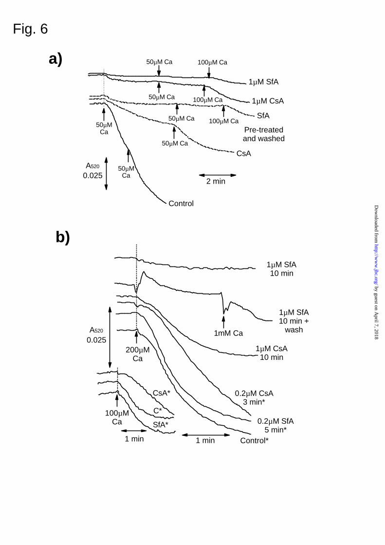
In Fig. 6a we investigated whether, following treatment of mitochondria with SfA, the drug
remains bound to CyP-D through extensive washing of mitochondria in the absence of SfA.
Mitochondria pre-treated with 1 µM CsA and then washed free of CsA exhibited greatly reduced
MPTP opening in response to Ca2+ when compared with untreated mitochondria, although the
inhibition was not as complete as when the CsA was added directly to the assay. These data
suggest that some CsA dissociated from CyP-D during the washing. In contrast, mitochondria
pretreated with 1 µM SfA maintained complete inhibition of the MPTP following washing. These
data indicate that SfA dissociates from CyP-D less readily than CsA. In addition, they imply that if
SfA has access to mitochondria within the perfused heart, this will be detected as an inhibition of
the MPTP in subsequently isolated mitochondria. In Fig. 6b we demonstrate that this is the case.
In hearts pretreated with 0.2 µM CsA for 3 min, the conditions found optimal for protecting
hearts from reperfusion injury (23), some inhibition of MPTP opening was detected at both 100 and
200 µM Ca2+. In contrast, treatment with 0.2 µM SfA for 5 min gave no detectable inhibition of the
by guest on April 7, 2018
http://ww
w.jbc.org/
Dow
nloaded from
Clarke et al page 9
MPTP, consistent with its inability to protect hearts from reperfusion injury under these conditions.
However, when hearts were perfused with 1 µM SfA for 10 min, almost total inhibition of the
MPTP could be demonstrated. This inhibition is unlikely to be due to the carry over of SfA into the
isolation buffer because the total amount of extracellular medium in the heart (about 0.5 ml)
contains only 500 pmoles SfA. This compares with a mitochondrial CyP-D content of the heart of
>2000 pmoles (calculated from data in (11, 31)). However, we have confirmed this conclusion
experimentally in two ways. First, when 1 µM SfA was perfused through the heart for only 1 min
prior to mitochondrial preparation opening of the MPTP was similar to control mitochondria (data
not shown). Secondly, when hearts were exposed to 1 µM SfA for 10 min and then the SfA
removed by a 30 second perfusion with SfA-free buffer, the MPTP opening in subsequently isolated
mitochondria remained inhibited even at 1 mM calcium (Fig. 6b). Thus it would be predicted that
these conditions should protect hearts from reperfusion injury and the data of Fig. 7 demonstrate
that this is indeed the case.
In Fig. 7a we show that pretreatment of hearts with 0.2 µM CsA for 3 min before 30 min
global ischemia, greatly improved function recovery of the heart during reperfusion, as reflected in
the greater LVDP (69 ± 8 % of pre-ischemic values c.f. control value of 26 ± 8%; p<0.01) and
lower LVEDP (37 ± 6 mm Hg cf control value of 64 ± 6; p<0.01). These data confirm previous
data from this and other laboratories (23, 30). Very similar results were obtained when hearts were
pre-treated with 1 µM SfA for 10 min, with recovery of LVDP being 79 ± 10% of pre-ischemic
values and the LVEDP being 33 ± 4 mm Hg (both p<0.01 versus controls). In order to confirm that
this functional protection reflects an inhibition of necrotic cell death during reperfusion, lactate
dehydrogenase (LDH) release was determined and results are reported in Fig. 7b. It is clear that
both SfA and CsA greatly reduce LDH release coincident with their protection of heart function.
by guest on April 7, 2018
http://ww
w.jbc.org/
Dow
nloaded from
Clarke et al page 10
DISCUSSION
The molecular mechanism of the MPTP
We have shown previously (6, 7, 18, 20, 21, 32) and confirm here that CsA binds to CyP-D,
inhibiting its PPIase activity and preventing its binding to the ANT. This would appear to be the
mechanism by which CsA inhibits opening of the MPTP as we originally proposed (6). In contrast,
SfA inhibits the PPIase activity of CyP-D (Fig. 1) and inhibits MPTP opening (Figs 2-4) without
preventing CyP-D binding to the ANT (Fig. 5). It seems probable that both CsA and SfA are acting
to inhibit the MPTP via their binding to CyP-D since the concentration of each required to inhibit
pore opening by > 80% is almost identical (Fig. 3a). However, our data imply that SfA and CsA
bind to CyP-D at different sites. In both cases the PPIase activity is inhibited but only for CsA does
binding of the inhibitor prevent binding of CyP-D to the ANT. Thus the SfA-CyP-D complex can
still bind to the ANT but is unable to facilitate the conformational change, triggered by [Ca2+], that
induces pore opening. This allows a further conclusion to be drawn. Binding of CyP-D alone is not
sufficient to promote pore opening; a subsequent conformational change, catalysed by the PPIase
activity of CyP-D, must then occur that is inhibited by SfA. CsA prevents the PPIase-dependent
conformational change because it prevents CyP-D binding, whilst SfA still allows CyP-D binding
but not the subsequent conformational change.
A further insight into the mechanism of the MPTP is provided by the sigmoidal nature of the
concentration dependence of pore opening by SfA (Figs. 3 and 4). These data imply that SfA only
inhibits pore opening once a significant proportion (about 50%) of the matrix CyP-D has bound
SfA. This contrasts with CsA where inhibition occurs directly in proportion to the amount bound to
CyP-D (Fig. 3 and (6, 7, 20). The most probable explanation of this observation is that the MPTP is
a multisubunit complex, with the CyP-D-mediated conformational change required to open the pore
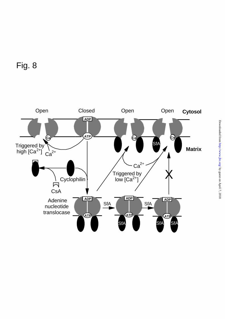
involving co-operative interactions between the subunits. The simplest model, illustrated in Fig. 8,
would be that the ANT, which exists as a dimer, must bind two CyP-D molecules to induce the
conformational change required to prime the MPTP to open upon triggering with Ca2+. Even if one
of the two CyP-D molecules has SfA bound, the conformational change will occur, but if both the
by guest on April 7, 2018
http://ww
w.jbc.org/
Dow
nloaded from

Clarke et al page 11
bound CyP-D molecules are associated with SfA, the conformational change cannot occur.
However, a CyP-D independent conformational change of the ANT can also induce pore opening at
high [Ca2+] (33) and this can explain why pore opening can still be observed in the presence of
maximally inhibiting concentration of SfA when higher [Ca2+] are employed (Fig. 4b).
It should be noted that our data do not eliminate other models for the MPTP pore. Thus a
membrane protein other than the ANT may be the true target, or an additional target, of CyP-D
binding that is responsible for MPTP formation. Indeed He and Lemasters have suggested that
many misfolded proteins may be capable of producing pores but are prevented from doing so by
binding of CyP-D and other chaperone proteins. This interaction may be disrupted by a calcium
mediated conformational change induced by the PPIase activity of CyP-D and blocked by CsA (28).
Our data could be reconciled with such a model by substituting a denatured protein aggregate for
the dimeric ANT shown in our scheme. However, the well documented effects on the MPTP of
adenine nucleotides and other ligands of the ANT, such as bongrekic acid and carboxyatractyloside
(18) are strongly suggestive that the ANT is the key membrane component that operates under most
pathophysiological conditions. So too does the ability of the purified reconstituted ANT to form a
non-specific channel at high [Ca2+] (33) that in the presence of CyP-D is opened at lower [Ca2+] in a
CsA-sensitive manner (2, 34).
SfA may be better than CsA as an inhibitor of MPTP-dependent cell death
In order to establish a role for the MPTP in apoptotic cell death, many workers have relied on the
use of either CsA or bongkrekic acid (BKA) as specific inhibitors of the MPTP. In reality, neither
of these agents is specific. The use of BKA is especially suspect, since its primary effect is to
inhibit ATP/ADP transport across the mitochondrial inner membrane, a process that is fundamental
to the metabolic and bioenergetic integrity of almost all cells. The use of CsA is open to criticism
because, when bound to CyP-A, it inhibits the calcium-dependent protein phosphatase, calcineurin,
which has many intracellular roles (8). This may, in part, explain why protection of hearts from
reperfusion injury by CsA is critically dependent on its concentration, with optimal effects being
by guest on April 7, 2018
http://ww
w.jbc.org/
Dow
nloaded from

Clarke et al page 12
observed at 0.2 µM (23, 29, 30). There are CsA analogues such as N-Me-Ala-6-cyclosporin A
and N-Me-Val-4-cyclosporin A that do not inhibit calcineurin activity yet still inhibit the PPIase
activity of CyP-D, antagonise the opening of the MPTP and protect cells from apoptotic and
necrotic cell death (11-13). SfA also lacks the ability to inhibit calcineurin (14,15), but has the
additional advantage that it binds more tightly to mitochondria than does CsA (Fig. 6). It thus
represents an important new specific tool for exploring the role of the MPTP in cell death that may
have advantages over CsA analogues as a protective agent against reperfusion injury and other
cellular stresses that cause cell death through MPTP opening. The immunosuppressant activity of
SfA is exerted by blocking T cell proliferation in response to interleukin 2 through a mechanism
involving an NFkB-mediated increase in the expression of the tumor suppressor genes p53 and p21,
with the latter binding to and inhibiting the appearance of cell cycle kinase activity cyclin E-Cdk2
(14, 16, 17). Whether this action of SfA on the cell cycle will have any detrimental effects when
using the drug to inhibit reperfusion injury is unknown, although the relatively short time scales
involved in reperfusion after ischemia would suggest not.
In conclusion, SfA provides new clues to the molecular mechanism of the MPTP, and
represents an important additional tool for studying the role of the MPTP in both necrotic and
apoptotic cell death. The lack of any effect of SfA on calcineurin activity may also make it an
appropriate therapeutic agent for the protection of tissues from reperfusion injury. Two situations
of particular importance in this regard are the treatment of stroke and coronary thrombosis where
reperfusion of the ischemic area following clot disruption may induce necrotic or apoptotic cell
death. Finally, our data demonstrate that there are two distinct sites on CyP-D that may be targeted
in the design of novel inhibitors of the MPTP that could be used therapeutically in this way.
Acknowledgements
This work was supported by a project grant from the British Heart Foundation and a CASE research
studentship to GPM from the Biotechnology and Biological Sciences Research Council in
by guest on April 7, 2018
http://ww
w.jbc.org/
Dow
nloaded from

Clarke et al page 13
conjunction with MitoKor (San Diego). We are grateful to Novartis Pharma AG (Basel) for
providing us with Sanglifehrin A.
References
1. Crompton, M. (1999) Biochem. J. 341, 233-249
2. Halestrap, A. P., Doran, E., Gillespie, J. P., and O’Toole, A. (2000) Biochem. Soc. Trans. 28, 170-177
3. Martinou, J. C. and Green, D. R. (2001) Nature Reviews Molecular Cell Biology 2, 63-67
4. Bernardi, P., Petronilli, V., DiLisa, F., and Forte, M. (2001) Trends Biochem. Sci. 26, 112-117
5. Crompton, M., Ellinger, H., and Costi, A. (1988) Biochem. J. 255, 357-360
6. Halestrap, A. P. and Davidson, A. M. (1990) Biochem. J. 268, 153-160
7. Griffiths, E. J. and Halestrap, A. P. (1991) Biochem. J. 274, 611-614
8. Schreiber, S. L. and Crabtree, G. R. (1992) Immunol. Today 13, 136-142
9. Rusnak, F. and Mertz, P. (2000) Physiol. Rev. 80, 1483-1521
10. Crabtree, G. R. (2001) J. Biol. Chem. 276, 2313-2316
11. Griffiths, E. J. and Halestrap, A. P. (1995) Biochem. J. 307, 93-98
12. Nicolli, A., Basso, E., Petronilli, V., Wenger, R. M., and Bernardi, P. (1996) J. Biol. Chem. 271, 2185-2192
13. Khaspekov, L., Friberg, H., Halestrap, A., Viktorov, I., and Wieloch, T. (1999) Eur. J. Neurosci. 11, 3194-3198
14. Zenke, G., Strittmatter, U., Fuchs, S., Quesniaux, V. F., Brinkmann, V., Schuler, W., Zurini, M., Enz, A., Billich, A., Sanglier, J. J., and Fehr, T. (2001) J. Immunol. 166, 7165-7171
15. Sanglier, J. J., Quesniaux, V., Fehr, T., Hofmann, H., Mahnke, M., Memmert, K., Schuler, W., Zenke, G., Gschwind, L., Maurer, C., and Schilling, W. (1999) J. Antibiot. (Tokyo) 52, 466-473.
16. Zhang, L. H. and Liu, J. O. (2001) J. Immunol. 166, 5611-5618
17. Zhang, L. H., Youn, H. D., and Liu, J. O. (2001) J. Biol. Chem. 276, 43534-43540
18. Halestrap, A. P., Woodfield, K. Y., and Connern, C. P. (1997) J. Biol. Chem. 272, 3346-3354
19. Owen, M. R., Doran, E., and Halestrap, A. P. (2000) Biochem. J. 348, 607-614
20. Connern, C. P. and Halestrap, A. P. (1994) Biochem. J. 302, 321-324
21. Woodfield, K., Ruck, A., Brdiczka, D., and Halestrap, A. P. (1998) Biochem. J. 336, 287-290
by guest on April 7, 2018
http://ww
w.jbc.org/
Dow
nloaded from

Clarke et al page 14
22. Garciaecheverria, C., Kofron, J. L., Kuzmic, P., and Rich, D. H. (1993) Biochem. Biophys. Res. Commun. 191, 70-75
23. Griffiths, E. J. and Halestrap, A. P. (1993) J. Mol. Cell. Cardiol. 25, 1461-1469
24. Novgorodov, S. A., Gudz, T. I., Milgrom, Y. M., and Brierley, G. P. (1992) J. Biol. Chem. 267, 16274-16282
25. Bernardi, P., Veronese, P., and Petronilli, V. (1993) J. Biol. Chem. 268, 1005-1010
26. Petronilli, V., Cola, C., Massari, S., Colonna, R., and Bernardi, P. (1993) J. Biol. Chem. 268, 21939-21945
27. Andreyev, A. and Fiskum, G. (1999) Cell Death Differ. 6, 825-832
28. He, L. and Lemasters, J. J. (2002) FEBS Lett. 512, 1-7
29. Nazareth, W., Yafei, N., and Crompton, M. (1991) J. Mol. Cell. Cardiol. 23, 1351-1354
30. DiLisa, F., Menabo, R., Canton, M., Barile, M., and Bernardi, P. (2001) J. Biol. Chem. 276, 2571-2575
31. Connern, C. P. and Halestrap, A. P. (1992) Biochem. J. 284, 381-385
32. Connern, C. P. and Halestrap, A. P. (1996) Biochemistry 35, 8172-8180
33. Brustovetsky, N. and Klingenberg, M. (1996) Biochemistry 35, 8483-8488
34. Crompton, M., Virji, S., and Ward, J. M. (1998) Eur. J. Biochem. 258, 729-735
by guest on April 7, 2018
http://ww
w.jbc.org/
Dow
nloaded from

Clarke et al page 15
Figure Legends
FIG. 1. Inhibition of PPIase activity of CyP-D by SfA. Recombinant CyP-D (6 pmoles) was
preincubated at 10oC for 5 min in 3.5ml PPIase assay buffer (see Experimental section) with the
concentration of SfA shown. The assay was started by addition of peptide and initial rates of
reaction were determined by first order regression analysis of the time course of change in A520.
The Ki (± S.E.) derived by fitting the data to the equation for a tight binding inhibitor(6) was 2.2 ±
0.7 nM.
FIG. 2. Inhibition of the MPTP by SfA. In Panel (a) MPTP opening in rat liver mitochondria
(1.5 mg protein / ml) was assayed under energised conditions (see Experimental section) by
monitoring the decrease in A520 with a split beam spectrophotometer after addition of 100 µM
CaCl2 to the sample cuvette. When present, SfA or CsA were added at the concentration shown 2
min before calcium addition. In Panel (b) the protocol was similar but de-energised conditions
were employed and pore opening initiated by addition of CaCl2 to give a calculated free
concentration of 100 µM.
FIG. 3. Concentration dependence of the inhibition of the MPTP by SfA. In both panels the
extent of MPTP opening was determined from the rate of swelling in experiments similar to those
shown in Fig 2b but with CsA or SfA added 2 min before CaCl2 at the concentrations of indicated.
In panel (a) the protein concentration was 1.5mg / ml whilst in panel (b) the concentration of
mitochondria was varied as indicated.
FIG. 4. The effect of SfA on the calcium dependence of the MPTP In both panels the extent of
MPTP opening was determined by measuring the rate of shrinkage of pre-swollen mitochondria
upon addition of 7% (w/v) PEG as described in the Experimental section. In panel (a) the pre-
swollen mitochondria were incubated for 2 min with 50 µM [Ca2+] and the concentration of SfA
by guest on April 7, 2018
http://ww
w.jbc.org/
Dow
nloaded from

Clarke et al page 16
shown before addition of PEG to initiated shrinkage. In panel (b) the concentration of Ca2+ was
varied as indicated in the absence or presence of 1 µM SfA.
FIG. 5. CsA but not SfA prevents CyP-D binding to SMPs and ANT binding to GST-Cyp-D.
In panel (a) recombinant CyP-D (10 µg) was pre-incubated for 15 min at 22oC with 50 µM CsA, 50
µM SfA or solvent (ethanol) as indicated before addition SMPs. After incubation for a further 15
min at 22oC, bound CyP-D was determined by SDS-PAGE and Western blotting as described in the
Experimental section. In Track 5, SfA and CsA were added together. In Track 6, CyP-D was
incubated with CsA for 15 min followed by SfA for 15 min whereas in Track 7, the order of
addition of the inhibitors was reversed. In Tracks 8 and 9, the CsA and SfA were added after
incubation of SMPs with CyP-D and incubation continued for a further 15 min. In panel (b), inner
mitochondrial membranes were prepared from rat liver mitochondria after incubation in the
presence or absence of 1 mM diamide for 10 min at 22oC. After detergent solubilisation they were
passed down a GST-CyP-D affinity column that had been pre-incubated with or without 25 µM
CsA or SfA as described in the Experimental section. Following extensive washing, bound proteins
were eluted with glutathione and analysed by SDS-PAGE and Western blotting with anti-ANT
antibodies.
FIG. 6. SfA and CsA inhibit the MPTP in the perfused heart.
In panel (a) isolated rat heart mitochondria (2 mg/ml in isolation buffer) were incubated in the
presence or absence of 1 µM CsA or SfA for 10 min at 0oC before sedimentation and 2 washes at
0.4 mg/ml. MPTP opening under de-energised conditions (1 mg protein / ml) was monitored (A520)
following addition of Ca2+ at the concentration shown. The top 2 traces represent control
mitochondria to which 1 µM CsA and SfA have been added directly to the swelling assay whereas
the middle traces show mitochondria pre-treated with the drugs and then washed. In panel (b)
mitochondria were isolated from hearts perfused (Langendorff mode) for 30 min in the absence of
by guest on April 7, 2018
http://ww
w.jbc.org/
Dow
nloaded from

Clarke et al page 17
drug and then with the concentration of CsA or SfA indicated for the time shown. Where indicated,
extracellular SfA was washed from the heart prior to mitochondrial preparation by perfusing for 30
seconds with SfA-free buffer (5 ml). Assay of MPTP under de-energised conditions was performed
as in (a) at a final protein concentration of 0.5mg/ml. The inset traces were performed with the
same mitochondria as the traces marked with an asterisk but with swelling initiated by addition of
[Ca2+] at 100 µM as opposed to 200 µM.
FIG. 7. SfA and CsA protect the ischemic rat heart from reperfusion injury
Rat hearts were perfused in the Langendorff mode and left ventricular pressure continuously
monitored as described in the Experimental Section. After 50 min perfusion hearts were subject to
30 min global isothermic ischemia followed by reperfusion. Where required, 0.2 µM CsA or 1 µM
SfA were added 3 min or 10 min prior to ischemia respectively. In panel (a) greater functional
recovery of the SfA and CsA treated hearts after 30 min reperfusion is reflected in higher values for
the LVDP and lower values for the left ventricular end diastolic pressure LVEDP. Preischemic
values for the LVDP and LVEDP were 76 ± 3.5 and 2.4 ± 0.3 mm Hg respectively (n=9) and were
unaltered by either CsA or SfA. Values of the LVDP after reperfusion are presented as a
percentage of the preischemic value and were significantly greater (p<0.001) with either SfA or
CsA treatment, whilst values for the LVEDP were significantly lower (p<0.001). In panel (b) the
release of LDH into the perfusate from the same hearts as used in panel (a) was measured as an
indicator of necrotic cell death (* p<0.05 for CsA- or SfA-treated hearts versus controls).
FIG. 8. A scheme to account for the different mode of action of CsA and SfA on the MPTP
by guest on April 7, 2018
http://ww
w.jbc.org/
Dow
nloaded from
Fig. 1
0 5 10 15 20 25 100
[Sanglfehrin] nM
0
1
2
3
4
5
6
Rat
e co
nst
ant
for
pep
tid
e h
ydro
lysi
s (s
-1)
[Sanglifehrin] (nM)
Rat
e co
nsta
nt fo
rpe
ptid
e hy
drol
ysis
(s )
0
1
2
3
4
5
6
0 5 10 15 20 25 100
-1
by guest on April 7, 2018
http://ww
w.jbc.org/
Dow
nloaded from
30 sA520
0.05
Control
150 nM SfA
150 nM CsA
500 nM SfA
1 µM SfA1 µM CsA
500 nM CsA
Ca2+
Fig. 2
1 min
A520
0.05
100 µM Ca2+
300 nM SfA
400 nM SfA
500 nM SfA
200 nM SfA
2 µM SfA
100 nM SfAControl
De-energised
Energiseda)
b) by guest on April 7, 2018
http://ww
w.jbc.org/
Dow
nloaded from
0 100 200 300 400 500 1000
[Inhibitor] (nM)
0
20
40
60
80
100
120
Rat
e of
sw
ellin
g as
% c
ontr
ol
SanglifehrinCyclosporin
[Inhibitor] (nM)0 100 200 300 400 500 1000R
ate
of s
wel
ling
(% c
ontro
l)
0
20
40
80
100
60
120
Sanglifehrin
Cyclosporin
Fig. 3
a)
b)
0 100 200 300 400
[Sanglifehrin] nM
0
20
40
60
80
100
Rat
e of
sw
ellin
g as
% c
ontr
ol
0.5 mg/ml 1.0 mg/ml 1.5 mg/mlMitochondrial protein
concentration
[Sanglifehrin] (nM)0 100 200 300 400
Rat
e of
sw
ellin
g (%
con
trol)
0
20
40
80
100
60Mitochondrial protein concentration (mg/ml)0.5 1.0 1.5
by guest on April 7, 2018
http://ww
w.jbc.org/
Dow
nloaded from

b)
[Sanglifehrin] (nM)
Rat
e of
shr
inka
ge (
A52
0.s
.10
)
-14
0
20
40
10
30
40 80 10000 20 60 100
Fig. 4
a)
Control1 µM SfA
0 20 40 60 80
[Ca2+] µM
0
10
20
30
40
50
60
70
Rat
e o
f S
hri
nka
ge
(A52
0.s-1
x10
4 )
10
20
30
40
50
60
0
70
Rat
e of
shr
inka
ge (
A52
0.s
.10
)-1
4
0 20 40 60 80
[Ca 2+] µM
by guest on April 7, 2018
http://ww
w.jbc.org/
Dow
nloaded from

DiamideCsASfA
---
-+-
--+
+--
++-
+-+
CyP-DCsASfATrack
---1
+--2
++-3
+-+4
+++5
+++6
+++7
++-8
+-+9
20kDa
30kDa
a)
b)
Fig. 5
by guest on April 7, 2018
http://ww
w.jbc.org/
Dow
nloaded from
A520
0.025
1 min Control*1 min
200µM Ca
100µM Ca
1µM CsA 10 min
0.2µM CsA 3 min*
C*
SfA*
CsA*
0.2µM SfA 5 min*
50µM Ca
SfA
A520
0.0252 min
50µM Ca
50µM Ca
50µM Ca 100µM Ca
50µM Ca 100µM Ca
50µM Ca 100µM Ca
Control
CsA
1µM SfA
1µM CsA
Pre-treated and washed
a)
b)
Fig. 6
1µM SfA 10 min
1µM SfA 10 min +
wash1mM Ca
by guest on April 7, 2018
http://ww
w.jbc.org/
Dow
nloaded from

a)
Fig. 7
b)
0
20
40
60
80
100
Rec
over
y of
LV
DP
(% p
re-ie
chae
mic
val
ue)
0
20
40
60
80
100
LVE
DP
(mm
Hg)
LVDP LVEDP
Con SfA CsA Con SfA CsA
80
60
40
100
0
20
80
60
40
100
0
20
LV
DP
(%
pre
isch
aem
ic v
alu
e) LEV
DP
(mm
Hg)
70 80 90 100 110
Time (min)
0
10
20
30
40
50
60
LDH
(m
unit
s/m
l)
Control
SfA
CsA
30 40 80 85 90 95 100 105 110
30 m
in Is
chem
ia
LDH
rel
ease
d (m
units
/ml p
erfu
sate
)
60
50
40
30
10
0
20
Time (min)
*
**
*
* *
by guest on April 7, 2018
http://ww
w.jbc.org/
Dow
nloaded from
ATP
ADP
Adeninenucleotidetranslocase
Ca
CsA
Cyclophilin X
ATP
ADP
ATP
ADP
ATP
ADP
SfA SfA SfA
SfASfA
Cytosol
Matrix2+Ca
2+CaTriggered by low [Ca ]2+
Triggered by high [Ca ]2+
Ca Ca
SfA
Closed Open
Fig. 8
OpenOpen
by guest on April 7, 2018
http://ww
w.jbc.org/
Dow
nloaded from
Samantha J. Clarke, Gavin P. McStay and Andrew P. Halestrapdifferent site from Cyclosporin A
transition and reperfusion injury of the heart by binding to cyclophilin-D at a Sanglifehrin A acts as a potent inhibitor of the mitochondrial permeability
published online July 2, 2002J. Biol. Chem.
10.1074/jbc.M202191200Access the most updated version of this article at doi:
Alerts:
When a correction for this article is posted•
When this article is cited•
to choose from all of JBC's e-mail alertsClick here
by guest on April 7, 2018
http://ww
w.jbc.org/
Dow
nloaded from


