
© CNJ McGhee 2017
Cataract surgery in New Zealand /Aotearoa approaching 2020:
demand, supply, politics, economics & shared careProfessor Charles NJ McGheeMB.ChB, BSc(Hons), PhD, DSc, FRCS, FRCOphth, FRANZCO
Maurice Paykel Professor & Chair of OphthalmologyDirector, New Zealand National Eye Centre,SMO, Auckland District Health Board,Consultant, Eye Institute, Auckland

© CNJ McGhee 2017
Introduction: cataract
The most common cause of visual impairment
The most common surgical procedure in the developed world
Extremely efficient, effective and safe health-dollar investment!
Why can we never meet the annual demand?
New Zealand > 30,000
Australia > 160,000
United Kingdom > 390,000
United States > 3,600,000
Globally > 20 million

© CNJ McGhee 2017
Political solutions or misdirection
Cataracts and hip replacement surgery the currency of election healthcare policies

Considering Cataract Outcomes in NZThe Auckland Cataract Studies 2000-2017

OUTLINE OF LECTURE
Cataract surgery is most common surgical procedure in New Zealand
Typically presents to optometrists and general practitioners

© CNJ McGhee 2017
International Cataract CostsWe are more, not less, expensive!
xx

© CNJ McGhee 2017
Potential barriers to successful cataract surgery & visual rehabilitation in NZ
1. Identifying visually significant cataract
2. Referral and first specialist visit
3. Assessing suitability, eligibility, listing
4. Pre-operative assessment
5. Provision of excellent surgical services
6. Post-operative care
7. Dealing with complications
8. Potential barriers to discharge*
9. Long term Optical correction
10. Funding the process: Government, Health Insurers, Self

Aetiology of cataract
Congenital
Inherited
Age-related (the majority)
Metabolic – e.g. diabetes
Toxic – e.g. corticosteroids
Traumatic – e.g. irradiation
Secondary – e.g. uveitis
9

• Reduced visual acuity– Snellen, glare, contrast
sensitivity, driving
• Entirely clinical diagnosis– Distance & near vision
– Ophthalmoscope
– Slit lamp microscope
– Significant lens opacity
• Exclude other ocular pathologies*
10
Diagnosing cataract

Effect of differing cataracts
• Nuclear Cataract
– Common, VA may be preserved
– may induce myopia
• Cortical Cataract
– Often associated with nuclear
– May cause distortion/glare
• Subcapsular Cataract
– May have good vision in test conditions
– Variable vision particularly in low light
– Consider history and test glare vision11
Posterior Subcapsular
Cortical cataract

Burden of cataracts: visual impairment
http://www.who.int/blindness/data_maps/cataract_surgery_rate/en/
“cataract is responsible for
51% of world blindness,
which represents about
20 million people (2010)”
World Health Organisation
Global cataract surgical rates (2004)

Mean age 77.2 years
Mean wait 18.2+/-11.6 mths
Mean BCVA 6/36*
Outcome
Still waiting 49%*
Expedited Rx 4%
Private surgery 21%
Deceased 12%
Declined surgery 2%
The Auckland Cataract Study 1:2000-2001: Waiting – the harbor bridge study N=193
The Waiting Game: The natural history of a cataract waiting list in New Zealand AF Riley, C Grupcheva, TY Malik, JP Craig, CN McGhee. Clin Expt Ophthalmol 2001

14
Pre-proliferative diabetic retinopathy
The majority of those with significant cataract have General Health issues
Hypertension 25%Cerebral vascular disease 12%Diabetes Melitus 11%Ischaemic Heart disease 10%RxAspirin 42%Warfarin 6%
Auckland Cataract Study Systemic Disease
The Auckland Cataract Study: Demographic, Corneal Topographic and Ocular Biometric Parameters. AF Riley, CN Grupcheva, TY Malik, JP Craig, CN McGhee. Clinical & Experimental Ophthalmology 2001
15
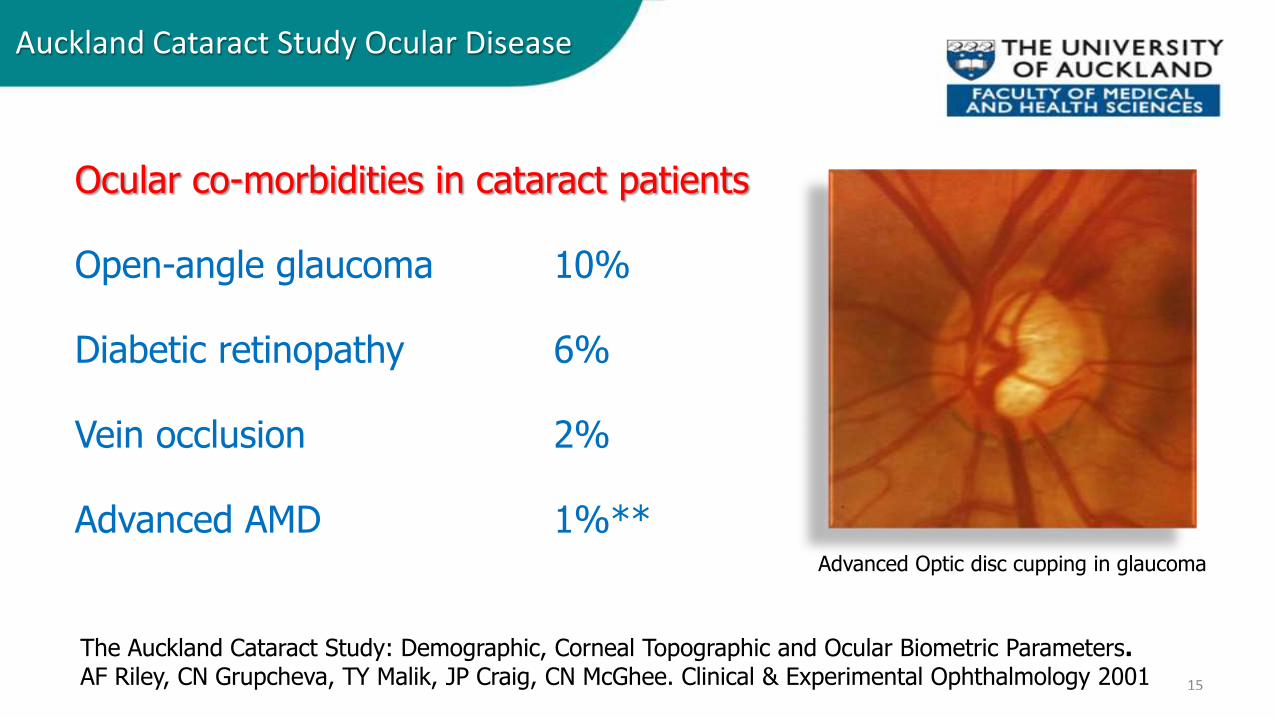
Advanced Optic disc cupping in glaucoma
Ocular co-morbidities in cataract patients
Open-angle glaucoma 10%
Diabetic retinopathy 6%
Vein occlusion 2%
Advanced AMD 1%**
The Auckland Cataract Study: Demographic, Corneal Topographic and Ocular Biometric Parameters. AF Riley, CN Grupcheva, TY Malik, JP Craig, CN McGhee. Clinical & Experimental Ophthalmology 2001
Auckland Cataract Study Ocular Disease

Cataract Surgery
Ancient Techniques
– Couching
Current cataract techniques
Intra-capsular –
Now mainly in developing world
Extra-capsular –
Some use in developed world
Phacoemulsification –
most popular technique
Femto-laser assisted
Use increasing developed world
16

1990’s small incision phaco-emulsification and the evolution of foldable/injectable Intraocular lenses revolutionized cataract surgery in developed world
17
Contemporary Cataract Surgery

Extracapsular surgery: ECCE vs Phaco

Phacoemulsification in practice

Auckland Cataract Study 1: 2000-2001
Outcome:
Mean BSCVA 6/7.5 (88% > 6/12)
Mean SphEq –0.46+/-0.89D
Complications:
4.9% capsular tears
3.7% cystoid macular oedema
0.2% endophthalmitis
1.5% of eyes red’n BSCVA due to surgery
The Auckland Cataract Study: co-morbidity, surgical techniques and clinical outcomes in a Public Hospital Service. Andrew Riley, Tahira Malik, Christina Grupcheva, Michael Fisk, Jennifer Craig, Charles McGhee. BJO 2002

Cataract Pathway 2017
The timing of referral varies significantly with available local DHB funding,however, many would consider referring when vision ≤6/12
Current cataract referral waiting times:1. From referral to FSA (4 months)2. From FSA to surgery (4 months)
Cataract referrals at ADHB via three main routes: General Practice Optometrist* Ophthalmologist
21

© CNJ McGhee 2017
Efficiency:Radical streamlining of existing systems?
1. Immediate listing from the community
2. Combined Optometry/General Practitioner electronic referrals
Enables collection of demographic data and risk analysis
3. Same day FSA clinic review and surgery
4. Electronic surgical records
Enables ongoing audit and creates discharge
5. Bilateral same day cataract surgery
6. Immediate discharge to optometric shared care

© CNJ McGhee 2017
One stop cataract clinic N=4657 (Bristol, 2008-2010)
Two referral routes – traditional GP route and Refined Direct Optometrist (RDO) pathway
Operative rates higher for RDO (92%) than GP (82%) routes
“ By combining referral information from optometrists and GPs, a high-quality and efficient cataract surgery patient pathway can be established.
This has major economic advantages, and this scheme could be adopted at a national level.”
J Cataract Refract Surg 2013; 39:712–715

© CNJ McGhee 2017
Uncomplicated surgery and no significant ocular comorbidity - same day discharge to community optometrists.
Over 2 years, 1492 of 2461 (61%) Cambridgeshire patients discharged to community on day of cataract surgery.
Complete feedback in 97%, uneventful in 94% and 3% of patients re-referred. CMO 0.6%, uveitis 1.0% and raised IOP 0.1%.
No patients had sight-threatening complications in this study

Preoperative RISK stratificationmaximizing outcomes, safety and audit
• Scores for patient risk factors documented in the clinical notes
• A validated system devised by Muhtaseb et al. (2004, UK)
1 point 3 points
Age >88 yearsBrunescent/white/dense/total cataract/
no fundus view
Ametropia (>6D of myopia or hyperopia) Pseudoexfoliation
Corneal scar
Posterior capsule plaque
Posterior polar cataract
Previous vitrectomy
Shallow AC <2.5mm
Small pupil <3mm
Miscellaneous (poor position, etc)

Auckland Cataract Study IIa and IIbRisk of intraoperative complications
• Observed higher complication rates with high risk scores
• Significant increase for scores >3
0
50
100
150
0 1 2 3 4 5 6 7
Intr
aop
erat
ive
com
plic
atio
n r
ate
(%)
Risk Score
Dr Bia Kim MD

Intraoperative complications in cataract surgery (N=1000) Following introduction of risk analysis in ADHB
2015 2016 P-value fordifference
Intraoperativecomplications
8.4%(N=500)
5.0%(N=500)
0.042
PC tear ± vitreous loss 2.6% 1.8% 0.258
Anterior capsular tear 1.2% 0.4% 0.224
Zonular dialysis 1.6% 0.6% 0.224
Iris prolapse 2.6% 1.4% 0.258
Iris trauma 1.0% 1.4% 0.773
Dropped nucleus fragment 0.2% 0.4% 1.000
The Auckland Cataract Study II: Reducing Complications by Preoperative Risk Stratification and Case Allocation in a Teaching Hospital. Kim BZ, Patel DV, McKelvie J, Sherwin T, McGhee CNJ. Am J Ophthalmol. 2017 Sep;181:20-25.

2015 2016 P-value fordifference
Postoperative complications 8.1%(N=479)
6.1%(N=472)
0.258
Cystoid macular oedema 3.5% 3.8% 0.865
Auckland Cataract Study 2:Postoperative complications

Auckland Cataract Study 2:Postoperative complications: Cystoid macular oedema
• Rare• Endophthalmitis
• Pain, redness, decreased vision in first week
• Intra-ocular haemorrhage
• Severe uveitis
• More common• Cystoid macular oedema
• Typically in first month – suspect if vision has been good and deteriorates moderately in quiet eye
• Mild uveitis beyond 4 week

RADICAL ROUTES TO IMPROVED CATARACT SURGERY PROVISION: Excellence, Efficiency, Economy top ten
1. Fully electronic pathway
2. Optometry/GP referral / listing
3. Risk stratification analysis and audit
4. Consider one-stop surgical approach
5. Bilateral same day surgery where appropriate
6. In teaching units create (hi-volume) service vs training lists
7. Day-one discharge to community optometry in >50% cases
8. Standardised data set pre and post-op
9. Unified national program and agreed threshold
10. Continuous audit cycle – Local, DHB and National level

Shared care in cataract surgery
Identifying
Appropriately referring
Pre-operative assessment
Post operative management*
Unexpected complications*
32

The fine-print of co-management

Striate keratopathy Elevated IOP Aqueous leak Ant. Uveitis Loose sutures*
Rare complications Retinal detachment Choroidal haemorrhage Filtering bleb
34
Common complications

94,653 cataract procedures in 19 years
Endophthalmitis in 188 patients
Serious visual impairment in 70.6%
Incidence of cataract surgery increased x3
However, endophthalmitis rate constant at 2 per 1000
35
Endophthalmitis – always a risk?
Semmens JB, Li J, Morlet N, Ng J. Trends in cataract surgery and post-operative endophthalmitis in Western Australia (1980-1998):The endophthalmitis Population Study of Western Australia. Clinical & Experiment Ophthalmol June 2003

Day 1 review
Symptoms
Unaided VA
Pinhole VA*
Assess: Cornea
Pupil
Media
IOL position
IOP36
Post-operative management

Usually 2-4 weeks post-operative topical medication
Typically an antibiotic and a steroid
E.G. g. Chloramphenicol QDS / g. ciprofloxacin TDS*
E.G. g. Predforte QDS / g. Maxidex QDS
Or Maxitrol (dexamethasone + neomycin + polymyxin B)
Or occasionally NSAIDS e.g. ketorolac/diclofenac
37
Post-operative management

Post-op vision & visual acuity
Day one and day seven:
Usually 6/6 to 6/18
If less than 6/12 unaided – refract
More than 1.5D residual error - consult
38
Post-operative management

1/12 post-op vision & best corrected visual acuity
Generally BSCVA ≥ 6/7.5
BSCVA <6/12, exclude pathology:
Cystoid macular oedema
Posterior capsule opacity
Macular degeneration
Diabetic maculopathy39
Post-operative management
Post-operatively they all obtained at least counting cats unaided vision

Post-operative refractive error
Contact ophthalmologist if (*):
≥1.50D from intended refraction
≥1.50D of induced astigmatism
40
Post-operative management

Wound appearance and aqueous leak
Day 1 wound should well apposed, if anterior chamber shallow or IOP <10mmHg exclude wound leak:
Check for phaco burn or wound retraction
Perform fluorescein test
Spontaneous leak
Leak to gentle compression
Check pupil is round
Exclude iris prolapse
If AC compromised refer41
Post-operative management
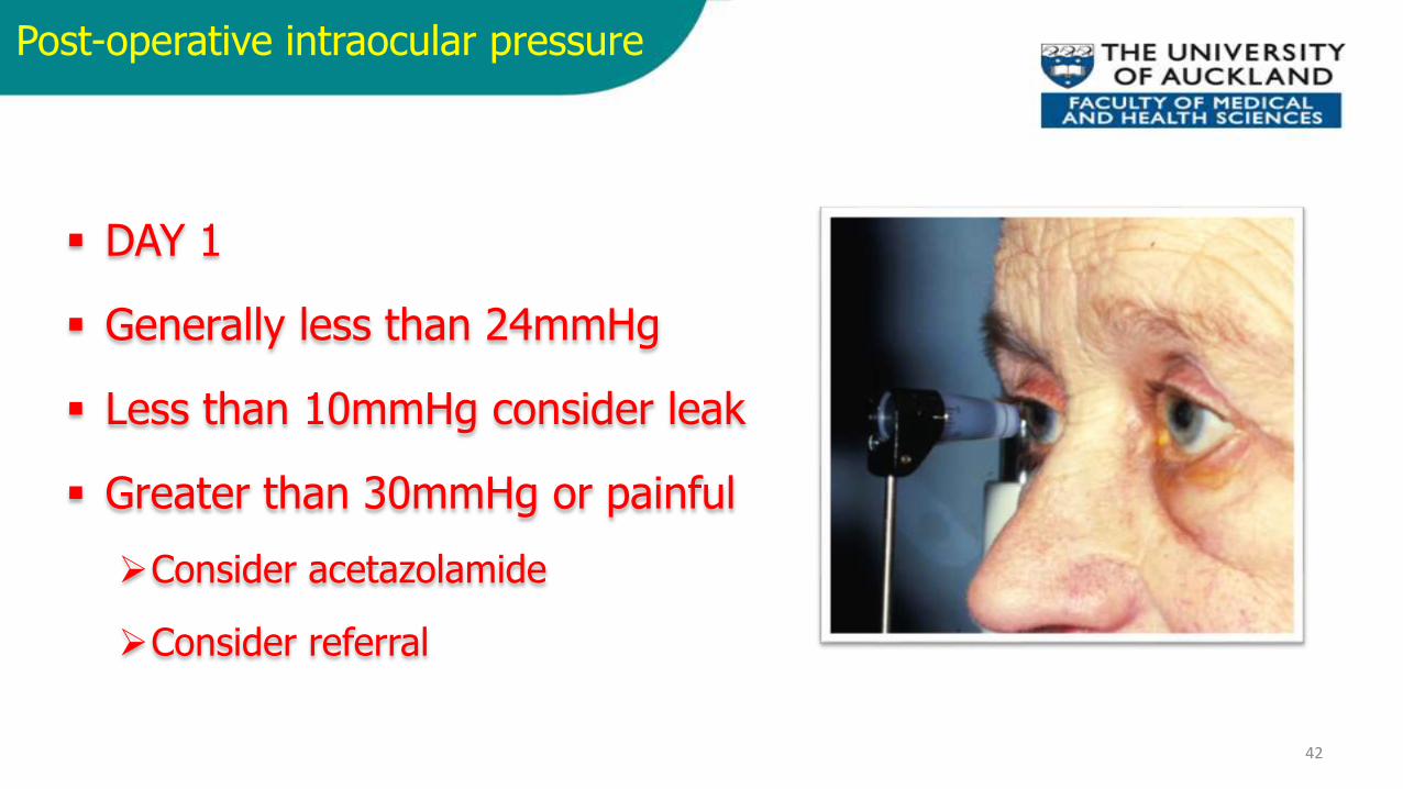
42
DAY 1
Generally less than 24mmHg
Less than 10mmHg consider leak
Greater than 30mmHg or painful
Consider acetazolamide
Consider referral
Post-operative intraocular pressure

Appearance related to cataract density, difficulty of case, nucleus density, FECD and phaco energy used:
1. Entirely clear
2. Occasional effete endothelial cells
3. Focal striae & oedema at incisions
4. Extensive striae & oedema
Generally resolves - <0.5% develop PBK
Corneal appearance day 1

Should be round, up to mid-dilated,
If distorted exclude:
• Iris prolapse
• Vitreous to wound
• Vitreous in anterior chamber
• IOL displacement
Postoperative pupil

Day 1
Flare +
Cells + to ++
If heavy flare or cells +++ consider endophthalmitis
Day 28
Usually no activity
5% may have persisting low-grade inflammation
Anterior chamber inflammation

Auckland Cataract Study 2 (2015-16)Postoperative visual outcomes (N=1000)
Visual acuity 2015(N=476)
2016(N=472)
P-value fordifference
Unaided 6/12 6/12 0.262
Best-corrected 6/9 6/9 0.648

IOL should be well centred
Relative to capsular bag / rhexis
Relative to pupil
If > 1.0mm IOL displacement
Consider haptic position
Assess for vitreous in AC
Discuss with ophthalmologist
Postoperative IOL position

If BSCVA less than expected
Assess macula
Exclude retinal detachment
Exclude vitreous/retinal
haemorrhage
Fundal examination

Day 28 review
Symptoms
Unaided VA
Refraction
Assess: Cornea
Pupil
Media
IOL position
IOP
Dilated fundus
Post-operative day 28

Posterior capsular thickening 2-5%
Rx YAG laser
Capsular phimosis
Retinal detachment 1%
Delayed complications

Posterior Capsular Opacification

YAG Laser capsulotomy
52
Posterior capsule opacification

Co-management Summary
Assessment Normal Action
Unaided vision 6/18 - 6/5 Refract if <6/12
Corrected vision 6/6 – 6/12 Exclude causes of reduced BSCVA if <6/12
Refractive error +/-1.00D
of intended
If greater than 1.50D deviation from intended
endpoint contact ophthalmologist
Exclude wound leak None If spontaneous & AC compromised - refer
Wound appearance Closed exclude phaco burn, retraction, or leak
Goldman IOP <24mmHg If painful or >30mmHg Rx diamox or refer
Corneal striae or oedema None/Minimal Observe, should settle in few days
Anterior chamber activity Flare+,
cells + to ++
If marked activity consider endophthalmitis or
secondary uveitis - refer
Pupil Round, up to
mid-dilated
If distorted: exclude iris prolapse, vitreous in
AC or to wound, or IOL displacement - refer
IOL position Well-centred
in the bag
If greater than 1.0mm decentred - refer
Fundal examination Clear view Exclude haemorrhage, detachment

What to tell your patients?
• Public surgery at ≤ 6/12 vision (Private insurers typically ≤ 6/9)
• Will need to complete “Impact on life” questionnaire
• Cataract surgery is safe and typically takes < 30 minutes
• Small risk of intra/post-operative compilations < 5%
• More than 95% of patients will have significant improvement
54
Establishing eligibility

Thank you


![refractive error, cataract, age-related macular degeneration, diabetic retinopathy, glaucoma, and corneal opacity.[1,2] Similarly, top causes of blindness in the United States include](https://cdn.vdocument.in/doc/165x107/5f780010f1163d15b07111eb/-refractive-error-cataract-age-related-macular-degeneration-diabetic-retinopathy.jpg)
