Download - Starter - LT Scotland
Starter

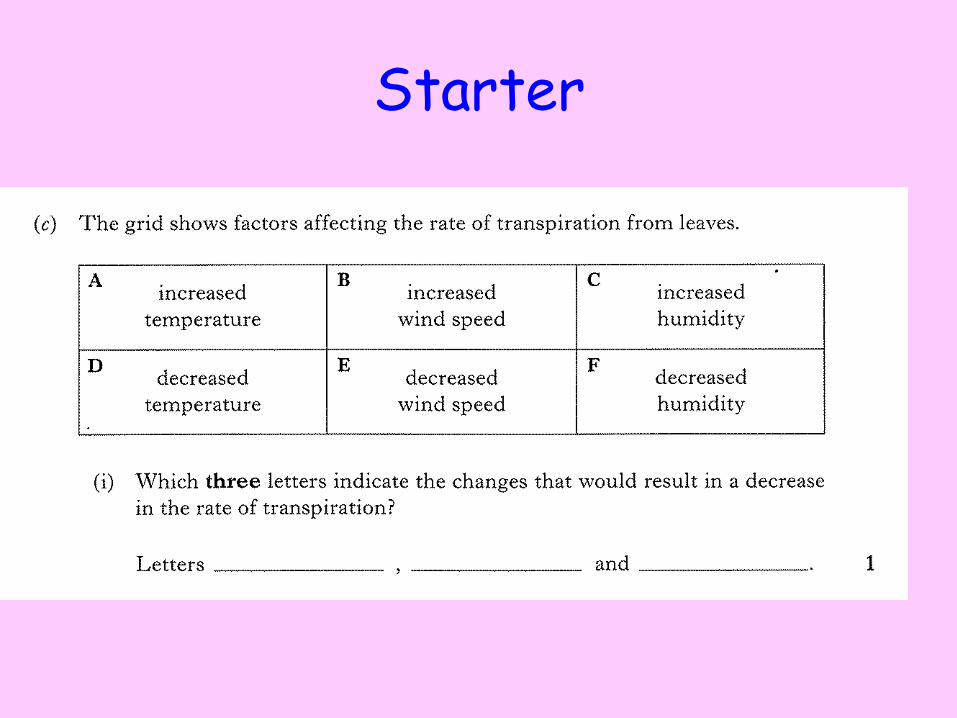
Starter
C D E

Animal transport systems: The circulatory system
We are learning to
• Identify the different organs of the circulatory system.
• Identify the different organs of the heart.
I can
• Label the chambers, blood vessels, valves and direction of blood flow through the heart.

Animal Transport
• In mammals the transport and exchange of nutrients and gases occurs through out the body.
• To allow this to occur the mammalian body has a complex network of transport pathways.
• The three main body systems we’ll focus on are the Circulatory, Respiratory and Digestive systems.
• Do you know what each of these systems does??

Circulatory system
• The circulatory system is made up of the heart, blood and blood vessels.
• Its main function is to transport oxygen and nutrients to the cells of the body.

Heart structure • The heart is made up of
four chambers separated by valves.
• These are the right and left, Atria and Ventricles.
• The right hand side of the heart pumps deoxygenated blood while the left side pumps oxygenated blood.
Lungs
Body
Coronary Arteries

Re-Cap 1.
2.
3.
4.
5.
6.
7.
8.
9.
10.
11.

On your diagram
1. Colour in the side that deals with oxygenated blood red and the side that deals with deoxygenated blue
2. Label the:
• Right Atrium
• Right Ventricle
• Left Atrium
• Left Ventricle
• Pulmonary vein
• Pulmonary artery
3. Draw arrows to show the flow of blood through the heart.
• Aorta • Vena cava • A-V Valves • Semi- lunar
valves • Coronary
arteries

The Heart
Aorta
Vena cava
Pulmonary Artery
Pulmonary Veins
Thick muscular wall
Coronary Arteries

Heart Dissection
• Pig heart dissection-
• Heart Dissection
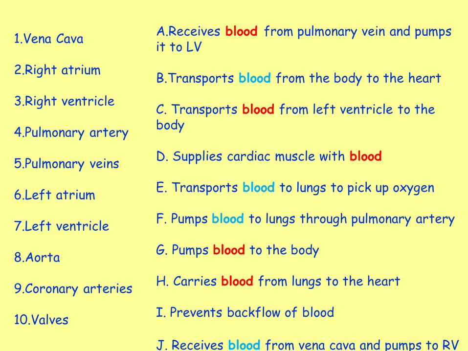
1.Vena Cava 2.Right atrium 3.Right ventricle 4.Pulmonary artery 5.Pulmonary veins 6.Left atrium 7.Left ventricle 8.Aorta 9.Coronary arteries 10.Valves
A.Receives blood from pulmonary vein and pumps it to LV B.Transports blood from the body to the heart C. Transports blood from left ventricle to the body D. Supplies cardiac muscle with blood E. Transports blood to lungs to pick up oxygen F. Pumps blood to lungs through pulmonary artery G. Pumps blood to the body H. Carries blood from lungs to the heart I. Prevents backflow of blood J. Receives blood from vena cava and pumps to RV

Vena Cava Right atrium Right ventricle Pulmonary artery Pulmonary veins Left atrium Left ventricle Aorta Coronary arteries Valves
Transports blood from the body to the heart
Receives blood from vena cava and pumps to RV
Pumps blood to lungs through pulmonary artery
Transports blood to lungs to pick up oxygen
Transports blood from lungs to the heart
Receives blood from pulmonary vein and pumps it to LV
Pumps blood to the body
Transports blood from left ventricle to the body
Supplies cardiac muscle with blood
Prevents backflow of blood


Multiple Choice Quiz


Heart Bingo
Pick any 4 features from your heart diagram and write them in your jotter

Quick Questions
1. What are the two chambers at the bottom of the heart called?
2. What are the two chambers at the top of the heart called?
3. Which blood vessels carry blood into the heart?
4. Which blood vessels carry blood away from the heart?
5. What is the function of valves?
6. Where does the left hand side of the heart pump blood to?
7. Where does the right hand side of the heart pump blood to?

Animal transport systems: Blood Vessels
We are learning to
• Identify the structure and function of the blood vessels in the circulatory system.
I can
• Describe the function of arteries, veins and capillaries
• Describe how the structure of blood vessels is related to their function.

Blood vessels
• There are three types of blood vessel found in the body.
• Arteries- carry blood away from the heart.
• Veins- carry blood in to the heart.
• Capillaries- connect arteries and veins.

Arteries
•Arteries have a pulse – this is the movement of blood being pushed out of the heart along an artery.
•Arteries have thick muscular walls to cope with the blood travelling under high pressure.

Veins
• Veins carry blood under lower pressure so have thinner muscular walls.
• Veins have valves to prevent the backflow of blood.

Capillaries • Capillaries allow exchange of materials between the blood and the cells of the body (O2, glucose).
• Capillaries are only one cell thick to allow easier/quicker diffusion of materials.
•Capillaries also provide a large surface area for exchange.


Carries blood away from the heart
Carries blood towards the heart
Allows exchange of materials
Place these features of blood vessels in a table with the headings arteries, veins and capillaries
Thick muscular walls
Thinner walls
Walls are 1 cell thick
High pressure
Low pressure
Has large surface area for exchange
Narrow central cavity
Wider central cavity
Forms network at tissues
Contains valves

Carries blood away from the heart Carries blood towards the heart
Allows exchange of materials
Arteries
Thick muscular walls Thinner walls
Walls are 1 cell thick
High pressure Low pressure
Has large surface area for exchange
Narrow central cavity Wider central cavity
Forms network at tissues
Contains valves
Veins
Capillaries
Task
• Complete the worksheet on blood vessels.


2 truths and a lie
1. The vena cava carries blood from the lungs to the heart
2. The pulmonary artery carries blood from the heart to the lungs
3. The coronary arteries supply the cardiac muscle with blood
1. The left ventricle has a thicker wall than the right ventricle
2. The left ventricle carries deoxygenated blood
3. The left ventricle pumps blood to the body through the aorta
Work out which are the truths and which Are the lies in the statements below then
Come up with two truths and a lie of your own.

Animal transport systems: The blood
We are learning to
• Identify the components of blood
• Describe the function of red and white blood cells
• Describe in detail the action of lymphocytes and phagocytes.
I can
• Name the 4 components of blood
• Identify how red blood cells area adapted for their function
• Detail the steps in phagocytosis and explain the function of lymphocytes.


Blood Composition
• Blood is made up of three main parts:
– plasma
– red blood cells
– white blood cells
• Plasma is the liquid part of blood that carries many substances such as sugars, salts, amino acids, proteins, vitamins, water, carbon dioxide and urea.
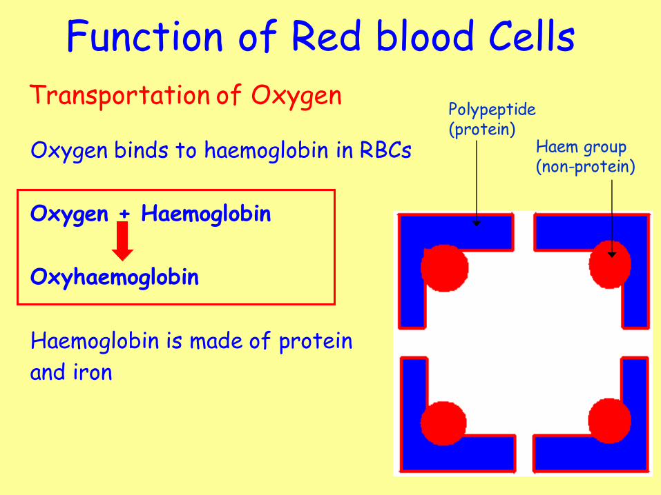
Function of Red blood Cells
Oxygen binds to haemoglobin in RBCs
Oxygen + Haemoglobin
Oxyhaemoglobin
Haemoglobin is made of protein
and iron
Polypeptide (protein)
Haem group (non-protein)
Transportation of Oxygen
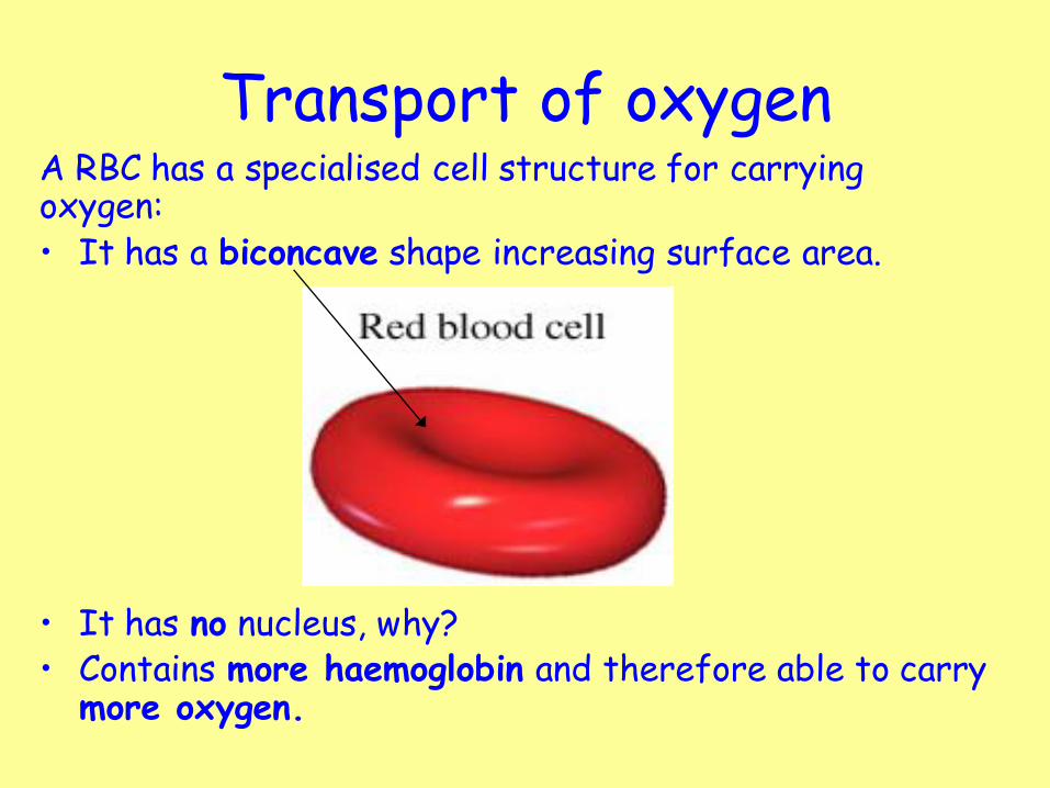
Transport of oxygen A RBC has a specialised cell structure for carrying oxygen: • It has a biconcave shape increasing surface area.
• It has no nucleus, why? • Contains more haemoglobin and therefore able to carry
more oxygen.

Pathway of blood •The pathway of blood through the heart, lungs and body follows the path shown in the diagram opposite.
•You should be able to start at any point and name the chambers and blood vessels that the blood travels through.
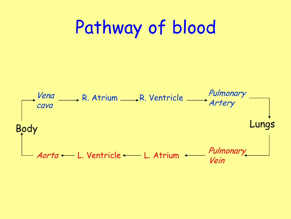
Pathway of blood
Body
Vena cava
R. Atrium R. Ventricle Pulmonary Artery
Lungs
Pulmonary Vein
L. Atrium L. Ventricle Aorta

Quick Questions
1. What are the three components of blood
2. What is the function of red blood cells
3. What does oxygen join to in a red blood cell and what molecule does it form?
4. What are the specialised features of a red blood cell?
5. How do these features ensure it can carry out it’s function?

White Blood Cells
• White blood cells make up our immune system.
• Our immune system keeps us healthy by seeking out and killing foreign cells.

White blood cells • The 2 main types of WBC’s are Phagocytes and
Lymphocytes.
Phagocyte
Lymphocyte

Phagocytosis
• Glow Phagocyte Video
• Phagocytosis is the process by which pathogens are engulfed and destroyed by phagocytes.
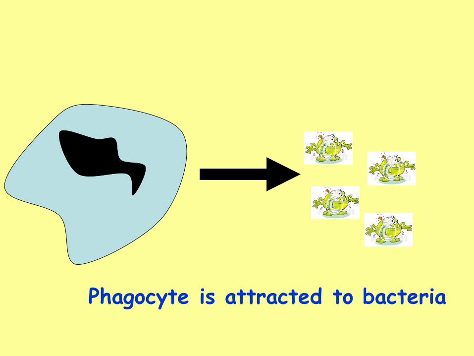
Phagocyte is attracted to bacteria

Phagocyte detects bacteria

White blood cells and immunity
Phagocyte engulfs bacteria

White blood cells and immunity
Phagocyte digests bacteria

Put the phagocytosis steps in order. (draw a picture to illustrate)
• The phagocyte moves forward and adheres the bacterial cell
• A phagocytic cell detects chemicals that bacteria release.
• Once inside the phagocyte, powerful digestive enzymes from lysosomes are released and these digest the bacteria
• The phagocyte then engulfs the bacteria by trapping it inside a vacuole inside its cytoplasm.

Lymphocytes
• Glow Video - immune response
• Antigens are found on the surface of pathogens.
• Antigen presence stimulates lymphocytes to produce molecules called antibodies. Each antibody is specific to a particular antigen.
• The lymphocytes can then destroy the pathogen in a number of ways.

White blood cells and defence • Bacteria in the blood make unique_________ on their
surfaces called ______.
• _________ are made by ___________ which recognise them as different. They then bind on to the _________ on the surface of the bacteria.
• _________ will engulf the invading bacterial cells and ______ them using ________.
• Your white blood cells remember the right ________ needed to tackle a particular ________. If you meet that ______ again they make the same __________ very quickly, so you become _________ to that disease.
Immune antigen antibody lymphocyte phagocyte proteins digest Enzymes pathogen

White blood cells and defence • Bacteria in the blood make unique proteins on their
surfaces called antigens.
• Antibodies are made by lymphocytes which recognise them as different. They then bind on to the antigen on the surface of the bacteria.
• Phagocytes will engulf the invading bacterial cells and digest them using enymes.
• Your white blood cells remember the right antibody needed to tackle a particular antigen. If you meet that pathogen again they make the same antibody very quickly, so you become immune to that disease.
Immune antigen antibody lymphocyte phagocyte proteins digest Enzymes pathogen

Starter

Starter
To stop backflow of blood.
P M

Animal transport systems: Diffusion at the alveoli
We are learning to
• Identify the structures and functions of the respiratory system.
• Describe how alveoli are adapted for the process of diffusion.
I can
• Label the diagram of the lungs
• Identify 3 ways in which the alveoli are adapted for efficient gas exchange.
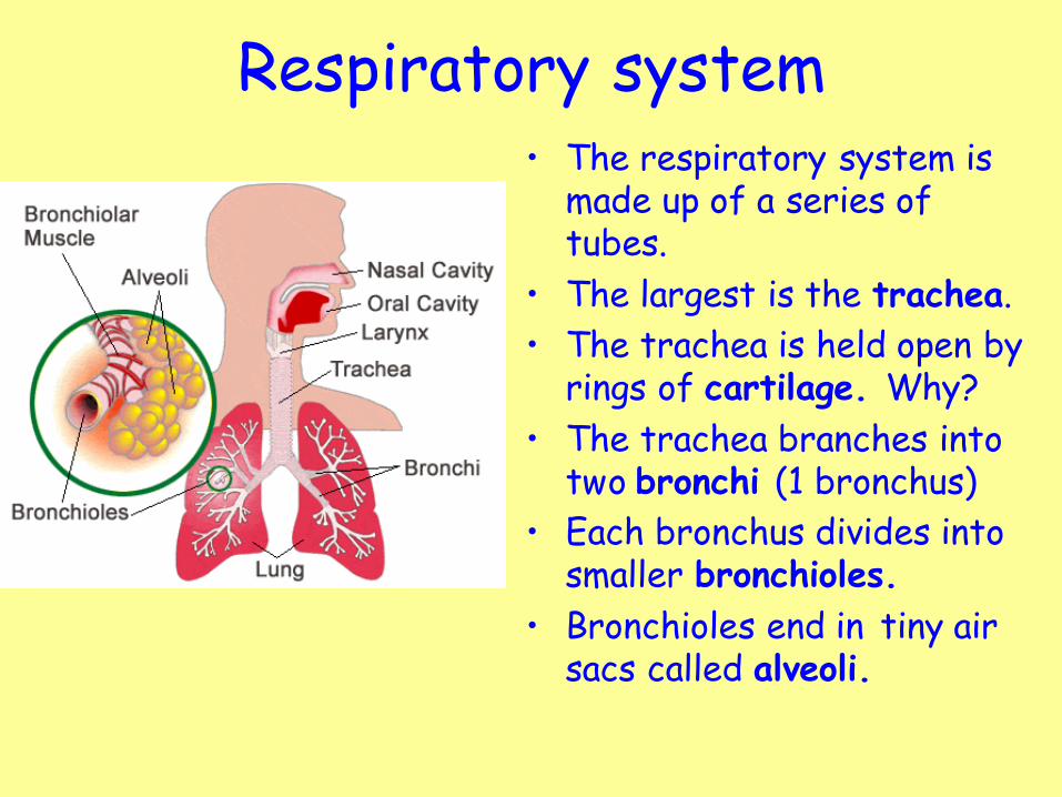
Respiratory system • The respiratory system is
made up of a series of tubes.
• The largest is the trachea.
• The trachea is held open by rings of cartilage. Why?
• The trachea branches into two bronchi (1 bronchus)
• Each bronchus divides into smaller bronchioles.
• Bronchioles end in tiny air sacs called alveoli.

Respiratory System • The trachea contains cartilage
rings to prevent it from sticking closed.
• Mucus is produced by goblet cells to trap dirt and bacteria.
Mucus producing cells
u
• Cilia are hair like projections that move the mucus up to back of the throat to be swallowed or spat out. •Alveoli are the site of gas exchange.


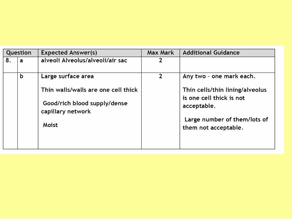
Alveoli
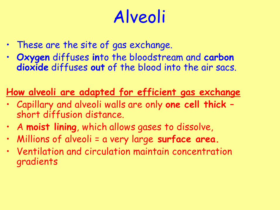
• These are the site of gas exchange. • Oxygen diffuses into the bloodstream and carbon
dioxide diffuses out of the blood into the air sacs.
How alveoli are adapted for efficient gas exchange • Capillary and alveoli walls are only one cell thick –
short diffusion distance. • A moist lining, which allows gases to dissolve, • Millions of alveoli = a very large surface area. • Ventilation and circulation maintain concentration
gradients

Questions 1. What cells produce mucus in the respiratory system
and where are these cells found?
2. What is the function of the mucus?
3. Which specialised cells sweep the mucus out of the lungs?
4. What are the small air sacs at the bottom of the lungs known as?
5. Label your air sac diagram to show:
A red blood cell, The capillary, the wall of the alveoli, the direction oxygen and carbon dioxide flow from the capillary to the air sac.
6. List at least two adaptations of the alveoli to ensure rapid diffusion of gases.

Starter
Starter
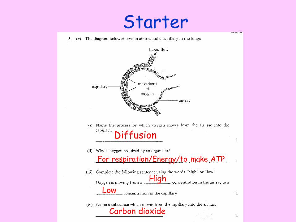
Diffusion
High
Low
Carbon dioxide
For respiration/Energy/to make ATP

Animal transport systems: Diffusion at the villus
We are learning to
• Identify where diffusion of substances occurs in the digestive system.
• Describe ways in which parts of the digestive system are adapted for diffusion of materials.
I can
• Identify where diffusion occurs in the digestive system
• Identify how this organ is adapted for the absorption of materials.

3.
1.
2.
4.
5.
7.
9.
6.
8.
10.
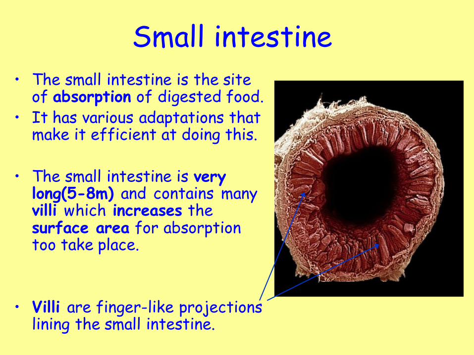
Small intestine • The small intestine is the site
of absorption of digested food. • It has various adaptations that
make it efficient at doing this.
• The small intestine is very long(5-8m) and contains many villi which increases the surface area for absorption too take place.
• Villi are finger-like projections lining the small intestine.

Villi Each Villus wall is only 1 cell thick. There are approximately 4-5 million Villi in the small intestine.
The lacteal absorbs products of fat digestion (fatty acids/ glycerol).
The blood vessels provide a good blood supply to aid absorption of glucose and amino acids.
Tasks
1. What is the function of the small intestine?
2. Draw and label a diagram of the villus.
3. Where in the villus do the products of fat digestion get absorbed?
4. Name a substance that is absorbed into the bloodstream.
5. Identify 2 ways in which the villus is adapted for efficient diffusion.

Model Gut experiment
• The following experiment represents how the small intestine functions. (The visking tubing is the intestine, the water is the blood.)
• The lining or membrane, of the small intestine is selectively permeable.
• This means only certain, small, soluble molecules are able to diffuse across the lining of the small intestine.
• Eg, glucose, water, amino acids

Model Gut experiment • Aim: To show membranes are selectively permeable.
• Method:
1. Soak a 20cm long piece of visking tubing in water to soften. Tie a knot in one end.
2. Using syringes, add 10ml of starch and 10ml of glucose solution to the visking tubing bag.
3. Tie a knot in the other end, trim excess tubing to 1cm length and wash thoroughly under running water.
4. Place the bag in a boiling tube and fill the remaining space with water.
5. Immediately remove a few mls of water using a dropper. 6. Put 2 drops in a dimple on a spotting tile and the rest in a
small test tube. 7. Add iodine to the spotting tile. Add Benedicts to the small
test tube and place in a the water bath at 90°C. 8. Repeat steps 6 and 7 after 15 minutes and 30 minutes.

Model Gut experiment • Results:
• What do you think will happen? Why?
• Starch should remain in the visking tubing and glucose should diffuse out.
• Starch is a large complex molecule and is to big to pass through the membrane.
• Conclusion: now write out your own conclusion
Time (mins) Starch test Glucose test
0
15
30