Sudden Cardiac Death (SCD):Causes, Clinical Evaluation, and Treatment
William Whang, MD, MS

Outline
• Definition of SCD• Conditions associated with SCD• Evaluation for risk prediction• Therapy for prevention of SCD

Definition
• Death within 1 hour of the onset of symptoms, without preceding heart failure
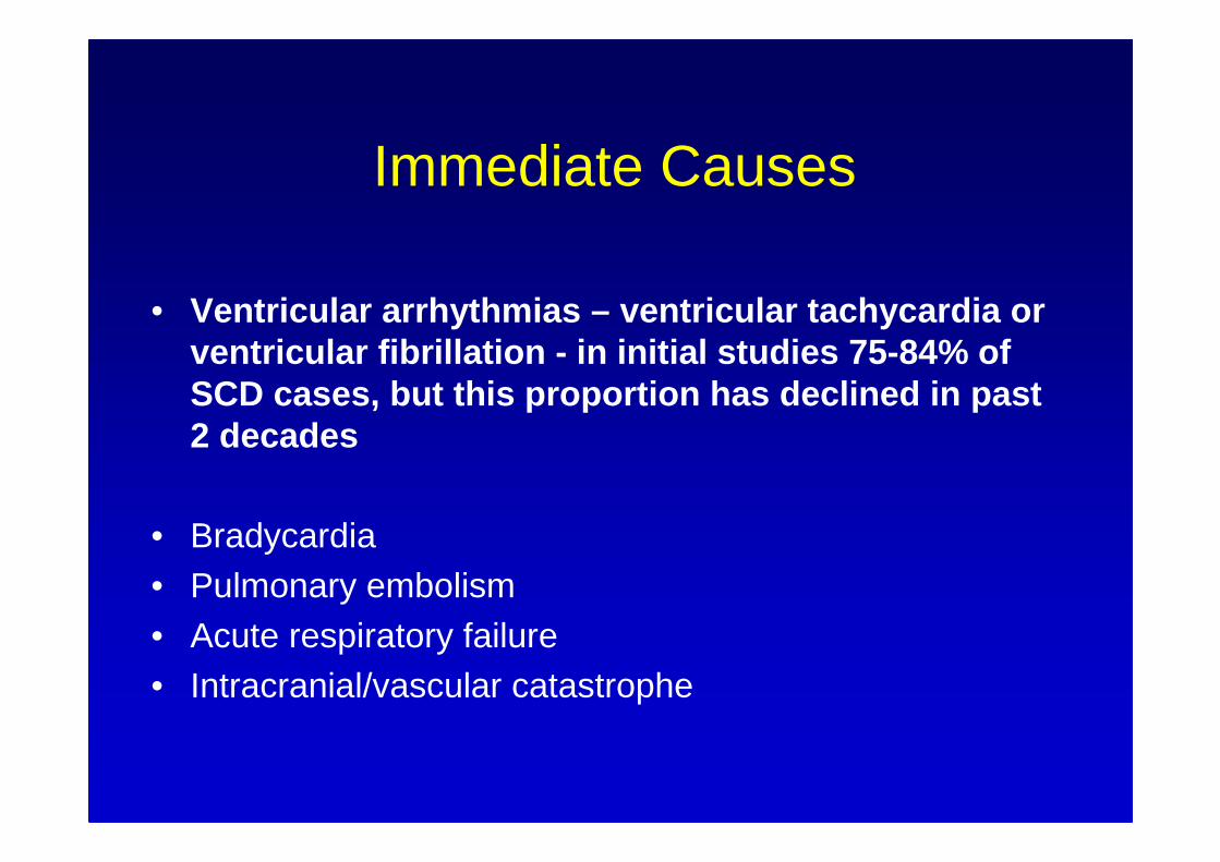
Immediate Causes
• Ventricular arrhythmias – ventricular tachycardia or ventricular fibrillation - in initial studies 75-84% of SCD cases, but this proportion has declined in past 2 decades
• Bradycardia• Pulmonary embolism• Acute respiratory failure• Intracranial/vascular catastrophe

Burden of SCD
• Incidence: 41-89 per 100,000 per year in general population
• 180,000 to 250,000 per year in US
• Survival to hospital discharge: 4.4%*
*Nichol G, Thomas E, Callaway CW, Hedges J, Powell JL, Aufderheide TP, Rea T, Lowe R, Brown T, Dreyer J, Davis D, Idris A, Stiell I. Regional variation in out-of-hospital cardiac arrest incidence and outcome. JAMA. 2008 Sep 24;300(12):1423-31.

Normal Sinus Rhythm
Ventricular Tachycardia

Typical sequence of SCD episode

Chugh SS, Reinier K, Teodorescu C, Evanado A, Kehr E, Al Samara M, Mariani R, Gunson K, Jui J. Epidemiology of sudden cardiac death: clinical and research implications. Prog Cardiovasc Dis. 2008;51(3):213-228.
Etiologies of SCD

S.S. Chugh, J. Jui and K. Gunson et al., Current burden of sudden cardiac death: Multiple source surveillance versus retrospective death certificate–based review in a large U.S. community, J Am Coll Cardiol 44 (2004), pp. 1268–1275.
SCD and Age

S.S. Chugh, K. Chung and Z.J. Zheng et al., Cardiac pathologic findings reveal a high rate of sudden cardiac death of undetermined etiology in younger women, Am Heart J 146 (2003), pp. 635–639.
Spectrum of SCD in Young (35-44)

• Post-myocardial infarction– fibrous tissue replaces necrotic myocardium– collagen and myocardium intermingled, results in
conduction delay– Reduced/redistributed connexin expression– Reduced Na+ channel density
Arrhythmic Substrate in Coronary Artery Disease
deBakker 2006.
Kostin SM 2003.

Arrhythmic Substrate in Coronary Artery Disease
Stevenson WG. JACC 1997.

Non CAD-related Causes of SCD
• Structural Heart Disease– Hypertrophic Cardiomyopathy– Arrhythmogenic Right Ventricular Cardiomyopathy– Cardiac Sarcoidosis– Cardiac Amyloidosis– Anomalous Coronary Arteries– Adult Congenital Heart Disease
• Electrical Disease– Long QT syndromes– Brugada syndrome– Familial catecholaminergic polymorphic VT– Short QT syndrome– Wolff-Parkinson-White syndrome

Structural heart disease: Hypertrophic Cardiomyopathy
• Incidence of SCD ~1-5 % per year depending on number of risk factors
• Risk factors for SCD – family history of SCD, history of ventricular tachycardia by ambulatory monitoring, history of syncope, septal thickness >30 mm, drop in BP with exercise

Electrical: Brugada Syndrome
• ~20% of sudden deaths in patients with a structurally normal heart
• SCN5A mutation– Reduced channel function– Coved ST segment on
ECG• SCD risk higher with
spontaneous ST coving, history of syncope

Electrical: Wolff-Parkinson-White
Accessory pathways - aberrant cardiac muscle bundles capable of rapid conduction that connect the atrium to the ventricle.

Electrical: WPW


Link MS et al. N Engl J Med 1998;338:1805-11.
Electrical ‘malfunction’: Commotio Cordis
Maron BJ et al. N Engl J Med 1995;333:337-42.
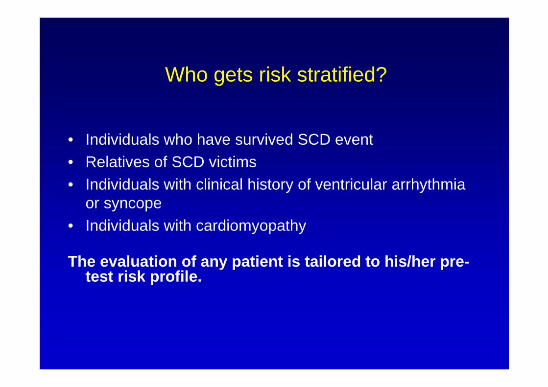
Who gets risk stratified?
• Individuals who have survived SCD event• Relatives of SCD victims• Individuals with clinical history of ventricular arrhythmia
or syncope• Individuals with cardiomyopathy
The evaluation of any patient is tailored to his/he r pre-test risk profile.

Workup for SCD risk
• History and physical, family history• Electrocardiogram (ECG)
Electrical substrate• Holter monitor (ambulatory ECG monitoring)• T wave alternans test• Signal-averaged ECG
Structural• Echocardiogram/cardiac MRI
Provocative • Exercise test• Electrophysiology study
Genetic testing

History
• Documented arrhythmia, particularly ventricular tachycardia– History of fainting (syncope) without warning,
especially with exertion– Palpitations/lightheadedness
• Structural heart disease – history of myocardial infarction– Symptoms of shortness of breath
• Family history of SCD
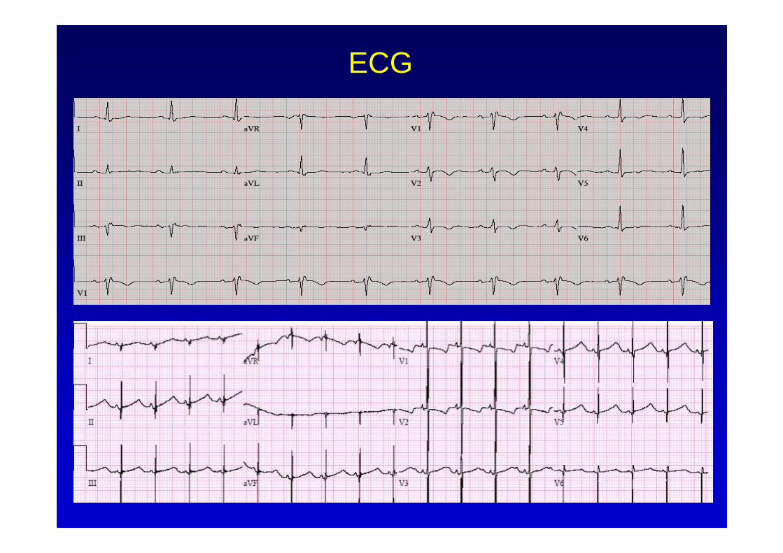
ECG

Structural Disease
• Left ventricular dysfunction is best available risk predictor based on positive predictive value/specificity, especially in CAD
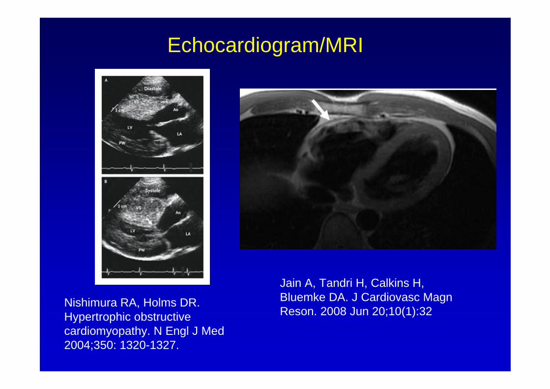
Nishimura RA, Holms DR. Hypertrophic obstructive cardiomyopathy. N Engl J Med 2004;350: 1320-1327.
Jain A, Tandri H, Calkins H, Bluemke DA. J Cardiovasc MagnReson. 2008 Jun 20;10(1):32
Echocardiogram/MRI

Provocative testing: Exercise test
• Purpose– to evaluate for evidence of coronary artery disease– To evaluate for exercise-induced arrhythmias (e.g.
catecholaminergic VT)


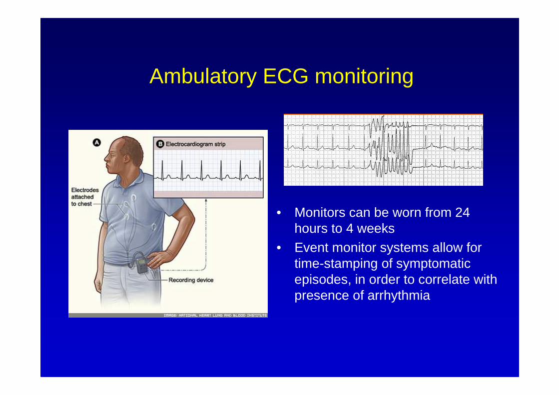
Ambulatory ECG monitoring
• Monitors can be worn from 24 hours to 4 weeks
• Event monitor systems allow for time-stamping of symptomatic episodes, in order to correlate with presence of arrhythmia
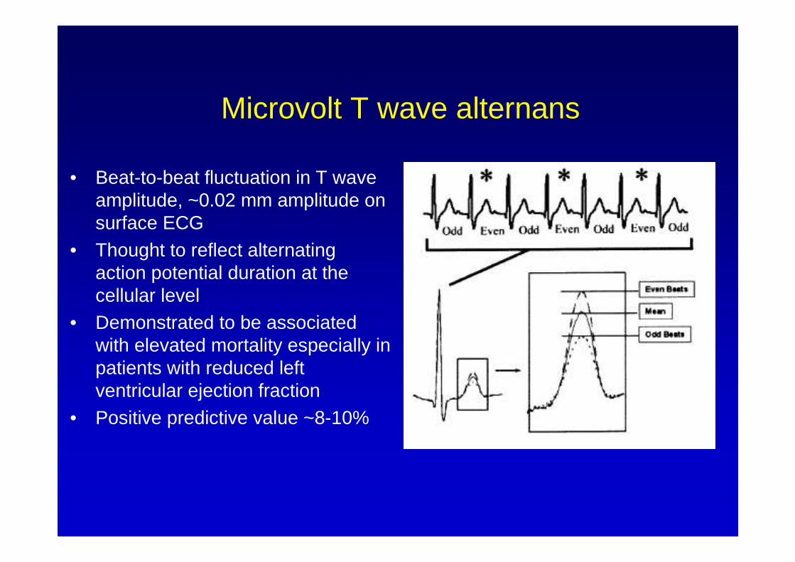
Microvolt T wave alternans
• Beat-to-beat fluctuation in T wave amplitude, ~0.02 mm amplitude on surface ECG
• Thought to reflect alternating action potential duration at the cellular level
• Demonstrated to be associated with elevated mortality especially in patients with reduced left ventricular ejection fraction
• Positive predictive value ~8-10%

Signal-averaged ECG
• Seek to detect late activation in myocardium (late potentials)
• 3 parameters are assessed – duration of the filtered QRS, duration of the low-amplitude signal, and root mean square voltage in the last 40 msec of the QRS complex.
• May be particularly useful for diagnosis in arrhythmogenic right ventricular cardiomyopathy

Electrophysiology (EP) Study
• Electrical stimulation of the ventricle via pacing catheters• Attempt to induce ventricular tachycardia by programmed
ventricular stimulation
• Sustained ventricular tachycardia at EP study is predictive of SCD in patients with reduced left ventricular ejection fraction and coronary artery disease
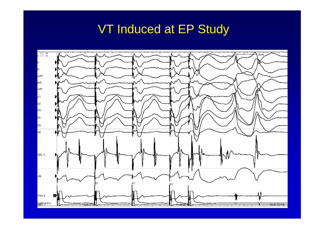
VT Induced at EP Study

VT Induced at EP Study

Genetic testing
• Commercially available for long QT, hypertrophic cardiomyopathy, Brugada syndrome, ARVC
• Yield varies depending on population studied: – ~35% in patients referred for long QT testing– Limited by lack of sensitivity
• Mostly used to attempt diagnosis/risk stratification in relatives of individuals with known conditions

Treatments
• Treatment for underlying heart disease – e.g. aspirin, beta blockers, angiotensin converting-enzyme inhibitors for coronary artery disease

Treatments
• Antiarrhythmic therapy
• Defibrillator therapy
• Catheter ablation

Anti-arrhythmic medications
• Sodium channel blockers – lidocaine (IV), quinidine
• Potassium channel blockers – sotalol, dofetilide
• Sodium/potassium blockers – amiodarone
No medication is efficacious enough (~50-70%) for most conditions that predispose to SCD

Early ICDs
• Short-lived• Shock only• 250 g• Nonprogrammable• Required thoracotomy
abdominal implant
Built from over-the-counter electronics

Modern ICDs

ICD Benefit
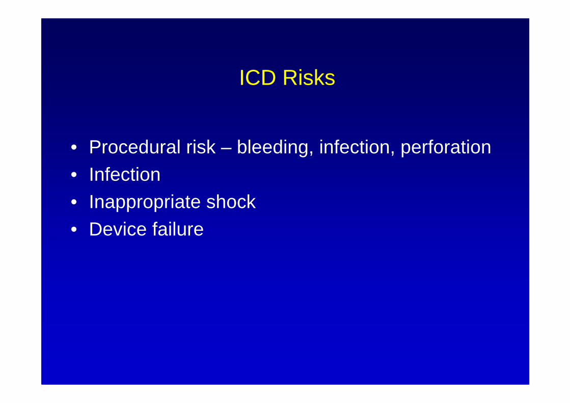
ICD Risks
• Procedural risk – bleeding, infection, perforation
• Infection• Inappropriate shock
• Device failure

How does workup get integrated? Brugada syndrome

• Among all patients with coronary artery disease, depending on the age group, only 13% to 20% will have sudden cardiac arrest.
• Using left ventricular ejection fraction as a risk stratifier, ~15 ICDs need to be implanted to save one person’s life.
• How to identify those who are at high risk?
The Conundrum of Risk Stratification in CAD

28.9
34.1 35.8
05
10
15202530
354045
ICD Amiodarone Placebo
Mor
talit
y (%
)SCD-HeFT
All-cause mortality at 5 years
Amiodarone vs placeboHR 1.06, p=0.529ICD vs placebo
HR 0.77, p=0.007
Presented at ACC Scientific Sessions 2004

Myerburg RJ, Kessler KM, Castellanos A. Sudden cardiac death. Structure, function, and time-dependence of risk. Circulation. 1992;85(1 Suppl):I2-10.
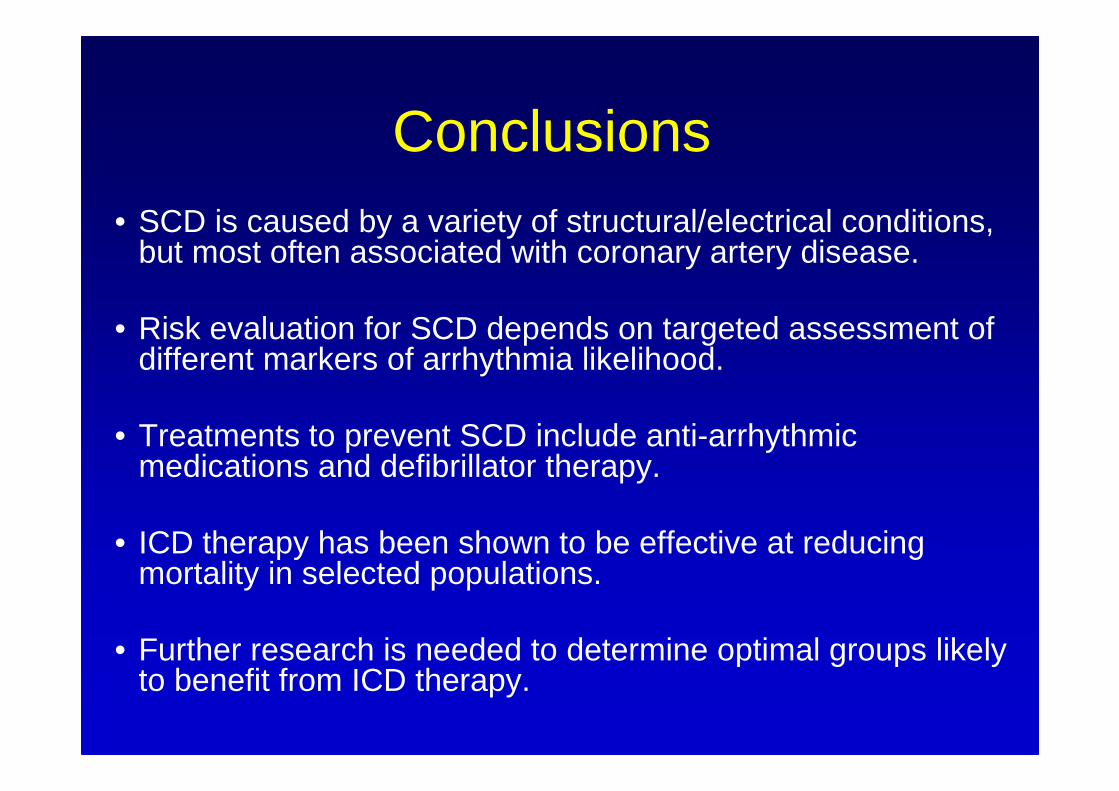
Conclusions• SCD is caused by a variety of structural/electrical conditions,
but most often associated with coronary artery disease.
• Risk evaluation for SCD depends on targeted assessment of different markers of arrhythmia likelihood.
• Treatments to prevent SCD include anti-arrhythmic medications and defibrillator therapy.
• ICD therapy has been shown to be effective at reducing mortality in selected populations.
• Further research is needed to determine optimal groups likely to benefit from ICD therapy.


