1
eLS A26071 1
Supercoiled DNA: Structure 2
Richard Peter Bowater, University of East Anglia, Norwich, UK 3
4
Based in part on the previous version of this eLS article ‘Supercoiled DNA: Structure’ (2005) 5
6
Advanced Article 7
(Advanced articles are aimed at advanced undergraduates, graduate students, postgraduates, 8
and researchers reading outside their field of expertise.) 9
Abstract 10
Supercoiling is introduced into DNA molecules when the double helix is twisted around its 11
own axis in three‐dimensional space. Experimental techniques that are sensitive to 12
molecular shape can be used to analyse the topological states of DNA, but the approaches 13
used most successfully are high-speed centrifugation, high-resolution microscopy and gel 14
electrophoresis. Generally, DNA molecules are negatively supercoiled inside cells, although 15
the level of supercoiling is not equal throughout the genome and many supercoils may be 16
constrained by bound proteins. Supercoiling increases the free energy of DNA and 17
influences DNA metabolism by promoting or hindering specific enzymatic processes. DNA 18
topoisomerases are the main enzymes that regulate DNA topology and several different 19
types of enzymes are present in all cells. 20
Keywords: DNA; linking number; supercoiling; topology; twist; writhe 21
22
Key Concepts 23
Double stranded DNA helices can wind in three-dimensional space to form further 24
helices of higher order, forming supercoiled DNA. 25
Since the early 1960s the importance of DNA supercoiling to cellular processes has 26
been apparent, with its most obvious consequence being that it aids compaction of 27
large DNA molecules into the relatively small volume of cells. 28
The extent of supercoiling in a DNA molecule is influenced by environmental 29
conditions, such as ionic strength and temperature; since supercoiling of DNA 30
influences the biological pathways in which it is involved, the level of DNA 31
supercoiling inside cells is tightly regulated. 32
Supercoiling provides a significant amount of free energy to DNA molecules and, 33
inside cells, this can be used to drive structural transitions and other metabolic 34
processes that would normally be thermodynamically unfavourable, such as opening 35
of the DNA helix during replication and transcription. 36

2
Mathematical and modelling studies have provided insight for quantitative analyses 1
of DNA supercoiling, leading to definitions for twist, which describes how the 2
individual strands of DNA coil around its axis, and writhe, which describes how the 3
helix axis coils in three-dimensional space. 4
DNA inside cells contains supercoils of two types: interwound, which is when circular 5
DNA winds around its own axis; toroidal supercoiling occurs when the DNA helix 6
forms a series of spirals around an imaginary ring. 7
Any technique that is sensitive to molecular shape will be useful for experimental 8
analysis of supercoiled DNA, but the large size of the molecules mean few 9
techniques have been used successfully; those that have been widely used include 10
high-speed centrifugation, high-resolution microscopy (e.g. electron microscopy and 11
scanning-force microscopy) and agarose gel electrophoresis. 12
A wide variety of proteins that bind to DNA alter the local geometry of its helix and 13
influence DNA topology; an important characterised example of this effect is the 14
winding of DNA around the eukaryotic histone octamer to form the nucleosome. 15
A fundamental feature of closed domains of DNA, such as a circular molecule, is that 16
the two strands of DNA are topologically linked and strand separation can be 17
achieved only by breakage of one of the strands; the main enzymes that regulate 18
DNA topology are DNA topoisomerases and they may act to remove or introduce 19
negative supercoils or they may remove both positive and negative supercoils. 20
Cellular processes that move macromolecular assemblies along DNA may generate 21
localized DNA supercoiling since, as the large protein complex moves along the 22
DNA, its rotation around the DNA may be inhibited. 23
24
Introduction 25
Normally, DNA occurs as a helical, double-stranded molecule in which the two strands pair 26
up in antiparallel fashion; this is the classical B-type helical structure first solved in 1953 by 27
Watson and Crick (Watson and Crick, 1953) using a range of experimental data obtained by 28
many other scientists (Chargaff et al, 1950; Franklin and Gosling, 1953; Wilkins et al, 1953). 29
The DNA helix is usually visualized in a linear form, but, frequently, the helix axis is curved 30
and numerous ‘unusual DNA structures’ form under specific sequence and environmental 31
conditions. See also: DNA Structure, DOI: 10.1002/9780470015902.a0006002.pub2; DNA 32
Structure: A-, B- and Z-DNA Helix Families, DOI: 10.1038/npg.els.0003122; DNA Structure: 33
Sequence Effects, DOI: 10.1002/9780470015902.a0002976.pub2; Non‐B DNA Structure 34
and Mutations Causing Human Genetic Disease, DOI: 10.1002/9780470015902.a0022657; 35
Crick, Francis Harry Compton, DOI: 10.1038/npg.els.0002392; Watson, James Dewey, DOI: 36

3
10.1038/npg.els.0002445; Franklin, Rosalind Elsie, DOI: 10.1038/npg.els.0003559; Wilkins, 1
Maurice Hugh Frederick, DOI: 10.1038/npg.els.0002954. 2
Notably, since the B-form of DNA is a configuration of minimum energy, any bending or 3
twisting of the DNA molecule will increase its free energy. In addition to varying secondary 4
structures, the DNA helix can wind in three-dimensional space to form further helices of 5
higher order. DNA in this conformation is termed supercoiled and changes to this tertiary 6
structure of a DNA molecule have dramatic consequences for the free energy and biology of 7
the molecule. See also: Nucleic Acids: General Properties, DOI: 10.1038/npg.els.0001335; 8
DNA Topology: Fundamentals, DOI: 10.1038/npg.els.0001038; DNA Topology: Supercoiling 9
and Linking, DOI: 10.1038/npg.els.0003904. 10
In a linear double-stranded DNA molecule, the two strands of the helix are free to rotate 11
around each other and, indeed, may unwind completely to give two separate strands. 12
Complete separation is unlikely to happen inside cells because of the large number of base 13
pairs contained within genomic DNA. Complete separation of the DNA helix may also be 14
prevented because the molecule may exist within closed domains, for example by covalent 15
joining of the DNA strands to give a circular molecule. The binding of proteins may also 16
separate the DNA molecule into different domains, particularly if a loop of DNA is formed 17
due to the same protein complex binding at two distinct sites on the DNA. A fundamental 18
feature of closed domains in DNA is that the strands are topologically linked and strand 19
separation can be achieved only by breakage of one of the strands. Unlike the open-ended 20
DNA molecule, within closed domains of DNA the three-dimensional conformation of any 21
base pair cannot be changed without influencing the structure of the remainder of the 22
domain. 23
Studies of topological isoforms (topoisomers) of DNA began during the early 1960s with the 24
demonstration that polyomavirus DNA was consistently isolated in linear and closed-circular 25
forms. When Vinograd's laboratory showed that DNA extracted from cells was negatively 26
supercoiled, the importance of DNA supercoiling to cellular processes became apparent 27
(Lebowitz, 1990). Supercoiling of DNA has dramatic consequences for the biological 28
pathways in which it is involved and, thus, the level of DNA supercoiling inside cells is tightly 29
regulated. 30
Definition of Supercoiling 31
Quantitative measurements and analyses of DNA supercoiling have been defined. 32
Mathematical studies have provided enormous insight for these definitions and are 33
4
discussed in more detail in other reviews and monographs (Bates and Maxwell, 2005; Bauer 1
et al, 1980; Benham and Mielke, 2005; Schlick, 1995). The basic ideas are described below 2
(Table 1) in relation to covalently closed-circular DNA (cccDNA) molecules, but similar 3
principles apply to any closed domain of DNA. See also: DNA Topology: Fundamentals, 4
DOI: 10.1038/npg.els.0001038; DNA Topology: Supercoiling and Linking, DOI: 5
10.1038/npg.els.0003904. 6
<Table 1 near here> 7
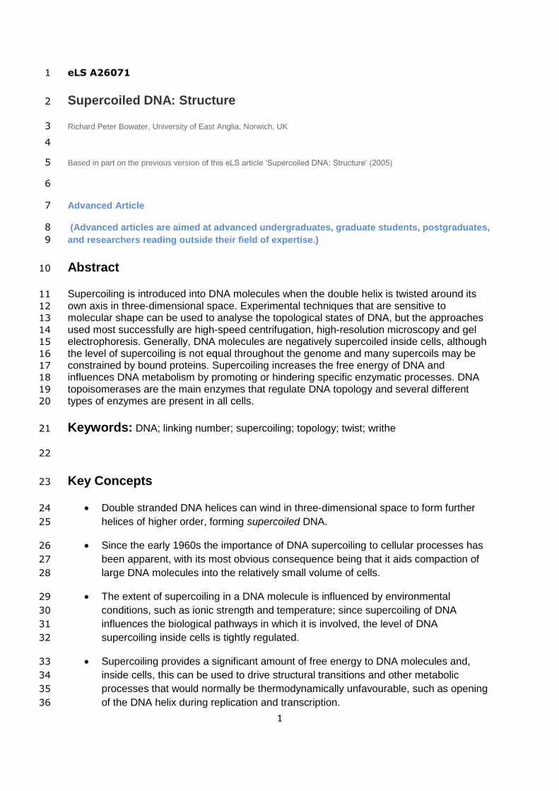
Linear double-stranded DNA molecules can be closed into a circle by the formation of 5′–3′ 8
phosphodiester bonds to seal each strand. Due to the helical nature of the DNA backbone, 9
after circularization the two strands of the helix cannot be separated without breaking one of 10
them; the backbone strands are linked topologically. The number of links between the 11
strands corresponds to the number of double-helical turns (twists) in the original DNA 12
molecule (Figure 1). Upon circularization, this number must be an integer and is known as 13
the linking number of the cccDNA molecule, abbreviated as Lk. (Note that earlier literature 14
refers to the topological winding number of DNA, α, which is identical to Lk. Previously this 15
term has also been abbreviated to L.) Lk is a topological property of cccDNA that does not 16
depend on its particular conformation. 17
<Figure 1 near here> 18
Linking number is a fundamental property of any two closed curves in three-dimensional 19
space and is equal to the number of times that one strand intersects the plane of the other. 20
An intersection may act to increase or decrease the number of links between the two 21
strands. By convention, positive values are given to links arising from forming a closed circle 22
with a completely right-handed double helix (such as B-DNA). Consequently, links with a 23
negative value arise from the formation of a cccDNA molecule from a left-handed DNA helix 24
(such as Z-DNA). The overall Lk of a DNA molecule is equal to the sum of the sign-25
dependent intersections. 26
A given length of DNA has an inherent number of double-helical turns, which is equivalent to 27
the length of the DNA (defined as N base pairs) divided by the number of base pairs per turn 28
of the helix (defined as h). Values of h depend upon environmental conditions and 29
sequence, but an average value is specified from standard conditions and is usually taken to 30
be 10.5 bp per turn for B-DNA. The linking number corresponding to an unconstrained state 31
is termed Lk0 and, for any DNA: 32
(1) 33
5
Since N/h need not necessarily be an integer, Lk0 is not a true linking number (it is 1
sometimes called the ‘hypothetical linking number’). For DNA with N/h that is not an integer, 2
some deformation of the molecule's conformation will be required to line up the strands to 3
allow their closure into a circle, which leads to torsional stress within the cccDNA. In this 4
situation, the most unconstrained DNA circle is referred to as the relaxed topoisomer, 5
defined Lkm. Note that, if N/h is an integer, when the linear DNA is bent to form a simple, 6
planar circle, the strands will line up precisely and Lk = Lk0 = Lkm. 7
It is also possible to add or remove turns to the DNA helix before it is closed into a circle, 8
leading to the molecule having Lk that deviates from Lkm (Figure 1). Since the average 9
conformation of DNA (B-type helix) is defined to have positive Lk, twisting up of the helix 10
before closure leads to an increase in linking number above Lkm and is defined as positive 11
supercoiling. Analogously, unwinding of the helix before closure is defined as negative 12
supercoiling. DNA molecules with positive and negative supercoiling may also be referred to 13
as helices that are over- or underwound, respectively. Note that an underwound helix has an 14
increased value of h. 15
It is clear that Lk is related to the number of turns of the helix, but these two parameters are 16
not equivalent. This can be shown using mathematical analysis, which defines supercoiling 17
in the form of topological and geometric parameters. Lk is a topological property and its 18
value can only be applied to the complete DNA molecule. The twist (Tw) of DNA is a 19
geometric parameter and its values have importance for local regions of the molecule; 20
indeed, the value of Tw of the whole molecule is equal to the sum of individual sections of 21
the molecule. There is a geometrical significance to any difference between Lk and Tw and 22
this is named the writhe (Wr) of the molecule. 23
In terms of cccDNA, Tw and Wr are complementary geometric parameters, and each may 24
be defined and described (Table 1): Tw describes how the individual strands of DNA coil 25
around the axis of the DNA helix and Wr describes how the helix axis coils in space. Both 26
are complex geometric functions whose values need not be an integer. The important finding 27
in relation to studies of DNA supercoiling is that: 28
(2) 29
The main consequence of this equation is that because Lk is invariant for a given cccDNA, 30
any change in Tw of the molecule must be accompanied by an equal and opposite change in 31
Wr, and vice versa. 32
6
Supercoiling of DNA can occur in two forms that produce different overall shapes for the 1
molecule. Circular DNA that winds around itself, as shown in Figure 1, is called interwound 2
(also referred to as plectonemic supercoils) and purified cccDNA in solution usually has this 3
type of supercoiling (Benham and Mielke, 2005). Note that the sense of interwinding is right-4
handed in the case of negatively supercoiled DNA. Supercoiling can also be achieved if the 5
DNA helix forms a series of spirals around an imaginary ring, taking a shape similar to a 6
telephone flex. This kind of supercoiling is known as toroidal (Hud and Vilfan, 2005; Schlick, 7
1995). It corresponds most closely to the term ‘superhelix’ since a left-handed untwisting of 8
the DNA helix (i.e. negative supercoiling) is manifested as a left-handed helix of higher order 9
wound around the torus. Toroidal supercoiling is formed when DNA is wrapped around 10
proteins, as in nucleosomes (Luger and Richmond, 1998; Richmond and Davey, 2003). In 11
reality, DNA inside cells contains supercoils of both interwound and toroidal geometries. See 12
also: Cell Biophysics, DOI: 10.1038/npg.els.0001271; DNA Topology: Fundamentals, DOI: 13
10.1038/npg.els.0001038; DNA Topology: Supercoiling and Linking, DOI: 14
10.1038/npg.els.0003904. 15
Specific Linking Difference, Superhelical 16
Density 17
Changes in Lk of a DNA produce alterations to the level of supercoiling of the molecule. The 18
change in linking number from Lkm is a measurement of the extent of supercoiling of the 19
molecule and is termed the linking difference of the DNA, or ΔLk. The addition of turns to the 20
DNA helix (positive supercoiling) leads to an increase in Lk over Lkm, giving a positive ΔLk. 21
Conversely, the removal of turns from the DNA helix (negative supercoiling) gives a value of 22
Lk lower than Lkm, giving a negative ΔLk. 23
If Lkm is not equal to Lk0, the ‘relaxed’ topoisomer will contain a small amount of torsional 24
strain, which should really be counted towards the total supercoiling of the DNA. Thus, an 25
exact definition of ΔLk is: 26
(3) 27
Furthermore, since changes in Lk produce corresponding changes in Tw and/or Wr, 28
(4) 29
Specific values of ΔLk produce more torsional stress in small DNA molecules than in large 30
ones because they comprise a larger proportion of the overall Lk. To allow comparison of the 31

7
degree of supercoiling in molecules of different sizes, it is useful to normalize measurements 1
of supercoiling to give the specific linking difference (σ); frequently, this is referred to as 2
superhelical density (Muskhelishvili and Travers, 2003). Since cccDNA molecules of Lk0 do 3
not contain supercoils, this serves as a good reference point for such normalization: 4
(5) 5
The specific linking difference allows meaningful comparison between DNA molecules. For 6
example, natural cccDNA molecules, such as bacterial plasmids, vary widely in size, but, 7
when isolated in vitro, the majority have values for σ of −0.05 to −0.06. 8
Energetics of Supercoiled DNA 9
Like all molecules, DNA will assume a configuration of minimum energy, and this is usually a 10
helix of the B-form. Upon bending or twisting of the molecule, its energy is increased. For a 11
cccDNA with a surplus or deficit in Lk, conformational modifications introduce specific 12
changes to the free energy of the molecule. For example, to accommodate the same length 13
of DNA in fewer helical turns, the double helix must be untwisted, leading to a substantial 14
increase in the deformation energy of the molecule. By taking an appropriate writhed 15
configuration, the cccDNA minimizes the amount by which it departs from the B configuration 16
and reduces its deformation energy. On the other hand, writhing always introduces some 17
curvature, and so it increases the bending contribution to the energy of the molecule. Since 18
Wr and Tw are interconvertible, it is apparent that the underwound DNA molecule will 19
assume a configuration that optimizes twist while introducing the smallest possible amount 20
of bending (Benham and Mielke, 2005; Vologodskii and Cozzarelli, 1994a). 21
Experimental studies during the 1970s established that the free energy of a supercoiled DNA 22
sample (ΔGsc) has a quadratic dependence on ΔLk: 23
(6) 24
where K is a DNA length-dependent constant. Thus, samples of cccDNA exist in a normal 25
(Gaussian) distribution of topoisomers (i.e. molecules have a continuous, symmetrical 26
distribution of Lk around the most intensely populated topoisomer – see Figure 2). 27
Theoretical simulations suggest it is likely that ΔGsc varies with ionic conditions and, in fact, it 28
may not be a quadratic function of ΔLk under all conditions (Schlick, 1995; Vologodskii and 29
Cozzarelli, 1994a). The influence of environmental conditions on DNA supercoiling is due, at 30
8
least in part, to the fact that ionic strength and temperature alter Tw of double-helical DNA. 1
Effects of ionic environment on the three-dimensional structure of DNA are to be expected 2
because DNA is a polyelectrolyte with a net negative charge at every nucleotide residue. 3
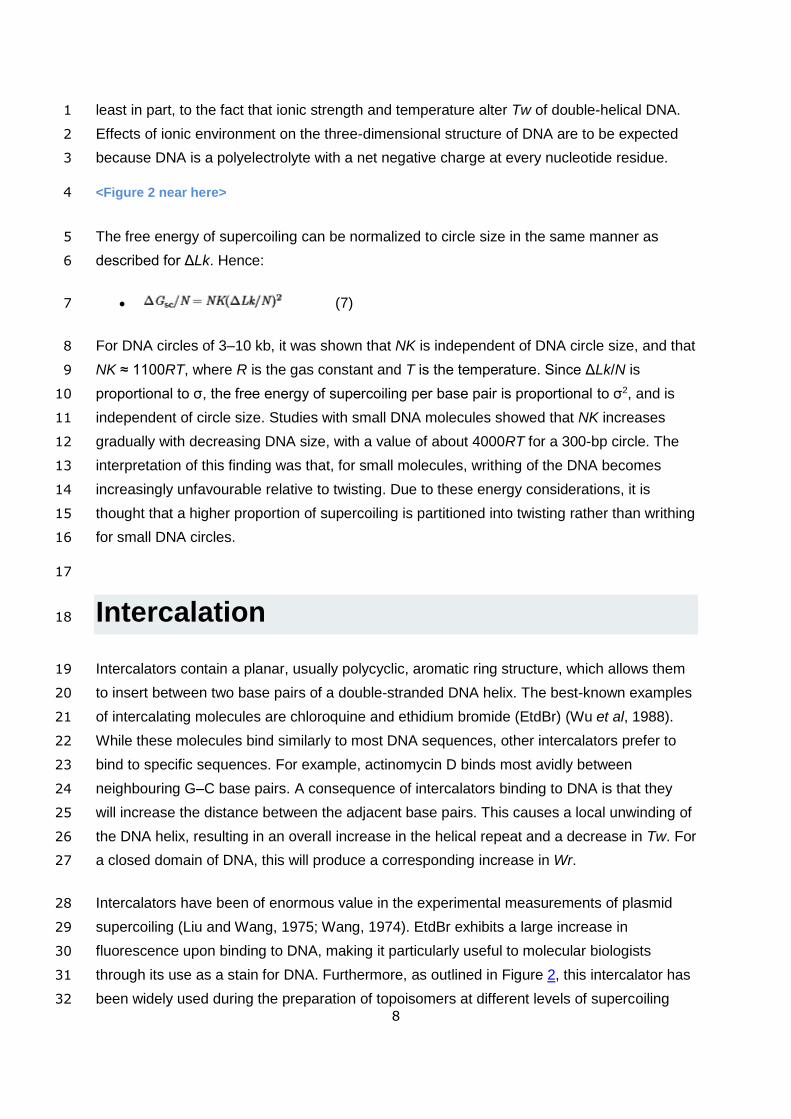
<Figure 2 near here> 4
The free energy of supercoiling can be normalized to circle size in the same manner as 5
described for ΔLk. Hence: 6
(7) 7
For DNA circles of 3–10 kb, it was shown that NK is independent of DNA circle size, and that 8
NK ≈ 1100RT, where R is the gas constant and T is the temperature. Since ΔLk/N is 9
proportional to σ, the free energy of supercoiling per base pair is proportional to σ2, and is 10
independent of circle size. Studies with small DNA molecules showed that NK increases 11
gradually with decreasing DNA size, with a value of about 4000RT for a 300-bp circle. The 12
interpretation of this finding was that, for small molecules, writhing of the DNA becomes 13
increasingly unfavourable relative to twisting. Due to these energy considerations, it is 14
thought that a higher proportion of supercoiling is partitioned into twisting rather than writhing 15
for small DNA circles. 16
17
Intercalation 18
Intercalators contain a planar, usually polycyclic, aromatic ring structure, which allows them 19
to insert between two base pairs of a double-stranded DNA helix. The best-known examples 20
of intercalating molecules are chloroquine and ethidium bromide (EtdBr) (Wu et al, 1988). 21
While these molecules bind similarly to most DNA sequences, other intercalators prefer to 22
bind to specific sequences. For example, actinomycin D binds most avidly between 23
neighbouring G–C base pairs. A consequence of intercalators binding to DNA is that they 24
will increase the distance between the adjacent base pairs. This causes a local unwinding of 25
the DNA helix, resulting in an overall increase in the helical repeat and a decrease in Tw. For 26
a closed domain of DNA, this will produce a corresponding increase in Wr. 27
Intercalators have been of enormous value in the experimental measurements of plasmid 28
supercoiling (Liu and Wang, 1975; Wang, 1974). EtdBr exhibits a large increase in 29
fluorescence upon binding to DNA, making it particularly useful to molecular biologists 30
through its use as a stain for DNA. Furthermore, as outlined in Figure 2, this intercalator has 31
been widely used during the preparation of topoisomers at different levels of supercoiling 32
9
and in agarose gel electrophoresis analysis of Lk (Bowater et al, 1992). See also: Staining, 1
Viewing and Photography of Gels and Estimation of Fragment Sizes, DOI: 2
10.1038/npg.els.0003777. 3
Intercalation of one molecule of EtdBr to DNA causes a local unwinding of adjacent base 4
pairs of 26°. Some classes of intercalator affect the helix in the opposite manner, leading to 5
a localized increase in the twist of the helix. The best-characterized example of such a 6
molecule is netropsin, which binds to the minor groove of AT-rich DNA and increases 7
winding of the helix by approximately 9° for each molecule bound (Schlick and Olson, 1992). 8
Assays 9
In principle, any technique that is sensitive to molecular shape will be useful for experimental 10
analysis of supercoiled DNA. However, because of the large size of these molecules, few 11
techniques have been used successfully to provide direct structural information. 12
Of central importance to the discovery and initial characterization of supercoiled DNA was 13
the use of high-speed centrifugation (Lebowitz, 1990). The sedimentation velocity and 14
buoyant density of polyoma viral DNA was monitored after various treatments that induce 15
strand separation or cleavage of the DNA helix. These experiments clearly identified that 16
DNA molecules could exist in a variety of forms that differed only by their shape, and the 17
concept of supercoiled DNA was founded. 18
High-resolution microscopy provides explicit analysis of molecular structure and electron 19
microscopy (EM), cryo-EM and scanning-force microscopy have been used to analyse 20
supercoiled forms of DNA (Vologodskii and Cozzarelli, 1994a). Each of these techniques 21
has confirmed that supercoiled DNA has a compact shape and that the interwound form 22
predominates in naked DNA. Moreover, high-resolution microscopy clearly shows that 23
supercoiled DNA is often branched and that its conformational and thermodynamic 24
properties depend on ionic conditions (Vologodskii and Cozzarelli, 1994a). 25
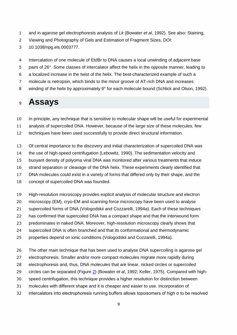
The other main technique that has been used to analyse DNA supercoiling is agarose gel 26
electrophoresis. Smaller and/or more compact molecules migrate more rapidly during 27
electrophoresis and, thus, DNA molecules that are linear, nicked circles or supercoiled 28
circles can be separated (Figure 2) (Bowater et al, 1992; Keller, 1975). Compared with high-29
speed centrifugation, this technique provides a higher resolution for distinction between 30
molecules with different shape and it is cheaper and easier to use. Incorporation of 31
intercalators into electrophoresis running buffers allows topoisomers of high σ to be resolved 32

10
(Figure 2a,b) and their exploitation in two-dimensional gel electrophoresis allows a wide 1
range of topoisomers to be analysed on a single gel (Figure 2c) (Bowater et al, 1992). See 2
also: Gel Electrophoresis, DOI: 10.1002/9780470015902.a0005335.pub2; Staining, Viewing 3
and Photography of Gels and Estimation of Fragment Sizes, DOI: 10.1038/npg.els.0003777. 4
Other experimental techniques have been used to provide less direct information about the 5
structure of supercoiled DNA, including a variety of spectroscopic methods (circular 6
dichroism, static and dynamic light scattering) (Lilley and Dahlberg, 1992; Vologodskii and 7
Cozzarelli, 1994a). Utilization of complementary techniques, particularly in combination with 8
theoretical methods, has provided significant information on the three-dimensional structure 9
of supercoiled DNA. 10
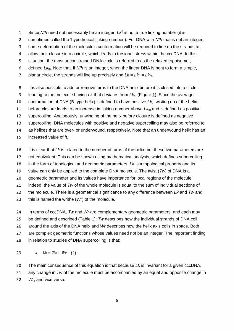
Catenanes and Knots 11
Knots and catenanes (Figure 3) occur frequently in DNA, primarily as a consequence of the 12
complex biochemical reactions that take place within closed topological domains. These 13
structures can influence processes occurring on the DNA molecule, such as replication and 14
transcription, and they are also utilized as intermediates in some types of genetic 15
recombination (Wasserman and Cozzarelli, 1986). See also: DNA Topology: Fundamentals, 16
DOI: 10.1038/npg.els.0001038; DNA Topology: Supercoiling and Linking, DOI: 17
10.1038/npg.els.0003904. 18
<Figure 3 near here> 19
Knots were first detected in DNA treated with topoisomerases in vitro (Wasserman and 20
Cozzarelli, 1986). They have also been observed in native DNA, although they are not 21
particularly common. Catenanes are more prevalent and are utilized in a number of diverse 22
biological systems. Catenated molecules were first observed in mitochondrial DNA from 23
human cells (Wasserman and Cozzarelli, 1986) and their most common occurrence is as 24
intermediates during the replication of circular DNA. 25
Topological knots of a wide degree of complexity can be formed (Wasserman and 26
Cozzarelli, 1986; Witz and Stasiak, 2010). Mathematical methods allow unique description of 27
different knots and catenanes, although these descriptions become complex for highly 28
knotted structures and for catenanes involving many loops. Theoretical analyses of these 29
types of DNA conformations have been particularly valuable in the development of molecular 30
models of supercoiled DNA (Benham and Mielke, 2005; Schlick, 1995; Vologodskii and 31
Cozzarelli, 1994b). 32

11
Protein Binding and DNA Topology 1
A wide variety of proteins that bind to DNA alter the local geometry of its helix and influence 2
DNA topology. The best-known example of this effect is the winding of DNA around the 3
eukaryotic histone octamer to form the nucleosome (Luger and Richmond, 1998; Richmond 4
and Davey, 2003). Histone proteins are positively charged and have no enzymatic activity, 5
but they allow extremely large DNAs to be compacted to fit within each eukaryotic cell. Each 6
histone core envelops approximately 1.8 left-handed turns of DNA and stabilizes negative 7
writhing within the complex. Supercoiling of this type is constrained because it is not 8
available to influence the structure of the remainder of the DNA. See also: Chromosomes 9
and Chromatin, DOI: 10.1002/9780470015902.a0005766.pub2; Chromosome Structure, 10
DOI: 10.1002/9780470015902.a0001486.pub2; DNA Coiling and Unwinding, DOI: 11
10.1038/npg.els.0005967; Nucleosomes: Structure and Function, DOI: 12
10.1038/npg.els.0001155. 13
Prokaryotes do not contain histone proteins, but they do harbour proteins that influence DNA 14
architecture. The two most abundant of these in the nucleoid of Escherichia coli are H-NS 15
(H1) and HU (Drlica and Rouviere-Yaniv, 1987). These proteins constrain supercoils and 16
have highly pleotropic effects, affecting genome stability, and recombination- and 17
transcription-related events (Dillon and Dorman, 2010). Proteins that have more specific 18
cellular functions also exhibit differential binding to DNA templates at various levels of 19
supercoiling. Included among these are polypeptides that bind to specific DNA sequences, 20
such as integration host factor (IHF), which is involved in site-specific recombination 21
(Swinger and Rice, 2004), and factor for inversion stimulation (FIS), which influences 22
transcription at certain promoters (Travers et al, 2001). Other proteins that bind to DNA 23
independent of its sequence become abundant under specific growth conditions. For 24
example, Dps is induced upon starvation of E. coli and is important for coordinating cellular 25
responses to such stress (Chiancone and Ceci, 2010). See also: Chromosomes: Nonhistone 26
Proteins, DOI: 10.1038/npg.els.0001158. 27
Enzymatic Modulations of DNA Topology: 28
Global and Local DNA Supercoiling 29
As a fundamental component of the three-dimensional structure of DNA, it is essential that 30
cells regulate the overall amount of supercoiling that persists within chromatin – frequently 31
referred to as the global level. The main enzymes involved in control mechanisms are the 32
DNA topoisomerases, which can alter Lk of DNA (Schoeffler and Berger, 2008; Vos et al, 33

12
2011). General DNA topoisomerases function with little regard for DNA sequence. Additional 1
enzymes that are involved in site-specific recombination reactions have considerable amino 2
acid homology to some topoisomerases and have similar reaction mechanisms (Wasserman 3
and Cozzarelli, 1986). 4
Topoisomerases with a wide variety of activities have been identified (Corbett and Berger, 5
2004; Schoeffler and Berger, 2008): they may act to remove or introduce negative supercoils 6
or they may remove both positive and negative supercoils. In some cases these topological 7
changes are coupled to the hydrolysis of ATP, as is the case for DNA gyrase and reverse 8
gyrase (Gubaev and Klostermeier, 2014; Lulchev and Klostermeier, 2014). DNA gyrases are 9
well-characterised essential enzymes in bacteria that are able to add negative supercoils to 10
DNA. By contrast, reverse gyrases are able to positively supercoil a circular DNA; these 11
atypical topoisomerases are present in some hyperthermophilic organisms (Forterre et al, 12
2007). See also: DNA Coiling and Unwinding, DOI: 10.1038/npg.els.0005967; 13
Topoisomerases, DOI: 10.1038/npg.els.0001039. 14
Topoisomerases are classed as type I or II, according to the mechanism by which they 15
produce topological changes of DNA (Schoeffler and Berger, 2008). Type I topoisomerases 16
are further subdivided into two groups, types IA and IB, which exhibit dissimilar structures 17
and distinct reaction characteristics. Type I enzymes transiently cleave one strand of the 18
helix, pass the intact strand through and seal the break. Type II enzymes also make 19
transient breaks in the helix, but they cleave both complementary strands of the molecule 20
before passing another intact double-stranded molecule through the break. A consequence 21
of these different reaction mechanisms is that type I enzymes change Lk in steps of 1, 22
whereas type II enzymes change Lk in multiples of two. 23
Global DNA supercoiling varies for different cell types and growth conditions and DNA 24
topoisomerases maintain levels within strict physiological boundaries (Baranello et al, 2012; 25
Gilbert and Allan, 2014; Roca, 2011). Cellular processes that involve movement of 26
macromolecular assemblies along DNA may also generate localized DNA supercoiling. As a 27
large protein complex moves along the DNA, its rotation around the DNA may be inhibited 28
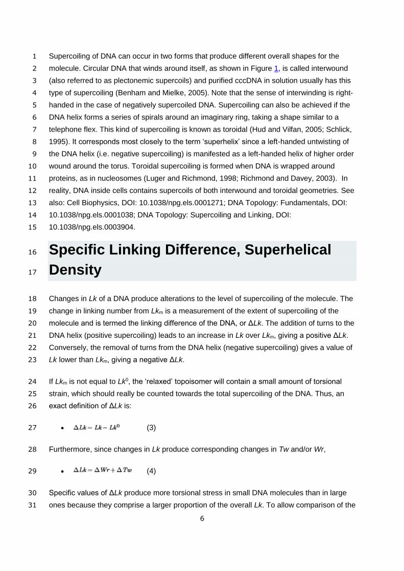
(Liu and Wang, 1987). Instead, the DNA will rotate upon its axis, causing an increase in twist 29
ahead of the complex and a reduction in twist behind; these twist changes are equivalent to 30
positive and negative DNA supercoiling, respectively. This process is named ‘twin domains 31
of supercoiling’, in recognition of the two regions of supercoiling that border the large protein 32
complex. This phenomenon is best characterized for transcription (Figure 4), although it is 33
also likely to occur during the action of DNA polymerases and DNA helicases. Such local 34
topological changes in vivo have been shown to have a significant impact on the 35

13
conformation and function of important DNA sequence elements, such as promoters and 1
DNA replication origins (Travers and Muskhelishvili, 2005; Wu and Fang, 2003). 2
<Figure 4 near here> 3
Since DNA strands are not broken during processes generating twin domains of 4
supercoiling, there is no overall change of Lk. In a linear molecule such transient 5
supercoiling will diffuse away and on a circular molecule the negative and positive supercoils 6
will cancel out by diffusion around the circle. However, since chromatin is organized into 7
discrete domains that are topologically independent, the diffusion of supercoils may be 8
blocked and elevated levels of DNA supercoiling may build up (Gilbert and Allan, 2014). The 9
relative orientation of neighbouring promoters may also influence the formation of 10
transcription-induced supercoiling. For example, highly negatively supercoiled DNA may 11
form between two divergent promoters that transcribe away from each other, whereas DNA 12
that is between two convergent promoters may be positively supercoiled. DNA 13
topoisomerases prevent increases in localized DNA supercoiling: in bacteria, negative and 14
positive supercoils are removed by topoisomerase I and DNA gyrase, respectively (Corbett 15
and Berger, 2004; Schoeffler and Berger, 2008). Inhibition of the activity of either of these 16
enzymes, for example, by mutation, can lead to significant changes to cellular DNA topology 17
(Hatfield and Benham, 2002; Wu and Fang, 2003). See also: DNA Coiling and Unwinding, 18
DOI: 10.1038/npg.els.0005967; DNA Topology: Fundamentals, DOI: 19
10.1038/npg.els.0001038; DNA Topology: Supercoiling and Linking, DOI: 20
10.1038/npg.els.0003904; Topoisomerases, DOI: 10.1038/npg.els.0001039. 21
Biological Functions 22
The unconstrained σ of chromatin is believed to be about −0.02 to −0.03 in prokaryotic cells 23
and is probably less negatively supercoiled in eukaryotes (Drlica, 1992). When localized 24
variations to DNA topology are considered, it is clear that supercoiling provides a significant 25
amount of free energy to DNA molecules inside cells. This increase in free energy can be 26
used to drive structural transitions and other metabolic processes that would normally be 27
thermodynamically unfavourable. For example, DNA can adopt a wide range of “unusual” 28
structures that are different to the standard B-form helix, and many of these are more likely 29
to form in molecules that negatively supercoiled (Kouzine and Levens, 2007). Importantly, 30
some of these non-B-DNA structures have been linked with physiological consequences, 31
including some types of human diseases. See also: Base Pairing in DNA: Unusual Patterns, 32
DOI: 10.1038/npg.els.0003127; DNA Structure, DOI: 33
14
10.1002/9780470015902.a0006002.pub2; Non‐B DNA Structure and Mutations Causing 1
Human Genetic Disease, DOI: 10.1002/9780470015902.a0022657. 2
The most obvious consequence of DNA supercoiling is that it aids compaction of very large 3
DNA molecules into the relatively small volume of cells. The most efficient form of length 4
reduction arises from toroidal winding. Notably, size problems are particularly acute in 5
eukaryotic cells and are overcome by the binding of DNA into toroids (nucleosomes and 6
higher order structures). In addition to these effects, DNA supercoiling has a direct influence 7
on many aspects of DNA metabolism in vivo. The binding of proteins to DNA is often 8
influenced by supercoiling. Conversely, the binding of proteins that remove DNA supercoils 9
can be used to relieve excess energy associated with supercoiling and prevent unfavourable 10
deformations within the DNA. See also: Cell Biophysics, DOI: 10.1038/npg.els.0001271; 11
Protein–DNA Interactions: Structure and Energetics, DOI: 10.1038/npg.els.0001349. 12
DNA topology plays a fundamental role in facilitating site-specific recombination reactions. 13
Furthermore, analysis of the topology of reaction products has provided significant 14
information towards understanding the recombination reaction, particularly for processes 15
involving IHF and resolvases (Swinger and Rice, 2004; Wasserman and Cozzarelli, 1986). 16
Increased free energy associated with negative supercoiling can also be used to separate 17
the strands of the DNA helix (Figure 1), which is usually unfavourable under physiological 18
conditions. Thus, negatively supercoiled DNA templates assist processes that require 19
opening of the DNA helix, such as replication and transcription. In general, these processes 20
are increased at higher levels of negative supercoiling, but the relationship between σ and 21
efficiency of transcription is complex. Some promoters are inhibited by increases in negative 22
DNA supercoiling, suggesting that sequence or chromatin context are also important (Gilbert 23
and Allan, 2014). 24
Current Research Topics 25
The influence of supercoiling upon the three-dimensional structure of DNA is well 26
understood in vitro. The relationship of these observations to the structure of DNA in vivo is 27
less clear. For example, many experiments show that twin domains of supercoiling can be 28
generated when macromolecular protein assemblies translocate along DNA, and these 29
could have profound effects on DNA metabolism inside cells. Although localized levels of 30
DNA supercoiling are observed to vary in vivo, it seems that DNA topoisomerases normally 31
keep these variations within well-defined limits (Baranello et al, 2012; Gilbert and Allan, 32
2014; Roca, 2011). Evidence is growing to show that variable levels of supercoiling may 33

15
impact on specific reactions that involve DNA metabolism. Ongoing research is evaluating 1
the extent by which DNA topology exerts regulatory influences over DNA metabolism. 2
Developments of new scientific technologies have been particularly useful for visualising 3
how proteins influence DNA topology inside cells and at the single-molecule level in vitro (De 4
Vlaminck and Dekker, 2012; Koster et al, 2010; Neuman, 2010). See also: Magnetic 5
Tweezers, DOI: 10.1002/9780470015902.a0023173. 6
There are reciprocal interactions between virtually every reaction involving DNA and DNA 7
topology. In other words, DNA topology influences its metabolism and DNA metabolism 8
influences its topology (Fogg et al, 2012). Thus, there is obvious potential for DNA topology 9
to be used in the regulation of gene expression (Ptacin and Shapiro, 2013). There is 10
significant evidence that this occurs under some physiological conditions, but the extent to 11
which this happens remains unclear. It seems likely that because global DNA supercoiling is 12
an intrinsic property of the DNA template, it may be too universal to provide the fine control 13
of expression of all genes. Perhaps the cell has evolved mechanisms that sever the links 14
between DNA topology and transcription in some circumstances? The situation could be 15
dramatically different for local DNA supercoiling where the surrounding DNA determines the 16
topological changes. The manner by which these interactions take place are still unresolved. 17
Well-characterized experimental systems are now available to monitor reactions such as 18
replication, transcription and recombination, from both pro- and eukaryotes. Continued 19
development and application of in vitro and in vivo approaches will provide significant 20
advances in our understanding of how the three-dimensional structure of DNA integrates 21
within cellular metabolism. 22
23
Glossary 24
Catenane 25
Interlinked double-stranded DNA circles, i.e. that cannot be unlinked without 26
breakage of both strands of one double-stranded helix. 27
Closed-circular DNA 28
Double-helical DNA with no free ends, i.e. both strands are closed circles with no 29
discontinuities in their phosphodiester backbones. 30
Linking difference 31
Difference between the linking number of a particular topoisomer of closed-circular 32
DNA and the average linking number of relaxed DNA; can have positive or negative 33
values. 34

16
Linking number 1
Number of times two strands of closed-circular DNA are connected. It is distributed 2
between the two geometric parameters twist and writhe. 3
Open-circular DNA 4
Double-helical DNA with one strand containing a broken phosphodiester bond; also 5
referred to as ‘nicked DNA’. 6
Relaxed DNA 7
Closed-circular DNA formed with minimal torsional strain of the DNA helix. 8
Supercoiled DNA 9
DNA containing coiling in addition to its normal helical path; closed-circular DNA 10
molecules formed under torsional stress have average linking difference not equal to 11
zero – can be positive or negative. 12
Topoisomer 13
(from topological isomer) Closed-circular DNA molecule of unique linking number. 14
Topoisomerase 15
Enzyme that catalyses changes in the linking number of closed-circular DNA. 16
Twist 17
Number of double-helical turns in a given length of DNA, measured relative to the 18
DNA helix axis. 19
Writhe 20
Geometric parameter that describes the path of a DNA helix in three-dimensional 21
space. 22
23
References 24
Baranello L, Levens D, Gupta A and Kouzine F (2012) The importance of being supercoiled: 25
how DNA mechanics regulate dynamic processes. Biochim Biophys Acta 1819: 632-638 26
27
Bates AD and Maxwell A (2005) DNA topology, Chapter 2, pp. 25-81, Oxford: IRL Press. 28
29
Bauer WR, Crick FHC and White JH (1980) Supercoiled DNA. Sci Amer 243: 100-113 30
31
Benham CJ and Mielke SP (2005) DNA mechanics. Annu Rev Biomed Eng 7: 21-53 32
33
17
Bowater R, Aboul-Ela F and Lilley DMJ (1992) Two-dimensional gel electrophoresis of 1
circular DNA topoisomers. Meth Enzymol 212: 105-120 2
3
Chargaff E, Zamenhof S and Green C (1950) Composition of human desoxypentose nucleic 4
acid. Nature 165: 756-757 5
6
Chiancone E and Ceci P (2010) The multifaceted capacity of Dps proteins to combat 7
bacterial stress conditions: Detoxification of iron and hydrogen peroxide and DNA binding. 8
Biochim Biophys Acta 1800: 798-805 9
10
Corbett KD and Berger JM (2004) Structure, molecular mechanisms, and evolutionary 11
relationships in DNA topoisomerases. Annu Rev Biophys Biomol Struct 33: 95-118 12
13
De Vlaminck I and Dekker C (2012) Recent advances in magnetic tweezers. Annu Rev 14
Biophys 41: 453-472 15
16
Dillon SC and Dorman CJ (2010) Bacterial nucleoid-associated proteins, nucleoid structure 17
and gene expression. Nat Rev Microbiol 8: 185-195 18
19
Drlica K (1992) Control of bacterial DNA supercoiling. Mol Microbiol 6: 425-433 20
21
Drlica K and Rouviere-Yaniv J (1987) Histone like proteins of bacteria. Microbiol Reviews 22
51: 301-319 23
24
Fogg JM, Randall GL, Pettitt BM et al (2012) Bullied no more: when and how DNA shoves 25
proteins around. Q Rev Biophys 45: 257-299 26
27
Forterre P, Gribaldo S, Gadelle D and Serre MC (2007) Origin and evolution of DNA 28
topoisomerases. Biochimie 89: 427-446 29
30
Franklin RE and Gosling RG (1953) Molecular configuration in sodium thymonucleate. 31
Nature 171: 740-741 32
33
Gilbert N and Allan J (2014) Supercoiling in DNA and chromatin. Curr Opin Genet Dev 25: 34
15-21 35
36
Gubaev A and Klostermeier D (2014) The mechanism of negative DNA supercoiling: a 37
cascade of DNA-induced conformational changes prepares gyrase for strand passage. DNA 38
Repair (Amst) 16: 23-34 39
40
Hatfield GW and Benham CJ (2002) DNA topology-mediated control of global gene 41
expression in Escherichia coli. Annu Rev Genet 36: 175-203 42
43

18
Hud NV and Vilfan ID (2005) Toroidal DNA condensates: unraveling the fine structure and 1
the role of nucleation in determining size. Annu Rev Biophys Biomol Struct 34: 295-318 2
3
Keller W (1975) Determination of the number of superhelical turns in simian virus 40 DNA by 4
gel electrophoresis. Proc Natl Acad Sci U S A 72: 4876-4880 5
6
Koster DA, Crut A, Shuman S, Bjornsti MA and Dekker NH (2010) Cellular strategies for 7
regulating DNA supercoiling: a single-molecule perspective. Cell 142: 519-530 8
9
Kouzine F and Levens D (2007) Supercoil-driven DNA structures regulate genetic 10
transactions. Front Biosci 12: 4409-4423 11
12
Lebowitz J (1990) Through the looking glass: the discovery of supercoiled DNA. Trends 13
Biochem Sci 15: 202-207 14
15
Lilley DMJ and Dahlberg JE (1992) DNA structures. Meth Enzymol 211 and 212 16
17
Liu LF and Wang JC (1975) On the degree of unwinding of the DNA helix by ethidium. II. 18
Studies by electron microscopy. Biochim Biophys Acta 395: 401-412 19
20
Liu LF and Wang JC (1987) Supercoiling of the DNA template during transcription. Proc 21
Natl Acad Sci USA 84: 7024-7027 22
23
Luger K and Richmond TJ (1998) DNA binding within the nucleosome core. Curr Opinion 24
Struct Biol 8: 33-40 25
26
Lulchev P and Klostermeier D (2014) Reverse gyrase--recent advances and current 27
mechanistic understanding of positive DNA supercoiling. Nucleic Acids Res 42: 8200-8213 28
29
Muskhelishvili G and Travers A (2003) Transcription factor as a topological homeostat. 30
Front Biosci 8: d279-285 31
32
Neuman KC (2010) Single-molecule measurements of DNA topology and topoisomerases. J 33
Biol Chem 285: 18967-18971 34
35
Ptacin JL and Shapiro L (2013) Chromosome architecture is a key element of bacterial 36
cellular organization. Cell Microbiol 15: 45-52 37
38
Richmond TJ and Davey CA (2003) The structure of DNA in the nucleosome core. Nature 39
423: 145-150 40
41
Roca J (2011) The torsional state of DNA within the chromosome. Chromosoma 120: 323-42
334 43
19
1
Schlick T (1995) Modeling superhelical DNA: recent analytical and dynamic approaches. 2
Curr Opin Struct Biol 5: 245-262 3
4
Schlick T and Olson WK (1992) Supercoiled DNA energetics and dynamics by computer 5
simulation. J Mol Biol 223: 1089-1119 6
7
Schoeffler AJ and Berger JM (2008) DNA topoisomerases: harnessing and constraining 8
energy to govern chromosome topology. Q Rev Biophys 41: 41-101 9
10
Swinger KK and Rice PA (2004) IHF and HU: flexible architects of bent DNA. Curr Opin 11
Struct Biol 14: 28-35 12
13
Travers A and Muskhelishvili G (2005) DNA supercoiling - a global transcriptional regulator 14
for enterobacterial growth? Nat Rev Microbiol 3: 157-169 15
16
Travers A, Schneider R and Muskhelishvili G (2001) DNA supercoiling and transcription in 17
Escherichia coli: The FIS connection. Biochimie 83: 213-217 18
19
Vologodskii AV and Cozzarelli NR (1994a) Conformational and thermodynamic properties of 20
supercoiled DNA. Annu Rev Biophys Biomol Struct 23: 609-643 21
22
Vologodskii AV and Cozzarelli NR (1994b) Supercoiling, knotting, looping and other large-23
scale conformational properties of DNA. Curr Opinion Struct Biol 4: 372-375 24
25
Vos SM, Tretter EM, Schmidt BH and Berger JM (2011) All tangled up: how cells direct, 26
manage and exploit topoisomerase function. Nat Rev Mol Cell Biol 12: 827-841 27
28
Wang JC (1974) The degree of unwinding of the DNA helix by ethidium. I. Titration of twisted 29
PM2 DNA molecules in alkaline cesium chloride density gradients. J Mol Biol 89: 783-801 30
31
Wasserman SA and Cozzarelli NR (1986) Biochemical topology: applications to DNA 32
recombination and replication. Science 232: 951-960 33
34
Watson JD and Crick FC (1953) Molecular structure of nucleic acids: a structure for 35
deoxyribose nucleic acids. Nature 171: 737-738 36
37
Wilkins MH, Stokes AR and Wilson HR (1953) Molecular structure of deoxypentose nucleic 38
acids. Nature 171: 738-740 39
40
Witz G and Stasiak A (2010) DNA supercoiling and its role in DNA decatenation and 41
unknotting. Nucleic Acids Res 38: 2119-2133 42
43

20
Wu HY and Fang M (2003) DNA supercoiling and transcription control: a model from the 1
study of suppression of the leu-500 mutation in Salmonella typhimurium topA- strains. Prog 2
Nucleic Acid Res Mol Biol 73: 43-68 3
4
Wu P, Song L, Clendenning JB et al (1988) Interaction of chloroquine with linear and 5
supercoiled DNAs. Effect on the torsional dynamics, rigidity, and twist energy parameter. 6
Biochemistry 27: 8128 - 8144 7
8
Further Reading 9
Calladine CR, Drew HR, Luisi B and Travers AA (2004) Understanding DNA: 10
The Molecule and How it Works, 3rd edn. Academic Press: London. 11
Cozzarelli NR and Wang JC (eds) (1990) DNA Topology and its Biological 12
Effects, Cold Spring Harbor, NY: Cold Spring Harbor Press. 13
Frank-Kamenetskii MD (1997) Unraveling DNA: The Most Important 14
Molecules of Life, Reading, USA: Addison Wesley. 15
Neidle S (2008) Principles of Nucleic Acid Structure, UK: Academic Press: 16
London. 17
Saenger W (1984) Principles of Nucleic Acid Structure. New York, NY: 18
Springer-Verlag. 19
Sinden RR (1994) DNA Structure and Function. San Diego, USA: Academic 20
Press. 21
Travers A (1993) DNA-Protein Interactions. UK: Chapman & Hall. 22
Wang JC (1994) Appendix I: an introduction to DNA supercoiling and DNA 23
topoisomerase-catalyzed linking number changes of supercoiled DNA. In: Liu LF (ed.) 24
DNA Topoisomerases: Topoisomerase-targeting Drugs, pp. 257–270. San Diego: 25
Academic Press. 26
Vologodskii A (1992) Topology and Physics of Circular DNA. Boca Raton, 27
USA: CRC Press. 28
29

21
1
Table 1. Definitions of abbreviation of DNA topological parameters 2
3
Abbreviation Term Definition
cccDNA Covalently closed circular DNA
Double-helical DNA without free ends, i.e. both strands are closed circles
N Length Total number of base pairs in DNA
h Helical repeat (helical pitch) Number of base pairs per turn of DNA helix
Lk Linking number For cccDNA, number of times one DNA strand intersects the plane of the other
Lk0 Hypothetical linking number cccDNA without torsional strain (equal to N/h)
Lkm Relaxed linking number Linking number of topoisomer with least torsional strain (may not be equal to N/h)
Lk Linking difference For supercoiled DNA, difference between Lk and Lk0
σ Specific linking difference Lk normalized to length of DNA
(equal to Lk/Lk0)
Tw Twist Number of turns within DNA double helix
Wr Writhe Number of times DNA double helix crosses its own path
Gsc Free energy of supercoiling Additional free energy contained within DNA due to presence of supercoils
4
22
1
2 3
Figure 1. Relationship of linking number, twist and writhe of closed circular forms of 4
DNA. Closed DNA circles can be made by formation of covalent 5′–3′ 5
phosphodiester bonds on each strand of a linear molecule. For a linear molecule 6
with 36 helical turns, the linking number of this unconstrained state (Lk0) is 36. 7
23
Closure into an unconstrained planar circle, as shown on the left side of the figure, 1
produces a molecule with twist (Tw) = 36 and writhe (Wr) = 0. If the number of helical 2
turns is altered before closure, the DNA molecule adopts a supercoiled 3
conformation. On the right side of the figure, four helical turns are removed from the 4
molecule, reducing the linking number (Lk) to 32. For simplicity, the figure shows all 5
unwinding partitioned as Wr, although such changes are usually partitioned between 6
Tw and Wr. Unwinding of helical turns produces negatively supercoiled DNA (or 7
−ΔLk) as shown, whereas the inclusion of additional turns produces positively 8
supercoiled DNA. For DNA with −ΔLk in the interwound form, the superhelical turns 9
are right-handed. Note that separation of DNA strands removes negative supercoils 10
(equivalent to the addition of positive supercoils). 11

24
1
Figure 2. Measurement of linking number by gel electrophoresis. (a) Schematic 2
illustration of a DNA sample separated by electrophoresis through an agarose gel 3
with and without an intercalator. DNA isolated from bacterial cells contains molecules 4
with different topology: some have their backbones unbroken and are negatively 5
supercoiled (SC), some have one strand broken and referred to as ‘nicked’ (N) and 6

25
some have both strands broken to produce a linear molecule (L). Note that the 1
supercoiled DNA consists of a Gaussian distribution of different topoisomers. Upon 2
addition of intercalator, the migration of intact molecules is altered, but that of nicked 3
and linear molecules is not changed. (b) Enzymatic relaxation of plasmid DNA in the 4
presence of varying concentrations of intercalator produces samples containing 5
topoisomers at different levels of supercoiling. Utilization of multiple gels with 6
different concentrations of intercalator allows measurement of ΔLk. For each sample, 7
average superhelical density (σ) is shown above the lane. Note that in each gel, 8
samples can have positively or negatively supercoiled topoisomers. The inclusion of 9
intercalator in the running buffer alters the electrophoretic mobility of all topoisomers 10
equivalently. Superhelical density can be measured for experimental samples 11
(‘native’) by comparison with those of known σ. (c) Two-dimensional agarose gel 12
electrophoresis of topoisomers ranging from high negative σ to moderate positive σ. 13
A DNA sample is loaded in a single well in a large agarose gel and electrophoresis is 14
performed under specific conditions (usually without intercalator) in direction D1. 15
After soaking of the gel in buffer containing intercalator, electrophoresis is continued 16
in direction D2 (90° to D1). The gel shown contained 20 μg mL−1 chloroquine during 17
the second electrophoresis, resulting in all topoisomers having positive σ. Deviation 18
of topoisomers from a smooth curve indicates that structural transitions in the DNA 19
molecules reduced their negative σ during the first direction of electrophoresis. Spots 20
marked ‘N’ and ‘L’ indicate the position of migration of ‘nicked’ and ‘linear’ DNA 21
molecules, respectively. 22
23
26
1
2 3
Figure 3. Representation of knots and catenanes. (a) Topological knots may be 4
formed in closed circles of DNA. The simplest knot that can be formed is called a 5
trefoil because there are three lobes to the structure when it is laid flat. Two isomers 6
of the trefoil knot are shown. Many other more complex knots may be formed within 7
cccDNA molecules. (b) Catenanes are formed when two circular DNA molecules are 8
interlocked. Catenanes containing complex, multiple links and involving many DNA 9
circles have been observed in naturally occurring DNA. The arrows indicate that the 10
polarity of a knot or catenane is influenced by the directionality of the sequence in 11
the DNA molecule. 12

27
1
Figure 4. Twin domains of supercoiling are generated during transcription. (a) The 2
shaded cylinders flank a closed domain of DNA containing eight helical turns. (b) To 3
accommodate the transcriptional complex, some unwinding of the DNA helix occurs 4
producing slight overwinding of the remaining DNA within each closed domain. (c) 5
During transcription elongation, rotation of the large transcriptional complex around 6
the DNA is hindered and positive and negative supercoiling are generated ahead 7
and behind the polymerase, respectively. In this diagram, positive and negative 8
supercoiling is represented by the presence of the same number of helical turns over 9
a shorter and longer distance of DNA, respectively. Several biological mechanisms 10
exist to remove these supercoils. 11
12
Permissions 13
14
Figures 1-4 have been published in the previous version of this eLS article, so no 15
permissions are required. 16
17
18


