
Petrotympanicfissure
Articulartubercle
Mandibularfossa of TMJ
Externalacoustic meatus(auditory canal)
Zygomatic processof temporal bone
Styloid process
Mastoid process
Postglenoidtubercle
Atlanto-occipital joint
Stylomandibular ligament
Joint capsule
Lateral ligament
Mylohyoid groove
Mandibular foramen
Lingula
Neck of mandible
Neck ofmandible
Coronoidprocess
Pterygoidfovea
Head (condyle)of mandible
A B
Fig. 9.27 Mandibular fossa of the TMJInferior view of skull base. The head (condyle) of the mandible artic-ulates with the mandibular fossa of the temporal bone via an articu-lar disk. The mandibular fossa is a depression in the squamous part of the temporal bone, bounded by an articular tubercle and a postglenoid
Fig. 9.28 Head of the mandible in the TMJA Anterior view. B Posterior view. The head (condyle) of the mandible is markedly smaller than the mandibular fossa and has a cylindrical shape. Both factors increase the mobility of the mandibular head, allowing rotational movements about a vertical axis.
Fig. 9.29 Ligaments of the lateral TMJLeft lateral view. The TMJ is surrounded by a relatively lax capsule that permits physiological dislocation during jaw opening. The joint is stabi-lized by three ligaments: lateral, stylomandibular, and sphenomandib-ular (see also Fig. 9.30). The strongest of these ligaments is the lateral ligament, which stretches over and blends with the joint capsule.
tubercle. Unlike other articular surfaces, the mandibular fossa is cov-ered by fi brocartilage, not hyaline cartilage. As a result, it is not as clearly delineated on the skull (compare to the atlanto-occipital joints). The external auditory canal lies just posterior to the mandibular fossa. Trauma to the mandible may damage the auditory canal.
194
Regions of the Head 9. Oral Cavity & Perioral Regions
Temporomandibular Joint (TMJ)
7050003C09.indd 1947050003C09.indd 194 10/20/09 12:28:10 PM10/20/09 12:28:10 PM

Pterygoid process,medial plate
Pterygoid process,lateral plate
Stylomandibularligament
Sphenomandib-ular ligament
Mandibularnotch
Pterygospinousligament
Joint capsule
Stylomandibular ligament
Head (condyle)of mandible
Articular disk
Articulartubercle
Postglenoidtubercle
Mandibular nerve (CN V3)
Masseteric nerve
Deep temporal nerve, posterior branch
Auriculotemporalnerve
Posterior division
Anterior division
Fig. 9.33 Sensory innervation of the TMJ capsuleSuperior view. The TMJ capsule is supplied by articular branches aris-ing from three nerves from the mandibular division of the trigeminal nerve (CN V3):
• Auriculotemporal nerve• Deep temporal nerve, posterior branch• Masseteric nerve
Fig. 9.30 Ligaments of the medial TMJLeft medial view. Note the sphenomandibular ligament. The variable pterygospinous ligament is also present.
Fig. 9.31 Opened TMJLeft lateral view. The TMJ capsule begins at the articular tubercle and extends posteriorly to the petrotympanic fi ssure (see Fig. 9.27). Inter-posed between the mandibular head and fossa is the fi brocartilaginous articular disk, which is attached to the joint capsule on all sides.
Fig. 9.32 Dislocation of the TMJThe head of the mandible may slide past the articular tubercle when the mouth is opened, dislocating the TMJ. This may result from heavy yawning or a blow to the opened mandible. When the joint dislocates, the mandible becomes locked in a protruded posi tion and can no longer be closed. This condition is easily diagnosed clinically and is reduced by pressing on the mandibular row of teeth.
195
Regions of the Head 9. Oral Cavity & Perioral Regions
7050003C09.indd 1957050003C09.indd 195 10/20/09 12:28:15 PM10/20/09 12:28:15 PM

Median plane
Head of mandible
Transverse axis throughhead of mandible(axis of rotation)
150°
AProtrusion
Retrusion
B
Working side(laterotrusion)
Balance side(mediotrusion)
Bennett angle
Axis ofrotation
Resting condyle
Swingingcondyle
C
Balance sideWorking side
D
Axis ofrotation
Fig. 9.34 Movements of the mandible in the TMJSuperior view. Most of the movements in the TMJ are complex motions that have three main components:
• Rotation (opening and closing the mouth)• Translation (protrusion and retrusion of the mandible)• Grinding movements during mastication
A Rotation. The axis for joint rotation runs transversely through both heads of the mandible. The two axes intersect at an angle of approxi mately 150 degrees (range of 110 to 180 degrees between individuals). During this movement the TMJ acts as a hinge joint (abduction/depression and adduction/elevation of the mandible). In humans, pure rotation in the TMJ usually occurs only during sleep with the mouth slightly open (aperture angle up to approximately 15 degrees). When the mouth is opened past 15 degrees, rotation is combined with translation (gliding) of the mandibular head.
B Translation. In this movement the mandible is advanced (protruded) and retracted (retruded). The axes for this movement are parallel to the median axes through the center of the mandibular heads.
C Grinding movements in the left TMJ. In describing these lateral movements, a distinction is made between the “resting condyle” and the “swinging condyle.” The resting condyle on the left working side rotates about an almost vertical axis through the head of the mandible (also a rotational axis), whereas the swinging condyle on the right balance side swings forward and inward in a translational movement. The lateral excursion of the mandible is measured in de-grees and is called the Bennett angle. During this movement the mandible moves in laterotrusion on the working side and in medio-trusion on the balance side.
D Grinding movements in the right TMJ. Here, the right TMJ is the working side. The right resting condyle rotates about an almost ver-tical axis, and the left condyle on the balance side swings forward and inward.
196
Regions of the Head 9. Oral Cavity & Perioral Regions
Temporomandibular Joint (TMJ): Biomechanics
7050003C09.indd 1967050003C09.indd 196 10/20/09 12:28:20 PM10/20/09 12:28:20 PM

Lateral pterygoid muscle, inferior head
Head of mandible
Joint capsule
Articular disk
Articulartubercle
Lateral pterygoid muscle,superior head
A
Mandibular fossa
Lateral pterygoid muscle, inferior head
Joint capsule
Head of mandible
Lateral pterygoid muscle,superior head
B
Articular disk
Axis of rotation15°
Lateral pterygoid muscle, inferior head
Joint capsule
Articular disk
Lateral pterygoid muscle,superior head
C
Mandibular fossa
>15°
Fig. 9.35 Movements of the TMJLeft lateral view. Each drawing shows the left TMJ, including the articu-lar disk and capsule and the lateral pterygoid muscle. Each schematic diagram at right shows the corresponding axis of joint movement. The muscle, capsule, and disk form a functionally coordinated musculo-disco-capsular system and work closely together when the mouth is opened and closed. Note: The space between the muscle heads is greatly exaggerated for clarity.
A Mouth closed. When the mouth is in a closed position, the head of the mandible rests against the mandibular fossa of the temporal bone with the intervening articular disk.
B Mouth opened to 15 degrees. Up to 15 degrees of abduction, the head of the mandible remains in the mandibular fossa.
C Mouth opened past 15 degrees. At this point the head of the man-dible glides forward onto the articular tubercle. The joint axis that runs transversely through the mandibular head is shifted forward. The articular disk is pulled forward by the superior part of the lateral pterygoid muscle, and the head of the mandible is drawn forward by the inferior part of that muscle.
197
Regions of the Head 9. Oral Cavity & Perioral Regions
7050003C09.indd 1977050003C09.indd 197 10/20/09 12:28:21 PM10/20/09 12:28:21 PM

①
②
④
③
⑧
⑤
⑥
⑦
Table 9.6 Masseter and temporalis muscles
Muscle Origin Insertion Innervation* Action
Masseter ① Superfi cial head
Zygomatic bone (maxillary process) and zygomatic arch (lateral aspect of anterior ⅔)
Mandibular angle and ramus (lower posterior lateral surface)
Masseteric n. (anterior division of CN V3)
Elevates mandible; also assists in protraction, retraction, and side-to-side motion
Middle head Zygomatic arch (medial aspect of anterior ⅔)
Mandibular ramus (central part)
② Deep head Zygomatic arch (deep surface of posterior ⅓)
Mandibular ramus (upper portion) and lateral side of coronoid process
Temporalis ③ Superfi cial head
Temporal fascia Coronoid process of mandible (apex and medial and anterior surfaces)
Deep temporal nn. (anterior division of CN V3)
Vertical (anterior) fi bers: Elevate mandibleHorizontal (posterior) fi bers: Retract (retrude) mandibleUnilateral: Lateral movement of mandible (chewing)
④ Deep head Temporal fossa (inferior temporal line)
*The muscles of mastication are innervated by motor branches of the mandibular nerve (CN V3), the 3rd division of the trigeminal nerve (CN V).
Table 9.7 Lateral and medial pterygoid muscles
Muscle Origin Insertion Innervation Action
Lateral pterygoid
⑤ Superior head Greater wing of sphenoid bone (infratemporal crest)
Mandible (pterygoid fovea) and temporomandibular joint (articular disk)
Mandibular n. (CN V3) via lateral ptery goid n. (from anterior division of CN V3)
Bilateral: Protrudes mandible (pulls articular disk forward)Unilateral: Lateral movements of mandible (chewing)⑥ Inferior head Lateral pterygoid plate (lateral
surface)Mandible (pterygoid fovea and condylar process)
Medial pterygoid
⑦ Superfi cial head Maxilla (maxillary tuberosity) and palatine bone (pyramidal process)
Pterygoid tuberosity on medial surface of the mandibular angle
Mandibular n. (CN V3) via medial pterygoid n. (from trunk of CN V3)
Raises (adducts) mandible
⑧ Deep head Medial surface of lateral ptery goid plate and pterygoid fossa
The muscles of mastication are derived from the fi rst branchial arch and are located at various depths in the parotid and infratemporal re-gions of the face. They attach to the mandible and receive their motor
Fig. 9.36 Masseter Fig. 9.37 Temporalis Fig. 9.38 Pterygoids
innervation from the mandibular division of the trigeminal nerve (CN V3). The muscles of the oral fl oor (mylohyoid and geniohyoid) are found on pp. 178, 203.
198
Regions of the Head 9. Oral Cavity & Perioral Regions
Muscles of Mastication: Overview
7050003C09.indd 1987050003C09.indd 198 10/20/09 12:28:28 PM10/20/09 12:28:28 PM

Coronoidprocess
Joint capsule
Lateralpterygoid
Masseter
Lateral ligament
Temporalis
B
Zygomaticarch
Superiortemporal
line
Frontal bone Parietal boneZygomaticarch
External acoustic meatus
Mastoid process
Joint capsule
Lateral ligament
Styloid process
Masseter,deep head
Masseter,superficial head
Temporalis
A
Superior temporal line
Fig. 9.39 Temporalis and masseterLeft lateral view.
A Superfi cial dissection.B Deep dissection. The masseter and zygo-
matic arch have been partially removed to show the full extent of the temporalis.
The temporalis is the most powerful muscle of mastication, doing approximately half the work. It works with the masseter (consisting of a superfi cial and a deep part) to elevate the mandible and close the mouth. Note: A small portion of the lateral pterygoid is visible in B.
199
Regions of the Head 9. Oral Cavity & Perioral Regions
7050003C09.indd 1997050003C09.indd 199 10/20/09 12:28:29 PM10/20/09 12:28:29 PM

Masseter (cut)
Medial pterygoid,superficial and deep heads
Lateral pterygoid,superior andinferior heads
Temporalis (cut)
A
Lateral plate, pterygoid process (sphenoid bone)
Medial pterygoid,deep head
Lateral pterygoid,inferior head (cut)
Articulardisk
Lateral pterygoid,superior head (cut)
BMedial pterygoid,superficial head
Articular disk
Head of mandible,articular surface
Masseter, superficial part
Medial pterygoid,deep head
Masseter, deep part
Lateral pterygoid, inferior head
Lateral pterygoid, superior head
Temporalis
Mandibular angle
Pterygoid process, medial plate
Mandibular fossa, articular surface
Lateral pterygoid, inferiorhead, in pterygoid fovea
Coronoid process(with temporalis)
Fig. 9.40 Lateral and medial pterygoid musclesLeft lateral views.A The coronoid process of the mandible has been removed here along
with the lower part of the temporalis so that both pterygoid muscles can be seen.
B Here the temporalis has been completely removed, and the supe-rior and inferior heads of the lateral pterygoid have been windowed. The lateral pterygoid initiates mouth opening, which is then contin-ued by the digastric and the suprahyoid muscles, along with gravity.
With the temporomandibular joint opened, we can see that fi bers from the lateral pterygoid blend with the articular disk. The lateral pterygoid functions as the guide muscle of the temporomandibular joint. Because both its superior and inferior heads are active during all movements, its actions are more complex than those of the other muscles of mastication. The medial pterygoid runs almost perpen-dicular to the lateral pterygoid and contributes to the for mation of a muscular sling, along with the masseter, that partially encompasses the mandible (see Fig. 9.41).
Fig. 9.41 Masticatory muscular sling
Oblique posterior view. The masseter and medial pterygoid form a muscu-lar sling in which the mandible is sus-pended. By combining the actions of both muscles into a functional unit, this sling enables powerful closure of the jaws.
200
Regions of the Head 9. Oral Cavity & Perioral Regions
Muscles of Mastication: Deep Muscles
7050003C09.indd 2007050003C09.indd 200 10/20/09 12:28:36 PM10/20/09 12:28:36 PM

Ethmoidair cells
Sphenoidsinus
Nasopharynx
Parotid gland
Oropharynx
Tongue
Submandibulargland
Mylohyoidmuscle
Digastric muscle,anterior belly
Platysma
Masseter, superficial head
Masseter, deep head
Lateral pterygoid, inferior head
Temporalis, superficial and deep heads
Lateral pterygoid, superior head
Medial pterygoid,deep and superficial heads
Superior sagittal sinus
Dura materTemporal
lobe
Optic nerve (CN II)
Frontallobe
Falx cerebri
Mandible
Geniohyoidmuscle
Temporalfascia
Zygomaticarch
Pterygoidprocess,
lateral plate
Hyoglossus
Inferior alveolar nerve(CN V3) in mandibularcanal
Lingual septum
Inferiorsagittal sinus
Fig. 9.42 Muscles of mastication, coronal section at the level of the sphenoid sinusPosterior view.
201
Regions of the Head 9. Oral Cavity & Perioral Regions
7050003C09.indd 2017050003C09.indd 201 10/20/09 12:28:41 PM10/20/09 12:28:41 PM

A
Auriculotemporalnerve
Massetericnerve
Medial pterygoidmuscle
Lingual nerve
Masseter muscle Inferior alveolarnerve
Mental nerve
Deep temporalnerves
Mandibulardivision
Trigeminalganglion
Nerve to medial pterygoid
Buccal nerve(long buccal nerve)
Mandibularcanal
Inferior dentalbranches
Mentalforamen
Middlemeningeal
artery
Foramenovale
Lateral pterygoidmuscle
Infraorbitalforamen
Recurrent meningealnerve (nervus
spinosus)
B
Nerve of tensortympani
Facialnerve
Stylomastoidforamen
Auriculotemporalnerve
Lesserpetrosal
nerve
Communicating branchto auriculotemporal nerve
Chordatympani
Nerve to mylohyoid
Lingual nerve
Medial pterygoidmuscle
Oticganglion
Nerve of tensorveli palatini
CN V1 (mandibular nerve)
Foramenovale
SubmandibularganglionInferior
alveolar nerve
Nerve of medialpterygoid
Fig. 4.32 Mandibular division (CN V3) of the trigeminal nerve
Right lateral view. A Partially opened mandible with middle cranial fossa windowed. B Opened oral cavity (right half of mandible removed).The trunk of CN V3 gives off two branches (recur-rent meningeal and medial pterygoid nerves) before splitting into an anterior and a posterior division (see Table 4.21). The nerve to the me-dial pterygoid conveys branchio motor fi bers to the otic ganglion; these fi bers pass with-out synapsing to innervate the tensors tym-pani and veli palatini. The otic ganglion is the parasympathetic ganglion of the glossopha-ryngeal nerve (CN IX). Preganglionic fi bers en-ter via the lesser petrosal nerve (reconstituted from the tympanic plexus; see pp. 160–161). Postganglionic fi bers leave with the auricu-lotemporal nerve (CN V3) to innervate the buc-cal gland. Taste fi bers of CN VII travel in the lingual nerve (CN V3) to the chorda tympani (which they enter either directly or indirectly via the otic ganglion). These fi bers ascend in the chorda tympani via the tympanic cavity to the facial nerve (CN VII; see p. 83).
80
Head 4. Innervation of the Head & Neck
CN V3: Trigeminal Nerve, Mandibular Division
7050003C04.indd 807050003C04.indd 80 10/20/09 1:14:05 PM10/20/09 1:14:05 PM

Table 4.21 Mandibular nerve (CN V3)
The mandibular nerve (CN V3) is the mixed aff erent-eff erent branch of CN V, containing general sensory fi bers and branchiomotor fi bers to the eight muscles derived from the 1st pharyngeal arch. The large sensory and small motor roots of CN V leave the middle cranial fossa via the foramen ovale. In the infratemporal fossa, they unite to form the CN V3 trunk. The trunk gives off two branches before splitting into an anterior and a posterior division. Of the eight branchial arch muscles, three are supplied by the trunk, three by the anterior division, and two by the posterior division.
Trunk: The trunk of CN V3 gives off one sensory and one motor branch. The motor branch conveys branchiomotor fi bers to three of the eight muscles of the 1st pharyngeal arch.
R Recurrent meningeal branch (nervus spinosum)
Sensory: Dura of the middle cranial fossa (also anterior cranial fossa and calvarium). The nervus spinosum arises in the infratemporal fossa and re-enters the middle cranial fossa via the foramen spinosum.
MP Medial pterygoid n. Branchiomotor: Directly to the medial pterygoid. Certain fi bers enter the otic ganglion via the motor root and pass without synapsing to:• N. to tensor veli palatini: Tensor veli palatini.• N. to tensor tympani: Tensor tympani.
Anterior division: The anterior division of CN V3 contains predominantly eff erent fi bers (with one sensory branch, the buccal nerve.) The branchio-motor fi bers innervate three of the eight muscles of the 1st pharyngeal arch.
M Masseter n. Branchiomotor: Masseter.
Sensory: Temporomandibular joint (articular branches).
T Deep temporal nn. Branchiomotor: Temporalis via two branches:• Anterior deep temporal n.• Posterior deep temporal n.
LP Lateral pterygoid n. Branchiomotor: Lateral pterygoid.
B Buccal (long buccal) n. Sensory: Cheek (skin and mucosa) and buccal gingivae of the molars.
Posterior division: The larger posterior division of CN V3 contains predominantly aff erent fi bers (with one motor branch, the mylohyoid nerve). The mylohyoid nerve arises from the inferior alveolar nerve and supplies the remaining two muscles of the 1st pharyngeal arch.
A Auriculotemporal n. Sensory: Skin of the ear and temple. Fibers pass through the parotid gland, behind the temporomandibular joint, and into the infratemporal fossa. The nerve typically splits around the middle meningeal artery (a branch of the maxillary artery) before joining the posterior division.Distributes postganglionic parasympathetic fi bers from the otic ganglion.
L Lingual n. Sensory: Mucosa of the oral cavity (presulcal tongue, oral fl oor, and gingival covering of lingual surface of mandibular teeth). In the infratemporal fossa, the lingual nerve combines with the chorda tympani (CN VII).
I Inferior alveolar n. Sensory: Mandibular teeth and chin:• Incisive branch: Incisors, canines, and 1st premolars (with associated labial gingivae).• Mental n.: Labial gingivae of the incisors and the skin of the lower lip and chin.The mental nerve enters the mental foramen and combines with the incisive branch in the mandibular canal. The inferior alveolar nerve exits the mandible via the mandibular foramen and combines to form the posterior division of CN V3.Note: 2nd premolars and mandibular molars are supplied by the inferior alveolar nerve before it splits into its terminal branches.
Branchiomotor: Fibers branch just proximal to the mandibular foramen:• Mylohyoid n.: Mylohyoid and anterior belly of the digastric.
Autonomic scaff olding: The parasympathetic ganglia of CN VII (submandibular ganglion) and CN IX (otic ganglion) are functionally associated with CN V3.
Submandibular ganglion (CN VII)
Parasympathetic root Preganglionic parasympathetic fi bers from the facial nerve (CN VII) travel to the ganglion in the chorda tympani, facial nerve, and lingual nerve (CN V3).
Sympathetic root Sympathetic fi bers from the superior cervical ganglion ascend (via the internal carotid plexus) and travel in a plexus on the facial artery.
Otic ganglion (CN IX) Parasympathetic root Preganglionic parasympathetic fi bers enter from CN IX via the lesser petrosal nerve.
Sympathetic root Postganglionic sympathetic fi bers from the superior cervical ganglion enter via a plexus on the middle meningeal artery.
• Parotid gland: Postganglionic parasympathetic fi bers from the otic ganglion travel to the parotid gland via the auriculotemporal n. (CN V3).• Submandibular and sublingual glands: Postganglionic autonomic fi bers to the submandibular and sublingual glands travel from the submandibular
ganglion via glandular branches.
• Taste (CN VII): Taste fi bers (special viscerosensory fi bers) to CN VII may travel via the lingual nerve (CN V3) to the chorda tympani (CN VII).
Note: Nerve courses are traditionally described proximal to distal (CNS to periphery). However, for sensory nerves, the sensory relay is in the opposite direction. It is more appropriate to talk of sensory nerves collecting fi bers than to talk of them branching to supply a region.
81
Head 4. Innervation of the Head & Neck
7050003C04.indd 817050003C04.indd 81 10/20/09 1:14:09 PM10/20/09 1:14:09 PM

Nervusintermedius
Stylomastoidforamen
Geniculateganglion
Nucleusof solitary
tract
PonsNucleus of the
abducent nerve(CN VI)
Superiorsalivatory
nucleus
Facial motornucleus
Branches ofthe parotid
plexusA
Facial motornucleus
Nucleus ofsolitary tract,superior part
Superiorsalivatory nucleus
Nucleus of theabducent nerve (CN VI)
B
Internal genuof facial nerve
Facial nerve
Table 4.22 Facial nerve (CN VII)
Nuclei, ganglia, and fi ber distribution
Branchiomotor (purple)
Facial motor nucleus
Lower motor neurons innervate all muscles of the 2nd branchial (pharyngeal) arch:• Muscles of facial expression• Stylohyoid• Digastric, posterior belly• Stapedius
Parasympathetic (blue)
Superior salivatory nucleus
Preganglionic neurons synapse in the pterygo-palatine or submandibular ganglion.
Postganglionic neurons innervate:• Lacrimal gland• Submandibular and sublingual glands• Small glands of the oral and nasal cavities
Special visceral aff erent (light green)
Nucleus of the solitary tract, superior part
First-order pseudounipolar cells in the geniculate ganglion relay taste sensation from the presulcal tongue and soft palate (via the chorda tympani and greater petrosal nerve).
General somatic aff erent (not shown)
First-order pseudounipolar cells in the geniculate ganglion relay general sensation from the external ear (auricle and skin of the auditory canal) and lateral tympanic membrane.
Course
Emergence: Axons from the superior salivatory nucleus and the nucleus of the solitary tract form the nervus intermedius. These combine with the branchiomotor and somatosensory fi bers to emerge from the brainstem as CN VII.Internal branches: CN VII enters the petrous bone via the internal acoustic meatus. Within the facial canal, it gives off one branchio-motor branch (nerve to the stapedius) and two nerves (greater petrosal nerve and chorda tympani) containing both parasympathetic and taste fi bers.External branches: The remaining fi bers emerge via the stylomas-toid foramen. Three direct branches arise before the fi bers enter the parotid gland (nerve to posterior digastric, nerve to stylohyoid, and posterior auricular nerve). In the gland, the branchiomotor fi bers branch to form the parotid plexus, which innervates the muscles of the 2nd branchial arch.
Lesions
CN VII is most easily injured in its distal portions (after emerging from the parotid gland). Nerve lesions of the parotid plexus cause muscle paralysis. Temporal bone fractures may injure the nerve within the facial canal, causing disturbances of taste, lacrimation, salivation, etc. (see Fig. 4.34).
Fig. 4.33 Facial nerve (CN VII)A Anterior view of brainstem. B Superior view of cross section through pons.Fibers: The facial nerve provides branchiomotor innervation to the muscles of the second branchial arch and parasympathetic motor in-nervation to most salivary glands (via the pterygopalatine and subman-dibular ganglia). Taste fi bers are conveyed via pseudounipolar sensory neurons with cell bodies in the geniculate ganglion. The facial nerve also receives general sensation from the external ear.Branches: The superfi cial branches of CN VII are primarily branchio-motor (only the posterior auricular nerve may contain sensory fi bers as well as motor). Taste and preganglionic parasympathetic fi bers travel in both the chorda tympani and greater petrosal nerves. These fi bers con-verge in the external genu and enter the brainstem together as the ner-vus intermedius.
82
Head 4. Innervation of the Head & Neck
CN VII: Facial Nerve, Nuclei & Internal Branches
7050003C04.indd 827050003C04.indd 82 10/20/09 1:14:09 PM10/20/09 1:14:09 PM

Posteriorauricular nerve
Stapedialnerve and
muscle
Facial nerve(CN VII) infacial canal
Geniculateganglion
Chordatympani
Greaterpetrosal nerve
CN V3
CN V2
CN V1
Trigeminalganglion
Trigeminalnerve (CN V)
Stylohyoid muscle with nerve
Pterygo-palatineganglion
Tympaniccavity
Hiatus of greaterpetrosal canal
Petro-tympanicfissure
Lingual nerve(CN V3)
Digastric muscle, posterior belly with nerve
Stylo-mastoidforamen
Branchiomotorfibers to
parotid plexus
CN VIII
Stylo-mastoidforamen
Posteriorauricular
nerve
Greater petrosalnerve
Internal acousticmeatus
1
2
3
4
5
Chordatympani
Nerve to thestapedius
Nerves to thestylohyoid and
posterior digastricParotid plexus
Fig. 4.34 Branches of the facial nerveThe facial nerve enters the facial canal of the petrous bone via the inter-nal acoustic meatus. Most branchiomotor fi bers and all somatosensory fi bers emerge via the stylomastoid foramen. Within the facial canal, CN VII gives off one branchiomotor branch and two nerves contain-ing both parasympathetic and taste fi bers (greater petrosal nerve and chorda tympani). Temporal bone fractures may injure the facial nerve at various levels:
1 Internal acoustic meatus: Lesions aff ect CN VII and the vestibulo-cochlear nerve (CN VIII). Peripheral motor facial paralysis is accom-panied by hearing loss and dizziness.
2 External genu of facial nerve: Peripheral motor facial paralysis is ac-companied by disturbances of taste sensation, lacrimation, and sali-vation (greater petrosal nerve).
3 Motor paralysis is accompanied by disturbances of salivation and taste (chorda tympani). Paralysis of the stapedius causes hyperacu-sis (hypersensitivity to normal sounds).
4 Facial paralysis is accompanied by disturbances of taste and saliva-tion (chorda tympani).
5 Facial paralysis is the only manifestation of a lesion at this level.
Fig. 4.35 Course of the facial nerveRight lateral view of right temporal bone (pe-trous part). Both the facial nerve and vestibulo-cochlear nerve (CN VIII, not shown) pass through the internal acoustic meatus on the posterior surface of the petrous bone. The facial nerve courses laterally in the bone to the external genu, which contains the geniculate ganglion (cell bodies of fi rst-order pseudounipolar sensory neurons). At the genu (L. genu = knee), CN VII bends and descends in the facial canal. It gives off three branches between the geniculate gan-glion and the stylomastoid foramen:
• Greater petrosal nerve: Parasympathetic and taste (special visceral aff erent) fi bers branch from the geniculate ganglion in the greater petrosal canal. They emerge on the anterior surface of the petrous pyramid and continue across the surface of the foramen lacerum. The greater petrosal nerve combines with the deep petrosal nerve in the pterygoid ca-nal (nerve of the pterygoid canal, vidian). The greater petrosal nerve contains the fi bers that form the motor root of the pterygopalatine ganglion (the parasympathetic ganglion of CN VII). The pterygopalatine ganglion distrib-utes autonomic fi bers via the trigeminal nerve (primarily the maxillary division, CN V2).
• Stapedial nerve: Branchiomotor fi bers in-nervate the stapedius muscle.
• Chorda tympani: The remaining parasym-pathetic and taste fi bers leave the facial nerve as the chorda tympani. This nerve runs through the tympanic cavity and petrotym-panic fi ssure to the infratemporal fossa, where it unites with the lingual nerve (CN V3).
The remaining fi bers (branchiomotor with some general sensory) exit via the stylomastoid fora-men.
83
Head 4. Innervation of the Head & Neck
7050003C04.indd 837050003C04.indd 83 10/20/09 1:14:11 PM10/20/09 1:14:11 PM

Temporal branches
Zygomaticbranches
Buccal branches
Marginalmandibular
branch
Cervicalbranch
Nerve to digastric(posterior belly)
Parotid plexus
Posteriorauricular nerve
Facial nerve
Nerve tostylohyoid
A C
B
Cortico-nuclear
fibers
Precentralgyrus
Facial motornucleus
Facialnerve
(CN VII)
Fig. 4.36 Innervation of the second branchial arch muscles
Left lateral view. The branchiomotor fi bers of CN VII innervate all the muscles derived from the second branchial arch. With the excep-tion of the stapedial nerve (to the stapedius), all branchiomotor fi bers in the facial nerve emerge from the facial canal via the stylomas-toid foramen. Three branches arise before the parotid plexus:
• Posterior auricular nerve (Note: This may also contain general somatosensory fi bers.)
• Nerve to the digastric (posterior belly)• Nerve to the stylohyoid
The remaining branchiomotor fi bers then en-ter the parotid gland where they divide into two trunks (temporofacial and cervicofacial) and fi ve major branches, which innervate the muscles of facial expression:
• Temporal• Zygomatic• Buccal• Mandibular (marginal mandibular)• Cervical
The branching of the plexus is variable.
Fig. 4.37 Facial paralysisA Upper motor neurons in the primary somatomotor cortex (pre-
central gyrus) descend to the cell bodies of lower motor neurons in the facial motor nucleus. The axons of these lower motor neurons innervate the muscles derived from the second branchial arch. The facial motor nucleus has a “bipartite” structure: its cranial (upper) part supplies the muscles of the calvaria and palpebral fi ssure, and its caudal (lower) part supplies the muscles of the lower face. The cranial part of the nucleus receives bilateral innervation (from upper motor neurons in both hemispheres). The caudal part receives con-tralateral innervation (from cortical neurons on the other side).
B Central (supranuclear) paralysis: Loss of upper motor neurons (shown here for the left hemisphere) causes contralateral paralysis in the lower half of the face but no paralysis in the upper half. For example, the patient’s mouth will sag on the right (contralateral paralysis of lower muscles), but the ability to wrinkle the forehead and close the eyes is intact.
C Peripheral (infranuclear) paralysis: Loss of lower motor neurons (shown here for right brainstem) causes complete ipsilateral pa-ralysis. For example, the whole right side of the face is paralyzed. Depending on the site of the lesion, additional defi cits may be pres-ent (decreased lacrimation or salivation, loss of taste sensation in the presulcal tongue).
84
Head 4. Innervation of the Head & Neck
CN VII: Facial Nerve, External Branches & Ganglia
7050003C04.indd 847050003C04.indd 84 10/20/09 1:14:14 PM10/20/09 1:14:14 PM

Fig. 4.38 Facial nerve gangliaAutonomic and taste fi bers often travel with sensory fi bers from other nerves to reach their targets. Parasympathetic and taste fi bers leave the facial nerve via two branches: the greater petrosal nerve and the chorda tympani.
• Greater petrosal nerve: Preganglionic parasympathetic and taste fi bers from the geniculate ganglion course in the greater petrosal canal. They are joined by the deep petrosal nerve, which conveys postganglionic sympathetic fi bers from the superior cervical gan-glion (via the internal carotid plexus). The greater and deep petrosal nerves combine to form the nerve of the pterygoid canal (vidian), which conveys sympathetic, parasympathetic, and taste fi bers to the pterygopalatine ganglion (only parasympathetics will synapse at the ganglion; all other fi ber types pass through without synapsing). Branches of CN V2 then distribute the fi bers to their targets:
◦ Lacrimal gland: Autonomic fi bers (sympathetic and parasympa-thetic) run with branches of CN V2 (zygomatic and zygomatico-temporal nerves) to a communicating branch, which conveys them to the lacrimal nerve (CN V1) and thus to the lacrimal gland.
◦ Small glands of the nasal and oral cavities: Autonomic fi bers run with branches of CN V2 to the small glands in the mucosa of the nasal cavity, maxillary sinuses, and palatine tonsils.
◦ Taste: Taste fi bers run with branches of CN V2 to the soft palate.
• Chorda tympani: Preganglionic parasympathetic and taste fi bers course through the chorda tympani. They emerge from the petro-tympanic fi ssure and combine with the lingual nerve (CN V3) in the infratemporal fossa. They are conveyed to the submandibular gan-glion by the lingual nerve, and from there, postganglionic branches travel to their targets via branches of CN V3.
◦ Submandibular and sublingual glands: Postganglionic parasym-pathetic fi bers run with branches of CN V3 to the glands.
◦ Taste buds of tongue: The taste buds on the presulcal portion of the tongue receive taste fi bers from the chorda tympani via the lingual nerve (CN V3). Note: The postsulcal portion of the tongue and the oropharynx receive taste fi bers from CN IX. The root of the tongue and epiglottis receive taste fi bers from CN X.
Note: The lesser petrosal nerve runs in the lesser petrosal canal roughly parallel to the greater petrosal nerve. The lesser petrosal nerve conveys preganglionic parasympathetic fi bers from the tympanic plexus (CN IX) to the otic ganglion. These fi bers innervate the parotid, buccal, and in-ferior labial glands, with postganglionic fi bers distributed via branches of CN V3.
85
Head 4. Innervation of the Head & Neck
7050003C04.indd 857050003C04.indd 85 10/20/09 1:14:18 PM10/20/09 1:14:18 PM

Vestibular ganglion,superior part
Cochlear root
Spiral ganglia
Vestibular root
Saccularnerve
Utricularnerve
Posteriorampullary nerve
Anteriorampullary nerve
Vestibulocochlearnerve (CN VIII)
Vestibular ganglion,inferior part
Lateralampullary
nerve
Table 4.23 Vestibulocochlear nerve (CN VIII)
Nuclei, ganglia, and fi ber distribution
Special somatic aff erent (orange): Special somatic sensory neurons convey sensory fi bers from the vestibular apparatus (balance) and auditory apparatus (hearing). Both parts of the nerve contain fi rst-order bipolar sensory neurons.
Neurons Vestibular root Cochlear root
Peripheral processes
In the sensory cells of the semicircular canals, the saccule, and the utricle.
In the hair cells of the organ of Corti.
Cell bodies Vestibular ganglion• Inferior part: Peripheral processes from saccule and
posterior semicircular canal.• Superior part: Peripheral processes from anterior and
lateral semicircular canals and utricle.
Spiral ganglia. The peripheral processes from the neurons in these myriad ganglia radiate outward to receive sensory input from the spiral modiolus.
Central processes (axons)
To four vestibular nuclei in the medulla oblongata (fl oor of the rhomboid fossa). A few pass directly to the cerebellum via the inferior cerebellar peduncle.
To two cochlear nuclei lateral to the vestibular nuclei.
Nuclei Superior, lateral, medial, and inferior vestibular nuclei. Anterior and posterior cochlear nuclei.
Lesions Dizziness. Hearing loss (ranging to deafness).
Course
The vestibular and cochlear roots unite in the internal acoustic meatus to form the vestibulocochlear nerve, which is covered by a common connective tissue sheath. The nerve emerges from the internal acoustic meatus on the medial surface of the petrous bone and enters the brainstem at the level of the pontomedullary junction, in particular at the cerebellopontine angle.
Fig. 4.39 Vestibulocochlear nerve (CN VIII)The vestibulocochlear nerve consists of two parts. The vestibular root conveys aff erent impulses from the vestibular apparatus (balance). The cochlear root conveys aff erent impulses from the auditory apparatus (hearing).
86
Head 4. Innervation of the Head & Neck
CN VIII: Vestibulocochlear Nerve
7050003C04.indd 867050003C04.indd 86 10/20/09 1:14:22 PM10/20/09 1:14:22 PM

Cerebello-pontine angle
Acousticneuroma(vestibularschwannoma)
Medialvestibularnucleus
Lateralvestibular
nucleus
Superiorvestibular
nucleus
B
Anteriorcochlearnucleus
Posteriorcochlearnucleus
B
Flocculus ofcerebellum
Inferior vestibularnucleus
Medial vestibularnucleus
Lateralvestibularnucleus
Superiorvestibularnucleus
Vestibulo-cochlear nerve
(CN VIII)
A
Semi-circular
canals
Vestibularganglion,
superior andinferior parts
Vestibularroot
Direct fibersto cerebellum
Posteriorcochlear nucleus
Anteriorcochlear nucleus
A
Vestibulo-cochlear nerve
(CN VIII)
Cochlear root Cochlea with
spiral ganglia
Fig. 4.41 Cochlear root and nucleiA Anterior view of the medulla oblongata and pons. B Cross section through the upper me-dulla oblongata.
Fig. 4.42 Acoustic neuroma in the cerebellopontine angleAcoustic neuromas (more accurately, vestibular schwannomas) are be-nign tumors of the cerebellopontine angle arising from the Schwann cells of the vestibular root of CN VIII. As they grow, they compress and displace the adjacent structures and cause slowly progressive hearing loss and gait ataxia. Large tumors can impair the egress of CSF from the 4th ventricle, causing hydrocephalus and symptomatic intracranial hyper tension (vomiting, impairment of consciousness).
Fig. 4.40 Vestibular root and nucleiA Anterior view of the medulla oblongata and pons. B Cross section through the upper me-dulla oblongata.
87
Head 4. Innervation of the Head & Neck
7050003C04.indd 877050003C04.indd 87 10/20/09 1:14:25 PM10/20/09 1:14:25 PM

Nucleus of thesolitary tract,
inferior part
Jugularforamen
Pharyngealbranches
Carotidbranch
Muscularbranch
Tympanic nerve
Inferiorganglion
Superiorganglion
Nucleus of thesolitary tract,superior part
Nucleus ambiguus
Inferior saliva-tory nucleus
ASpinal nucleus
of trigeminal nerve
Nucleusambiguus
Nucleus of thesolitary tract,
inferior part
Inferiorsalivatorynucleus
B
Nucleus of thesolitary tract,superior part
Glosso-pharyngealnerve
A
FE
B
C D
Table 4.24 Glossopharyngeal nerve (CN IX)
Nuclei, ganglia, and fi ber distribution
Branchiomotor (purple)
Nucleus ambiguus
Lower motor neurons innervate the muscles derived from the 3rd, 4th, and 6th branchial arches via CN IX, X, and XI.• CN IX innervates the derivative of the 3rd
branchial arch (stylopharyngeus)
Parasympathetic (blue)
Inferior salivatory nucleus
Preganglionic neurons synapse in the otic ganglion.
Postganglionic neurons innervate:• Parotid gland (Fig. 4.44A)• Buccal glands• Inferior labial glands
General somatic aff erent (yellow)
Spinal nucleus of CN V
First-order pseudounipolar cells in the superior ganglion of CN IX innervate:• Nasopharynx, oropharynx, postsulcal tongue,
palatine tonsils, and uvula (Fig. 4.44B,C). These fi bers include the aff erent limb of the gag refl ex.
• Tympanic cavity and pharyngotympanic tube (Fig. 4.44D).
Viscerosensory (green)
First-order pseudounipolar cells in the inferior ganglion relay taste and visceral sensation to the nucleus of the solitary tract. This nuclear complex consists of a superior part (taste) and inferior part (general visceral sensation).
Nucleus of the solitary tract
Taste (Fig. 4.44E): Special viscerosensory fi bers from the postsulcal tongue synapse in the superior part.
Visceral sensation (Fig. 4.44F): General viscero sensory fi bers from the carotid body (chemoreceptors) and carotid sinus (pressure receptors) synapse in the inferior part.
Course
The glossopharyngeal nerve arises from the medulla oblongata and exits the skull by passing through the jugular foramen. It has two sensory ganglia with fi rst-order pseudounipolar sensory cells: the superior ganglion (somatosensory) is within the cranial cavity, and the inferior ganglion (viscerosensory) is distal to the jugular foramen.
Lesions
Isolated CN IX lesions are rare. Lesions tend to occur during basal skull fractures, which disrupt the jugular foramen. Such injuries would aff ect CN IX, X, and XI.
Fig. 4.43 Glossopharyngeal nerve nucleiA Anterior view of brainstem. B Cross section through the medulla oblongata.
Fig. 4.44 Distribution of CN IX fi bers
88
Head 4. Innervation of the Head & Neck
CN IX: Glossopharyngeal Nerve
7050003C04.indd 887050003C04.indd 88 10/20/09 1:14:28 PM10/20/09 1:14:28 PM
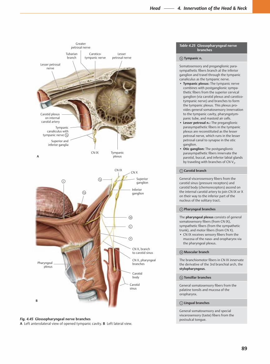
Carotidbody
CN IX
Inferiorganglion
Superior ganglion
CN X
CN X, branchto carotid sinus
CN X, pharyngeal branchesPharyngeal
plexus
B
LTy
To
M
C
P
Carotidsinus
Tubarianbranch
Carotid plexuson internal
carotid artery
CN IX
Tympaniccanaliculus with
tympanic nerve
Tympanicplexus
Lesser petrosalnerve
Carotico-tympanic nerve
Superior andinferior ganglia
Ty
Lesserpetrosal nerve
A
Greaterpetrosal nerve Table 4.25 Glossopharyngeal nerve
branches
Ty Tympanic n.
Somatosensory and preganglionic para- sympathetic fibers branch at the inferior ganglion and travel through the tympanic canaliculus as the tympanic nerve.• Tympanic plexus: The tympanic nerve
combines with postganglionic sympa- thetic fibers from the superior cervical ganglion (via carotid plexus and carotico- tympanic nerve) and branches to form the tympanic plexus. This plexus pro- vides general somato sensory innervation to the tympanic cavity, pharyngotym-panic tube, and mastoid air cells.
• Lesser petrosal n.: The preganglionic parasympathetic fibers in the tympanic plexus are reconstituted as the lesser petrosal nerve, which runs in the lesser petrosal canal to synapse in the otic ganglion.
• Otic ganglion: The postganglionic parasympathetic fibers innervate the parotid, buccal, and inferior labial glands by traveling with branches of CN V3.
C Carotid branch
General viscerosensory fibers from the carotid sinus (pressure receptors) and carotid body (chemoreceptors) ascend on the internal carotid artery to join CN IX or X on their way to the inferior part of the nucleus of the solitary tract.
P Pharyngeal branches
The pharyngeal plexus consists of general somatosensory fibers (from CN IX), sympathetic fibers (from the sympathetic trunk), and motor fibers (from CN X).• CN IX receives sensory fibers from the
mucosa of the naso- and oropharynx via the pharyngeal plexus.
M Muscular branch
The branchiomotor fibers in CN IX innervate the derivative of the 3rd branchial arch, the stylopharyngeus.
To Tonsillar branches
General somatosensory fibers from the palatine tonsils and mucosa of the oropharynx.
L Lingual branches
General somatosensory and special viscerosensory (taste) fibers from the postsulcal tongue.Fig. 4.45 Glossopharyngeal nerve branches
A Left anterolateral view of opened tympanic cavity. B Left lateral view.
89
Head 4. Innervation of the Head & Neck
7050003C04.indd 89 11/9/09 2:39:31 PM

Spinal nucleusof trigeminal nerve
Dorsal motornucleus
Superiorlaryngeal nerve
Pharyngealbranch
Inferior ganglion
Nucleus ambiguus
Jugularforamen
Superior ganglion
Nucleus of thesolitary tract,superior part
A
Nucleus of thesolitary tract,
inferior part
Spinal nucleus oftrigeminal nerve
Nucleus of thesolitary tract,
inferior part
Dorsal motornucleus
B
Nucleus of thesolitary tract,superior part
Nucleusambiguus
Olive
Table 4.26 Vagus nerve (CN X)
Nuclei, ganglia, and fi ber distribution
Branchiomotor (purple)
Nucleus ambiguus
Lower motor neurons innervate the muscles derived from the 3rd, 4th, and 6th branchial arches via CN IX, X, and XI. CN X innervates the derivatives of the 4th and 6th branchial arches:• Pharyngeal muscles (pharyngeal constrictors)• Muscles of the soft palate (levator veli palatini,
musculus uvulae, palatoglossus, palatopharyngeus)• Intrinsic laryngeal muscles
Parasympathetic (blue)
Dorsal motor nucleus
Preganglionic neurons synapse in small, unnamed ganglia close to target structures.
Postganglionic neurons innervate:• Smooth muscles and glands of thoracic and
abdominal viscera (Fig. 4.48G)
General somatic aff erent (yellow)
Spinal nucleus of CN V
First-order pseudounipolar cells in the superior (jugular) ganglion innervate:• Dura of the posterior cranial fossa (Fig. 4.48F)• Auricle, external auditory canal, and lateral tympanic
membrane (Fig. 4.48B,C)• Mucosa of the oropharynx and laryngopharynx
Viscerosensory (green)
First-order pseudounipolar cells in the inferior (nodose) ganglion relay taste and visceral sensation to the nucleus of the solitary tract. This nuclear complex consists of a superior part (taste) and inferior part (general visceral sensation).
Nucleus of the solitary tract
Taste (Fig. 4.48D): Fibers from the epiglottis and the root of the tongue are conveyed to the superior part of the nucleus of the solitary tract.
Visceral sensation (Fig. 4.48G): Fibers are relayed to the inferior part of the nucleus of the solitary tract from:• Mucosa of the laryngopharynx and larynx (Fig.
4.48A)• Aortic arch (pressure receptors) and para-aortic body
(chemoreceptors) (Fig. 4.48E)• Thoracic and abdominal viscera (Fig. 4.48G)
Course
The vagus nerve arises from the medulla oblongata and emerges from the skull via the jugular foramen. It has two sensory ganglia with fi rst-order pseudounipolar cells: the superior (jugular) ganglion (soma-tosensory) is within the cranial cavity, and the inferior (nodose) ganglion (viscerosensory) is distal to the jugular foramen.
Lesions
The recurrent laryngeal nerve supplies parasympathetic innervation to the intrinsic laryngeal muscles (except the cricothyroid). This includes the posterior cricoarytenoid, the only muscle that abducts the vocal cords. Unilateral lesions of this nerve cause hoarseness; bilateral destruction leads to respiratory distress (dyspnea).
Fig. 4.46 Vagus nerve nucleiA Anterior view of medulla oblongata. B Cross section through the medulla oblongata.The vagus nerve has the most extensive distribution of all the cranial nerves (L. vagus = vagabond). Parasympathetic fi bers descend into the thorax and abdomen. These fi bers form autonomic plexuses with post-ganglionic sympathetic fi bers (from the sympathetic trunk and abdom-inal ganglia). The plexuses extend along organs and blood vessels and provide motor innervation to the thoracic and abdominal viscera. Gen-eral viscerosensory fi bers ascend via CN X to the inferior part of the nu-cleus of the solitary tract.
90
Head 4. Innervation of the Head & Neck
CN X: Vagus Nerve
7050003C04.indd 907050003C04.indd 90 10/20/09 1:14:40 PM10/20/09 1:14:40 PM

A
F
B C
E GD
Left recurrentlaryngeal nerve
Left recurrentlaryngeal nerve
Thyrohyoidmembrane
CN X
Internal laryngeal nerve
Pharyngeal branches
Subclavian artery
Cricothyroidmuscle
Cervicalcardiac
branches
Aortic arch
Brachio-cephalic
trunk
Right recurrentlaryngeal nerve
Superiorlaryngeal
nerve
Externallaryngeal
nerve
Anterioresophageal
plexus
Table 4.27 Vagus nerve branches
Meningeal branches
General somatosensory fi bers from the dura of the posterior cranial fossa.
Auricular branch
General somatosensory fi bers from external ear (auricle, external acoustic canal, and part of lateral side of tympanic membrane).
Pharyngeal branches
The pharyngeal plexus consists of general somatosensory fi bers (from CN IX), sympathetic fi bers (from the sympathetic trunk), and motor fi bers (from CN X).• CN X conveys branchiomotor fi bers to the pharyngeal muscles.
Carotid branch
General viscerosensory fi bers from the carotid body (chemoreceptors) ascend on the internal carotid artery to join CN IX or X on their way to the inferior part of the nucleus of the solitary tract.
Superior laryngeal n.
Combines with a sympathetic branch from the superior cervical ganglion and divides into:• Internal laryngeal n.: Sensory fi bers from the mucosa of the
laryngopharynx and larynx.• External laryngeal n.: Parasympathetic motor innervation to the
cricothyroid.
Recurrent laryngeal n.
The recurrent laryngeal nerve is asymmetrical:• Right recurrent laryngeal n.: Recurs behind the right subclavian
artery.• Left recurrent laryngeal n.: Recurs behind the aortic arch.Ascends between the trachea and esophagus. The recurrent laryngeal nerves supply:• Motor innervation to the laryngeal muscles (except the cricothyroid).• Viscerosensory innervation to the laryngeal mucosa.
Branches to the thorax and abdomen
The vagus nerve also conveys parasympathetic and general viscerosensory fi bers from the cardiac, pulmonary, esophageal, celiac, renal, hepatic, and gastric plexuses (Fig. 4.48G)
Fig. 4.48 Distribution of the vagus nerve (CN X)
Fig. 4.47 Vagus nerve branches in the neck Anterior view.
91
Head 4. Innervation of the Head & Neck
7050003C04.indd 917050003C04.indd 91 10/20/09 1:14:41 PM10/20/09 1:14:41 PM

To laryngeal muscles via pharyngeal plexus and recurrentlaryngeal nerve
Trapezius
Cortico-bulbarfibers
Cranialroot
Foramenmagnum
Accessorynerve (CN XI), external branch
Vagus nerve(CN X)
Nucleusambiguus
Sternocleido-mastoid
Jugularforamen
Spinalroot
Spinal nucleus ofaccessory nerve
A B
A
B
Table 4.28 Accessory nerve (CN XI)
Nuclei, ganglia, and fi ber distribution
Branchiomotor (purple)
Nucleus ambiguus
Lower motor neurons innervate the muscles derived from the 3rd, 4th, and 6th branchial arches via CN IX, X, and XI.• CN XI innervates the
laryngeal muscles (except cricoarytenoid).
General somatomotor (red)
Spinal nucleus of CN XI
Lower motor neurons in the lateral part of the ventral horn of C2–C6 spinal cord segments innervate:• Trapezius (upper part).• Sternocleidomastoid.
Course
CN XI arises and courses in two parts that unite briefl y distal to the jugular foramen:
Cranial root: Branchiomotor fi bers emerge from the medulla oblongata and pass through the jugular foramen. They briefl y unite with the spinal root before joining CN X at the inferior ganglion. CN X distributes the branchiomotor fi bers via the pharyngeal plexus and the external and recurrent laryngeal nerves.
Spinal root: General somatomotor fi bers emerge as rootlets from the spinal medulla. They unite and ascend through the foramen magnum. The spinal root then passes through the jugular foramen, courses briefl y with the cranial root, and then descends to innervate the sternocleidomas-toid and trapezius.
Lesions
The sternocleidomastoid is exclusively innervated by CN XI, and the lower portions of the trapezius are innervated by C3–C5. Accessory nerve lesions therefore cause complete (fl accid) sternocleidomastoid paralysis but only partial trapezius paralysis.
Trapezius paralysis: Unilateral lesions may occur during operations in the neck (e.g., lymph node biopsies), causing:• Drooping of the shoulder on the aff ected
side.• Diffi culty raising the arm above the
horizontal.
Sternocleidomastoid paralysis:• Unilateral lesions: Flaccid paralysis causes
torticollis (wry neck, i.e., diffi culty turning the head to the opposite side).
• Bilateral lesions: Diffi culty holding the head upright.
Fig. 4.49 Accessory nerveA Posterior view of brainstem. B Right lateral view of sternocleidomastoid and trapezius.
Fig. 4.50 Accessory nerve lesionsAccessory nerve lesions cause partial paraly-sis of the trapezius and complete (fl accid) pa-ralysis of the sternocleidomastoid (see Table 4.28). Both lesions shown here are unilateral
(Note: For didactic reasons, the right muscles are displayed though they are innervated by the right cranial nerve nuclei.)
(right side). A Posterior view. Partial paralysis of the trapezius causes drooping of the shoul-der on the aff ected side. B Right anterolateral view. Flaccid paralysis of the sternocleidomas-toid causes torticollis (wry neck).
92
Head 4. Innervation of the Head & Neck
CN XI & XII: Accessory Spinal & Hypoglossal Nerves
7050003C04.indd 927050003C04.indd 92 10/20/09 1:14:50 PM10/20/09 1:14:50 PM

Hypoglossal trigone(in rhomboid fossa)
Nucleus ofthe hypoglossalnerve (CN XII)
A
OlivePyramid
Foramenmagnum
C1 spinal nerve
CN XII
Anteriorcondylar canal
Nucleus ofthe hypo-glossal nerve
B
Olive Pyramid
CN XII
Hyoglossusmuscle
Cortico-bulbarfibers
Styloglossusmuscle
Anteriorcondylar
canal
Nucleus ofthe hypoglossal
nerve
A
B C
Precentralgyrus
Left and rightgenioglossus muscles
Paralyzedgenioglossus
Tongue
CN X
C1
Genioglossus muscle
Table 4.29 Hypoglossal nerve (CN XII)
Nuclei, ganglia, and fi ber distribution
General somatomotor (red)
Nucleus of CN XII
Lower motor neurons innervate:• Extrinsic lingual muscles
(except palatoglossus).• Intrinsic lingual muscles.
Course
The hypoglossal nerve emerges from the medulla oblongata as rootlets between the olive and pyramid. These rootlets combine into CN XII, which courses through the hypoglossal (anterior condylar) canal. CN XII enters the root of the tongue superior to the hyoid bone and lateral to the hyoglossus.• C1 motor fi bers from the cervical plexus
travel with the hypoglossal nerve: some branch to form the superior root of the ansa cervicalis (not shown), whereas others continue with CN XII to supply the geniohyoid and thyrohyoid muscles.
Lesions
Upper motor neurons innervate the lower motor neurons in the contralateral nucleus of the hypoglossal nerve. Supranuclear lesions (central hypoglossal paralysis) will therefore cause the tongue to deviate away from the aff ected side. Nuclear or peripheral lesions will cause the tongue to deviate toward the aff ected side (Fig. 4.52C).
Fig. 4.52 Hypoglossal nerveA Course of the hypoglossal nerve. Upper mo-tor neurons synapse on lower motor neurons on the contralateral nucleus of the hypoglossal nerve. Supranuclear lesions will therefore cause contralateral paralysis; peripheral lesions will cause ipsilateral paralysis (same side). B The functional genioglossus extends the tongue anteriorly. C Unilateral paralysis due to a pe-ripheral lesion causes the tongue to deviate toward the aff ected side (dominance of the in-tact genioglossus).
Fig. 4.51 Hypoglossal nerve nucleiThe nucleus of the hypoglossal nerve is located in the fl oor of the rhomboid fossa. Rootlets emerge between the pyramid and the olive.
A Cross section through the medulla oblon-gata. The proximity of the nuclei to the mid-line causes extensive lesions to involve both nuclei. B Anterior view of medulla oblongata.
93
Head 4. Innervation of the Head & Neck
7050003C04.indd 937050003C04.indd 93 10/20/09 1:14:55 PM10/20/09 1:14:55 PM

①
Incisive canal
Nasopalatine n. and a., greater palatine a.
Mastoid foramen
Emissary v.
Hypoglossal canal
CN XII, venous plexus of hypoglossal canal
(Posterior) condylar canal
Condylar emissary v.
Foramen spinosum
Carotid canal
Petrotympanicfissure
Anterior tympanic a., chorda tympani
Stylomastoidforamen
Facial n., stylo-mastoid a.
Jugular foramen
– See opposite
Internal jugular v.
Cribriform plate
CN I, ethmoidal aa. and vv.
Internal acoustic meatus
CN VIII
CN VII
Labyrinthine a. and v.
Foramen rotundum
CN V2
Carotid canal
Internal carotid a. with sympathetic plexus
Foramen spinosum
Middle meningeal a., recurrent meningeal branch of CN V3
Hiatus of canal forlesser petrosal n.
Lesser petrosal n.,superior tympanic a.
Hiatus of canal forgreater petrosal n.
Greater petrosal n.
Jugular foramen
Posteriormeningeal a.
Inferiorpetrosal sinus
CN XI
CN X
CN IX
Sigmoid sinus Foramen magnum
CN XI, spinal root
Spinal cord
Vertebral a.
Anterior and posterior spinal aa.
Emissary vv.
Optic canal
CN II, ophthalmic a.
Superior orbital fissure
CN III
Frontal n.
Lacrimal n.
Superior andinferior ophthalmic v.
CN VI
CN IV
Foramen ovale
CN V3, lesser petrosal n. (CN IX)
Foramen lacerum
Deep and greater petrosal nn. (across superior surface)
Greater palatine foramen
Greater palatine n. and a.
Lesser palatine foramina
Lesser palatine n. and a.
②③⑤⑥⑦
④
②③④⑥
①
⑤
③② ①
⑤④
①
⑥⑤③②
④A
B
①
②
③
④
⑤
⑥
A
B
①
②
③
④
⑤
⑥①
②
③
④
⑤
⑥
①
②
A B
Internal carotid a., internal carotid plexus
Nasociliary n.⑦
Fig. 4.53 Passage of neurovascular structures through the skull baseA Superior view of cranial cavity. B Inferior view of base of skull. This image and the corresponding table only address structures entering
and exiting the skull. Many neuro vascular structures pass through bony canals within the skull (to pterygopalatine fossa, infratemporal fossa, etc.).
94
Head 4. Innervation of the Head & Neck
Neurovascular Pathways through the Skull Base
7050003C04.indd 94 11/9/09 2:40:16 PM

Table 4.30 Openings in the skull base
Cranial cavity Opening Transmitted structures
Nerves Arteries and veins
Internal view, base of the skull
Anterior cranial fossa
Cribriform plate • CN I (olfactory fi bers collected to form olfactory n.)
• Anterior and posterior ethmoidal aa. (from ophthalmic a.)• Ethmoidal vv. (to superior ophthalmic v.)
Middle cranial fossa
Optic canal • CN II (optic n.) • Ophthalmic a. (from internal carotid a.)
Superior orbital fi ssure • CN III (oculomotor n.)• CN IV (trochlear n.)• CN IV (abducent n.)• CN V1 (ophthalmic n.) divisions
(lacrimal, frontal, and nasociliary nn.)
• Superior and inferior ophthalmic vv. (to cavernous sinus) (Note: The inferior ophthalmic v. also drains through the inferior orbital fi ssure to the pterygoid plexus.)
Foramen rotundum* • CN V2 (maxillary n.)
Foramen ovale • CN V3 (mandibular n.)• Lesser petrosal n. (CN IX)
• Accessory meningeal a. (from mandibular part of maxillary a.)
Foramen spinosum • CN V3, recurrent meningeal branch • Middle meningeal a. (from mandibular part of maxillary a.)
Carotid canal • Carotid plexus (postganglionic sympathetics from superior cervical ganglion)
• Internal carotid a.
Hiatus of canal for greater petrosal n.
• Greater petrosal n. (CN VII)
Hiatus of canal for lesser petrosal n.
• Lesser petrosal n. (CN IX) • Superior tympanic a. (from middle meningeal a.)
Posterior cranial fossa
Internal acoustic meatus
• CN VII (facial n.)• CN VIII (vestibulocochlear n.)
• Labyrinthine a. (from vertebral a.)• Labyrinthine vv. (to superior petrosal or transverse sinus)
Jugular foramen • CN IX (glossopharyngeal n.)• CN X (vagus n.)• CN XI (accessory n., cranial root)
• Internal jugular v. (bulb)• Sigmoid sinus (to bulb of internal jugular v.)• Posterior meningeal a. (from ascending pharyngeal a.)
Hypoglossal canal • CN XII (hypoglossal n.) • Venous plexus of hypoglossal canal
Foramen magnum • Medulla oblongata with meningeal coverings
• CN XI (accessory n.)
• Vertebral aa.• Anterior and posterior spinal aa. (from vertebral a.)• Emissary vv.
External aspect, base of the skull (where diff erent from internal aspect)
Incisive canal • Nasopalatine n. (from CN V2) • Branch of greater palatine a.
Greater palatine foramen
• Greater palatine n. (from CN V2) • Greater palatine a. (from pterygopalatine part of maxillary a.)
Lesser palatine foramen
• Lesser palatine n. (from CN V2) • Lesser palatine aa. (from pterygopalatine part of maxillary a. or as branch of greater palatine a. or descending palatine a.)
Foramen lacerum** • Deep petrosal n. (from superior cervical ganglion via carotid plexus)
• Greater petrosal n. (from CN VII)
Petrotympanic fi ssure • Chorda tympani (from CN VII) • Anterior tympanic a. (from mandibular part of maxillary a.)
Stylomastoid foramen • Facial n. (CN VII) • Stylomastoid a. (from posterior auricular a.)
(Posterior) condylar canal
• Condylar emissary v. (to sigmoid sinus)
Mastoid foramen • Mastoid emissary v. (to sigmoid sinus)
*The external opening of the foramen rotundum is located in the pterygopalatine fossa, which is located deep on the lateral surface of the base of the skull and is not visible here.
**Structures travel over the superior surface of the foramen lacerum, not through it (with the possible exception of lymphatic vessels and emissary veins).
95
Head 4. Innervation of the Head & Neck
7050003C04.indd 957050003C04.indd 95 10/20/09 1:15:06 PM10/20/09 1:15:06 PM


![24 D...Research Data Air-ow rate[m3/h] 439 Pressure[Pa] 1 1 8.6 Pressure type P staPower supply al l Density[m3/h] 1 .205 Label Code K224 P re ssure To ta l P re ssure Sta tic 0 200](https://cdn.vdocument.in/doc/165x107/60e5e7d99f08c372702263b6/24-d-research-data-air-ow-ratem3h-439-pressurepa-1-1-86-pressure-type.jpg)


