
Tendinopathies aroundfoot and ankle
• M Maestro,• Y Tourné ,J Cazal• ,B Schlaterer, B Ferré,• J M Parisaux, P Ballerio
AFCP22 nov 2014 Novotel Monaco

PERMANENT REMODELINGEVERSION
STABLE
INVERSION
FLEXIBLE
ENERGY
VISCO-ELASTIC STRUCTURE
Stocking Distribution
flatcavus
28 muscles – 33 articulations

Jonction myo-tendineuse
Zone moyenne hypovasculaire
Gaine
Tunnel ostéo-fibreuxPoulie fibreuse
Insertion osseuseenthésopathies
Tendon tendinopathies
Os et articulations
MuscleSd de loge chron.
Peau ,ongle, bourses séreusesaponévroses
Capitongraisseux
Paquets vasculo-nerveux
pied= COMPLEXITE STRUCTURELLE
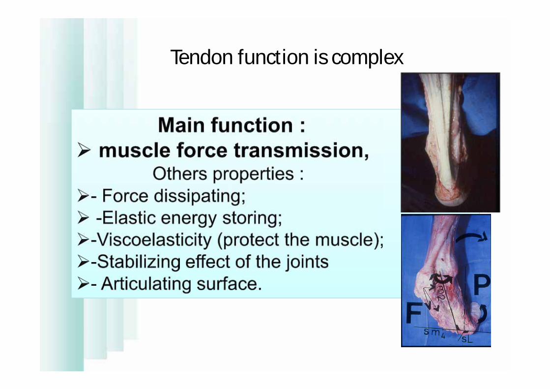
Tendon function is complex

aponeurosis and tendons= 2/3 of lesions
Novachek JBJS 1998; 80-A: 8
James Jones 1990
30 to 50% of all sports injuryRenstrom 1991

SésamoïdesRenander, frac fatigue
Système SCPAchille, ap. Plant.
Fractures de fatigue
Talonades
Tendons péroniersTA, TP,
HV, HR, Freiberg
Peau, Ongle
Toutes les structurespeuvent être atteintes (le maillon le plus faible cède )
Impingement
bursopathiesFOOT BALL= 50%TENNISCOURSEMARCHE
CHRONIC Pathology,NON INFLAMMATORYÀ Caractère mécanique
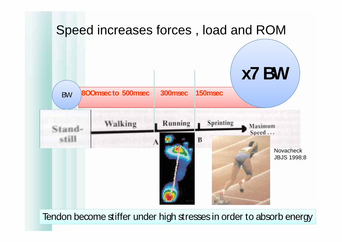
8OOmsec to 500msec 300msec 150msec BW
x7 BW
Speed increases forces , load and ROM
Tendon become stiffer under high stresses in order to absorb energy
Novacheck JBJS 1998;8

Harmfull forcesare not during heel strike,
but just after during loading response and midstance
gait
running
TENSEGRITY= tension-intregrity(Buckminster Fuller -1895-1983 architect)BIOTENSEGRITY(Stephen M Levin MD orthopedic surgeon 1970)
Concept de « bio-tenségrité »Équilibre tension-compressionStructures légères et résistantes
1 Seule Réponse face aux diverses contraintes= défDéformation = pathologie
MOBILITEFORCESTABILITE
Une autre vision de la biomécanique

Mécanique des axes et leviers Biotenségrité
Les os ne se touchent jamaisIls sont en suspension dans les fascia en assurant leur mise en tension
• Non linéaire (tjs sous tension)
• Structure continue• Indépendant de la
gravité• Pluridirectionnel• Basse énergie• Articulations axes
évolutifs
• Linéaire (retour à zéro)• Structure discontinue• Dépendant de la
gravité• Unidirectionnel• Haute énergie• Articulation à axe fixe
icosahèdre

Tenségrité= déformabilité sous la contrainte
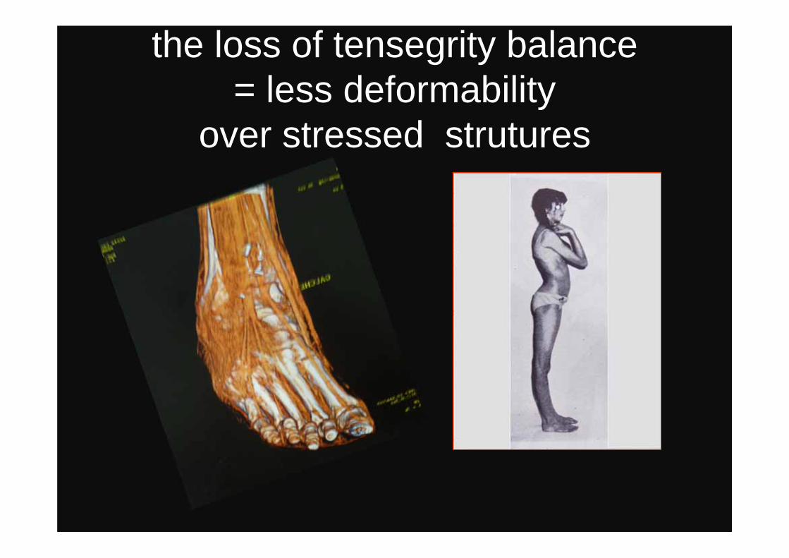
the loss of tensegrity balance= less deformability
over stressed strutures

Pe: gastrocnemius tightness
Enability or difficulties for bare foot walkingLack of endurance Difficulties of climbing speedDifficulties to run or accelerate, to jumpVeinous problems
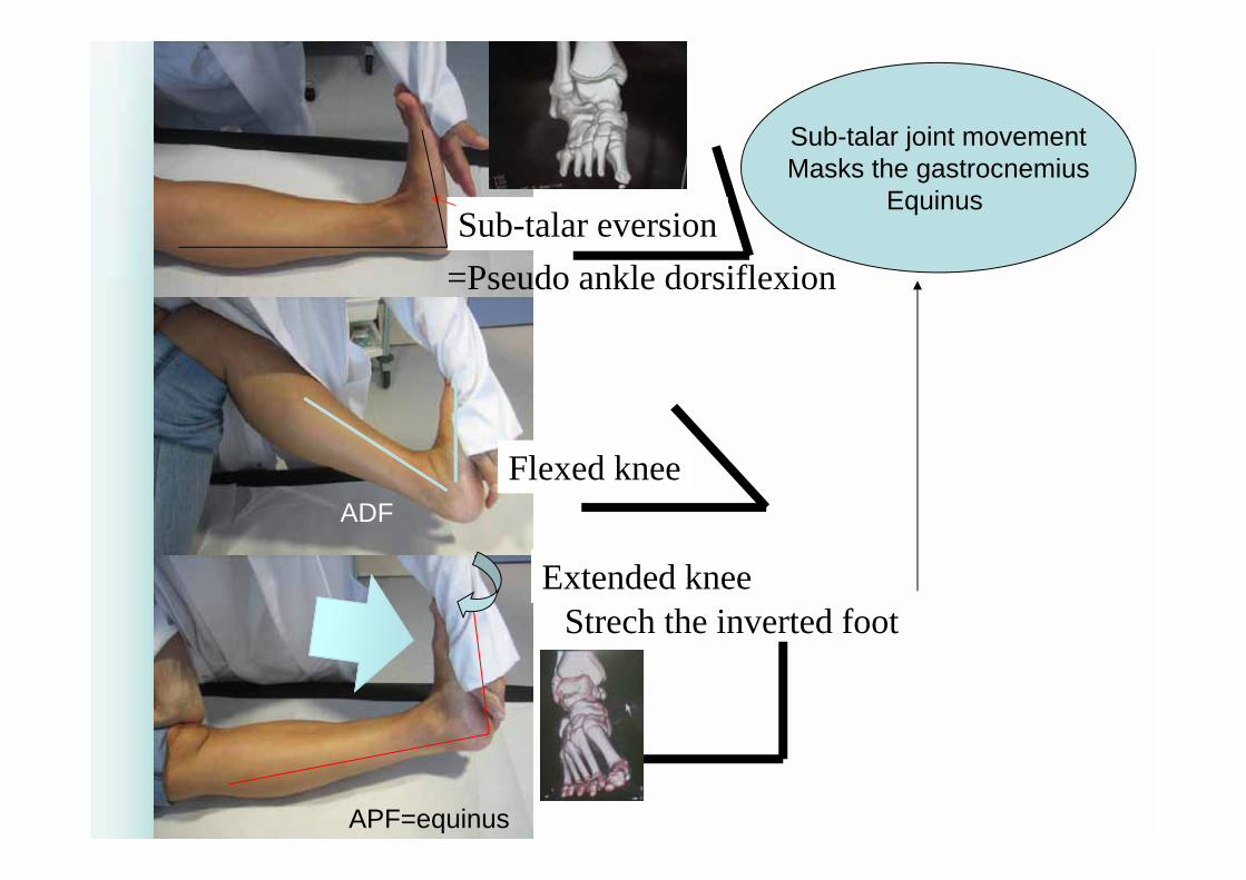
Strech the inverted foot
Sub-talar joint movementMasks the gastrocnemius
Equinus
Flexed knee
Sub-talar eversion=Pseudo ankle dorsiflexion
ADF
APF=equinus
Extended knee
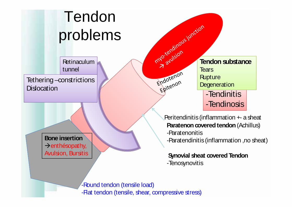
Tendon problems
-Tendinitis-Tendinosis
Synovial sheat covered Tendon-Tenosynovitis
Peritendinitis (inflammation +- a sheat
-Round tendon (tensile load)-Flat tendon (tensile, shear, compressive stress)
Paratenon covered tendon (Achillus)-Paratenonitis-Paratendinitis (inflammation ,no sheat)
Tethering –constrictionsDislocation
Tendon substanceTearsRuptureDegeneration
Retinaculumtunnel
Bone insertionenthésopathy,Avulsion, Bursitis

Dérangement interne
Modification des CIR
Surfaces anormales
Lésions articulaires
Réponse bio:Hypertrophie synovialeostéophytes
Dégénérescencearticulaire
Frankel 1971
Surcharge articulaire
Leadbetter 1992, Novachek 1998

Non symptomatic-------symptomatic
• TENDINOSIS • degenerative process• absence of inflammatory
cells,• fibers desorientation• collagene degeneration,• hypercellularity and vascular
nerve ingrowth
• TENDINITIS• vascular disruption• “inflammatory “ repair process

ImbalanceExcess of contrains: extrinsic F•Trainning errors•Ground structure (hard)•Shoes (pronation control)•Iatrogenic (fluoroquinolones
steroids …)
Tendon weakness: intrinsic F• Age, weight • Compliance and length• Anatomical particularities • Lower extremity
malalignment• Leg length discrepancy• Metabolic factors (ph, H2O)• Fatigue,muscular weakness
Combination of extrinsic and intrinsic factors
Ethiologic factors

PhysiotherapyHighenergyECSWT
Treatment ?
ReeducationCompliance
Muscular strength
Proprioception
reconditioning
Médications
NSAIDs
Insoles
Shoe wear modifSurgeryTenoscopyOpen surg
Tendinopathy
Infiltrations
Growth factors
Their treatment are challenging problemsCareful evaluation prior to embarking on a specific treatment

1970’s 1990’simmobility active approach
No study to date provideConcensus Treatment trend
Healing process: Inflammatory—Proliferative (fibroblastic)--Remodeling

Clinical assessmentsphysical examination
Joint laxityprovocative pain maneuverLoss of power
patient history Sprain Injuries swelling fluoroquinolones abs
Imaging assessmentsconventional X-Rays of foot and ankle
in WB position and bilateralAP, lateral and AP oblique views: achitecture,, avulsion fractures…Meary’s viewstress views
MRI: modality of choice multiplanar imaging + soft and bone tissues
- cartilage US
competitive for tendons but operator-dependent
CT scan choice for ankle laxity : ligt &
cartilage damages, associated tears of PT
limited use with teno-sheath injection

Tibialis Anterior
Insertional tendinopathy
- Sports with pulse (danse,ballet, jump, skating…)
- medial cuneiform pain
- gait abnormality (heel strike-initial loading,swing)
- Pain with passive stretching+isometric contraction
Spontaneous rupture
-rare, aging
-Foot drop
Tenosynovitis (stenosing, crepitans)- Windsurf, treck,marathon
- Relatively rare

> Mechanical Tenosynovitis> Insertional
>Degenerative TendinopathyVery - frequent, evolutive
- Pes valgus ++
- Gait abnormality: stance
>Spontaneous rupture- Frequent :2/3 women,
>40years
- Chronic insidious
Tibialis Posterior ++
Stage I
Stage II
StageIIIstageIV
Johnson & Strom1989
Jhonson & Strom Clin Orthop 1989

Foot pronator is at risk. Bussenil C. et Coll.
Rearfoot – forefoot orientation and traumatic risk for runners.Foot and Ankle Intern 1998 ; 19 (1) : 32 – 37
GastrocnemiusRetraction associated++

- Rare, inversion painfull trauma
- X ray : possible fracture
- MRI++
- surgical treatment
Tibialis Posterior luxation
Sheat and retinaculum lesion
PRF
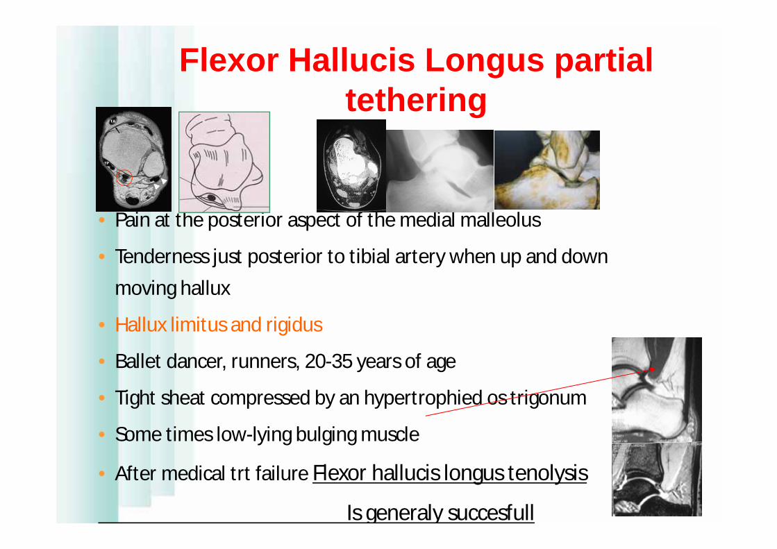
• Pain at the posterior aspect of the medial malleolus
• Tenderness just posterior to tibial artery when up and down moving hallux
• Hallux limitus and rigidus
• Ballet dancer, runners, 20-35 years of age
• Tight sheat compressed by an hypertrophied os trigonum
• Some times low-lying bulging muscle
• After medical trt failure Flexor hallucis longus tenolysis
Is generaly succesfull
Flexor Hallucis Longus partial tethering

Peroneal tendonsanatomy and biomechanics
both the tendons everse the hind foot and plantarflex the ankle
the PL lock the transversal arch and plantarflex the first ray.

Peroneal tendon disorders
• Clinical conditions are numerous: pain in the postero lateral aspect of the ankle+++
• Hindfoot in varus and ankle instability must be pointed out, predisposing factors
• MRI remains mandatory to achieve a precise diagnosis
• Surgical procedures must be combined• Return to maximal function after surgery is prolonged• Early ROM with protected ambulation to prevent
adhésions
Selmani et Al. Foot & Ankle Intern.2006;27:221

avascular zones are behind the lateral malleolus And cuboid bone : most frequent location of tendinosis
Critic point

Peroneus brevistendinosis
Low-lying muscle
repaired

tendinosisLateral tubercle hypertrophy
Strain mechanism
Low-lyingMuscle belly
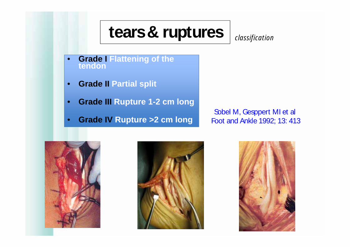
• Grade I Flattening of the tendon
• Grade II Partial split
• Grade III Rupture 1-2 cm long
• Grade IV Rupture >2 cm long
tears & ruptures classification
Sobel M, Gesppert MI et alFoot and Ankle 1992; 13: 413
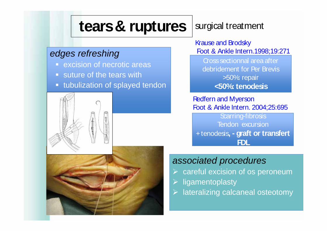
edges refreshing excision of necrotic areas suture of the tears with tubulization of splayed tendon
tears & ruptures surgical treatment
associated procedures careful excision of os peroneum ligamentoplasty lateralizing calcaneal osteotomy
Cross sectionnal area afterdebridement for Per Brevis
>50%: repair<50%: tenodesis
Krause and BrodskyFoot & Ankle Intern.1998;19:271
Redfern and MyersonFoot & Ankle Intern. 2004;25:695
Scarring-fibrosisTendon excursion
+ tenodesis, - graft or transfert FDL

• overuse on the insertion• tendoperiostial microruptures and
calcifications
Insertional tendinopathy enthesopathies

pathomechanics disruption of the
retinaculum eversion and
dorsiflex of the ankle combined with a
forceful contractionof PT
elective lesion in downhill skiers(slalom,bumps)
dislocation clinical conditions
Eckert and Davies ‘ staging
1 2
3
4
acute conditions: diagnosis the more often missed« severe ankle sprain-like » but no laxity
chronic conditions« lateral chronic instability of the ankle-like » but no laxity

ACHILLUS TENDONPOWER= TRICEPS (Gastrocnemius+soleus)TRANSMISSION=achilleo-calcaneo-plantar system
Windlass mechanismMécanisme du treuil
MGN
Soléaire

ETUDE DE LA MARCHE APRES TEST DE FATIGUE ET TRAITEMENT AU LPG CHEZ 18 GOLFEURS PROFESSIONELS
RIVET J.J. MAISETTI O. MAESTRO M.
A.F.C.P. 11 nov. 2000 Paris SOFCOT

Pression Pieds/Jumeaux Rétractés
02468
1012
avant fat après fat après LPG
** *
Chez les sujets ayant tendance à la rétraction des jumeaux : Les pics d’appuis talonniers lors de la marche sont tous latéralisés .
Ils migrent vers le centre calcanéen après fatigue , puis reviennent à leurs données initiales après LPG.( 1 )
Figure 2 : médialisation des appuis calcanéens après fatigue.( ** pour significatif à p = 0.01 - * pour significatif à p = 0.05

CONCLUSION
Chez un groupe de sujets sportifs professionnels, ayant tous des pieds considérés normaux sans callosités plantaires, cette étude permet de dégager 2 groupes de sujets :- Un groupe qualifié de normal : 10 sujets soit 54%- Un groupe ayant une tendance à la rétraction des muscles jumeaux représentant 8 sujets soit 46%
- Cela est en accord avec l’étude Kowalski sur la rétraction des gastrocnémiens .

Achillus tendon-Acute ruptures:- early trt,anatomic restoration,-meticulous attention to detail to avoid wound complications,-early rehabilitation to minimize recovery time and maximize function and strenght
Chiodo and Den Hartog, Foot & Ankle Intern.2008;29:114
-Non insertional tendinopathy-Non operative trt 6months-Age and long term duration less favorable-Percutaneous_open debridment_ transfert FHL augmentation
Scott et al. Foot & Ankle Intern.2008.29:759
-Insertional tendinopathy-Direct posterior approach-Early weight bearing and mobilisation if less than 50% tendon excised-If more FHL tranfert
Deorio and Easley Foot & Ankle Intern.2008;29:542

TM junction: 14,5%TM junction: 14,5%
Body: 75%Body: 75%
Insertion:10,5%Insertion:10,5%
30 Y_50Y: sports activity
>50Y: degenerative
Evaluation:
- Site- -gap (echo)- -degenerative lesions
Trt- NON OPEN: rerupture 10%- OPEN: complications
(infection, adhernce, nerve)-PER-CUT
Early MOB
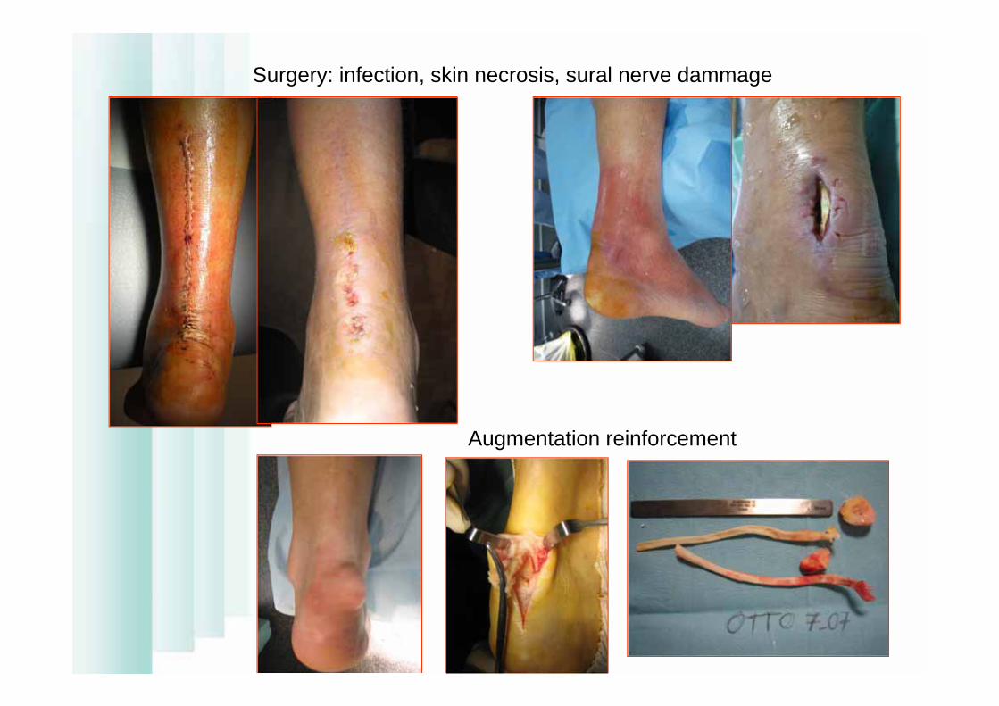
Surgery: infection, skin necrosis, sural nerve dammage
Augmentation reinforcement

Shoe shape and lateral movements (Hoffmann 1997)

Intratendinous micro-tears
thickness
Nodules
JL Besse
Plantairegrele
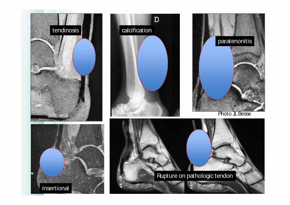
tendinosis calcification
paratenonitis
insertional
Rupture on pathologic tendon
Photo JL Besse

Functionnal training programstability= mobility
Phase I:recovering-Mobility-Length-Flexibility
Phase IIStrength-balance control
Powerfull contractile action-Gradual process-Proprioception (compensation can occur for up than 1 year)-Repetitive movment to replicate functional movt-Close kinematic chain exercises+++
Phase IIIAerobic reconditioning-Treadmill,stairmaster,elliptical unitOptions:swimming,walk-jog,-posture and reflexes-reinforcement
Phase IVReturn to activity-Maintenance strength-flexibility-Reinforcement,aerobic exercises-Activity modification(Bernstein concept)
Age +++Assessment
PainInflammationTendon irritability=progression of
program

Malgré des années de pratique et d’entraînement , les athlètes sont
incapables de répéter un mouvement invariant
Variabilité intra et inter individuelle des performances
Mvts systems as dynamical systemsBernstein’s pb (1967)
« maîtriser la redondance des DOF »

Neuro-muscular control
Joint stability
Tendon-Muscular-bone
unit
SkillPhysical fitnessTactile sensing
Psychologymeteorology
Winning shot!
Winning shot!
Monte-carlo 230406
Conclusion
Antibes 70908
Conservative management= high success rate Assessment critical Active approach (biomechanics+++) Don’t miss surgery time Pain is a signal !
Thankyou
MERCI


