
©2014 MFMER | slide-1 ©2014 MFMER | slide-1
Paul J. Kurtin, M.D. Rochester, MN
Test Utilization: • Chronic Lymphocytic Leukemia
Initial Evaluation • Diagnostic Criteria • Selection of Tests for Prognosis Response to Therapy • Challenges • Assessment for persistent disease

©2014 MFMER | slide-2
DISCLOSURES:
Relevant Financial Relationship(s) None
Off Label Usage
None

©2014 MFMER | slide-3
Chronic Lymphocytic Leukemia Development of Test Utilization Algorithms
• Close collaboration with CLL disease oriented group
• Typical points for blood / bone marrow analysis in CLL patients
• Diagnosis • Assessment of initial prognosis • Assessment of response to therapy • Assessment of progressive disease
• Genetic progression • Transformation
• Assessment of new cytopenias/systemic symptoms

©2014 MFMER | slide-4
CLL-Initial Evaluation on Blood
Challenge: --Correctly diagnose
CLL --Optimize prognostic
testing

©2014 MFMER | slide-5
• Establish CLL Dx • Lymphocyte morphology • Flow cytometry results • Lymph node biopsy
• Consider Absolute B cell Count • Chronic lymphocytic
leukemia • Monoclonal B cell
lymphocytosis
CLL Initial Evaluation on Blood It is important to get the diagnosis correct
right from the beginning

©2014 MFMER | slide-6
CLL Blood Morphology

©2014 MFMER | slide-7
Diagnostic Criteria for CLL Flow Cytometry
CD5+ B cells
Dim monotypic light chain
CD23+
Dim CD20

©2014 MFMER | slide-8
CLL Lymph Node Morphology
Proliferation Center

©2014 MFMER | slide-9
CD20 CD3 CD5 CD43
CD23 Cyclin D1 lambda kappa
CLL Lymph Node Immunohistochemistry

©2014 MFMER | slide-10
B-cell count <1000 AND
NO SLL presentation
STOP
B-cell count 1000-5000 OR
B-cell count <1000 AND
SLL presentation (i.e. lymphadenopathy)
B-cell count >5000
Flow Cytometry CD38 CD49d Zap-70
FISH: B-CLL Panel
or Array CGH
IGVH sequencing
CLL Initial Evaluation on Blood Prognostic Studies

©2014 MFMER | slide-11
Typical CLL FISH Panel* • 6q-, MYB/Cen6
• 11q-, ATM/Cen11
• +12, D12Z3/MDM2
• 13q-, D13S319/LAMP1
• 17p-, TP53/Cen17
• t(11;14), CCND1/IGH
• IGH BAP reflex to: • t(14;18), IGH/BCL2 • t(14;18), IGH/BCL3
Prognosis
Exclude Mantle Cell Lymphoma
Unusual CLL
Subgroups
*For Prognosis NOT Diagnosis

©2014 MFMER | slide-12
Survival Significance of Standard CLL Risk Factors
IGVH mutated IGVH
unmutated
• CD38 hi • Zap-70 hi • p53 mutations • High risk FISH
(17p-, 11q-)
• CD38 lo • Zap-70 lo • p53 normal • Low risk FISH
(13q-)
Median survival 8-10 years Median survival 25 years
After: Shanafelt TD, et al Blood (2004) 103:1202-10

©2014 MFMER | slide-13
CLL Bone Marrow Assessment for Response to Therapy
• Previously diagnosed CLL patients who have been treated
• No absolute lymphocyte count progression • No progression of adenopathy or hepatosplenomegaly • Minimal risk for MDS • Assess bone marrow to determine:
• Complete response (CR), minimal residual disease (MRD) negative or
• CR, but MRD positive or • Partial response (nodular) or • Persistent disease

©2014 MFMER | slide-14
CLL Bone Marrow Assessment for Response to Therapy—Challenges
Is Minimal Residual Disease Testing Necessary?
• Level of MRD predicts progression free survival and overall survival: • After routine chemotherapy • After immunochemotherapy
(FCR/alemtuzumab) • After stem cell transplantation
*Moreton C, et al JCO 2005;23:2971-9
Aletuzumab therapy; overall survival; MRD flow cytometry*
©2014 MFMER | slide-15
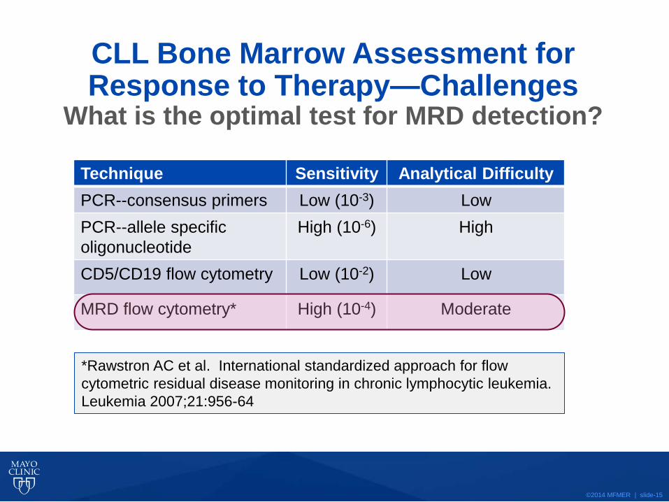
CLL Bone Marrow Assessment for Response to Therapy—Challenges
What is the optimal test for MRD detection?
Technique Sensitivity Analytical Difficulty PCR--consensus primers Low (10-3) Low PCR--allele specific oligonucleotide
High (10-6)
High
CD5/CD19 flow cytometry Low (10-2) Low
MRD flow cytometry* High (10-4) Moderate
*Rawstron AC et al. International standardized approach for flow cytometric residual disease monitoring in chronic lymphocytic leukemia. Leukemia 2007;21:956-64
©2014 MFMER | slide-16
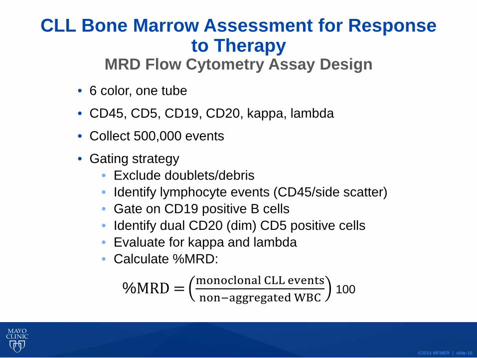
CLL Bone Marrow Assessment for Response to Therapy
MRD Flow Cytometry Assay Design • 6 color, one tube
• CD45, CD5, CD19, CD20, kappa, lambda
• Collect 500,000 events
• Gating strategy • Exclude doublets/debris • Identify lymphocyte events (CD45/side scatter) • Gate on CD19 positive B cells • Identify dual CD20 (dim) CD5 positive cells • Evaluate for kappa and lambda • Calculate %MRD:
%MRD = monoclonal CLL eventsnon−aggregated WBC
100

©2014 MFMER | slide-17
CLL MRD By Flow Cytometry
MRD=0.35% MRD=0.02%

©2014 MFMER | slide-18
CLL MRD By Flow Cytometry MRD Negative
MRD=0.00%

©2014 MFMER | slide-19
CLL Minimal Residual Disease Can Immunohistochemistry Substitute for Flow
Cytometry?
• 82 patients
• Confirmed CLL
• Treatment with chemoimmunotherapy
• Bone marrow aspirates and biopsies: • Morphology assessment • MRD flow cytometry • Immunohistochemistry (IHC) for
CD3, CD5, CD23 and PAX-5
• Compare MRD flow and IHC results
Amador-Ortiz C, et al; 2013

©2014 MFMER | slide-20
MRD Flow Cytometry vs. IHC Concordance
PAX-5+ CD3- CD5+ CD23+
Amador-Ortiz C, et al; 2013

©2014 MFMER | slide-21
CLL Minimal Residual Disease MRD flow cytometry vs. Immunohistochemistry
Concordance = 87%
Discrete IHC positive lymphoid aggregate, hemodilute aspirate
Very low flow MRD: IHC negative: 0.005 - 0.200% IHC suspicious: 0.040 - 0.890%
Conclusion: with a slight loss in sensitivity, IHC can substitute for MRD flow cytometry
Amador-Ortiz C, et al; 2013

©2014 MFMER | slide-22
CLL Bone Marrow Assessment for Response to Therapy—Challenges
What is the significance of lymphoid aggregates?
Residual involvement by CLL (nodular partial remission) VS.
Reactive lymphoid aggregates VS.
Rituximab effect—T cell rich lymphoid aggregates

©2014 MFMER | slide-23
CLL—Assessment of Response to Therapy

©2014 MFMER | slide-24
CLL Bone Marrow Assessment for Response to Therapy

©2014 MFMER | slide-25
CLL Bone Marrow Assessment for Response to Therapy
Morphologic involvement by CLL/SLL >30%
No Yes
Flow: CLL MRD Diagnosis: “Residual
involvement by CLL/SLL (%),
nodules present/absent”
NO ancillary studies

©2014 MFMER | slide-26
CLL Bone Marrow Assessment for Response to Therapy
Negative Positive
Lymphoid aggregates/inters
titial infiltrates
Flow: CLL MRD
No Yes
Diagnosis: “Negative for CLL/SLL by
morphologic and MRD assessments”
Diagnosis: “Minimally involved
by SLL/CLL (%), nodules absent”
BM lymphoid nodules present?
Concordant % involvement
MRD flow and morphology
No Yes
Perform IHC—see next slide No
Yes
Diagnosis: “Involved by
SLL/CLL (%), nodules present”

©2014 MFMER | slide-27
CLL Bone Marrow Assessment for Response to Therapy
Diagnosis: “Negative for CLL/SLL by
morphologic and MRD assessments”
Immunohistochemistry CD3, CD5, CD20,
CD23, PAX-5
Negative
Lymphoid aggregates/inters
titial infiltrates
Flow: CLL MRD
Yes
? Do the lymphoid aggregates represent
CLL missed by the MRD flow assay or
are they T cell aggregates in a
Rituximab treated patient
Yes No
CLL Phenotype
Diagnosis: “Residual
involvement by CLL/SLL (%)
nodules present/absent”

©2014 MFMER | slide-28
CLL Bone Marrow Assessment for Response to Therapy
Positive Flow: CLL MRD
Yes
BM lymphoid nodules present?
Concordant % involvement
MRD flow and morphology
No
? What is the degree of BM involvement by
CLL: --CLL involvement underestimated by
the MRD flow OR
--MRD with abundant reactive T cells
Immunohistochemistry CD3, CD5, CD20,
CD23, PAX-5 Nodules rich in CLL B
cells Yes No
Diagnosis: “Residual
involvement by CLL/SLL(%)
nodules present”
Diagnosis: “Minimal residual
involvement by CLL(%)”

©2014 MFMER | slide-29
• Initial diagnosis • Get it right
• Strict flow cytometry criteria (histograms!) • LN biopsy • Be patient
• Judiciously order prognostic studies • Evaluation of Response to Therapy
• Importance of MRD testing • Use a highly sensitive flow cytometry assay possibly
supplemented by IHC • Recognize the non-specificity of lymphoid
aggregates post-therapy • Clinican/Hematopathologist joint algorithm
development for evaluation of MRD
CLL Test Utilization Summary


