Download - The anatomy of the arm

The Anatomy of the Arm
(or The Brachium)

Biceps brachii Origin Short head: tip of coracoid process of
scapula; Long head: supraglenoid tubercle of scapula, passes through the shoulder joint and emerges from the joint through the intertubercular groove.
Insertion Tuberosity of radius and fascia of forearm via bicipital aponeurosis
Action Supinates forearm and, when it is
supine, flexes forearm Weak flexion of arm Innervation Musculocutaneous nerve
(C5, C6)

The bicipital aponeurosis• It is a broad aponeurosis of the
biceps brachii located in the cubital fossa .
• It separates superficial from deep structures in the fossa.
• It originates from the distal insertion of the biceps brachii and runs across the brachial artery.
• It is inserted into the antebrachial fascia of the forearm.
• It helps to protect the brachial artery and the median nerve running underneath( during venipuncture ) from the median cubital vein.
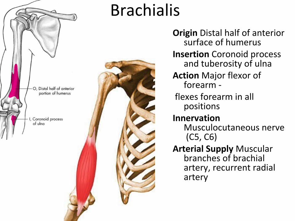
BrachialisOrigin Distal half of anterior
surface of humerusInsertion Coronoid process
and tuberosity of ulnaAction Major flexor of
forearm - flexes forearm in all
positionsInnervation
Musculocutaneous nerve (C5, C6)
Arterial Supply Muscular branches of brachial artery, recurrent radial artery

CoracobrachialisOriginTip of coracoid process of scapulaInsertion Middle third of medial surface of
humerusAction Helps to flex and adduct armInnervation Musculocutaneous nerve (C5,
C6, C7)• Arterial Supply Muscular branches of
brachial arteryNote: Pierced by musculocutaneous nerve. Ligament of Struthers• It is a fibrous band which connects
medial epicondyle of humerus and supratrochlear spur (an occasional downward projection from anterior surface of lower part of humerus). This represents the remains of the third head of coracobrachialis.

Musculocutaneous nerve(C,5&6) It arises from lateral cord
of brachial plexus
It pierces the coracobrachialis and supplies
Biceps brachii
Brachialis
Coracobrachialis
It continues as lateral cutaneous nerve of forearm.

Brachial artery Begins at the lower margin of
the tendon of the Teres major as continuation of Axillary.
It terminates just below the elbow by dividing into the radial and ulnar arteries.
Branches
Profunda Brachii.
Superior Ulnar Collateral.
Nutrient artery of Humerus
Inferior Ulnar Collateral.
Muscular.

Triceps brachiiOrigin Long head: infraglenoid tubercle of
scapula; Lateral head: posterior surface of
humerus, superior to radial groove; Medial head: posterior surface of
humerus, inferior to radial groove InsertionProximal end of olecranon process of
ulna and fascia of forearmActionChief extensor of forearm; long head
steadies head of abducted humerusInnervation Radial nerve (C6, C7, C8)Arterial SupplyBranches of deep brachial artery

Radial nerve• In the posterior compartment
winds in the spiral groove of the humerus with the profunda brachii vessels.
• Just above the elbow, it pierces the lateral intermuscular septum and continues downward into the cubital fossa
• At the level of the elbow (lateral epicondyle), it divides into superficial and deep branches.
• Superficial branch , a sensory nerve of the hand is a content of cubital fossa.
• The deep branch of the radial nerve enters the posterior compartment of the forearm.

Branches of radial nerve Branches in the axilla
– Cutaneous branch - Posterior brachial cutaneous nerve
– Muscular branches - Long and medial heads of triceps
Branches in the spiral groove – Cutaneous branches - Lower
lateral brachial cutaneous nerve, posterior antebrachial cutaneous nerve
– Muscular branches - Lateral and medial heads of the triceps, anconeus
• Branches in the arm – Articular branch - Elbow joint – Muscular branches - Brachialis,
brachioradialis, extensor carpi radialis longus

Radial nerve Palsy Causes: fracture of midshaft of humerusIntramuscular injectionSaturday night palsyCrutch paralysisFractures of shaft of humerus
Results in a loss of function in the extensors of forearm, hand, metacarpals and phalanges.
Results in loss of wrist extension leading to Wrist Drop and producing a weakness of adduction and abduction of hand.

Arrangement of nerve in the cubital fossa

The elbow joint• Humerus, radius and ulna form a hinge
joint.• The capitulum of the humerus articulates
with the upper aspect of the head of the radius (humeroradial joint)
• The trochlea of the humerus articulates with the trochlear notch of the ulna (humero-ulnar joint).
• These two parts of the elbow joint are continuous with each other and share a common cavity with the proximal radio-ulnar joint.
Movements• Flexion is done by biceps, brachialis and
coracobrachialis.• Extension is performed by triceps,
particularly medial head.• The extended ulna makes with the
humerus an angle of 170 degrees. This is carrying angle. It is 10-15degrees It fits the elbow into the waist when the arm is at the side.

Proximal radioulnar joint• The circumference of the head of the
radius fits into the radial notch of ulna to form a pivot joint.
• Strong annular ligament :attached to the anterior and posterior margins of the notch.
• Some fibres extend from the lower margin of the notch to the neck of radius(quadrate ligament)
• The synovial membrane is continuous with that of the elbow joint.
Movements-Supination &Pronation• The axis of movement extends from the
middle of the head of the radius to the lower end of the ulna.
• Although the head of the radius merely rotates within the annular ligament, its inferior end describes an arc around the lower end of the ulna and carries the hand with it.
• The supinators are supinator and biceps.• The pronators are pronator quadratus and
pronator teres.

The Cubital Fossa
Boundaries• Lateral: medial border of
Brachioradialis• Medial : Lateral border of
pronator teres• Base: Imaginary line
connecting medial and lateral epicondyles of humerus
• Apex: Site of overlap of pronator teres by brachioradialis

Structures in roof of cubital fossa
• • Skin• Superficial fascia• Deep fascia with bicipital
aponeurosis• Cephalic vein• Basilic vein• Median cubital vein
connecting the cephalic and basilic veins
• Lateral and medial cutaneous nerves of forearm

Contents of cubital fossa
• Median nerve • Brachial artery dividing into
radial and ulnar arteries• Tendon of biceps brachii• Radial nerve and its two
terminal branches (superficial and deep)
• Applied Aspect• The median cubital vein is
often the vein of choice for intravenous injections.


