The antigenotoxic potential of dietary flavonoids
Vlad Simon Luca . Anca Miron . Ana Clara Aprotosoaie
Received: 13 December 2015 / Accepted: 18 February 2016 / Published online: 2 March 2016
© Springer Science+Business Media Dordrecht 2016
Abstract Human exposure to genotoxic agents has
dramatically increased. Both endogenous (reactive
species generated during physiological and patholog-
ical processes) and exogenous (UV light, ionizing
radiation, alkylating agents, antimetabolites and topoi-
somerase inhibitors, air, water and food pollutants)
factors can impair genomic stability. The cumulative
DNA damage causes mutations involved in the devel-
opment of cancer and other disorders (neuromuscular
and neurodegenerative diseases, immune deficiencies,
infertility, cardiovascular diseases, metabolic syn-
drome and aging). Dietary flavonoids have protective
effects against DNA damage induced by different
genotoxic agents such asmycotoxins, food processing-
derived contaminants (polycyclic aromatic hydrocar-
bons, N-nitrosamines), cytostatic agents, other
medications (estrogenic and androgenic hormones),
nicotine, metal ions (Cd2+, Cr6+), radiopharmaceuti-
cals and ionizing radiation. Dietary flavonoids exert
their genoprotection by reducing oxidative stress and
modulation of enzymes responsible for bioactivation
of genotoxic agents and detoxification of their reactive
metabolites. Data on structure–activity relationship is
sometimes contradictory. Free hydroxyl groups on the
B ring (catechol moiety) and C-3 position of the C ring
are important structural features for the antigenotoxic
activity. As dietary flavonoids are extensively metab-
olized, more in vivo studies are needed for a better
characterization of their antigenotoxic potential.
Keywords Genotoxic damage · Oxidative stress ·
Quercetin · Epigallocatechin gallate ·
Cyanidin
Abbreviations2-AA 2-Aminoanthracene
2-AAAF 2-Acetoxyacetylaminofluorene
4,8-
diMeIQx
2-Amino-3,4,8-trimethylimidazo[4,5-f]
quinoxaline
8-MeIQx 2-Amino-3,8-dimethylimidazo[4,5-f]
quinoxaline99mTc-
MIBI
Technetium-99 m methoxy-isobutyl-
isonitrile
AC Acridine
AFB1 Aflatoxin B1
ARE Antioxidant responsive element
B(a)P Benzo(a)pyrene
tBHP tert-ButylhydroperoxideBLM Bleomycin
CAT Catalase
COL Colchicine
CPA Cyclophosphamide
CYP450 Cytochrome P450
DEB Diepoxybutane
DEN Diethylnitrosamine
V. S. Luca · A. Miron (&) · A. C. Aprotosoaie
Department of Pharmacognosy, Faculty of Pharmacy,
University of Medicine and Pharmacy Grigore T. Popa-
Iasi, Universitatii Str. 16, 700115 Iasi, Romania
e-mail: [email protected];
123
Phytochem Rev (2016) 15:591–625
DOI 10.1007/s11101-016-9457-1

DMBA Dimethylbenz(a)anthracene
DXR Doxorubicin
ED50 Efficient dose 50
EGCG Epigallocatechin-3-gallate
EMS Ethyl methanesulfonate
Endo III Endonuclease III
EpRE Electrophile responsive element
EtE Ethinylestradiol
Fpg Formamidopyrimidine-N-glycosylase
GPx Glutathione peroxidase
GR Glutathione reductase
GSC Glutamylcysteine synthetase
GSH Reduced glutathione
GST Glutathione S-transferaseGSTalpha2 Glutathione S-transferase alpha 2
HO-1 Heme oxygenase 1
IARC International Agency for Research on
Cancer
IQ 2-Amino-3-methylimidazo[4,5-f]
quinoline
MAPK Mitogen-activated protein kinase
MDA Malondialdehyde
MMC Mitomycin C
MMS Methyl methanesulfonate
MNU Methyl nitrosourea, N-nitroso-N-methylurea
NAD Nicotinamide adenine dinucleotide
NADPH Nicotinamide adenine dinucleotide
phosphate hydrogen
NDBA N-Nitrosodibutylamine
NDEA N-Nitrosodiethylamine
NDMA N-Nitrosodimethylamine
NNK 4-(Methylnitrosamino)-1-(3-pyridyl)-
1-butanone
NorA Norethandrolone
NPIP N-NitrosopiperidineNPYR N-NitrosopyrrolidineNQO 4-Nitroquinoline 1 oxide
NQO1 NAD(P)H-quinone oxidoreductase 1
Nrf2 Nuclear factor E 2-related factor 2
OTA Ochratoxin A
OxA Oxandrolone
PAT Patulin
PhIP 2-Amino-1-methyl-6-phenylimidazo
[4,5-b]pyridine
QR Quinone reductase
RNS Reactive nitrogen species
ROS Reactive oxygen species
SOD Superoxide dismutase
TBARS Thiobarbituric acid reactive substances
TEAC Trolox equivalent antioxidative activity
test
UDP Uridyl diphosphate
UGT UDP-glucuronyltransferase
URE Urethane
Introduction
Within the human body, DNA is continuously
exposed to damaging agents. It is estimated that tens
of thousands of DNA lesions occur daily in each cell
of the human body (Jackson and Bartek 2009). DNA
damage is induced by both endogenous (reactive
species produced during physiological and patholog-
ical processes) and exogenous agents (UV light,
ionizing radiation, anticancer drugs such as alkylating
agents, antimetabolites and topoisomerase inhibitors,
air, water and food pollutants) (Swift and Golsteyn
2014). To a lesser extent, DNA lesions may occur
occasionally during replication or as result of
abortive topoisomerase activity (Jackson and Bartek
2009). There are several lesions affecting DNA: base
depurination, base deamination, base alkylation, base
oxidation, DNA adducts, DNA single- and double-
strand breaks, DNA–DNA and DNA–protein cross-
links (Swift and Golsteyn 2014). Among the DNA
lesions, DNA double-strand breaks are highly cyto-
toxic (Jackson and Bartek 2009). Cells have
developed multiple pathways to detect and repair
DNA damage. In case DNA lesions are not properly
repaired or are not repaired, they lead to gene
mutations and chromosomal aberrations which play
an important role in cancer development (Jackson
and Bartek 2009; Swift and Golsteyn 2014). DNA
damage is also associated with several neuromuscular
and neurodegenerative disorders, immune deficien-
cies, infertility, cardiovascular diseases, metabolic
syndrome and aging (Jackson and Bartek 2009).
There are several types of tests to evaluate both
genotoxicity and antigenotoxicity. Tests on non-
mammalian cells use either prokaryotic (Salmonellatyphimurium, Escherichia coli) or eukaryotic organ-
isms (Saccharomyces cerevisiae, Aspergillus nidu-lans). The Ames test, a bacterial reverse mutation
592 Phytochem Rev (2016) 15:591–625
123

assay, is the most widely used non-mammalian cell
model (Ouedraogo et al. 2012; Vedmaurthy et al.
2012). Micronucleus, unscheduled DNA synthesis,
sister chromatid exchanges, mouse lymphoma, sin-
gle-cell gel electrophoresis (comet) and γH2AXassays are other in vitro models used to detect
OHO
OH
OH
OH
OQuercetin
OH
A C
B
2
345
OHO
OH
OH
OH
OH
OMyricetin
OH
A C
B
OHO
OH
OH
OH
O
O OHO
OH
OH
O
O
OHHO
HO
H3C
Rutin
3
A C
B
OHO
OH
OH
OApigenin
A C
B
3
OHO
OH OChrysin
OHO
OH OLuteolin
OH
OH
OO
OH OLuteolin-7-O-glucoside
OH
OH
O
OH
HO
HOOH
Fig. 1 Dietary flavonoids
with antigenotoxic
properties
Phytochem Rev (2016) 15:591–625 593
123

different types of DNA damage in mammalian cells.
Some of these assays may also be performed in vivo
as well as the mouse spot, transgenic rodent mutation
and somatic mutation and recombination tests. The
micronucleus and comet assays are rapid and sensi-
tive methods for assessing DNA alterations:
chromosomal aberrations (micronucleus assay) and
DNA lesions such as strand breaks, alkali-labile sites,
DNA–DNA and DNA–protein crosslinks (comet
assay) (Ouedraogo et al. 2012; Patil et al. 2014).
Many genotoxic agents such as ochratoxin A (Pfohl-
Leszkowicz and Manderville 2012), aflatoxin B1
(Bahari et al. 2014), benzo(a)pyrene (Delgado et al.
2008; Hassan et al. 2011), diethylnitrosamine (Gupta
et al. 2010) undergo CYP450-mediated bioactivation.
In this regard, cell-based assays can also be per-
formed in the presence of rat liver S9 fraction,
containing phase I and phase II metabolizing
OO
OH OLuteolin-7-O-glucuronide
OH
OH
OHO
HOOH
HOOC
OO
OH O
Luteolin-7-O-rutinoside
OH
OH
OHO
HOOH
OO
HO
HO
HOH3C
OHO
OH
OCH3
OHesperetin
OH
Fig. 1 continued
594 Phytochem Rev (2016) 15:591–625
123

enzymes (metabolic activation) (Duffus et al. 2007;
Ouedraogo et al. 2012). Pre-, simultaneous and post-
treatment protocols (treatment with antigenotoxic
agent before, during and after exposure to the
genotoxicant, respectively) are usually used in the
antigenotoxic assays. Regarding pre-treatment proto-
cols, antigenotoxic activity is determined due to a
direct or indirect inactivation of the genotoxic agent
or its products of metabolization. In case of applica-
tion of a post-treatment protocol, the detection of
antigenotoxic activity is based on the up-regulation of
mechanisms involved in DNA protection and reversal
of genotoxic effects while in the simultaneous
treatment all the mechanisms mentioned above might
occur (Barcelos et al. 2011).
Human exposure to genotoxic agents has dramat-
ically increased in the last decades. Mycotoxins,
nitrosamines, heterocyclic aromatic amines and poly-
cyclic aromatic hydrocarbons in foods, nicotine in
tobacco smoke, UV light are only few examples of
genotoxic agents people are exposed to every day
(Jackson and Bartek 2009; Gupta et al. 2010;
Barcelos et al. 2011; Hassan et al. 2011; Ramyaa
and Padma 2013). The degree of UV exposure is
enhanced due to the depletion of the stratospheric
ozone layer (Norval et al. 2011). Besides, cancer
OO
OH O
Hesperidin
OH
OCH3
OHO
HOOH
OO
HO
HO
HOH3C
OHO
OH
OH
ONaringenin
OO
OH
OH
O
Naringin
O
OH
O
HOHO
O
OHOH
HO
H3C
Fig. 1 continued
Phytochem Rev (2016) 15:591–625 595
123

OHHO
OCH3
OH
Xanthohumol
CH3H3C
O
OHO
OH
OH
(+)-Catechin
OH
OH
A C
B
2
345
(-)-Epicatechin
OHO
OH
OH
OH
OH
A C
B
2
345
OHO
OH
OH
(-)-Epigallocatechin gallate
OH
O
OH
OOH
OH
OH
OHO
OHODaidzein
OHO
OHOH
OGenistein
Fig. 1 continued
596 Phytochem Rev (2016) 15:591–625
123

patients undergoing radiotherapy and chemotherapy
are overexposed to genotoxic agents (Attia 2012;
Patil et al. 2014; Swift and Golsteyn 2014). There-
fore, identification of substances, especially in dietary
and medicinal plants, that can protect against geno-
toxic damage, is of great interest. A large number of
studies have been performed in this respect and many
phytochemicals have been investigated. This review
summarizes data on the antigenotoxic potential of
major dietary flavonoids. The mechanisms of
antigenotoxic activity, structure–activity relationship
and in vivo antigenotoxicity with respect to bioavail-
ability are reviewed for different classes of
flavonoids.
Flavonols and flavonol glycosides
Quercetin (Fig. 1; Table 1) is ubiquitously present in
plant foods (Boots et al. 2008; D’Andrea 2015). The
major dietary sources of quercetin are capers,
peppers, onions, berries, cherries, apples, grapes, tea
and wine (Erlund 2004; D’Andrea 2015). Regarding
the mean daily intake of quercetin, the values
reported by national cohort assessments vary from
\5 to 40 mg (Gupta et al. 2010). Daily values of
200–500 mg can be reached in case of a high intake
of fruits and vegetables (D’Andrea 2015). Quercetin
has been reported to possess antioxidant (Boots et al.
2008; D’Andrea 2015), anti-inflammatory (Boots
OHO
OH
OH
OH
OH Cyanidin
OHO
O
OH
OH Cyanidin-3-O-glucoside
OH
O
HO HO
OH
OH
OHO
OH
OH
OH
OH Delphinidin
OH
OHO
OH
OH
OH Pelargonidin
Fig. 1 continued
Phytochem Rev (2016) 15:591–625 597
123

Tab
le1
Theantigenotoxic
potential
offlavonolsandflavonolglycosides
Compound
Genotoxic
agent
Antigenotoxic
assay
Cell
culture/
anim
als
Experim
entalprotocol
Outcome
References
Quercetin
OTA
CBMN
SCGE
HepG2
OTA
(10μM,48h);quercetin(10μM)pre-treatment(24h)
Reductionof%
DNA
intail,OTM
andMN
frequency
Ram
yaa
etal.
(2014)
CBMN
SCGE
Vero
OTA
(10μM,24h);quercetin(10μM)pre-treatment(24h)
Reductionof%
DNA
intail,OTM
andMN
frequency
Ram
yaa
and
Padma
(2013)
AFB1
SCGE
HepG2
AFB1(1
μM,2h);quercetin(0.1–5μg/m
l,2h)pre-;
simultaneous;post-treatment
Reductionofcomet
score
Barcelosetal.
(2011)
CBMN
Wistar
rats
AFB1(4
mg/kg,i.p.);quercetintreatm
ent(300mg/kg,i.p.,before
andsimultaneouswithAFB1)
ReductionofMN
frequency
inbonemarrow
Kohliet
al.
(2002)
B(a)P
32P- postlabelling
HPBLC
B(a)P
(1µM,18h);quercetin(1–100µM)pre-treatment(1
h)
Dose-dependentreductionofBPDE-D
NA
adduct
level
Wilmset
al.
(2005)
SCGE
HepG2
B(a)P
(50μM,24h);quercetin(1–10μM)pre-treatment(24h)
followed
bysimultaneoustreatm
ent(24h)
ReductionofEndoIIIsensitivesiteform
ationat
alltested
concentrationsandFpgsensitive
site
form
ationat
5–10µM
Delgadoet
al.
(2008)
NDMA
SCGE
HepG2
NDMA
(27mM
withEndoIIIorFpgand135mM
without
enzymes,24h);quercetin(0.1–5μM)pre-treatment(24h)
followed
bysimultaneoustreatm
ent(24h)
ReductionofEndoIIIsensitivesite
form
ation
Delgadoet
al.
(2008)
NPIP
SCGE
HepG2
NPIP
(44mM,24h);quercetin(0.1
μM)pre-treatment(24h)
followed
bysimultaneoustreatm
ent(24h)
ReductionofEndoIIIandFpgsensitivesite
form
ation
Delgadoet
al.
(2009)
NPYR
SCGE
HepG2
NPYR(5
mM
withEndoIIIorFpgand50mM
withoutenzymes,
24h);quercetin(0.1–5μM)pre-treatment(24h)followed
by
simultaneoustreatm
ent(24h)
ReductionofEndoIIIsensitivesiteform
ationat
1–5µM
andFpgsensitivesiteform
ationatall
tested
concentrations
Delgadoet
al.
(2008)
DXR
MMS
SCGE
HepG2
DXR(0.3
µM,2h)orMMS(400μM,2h);quercetin(0.1–5μg/
ml,2h)pre-;simultaneous;post-treatment
Reductionofcomet
score
Barcelosetal.
(2011)
MMC
SCGE
HPBLC
MMC(0.1
µg/m
l);quercetin(0.03–3mM)simultaneoustreatm
ent
(30min)
Reductionofcomet
score
Undeger
etal.
(2004)
H2O2
SCGE
Hep
G2
Caco-2
H2O2(50μM,30min);quercetin(10–200μM)pre-treatment
(24h)
Reductionofcomet
score
Aherneand
O’Brien
(1999,
2000a)
SCGE
V79
H2O2(50μM,30min);quercetin(50μM)pre-treatment(10h)
ReductionofDNA
strandbreaks
Aherneand
O’Brien
(2000a)
598 Phytochem Rev (2016) 15:591–625
123

Tab
le1
continued
Compound
Genotoxic
agent
Antigenotoxic
assay
Cell
culture/
anim
als
Experim
entalprotocol
Outcome
References
Quercetin
H2O2
SCGE
HPBLC
H2O2(25µM,1h);quercetin(1–100µM)
pre-treatment(1
h)
Dose-dependentreductionof
TM
Wilmset
al.(2005)
SCGE
HPBLC
H2O2(100µmol/l,5min
onice);quercetin
(7.6–279.4
µmol/l)pre-treatment(30min)
Dose-dependentreductionof
comet
score
Noroozi
etal.(1998)
tBHP
SCGE
HepG2
tBHP(200μM,1h);quercetin(12.5–
50μM)pre-(24h);simultaneous
treatm
ent(1
h)
ReductionofDNA
strand
breaks
Ram
oset
al.(2008)
SCGE
Caco-2
tBHP(50μM,30min);quercetin(50μM)
pre-treatment(24h)
ReductionofDNA
strand
breaks
AherneandO’Brien
(2000b)
Menadione
SCGE
Caco-2
Menadione(10μM,30min);quercetin
(50μM)pre-treatment(24h)
ReductionofDNA
strand
breaks
AherneandO’Brien
(2000b)
DEN
SCGE
SD
rats
DEN
(200mg/kg,i.p.);quercetin(10–
100mg/kg,i.p.)post-treatment(5
days)
Dose-dependentreductionof
TL,TM
and%
DNAin
tail
inhepatocytes
Gupta
etal.(2010)
Nicotine
SCGE
Wistarrats
Nicotine(2.5
mg/kg,p.o.,5daysaweek);
quercetin(50mg/kg,p.o.)simultaneous
treatm
ent(22weeks)
ReductionofTL,TM,OTM
and%
DNA
intailin
peripheral
blood
Muthukumaran
etal.(2008)
Gam
maradiation
CA
CBMN
SCGE
Swissalbinomice
3Gywhole-bodygam
mairradiation;
quercetin(20mg/kg,p.o.)pre-treatment
(5days)
Reductionof%
DNA
intail
andOTM
inblood
leukocytes;
reductionof
CA
andMN
frequency
in
bonemarrow
Patilet
al.(2014)
SCGE
Murinesplenocytes
3Gygam
mairradiation;quercetin(100μM)
pre-treatment(2
h)
ReductionofTL,TM,OTM
and%
DNA
intail
Richiet
al.(2012)
Chromium
CBMN
CD-1
mice
CrO
3(20mg/kg,i.p.);quercetin(100mg/kg,
i.p.)pre-treatment(4
h)
ReductionofMN
frequency
inperipheral
blood
Garcıa-Rodrıguez
etal.
(2014)
Alloxan
SCGE
CBA
inbredmice
Alloxan
(75mg/kg,i.v.);quercetin(50mg/
kg,i.p.)post-treatment(7
days,started
2daysafteralloxan
injection)
ReductionofTM
andTIin
lymphocytes
Orsolicet
al.(2011)
NNK
SCGE
A549
NNK(700µM,4h);quercetin(23µM)pre-
treatm
ent(1
h)
ReductionofTM
Yeh
etal.(2006)
Myricetin
H2O2
SCGE
HepG2
Caco-2
H2O2(50μM,30min);myricetin(10–
200μM)pre-treatment(24h)
Reductionofcomet
score
AherneandO’Brien
(1999,
2000a)
SCGE
HPBLC
H2O2(100µmol/l,5min
onice);myricetin
(7.6–279.4
µmol/l)pre-treatment(30min)
Dose-dependentreductionof
comet
score
Noroozi
etal.(1998)
Phytochem Rev (2016) 15:591–625 599
123

Tab
le1
continued
Compound
Genotoxic
agent
Antigenotoxic
assay
Cell
culture/
anim
als
Experim
entalprotocol
Outcome
References
Myricetin
NPIP
SCGE
HepG2
NPIP
(44mM,24h),myricetin(0.1–5μM)pre-treatment
(24h)followed
bysimultaneoustreatm
ent(24h)
ReductionofDNA
strand
breaksat
0.1
µM
andFpg
sensitivesite
form
ationat
5µM
Delgadoet
al.(2009)
NDBA
SCGE
HepG2
NDBA
(3mM,24h);myricetin(0.1
μM)pre-treatment
(24h)followed
bysimultaneoustreatm
ent(24h)
ReductionofEndoIIIand
Fpgsensitivesite
form
ation
Delgadoet
al.(2009)
NPYR
SCGE
HepG2
NPYR(5
mM
withEndoIIIorFpgand50mM
without
enzymes,24h);myricetin(0.1–5μM)pre-treatment
(24h)followed
bysimultaneoustreatm
ent(24h)
ReductionofEndoIII
sensitivesite
form
ationat
1–5µM
andFpgsensitive
site
form
ationat
alltested
concentrations
Delgadoet
al.(2008)
NDMA
B(a)P
SCGE
HepG2
NDMA(27mM
withEndoIIIorFpgand135mM
without
enzymes,24h)orB(a)P
(50μM,24h);myricetin(0.1–
5μM)pre-treatment(24h)followed
bysimultaneous
treatm
ent(24h)
ReductionofEndoIII
sensitivesite
form
ation
Delgadoet
al.(2008)
Rutin
AFB1
DXR
MMS
SCGE
HepG2
AFB1(1
μM,2h),DXR(0.3
μM,2h)orMMS(400μM,
2h);rutin(0.1–5μg/m
l,2h)pre-;simultaneous;
post-
treatm
ent
Reductionofcomet
score
Barceloset
al.(2011)
MMC
SCGE
HPBLC
MMC(0.1
µg/m
l,30min);rutin(0.08–0.82mM)
simultaneoustreatm
ent(30min)
Reductionofcomet
score
Undeger
etal.(2004)
B(a)P
CBMN
HTC
B(a)P
(80μM);rutin(90–810μM)simultaneoustreatm
ent
(24h)
ReductionofMN
frequency
Marcariniet
al.(2011)
tBHP
SCGE
Caco-2
tBHP(50μM,30min);rutin(50μM)pre-treatment(24h)
ReductionofDNA
strand
breaks
AherneandO’Brien
(2000b)
H2O2
SCGE
HepG2
Caco-2
H2O2(50μM,30min);rutin(10–200μM)pre-treatment
(24h)
Reductionofcomet
score
AherneandO’Brien
(1999,
2000a)
SCGE
V79
H2O2(50μM,30min);rutin(50μM)pre-treatment(10h)
ReductionofDNA
strand
breaks
AherneandO’Brien
(2000a)
SCGE
HPBLC
H2O2(100µmol/l,5min
onice);rutin(7.6–279.4
µmol/l)
pre-treatment(30min)
Dose-dependentreductionof
comet
score
Noroozi
etal.(1998)
600 Phytochem Rev (2016) 15:591–625
123

Tab
le1
continued
Compound
Genotoxic
agent
Antigenotoxic
assay
Cell
culture/
anim
als
Experim
entalprotocol
Outcome
References
Gam
maradiation
CA
CBMN
SCGE
Swissalbinomice
3Gywhole-bodygam
ma
irradiation;rutin(10mg/kg,p.
o.)pre-treatment(5
days)
Reductionof%
DNA
intail
andOTM
inblood
leucocytes;reductionof
CA
andMN
frequency
in
bonemarrow
Patilet
al.(2014)
Rutin
Chromium
CBMN
CD-1
mice
CrO
3(20mg/kg,i.p.);rutin
(625mg/kg,i.p.,tworepetead
dosesat
24hinterval)pre-
treatm
ent(4
h)
ReductionofMN
frequency
inperipheral
blood
Garcıa-Rodrıguez
etal.
(2014)
NNK
SCGE
A549
NNK
(700µM,4h);rutin
(23µM)pre-treatment(1
h)
ReductionofTM
Yeh
etal.(2006)
A54
9human
adenocarcinomicalveolarbasalepithelialcells,AFB1aflatoxin
B1,B(a)P
benzo(a)pyrene,BPDEbenzo(a)pyrenediolepoxide,CAchromosomalaberrations,Caco-2
human
intestinal
cells,
CBMN
cytokinesis-block
micronucleus,
CrO
3chromium
trioxide,
DEN
diethylnitrosamine,
DNA
deoxyribonucleicacid,DXR
doxorubicin,End
oIII
endonuclease
III,Fpg
form
amidopyrimidine-DNA
glycosylase,H2O2hydrogen
peroxide,
HepG2human
hepatocellularliver
carcinomacells,HPBLChuman
peripheral
blood
lymphocytecells,HTCrathepatomacells,i.p
.intraperitoneal,MMCmitomycinC,MMSmethylmethanesulfonate,MNmicronuclei,OTA
Ochratoxin
A,OTM
olivetailmoment,
NDBAN-nitrosodibutylamine,NDMAN-nitrosodim
ethylamine,NNK4-(methylnitrosamino)-1-(3-pyridyl)-1-butanone,NPIP
N-nitrosopiperidine,NPYRN-nitrosopyrrolidine,p.
o.orally,SC
GEsingle
cellgel
electrophoresis,SD
Sprague–Daw
ley,tBHPtert-butylhydroperoxide,
TItailintensity,TLtaillength,TM
tailmoment,V79
Chineseham
ster
lung
fibroblastcells,Vero
African
green
monkey
kidney
cells
Phytochem Rev (2016) 15:591–625 601
123
et al. 2008; Chirumbolo 2010), antibacterial (Cushnie
and Lamb 2005), anticoagulant (Yu et al. 2013),
antiatherogenic (Kleemann et al. 2011), antihyper-
tensive (Perez-Vizcaıno et al. 2009), antifibrotic
(Marcolin et al. 2012) and antiproliferative effects
(Wu et al. 2011; Delgado et al. 2014).
Antigenotoxic activity of quercetin is based, in
part, on its ability to protect against oxidative stress.
In cell-free based assays, quercetin showed excellent
free radical scavenging and transition metal ion
binding activities. Quercetin scavenged both reactive
oxygen species (ROS; superoxide anion and hydroxyl
radicals, H2O2, singlet oxygen and lipid peroxyl
radicals) and reactive nitrogen species (RNS; NO and
peroxinitrite anion) (Boots et al. 2008; Gupta et al.
2010). In the Trolox equivalent antioxidative activity
test (TEAC), quercetin was reported to have an
antioxidant activity of 4.7 ± 0.1 mM. Among other
phenolic phytochemicals tested in this assay, only
epigallocatechin gallate and epicatechin gallate
showed higher activity (4.75 ± 0.06 and
4.93 ± 0.02 mM, respectively) (Rice-Evans et al.
1996). The most important structural features for the
antiradicalar properties of quercetin are the o-diphe-nolic structure of the B ring, 2,3-double bond in
conjunction with 4-keto group in the C ring, both of
them facilitating electron delocalization, free 3- and
5-hydroxyl groups in the C and A ring, respectively
(Sroka 2005). Quercetin strongly binds transition
metal ions (Cu+, Fe2+). The o-diphenolic (catechol)
moiety, 3- and 5-hydroxyl groups along with 4-keto
group are important for metal chelation. Thus,
quercetin suppresses Fe2+-triggered generation of
very harmful ROS such as hydroxyl radical and final
products of lipid peroxidation [4-hydroxynonenal,
malondialdehyde (MDA)] (Guo et al. 2007; Delgado
et al. 2009). Quercetin showed significant protective
effects against oxidative stress in cell-based and
animal models by activating cellular defense systems
such as superoxide dismutase (SOD), catalase (CAT),
glutathione reductase (GR), glutathione peroxidase
(GPx) and reduced glutathione (GSH) (Han et al.
2007; Ramyaa and Padma 2013; Ramyaa et al. 2014).
Antioxidant-responsive element/electrophile-respon-
sive element (ARE/EpRE) plays a key role in the
cellular defense as it mediates the expression of
antioxidant and phase II detoxifying enzymes (Kluth
et al. 2007; Granado-Serrano et al. 2012). Quercetin
has been reported to up-regulate nuclear factor E2-
related factor 2 (Nrf2) in human hepatoblastoma
HepG2 cells; Nrf2 binds to ARE/EpRE with a
subsequent increase in the expression of several
enzymes such as NAD(P)H-quinone oxidoreductase 1
(NQO1), heme oxygenase-1 (HO-1), GPx, GR and
glutamylcysteine synthetase (GSC), the latter being
involved in GSH synthesis (Tanigawa et al. 2007;
Granado-Serrano et al. 2012). p38 Mitogen-activated
protein kinase (p38 MAPK) was found to play a key
role in Nrf2/ARE-modulation of GSH-related
enzymes (Granado-Serrano et al. 2012).
Quercetin showed antigenotoxic effects in cell-
based assays as well as in animal studies. In different
cell cultures, quercetin protected against the geno-
toxicity induced by some food contaminants.
Ochratoxin A (OTA) and aflatoxin B1 (AFB1) are
very common food-borne mycotoxins with a high
carcinogenic potential. According to International
Agency for Research on Cancer (IARC), they belong
to group 2B (OTA) and group 1 (AFB1) human
carcinogens. OTA is produced by several Aspergillusand Penicillium species while AFB1 is a secondary
metabolite of Aspergillus flavus and Aspergillusparasiticus. They both cause cancer in different
organs: OTA in kidney and liver, AFB1 in liver,
lungs and colon (Bahari et al. 2014; Ramyaa et al.
2014). Their genotoxic potential is due to both direct
and indirect effects. OTA bioactivation products
(electrophilic species generated by CYP450 isoen-
zymes) bind covalently to DNA generating adducts.
Additionally, these electrophilic species reduce GSH
level and increase ROS production and oxidative
DNA damage (Pfohl-Leszkowicz and Manderville
2012). Besides, in Vero cells (normal African green
monkey kidney cell line) and HepG2 cells, OTA
increased the intracellular level of calcium and down-
regulated the expression of Nrf2, transcription factor
responsible for the induction of genes encoding
antioxidant enzymes. These events led to an increase
in ROS followed by oxidative DNA damage (Ramyaa
and Padma 2013; Ramyaa et al. 2014). According to
Ramyaa and Padma (2013), the protective activity of
quercetin against OTA-induced DNA damage is due
to its antioxidant potential but also to the ability to
bind to DNA thus blocking the reaction between
DNA and free radicals. Quercetin also counteracted
the effects induced by mycotoxin [increase in the
602 Phytochem Rev (2016) 15:591–625
123
intracellular calcium level, down-regulation of Nrf2,
NO release, ROS generation, elevation of lipid
peroxidation, GSH depletion, decrease of antioxidant
enzymes such as SOD, CAT, GPx and glutathione-S-
transferase (GST)] (Ramyaa and Padma 2013;
Ramyaa et al. 2014). AFB1 undergoes CYP-mediated
metabolic activation to its epoxide; the exo-isomer of
AFB1-epoxide leads to DNA damage by irreversible
binding to N7-guanine residue. Several CYP isoen-
zymes (CYP3A4, CYP1A1, CYP1A2, CYP1B1,
CYP2A13, CYP2A6) are involved in this activation
(Bahari et al. 2014). The metabolic activation of
AFB1 is accompanied by a significant production of
ROS (H2O2, hydroxyl radical) and other secondary
free radicals responsible for DNA damage (Towner
et al. 2003; Barcelos et al. 2011). Quercetin protected
against AFB1-induced DNA damage in both cell- and
animal-based studies (Kohli et al. 2002; Barcelos
et al. 2011). The antigenotoxic activity of quercetin
seems to be based on its antioxidant effects but also
on its ability to inhibit CYP450 isoenzymes.
Quercetin has been reported to down-regulate several
CYP450 isoenzymes, including those involved in the
bioactivation of AFB1 such as CYP3A4 (Choi et al.
2011), CYP1A2 (Yeh and Wu 2006), CYP1A1 and
CYP1B1 (Choi et al. 2012).
Benzo(a)pyrene (B(a)P), a polycyclic aromatic
hydrocarbon found in grilled foods, cigarette smoke
but also in automobile and industrial emissions, is a
systemic and local carcinogen whose genotoxicity
significantly increases by bioactivation (Delgado
et al. 2008; Hassan et al. 2011). Different CYP450
isoenzymes (CYP1A1, CYP1A2, CYP1B1) metabo-
lize B(a)P to diol epoxides and ROS that damage
DNA by alkylation and oxidation, respectively (Has-
san et al. 2011). In cells exposed to B(a)P, quercetin
reduced the level of DNA adducts as assessed by 32P-
postlabelling assay. In HepG2 cells, the reduction of
DNA adducts was found to be related to quercetin
ability to inhibit CYP1A1 gene expression with a
consequent decrease in B(a)P bioactivation. Other
mechanisms (antioxidant activity, inactivation of B
(a)P metabolites) have also been suggested for DNA
adduct reduction induced by quercetin (Kang et al.
1999; Wilms et al. 2005). Quercetin (pre-treatment
followed by simultaneous treatment) attenuated
purine and pyrimidine oxidation induced by B(a)P
in HepG2 cells (Delgado et al. 2008).
N-Nitrosamines have been detected in a wide variety
of foods and smoked tobacco. Their carcinogenic
potential substantially increases afterCYP450 (CYP2E1,
CYP2A6, CYP1A1)-mediated transformation to DNA
alkylating reactive metabolites; concomitantly, ROS are
generated. The metabolites and ROS are responsible for
the generation of alkali labile adducts, abasic sites, DNA
strand breaks and oxidative DNA damage. Quercetin
attenuated pyrimidine oxidation induced by N-ni-trosodimethylamine (NDMA) and both purine and
pyrimidine oxidation induced by N-nitrosopiperidine(NPIP) andN-nitrosopyrrolidine (NPYR) inHepG2 cellsbut showed no protection against N-nitrosodibutylamine
(NDBA)-induced DNA damage. ROS scavenging activ-
ity of quercetin and also CYP2E1 inhibition might be
involved in DNA protection (Delgado et al. 2008, 2009).
Quercetin has been reported to suppress ethanol-stimu-
latedCYP2E1 expression throughHO-1 induction (Tang
et al. 2013).
In cell-based assays, quercetin reduced the geno-
toxic effects of other DNA destabilizing agents such
as intercalating (doxorubicin) and alkylating (mito-
mycin C, methyl methanesulfonate) agents (Undeger
et al. 2004; Barcelos et al. 2011). Both doxorubicin
(DXR) and mitomycin C (MMC) are used in cancer
chemotherapy (Lenglet and David-Cordonnier 2010)
whereas methyl methanesulfonate (MMS) is com-
monly used in experimental research (Nicolella et al.
2014). DXR (anthracycline antibiotic originally iso-
lated from Streptomyces pencetius) induces DNA
damage by different mechanisms: intercalation
between DNA bases thus interfering with DNA
synthesis, inhibition of topoisomerase II with subse-
quent DNA strand breakages and stimulation of ROS
production. DXR generates superoxide anion radicals
(reduction catalyzed by NADPH-dependent reduc-
tases followed by redox cycling), H2O2 and other
ROS (iron-mediated redox reactions) (Quiles et al.
2002; Yurtcu et al. 2014). MMC (aziridine antibiotic
initially obtained from Streptomyces caespitosus)induces significant DNA damage after bioactivation
to DNA alkylating and cross-linking metabolites and
ROS (superoxide anion and hydroxyl radicals, H2O2).
The process is mainly catalyzed by NADPH-cy-
tochrome P450 (oxido)reductase (Gustafson and
Pritsos 1993; Wang et al. 2007). To our knowledge,
the mechanisms by which quercetin protects against
the genotoxic effects of DXR and MMC have not
Phytochem Rev (2016) 15:591–625 603
123

been elucidated yet. However, its antioxidant effects
(free radical scavenging, iron chelation) might play a
major role.
The involvement of antioxidant activity is sup-
ported by the protective effects of quercetin against
genotoxic agents that cause major oxidative DNA
injury such as H2O2 (Noroozi et al. 1998; Aherne and
O’Brien 1999), tert-butylhydroperoxide (Aherne and
O’Brien 2000b; Ramos et al. 2008) and menadione
(Aherne and O’Brien 2000b). H2O2 damages DNA
both directly and indirectly through hydroxyl radicals
generated in a Fenton-type reaction (iron-dependent
reaction) and causes DNA single-strand breaks,
chromosomal aberrations and gene mutations (Ah-
erne and O’Brien 1999). tert-Butylhydroperoxide(tBHP) decomposes to more reactive species such
as H2O2, alkoxyl and peroxyl radicals (transition
metal catalyzed decomposition), causes GSH deple-
tion and DNA strand breaks. Menadione (vitamin K3)
interacts with thiol groups of essential proteins and
undergoes redox-cycling with the generation of ROS
(superoxide anion radical, H2O2). Ferric ions have
been reported to play an important role in menadione
genotoxicity (Aherne and O’Brien 2000b). In several
cell lines, quercetin reduced the extent of DNA
damage induced by H2O2, tBHP and menadione. This
protection may be attributed, at least in part, to ROS
scavenging activity of quercetin. Metal chelation
might also be involved as transition metal ions play a
key role in DNA damage caused by all three
genotoxic agents (Noroozi et al. 1998; Aherne and
O’Brien 1999, 2000b).
Quercetin acted as a protector against tobacco
carcinogens involved in lung cancer development
(Yeh et al. 2006; Muthukumaran et al. 2008).
4-(Methylnitrosamino)-l-(3-pyridyl)-l-butanone (NNK),
a tobacco specific carcinogen, is bioactivated by
CYP450 isoenzymes (CYP1A1, CYP1A2, CYP2B1,
CYP2D6, CYP2E1) to reactive metabolites (O6- and
N7-methylguanine, O6- and N7-pyridyloxobutylgua-
nine) and ROS which cause DNA damage (DNA
adducts, oxidative lesions). Quercetin markedly
reduced DNA strand breaks in A549 (human lung
cancer) cells exposed to NNK via decreasing ROS
level but showed no significant effect on NNK
bioactivation (Yeh et al. 2006).
Normally, DNA lesions are detected and repaired.
Cells developed several pathways (O6-methylgua-
nine-DNA methyltransferase, nucleotide excision
repair, base excision repair, DNA single and double
strand break repair) to repair DNA damage (Iyama
and Wilson III 2013). When the repair mechanisms
fail and damaged cells divide, genetic alterations, that
may promote carcinogenesis, are triggered. The
ability of quercetin to enhance DNA repair mecha-
nisms was evaluated in Caco-2 (human colonic
adenocarcinoma), HepG2 and V79 (Chinese hamster
lung fibroblast) cells exposed to H2O2. An enhance-
ment in DNA repair causes a reduction in DNA
strand breaks due to strand break rejoining. Surpris-
ingly, no influence on DNA repair mechanisms was
detected. In view of these data, it is obvious that
quercetin protects against DNA damage but lacks the
capacity to enhance DNA repair mechanisms (Aherne
and O’Brien 2000a). Further in vitro and in vivo
studies are needed to confirm this lack of activity.
The antigenotoxic potential of quercetin has been
confirmed in animal models against potent genotoxic
agents: diethylnitrosamine (Gupta et al. 2010),
nicotine (Muthukumaran et al. 2008), AFB1 (Kohli
et al. 2002), alloxan (Orsolic et al. 2011), CrO3
(Garcıa-Rodrıguez et al. 2014) and gamma radiation
(Patil et al. 2014). There are several sources of
exposure to diethylnitrosamine (DEN): food products
(especially cheese, soybean, meat and meat products
undergoing curing, baking or frying), drinking water
having high nitrate levels, tobacco smoke and
metabolization of certain drugs (Gupta et al. 2010).
DEN can also be generated from protein-rich foods in
the acidic medium of the stomach (Bingul et al.
2013). It is a potent hepatocarcinogen belonging to
group 2A human carcinogens (Gupta et al. 2010).
DEN is metabolized by CYP450 isoenzymes
(CYP2E1) to reactive metabolites which bind cova-
lently to DNA. Additionally, ROS are generated.
DEN produces DNA adducts, DNA strand breaks and
chromosomal aberrations (Gupta et al. 2010; Ali et al.
2014). In animals exposed to DEN, quercetin (post-
treatment) minimized DNA damage in hepatocytes
and other liver toxic effects such as decrease in GSH
(antioxidant, detoxification agent) and increase in
MDA (marker of lipid peroxidation) levels. DEN
increased the oxidative stress in vivo and this effect
was efficiently counteracted by quercetin. The DNA
protective effect might be due to the antioxidant
activity of quercetin but also to CYP2E1 inhibition
and a consequent reduction in DEN bioactivation
(Gupta et al. 2010).
604 Phytochem Rev (2016) 15:591–625
123

The genotoxicity of nicotine is mainly due to its
pro-oxidant effects. Lipid peroxidation products of
polyunsaturated fatty acids are important mediators
of nicotine genotoxic effects: lipid hydroperoxides,
alkoxyl and peroxyl radicals produce DNA strand
breaks and guanine hydroxylation, MDA and 2-alke-
nals cause DNA single-strand breaks, sister
chromatid exchanges, chromosome fragmentation
and micronuclei; MDA also reacts with DNA to
generate adducts. Nicotine exposure results in oxida-
tive stress and severe DNA damage (Burcham 1998;
Muthukumaran et al. 2008). The extent of DNA
damage in the peripheral blood of rats exposed to
nicotine was significantly reduced in case of simul-
taneous treatment with quercetin. The results of
additional biochemical investigations suggested that
DNA protective activity of quercetin was based on
enhancement of antioxidant defense system (GSH
level, SOD, CAT and GPx activities) with a conse-
quent decrease in oxidative stress parameters such as
thiobarbituric acid-reacting substances (TBARS),
hydroperoxides and NO levels (Muthukumaran
et al. 2008).
Cr(VI) is a potent genotoxic agent. Inside cells, Cr
(VI) is reduced to Cr(III), the process being accom-
panied by a high production of free radicals and ROS
leading to genetic damage. Pre-treatment with
quercetin reduced the frequency of CrO3-induced
micronucleated polychromatic erythrocytes in mouse
peripheral blood by 80.9 and 46.3 % at 24 and 48 h,
respectively after treatment with CrO3. Free radical
and ROS scavenging and also metal chelation have
been proposed as possible mechanisms for quercetin
protective effects against genotoxicity of CrO3 in
mice (Garcıa-Rodrıguez et al. 2014).
Gamma radiation is used for both diagnostic and
therapeutic purposes.Gamma rays induceDNAdamage
through an increase in the production of ROS and lipid
peroxidation; ROS react with DNA bases and deoxyri-
bose generating base and deoxyribose radicals and
consequently, single- and double-strand breaks (Srini-
vasan et al. 2007; Hosseinimehr et al. 2009; Patil et al.
2014). Quercetin was efficient in protecting murine
splenocytes against gamma radiation-induced DNA
damage (Richi et al. 2012). Protective effects against
genotoxicity induced by gamma radiation were also
detected in in vivo studies. Pre-treatment with quercetin
before whole body gamma irradiation of mice signifi-
cantly decreased the frequency of chromosomal
aberrations (dicentrics, dicentrics plus rings),
micronucleated polychromatic and normochromatic
erythrocytes and DNA breakage in the bone marrow
and peripheral blood leukocytes, respectively. The
radioprotective activity of quercetin has been ascribed
to its capacity to decrease oxidative stress (Patil et al.
2014).
In light of these literature data, it appears that
quercetin exerts its genoprotective effect mainly by
alleviating oxidative stress and inhibiting enzymes
responsible for bioactivation of genotoxic agents
(Fig. 2).
Myricetin (Fig. 1; Table 1), flavonol occurring in
many vegetables, fruits and beverages (red wine, tea)
(Li and Ding 2012; Hobbs et al. 2015), has numerous
health benefits as it is endowed with antioxidant,
hypoglycemic, anti-platelet, antimicrobial, antiviral
and antitumor effects (Li and Ding 2012). Surpris-
ingly, the presence of a third hydroxyl group in the B
ring (galloyl moiety) does not improve the antirad-
icalar properties of myricetin; myricetin is a weaker
free radical scavenger than quercetin. This strange
behavior of myricetin was attributed to an increased
susceptibility to oxidation due to its higher degree of
hydroxylation (Rice-Evans et al. 1996; Sroka 2005).
Myricetin possesses antigenotoxic effects. In HepG2
cells, there were detected no significant differences in
protective effects against H2O2-induced DNA dam-
age between myricetin and quercetin (Aherne and
O’Brien 1999). In human lymphocytes exposed to
H2O2, the DNA protective effect of myricetin was
lower than that of quercetin (ED50 = 64 vs. 47 μmol/
l) (Noroozi et al. 1998). Apart from quercetin,
myricetin protected HepG2 cells against NPIP-in-
duced DNA strand breaks and mitigated the oxidation
of purines and pyrimidines caused by NDBA and
NPYR but also NPIP-induced purine oxidation and
NDMA- and B(a)P-induced pyrimidine oxidation
(Delgado et al. 2008, 2009). Studies on myricetin
genoprotective activity clearly show the lack of a
direct relationship between the free radical scaveng-
ing and antigenotoxic effects. Other biological
activities (transition metal ion chelation, modulation
of enzymes involved in nitrosamine bioactivation and
detoxification of reactive metabolites) might be
involved in the antigenotoxicity of myricetin. In
addition, the different susceptibilities to genetic
alterations of cell lines (high sensitivity to H2O2 of
V79 cells due to low CAT activity and strand break
Phytochem Rev (2016) 15:591–625 605
123

rejoining capacity) must be taken into consideration
too (Aherne and O’Brien 1999, 2000a; Delgado et al.
2009). Similar to quercetin, myricetin had no effect
on DNA strand break rejoining after H2O2 exposure
in Caco-2, HepG2 and V79 cells (Aherne and
O’Brien 2000a) but induced DNA excision-repair
mechanisms in primary rat hepatocytes exposed to
ferric nitrilotriacetate (ROS-inducing genotoxic
agent) via activation of DNA polymerase beta
(Abalea et al. 1999).
Rutin (quercetin-3-rutinoside, rutoside) (Fig. 1;
Table 1), one of the most common dietary flavonol
glycosides, is present in many fruits (grapes, apples,
figs, berries, citrus fruits) (Veberic et al. 2005, 2008;
Iacopini et al. 2008; Chua 2013), vegetables (caper,
onion, asparagus) (Makris and Rossiter 2001; Musal-
lam et al. 2012), pseudocereals (buckwheat) (Kreft
et al. 2006) and beverages (unfermented rooibos tea)
(Bramati et al. 2003). In different in vitro and in vivo
experimental models, rutin showed antioxidant, anti-
inflammatory, antidiabetic, lipid-lowering, neuropro-
tective, vasoprotective, cardioprotective and
anticancer effects (Chua 2013). The antigenotoxic
potential of rutin was also investigated. In cell-based
assays, rutin reduced the DNA damage induced by
several genotoxic agents such as AFB1, DXR and
MMS (Barcelos et al. 2011), MMC (Undeger et al.
2004), NNK (Yeh et al. 2006), B(a)P (Marcarini et al.
2011), tBHP (Aherne and O’Brien 2000b) and H2O2
(Noroozi et al. 1998; Aherne and O’Brien 1999).
Except MMS which is a direct monofunctional alky-
lating agent (Nicolella et al. 2014), DNA damaging
effects of H2O2 (Aherne and O’Brien 1999, 2000a),
tBHP (Aherne and O’Brien 2000b), AFB1 (Towner
et al. 2003), DXR (Quiles et al. 2002; Yurtcu et al.
2014), MMC (Gustafson and Pritsos 1993; Wang et al.
2007), NNK (Yeh et al. 2006) andB(a)P (Delgado et al.
2008; Hassan et al. 2011) are mediated, in part, by
generation of ROS.Besides,AFB1 (Bahari et al. 2014),
B(a)P (Delgado et al. 2008; Hassan et al. 2011) and
MMC (Gustafson and Pritsos 1993; Wang et al. 2007)
are bioactivated by phase I enzymes to reactive
metabolites. The antigenotoxic activity of rutin might
involve ROS scavenging and down-regulation of
bioactivating enzymes. Due to glycosylation of 3-hy-
droxyl group, rutin has weaker free radical scavenging
and transitionmetal ion chelating effects than quercetin
(Rice-Evans et al. 1996; Sroka 2005). Rutin has been
reported to inhibit CYP1A1 and CYP1B1, enzymes
involved in the bioactivation of AFB1 and B(a)P
RNS(NO,
ONOO–) LPO
ROS (O2
.–, HO., H2O2, 1O2)
metal ion chelation(Fe2+, Cu+)
cellular defense systems(GR, GPx, GST, GSH, SOD,
CAT, NQO1, HO-1)
free radical scavenging
oxidative stress CYP450 isoenzymes
(1A1, 1A2, 1B1, 2E1, 3A4)
QUERCETIN
bioactivation of genotoxic
agentsoxidative DNA damagei
GENOTOXICITY
1O2 singlet oxygen, CAT catalase, GPx glutathione peroxidase, GR glutathione reductase, GSH reduced
glutathione, GST glutamylcysteine synthetase, H2O2 hydrogen peroxide, HO. hydroxyl radical, HO-1 heme
oxygenase-1, LPO lipid peroxidation, NO nitric oxide, NQO1 NAD(P)H– quinone oxidoreductase 1, O2. –
superoxide anion radical, ONOO– peroxynitrite anion, RNS reactive nitrogen species, ROS reactive oxygen
species, SOD superoxide dismutase
Fig. 2 Mechanisms of antigenotoxic activity of quercetin
606 Phytochem Rev (2016) 15:591–625
123

(Hassan et al. 2011;Choi et al. 2012) but surprisingly, it
did not influence CYP450-mediated bioactivation of
NNK in A549 cells (Yeh et al. 2006). A comparison
between the antigenotoxic effects of rutin and querce-
tin revealed contradictory aspects. Thus, there were no
significant differences between the protective activities
of the two phytochemicals in HepG2 cells exposed to
H2O2 (Aherne andO’Brien 1999).On the other hand, in
human lymphocytes, rutin showed considerably
weaker protective effects against H2O2-induced DNA
damage thanquercetin (ED50= 43mmol/l vs. 47μmol/
l) (Noroozi et al. 1998). A similar result was obtained in
Caco-2 cells exposed to tBHP: rutin reduced DNA
damage by 18 % whereas quercetin exhibited 50 %
protection (Aherne and O’Brien 2000b). Rutin
afforded less DNA protection most probably due to
its weaker free radical scavenging and metal chelating
effects in comparison with quercetin. Alike quercetin
and myricetin, rutin lacked the ability to stimulate the
DNA repair mechanisms in Caco-2, HepG2 and V79
cells (Aherne and O’Brien 2000a).
The antigenotoxic potential of rutin was confirmed
by in vivo studies. In animal models, rutin protected
against gamma radiation and CrO3 induced-genomic
instability (Garcıa-Rodrıguez et al. 2014; Patil et al.
2014). Rutin decreased the frequency of micronucle-
ated polychromatic erythrocytes in mouse peripheral
blood by 83.3 and 82.9 % at 24 and 48 h, respectively
after treatment with CrO3. As treatment doses for
rutin and quercetin were different (rutin: 625 mg/kg,
dose repeated after 24 h, 20 mg/kg CrO3 after 4 h;
quercetin: 100 mg/kg, 20 mg/kg CrO3 after 4 h)
(Garcıa-Rodrıguez et al. 2014), a comparison of their
protective effects against genotoxicity induced by
CrO3 is not feasible.
Flavones and flavone glycosides
Apigenin (Fig. 1; Table 2) is one of the most
common flavones in vegetables, spices and fruits; it is
found in parsley, celery, peppermint, oregano, basil,
tarragon, cilantro, lemon, apples and berries (Noel
et al. 2006; Siddique et al. 2008). Both in vitro and
in vivo studies have demonstrated antioxidant, anti-
inflammatory and antitumor properties for apigenin
(Ali et al. 2014). Apigenin’s capacity to protect
against DNA damaging agents has been widely
investigated in bacterial, mammalian cell-based and
animal assays. Mammalian cell-based assays showed
protective effects against H2O2− (Noroozi et al. 1998;
Siddique and Afzal 2009a), MMC- (Siddique et al.
2008, Siddique and Afzal 2009b), cyclophosphamide
(CPA)- (Siddique et al. 2008) and ethinylestradiol
(EtE)-induced genotoxicity (Siddique et al. 2010).
Alike MMC, CPA is used in cancer chemotherapy.
CPA is an alkylating agent which undergoes CYP450
(CYP2B6, CYP3A4, CYP2C9, CYP2C19)-mediated
metabolic activation to phosphoramide mustard (Sid-
dique et al. 2008). Widely used in combination with
progestogens in oral contraceptive formulations, EtE
has been reported to induce aneuploidy, polyploidy,
unscheduled DNA synthesis, DNA adduct formation,
chromosomal aberrations, sister chromatid exchanges
and micronucleus formation (in vitro and in vivo
studies) (Hundal et al. 1997; Siddique et al. 2010).
ROS might be involved in EtE-induced genotoxicity
(Wellejus et al. 2004; Siddique et al. 2005). EtE
becomes genotoxic after CYP450-mediated bioacti-
vation (Wellejus et al. 2004; Siddique et al. 2005,
2010). The antigenotoxic effects of apigenin were
assessed in both absence (MMC) and presence (CPA,
EtE) of metabolic activation (rat liver S9 homogenate
activation system containing CYP450 isoenzymes).
Apigenin showed antigenotoxic effects against MMC
as well as CPA and EtE. The DNA protective activity
of apigenin probably involves inactivation of
CYP450 isoenzymes but also free radical scavenging
effects (Siddique et al. 2008, 2010). Due to lack of o-dihydroxy structure/3-hydroxyl group in the B/C ring,
apigenin is a weaker free radical scavenger than
quercetin (TEAC = 1.45 ± 0.08 vs. 4.7 ± 0.1 mM)
(Rice-Evans et al. 1996).
Several in vivo studies confirmed the DNA
protective effects of apigenin and revealed possible
mechanisms responsible for the antigenotoxic activity
(Khan et al. 2006; Siddique and Afzal 2009b; Ali
et al. 2014). In Swiss albino mice exposed to B(a)P,
apigenin significantly reduced DNA damage in the
liver. Apigenin also down-regulated enzymes respon-
sible for B(a)P bioactivation (aryl hydrocarbon
hydroxylase, CYP450 isoenzymes), induced phase
II detoxifying enzymes such as quinone reductase
(QR) and GST and restored the normal level of GSH.
It appears from this study that in vivo antigenotoxic
activity of apigenin is based on its ability to suppress
the metabolic bioactivation, induce detoxification
mechanisms and protect against oxidative stress
Phytochem Rev (2016) 15:591–625 607
123

(Khan et al. 2006). In male Wistar rats, apigenin
mitigated DNA damage induced by N-nitrosodiethy-lamine (NDEA). This is mainly present in fried meals
but also in meat and milk products, preserved juices,
cigarette smoke and nitrate-rich water (Ali et al.
2014). In food stuffs, NDEA is generated by protein
and lipid oxidation. As many other genotoxic agents,
NDEA undergoes CYP450-mediated bioactivation to
a toxic ethyl radical metabolite; this process is
accompanied by a high production of ROS
Table 2 The antigenotoxic potential of flavones and flavone glycosides
Compound Genotoxic
agent
Antigenotoxic
assay
Cell
culture/
animals
Experimental protocol Outcome References
Apigenin H2O2 CBMN
SCE
HPBLC H2O2 (150 μM); apigenin treatment (5–20 μM) Reduction of SCE and MN
frequency
Siddique
and Afzal
(2009a)
EtE CA CCK
SCE
HPBLC EtE (10 μM, 24 h); apigenin (5–20 μM) post-
treatment (6 h); S9 mix (0.5 ml, 6 h)
Reduction of SCE, gaps,
chromatid and
chromosome breaks;
increase in RI
Siddique
et al.
(2010)
SCGE HPBLC H2O2 (100 µmol/l, 5 min on ice); apigenin (7.6–
279.4 µmol/l) pre-treatment (30 min)
Dose-dependent reduction
of comet score
Noroozi
et al.
(1998)
CPA
MMC
CA CCK
SCE
HPBLC CPA (0.16 μg/ml) with S9 mix (0.5 ml, 6 h) or
MMC (6 µM); apigenin (1–20 μM)
simultaneous treatment (24 h)
Reduction of SCE, gaps,
chromatid and
chromosome breaks;
increase in RI
Siddique
et al.
(2008)
MMC CA
SCE
Swiss
albino
mice
MMC (2 mg/kg, i.p.); apigenin (10–40 mg/kg, i.
p.) simultaneous treatment
Reduction of SCE and CA
frequency in bone
marrow
Siddique
and Afzal
(2009b)
NDEA CBMN
SCGE
Wistar
rats
NDEA (0.1 mg/ml, p.o.); apigenin (10–40 mg/
ml p.o.) simultaneous treatment (21 days)
Reduction of TL and MN
frequency in liver
Ali et al.
(2014)
B(a)P SCGE Swiss
albino
mice
B(a)P (125 mg/kg, p.o.); apigenin (2.5 and 5 mg/
kg, p.o.) pre-treatment (7 days)
Reduction of DNA
fragmentation in liver
Khan et al.
(2006)
Ames STTA100
B(a)P (2 µg/plate); apigenin (100 µg/plate) Reduction of mutagenicity Uhl et al.
(2003)
Chrysin B(a)P
PhIP
Ames ST TA98
and
TA100
B(a)P or PhIP (2 µg/plate); chrysin (25–
100 µg/plate or 20–50 µg/plate, respectively)Dose-dependent reduction
of mutagenicity
Uhl et al.
(2003)
CBMN HepG2 B(a)P or PhIP (0.6 mM, 24 h); chrysin (5–35 µg/ml) pre-treatment (24 h)
Reduction of MN frequency Uhl et al.
(2003)
Luteolin H2O2 SCGE HPBLC H2O2 (100 µmol/l, 5 min on ice); luteolin (7.6–
279.4 µmol/l) pre-treatment (30 min)
Dose-dependent reduction
of comet score
Noroozi
et al.
(1998)
SCGE Caco-2 H2O2 (75 µM, 5 min on ice); luteolin (10 and
20 µM) pre-treatment (2 h)
Reduction of % DNA in tail Ramos et al.
(2010)
B(a)P Ames STTA100
B(a)P (2 µg/plate); luteolin (100 µg/plate) Reduction of mutagenicity Uhl et al.
(2003)
L-7-Gc
L-7-Gu
L-7-Ru
AC
EMS
DEL
recombinant
SCRS112
AC or EMS (100 μg/ml); L-7-Gc or L-7-Gu or
L-7-Ru (2–16 μM/ml)
Reduction of DEL and ICR
events
Orhan et al.
(2013)
AC acridin, B(a)P benzo(a)pyrene, CA chromosomal aberrations, Caco-2 human intestinal cells, CBMN cytokinesis-block
micronucleus, CCK cell cycle kinetics, CPA cyclophosphamide, DEL deletion, DNA deoxyribonucleic acid, EMS ethyl
methanesulfonate, EtE ethinylestradiol, H2O2 hydrogen peroxide, HepG2 human hepatocellular liver carcinoma cells, HPBLChuman peripheral blood lymphocyte cells, ICR intrachromosomal recombination, i.p. intraperitoneal, L-7-Gc luteolin-7-O-glucoside,L-7-Gu luteolin-7-O-glucuronide, L-7-Ru luteolin-7-O-rutinoside, MMC mitomycin C, MN micronuclei, NDEA N-nitrosodiethylamine, p.o. orally, PhIP 2-amino-1-methyl-6-phenylimidazo[4,5-b]pyridine, RI replication index, SC Saccharomycescerevisiae, SCE sister chromatid exchange, SCGE single cell gel electrophoresis, ST Salmonella typhimurium, TL tail length
608 Phytochem Rev (2016) 15:591–625
123

responsible for DNA damage. Possible mechanisms
for the protective activity of apigenin against NDEA-
induced genotoxicity have been proposed to be direct
scavenging of ethyl radical and up-regulation of
antioxidant enzymes (Ali et al. 2014).
Antigenotoxic assays on chrysin (Fig. 1; Table 2), aflavone contained in fruits, vegetables and beverages,
showed contradictory results. Chrysin has been
reported to be highly active against several genotoxic
agents (AFB1, B(a)P) in bacterial assays. Chrysin, in
the presence of metabolic activation, protected against
B(a)P and 2-amino-1-methyl-6-phenylimidazo[4,5-b]
pyridine (PhIP) in S. typhimurium strains (TA98,
TA100) showing higher protection against B(a)P in S.typhimurium TA100 than apigenin and luteolin. The
antigenotoxic activity was assessed as reduction in the
number of histidine revertant colonies in Salmonellastrains exposed to genotoxic agents (Uhl et al. 2003).
PhIP is one of themost abundant heterocyclic amines in
cooked meat. Heterocyclic amines, produced during
heating of protein-rich products, are highly mutagenic
and carcinogenic after bioactivation. They are metab-
olized by CYP1A (CYP1A1, CYP1A2), N-acetyltransferases and sulfotransferases to reactive
derivatives which further bind to C-8 of guanine
generatingDNAadducts (Alaejos et al. 2008;Haza and
Morales 2011). In bacterial assays with exogenous
activation mix, chrysin inhibited CYP1A1/2. Surpris-
ingly, in HepG2 cells, chrysin showed DNA protection
against B(a)P- and PhIP-induced damage in a very
narrow and low dose range. Moreover, it was muta-
genic at doseswhich provedhigh protection in bacterial
assays. The mechanisms involved in DNA protection
in mammalian cells differ from the ones chrysin
protects bacterial cells. In HepG2 cells, chrysin inhib-
ited enzymes involved in PhIP bioactivation
(sulfotransferases) and activated enzymes responsible
for detoxification of B(a)P and PhIP reactive metabo-
lites (NAD-quinone reductase, UDP-glucuronosyl
transferase-1, N-acetyltransferase-1). Other antigeno-toxic mechanisms (ROS inactivation, epoxide
hydrolase induction) have also been proposed. In
addition, chrysin induced CYP1A in HepG2 cells (Uhl
et al. 2003).
Luteolin (Fig. 1; Table 2), a common dietary
flavone present in carrots, celery, peppers, pepper-
mint, oregano, thyme, rosemary (Lopez-Lazaro 2009)
but also in red wine and tea (Ramos et al. 2010), is
known to possess antioxidant, anti-inflammatory,
anticancer and antimicrobial effects (Lopez-Lazaro
2009; Ramos et al. 2010; Orhan et al. 2013). With
regard to its antigenotoxic potential, in mammalian
cell-based assays, luteolin protected DNA from
H2O2-induced oxidative damage (Noroozi et al.
1998; Ramos et al. 2010). The ability of luteolin to
protect cells against oxidative DNA damage might be
due, at least in part, to its antioxidant effects. Luteolin
has strong free radical scavenging effects (Sroka
2005). Additionally, luteolin has been reported to
attenuate the decrease in antioxidant enzyme (GPx,
GR, GST, SOD, CAT) activities and GSH level
induced by toxic agents (Ramos et al. 2010).
Moreover, in Caco-2 cells, luteolin specifically
protected against DNA base oxidation by reducing
the generation of 8-oxo-7,8-dihydroguanine under
exposure to photosensitizer Ro 19-8022 and visible
light. In pre-treatment protocol, luteolin increased the
rate of rejoining of H2O2-induced strand breaks while
as post-treatment it had no influence on cellular repair
mechanisms. It is most likely that luteolin acted by
inducing repair mechanisms not activating them
(Ramos et al. 2010).
Several luteolin derivatives (luteolin-7-O-glucoside,luteolin-7-O-rutinoside, luteolin-7-O-glucuronide) iso-lated from horse mint have been reported to protect
against deletion and interchromosomal recombination
caused by ethyl methanesulfonate (EMS) and acridine
(AC) inS. cerevisiae strainRS112.EMS is an alkylating
agent which also causes DNA depurination and DNA
strand breaks. AC binds reversibly to DNA (intercala-
tion); at high concentrations, it induces DNA strand
breaks. Both EMS and AC decreased the level of
antioxidants (GSH, GPx) in yeast cells. This effect was
efficiently counteracted by luteolin derivatives (Orhan
et al. 2013). Overall, the antigenotoxic potential of the
three luteolin derivatives might be attributed to their
capacity to interfere with DNA alkylation and interca-
lation processes, reduce DNA strand breaks formation
and increase antioxidant activity.
Flavanones and flavanone glycosides
Flavanones are almost exclusively found in citrus
fruits, principally in the peel and membranous parts
but they can reach high levels (hundreds of mg/l) in
the juice as well. Hesperidin (hesperetin-7-rutinoside)
and naringin (naringenin-7-neohesperidoside) are the
Phytochem Rev (2016) 15:591–625 609
123

major flavanone glycosides in oranges, mandarins
and grapefruit, respectively. Low amounts of narin-
genin are present in tomatoes and tomato-based
products. Mean daily intake values for hesperetin and
naringenin have been estimated at 28.3 and 8.3 mg,
respectively (Erlund 2004).
Antioxidant, anti-inflammatory and anticarcino-
genic effects have been reported for both hesperidin(Kalpana et al. 2009, 2011) and its aglycone,
hesperetin (Trivedi et al. 2011) (Fig. 1; Table 3).
Hesperetin also has anti-atherogenic, antihyperten-
sive and neuroprotective properties (Trivedi et al.
2011) whereas hesperidin is an efficient vasoprotector
(Kalpana et al. 2009, 2011). Few studies have
investigated the antigenotoxic effects of hesperetin
and hesperidin. The major disadvantage of
chemotherapy is the toxicity to non-target tissues.
In case of DXR treatment, the structure and function
of reproductive organs, mainly male ones, are highly
impaired. Increase in oxidative stress and subsequent
oxidative DNA damage are involved, in part, in
DXR-induced toxicity in testes. Hesperetin protected
against DXR-induced DNA damage in both mouse
sperm and testicular cells due to its ability to reduce
DXR-induced oxidative stress (increase in lipid
peroxidation, decrease in GSH level) (Trivedi et al.
2011). Hesperidin efficiently attenuated DNA dam-
age induced by technetium-99 m methoxy-isobutyl-
isonitrile (99mTc-MIBI) and gamma radiation in both
mammalian cell-based and animal models. Besides
radiotherapy, radiopharmaceuticals, used for diag-
nostic and therapeutic purposes, are a source of
gamma irradiation and consequently, induce geno-
toxic effects. Human peripheral blood lymphocytes
are highly exposed to this type of irradiation as they
take up the radiopharmaceuticals and transport them
within the body. Hesperidin significantly reduced the
increase in micronuclei frequency induced by 99mTc-
MIBI in cultured human peripheral blood lympho-
cytes (Hosseinimehr et al. 2009). Hesperidin also
attenuated genetic damage in human peripheral blood
lymphocytes and mice (liver and bone marrow cells)
exposed to gamma radiation. An increase in antiox-
idant enzyme (SOD, CAT, GPx) activities and GSH
level as well as a decrease in lipid peroxidation were
mechanisms by which hesperidin protected against
genotoxic effects induced by gamma radiation (Kal-
pana et al. 2009, 2011).
Besides CYP450 inhibitory potential, naringeninand naringin (Fig. 1; Table 3) have various health
promoting effects (lipid-lowering, insulin-like,
antioxidant, anti-inflammatory, anticancer, antimicro-
bial) (Orsolic et al. 2011; Bacanli et al. 2015).
Naringin has shown protective effects on DNA
against damage induced by several genotoxic agents
such as NNK (Yeh et al. 2006), H2O2 (Bacanli et al.
2015), Cd (Yılmaz et al. 2012), ifosfamide (Alvarez-
Gonzalez et al. 2001) and gamma radiation (Jagetia
and Reddy 2002). Similar to NNK and H2O2, Cd
induces oxidative stress and subsequent DNA
damage: oxidative lesions, single strand breaks,
chromosomal aberrations, sister chromatid
exchanges, DNA–protein crosslinks. In addition, Cd
inhibits the activity of DNA repair systems (nu-
cleotide and base excision repair, mismatch repair).
Cd is highly genotoxic being classified as a group 1
human carcinogen by IARC (Yılmaz et al. 2012).
Ifosfamide, a cytostatic drug, is metabolized by
CYP450 isoenzymes to ifosforamide mustard, the
active DNA alkylating metabolite (Ajithkumar et al.
2007). The antigenotoxic potential of naringenin has
been less studied. To the best of our knowledge, only
protective effects against alloxan-induced DNA dam-
age in peripheral lymphocytes of diabetic mice have
been reported (Orsolic et al. 2011). DNA protective
effects of naringenin and naringin might be attrib-
uted, in part, to their ability to decrease intracellular
ROS levels. Both flavanones possess free radical
scavenging properties (Orsolic et al. 2011; Yılmaz
et al. 2012). Naringin has been reported to scavenge
ROS (superoxide, hydroxyl and peroxyl radicals), up-
regulate enzymatic (GPx, GR, SOD, CAT) and non-
enzymatic (GSH) antioxidants and inhibit lipid
peroxidation (Yılmaz et al. 2012).
Prenylated flavonoids
Xanthohumol (Fig. 1; Table 4) is the main
prenylated flavonoid in hops (female inflorescences
of the hop plant) being responsible for the flavor and
bitterness of beer which is the major dietary source of
xanthohumol (Plazar et al. 2007, 2008). The content
of xanthohumol in hops usually ranges from 0.1 to
1 % whereas in commercial beers xanthohumol does
not exceed 0.2 mg/l. The low xanthohumol levels in
610 Phytochem Rev (2016) 15:591–625
123

beer cannot provide real health benefits. Therefore,
different methods for enriching the xanthohumol
content of beer have been developed. Although the
daily intake of xanthohumol is estimated to be low, it
can induce systemic effects as it is reasonably
bioavailable due to its lipophilicity (Liu et al.
2015). Xanthohumol has been reported to have
numerous biological effects: anticancer, anti-inflam-
matory, hypoglycemic, anti-hyperlipidemic, anti-
adipogenic, bone remodeling, central nervous system
modulating, liver protective, anti-platelet, antimicro-
bial and antiparasite effects (Plazar et al. 2007, 2008;
Liu et al. 2015).
Xanthohumol is a very efficient chemopreventive
agent due to its antigenotoxic effects. Xanthohumol
showed protective effects against DNA damage
induced by menadione (Dietz et al. 2005), B(a)P,
2-amino-3-methylimidazo[4,5-f]quinoline (IQ) and
tBHP (Plazar et al. 2007, 2008). Menadione is known
to be a strong inducer of ROS causing severe
oxidative DNA damage. In Hepa 1c1c7 cells,
xanthohumol (pre-treatment) was able to reduce
menadione-induced DNA damage by up-regulating
QR (Dietz et al. 2005). IQ, a heterocyclic amine
found in cooked foods and cigarette smoke, acts
similar as PhIP. IQ undergoes enzyme (CYP1A2, N-
Table 3 The antigenotoxic potential of flavanones and flavanone glycosides
Compound Genotoxic
agent
Antigenotoxic
assay
Cell
culture/
animals
Experimental protocol Outcome References
Hesperetin DXR SCGE SD rats DXR (4 mg/kg, i.p., 1 day/week); hesperetin
(25–100 mg/kg, p.o., 5 days/week) pre-
treatment (5 weeks)
Reduction of TL, TM,
OTM and % DNA in
tail in sperm and testes
Trivedi et al.
(2011)
Hesperidin 99mTc-MIBI CBMN HPBLC 99mTc-MIBI (200 µCi, 3 h); hesperidin (50 and
100 μmol) pre-treatment (1 h)
Reduction of MN
frequency
Hosseinimehr
et al.
(2009)
Gamma
radiation
CA CBMN
SCGE
HBPLC 1–4 Gy gamma radiation; hesperidin (16.38 µM)
pre-treatment (30 min)
Reduction of TL, TM,
OTM, % DNA in tail,
MN and DC frequency
Kalpana et al.
(2009)
SCGE Swiss
albino
mice
10 Gy whole body gamma radiation; hesperidin
(12.5–100 mg/kg, p.o.) pre-treatment (7 days)
Reduction of TL, TM,
OTM and % DNA in
tail in liver and bone
marrow
Kalpana et al.
(2011)
Naringenin Alloxan SCGE CBA
inbred
mice
Alloxan (75 mg/kg, i.v.); naringenin (50 mg/kg,
i.p.) post-treatment (7 days, started 2 days
after alloxan injection)
Reduction of TM and TI
in lymphocytes
Orsolic et al.
(2011)
Naringin H2O2 CBMN
SCGE
HPBLC H2O2 (50 µM, 5 min on ice); naringin (50–
2000 µM) pre-treatment (30 min)
Reduction of TL, TM, TI
and MN frequency
Bacanli et al.
(2015)
CBMN
SCGE
V79 H2O2 (50 µM, 5 min on ice); naringin (50–
2000 µM) pre-treatment (1 h)
Reduction of TL, TM, TI
and MN frequency
Bacanli et al.
(2015)
NNK SCGE A549 NNK (700 µM, 4 h); naringin (23 µM) pre-
treatment (1 h)
Reduction of TM Yeh et al.
(2006)
Cadmium CA HPBLC CdCl2 (40 µM); naringin (1 and 2 µg/ml)
simultaneous treatment (24 h)
Reduction of total CA
frequency
Yılmaz et al.
(2012)
Ifosfamide CBMN Mouse Ifosfamide (60 mg/kg, p.o.); naringin (50–
500 mg/kg, p.o.) simultaneous treatment
Dose-dependent reduction
of MN frequency in
blood erythrocytes
Alvarez-
Gonzalez
et al.
(2001)
Gamma
radiation
CBMN Mouse 3 and 4 Gy gamma radiation; narigin (2 mg/kg)
pre-treatment
Reduction of MN
frequency in bone
marrow
Jagetia and
Reddy
(2002)
99m Tc-MIBI technetium-99m methoxy isobutyl isonitrile, A549 human adenocarcinomic alveolar basal epithelial cells, CAchromosomal aberrations, CBMN cytokinesis-block micronucleus, CdCl2 cadmium chloride, DC dicentric chromosomes, DXRdoxorubicin, H2O2 hydrogen peroxide, HPBLC human peripheral blood lymphocyte cells, i.p. intraperitoneal, i.v. intravenous, MNmicronuclei, NNK 4-(methylnitrosamino)-1-(3-pyridyl)-1-butanone, OTM olive tail moment, p.o. orally, SCGE single cell gel
electrophoresis, SD Sprague–Dawley, TI tail intensity, TL tail length, TM tail moment, V79 Chinese hamster lung fibroblast cells
Phytochem Rev (2016) 15:591–625 611
123

acetyltransferase-2)-mediated bioactivation to reac-
tive metabolites (N-hydroxy- and N-acetoxy-derivatives) that covalently bind DNA generating
adducts. Additionally, the reactive metabolites induce
DNA strand breaks, chromosomal aberrations and
sister chromatid exchanges. By spontaneous dissoci-
ation, N-acetoxy-derivative forms a nitrenium ion
with a high DNA binding capacity (National Toxi-
cology Program 2002; Plazar et al. 2007).
Xanthohumol significantly protected HepG2 cells
against B(a)P, IQ and tBHP. As the genotoxicity of B
(a)P and IQ is strongly dependent on CYP1A-
mediated biotransformation, the protective effects of
xanthohumol against B(a)P- and IQ-induced DNA
damage have initially been attributed to its capacity
to inhibit CYP1A isoenzymes (CYP1A1, CYP1A2).
With respect to tBHP, an induction of cellular
mechanisms involved in antioxidant defense might,
at least in part, explain the DNA protective effects of
xanthohumol (Plazar et al. 2007). Unexpectedly,
further assays performed in fresh liver tissue (cells
from rat liver slices) showed protective effects
against B(a)P, IQ and tBHP but no effect on
CYP1A1, CYP1A2 and QR expression and CYP1A
activity. It appears that the antigenotoxic effects of
xanthohumol are not dependent on CYP1A and QR
modulation (Plazar et al. 2008).
Flavanols and flavanol derivatives
(+)-Catechin and (−)-epicatechin (Fig. 1; Table 5),
the most common dietary flavanols, are present in
large amounts in fruits (grapes, apples, berries,
cherries, apricots, pears), broad beans, beverages
(tea, red wine), cocoa and cocoa based products
(chocolate). The reported values for estimated dietary
intake (9.5–50 mg/day) vary greatly due to different
lifestyles (diet, smoking) and distinct methods used
for intake estimation (Arts et al. 2001; Mullie et al.
2007; Gadkari and Balaraman 2015). High intake of
catechin-rich foods reduces the risk of mortality by
coronary heart disease. In addition to this, catechins
have shown many biological properties (antioxidant,
antibacterial, antifungal, anti-inflammatory, anti-hy-
percholesterolemic, anticarcinogenic, antiviral), some
of them accounting for the prevention of chronic
diseases; antigenotoxic effects have also been
Table 4 The antigenotoxic potential of prenylated flavonoids
Compound Genotoxic
agent
Antigenotoxic
assay
Cell
culture/
animals
Experimental protocol Outcome References
Xanthohumol B(a)P
IQ
SCGE HepG2 B(a)P (50 μM) or IQ (1 mM);
xanthohumol (0.01–10 μM)
simultaneous treatment (24 h)
Reduction of % DNA
in tail
Plazar
et al.
(2007)
SCGE Wistar
rat
liver
slices
B(a)P (100 μM) or IQ (1 mM);
xanthohumol (0.001–10 μM)
simultaneous treatment (24 h)
Reduction of % DNA
in tail
Plazar
et al.
(2008)
tBHP SCGE HepG2 tBHP (0.5 μM, 20 min); xanthohumol
(0.01–10 μM) pre-treatment (24 h)
Dose-dependent
reduction of %
DNA in tail
Plazar
et al.
(2007)
SCGE Wistar
rat
liver
slices
tBHP (0.5 mM, 20 min); xanthohumol
(0.01–1 μM) pre-treatment (24 h)
Reduction of % DNA
in tail
Plazar
et al.
(2008)
Menadione SCGE Hepa
1c1c7
Menadione (10 µM, 30 min);
xanthohumol (0–8 µM) pre-treatment
(48 h)
Dose-dependent
reduction of DNA
strand breaks
Dietz et al.
(2005)
B(a)P benzo(a)pyrene, DNA deoxyribonucleic acid, Hepa 1c1c7 murine hepatoma cells, HepG2 human hepatocellular liver
carcinoma cells, HPBLC human peripheral blood lymphocyte cells, IQ 2-amino-3-methylimidazo[4,5-f]quinoline, SCGE single cell
gel electrophoresis, tBHP tert-butylhydroperoxide
612 Phytochem Rev (2016) 15:591–625
123

Table 5 The antigenotoxic potential of flavanols and flavanol derivatives
Compound Genotoxic
agent
Antigenotoxic
assay
Cell
culture/
animals
Experimental protocol Outcome References
(+)-Catechin 8-MeIQx
4,8-diMeIQx
SCGE HepG2 8-MeIQx (500 μM, 24 h) or 4,8-diMeIQx
(200 μM, 24 h); catechin (10–25 μM)
pre-treatment (24 h) followed by
simultaneous treatment (24 h)
Reduction of Fpg sensitive site
formation
Haza and
Morales
(2011)
PhIP SCGE HepG2 PhIP (300 μM, 24 h); catechin (10–
50 μM) pre-treatment (24 h) followed
by simultaneous treatment (24 h)
Reduction of DNA strand breaks at
10 µM, Endo III and Fpg sensitive
site formation at all tested
concentrations
Haza and
Morales
(2011)
NDBA SCGE HepG2 NDBA (3 mM, 24 h); catechin (10–
50 μM) pre-treatment (24 h) followed
by simultaneous treatment (24 h)
Reduction of DNA strand breaks at
10 μM, Endo III sensitive site
formation at 5–10 µM and Fpg
sensitive site formation at 25–
50 μM
Delgado
et al.
(2009)
NPIP SCGE HepG2 NPIP (44 mM, 24 h); catechin (10–50 μM)
pre-treatment (24 h) followed by
simultaneous treatment (24 h)
Reduction of DNA strand breaks and
Fpg sensitive site formation at
10 µM, Endo III sensitive site
formation at 5–10 µM
Delgado
et al.
(2009)
NDMA
NPYR
SCGE HepG2 NDMA (27 mM with Endo III or Fpg and
135 mM without enzymes, 24 h) or
NPYR (5 mM with Endo III or Fpg and
50 mM without enzymes, 24 h);
catechin (10–50 μM) pre-treatment
(24 h) followed by simultaneous
treatment (24 h)
Reduction of DNA strand breaks,
Endo III and Fpg sensitive site
formation
Delgado
et al.
(2008)
B(a)P SCGE HepG2 B(a)P (50 μM, 24 h); catechin (10–50 μM)
pre-treatment (24 h) followed by
simultaneous treatment (24 h)
Reduction of DNA strand breaks at
25 and 50 μM and Endo III
sensitive site formation at all
tested concentrations
Delgado
et al.
(2008)
Ketoprofen 8-OHdG SD rats Ketoprofen (50 mg/kg, p.o.); catechin (14
and 35 mg/kg, p.o.) pre-treatment
(21 days)
Dose-dependent reduction of
8-OHdG levels in intestinal
mucosa tissue
Cheng et al.
(2013)
(−)-Epicatechin 8-MeIQx
4,8-diMeIQx
SCGE HepG2 8-MeIQx (500 μM, 24 h) or 4,8-diMeIQx
(200 μM, 24 h; epicatechin (50 μM)
pre-treatment (24 h) followed by
simultaneous treatment (24 h)
Reduction of Fpg sensitive site
formation
Haza and
Morales
(2011)
PhIP SCGE HepG2 PhIP (300 μM, 24 h); epicatechin (10–
50 μM) pre-treatment (24 h) followed
by simultaneous treatment (24 h)
Reduction of Endo III sensitive site
formation
Haza and
Morales
(2011)
NDBA SCGE HepG2 NDBA (3 mM, 24 h); epicatechin (10–
50 μM) pre-treatment (24 h) followed
by simultaneous treatment (24 h)
Reduction of DNA strand breaks at
10 μM, Fpg and Endo III
sensitive site formation at all
tested concentrations
Delgado
et al.
(2009)
NPIP SCGE HepG2 NPIP (44 mM, 24 h); epicatechin (10–
50 μM) pre-treatment (24 h) followed
by simultaneous treatment (24 h)
Reduction: of DNA strand breaks at
10–25 μM, Fpg and Endo III
sensitive site formation at all
tested concentrations
Delgado
et al.
(2009)
NDMA
NPYR
SCGE HepG2 NDMA (27 mM with Endo III or Fpg and
135 mM without enzymes, 24 h) or
NPYR (5 mM with Endo III or Fpg and
50 mM without enzymes, 24 h);
epicatechin (10–50 μM) pre-treatment
(24 h) followed by simultaneous
treatment (24 h)
Reduction of DNA strand breaks,
Endo III and Fpg sensitive site
formation
Delgado
et al.
(2008)
Phytochem Rev (2016) 15:591–625 613
123

Table 5 continued
Compound Genotoxic
agent
Antigenotoxic
assay
Cell culture/
animals
Experimental protocol Outcome References
(−)-Epicatechin B(a)P SCGE HepG2 B(a)P (50 μM, 24 h); epicatechin
(10, 25 and 50 μM) pre-
treatment (24 h) followed by
simultaneous treatment (24 h)
Reduction of DNA strand
breaks at all tested
concentrations, Endo III
sensitive site formation at
10–25 μM and Fpg
sensitive site formation at
at 50 μM
Delgado et al.
(2008)
Etoposide SCGE Brown Norway
rats
Etoposide (50 mg/kg, i.p., in the
last 3 days); epicatechin
treatment (20 and 40 mg/kg, p.
o.,7 days)
Reduction of % DNA in tail
in bone marrow
Papiez (2013)
EGCG BLM SCGE HPBLC BLM (20 μg/ml, 30 min); EGCG
(2 and 20 μM) pre-treatment
(18 h), simultaneous (30 min)
and post-treatment (15 min)
Reduction of DNA strand
breaks
Glei and Pool-Zobel
(2006)
SCGE HWBLC BLM (0.3 μg/ml); EGCG (1–
100 μM) simultaneous
treatment (20 min); pre-
treatment (20 min) and
simultaneous treatment
(20 min)
Reduction of TM Kanadzu et al.
(2006)
SCGE HPuBLC BLM (0.2 μg/ml); EGCG (0.01–
0.1 μM) simultaneous
treatment (20 min)
Reduction of TM Kanadzu et al.
(2006)
H2O2 SCGE HWBLC H2O2 (0.2 μM); EGCG (1–
100 μM) simultaneous
treatment (20 min)
Reduction of TM Kanadzu et al.
(2006)
CAP SCGE HPBLC CAP (50 μM); EGCG (20 μM)
simultaneous treatment (1 h)
Reduction of TL and % DNA
in tail
Pandir (2015)
Gamma
radiation
SCGE Murine
splenocytes
3 Gy gamma irradiation; EGCG
(50 and 100 μM) pre-treatment
(2 h)
Reduction of TL, TM, OTM
and % DNA in tail
Richi et al. (2012)
Chromium CBMN CD-1 mice CrO3 (20 mg/kg, i.p.); EGCG
(10 mg/kg, i.p.) pre-treatment
(4 h)
Reduction of MN frequency
in peripheral blood
Garcıa-Rodrıguez
et al. (2014)
DEB
MMC
PAT
CBMN HL-60 DEB (2.5 µM) or MMC (0.9 µM)
or PAT (0.25 µM); EGCG (0.5
and 5 µM) simultaneous
treatment (4 h)
Reduction of MN frequency Abraham et al.
(2012)
4,8-diMeIQx 2-amino-3,4,8-trimethylimidazo[4,5-f]-quinoxaline, 8-MeIQx 2-amino-3,8-dimethylimidazo[4,5-f]quinoxaline, 8-OHdG 8-hydroxy-2′-deoxyguanosine, B(a)P benzo(a)pyrene, BLM bleomycin, CAP capsaicin, CBMN cytokinesis-block
micronucleus, CrO3 chromium trioxide, DEB diepoxybutane, DNA deoxyribonucleic acid, EGCG (–)-epigallocatechin gallate,
Endo III endonuclease III, Fpg formamidopyrimidine-N-glycosylase, H2O2 hydrogen peroxide, HepG2 human hepatocellular liver
carcinoma cells, HL-60 human promyelocytic cells, HPBLC human peripheral blood lymphocyte cells, HPuBLC human purified
blood lymphocyte cells, HWBLC human whole blood lymphocyte cells, i.p. intraperitoneal, MMC mitomycin C, NDBA N-nitrosodibutylamine, NDMA N-nitrosodimethylamine, NPIP N-nitrosopiperidine, NPYR N-nitrosopyrrolidine, OTM olive tail
moment, p.o. orally, PAT patulin, PhIP 2-amino-1-methyl-6-phenyl-imidazo[4,5-b]pyridine, SCGE single cell gel electrophoresis, SDSprague–Dawley, TL tail length, TM tail moment
614 Phytochem Rev (2016) 15:591–625
123

reported (Gadkari and Balaraman 2015). As men-
tioned before, N-nitrosamines and heterocyclic
amines, generated during food processing, become
mutagenic and carcinogenic after bioactivation. Cat-
echin and epicatechin reduced DNA breakage,
oxidation of purines and pyrimidines in HepG2 cells
exposed to N-nitrosamines (NDBA, NPIP, NPYR and
NDMA) but selectively attenuated DNA damage
induced by heterocyclic amines such as 2-amino-3,8-
dimethylimidazo[4,5-f]quinoxaline (8-MeIQx), 2-amino-
3,4,8-trimethylimidazo[4,5-f]quinoxaline (4,8-diMeIQx)
and PhIP. Catechin and epicatechin protected against
PhIP-induced DNA damage (strand breaks, oxidized
purines and pyrimidines) and 8-MeIQx- and 4,8-
diMeIQx-induced purine oxidation showing no protec-
tive effects against strand breakage and pyrimidine
oxidation caused by the two quinoxalines. The DNA
protective effects of catechins seem to reside, at least in
part, in their ability to protect against ROS produced
during bioactivation of N-nitrosamines and heterocyclic
amines. Catechin and epicatechin protected against DNA
damage (strand breaks and oxidized pyrimidines)
induced by B(a)P in HepG2 cells, but only epicatechin
showed protective effects against purine oxidation
(Delgado et al. 2008, 2009; Haza and Morales 2011).
Catechin and epicatechin exerted in vivo antigenotoxic
effects against ketoprofen and etoposide, respectively
(Cheng et al. 2013; Papiez 2013). Both flavanols have
structural features necessary for free radical scavenging
and metal ion chelation: catechol moiety in the B ring
and free 3- and 5-hydroxyl groups in the C and A rings,
respectively but lack 2,3-double bond and 4-keto group
in the C ring, being less active (TEAC= 2.4± 0.05 and
2.5± 0.02 mM) than quercetin (TEAC= 4.7± 0.1 mM)
in scavenging free radicals (Rice-Evans et al. 1996;
Sroka 2005). Another mechanism by which catechin and
epicatechin protect DNA could be modulation of
enzymes responsible for bioactivation and detoxification
of genotoxic agents. Both flavanols inhibited CYP1A1
and activated UDP-glucuronyltransferase 1A4 (UGT
1A4) in microsomes from baculovirus-infected cells
expressing human CYP1A1 and UGT1A4 (Haza and
Morales 2011). CYP1A1 is involved in the activation of
N-nitrosamines and heterocyclic amines (Alaejos et al.
2008; Delgado et al. 2008, 2009) whereas UGT1A4 is
the major enzyme involved in the detoxification of PhIP
reactive intermediates (N-hydroxy-PhIP) (Haza and
Morales 2011). Catechin was more efficient than
epicatechin in suppressing CYP1A1 and inducing
UGT1A4. These differences in enzyme modulating
effects might explain the higher protection against PhIP-
induced DNA damage afforded by catechin in compar-
ison with epicatechin. Structural differences between
(+)-catechin and (−)-epicatechin (β-OH and α-OH in the
C ring, respectively conferring different torsional angles
between B and C rings) are minor but might significantly
affect their biological properties (Mendoza-Wilson and
Glossman-Mitnik 2006; Haza and Morales 2011).
Epigallocatechin-3-gallate (EGCG) (Fig. 1;
Table 5) is the most abundant polyphenol in green
tea accounting for 55–70 % of total polyphenols in
green tea extract (Richi et al. 2012). It is estimated
that a cup of green tea contains 30–130 mg of EGCG.
EGCG is also found in small amounts in berries, black
currants and grapes (Garcıa-Rodrıguez et al. 2014;
Gadkari and Balaraman 2015). Due to eight phenolic
hydroxyl groups, EGCG has a strong antioxidant
potential being more active than quercetin, vitamin E
and vitamin C. It also possesses anti-inflammatory,
anticancer and chemopreventive effects, the latter
being strongly related to its antigenotoxic properties
(Rice-Evans et al. 1996; Sroka 2005; Richi et al. 2012;
Gadkari and Balaraman 2015). In different cell lines,
EGCG protected against DNA damage caused by
gamma radiation (Richi et al. 2012), bleomycin
(BLM) (Glei and Pool-Zobel 2006; Kanadzu et al.
2006), capsaicin (Pandır 2015), H2O2 (Kanadzu et al.
2006), MMC, diepoxybutane (DEB) and patulin
(PAT) (Abraham et al. 2012). BLM, a glycopeptide-
derived antitumor antibiotic, causes an increase in
ROS levels and consequently, DNA damage (DNA
single and double strand breaks, oxidized bases) (Glei
and Pool-Zobel 2006; Aprotosoaie et al. 2016). A
three-phase experimental protocol aiming to elucidate
possible antigenotoxic mechanisms (induction of
detoxification systems, free radical scavenging, stim-
ulation of DNA repair processes) showed that EGCG
reduced BLM-induced DNA damage (strand breaks,
oxidized pyrimidines) in human leukocytes through
all three types of mechanisms mentioned above (Glei
and Pool-Zobel 2006). Increase in GST and SOD
activities, GSH level and reduction in oxidative stress
determined in terms of TBARS have been reported to
be involved in EGCG protection against gamma
radiation-induced DNA damage in murine spleno-
cytes. Within the last study, EGCG showed stronger
Phytochem Rev (2016) 15:591–625 615
123

antigenotoxic effects than quercetin and vitamin C
(Richi et al. 2012). The genotoxic effects of capsaicin,
major pungent component in hot red and chili peppers,
usually occur at high doses, for instance, 50, 100 and
200 μM in human erythrocytes and leukocytes (Pandir
2015), 1.94 mg/kg/day i.p. in mice during a 32-day
treatment (Dıaz Barriga Arceo et al. 1995) and are
mediated by an increase in oxidative stress (Pandir
2015). Capsaicin promotes DNA double strand
breaks, micronuclei formation and sister chromatid
exchanges. In human erythrocytes and leukocytes,
EGCG attenuated capsaicin-induced DNA damage
and other effects (decrease in SOD, CAT and GPx
activities, increase in oxidative stress assessed as
MDA level) (Pandir 2015). EGCG also exhibited
antigenotoxic effects in vivo by reducing the frequency
of CrO3-induced micronucleated polychromatic ery-
throcytes in mouse peripheral blood by 78.6, 9.8 and
42.9 % at 24, 48 and 72 h, respectively after treatment
with CrO3 (Garcıa-Rodrıguez et al. 2014). As men-
tioned before, in this assay, a comparison of quercetin,
rutin and EGCG potencies is not feasible due to
different treatment doses.
Anthocyanidins and their glycosides
Anthocyanidins and anthocyanins, their glycosides,
are predominantly present in fruits. Cyanidin, the
most common anthocyanidin in higher plants, is
found in berries, cherries, red apples, peaches and
nectarines while delphinidin and perlargonidin are
present mostly in berries (Fig. 1; Table 6). They have
numerous health benefits attributed, in great part, to
their strong antioxidant effects (free radical scaveng-
ing, activation of GSH-related enzymes) (Khandelwal
and Abraham 2014). Recently, their antigenotoxic
potential has been extensively investigated.
Cyanidin showed genoprotective effects against
H2O2− and cisplatin-induced oxidative DNA damage
in normal human lymphocytes (ex vivo) and HK-2
proximal tubular cells, respectively (Duthie 2007;
Gao et al. 2013). Alike other chemotherapeutic drugs,
the use of cisplatin is limited due to its severe adverse
effects, especially nephrotoxicity. DNA damage
(oxidative lesions, DNA adducts) play a major role
in cisplatin cytotoxicity in kidney proximal tubules.
Cyanidin reduced ROS overproduction induced by
cisplatin thus preventing DNA damage in HK-2
proximal tubular cells (Gao et al. 2013). Pelargonidin
showed significant protective effects against DNA
damage induced by NQO (4-nitroquinoline 1 oxide)
(Abraham et al. 2007), DEB, MMC and PAT
(mycotoxin produced by Penicillium, Aspergillusand Byssochlamys) in human promyelocytic HL-60
cells (Abraham et al. 2012). Cyanidin-3-glucoside,
the major anthocyanin in red orange juice, signif-
icantly reduced oxidative DNA damage caused by
different genotoxic agents: H2O2, EMS and colchi-
cine (COL) in normal human lymphocytes
(Fimognari et al. 2004; Duthie 2007), OTA in human
immortalized gingival fibroblasts HF1 (Russo et al.
2005). Free radical scavenging, metal ion chelation
and a direct protection based on binding to DNA and
generation of a stable copigmentation complex have
been proposed as possible mechanisms by which
cyanidin-3-glucoside exerts its genoprotective activ-
ity (Russo et al. 2005). Surprisingly, cyanidin-3-
glucoside was more efficient than cyanidin in reduc-
ing the percentage of DNA strand breaks caused by
H2O2 in normal human lymphocytes (59 vs. 63 %
DNA strand breakage) (Duthie 2007). In vivo studies
showed that anthocyanidins (pelargonidin, cyanidin,
delphinidin) can protect against DNA damage
induced by CPA and environmental genotoxicants
such as DEB and urethane (URE) (Azevedo et al.
2007; Khandelwal and Abraham 2014). DEB, a
bifunctional alkylating agent forming DNA–DNA
crosslinks and exocyclic adenine adducts, is a very
toxic metabolite of 1,3-butadiene found in cigarette
smoke and automobile waste gases. URE, a group 2A
human carcinogen, is generated during fermentation
processes and occurs in yeast breads and alcoholic
beverages; it undergoes bioactivation to vinyl carba-
mate, the reactive metabolite. Cyanidin was more
potent than pelargonidin in reducing DEB- and URE-
induced micronucleus formation in mouse bone
marrow. This might be due to the presence of an
additional phenolic hydroxyl group in cyanidin
(Khandelwal and Abraham 2014). Highly carcino-
genic N-nitrosamines, generated during processing of
protein containing foods, can be also produced in the
acidic conditions of the stomach through a reaction
involving nitrites and secondary amines or N-substi-tuted amides present in foods (Bingul et al. 2013;
Khandelwal and Abraham 2014). Generation of a
616 Phytochem Rev (2016) 15:591–625
123

highly genotoxic and carcinogenic N-nitrosamine,
methyl nitrosourea (MNU), was induced in vivo by
administration of a nitrosation reaction mixture
(sodium nitrite and methyl urea). Both anthocyani-
dins significantly reduced the genotoxic damage
caused by MNU generation (Khandelwal and Abra-
ham 2014). Cyanidin and its 3-glucoside reduced
DNA strand breaks in colon mucosal cells of male
Wistar rats treated with irinotecan. Irinotecan, a
topoisomerase I poison, is widely used in association
with 5-florouracil in the treatment of colorectal
cancer; it is also a treatment option in other types
of cancer (pancreatic, lung and cervical cancer).
Irinotecan is a prodrug being bioactivated to the
reactive derivative SN-38. The latter stabilizes the
topoisomerase I-DNA covalent complex causing
DNA double strand breaks, chromosomal aberrations
and sister chromatid exchange thus leading to cell
death. The antigenotoxic effects of both phytochem-
icals might reduce the gastrointestinal toxicity of
Table 6 The antigenotoxic potential of anthocyanidins and their glycosides
Compound Genotoxic
agent
Antigenotoxic
assay
Cell
culture/
animals
Experimental protocol Outcome References
Cyanidin Cisplatin SCGE HK-2 Cisplatin (8 µg/ml, 24 h); cyanidin (40 µg/ml)
pre-treatment (3 h)
Reduction of OTM Gao et al.
(2013)
DEB
Urethane
CBMN Swiss
albino
mice
DEB (40 mg/kg, i.p.) or urethane (800 mg/kg i.
p.); cyanidin (1–4 mg/kg, p.o.) pre-treatment
(30 min)
Reduction of MN
frequency in bone
marrow
Khandelwal
and
Abraham
(2014)
Irinotecan SCGE Wistar
rats
Irinotecan (100 mg/kg, i.p.); cyanidin (100 mg/
kg, p.o.) pre-treatment (2 h)
Reduction of % DNA in
tail in colon mucosal
cells
Esselen et al.
(2013)
C-3-Gc OTA SCGE HF1 OTA (50 µM); C-3-Gc (0.125 and 0.250 mM)
simultaneous treatment (72 h)
Reduction of TM Russo et al.
(2005)
SCGE SD rats OTA (200 ppb, p.o.); C-3-Gc (1 g/kg feed, p.o.)
simultaneous treatment (4 weeks)
Reduction of DNA
fragmentation in
kidney, liver and brain
Di Giacomo
et al.
(2007)
EMS
COL
CBMN HPBLC EMS (120 µg/ml, 24 h) or COL (0.1 µg/ml, 24 h);
C-3-Gc (50–100 µg/ml, 24 h) pre-;
simultaneous; post-treatment
Reduction of MN
frequency
Fimognari
et al.
(2004)
H2O2 CBMN HPBLC H2O2 (3.4 µg/ml, 30 min); C-3-Gc (25–100 µg/ml, 30 min) pre-; simultaneous; post-treatment
Reduction of MN
frequency
Fimognari
et al.
(2004)
Irinotecan SCGE Wistar
rats
Irinotecan (100 mg/kg, i.p.); C-3-Gc (150 mg/kg,
p.o.) pre-treatment (2 h)
Reduction of % DNA in
tail in colon mucosal
cells
Esselen et al.
(2013)
Delphinidin CPA MN assay Swiss
albino
mice
CPA (50 mg/kg, i.p.); delphinidin (2, 10 and
20 mg/kg, p.o.) pre-treatment (14 days)
Reduction of MN
frequency in bone
marrow
Azevedo et al.
(2007)
Pelargonidin NQO CBMN HL-60 NQO (2.63 µM); pelargonidin (0.5 µM)
simultaneous treatment (4 h)
Reduction of MN
frequency
Abraham
et al.
(2007)
DEB
MMC
PAT
CBMN HL-60 DEB (2.5 µM) or MMC (0.9 µM) or PAT
(0.25 µM); pelargonidin (0.05 and 0.5 µM)
simultaneous treatment (4 h)
Reduction of MN
frequency
Abraham
et al.
(2012)
DEB
Urethane
CBMN Swiss
albino
mice
DEB (40 mg/kg, i.p.) or urethane (800 mg/kg, i.
p.); pelargonidin (2.5–20 mg/kg, p.o.) pre-
treatment (30 min)
Reduction of MN
frequency in bone
marrow
Khandelwal
and
Abraham
(2014)
CPA cyclophosphamide, C-3-Gc cyanidin 3-O-glucoside, CBMN cytokinesis-block micronucleus, COL colchicine, DEBdiepoxybutane, EMS ethyl methanesulfonate, HF1 human gingival fibroblast cells, HK-2 normal human renal tubular cells, HL-60 human promyelocytic cells, HPBLC human peripheral blood lymphocyte cells i.p. intraperitoneal, MMC mitomycin C, MNmicronuclei, NQO 4-nitroquinoline 1-oxide, OTA ochratoxin A, OTM olive tail moment, p.o. orally, PAT patulin, SCGE single cell gel
electrophoresis, SD Sprague–Dawley, TM tail moment
Phytochem Rev (2016) 15:591–625 617
123

irinotecan but also its effectiveness in colon cancer
treatment. Further studies are needed to evaluate the
interactions between irinotecan and anthocyanin-rich
food supplements (Esselen et al. 2013). Cyanidin-3-
glucoside also protected against genotoxicity induced
by OTA by reducing oxidative stress and increasing
HO-1 expression (Di Giacomo et al. 2007).
Isoflavonoids
Isoflavonoids are found exclusively in plants belong-
ing to the Fabaceae family, soybean seeds being the
major dietary source of isoflavonoids. In Asia, where
the diet is based on soybean and soybean products,
the isoflavonoid intake can reach 100 mg/day while
in European western countries it is below 1 mg/day
(Raschke et al. 2006).Genistein and daidzein (Fig. 1;
Table 7), found in soybean seeds as glycosides, are
undoubtedly the most important dietary isoflavonoids
(Zhang et al. 1999; Beg et al. 2008). Genistein has
been reported to protect against different types of
cancer and disorders associated with estrogen defi-
ciency such as cardiovascular diseases, osteoporosis
and menopausal symptoms (Polkowski and Mazurek
2000; Ali et al. 2015). All these effects are based on
the ability of genistein to act on different molecular
targets (estrogen receptors, tyrosin kinases, phos-
phatidylinositol kinases, topoisomerase II, ABC
transporters) and modulate cellular processes (cell
cycle, apoptosis, cell differentiation and proliferation)
(Polkowski and Mazurek 2000; Polıvkova et al.
2006). Besides the above mentioned biological
effects, genistein has been reported to possess
antigenotoxic properties. In cell-based assays, genis-
tein showed protective effects against several
genotoxic agents such as norethandrolone (NorA)
(Beg et al. 2008), oxandrolone (OxA) (Beg et al.
2008), CPA (in the presence of metabolic activation)
(Beg et al. 2007), MMS (in the absence of metabolic
activation) (Beg et al. 2007), DXR (Lepri et al. 2013),
2-aminoanthracene (2-AA) (Lepri et al. 2013) and
H2O2 (Raschke et al. 2006; Kim et al. 2008). NorA
and OxA are anabolic androgenic steroids recom-
mended for treatment of androgen deficiency in men,
aplastic anemia (rare forms responsive to anabolic
androgens) and catabolic states. They belong to group
2A human carcinogens and are genotoxic as they bind
DNA to form adducts and generate free radicals that
induce DNA damage (Beg et al. 2008; Meireles et al.
2013). Genistein might scavenge free radicals gener-
ated by the genotoxic agents mentioned above thus
mitigating DNA damage (Beg et al. 2007). Genistein
prevented DNA damage in hepatoma cells exposed to
DXR and 2-AA by modulating GSTs, a major phase
II detoxification system; genistein increased total
cytoplasmic GST activity and up-regulated GSTal-
pha2 isoenzymes (Lepri et al. 2013). Genistein
showed a significant protection against oxidative
DNA damage in H2O2-treated NIH/3T3 fibroblasts
(reduction of pyrimidine and purine base oxidation)
(Kim et al. 2008). Oxidative stress plays a major role
in the development of prostate cancer (Raschke et al.
2006; Khandrika et al. 2009). In Asian countries,
where soybean and soybean based products represent
a major component of the diet, the risk of developing
prostate cancer is lower than in western Europe and
USA. It was hypothesized that genistein, the main
isoflavone in soybean, could protect against prostate
cancer. The result of a mammalian cell-based assay
supports this hypothesis: genistein protected LAPC-4
prostate carcinoma cells against H2O2-induced DNA
damage by up-regulating enzymatic and non-enzy-
matic antioxidants such as GR, microsomal GST and
metallothionein 1X (Raschke et al. 2006). Among the
genotoxic agents used in in vivo models, NDEA and
dimethylbenz(a)anthracene (DMBA) are present in
many food products. They both are activated by
CYP450 isoenzymes in the liver. NDEA produces
ROS and electrophile species that damage DNA (Ali
et al. 2015) while DMBA is metabolized to a diol
epoxide that oxidizes DNA bases and deoxyribose
(Pugalendhi et al. 2009). The antigenotoxic effects of
genistein against AFB1, IQ and MNU, proven in the
Ames test, were confirmed by in vivo studies.
Genistein significantly decreased the frequency of
micronuclei in the bone marrow cells of mice
exposed to each of the three genotoxicants (Polıvkova
et al. 2006). In vivo antigenotoxic effects of genistein
might be due to a restoration of the cellular antiox-
idant defense system (SOD, CAT, GPx, GR, GSH)
depleted by genotoxic agents (Pugalendhi et al. 2009;
Ali et al. 2015).
Daidzein contributes to health effects of soybean but
the longer elimination half-time of genistein results in a
higher efficacy of the latter (Zhang et al. 1999). With
618 Phytochem Rev (2016) 15:591–625
123

Table 7 The antigenotoxic potential of isoflavonoids
Compound Genotoxic
agent
Antigenotoxic
assay
Cell culture/
animals
Experimental protocol Outcome References
Genistein NorA
OxA
CA
SCE
HPBLC NorA or OxA (30 and 40 μM);
genistein (30 and 40 μM)
simultaneous treatment (48 h)
Reduction of SCE and
total CA (gaps,
chromatid and
chromosome
breaks) frequency
Beg et al.
(2008)
MMS
CPA
CA
SCE
HPBLC MMS (60 μM) or CPA (0.16 μg/ml); genistein (25 and 40 μM)
simultaneous treatment (48 h)
Reduction of SCE and
CA (total breaks
without gaps)
frequency
Beg et al.
(2007)
H2O2 SCGE LAPC-4 H2O2 (19 μM, 5 min at 4 °C);genistein (1–30 μM) pre-
treatment (24 h)
Reduction of TI Raschke
et al.
(2006)
SCGE NIH/3T3 H2O2 (50 µM); genistein (2.5 µg/ml) simultaneous treatment
(30 min)
Reduction of OTM Kim et al.
(2008)
AFB1
IQ
MNU
Ames test ST (TA98,
TA100)
AFB1 (0.1–10 μg/plate) or IQ(TA98: 0.001–0.1 μg/plate;TA100: 0.1–10 μg/plate) orMNU (10–1000 μg/plate);genistein (0.3–300 μg/plate)
Dose-dependent
reduction of
mutagenicity
Polıvkova
et al.
(2006)
CBMN Balb/c mice AFB1 (5 mg/kg, p.o.) or IQ
(20 mg/kg, p.o.) or MNU
(50 mg/kg, p.o.); genistein
(20 mg/kg, p.o.) pre-treatment
(3 days)
Reduction of MN in
bone marrow
Polıvkova
et al.
(2006)
NDEA CBMN
SCGE
Wistar rats NDEA (0.1 mg/ml, p.o.);
genistein (25–100 mg/ml p.o.)
simultaneous treatment
(21 days)
Reduction of TL and
MN frequency in
hepatocytes
Ali et al.
(2015)
DMBA CA
CBMN
Wistar rats DMBA (30 mg/kg, i.p.); genistein
(20 mg/kg, p.o.) pre-treatment
(5 days)
Reduction of MN and
CA frequency in
bone marrow
Pugalendhi
et al.
(2009)
2-AA
DXR
CBMN HTC 2-AA (13 µM) or DXR (0.2 µM);
genistein (10 µM)
simultaneous treatment (26 h)
Reduction of MN
frequency
Lepri et al.
(2013)
Daidzein 2-AAAF SCGE CHO 2-AAAF (800 nM); daidzein (50–
400 µg/ml) simultaneous
treatment (2 h)
Dose-dependent
reduction of TM
Plewa et al.
(2001)
H2O2 SCGE NIH/3T3 H2O2 (50 µM); daidzein (2.5 µg/ml) simultaneous treatment
(30 min)
Reduction of OTM Kim et al.
(2008)
2-AA 2-aminoanthracene, 2-AAAF 2-acetoxyacetylaminofluorene, AFB1 aflatoxin B1, CA chromosomal aberrations, CBMNcytokinesis-block micronucleus, CHO Chinese hamster ovary cells, CPA cyclophosphamide, DMBA 7,12-dimethylbenz[a]anthracene,
DXR doxorubicin, H2O2 hydrogen peroxide, HPBLC human peripheral blood lymphocyte cells, HTC rat hepatoma cells, i.p.intraperitoneal, IQ 2-amino-3-methylimidazo[4,5-f]quinoline, LAPC-4 human prostate carcinoma cells, MMS methyl
methanesulfonate, MN micronuclei, MNU N-nitroso-N-methylurea, NDEA N-nitrosodiethylamine, NIH/3T3 mouse fibroblast cells,
NorA norethandrolone, OTM olive tail moment, OxA oxandrolone, p.o. orally, SCE sister chromatid exchange, SCGE single cell gel
electrophoresis, ST Salmonella typhimurium, TI tail intensity, TL tail length, TM tail moment
Phytochem Rev (2016) 15:591–625 619
123
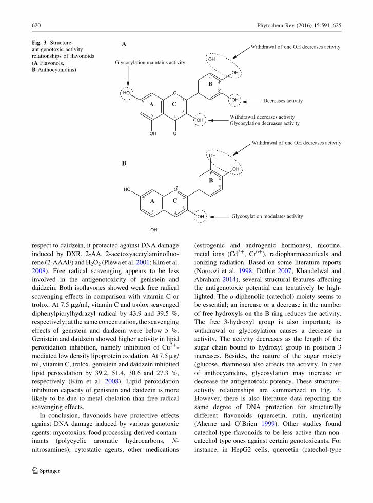
respect to daidzein, it protected against DNA damage
induced by DXR, 2-AA, 2-acetoxyacetylaminofluo-
rene (2-AAAF) andH2O2 (Plewa et al. 2001; Kim et al.
2008). Free radical scavenging appears to be less
involved in the antigenotoxicity of genistein and
daidzein. Both isoflavones showed weak free radical
scavenging effects in comparison with vitamin C or
trolox. At 7.5 μg/ml, vitamin C and trolox scavenged
diphenylpicrylhydrazyl radical by 43.9 and 39.5 %,
respectively; at the same concentration, the scavenging
effects of genistein and daidzein were below 5 %.
Genistein and daidzein showed higher activity in lipid
peroxidation inhibition, namely inhibition of Cu2+-
mediated low density lipoprotein oxidation. At 7.5 μg/ml, vitamin C, trolox, genistein and daidzein inhibited
lipid peroxidation by 39.2, 51.4, 30.6 and 27.3 %,
respectively (Kim et al. 2008). Lipid peroxidation
inhibition capacity of genistein and daidzein is more
likely to be due to metal chelation than free radical
scavenging effects.
In conclusion, flavonoids have protective effects
against DNA damage induced by various genotoxic
agents: mycotoxins, food processing-derived contam-
inants (polycyclic aromatic hydrocarbons, N-nitrosamines), cytostatic agents, other medications
(estrogenic and androgenic hormones), nicotine,
metal ions (Cd2+, Cr6+), radiopharmaceuticals and
ionizing radiation. Based on some literature reports
(Noroozi et al. 1998; Duthie 2007; Khandelwal and
Abraham 2014), several structural features affecting
the antigenotoxic potential can tentatively be high-
lighted. The o-diphenolic (catechol) moiety seems to
be essential; an increase or a decrease in the number
of free hydroxyls on the B ring reduces the activity.
The free 3-hydroxyl group is also important; its
withdrawal or glycosylation causes a decrease in
activity. The activity decreases as the length of the
sugar chain bound to hydroxyl group in position 3
increases. Besides, the nature of the sugar moiety
(glucose, rhamnose) also affects the activity. In case
of anthocyanidins, glycosylation may increase or
decrease the antigenotoxic potency. These structure–
activity relationships are summarized in Fig. 3.
However, there is also literature data reporting the
same degree of DNA protection for structurally
different flavonoids (quercetin, rutin, myricetin)
(Aherne and O’Brien 1999). Other studies found
catechol-type flavonoids to be less active than non-
catechol type ones against certain genotoxicants. For
instance, in HepG2 cells, quercetin (catechol-type
OHO
OH
OH
OH
O
OH
A C
B
2
345
OH7
4'
5'
3'
Withdrawal of one OH decreases activity
Decreases activity
Withdrawal decreases activityGlycosylation decreases activity
Glycosylation maintains activity
OHO
OH
OH
OH
OH
A C
B
2
345
7
4'
5'
3'
Withdrawal of one OH decreases activity
Glycosylation modulates activity
A
B
Fig. 3 Structure-
antigenotoxic activity
relationships of flavonoids
(A Flavonols,
B Anthocyanidins)
620 Phytochem Rev (2016) 15:591–625
123

flavonoid) was more efficient than myricetin (pyro-
gallol-type flavonoid) in protecting against NPIP-
induced oxidative DNA damage but less efficient in
affording protection against NDBA (Delgado et al.
2009). Similar behavior was detected for catechins.
Within the same cell line, (−)-epicatechin showed
higher protection against both NPIP- and NDBA-
induced oxidative DNA damage than (+)-catechin
(Delgado et al. 2009) whereas the latter was more
active in reducing PhIP-induced DNA damage (Haza
and Morales 2011). The mechanisms responsible for
these differences in effectiveness need to be clarified.
In vitro investigations on antigenotoxic potential
of flavonoids have to be followed by in vivo studies
as flavonoids undergo an extensive metabolization
and have a relatively low oral bioavailability.
Flavonoid metabolism is very complex involving a
wide array of reactions: deglycosylation, glu-
curonidation, sulfation, methylation, ring fission
which take place in the gastrointestinal lumen,
intestinal wall and liver (Erlund 2004). The metabo-
lites and their precursors develop different effects.
Although free radical scavenging and metal chelation
have been proposed as possible mechanisms for DNA
protective effects of flavonoids, both mechanisms
cannot be predominant in vivo due to conjugation of
free hydroxyl groups responsible for free radical
scavenging and metal chelation. In vivo antigenotox-
icity of flavonoids is based on other mechanisms such
as modulation of enzymes involved in the bioactiva-
tion of genotoxic agents and detoxification of their
reactive metabolites and up-regulation of antioxidant
defense systems which inactivate ROS generated
during the bioactivation of genotoxicants. Except
anthocyanidins, antigenotoxic effects of other flavo-
noids are concentration-dependent both in vitro and
in vivo. Flavonoids are antigenotoxic in a well-
defined concentration range becoming genotoxic at
higher concentrations and sometimes, surprisingly, at
low concentrations, most probably due to pro-oxidant
effects.
Overall, flavonoids have the ability to reduce the
genotoxic stress but further in vivo investigations are
needed for a better characterization of their metabolic
transformations, relation dose-biological response
and mechanisms involved in genetic protection.
Acknowledgments This work was supported by University
of Medicine and Pharmacy Grigore T. Popa-Iasi, Internal
Research Grant No. 1639/01.02.2013 (Investigations on the
radioprotective potential of some vegetal extracts).
References
Abalea V, Cillard J, Dubos MP et al (1999) Repair of iron-
induced DNA oxidation by the flavonoid myricetin in
primary rat hepatocyte cultures. Free Radic Biol Med
26:1457–1466
Abraham SK, Schupp N, Schmid U et al (2007) Antigenotoxic
effects of the phytoestrogen pelargonidin chloride and the
polyphenol chlorogenic acid. Mol Nutr Food Res 51:880–
887
Abraham SK, Eckhardt A, Oli RG et al (2012) Analysis in vitro
chemoprevention of genotoxic damage by phytochemi-
cals, as single agents or as combinations. Mutat Res
744:117–124
Aherne SA, O’Brien NM (1999) Protection by the flavonoids
myricetin, quercetin, and rutin against hydrogen peroxide-
induced DNA damage in Caco-2 and HepG2 cells. Nutr
Cancer 34:160–166
Aherne SA, O’Brien NM (2000a) Lack of effect of the flavo-
noids, myricetin, quercetin, and rutin, on repair of H2O2-
induced DNA single strand breaks in Caco-2, HepG2, and
V79 cells. Nutr Cancer 38:106–115
Aherne SA, O’Brien NM (2000b) Mechanism of protection by
the flavonoids, quercetin and rutin, against tert-butylhy-droperoxide- and menadione-induced DNA single strand
breaks in Caco-2 cells. Free Radic Biol Med 29:507–514
Ajithkumar T, Parkinson C, Shamshad F et al (2007) Iphos-
phamide encephalopathy. Clin Oncol 19:108–114
Alaejos MS, Pino V, Afonso AM (2008) Metabolism and
toxicology of heterocyclic aromatic amines when con-
sumed in diet: influence of the genetic susceptibility to
develop human cancer. A review. Food Res Int 41:327–
340
Ali F, Rahul Naz F et al (2014) Protective effect of apigenin
against N-nitrosodiethylamine (NDEA)-induced hepato-
toxicity in albino rats. Mutat Res 767:13–20
Ali F, Rahul Naz F et al (2015) Protective effect of genistein
against N-nitrosodiethylamine (NDEA)-induced hepato-
toxicity in Swiss albino rats. J Pharm Anal 5(1):51–57
Alvarez-Gonzalez I, Madrigal-Bujaidar E, Dorado V et al
(2001) Inhibitory effect of naringin on the micronuclei
induced by ifosfamide in mouse, and evaluation of its
modulatory effect on the Cyp3a subfamily. Mutat Res
480–481:171–178
Aprotosoaie AC, Mihai CT, Voichita G et al (2016)
Antigenotoxic and antioxidant activities of a polyphenolic
extract from European Dracocephalum moldavica L. Ind
Crop Prod 79:248–257
Arts ICW, Hollman PCH, Feskens EJM et al (2001) Catechin
intake might explain the inverse relation between tea
consumption and ischemic heart disease: the Zutphen
elderly study. Am J Clin Nutr 74:227–232
Attia SM (2012) Influence of resveratrol on oxidative damage
in genomic DNA and apoptosis induced by cisplatin.
Mutat Res 741:22–31
Phytochem Rev (2016) 15:591–625 621
123

Azevedo L, Alves de Lima PL, Gomes JC et al (2007) Dif-
ferential response related to genotoxicity between
eggplant (Solanum melanogena) skin aqueous extract and
its main purified anthocyanin (delphinidin) in vivo. Food
Chem Toxicol 45:852–858
Bacanli M, Basaran AA, Basaran N (2015) The antioxidant and
antigenotoxic properties of citrus phenolics limonene and
naringin. Food Chem Toxicol 81:160–170
Bahari A, Mehrzad J, Mahmoudi M et al (2014) Cytochrome
P450 isoforms are differently up-regulated in aflatoxin
B1-exposed human lymphocytes and monocytes.
Immunopharmacol Immunotoxicol 36:1–10
Barcelos GRM, Grotto D, Angeli JP et al (2011) Evaluation of
antigenotoxic effects of plant flavonoids quercetin and
rutin on HepG2 cells. Phytother Res 25:1381–1388
Beg T, Siddique YH, Ara G et al (2007) Antimutagenic eval-
uation of genistein, a polyphenol, in cultured human
peripheral blood lymphocytes. Biomed Res 18(2):139–
143
Beg T, Siddique YH, Ara G et al (2008) Antigenotoxic effect
of genistein and gingerol on genotoxicity induced by
norethandrolone and oxandrolone in cultured human
lymphocytes. Int J Pharmacol 4(3):177–183
Bingul I, Basaran-Kucukgergin C, Tekkesin MS et al (2013)
Effect of blueberry pretreatment on diethylnitrosamine-
induced oxidative stress and liver injury in rats. Environ
Toxicol Pharmacol 36:529–538
Boots AW, Haenen GRMM, Bast A (2008) Health effects of
quercetin: from antioxidant to nutraceutical. Eur J Phar-
macol 585:325–337
Bramati L, Aquilano F, Pietta P (2003) Unfermented rooibos
tea: quantitative characterization of flavonoids by HPLC-
UV and determination of the total antioxidant activity.
J Agric Food Chem 51:7472–7474
Burcham PC (1998) Genotoxic lipid peroxidation products:
their DNA damaging properties and role in formation of
endogenous DNA adducts. Mutagenesis 13:287–305
Cheng YT, Wu CH, Ho CY et al (2013) Catechin protects
against ketoprofen-induced oxidative damage of the gas-
tric mucosa by up-regulating Nrf2 in vitro and in vivo.
J Nutr Biochem 24:475–483
Chirumbolo S (2010) The role of quercetin, flavonols and
flavones in modulating inflammatory cell function.
Inflamm Allergy Drug Targets 9:263–285
Choi JS, Piao YJ, Kang KW (2011) Effects of quercetin on the
bioavailability of doxorubicin in rats: role of CYP3A4 and
P-gp inhibition of quercetin. Arch Pharm Res 34:607–613
Choi EJ, Kim T, Kim GH (2012) Quercetin acts as an
antioxidant and downregulates CYP1A1 and CYP1B1
against DMBA-induced oxidative stress in mice. Oncol
Rep 28:291–296
Chua LS (2013) A review on plant-based rutin extraction
methods and its pharmacological activities. J Ethnophar-
macol 150:805–817
Cushnie TP, Lamb AJ (2005) Antimicrobial activity of flavo-
noids. Int J Antimicrob Agents 26:343–356
D’Andrea G (2015) Quercetin: a flavonol with multifaceted
therapeutic applications? Fitoterapia 106:256–271
Delgado ME, Haza AI, Arranz N et al (2008) Dietary
polyphenols protect against N-nitrosamines and benzo(a)
pyrene-induced DNA damage (strand breaks and oxidized
purines/pyrimidines) in HepG2 human hepatoma cells.
Eur J Nutr 47:479–490
Delgado ME, Haza AI, Garcıa A et al (2009) Myricetin,
quercetin, (+)-catechin and (–)-epicatechin against N-ni-trosamines-induced DNA damage in human hepatoma
cells. Toxicol In Vitro 23:1292–1297
Delgado L, Fernandes I, Gonzalez-Manzano S et al (2014)
Anti-proliferative effects of quercetin and catechin
metabolites. Food Funct 5:797–803
Di Giacomo C, Acquaviva R, Piva A et al (2007) Protective
effect of cyanidin 3-O-β-D-glucoside on ochratoxin
A-mediated damage in the rat. Brit J Nutr 98:937–943
Dıaz Barriga Arceo S, Madrigal-Bujaidar E, Calderon Mon-
tellano E et al (1995) Genotoxic effects produced by
capsaicin in mouse during subchronic treatment. Mutat
Res 345:105–109
Dietz BM, Kang YH, Liu G et al (2005) Xanthohumol isolated
from Humulus lupulus inhibits menadione-induced DNA
damage through induction of quinone reductase. Chem
Res Toxicol 18:1296–1305
Duffus JH, Nordberg M, Templeton DM (2007) Glossary of
terms used in toxicology, 2nd edition (IUPAC recom-
mendations 2007). Pure Appl Chem 79:1153–1344
Duthie SJ (2007) Berry phytochemicals, genomic stability and
cancer: evidence for chemoprotection at several stages in
the carcinogenic process. Mol Nutr Food Res 51:665–674
Erlund I (2004) Review of the flavonoids quercetin, hesperetin,
and naringenin. Dietary sources, bioactivities, bioavail-
ability, and epidemiology. Nutr Res 24:851–874
Esselen M, Barth SW, Winkler S et al (2013) Anthocyanins
suppress the cleavable complex formation by irinotecan
and diminish its DNA-strand-breaking activity in the
colon of Wistar rats. Carcinogenesis 34:835–840
Fimognari C, Berti F, Cantelli-Forti G et al (2004) Effect of
cyanidin-3-O-β-glucopyranoside on micronucleus induc-
tion in cultured human lymphocytes by four different
mutagens. Environ Mol Mutagen 43:45–52
Gadkari PV, Balaraman M (2015) Catechins: sources, extrac-
tion and encapsulation: a review. Food Bioprod Process
93:122–138
Gao S, Chen T, ChoiMK et al (2013) Cyanidin reverses cisplatin-
induced apoptosis in HK-2 proximal tubular cells through
inhibitionofROS-mediatedDNAdamageandmodulationof
the ERK and AKT patways. Cancer Lett 333:36–46
Garcıa-Rodrıguez MC, Nicolas-Mendez T, Montano-Rodrı-
guez AR et al (2014) Antigenotoxic effects of (−)-epigallocatechin-3-gallate (EGCG), quercetin, and rutin
on chromium trioxide-induced micronuclei in the poly-
chromatic erythrocytes of mouse peripheral blood.
J Toxicol Environ Health Part A 77:324–336
Glei M, Pool-Zobel BL (2006) The main catechin of green tea
(–)-epigallocatechin-3-gallate (EGCG), reduces bleomy-
cin-induced DNA damage in human leucocytes. Toxicol
In Vitro 20:295–300
Granado-Serrano AB, Martın MA, Bravo L et al (2012)
Quercetin modulates Nrf2 and glutathione-related defen-
ses in HepG2 cells: involvement of p38. Chem Biol
Interact 195:154–164
Guo M, Perez C, Wei J et al (2007) Iron-binding properties of
plant phenolics and cranberry’s bioeffects. Dalton Trans
43:4951–4961. doi:10.1039/b705136k
622 Phytochem Rev (2016) 15:591–625
123

Gupta C, Vikram A, Tripathi DN et al (2010) Antioxidant and
antimutagenic effect of quercetin against DEN induced
hepatotoxicity in rat. Phytother Res 24:119–128
Gustafson DL, Pritsos CA (1993) Kinetics and mechanism of
mitomycin C bioactivation by xanthine dehydrogenase
under aerobic and hypoxic conditions. Cancer Res
53:5470–5474
Han X, Shen T, Lou H (2007) Dietary polyphenols and their
biological significance. Int J Mol Sci 8:950–988
Hassan AM, Alam SS, Abdel-Aziem SH et al (2011) Benzo-a-
pyrene induced genotoxicity and cytotoxicity in germ
cells of mice: intervention of radish and cress. J Genet
Eng Biotechnol 9:65–72
Haza AI, Morales P (2011) Effects of (+) catechin and (−)epicatechin on heterocyclic amines-induced oxidative
DNA damage. J Appl Toxicol 31:53–62
Hobbs CA, Swartz C, Maronpot R et al (2015) Genotoxicity
evaluation of the flavonoid, myricitrin, and its aglycone,
myricetin. Food Chem Toxicol 83:283–292
Hosseinimehr SJ, Ahmadi A, Beiki D et al (2009) Protective
effects of hesperidin against genotoxicity induced by99mTc-MIBI in human cultured lymphocyte cells. Nucl
Med Biol 36:863–867
Hundal BS, Dhillon VS, Sidhu IS (1997) Genotoxic potential
of estrogens. Mutat Res 389:173–181
Iacopini P, Baldi M, Storchi P et al (2008) Catechin, epicate-
chin, quercetin, rutin and resveratrol in red grape: content,
in vitro antioxidant activity and interactions. J Food
Compos Anal 21:589–598
Iyama T, Wilson DM III (2013) DNA repair mechanisms in
dividing and non-dividing cells. DNA Repair 12:620–636
Jackson SP, Bartek J (2009) The DNA damage response in
human biology and disease. Nature 461:1071–1078
Jagetia GC, Reddy TK (2002) The grapefruit flavanone nar-
ingin protects against the radiation-induced genomic
instability in the mice bone marrow: a micronucleus
study. Mutat Res 519:37–48
Kalpana KB, Devipriya N, Srinivasan M et al (2009) Investi-
gation of the radioprotective efficacy of hesperidin against
gamma-radiation induced cellular damage in cultured
human peripheral blood lymphocytes. Mutat Res 676:54–
61
Kalpana KB, Devipriya N, Srinivasan M et al (2011) Evalu-
ating the radioprotective effect of hesperidin in the liver of
Swiss albino mice. Eur J Pharmacol 658:206–212
Kanadzu M, Lu Y, Morimoto K (2006) Dual function of (–)-
epigallocatechin gallate (EGCG) in healthy human lym-
phocytes. Cancer Lett 241:250–255
Kang ZC, Tsai SJ, Lee H (1999) Quercetin inhibits benzo[a]
pyrene-induced DNA adducts in human Hep G2 cells by
altering cytochrome P-450 1A1 gene expression. Nutr
Cancer 35:175–179
Khan TH, Jahangir T, Prasad L et al (2006) Inhibitory effect of
apigenin on benzo(a)pyrene-mediated genotoxicity in
Swiss albino mice. J Pharm Pharmacol 58:1655–1660
Khandelwal N, Abraham SK (2014) Intake of anthocyanidins
pelargonidin and cyanidin reduces genotoxic stress in
mice induced by diepoxybutane, urethane and endogenous
nitrosation. Environ Toxicol Pharmacol 37:837–843
Khandrika L, Kumar B, Koul S et al (2009) Role of oxidative
stress in prostate cancer. Cancer Lett 282:125–136
Kim NY, Song EJ, Kwon DY et al (2008) Antioxidant and
antigenotoxic activities of Korean fermented soybean.
Food Chem Toxicol 46:1184–1189
Kleemann R, Verschuren L, Morrison M et al (2011) Anti-
inflammatory, anti-proliferative and anti-atherosclerotic
effects of quercetin in human in vitro and in vivo models.
Atherosclerosis 218:44–52
Kluth D, Banning A, Paur I et al (2007) Modulation of preg-
nane X receptor-and electrophile responsive element-
mediated gene expression by dietary polyphenolic com-
pounds. Free Radic Biol Med 42:315–325
Kohli E, Raj HG, Kumari R et al (2002) Comparison of the
prevention of aflatoxin B1-induced genotoxicity by
quercetin and quercetin pentaacetate. Bioorg Med Chem
Lett 12:2579–2582
Kreft I, Fabjan N, Yasumoto K (2006) Rutin content in
buckwheat (Fagopyrum esculentum Moench) food mate-
rials and products. Food Chem 98:508–512
Lenglet G, David-Cordonnier M-H (2010) DNA-Destabilizing
agents as an alternative approach for targeting DNA:
Mechanisms of action and cellular consequences. DOI, J
Nucleic Acids. doi:10.4061/2010/290935
Lepri SR, Luiz RC, Zanelatto LC et al (2013) Chemopreven-
tive activity of the isoflavones, genistein and daidzein on
mutagenicity induced by direct and indirect mutagens in
cultured HTC cells. Cytotechonology 65:213–222
Li Y, Ding Y (2012) Minireview: therapeutic potential of
myricetin in diabetes mellitus. Food Sci Human Wellness
1:19–25
Liu M, Hansen PE, Wang G et al (2015) Pharmacological
profile of xanthohumol, a prenylated flavonoid from hops
(Humulus lupulus). Molecules 20:754–779
Lopez-Lazaro M (2009) Distribution and biological activities
of the flavonoid luteolin. Mini Rev Med Chem 9:31–59
Makris DM, Rossiter JT (2001) Domestic processing of onion
bulbs (Allium cepa) and asparagus spears (Asparagusofficinalis): effect on flavonol content and antioxidant
status. J Agric Food Chem 49:3216–3222
Marcarini JC, Tsuboy MSF, Luiz RC et al (2011) Investigation
of cytotoxic, apoptosis-inducing, genotoxic and protective
effects of the flavonoid rutin in HTC hepatic cells. Exp
Toxicol Pathol 63:459–465
Marcolin E, San-Miguel B, Vallejo D et al (2012) Quercetin
treatment ameliorates inflammation and fibrosis in mice
with nonalcoholic steatohepatitis. J Nutr 142:1821–1828
Meireles JRC, Oliveira SV, Costa-Neto AO et al (2013)
Genotoxic and cytotoxic effects of testosterone cypionate
(deposteron®). Mutat Res 753:72–75
Mendoza-Wilson AM, Glossman-Mitnik D (2006) Theoretical
study of the molecular properties and chemical reactivity
of (C)-catechin and (K)-epicatechin related to their
antioxidant ability. J Mol Struct (Theochem) 761:97–106
Mullie P, Clarys P, Deriemaeker P et al (2007) Estimation of
daily human intake of food flavonoids. Plant Foods Hum
Nutr 62:93–98
Musallam I, Duwayri M, Shibli R et al (2012) Investigation of
rutin content in different plant parts of wild caper (Cap-paris spinosa L.) populations from Jordan. Res J Med
Plant 6:27–36
Muthukumaran S, Sudheer AR, Menon VP et al (2008) Pro-
tective effect of quercetin on nicotine-induced prooxidant
Phytochem Rev (2016) 15:591–625 623
123

and antioxidant imbalance and DNA damage in Wistar
rats. Toxicology 243:207–215
National Toxicology Program (2002). Report on Carcinogens,
Tenth Edition; U.S. Department of Health and Human
Services, Public Health Service, Research Triangle Park,
North Carolina. http://ntp.niehs.nih.gov/pubhealth/roc/
listings/a/iq/summary/index.html
Nicolella HD, de Oliveira PF, Munari CC et al (2014) Dif-
ferential effect of manool—A diterpene from Salvia
officinalis, on genotoxicity induced by methyl methane-
sulfonate in V79 and HepG2 cells. Food Chem Toxicol
72:8–12
Noel S, Kasinathan M, Rath SK (2006) Evaluation of apigenin
using in vitro cytochalasin blocked micronucleus assay.
Toxicol In Vitro 20:1168–1172
Noroozi M, Angerson WJ, Lean ME (1998) Effects of flavo-
noids and vitamin C on oxidative DNA damage to human
lymphocytes. Am J Clin Nutr 67:1210–1218
Norval M, Lucas RM, Cullen AP et al (2011) The human
health effects of ozone depletion and interactions with
climate change. Photochem Photobiol Sci 10:199–225
Orhan F, Gulluce M, Ozkan H et al (2013) Determination of
the antigenotoxic potential of some luteolin derivatives by
using a eukaryotic cell system, Saccharomyces cerevisiae.Food Chem 141:366–372
Orsolic N, Gajski G, Garaj-Vrhovac V et al (2011) DNA-
protective effects of quercetin or naringenin in alloxan-
induced diabetic mice. Eur J Pharmacol 656:110–118
Ouedraogo M, Baudoux T, Stevigny C et al (2012) Review of
current and “omics” methods for assessing the toxicity
(genotoxicity, teratogenity and nefrotoxicity) of herbal
medicines and mushrooms. J Ethnopharmacol 140:492–512
Pandir D (2015) Protective effect of—epigallocatechin-3-gal-
late on capsaicin-induced DNA damage and oxidative
stress in human erythrocytes and leucocytes in vitro.
Cytotechnology 67:367–377
Papiez MA (2013) The influence of curcumin and (–)-epicat-
echin on the genotoxicity and myelosuppression induced
by etoposide in bone marrow cells of male rats. Drug
Chem Toxicol 36(1):93–101
Patil SL, Rao NB, Somashekarappa HM et al (2014)
Antigenotoxic potential of rutin and quercetin in Swiss
mice exposed to gamma radiation. Biomed J 37(5):305–
313
Perez-Vizcaıno F, Duarte J, Jimenez R et al (2009) Antihy-
pertensive effects of the flavonoid quercetin. Pharmacol
Rep 61:67–75
Pfohl-Leszkowicz A, Manderville RA (2012) An update on
direct genotoxicity as a molecular mechanism of ochra-
toxin A carcinogenicity. Chem Res Toxicol 25:252–262
Plazar J, Zegura B, Lah TT et al (2007) Protective effects of
xanthohumol against the genotoxicity of benzo(a)pyrene
(BaP), 2-amino-3-methylimidazo[4,5-f]quinoline (IQ) and
tert-butyl hydroperoxide (t-BOOH)in HepG2 human
hepatoma cells. Mutat Res 632:1–8
Plazar J, Filipic M, Groothuis GMM (2008) Antigenotoxic
effect of xanthohumol in rat liver slices. Toxicol in Vitro
22:318–327
Plewa MJ, Berhow MA, Vaughn SF et al (2001) Isolating
antigenotoxic components and cancer cell growth
suppressors from agricultural by-products. Mutat Res
480–481:109–120
Polıvkova Z, Langova M, Smerak P et al (2006) Antimutagenic
effect of genistein. Czech J Food Sci 24:119–126
Polkowski K, Mazurek AP (2000) Biological properties of
genistein. A review of in vitro and in vivo data. Acta Pol
Pharm Drug Res 57:135–155
Pugalendhi P, Manoharan S, Panjamurthy K et al (2009)
Antigenotoxic effect of genistein against 7,12-dimethyl-
benz[a]anthracene induced genotoxicity in bone marrow
cells of female Wistar rats. Pharmacol Rep 61:296–303
Quiles JL, Huertas JR, Battino M et al (2002) Antioxidant
nutrients and adriamycin toxicity. Toxicology 180:79–85
Ramos AA, Lima CF, Pereira ML et al (2008) Antigenotoxic
effects of quercetin, rutin and ursolic acid on HepG2 cells:
evaluation by the comet assay. Toxicol Lett 177:66–73
Ramos AA, Pereira-Wilson C, Collins AR (2010) Protective
effects of ursolic acid and luteolin against oxidative DNA
damage include enhancement of DNA repair in Caco-2
cells. Mutat Res 692:6–11
Ramyaa P, Padma VV (2013) Ochratoxin-induced toxicity,
oxidative stress and apoptosis ameliorated by quercetin—
modulation by Nrf2. Food Chem Toxicol 62:205–216
Ramyaa P, Krishnaswamy R, Padma VV (2014) Quercetin
modulates OTA-induced oxidative stress and redox sig-
naling in HepG2 cells—up regulation of Nrf2 expression
and down regulation of NF-kB and COX-2. Biochim
Biophys Acta 1840:681–692
Raschke M, Rowland IR, Magee PJ et al (2006) Genistein protects
prostate cells against hydrogen peroxide-induced DNA dam-
age and induces expression of genes involved in the defence
against oxidative stress. Carcinogenesis 27:2322–2330
Rice-Evans CA, Miller JM, Paganga G (1996) Structure-an-
tioxidant activity relationship of flavonoids and phenolic
acids. Free Radic Biol Med 20:933–956
Richi B, Kale RK, Tiky AB (2012) Radio-modulatory effects
of green tea catechin EGCG on pBR322 plasmid DNA
and murine splenocytes against gamma-radiation induced
damage. Mutat Res 747:62–70
Russo A, La Fauci L, Acquaviva R et al (2005) Ochratoxin A-in-
duced DNA damage in human fibroblast: protective effect of
cyanidin 3-O-β-D-glucoside. J Nutr Biochem 16:31–37
Siddique YH, Afzal M (2009a) Protective effect of apigenin
against hydrogen peroxide induced genotoxic damage on
cultured human peripheral blood lymphocytes. J Appl
Biomed 7:35–43
Siddique YH, Afzal M (2009b) Antigenotoxic effect of api-
genin against mitomycin C induced genotoxic damage in
mice bone marrow cells. Food Chem Toxicol 47:536–539
Siddique YH, Beg T, Afzal M (2005) Genotoxic potential of
ethinylestradiol in cultured mammalian cells. Chem Biol
Interact 151:133–141
Siddique YH, Beg T, Afzal M (2008) Antigenotoxic effect of
apigenin against anti-cancerous drugs. Toxicol In Vitro
22:625–631
Siddique YH, Ara G, Beg T et al (2010) Anticlastogenic effect
of apigenin in human lymphocytes treated with
ethinylestradiol. Fitoterapia 81:590–594
Srinivasan M, Sudheer AR, Pillai KR et al (2007) Modulatory
effects of curcumin on γ-radiation-induced cellular
624 Phytochem Rev (2016) 15:591–625
123

damage in primary culture of isolated rat hepatocytes.
Environ Toxicol Pharmacol 24:98–105
Sroka Z (2005) Antioxidative and antiradicalar properties of
plant phenolics. Z Naturforsch 60c:833–843
Swift LH, Golsteyn RM (2014) Genotoxic anti-cancer agents
and their relationship to DNA damage, mitosis, and
checkpoint adaptation in proliferating cancer cells. Int J
Mol Sci 15:3403–3431
Tang Y, Tian H, Shi Y et al (2013) Quercetin suppressed
CYP2E1-dependent ethanol hepatotoxicity via depleting
heme pool and releasing CO. Phytomedicine 20:699–704
Tanigawa S, Fujii M, Hou D-X (2007) Action of Nrf2 and
Keap1 in ARE-mediated NQO1 expression by quercetin.
Free Radic Biol Med 42:1690–1703
Towner RA, Qian SY, Kadiiska MB et al (2003) In vivo
identification of aflatoxin-induced free radicals in rat bile.
Free Radic Biol Med 35:1330–1340
Trivedi PP, Tripathi DN, Jena GB (2011) Hesperetin protects
testicular toxicity of doxorubicin in rat: role of NFkB, p38
and capsase-3. Food Chem Toxicol 49:838–847
Uhl M, Ecker S, Kassie F et al (2003) Effect of chrysin, a
flavonoid compound, on the mutagenic activity of
2-amino-1-methyl-6-phenylimidazol[4,5-b]pyridine
(PhIP) and benzo(a)pyrene (B(a)P) in bacterial and human
hepatoma (HepG2) cells. Arch Toxicol 77:477–484
Undeger U, Aydın S, Basaran AA et al (2004) The modulating
effects of quercetin and rutin on the mitomycin C induced
DNA damage. Toxicol Lett 151:143–149
Vedmaurthy RB, Padmanabhan S, Vijayan M et al (2012)
Compatibility of different solvents with Salmonellatyphimurium mutant strains in bacterial reverse mutation
assay. Int J Pharm Pharm Sci 4:283–284
Veberic R, Trobec M, Herbinger K et al (2005) Phenolic
compounds in some apple (Malus domestica Borkh) cul-
tivars of organic and integrated production. J Sci Food
Agric 85:1687–1694
Veberic R, Colaric M, Stampar F (2008) Phenolic acids and
flavonoids of fig fruit (Ficus carica L.) in the northern
Mediterranean region. Food Chem 106:153–157
Wang S-L, Han J-F, He X-Y et al (2007) Genetic variation of
human cytochrome P450 reductase as a potential bio-
marker for mitomycin C-induced cytotoxicity. Drug
Metab Dispos 35:176–179
Wellejus A, Bornholdt J, Vogel UB et al (2004) Cell-specific
oxidative DNA damage induced by estrogen in rat tes-
ticular cells in vitro. Toxicol Lett 150:317–323
Wilms LC, Hollman PC, Boots AW et al (2005) Protection by
quercetin and quercetin-rich fruit juice against induction
of oxidative DNA damage and formation of BPDE-DNA
adducts in human lymphocytes. Mutat Res 582:155–162
Wu LC, Lu IW, Chung CF et al (2011) Antiproliferative
mechanisms of quercetin in rat activated hepatic stellate
cells. Food Funct 2:204–212
Yeh SL, Wu SH (2006) Effects of quercetin on beta-apo-8′-carotenal-induced DNA damage and cytochrome P1A2
expression in A549 cells. Chem Biol Interact 163:199–
206
Yeh SL, Wang WY, Huang CS et al (2006) Flavonoids sup-
presses the enhancing effect of β-carotene on DNA
damage induced by 4-(methylnitrosamino)-1-(3-pyridyl)-
1-butanone (NNK) in A549 cells. Chem Biol Interact
160:175–182
Yılmaz D, Aydemir NC, Vatan O et al (2012) Influence of
naringin on cadmium-induced genomic damage in human
lymphocytes in vitro. Toxicol Ind Health 28:114–121
Yu P, Zhou Q, Zhu W et al (2013) Effects of quercetin on LPS-
induced disseminated intravascular coagulation (DIC) in
rabbits. Thromb Res 131:e270–e273
Yurtcu E, ls˙eri OD, Sahin FI (2014) Genotoxic and cytotoxic
effects of doxorubicin and silymarin on human hepato-
cellular carcinoma cells. Hum Exp Toxicol 33:1269–1276
Zhang Y, Wang G-J, Song TT et al (1999) Urinary disposition
of the soybean isoflavones daidzein, genistein and glyci-
tein differs among humans with moderate fecal isoflavone
degradation activity. J Nutr 129:957–962
Phytochem Rev (2016) 15:591–625 625
123


