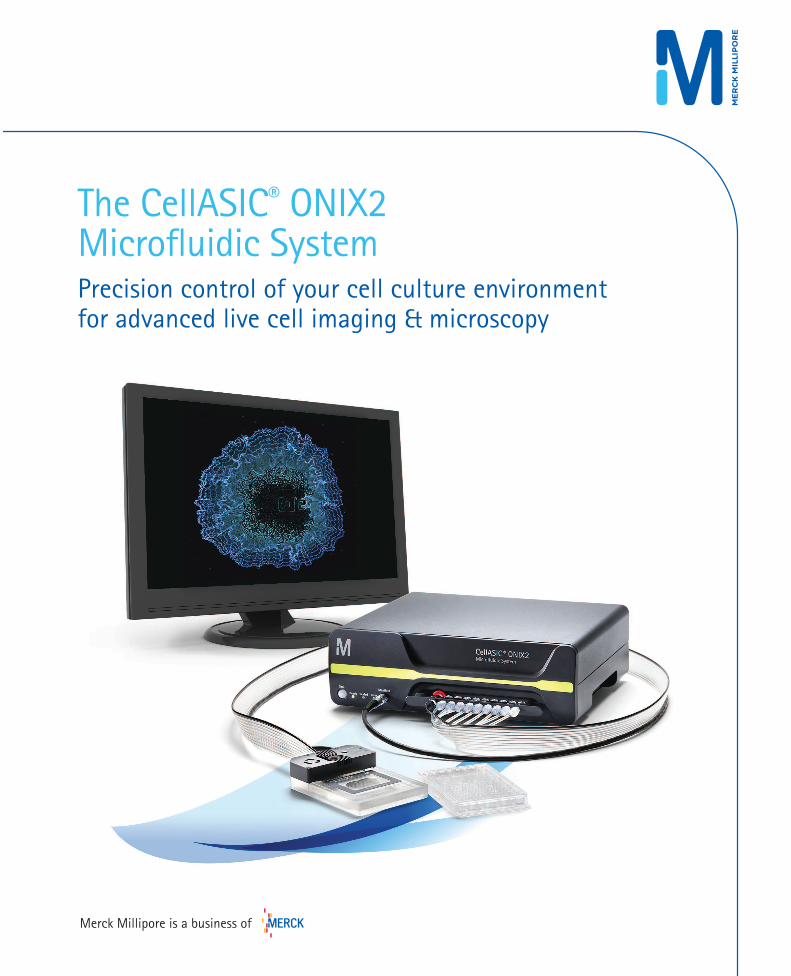
The CellASIC® ONIX2 Microfluidic SystemPrecision control of your cell culture environmentfor advanced live cell imaging & microscopy
Merck Millipore is a business of
Leading the way in science and technologyMerck KGaA, Darmstadt, Germany is a leading science and technology
company in healthcare, life science and performance materials.
Around 50,000 employees work to further develop technologies
that improve and enhance life – from biopharmaceutical therapies
to treat cancer or multiple sclerosis, cutting-edge systems for
scientific research and production, to liquid crystals for smartphones
and LCD televisions. Founded in 1668, we are the world’s oldest
pharmaceutical and chemical company.
Our Life Science team is dedicated to providing scientists and
engineers with best-in-class technologies, lab materials and services
with the intention of making research and production simpler, faster
and more successful.
Our solutions enable scientists to spend more time advancing
the promise of science through technologies that help detect the
previously undetectable, and products that make it possible to
monitor live cells in intricate detail.
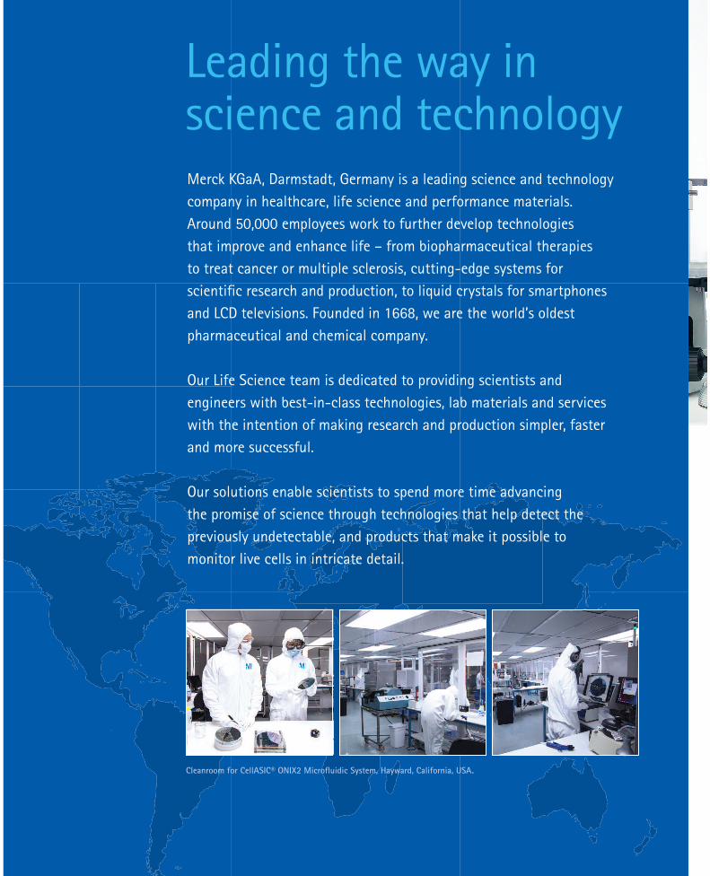
Cleanroom for CellASIC® ONIX2 Microfluidic System, Hayward, California, USA.

1
Quality, reliability and a growing array of publications. Introducing the CellASIC® ONIX2 Microfluidic System With a history of success, dedication to innovation and expertise in life science, Merck KGaA,
Darmstadt, Germany, is committed to providing the highest quality, most reliable and relevant
products for life science research and production.
The CellASIC® ONIX2 System incorporates extraordinary improvements to traditional live cell
observation and experimentation, using advanced microfluidics technology with computer
automation to manipulate the culture environment under controlled conditions while
allowing you to monitor the cells as they react in real-time.
This second generation CellASIC® ONIX2 Microfluidic System is backed by a strong history of
distinctive publications, stringent, globally-certified manufacturing and quality processes, and
a multinational organization committed to delivering the utmost in local service and support.

22
Why study living processes by killing your cells?Scientists recognize that live cell analysis yields necessary and unique insight into living
processes that are rapidly changing. While collecting data from a moment in time with
classical endpoint analyses has value, the increasingly common practice of observing and
measuring cellular changes over extended periods of time reveals more about the true
behavior of biological systems. The challenge has been in creating a dynamic cell culture
environment that is controllable, manipulatable, and reproducible.
Live cell imaging to transform your research
Move beyond the status quo and gain unprecedented insight into live cells using advanced
microfluidics technology. With the CellASIC® ONIX2 Microfluidic System, you too can
easily and efficiently create a stable, reproducible environment for imaging. See how it can
transform your research.
S. cerevisiae yeast cells with labeled mitochondria (red), cell wall (cyan), and tubulin (green), imaged in CellASIC® ONIX yeast haploid plate.
Image credit: Maja Bialecka-Fornal, University
of California, Irvine, Rafelski Lab.
Measuring a gene circuit in E. coli in a time-lapse experiment with the CellASIC® ONIX B04 bacterial microfluidic plate. Images were acquired at 100x magnification.
Culturing primary rat cortical neurons (GFP) cultured in the CellASIC® ONIX M04S mammalian microfluidic plate.
Life is Dynamic
Microfluidic technology is changing the way we perform live cell analysis.
The CellASIC ONIX Microfluidic System is amazingly easy to use yethighly adaptable, which greatly reduces the barrier to adopting this
emerging technology.
- Dr. Jintao Liu, Ph.D., UCSD
“
“
3
1
2
3
4
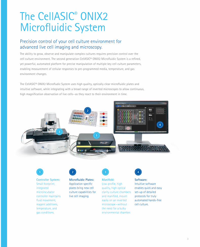
The CellASIC® ONIX2 Microfluidic SystemPrecision control of your cell culture environment for advanced live cell imaging and microscopy. The ability to grow, observe and manipulate complex cultures requires precision control over the
cell culture environment. The second generation CellASIC® ONIX2 Microfluidic System is a refined,
yet powerful, automated platform for precise manipulation of multiple key cell culture parameters,
enabling measurement of cellular responses to pre-programmed media, temperature, and gas
environment changes.
The CellASIC® ONIX2 Microfludic System uses high quality, optically clear microfluidic plates and
intuitive software, while integrating with a broad range of inverted microscopes to allow continuous,
high magnification observation of live cells—as they react to their environment in time.
Controller System:
Small footprint,
integrated
microincubator
controller maintains
fluid movement,
reagent additions,
temperature, and
gas conditions.
Microfluidic Plates:
Application specific
plates bring new cell
culture capabilities for
live cell imaging.
Manifold:
Low-profile, high
quality, high optical
clarity culture chambers
and manifold, mount
easily on an inverted
microscope—without
the need for a bulky
environmental chamber.
Software:
Intuitive software
enables quick and easy
set-up of detailed
protocols for truly
automated hands-free
cell culture.
1 2 3 4

4
... the key advantage of live cell imaging is that you get to actually watch
things unfold before your eyes. It’s very powerful to see, visually what the cells are
doing and how they change in space and as a function of time, because biology is a
dynamic process...and watching the cells do what they do is something that really appeals to us and is driving our science.
- Dr. Gurol Suel, UCSD
“
“
Nikon 139-Eclipse Ti-E System. Image Courtesy of Nikon, Inc.
The CellASIC® ONIX2 System turns your microscope into a powerful live cell culture and imaging system that works in conjunction with your capture and analysis software.
The boost your microscope needs for advanced live cell imaging.
You’ve invested in a powerful microscope; don’t limit its use to observing live cells in a
static fashion. Enhance your return on investment and obtain more meaningful data using
the CellASIC® ONIX2 Microfluidic System as your live cell imaging platform. The low-profile
manifold and plate assembly are easily positioned and removed from the microscope stage
providing maximum ease of use and flexibility.

5
Watch cells change through time and space, all with the precision of the CellASIC® ONIX2
System. Automatically control flow rates, gas and temperature shifts, standing gradients,
nutrient/drug additions, and media changes. With uninterrupted high resolution microscopic
culture observation and truly consistent, controlled cell culture, you’ll answer the questions
that set your research apart.
There is simply no better way to conduct live cell analysis.
Parameter If too low, can cause: If too high, can cause:
Temperature Decreased cell response Increased respiration / protein damage
Oxygen Level Decreased pH / increased glycolysis Increased ROS, membrane damage
Growth FactorsIncreased apoptosis / decreased protein synthesis
Increased angiogenesis and cell division
HumidityIncreased osmolarity / cell metabolism / oxidative stress
Could damage imaging equipment
pH Protein and membrane denaturation Increased alkalosis and dehydration
OsmolalityDecreased cell division / increased autophagic proteolysis and cell rupture
Increased oxidative stress, DNA breakage, and nutrient digestion
Glucose Decreased autophagy and metabolism Increased apoptosis and ROS
ECM and AdhesionDecreased angiogenesis / aberrant differentiation
Increased cell adhesion, chemotaxis, proliferation
Eight critical cell culture parameters can be controlled by the CellASIC® ONIX2 Microfluidic System:
Small footprint,
integrated
microincubator
controller maintains
fluid movement,
reagent additions,
temperature, and gas
mixture.
1
6
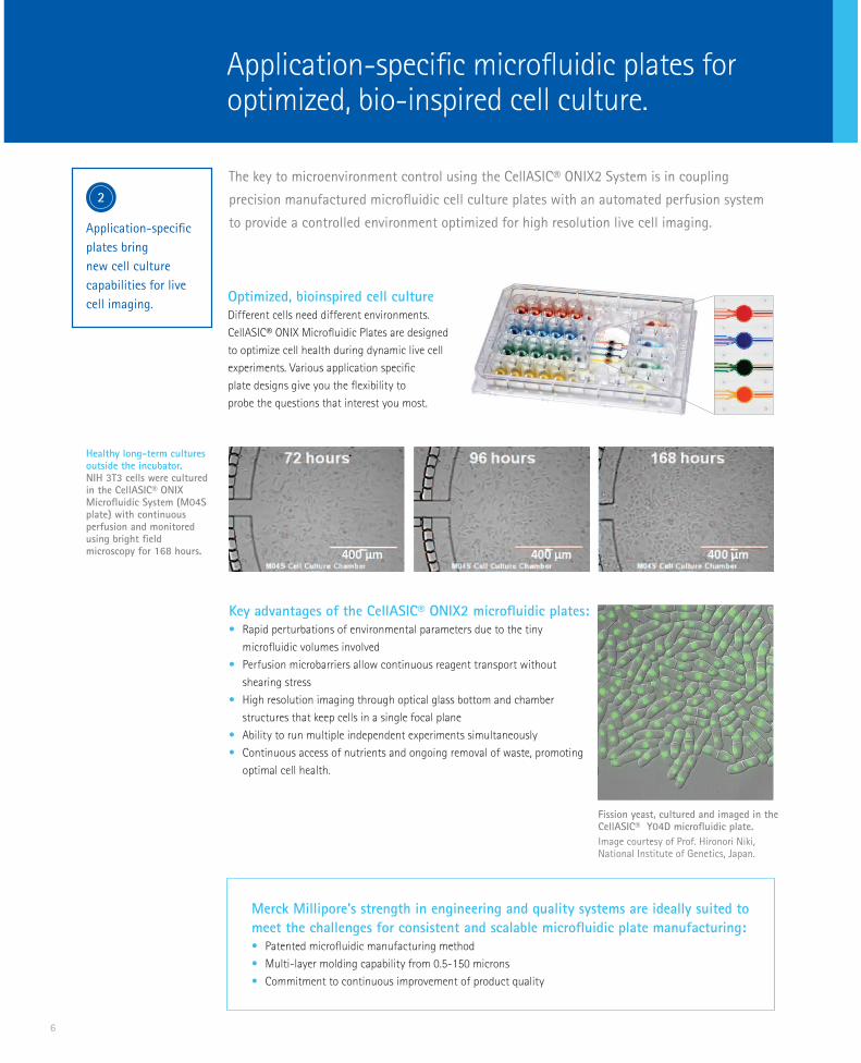
Application-specific
plates bring
new cell culture
capabilities for live
cell imaging.
The key to microenvironment control using the CellASIC® ONIX2 System is in coupling
precision manufactured microfluidic cell culture plates with an automated perfusion system
to provide a controlled environment optimized for high resolution live cell imaging.
Key advantages of the CellASIC® ONIX2 microfluidic plates:• Rapid perturbations of environmental parameters due to the tiny
microfluidic volumes involved
• Perfusion microbarriers allow continuous reagent transport without
shearing stress
• High resolution imaging through optical glass bottom and chamber
structures that keep cells in a single focal plane
• Ability to run multiple independent experiments simultaneously
• Continuous access of nutrients and ongoing removal of waste, promoting
optimal cell health.
Application-specific microfluidic plates for optimized, bio-inspired cell culture.
Healthy long-term cultures outside the incubator. NIH 3T3 cells were cultured in the CellASIC® ONIX Microfluidic System (M04S plate) with continuous perfusion and monitored using bright field microscopy for 168 hours.
Optimized, bioinspired cell cultureDifferent cells need different environments.
CellASIC® ONIX Microfluidic Plates are designed
to optimize cell health during dynamic live cell
experiments. Various application specific
plate designs give you the flexibility to
probe the questions that interest you most.
Fission yeast, cultured and imaged in the CellASIC® Y04D microfluidic plate.
Image courtesy of Prof. Hironori Niki, National Institute of Genetics, Japan.
Merck Millipore’s strength in engineering and quality systems are ideally suited to
meet the challenges for consistent and scalable microfluidic plate manufacturing:• Patented microfluidic manufacturing method
• Multi-layer molding capability from 0.5-150 microns
• Commitment to continuous improvement of product quality
2
7
Low profile, high
quality, high optical
clarity culture
chambers and
manifold, mount
easily on an inverted
microscope—without
the need for a bulky
environmental
chamber.
3
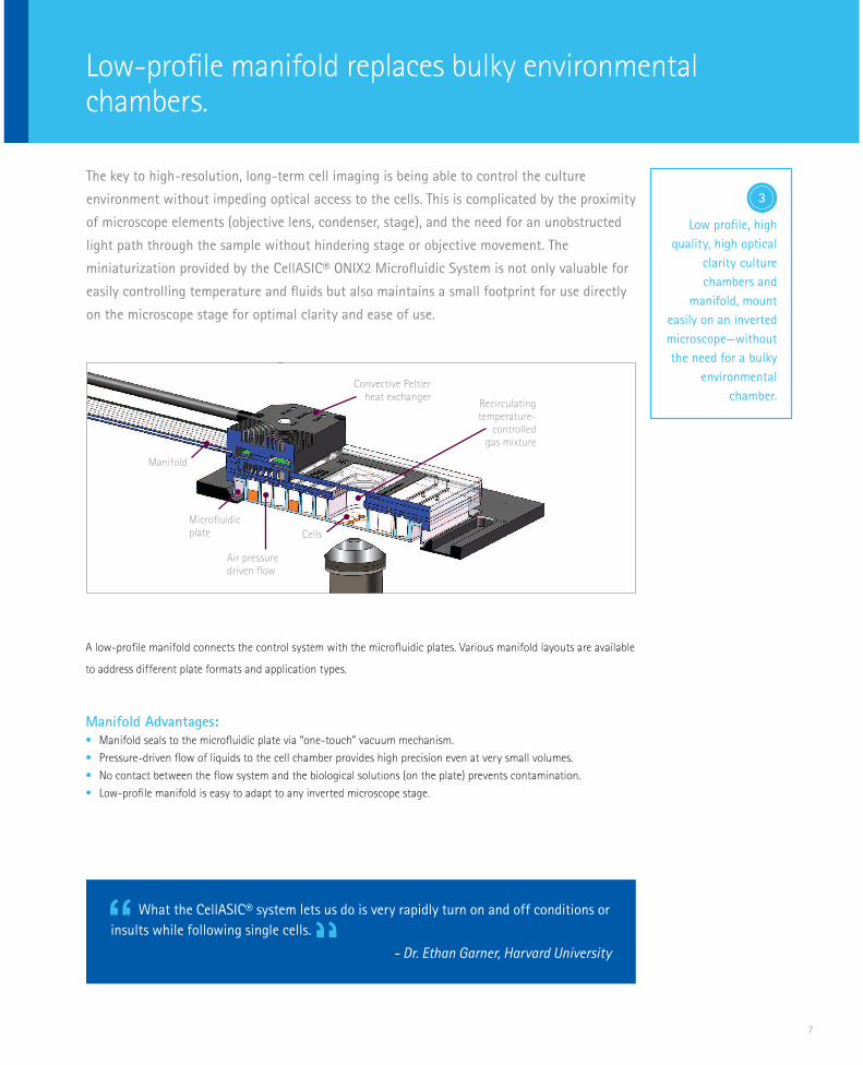
The key to high-resolution, long-term cell imaging is being able to control the culture
environment without impeding optical access to the cells. This is complicated by the proximity
of microscope elements (objective lens, condenser, stage), and the need for an unobstructed
light path through the sample without hindering stage or objective movement. The
miniaturization provided by the CellASIC® ONIX2 Microfluidic System is not only valuable for
easily controlling temperature and fluids but also maintains a small footprint for use directly
on the microscope stage for optimal clarity and ease of use.
A low-profile manifold connects the control system with the microfluidic plates. Various manifold layouts are available
to address different plate formats and application types.
Manifold Advantages:• Manifold seals to the microfluidic plate via “one-touch” vacuum mechanism.
• Pressure-driven flow of liquids to the cell chamber provides high precision even at very small volumes.
• No contact between the flow system and the biological solutions (on the plate) prevents contamination.
• Low-profile manifold is easy to adapt to any inverted microscope stage.
Low-profile manifold replaces bulky environmental chambers.
Convective Peltierheat exchanger
Recirculatingtemperature-
controlledgas mixture
Manifold
Microfluidic plate
Air pressure driven flow
Cells
What the CellASIC® system lets us do is very rapidly turn on and off conditions or
insults while following single cells.
- Dr. Ethan Garner, Harvard University
““
8
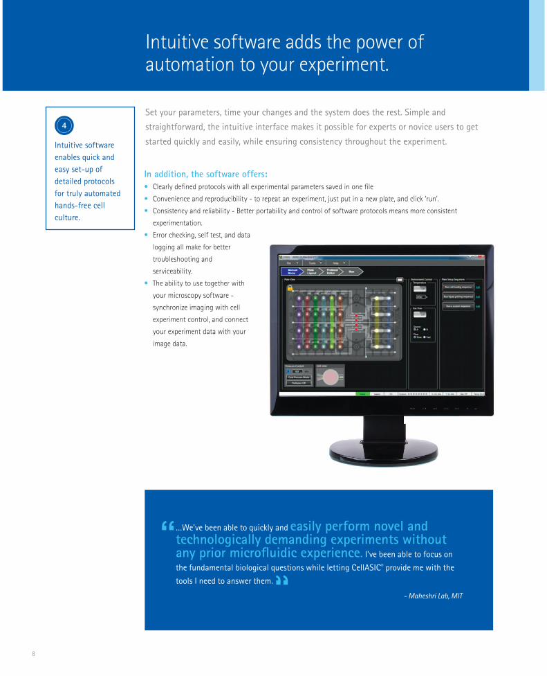
Intuitive software adds the power of automation to your experiment.
In addition, the software offers:
• Clearly defined protocols with all experimental parameters saved in one file
• Convenience and reproducibility - to repeat an experiment, just put in a new plate, and click ‘run’.
• Consistency and reliability - Better portability and control of software protocols means more consistent
experimentation.
• Error checking, self test, and data
logging all make for better
troubleshooting and
serviceability.
• The ability to use together with
your microscopy software -
synchronize imaging with cell
experiment control, and connect
your experiment data with your
image data.
Set your parameters, time your changes and the system does the rest. Simple and
straightforward, the intuitive interface makes it possible for experts or novice users to get
started quickly and easily, while ensuring consistency throughout the experiment.
…We’ve been able to quickly and easily perform novel and technologically demanding experiments without any prior microfluidic experience. I’ve been able to focus on
the fundamental biological questions while letting CellASIC® provide me with the
tools I need to answer them.
- Maheshri Lab, MIT
“
“
Intuitive software
enables quick and
easy set-up of
detailed protocols
for truly automated
hands-free cell
culture.
4
9
The CellASIC® ONIX2 Microfluidic System provides detailed information about live cell processes with ultra-high
quality images and complete environment control.
Discover new insights to dynamic cellular processes.
CellASIC® ONIX2 Advantage: Application
High resolution imaging in single focal plane with controlled nutrient flow for tracking single cells over time
Yeast single cell response
Measure multi-generational responses to live bacteria while maintaining cells in a single focal plane for days
Bacterial single cell response
Continuous monitoring of community dynamics and precise control of growth environment
Bacterial biofilm dynamics
S. cerevisiae cells expressing GFP-tubulin and SPC42- mCherry during alpha-factor exposure and arrest. Images were acquired at 60x magnification. Courtesy of S. Lacefield, U. Indiana
A gene circuit in E. coli was induced and visualized for a time-lapse experiment in the CellASIC® ONIX B04 microfluidic plate. Images were acquired at 100x magnification.
Time lapse, composite image of bacterial biofilm growth. Courtesy of the Suel lab, UCSD.
Dynamic assays on adherent and non-adherent cells
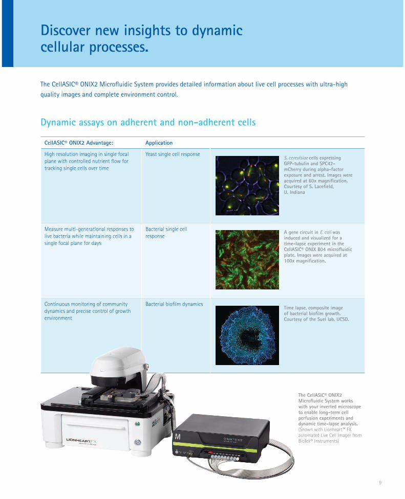
The CellASIC® ONIX2 Microfluidic System works with your inverted microscope to enable long-term cell perfusion experiments and dynamic time-lapse analysis.(Shown with Lionheart™ FX automated Live Cell Imager from BioTek® Instruments)
10
Rapid gas and media switching capabilities
CellASIC® ONIX2 Advantage: Application
Fast and precise control over culture conditions to induce hypoxic, starvation, or toxic microenvironments
Cell response to hypoxic environments
Microfluidic control of nutrient additions during continuous observation
Cell response to changing media conditions, drugs, and other stimulants
High resolution imaging of dynamic cellular interactions
Host-pathogen interactions
LAMP1-RFP/ LC3-GFP CHO reporter cells cultured on the CellASIC® ONIX system showing autophagosomes (green) and lysosomes (red) during a hypoxia-induced autophagy assay over 24 hours.
Long-term live cell microscopy of cellular cytoskeletal changes in HeLa cells with precise microenviron-ment control. Cells were stained for tubuliin (green) and actin (red) using “in-plate” immunostaining with mul-ti-solution, automated washing and exposure programs,in the CellASIC® ONIX M04S Microfluidic Plate. Image was acquired at 100X magnification.
Host-pathogen assay monitoring of M. tuberculosis-RFPinfection in macrophages
autophagosomes (green) and lysosomes (red) during a hypoxia-induced autophagy assay over 24 hours.
Long-term live cell microscopy of cellular cytoskeletal changes in HeLa cells with precise microenviron-ment control. Cells were stained for tubuliin (green) and actin (red) using “in-plate” immunostaining with mul-ti-solution, automated washing and exposure programs,in the CellASIC® ONIX M04S Microfluidic Plate. Image was acquired at 100X magnification.
Host-pathogen assay monitoring of M. tuberculosis-RFPinfection in macrophages
Control cell culture conditions to induce and observe reactions in real-time.
Since I aim to quantify mitochondrial morphology, I require constant,
stable imaging conditions that maintain the health of the cells, which the
CellASIC® ONIX System does very well.
- Marshall Lab, UCSF
““
11
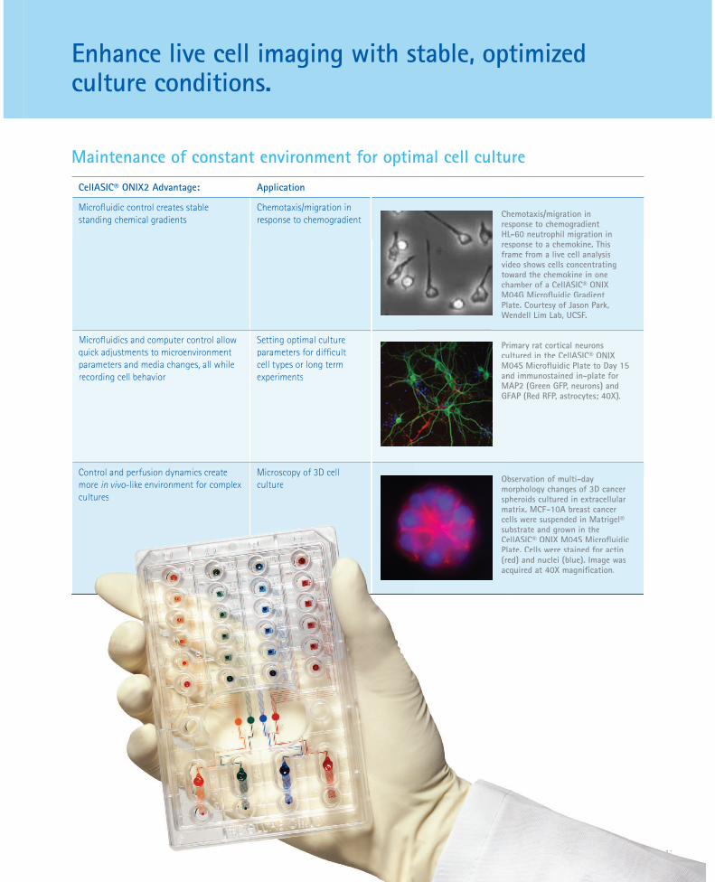
Maintenance of constant environment for optimal cell culture
CellASIC® ONIX2 Advantage: Application
Microfluidic control creates stable standing chemical gradients
Chemotaxis/migration in response to chemogradient
Microfluidics and computer control allow quick adjustments to microenvironment parameters and media changes, all while recording cell behavior
Setting optimal culture parameters for difficult cell types or long term experiments
Control and perfusion dynamics create more in vivo-like environment for complex cultures
Microscopy of 3D cell culture
Chemotaxis/migration in response to chemogradient HL-60 neutrophil migration in response to a chemokine. This frame from a live cell analysis video shows cells concentrating toward the chemokine in one chamber of a CellASIC® ONIX M04G Microfluidic Gradient Plate. Courtesy of Jason Park, Wendell Lim Lab, UCSF.
Primary rat cortical neurons cultured in the CellASIC® ONIX M04S Microfluidic Plate to Day 15 and immunostained in-plate for MAP2 (Green GFP, neurons) and GFAP (Red RFP, astrocytes; 40X).
Observation of multi-day morphology changes of 3D cancer spheroids cultured in extracellular matrix. MCF-10A breast cancer cells were suspended in Matrigel® substrate and grown in the CellASIC® ONIX M04S Microfluidic Plate. Cells were stained for actin (red) and nuclei (blue). Image was acquired at 40X magnification.
111111111111111111111111111111111111111111111111111111111111
Microfluidics and computer control allow quick adjustments to microenvironment parameters and media changes, all while recording cell behavior
Setting optimal culture parameters for difficult cell types or long term experiments
Control and perfusion dynamics create more in vivo-like environment for complex cultures
Microscopy of 3D cell culture
response to a chemokine. This frame from a live cell analysis video shows cells concentrating toward the chemokine in one chamber of a CellASIC® ONIX M04G Microfluidic Gradient Plate. Courtesy of Jason Park, Wendell Lim Lab, UCSF.
Primary rat cortical neurons cultured in the CellASIC® ONIX M04S Microfluidic Plate to Day 15 and immunostained in-plate for MAP2 (Green GFP, neurons) and GFAP (Red RFP, astrocytes; 40X).
Observation of multi-day morphology changes of 3D cancer spheroids cultured in extracellular matrix. MCF-10A breast cancer cells were suspended in Matrigel® substrate and grown in the CellASIC® ONIX M04S Microfluidic Plate. Cells were stained for actin (red) and nuclei (blue). Image was acquired at 40X magnification.
Enhance live cell imaging with stable, optimized culture conditions.
12
The CellASIC® ONIX2 Microfluidic System is the next generation of a well- established and
highly published technology. Here is a sampling of recent publications.
Find more by visiting our website at: www.merckmillipore.com/cellasic-publications
YeastKabeche R, Howard L, Moseley JB. Eisosomes provide
membrane reservoirs for rapid expansion of the yeast plasma
membrane. J. Cell Sci., Nov 2015; 128: 4057 - 4062.
http://www.ncbi.nlm.nih.gov/pubmed/26403204
Peroza EA, Ewald JC, Parakkal G, Skotheim JM, Zamboni N;
A genetically encoded FRET sensor for monitoring in vivo
trehalose-6-phosphate dynamics; Analytical Biochemistry
2015, Apr 1; 474:1-7. doi:10.1016/j. ab.2014.12.019
http://www.ncbi.nlm.nih.gov/pubmed/25582303
Kabeche R, Madrid M, Cansado J, Moseley JB. Eisosomes
Regulate Phosphatidylinositol 4,5-Bisphosphate (PI(4,5)P2)
Cortical Clusters and Mitogen-activated Protein (MAP) Kinase
Signaling upon Osmotic Stress. J. Biol. Chem., Oct 2015; 290:
25960 - 25973.
http://www.ncbi.nlm.nih.gov/pubmed/26359496
Mazo-Vargas A, Park H, Aydin M, Buchler NE; Measuring fast
gene dynamics in single cells with time-lapse luminescence
microscopy; Mol. Biol. Cell November 5, 2014 vol. 25 no. 22
3699-3708; doi: 10.1091/mbc.E14-07-1187
http://www.ncbi.nlm.nih.gov/pubmed/25232010
Burke TA, Christensen JR, Barone E, Suarez C, Sirotkin V, Kovar
DR. Homeostatic actin cytoskeleton networks are regulated by
assembly factor competition for monomers. Curr Biol. 2014
Mar 3;24(5):579-85.
http://www.ncbi.nlm.nih.gov/pubmed/24560576
Meyer RE, Kim S, Obeso D, Straight PD, Winey M, Dawson
DS. Mps1 and Ipl1/Aurora B act sequentially to correctly
orient chromosomes on the meiotic spindle of budding yeast.
Science. 2013 Mar 1;339(6123):1071-4.
http://www.ncbi.nlm.nih.gov/pubmed/23371552
Rafelski SM, Viana MP, Zhang Y, Chan YH, Thorn KS, Yam
P, Fung JC, Li H, Costa L da F, Marshall WF. Mitochondrial
network size scaling in budding yeast. Science. 2012 Nov
9;338(6108):822-4.
http://www.ncbi.nlm.nih.gov/pubmed/23139336
Kraft C, Kijanska M, Kalie E, Siergiejuk E, Lee SS, Semplicio
G, Stoffel I, Brezovich A, Verma M, Hansmann I, Ammerer G,
HofmannK, Tooze S, Peter M. Binding of the Atg1/ULK1 kinase
to the ubiquitin-like protein Atg8 regulates autophagy. EMBO
J. 2012 Sep 12;31(18):3691-703.
http://www.ncbi.nlm.nih.gov/pubmed/22885598
Kono K, Saeki Y, Yoshida S, Tanaka K, Pellman D. Proteasomal
degradation resolves competition between cell polarization
and cellular wound healing. Cell. 2012 Jul 6;150(1):151-64.
http://www.ncbi.nlm.nih.gov/pubmed/22727045
Sanchez-Diaz A, Nkosi PJ, Murray S, Labib K. The Mitotic
Exit Network and Cdc14 phosphatase initiate cytokinesis by
counteracting CDK phosphorylations and blocking polarised
growth. EMBO J. 2012 Aug 29;31(17):3620-34.
http://www.ncbi.nlm.nih.gov/pubmed/22872148
Wei P, Wong WW, Park JS, Corcoran EE, Peisajovich SG, Onuffer
JJ, Weiss A, Lim WA. Bacterial virulence proteins as tools to
rewire kinase pathways in yeast and immune cells. Nature. 2012
Aug 16;488(7411):384-8.
http://www.ncbi.nlm.nih.gov/pubmed/22820255
Bermejo C, Haerizadeh F, Takanaga H, Chermak D, Frommer WB.
Optical sensors for measuring dynamic changes of cytosolic
metabolite levels in yeast. Nat Protoc. 2011 Oct 27;6(11):1806-17.
http://www.ncbi.nlm.nih.gov/pubmed/22036883
Eser U, Falleur-Fettig M, Johnson A, Skotheim JM. Commitment
to a cellular transition precedes genome-wide transcriptional
change. Mol Cell. 2011 Aug 19;43(4):515-27.
http://www.ncbi.nlm.nih.gov/pubmed/21855792
Dechant R, Binda M, Lee SS, Pelet S, Winderickx J, Peter M.
Cytosolic pH is a second messenger for glucose and regulates
the PKA pathway through V-ATPase. EMBO J. 2010 Aug
4;29(15):2515-26.
http://www.ncbi.nlm.nih.gov/pubmed/20581803
Manzoni R, Montani F, Visintin C, Caudron F, Ciliberto A, Visintin
R. Oscillations in Cdc14 release and sequestration reveal a circuit
underlying mitotic exit. J Cell Biol. 2010 Jul 26;190(2):209-22.
http://www.ncbi.nlm.nih.gov/pubmed/20660629
Furuya K, Niki H. The DNA damage checkpoint regulates
a transition between yeast and hyphal growth in
Schizosaccharomyces japonicus. Mol Cell Biol. 2010
Jun;30(12):2909-17. doi: 10.1128/MCB.00049-10.
http://www.ncbi.nlm.nih.gov/pubmed/20368354
Octavio LM, Gedeon K, Maheshri N. Epigenetic and conventional
regulation is distributed among activators of FLO11 allowing
tuning of population-level heterogeneity in its expression. PLoS
Genet. 2009 Oct;5(10):e1000673.
http://www.ncbi.nlm.nih.gov/pubmed/19209350
A Growing Array of Distinguished Publications
13
BacteriaSutterlin HA, Shi H, May KL, Miguel A, Khare S, Huang KC,
and Silhavy TJ. Disruption of lipid homeostasis in the Gram-
negative cell envelope activates a novel cell death pathway.
PNAS. 2016 Feb; 10.1073/pnas.1601375113.
http://www.ncbi.nlm.nih.gov/pubmed/26929379
Prindle A, Liu J, Asally M, Ly S, Garcia-Ojalvo J, Süel GM.
Ion channels enable electrical communication in bacterial
communities. Nature. 2015 Nov 5;527(7576):59-63.
http://www.ncbi.nlm.nih.gov/pubmed/26503040
Grangeon R, Zupan JR, Anderson-Furgeson J, and Zambryski
PC. PopZ identifies the new pole, and PodJ identifies the old
pole during polar growth in Agrobacterium tumefaciens.
PNAS. 2015 Sep; 112:11666 - 11671.
http://www.ncbi.nlm.nih.gov/pubmed/26324921
Liu J, Prindle A, Humphries J, Gabalda-Sagarra M, Asally M, Lee
DD, Ly S, Gacia-Ojalvo J, Süel GM. Metabolic co-dependence
gives rise to collective oscillations within biofilms. Nature
2015 July 523:550-554.
http://www.ncbi.nlm.nih.gov/pubmed/26200335
Sieger B, Schubert K, Donovan C, Bramkamp M. The lipid
II flippase RodA determines morphology and growth
in Corynebacterium glutamicum. Mol Microbiol. 2013
Dec;90(5):966-82.
http://www.ncbi.nlm.nih.gov/pubmed/24118443
Gordon AJ, Satory D, Halliday JA, Herman C. Heritable change
caused by transient transcription errors. PLoS Genet. 2013
Jun;9(6):e1003595.
http://www.ncbi.nlm.nih.gov/pubmed/23825966
Donovan C, Schauss A, Kramer R, Bramkamp M. Chromosome
segregation impacts on cell growth and division site selection
in Corynebacterium glutamicum. PLOS One, February 2013;
8(2): eSS078.
http://www.ncbi.nlm.nih.gov/pubmed/23405112
Enrique Rojas, Julie A. Theriot, and Kerwyn Casey Huang.
Response of Escherichia coli growth rate to osmotic shock.
PNAS, May 2014; 111: 7807 - 7812.
http://www.ncbi.nlm.nih.gov/pubmed/24821776
Young JW, Locke JC, Elowitz MB. Rate of environmental
change determines stress response specificity. Proc Natl Acad
Sci U S A. 2013 Mar 5;110(10):4140-5.
http://www.ncbi.nlm.nih.gov/pubmed/23407164
AlgaeLudington WB, Shi LZ, Zhu Q, Berns MW, Marshall WF. Organelle
size equalization by a constitutive process. Curr Biol. 2012 Nov
20;22(22):2173-9.
http://www.ncbi.nlm.nih.gov/pubmed/23084989
MammalianChangou CA, Chen Y-R, Xing L, Yen Y, Chuang FYS, Cheng RH,
Bold RJ, Ann DK, Kung H-J; Arginine starvation-associated
atypical cellular death involves mitochondrial dysfunction,
nuclear DNA leakage, and chromatin autophagy. PNAS, Sep
2014; 111: 14147 - 14152.
http://www.ncbi.nlm.nih.gov/pubmed/25122679
Zambrano S, De Toma I, Piffer A, Bianchi ME, Agresti A; NF- B
oscillations translate into functionally related patterns of gene
expression; eLife 2016; 10.7554/eLife .09100.
http://www.ncbi.nlm.nih.gov/pubmed/26765569
Park JS, Rhau B, Hermann A, McNally KA, Zhou C, Gong D,
Weiner OD, Conklin BR, Onuffer J, Lim WA; Synthetic control
of mammalian-cell motility by engineering chemotaxis to an
orthogonal bioinert chemical signal. PNAS, Apr 2014; 111:
5896 - 5901.
http://www.ncbi.nlm.nih.gov/pubmed/24711398
View the updated list of publications, review protocols and application
data and watch video of live cells responding in real time by visiting:
www.merckmillipore.com/cellasic
A Growing Array of Distinguished Publications

14
Ordering InformationDescription Qty Catalog No.
CellASIC® ONIX2 Microfluidic System and Manifolds
CellASIC® ONIX2 Microfluidic System 1 CAX2-S0000
CellASIC® ONIX2 Manifold XT (Temperature Controlled) 1 CAX2-MXT20
CellASIC® ONIX2 Manifold Basic (No Temperature Control) 1 CAX2-MBC20
Microfluidic Plates
CellASIC® ONIX Plate for Haploid Yeast Cells 5 Y04C-02-5PK
CellASIC® ONIX Plate for Diploid Yeast Cells 5 Y04D-02-5PK
CellASIC® ONIX Plate for Bacteria Cells 5 B04A-03-5PK
CellASIC® ONIX Switching Plate for Mammalian Cells 5 M04S-03-5PK
CellASIC® ONIX Gradient Plate for Mammalian Cells 5 M04G-02-5PK
CellASIC® ONIX Open-Top Plate for Mammalian Cells 5 M04L-03-5PK
CellASIC® ONIX Plate for Chlamydomonas Cells 5 C04A-01-5PK
For a complete list of products and information, visit:
www.merckmillipore.com/cellasic
15
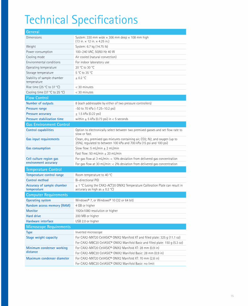
General
Dimensions System: 330 mm wide × 306 mm deep × 108 mm high (13 in. × 12 in. × 4.25 in.)
Weight System: 6.7 kg (14.75 lb)
Power consumption 100–240 VAC, 50/60 Hz 40 W
Cooling mode Air cooled (natural convection)
Environmental conditions For indoor laboratory use
Operating temperature 20 °C to 30 °C
Storage temperature 5 °C to 35 °C
Stability of sample chamber temperature
± 0.2 °C
Rise time (25 °C to 37 °C) < 30 minutes
Cooling time (37 °C to 25 °C) < 30 minutes
Flow Control
Number of outputs 8 (each addressable by either of two pressure controllers)
Pressure range -50 to 70 kPa (-7.25–10.2 psi)
Pressure accuracy ± 1.5 kPa (0.22 psi)
Pressure stabilization time within ± 5 kPa (0.73 psi) in < 5 seconds
Gas Environment Control
Control capabilities Option to electronically select between two premixed gasses and set flow rate to slow or fast.
Gas input requirements Clean, dry, premixed gas mixtures containing air, CO2, N2, and oxygen (up to 25%), regulated to between 100 kPa and 700 kPa (15 psi and 100 psi)
Gas consumption Slow flow: 5 mL/min ± 2 mL/min
Fast flow: 50 mL/min ± 20 mL/min
Cell culture region gas environment accuracy
For gas flow at 3 mL/min: < 10% deviation from delivered gas concentration
For gas flow at 30 mL/min: < 2% deviation from delivered gas concentration
Temperature Control
Temperature control range Room temperature to 40 °C
Control method Bi-directional PID
Accuracy of sample chamber temperature
± 1 °C (using the CAX2-ACT20 ONIX2 Temperature Calibration Plate can result in accuracy as high as ± 0.2 °C)
Computer Requirements
Operating system Windows® 7, or Windows® 10 (32 or 64 bit)
Random access memory (RAM) 4 GB or higher
Monitor 1920x1080 resolution or higher
Hard drive 200 MB or higher
Hardware interface USB 2.0 or higher
Microscope Requirements
Type Inverted microscope
Stage weight capacity For CAX2-MXT20 CellASIC® ONIX2 Manifold XT and filled plate: 325 g (11.1 oz)
For CAX2-MBC20 CellASIC® ONIX2 Manifold Basic and filled plate: 150 g (5.3 oz)
Minimum condenser working distance
For CAX2-MXT20 CellASIC® ONIX2 Manifold XT: 28 mm (0.9 in)
For CAX2-MBC20 CellASIC® ONIX2 Manifold Basic: 28 mm (0.9 in)
Maximum condenser diameter For CAX2-MXT20 CellASIC® ONIX2 Manifold XT: 70 mm (2.8 in)
For CAX2-MBC20 CellASIC® ONIX2 Manifold Basic: no limit
Technical Specifications
16
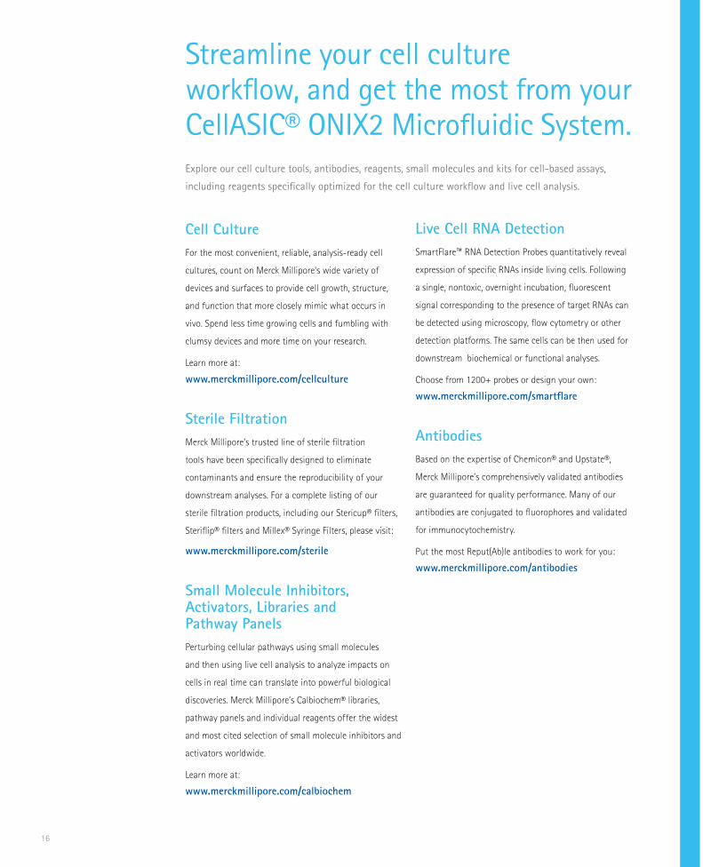
Streamline your cell culture workflow, and get the most from your CellASIC® ONIX2 Microfluidic System.
Cell Culture
For the most convenient, reliable, analysis-ready cell
cultures, count on Merck Millipore’s wide variety of
devices and surfaces to provide cell growth, structure,
and function that more closely mimic what occurs in
vivo. Spend less time growing cells and fumbling with
clumsy devices and more time on your research.
Learn more at:
www.merckmillipore.com/cellculture
Sterile Filtration
Merck Millipore’s trusted line of sterile filtration
tools have been specifically designed to eliminate
contaminants and ensure the reproducibility of your
downstream analyses. For a complete listing of our
sterile filtration products, including our Stericup® filters,
Steriflip® filters and Millex® Syringe Filters, please visit:
www.merckmillipore.com/sterile
Small Molecule Inhibitors, Activators, Libraries and Pathway Panels
Perturbing cellular pathways using small molecules
and then using live cell analysis to analyze impacts on
cells in real time can translate into powerful biological
discoveries. Merck Millipore’s Calbiochem® libraries,
pathway panels and individual reagents offer the widest
and most cited selection of small molecule inhibitors and
activators worldwide.
Learn more at:
www.merckmillipore.com/calbiochem
Live Cell RNA Detection
SmartFlare™ RNA Detection Probes quantitatively reveal
expression of specific RNAs inside living cells. Following
a single, nontoxic, overnight incubation, fluorescent
signal corresponding to the presence of target RNAs can
be detected using microscopy, flow cytometry or other
detection platforms. The same cells can be then used for
downstream biochemical or functional analyses.
Choose from 1200+ probes or design your own:
www.merckmillipore.com/smartflare
Antibodies
Based on the expertise of Chemicon® and Upstate®,
Merck Millipore’s comprehensively validated antibodies
are guaranteed for quality performance. Many of our
antibodies are conjugated to fluorophores and validated
for immunocytochemistry.
Put the most Reput(Ab)le antibodies to work for you:
www.merckmillipore.com/antibodies
Explore our cell culture tools, antibodies, reagents, small molecules and kits for cell-based assays,
including reagents specifically optimized for the cell culture workflow and live cell analysis.
17
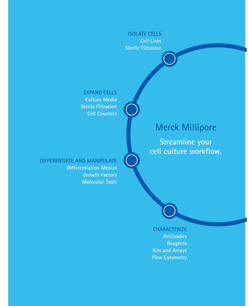
Merck Millipore
Streamline your cell culture workflow.
ISOLATE CELLS
Cell Lines
Sterile Filtration
EXPAND CELLS
Culture Media
Sterile Filtration
Cell Counters
DIFFERENTIATE AND MANIPULATE
Differentiation Medias
Growth Factors
Molecular Tools
CHARACTERIZE
Antibodies
Reagents
Kits and Assays
Flow Cytometry

To learn more about live cell imaging
and microfluidics technology, visit our
learning center at:
www.merckmillipore.com/cellasic-livecell
To request a demonstration,
or for more information on the
CellASIC® ONIX2 System, please visit:
www.merckmillipore.com/cellasic-livecell
The CellASIC® ONIX2 Microfluidic System
To place an order or receivetechnical assistance pleasecontact Merck
NEW ZEALAND:Ph: 0800 463 725Fax: (06) 356 7311 [email protected]
Australia:Ph: 1800 335 571Fax: 1300 360 418Orders: [email protected]


![arXiv:1804.09909v1 [physics.hist-ph] 26 Apr 2018 Johann Rafelski Fig. 2 View on introductory remarks in the W. Greiner and H. Diehl Electrodynamik (Verlag Harri Deutsch, Frankfurt](https://cdn.vdocument.in/doc/165x107/5ce662e688c993b62d8c828c/arxiv180409909v1-26-apr-2018-johann-rafelski-fig-2-view-on-introductory-remarks.jpg)


