
General rights Copyright and moral rights for the publications made accessible in the public portal are retained by the authors and/or other copyright owners and it is a condition of accessing publications that users recognise and abide by the legal requirements associated with these rights.
• Users may download and print one copy of any publication from the public portal for the purpose of private study or research. • You may not further distribute the material or use it for any profit-making activity or commercial gain • You may freely distribute the URL identifying the publication in the public portal
If you believe that this document breaches copyright please contact us providing details, and we will remove access to the work immediately and investigate your claim.
Downloaded from orbit.dtu.dk on: Dec 17, 2017
The role of African buffalos (Syncerus caffer) in the maintenance of foot-and-mouthdisease in Uganda
Ayebazibwe, C.; Mwiine, F. N.; Tjørnehøj, Kirsten; Balinda, S. N.; Muwanika, V. B.; Okurut, A. R. A.;Belsham, Graham; Normann, Preben; Siegismund, H. R.; Alexandersen, SørenPublished in:B M C Veterinary Research
Link to article, DOI:10.1186/1746-6148-6-54
Publication date:2010
Link back to DTU Orbit
Citation (APA):Ayebazibwe, C., Mwiine, F. N., Tjørnehøj, K., Balinda, S. N., Muwanika, V. B., Okurut, A. R. A., ... Alexandersen,S. (2010). The role of African buffalos (Syncerus caffer) in the maintenance of foot-and-mouth disease inUganda. B M C Veterinary Research, 6(54). DOI: 10.1186/1746-6148-6-54
This Provisional PDF corresponds to the article as it appeared upon acceptance. Fully formattedPDF and full text (HTML) versions will be made available soon.
The role of African buffalos (Syncerus caffer) in the maintenance offoot-and-mouth disease in Uganda
BMC Veterinary Research 2010, 6:54 doi:10.1186/1746-6148-6-54
Chrisostom Ayebazibwe ([email protected])Frank N Mwiine ([email protected])
Kirsten Tjornehoj ([email protected])Sheila N Balinda ([email protected])
Vincent B Muwanika ([email protected])Anna R Ademun Okurut ([email protected])
Graham J Belsham ([email protected])Preben Normann ([email protected])
Hans R Siegismund ([email protected])Soren Alexandersen ([email protected])
ISSN 1746-6148
Article type Research article
Submission date 21 December 2009
Acceptance date 11 December 2010
Publication date 11 December 2010
Article URL http://www.biomedcentral.com/1746-6148/6/54
Like all articles in BMC journals, this peer-reviewed article was published immediately uponacceptance. It can be downloaded, printed and distributed freely for any purposes (see copyright
notice below).
Articles in BMC journals are listed in PubMed and archived at PubMed Central.
For information about publishing your research in BMC journals or any BioMed Central journal, go to
http://www.biomedcentral.com/info/authors/
BMC Veterinary Research
© 2010 Ayebazibwe et al. , licensee BioMed Central Ltd.This is an open access article distributed under the terms of the Creative Commons Attribution License (http://creativecommons.org/licenses/by/2.0),
which permits unrestricted use, distribution, and reproduction in any medium, provided the original work is properly cited.
- 1 -
The role of African buffalos (Syncerus caffer) in the
maintenance of foot-and-mouth disease in Uganda
Chrisostom Ayebazibwe1, Frank N Mwiine
1, 5, Kirsten Tjørnehøj
3§, Sheila N Balinda
2,
Vincent B Muwanika2, Anna R Ademun Okurut
1, Graham J Belsham
3, Preben
Normann3, Hans R Siegismund
4, Soren Alexandersen
3, 6
1Ministry of Agriculture, Animal Industry and Fisheries, P.O. Box 513, Entebbe,
Uganda
2Makerere University Institute of Environment and Natural Resources, P.O. Box
7298, Kampala, Uganda
3National Veterinary Institute, Technical University of Denmark, Lindholm, DK-
4771, Kalvehave, Denmark
4Department of Biology, University of Copenhagen, Ole Maaløes Vej 5, DK-2200
Copenhagen N, Denmark
5 Work address: Department of Veterinary Medicine, Faculty of Veterinary Medicine,
Makerere University, Box 7062, Kampala,
Uganda.
6 Current address: National Centre for Foreign Animal Diseases, 1015 Arlington
Street, Winnipeg MB R3E 3M4, Canada
§Corresponding author
Email addresses:
- 2 -
ARAO [email protected]
Abstract
Background
To study the role of African buffalos (Syncerus caffer) in the maintenance of foot-
and-mouth disease in Uganda, serum samples were collected from 207 African
buffalos, 21 impalas (Aepyceros melampus), 1 giraffe (Giraffa camelopardalis), 1
common eland (Taurotragus oryx), 7 hartebeests (Alcelaphus buselaphus) and 5
waterbucks (Kobus ellipsiprymnus) from four major National Parks in Uganda
between 2005 and 2008. Serum samples were screened to detect antibodies against
foot-and-mouth disease virus (FMDV) non-structural proteins (NSP) using the
Ceditest® FMDV NS ELISA. Solid Phase Blocking ELISAs (SPBE) were used to
determine the serotype-specificity of antibodies against the seven serotypes of FMDV
among the positive samples. Virus isolation and sequencing were undertaken to
identify circulating viruses and determine relatedness between them.
Results
Among the buffalo samples tested, 85% (95% CI = 80−90%) were positive for
antibodies against FMDV non-structural proteins while one hartebeest sample out of
seven (14.3%; 95% CI = −11.6−40.2%) was the only positive from 35 other wildlife
samples from a variety of different species. In the buffalo, high serotype-specific
antibody titres (≥ 80) were found against serotypes O (7/27 samples), SAT 1 (23/29
samples), SAT 2 (18/32 samples) and SAT 3 (16/30 samples). Among the samples

- 3 -
titrated for antibodies against the four serotypes O, SAT 1, SAT 2 and SAT 3, 17/22
(77%; CI = 59.4−94.6%) had high titres against at least two serotypes.
FMDV isolates of serotypes SAT 1 (1 sample) and SAT 2 (2 samples) were obtained
from buffalo probang samples collected in Queen Elizabeth National Park (QENP) in
2007. Sequence analysis and comparison of VP1 coding sequences showed that the
SAT 1 isolate belonged to topotype IV while the SAT 2 isolates belonged to different
lineages within the East African topotype X.
Conclusions
Consistent detection of high antibody titres in buffalos supports the view that African
buffalos play an important role in the maintenance of FMDV infection within
National Parks in Uganda. Both SAT 1 and SAT 2 viruses were isolated, and
serological data indicate that it is also likely that FMDV serotypes O and SAT 3 may
be present in the buffalo population. Detailed studies should be undertaken to define
further the role of wildlife in the epidemiology of FMDV in East Africa.
Background
Foot-and-mouth disease (FMD) is a highly contagious viral disease that affects all
cloven-hoofed wild and domestic animals [1] and has serious socio-economic
consequences [2]. The epidemiology of FMD in Africa is unique, complex and poorly
understood. Seven FMDV serotypes have been defined: O, A, C, Asia 1, and the
Southern African Territories (SAT) 1, SAT 2 and SAT 3, of which all but Asia 1 have
occurred in most East African countries including Uganda [3]. Wildlife hosts,
especially African buffalos (Syncerus caffer), are believed to play an important role as
reservoirs for the SAT serotypes of FMDV [4] and the disease is sometimes
transmitted between and within different livestock and wildlife species [5−9].
- 4 -
In Africa, the epidemiology of FMD is complicated by the widespread movement of
animals, the wide host range of the virus involving wild and domestic animal
reservoirs and the presence of multiple strains and sub-strains. Moreover, the spread
of the disease is facilitated by the ability of the virus to survive for relatively long
periods in raw meat, raw milk or outside the host [1, 10, 11]. Infection of cloven-
hoofed animals can result in development of a carrier state in which case FMDV may
be found in such animals for more than 28 days after infection [12−14], and thus may
influence the epidemiology of the disease and interfere with its diagnosis and control.
The duration of the carrier state can be prolonged after recovery from acute disease; in
the case of cattle for up to 3.5 years [14]. The epidemiology of FMD in wildlife
populations has not been fully documented but it has been established that African
buffalo herds can harbour the infection for up to 24 years [15]. They act as long term
maintenance hosts for the SAT serotypes (SAT 1, SAT 2 and SAT 3) of FMDV with
no obvious clinical disease [4, 16]. Other cloven-hoofed wildlife species may develop
antibodies against FMD infections; however, their roles in excretion, transmission and
persistence of FMDV either have not been conclusively studied or have been shown
to be less important than the role of the buffalos [7, 17, 18]. In South Africa, the
impala (Aepyceros melampus) has been shown to play a potentially significant role in
the propagation of FMD outbreaks between livestock and wildlife [19].
FMD outbreaks are often encountered in cattle in Uganda but the roles of different
wild and domestic hosts in the maintenance and spread of FMDV have not been
exhaustively studied. Available data on seventy-three Ugandan FMD outbreaks,
mainly in cattle, and a few isolates from apparently healthy buffalos, indicate that
between the years 1958 and 2000, approximately 31% were attributed to serotype O,
26% to A, 25% to SAT 2, 14% to SAT 1, 3% to C and 1% to SAT 3 [3]. FMDV

- 5 -
serotypes SAT 1, SAT 2 and SAT 3 have been found in many other sub-Saharan
African countries, however, the viruses found in East Africa seem to belong to
distinct lineages [20, 21, 22]. The possible role played by the African buffalos in the
epidemiology of FMDV serotypes other than SATs has not been established, since
only one single study in Queen Elizabeth National Park has reported antibodies
against serotypes O and A [23], thus further research is required in this field.
This study was undertaken to evaluate the role of African buffalos and other wildlife
species in the maintenance of different FMDV serotypes under natural conditions in
selected National Parks in Uganda.
Results Antibodies elicited against FMDV NSP
Between 2005 and 2008, 207 samples were collected from African buffalos and 35
samples were collected from other wildlife species (21 impala (Aepyceros melampus),
1 giraffe (Giraffa camelopardalis), 1 common eland (Taurotragus oryx), 7 hartebeest
(Alcelaphus buselaphus) and 5 waterbuck, (Kobus ellipsiprymnus)) in Queen
Elizabeth National Park (QENP), Lake Mburo National Park (LMNP), Kidepo Valley
National Park (KVNP) and Murchison Falls National Park (MFNP). One hundred and
seventy-six out of 207 buffalo samples (85%; 95% CI = 80−90%) tested positive for
antibodies against FMDV NSP (Table 1), while only one of seven hartebeest samples
(14.3%; 95% CI = −11.6−40.2%) from among those of other wildlife species tested
positive in the NSP ELISA.
Screening for serotype-specific antibodies using the Solid Phase Blocking ELISA
(SPBE)
Ninety-six percent (131/137) of the buffalo samples tested were apparently positive
for antibodies against more than one serotype in the screening dilution 1:5 in SPBE.
The proportion of positive samples was higher for serotypes SAT 1, SAT 2, SAT 3
- 6 -
and to a lesser extent serotype O, than for serotypes A, C and Asia 1. One hartebeest
tested positive for SAT 1, SAT 2, and SAT 3 (data not shown). Cross reactivity
between the different serotypes is known to occur in such assays [17].
Titration of selected samples in relevant serotype-specific SPBEs
Samples from QENP, MFNP and LMNP were selected for titration on the basis of
positive screening results and sufficient volumes with the objective of comparison of
results across multiple years. A total of 37 buffalo samples were titrated in the
relevant serotype-specific SPBEs as follows; O (27), SAT 1 (29), SAT 2 (32) and
SAT 3 (30) as shown in Table 2. In this study, samples with titres of ≥ 80 were
considered positive based on the highest dilution at which non-specific reactions
tended to disappear and the results of a previous study [24]. All the sera titrated for
antibodies against serotypes A, C and Asia 1 had titres below 40 and were therefore
considered negative (data not shown), while titres of 80 and above were found in the
majority of sera titrated for antibodies against serotypes O (26%; 95% CI =
9.5−42.6%), SAT 1 (79%; 95% CI = 64.6−94.1%), SAT 2 (56%; 95% CI =
39.1−73.4%) and SAT 3 (53%; 95% CI = 35.45−71.2%). The samples positive for
antibodies against FMDV serotype O were also positive for at least two of the SAT
serotypes. Six of 22 (27%; 95% CI = 8.7−45.9%) samples titrated for antibodies
against all three SAT serotypes as well as against serotype O were positive for all four
serotypes, while 17 (77%; 95% CI = 59.5−94.6%) were positive for at least two
serotypes. Nine of the 24 samples titrated for antibodies against all three SAT
serotypes were positive for antibodies against all 3 serotypes, including at least one
buffalo in each of QENP, LMNP and MFNP.

- 7 -
Isolation and identification of FMDV
Three FMDV isolates were obtained in primary bovine thyroid cells from among nine
buffalo probang samples collected on the same day in January 2007 in QENP, and
were identified by antigen ELISA as SAT 1 (1 sample from BUF 17) and SAT 2
(from BUF 6 and BUF 10). BUF 17 had a higher titre of antibodies against SAT 1
(160) compared to those against SAT 2 (80) and SAT 3 (20) (Table 2), while the sera
of BUF 6 and BUF 10 were not titrated in the SPBE. Following RT-PCR, the near
complete genome sequences were obtained and blasted in the GenBank data base. The
sequencing data was entirely consistent with the antigen ELISA results in terms of
serotype identification. Due to the limited number of full length SAT serotype
sequences that are available, comparative analysis of the virus sequences was
restricted to the VP1 coding region. These sequences were compared to reference
strains for the defined topotypes [25] to assess the phylogenetic relationships (Figures
1 and 2). The SAT 1 isolate (SAT 1/UGA/1/07, [GenBank HM067706]) was most
closely related (pair wise identity of 83%) to a previous isolate obtained from a
buffalo in Uganda in 1970 (SAT 1/UGA BUFF/21/70, Knowles et al., unpublished)
belonging to the East African topotype IV (Figure 1). The two SAT 2 isolates were
closely related to each other (pair wise identity of 90.4%) and grouped with
representatives of the topotype X viruses (Figure 2). One of the isolates, SAT
2/UGA/1/07 [GenBank HM067705], was also related to an isolate from cattle in the
neighbouring country of Democratic Republic of Congo (pair wise identity 89.5%),
while the other, SAT 2/UGA/2/07 [GenBank HM067704], was related to a previous
isolate from a buffalo in Uganda (SAT 2/UGA/1998, accession number AY343969)
with pair wise identity of 89.6%. There were multiple amino acid differences between
the SAT 2 viruses within the G-H loop (residues 140−160) and the C-terminal region

- 8 -
of VP1 which correspond to known antigenic sites (Figure 3). The recent SAT 2
buffalo isolates had some amino acid differences, within the hyper-variable regions
surrounding the conserved RGD cell attachment motifs, compared to those obtained
from post-outbreak slaughtered cattle in Uganda in 2004 [26].
Discussion Antibodies against FMDV were detected by both the Ceditest
® FMDV NS kit and the
SPBE in over 80% of screened buffalo samples. Among the samples of wildlife
species other than the buffalos, it was only one from a hartebeest that had detectable
antibodies against FMDV. Due to small sample sizes in other tested wildlife species,
it is, at this stage, not possible to explain or conclude anything about the importance
of these other species relative to buffalos. However, the findings of this study do
relate to those of other studies done elsewhere. It has been indicated that a number of
wild ruminants become persistently infected with FMDV but it is only the African
buffalos that have been shown to spread the infection during the carrier state [16, 27].
The situation seems to be different within the impala population in the Kruger
National Park in South Africa, where clinical FMD has been reported, and subclinical
infections have been shown to occur much more regularly than previously suspected
[19]. It is hypothesized that during the acute state of the disease some species may act
as intermediaries in the transmission of FMD, mainly between buffalos and cattle [6,
18, 19]. The current findings concur with reports of very low seroprevalence of
antibodies against FMDV in non-buffalo wildlife species (4.4%) compared to buffalos
(67.7%) in Eastern Africa [17]. The Ceditest® NSP ELISA seemed to work well in
detecting antibodies against FMDV in buffalo samples, with estimates of sensitivity
and specificity at 87.7% and 87.3%, respectively [17].

- 9 -
In this study, the majority of the buffalos were positive for antibodies against FMDV
NSP during each of the sampling trips between the years 2005 and 2008. This
indicates that infection is almost always present in the sampled National Parks.
Persistent infections within buffalo herds have been reported to occur in Southern
Africa due to most calves becoming infected with the three SAT serotypes, when
maternal antibodies wane at 2-6 months of age, thereby creating an opportunity for
transmitting the infection to other susceptible species [28−30]. The current findings
justify the need to conduct much more in-depth age-stratified longitudinal studies to
confirm the serotypes and patterns of FMD in different localities in Uganda.
SPBE screening results (dilution 1:5) were difficult to interpret due to the large
percentage (96%) of animals apparently testing positive for antibodies against more
than one serotype. However, titrations showed that reactions in the serotype A, C and
Asia 1 antibody ELISAs were most likely cross-reactions. This fits well with the lack
of any reports of such serotypes in wildlife in Uganda, the almost complete
disappearance of serotype C from the world and the fact that serotype Asia 1 has
never been reported anywhere on the African continent [3].
This is the first time the SPBEs have been used in an unvaccinated animal population
like the buffalos, which probably harbour persistent infections with multiple
serotypes. For future studies in endemic conditions, sera should be screened in
dilution 1:10, and the SPBE ELISAs should be improved by using more purified
antigens and more recent FMDV strains representing the FMDV topotypes currently
circulating in Uganda for the production of reagents and positive sera, thereby
possibly enhancing the specificity.
Screening of samples by serotype-specific SPBE worked well for selection for further
titration, thereby significantly reducing the associated working time and expense.

- 10 -
Titrations demonstrated the highest antibody titres against serotypes SAT 1, SAT 2
and SAT 3 with the exception of one out of four buffalos sampled in MFNP in 2007
that had equally high titres against serotypes O and SAT 1.
It is thus evident from the present study, that buffalos were exposed to the FMDV
SAT serotypes, and in MFNP probably also to serotype O. These findings suggest that
African buffalos may play an important role as natural reservoirs of the SAT
serotypes of FMDV in East Africa and are consistent with what has been established
in Southern Africa [31−33]. Detection of antibodies against serotype O in this study
confirms previous reports of antibodies against other FMDV serotypes than the SATs
in buffalos in QENP [23].
The distribution of serotypes varied between the National Parks and between
sampling trips. In this study, a large proportion of the buffalo samples had high
antibody titres against more than one serotype of FMDV (77%), and this is consistent
with previous research findings [17, 23, 24]. The relative antibody prevalences found
in this study (SAT 1 > SAT 2 > SAT 3 > O) differ from those of Bronsvoort et al.
[17], who found that antibodies against SAT 2 were the most prevalent, followed by
SAT 1 and finally SAT 3, in African buffalos in Eastern Africa. This is likely due to
spatial and temporal differences in the distribution of the infection.
Three FMDV isolates consisting of one SAT 1 from a buffalo in one herd and two
SAT 2 from buffalos in another herd were obtained from three out of nine African
buffalo probang samples collected on the same day in 2007 in QENP indicating the
presence of either current or persistent infection. The three isolates were characterised
using antigen ELISA and by full-length sequencing. The VP1 coding regions of the
two SAT 2 isolates showed that these viruses belonged to the same topotype (X) but
different lineages, with 90.4% pair wise identity. One of the SAT 2 isolates (SAT

- 11 -
2/UGA/1/07) was most closely related with a previous isolate (SAT 2/ZAI/1/82
[AF367100]) from cattle in the neighbouring country of Democratic Republic of
Congo (89.5% pair wise identity) indicating a possibility of cross-border and wildlife-
livestock transmission. The SAT 1 sequence was closest to a representative of the
topotype IV isolate obtained in 1970 from a buffalo in Uganda (SAT 1/UGA
BUFF/21/70, N. Knowles, unpublished) with a pair wise identity of 83%. It is clear
from this study that the viruses obtained are different from each other. These
differences may be of particular significance during selection of strains that may be
considered for vaccine manufacture and effective control of foot-and-mouth disease
due to a range of viruses that may be shared between wildlife and livestock. The
isolation and characterization of these viruses from buffalo confirms the presence of
SAT 1 and SAT 2 types of FMDV as demonstrated serologically by SPBEs. More
molecular epidemiological studies are necessary for precise elucidation of the
diversity of FMDV genotypes and the possible challenges involved in matching such
strains with those included in vaccines produced for use in Uganda. Molecular studies
including the current SAT 1 virus in this study suggest that a unique group of SAT 1
viruses exist in Uganda and, may necessitate a regional approach for effective control
[34].
Consistent evidence of antibodies against multiple serotypes of FMDV in several
Ugandan National Parks and the isolation of SAT 1 and SAT 2 in QENP in 30% of
nine apparently healthy buffalos indicates that wildlife maintains FMDV infections,
and thus re-affirms recent findings in buffalo sera collected during 2001−2003 [34].
These findings combined with serological evidence of exposure of cattle grazing in
QENP to the SAT serotypes [35] emphasizes the need to study FMDV isolates from
- 12 -
these two populations to establish whether FMDV is transferred between them and at
which rate.
FMDV serotype SAT 3 was isolated from a buffalo in QENP in 1970 [36] and this
study indicates that this serotype may still be present. It is not clear why outbreaks
caused by serotype SAT 3 have never been confirmed in cattle, while outbreaks of
FMDV SAT 1 and SAT 2 are quite frequent in the region.
The findings of this study highlight the challenges involved in the diagnosis and
control of FMD in endemic areas and emphasize the need for optimization of the
methods used for serological diagnosis and for serotyping of FMDV outbreaks. There
is need for more studies to investigate detailed epidemiology of FMD in wildlife in
Uganda.
Conclusions African buffalos are important for the maintenance of FMDV within National Parks
of Uganda. They play an important epidemiological role in the circulation of FMDV
serotypes SAT 1 and SAT 2, and may also harbour serotype SAT 3 and O infections.
Methods Study area
The present study was kindly approved by Uganda Wildlife Authority
(UWA/PMR/RES/50) and wildlife samples were collected from four major National
Parks in Uganda, namely; QENP, LMNP, MFNP and KVNP (Figure 4). These
National Parks were chosen on the basis of the high chance of livestock-wildlife
interactions. Compared to other National Parks in Uganda, they are generally flat or
gently sloping and not densely covered by vegetation thereby facilitating the exercise
of darting and follow up of the sedated animals. Such National Parks are also home to
- 13 -
sizeable buffalo populations with estimates of about 6,807 animals in QENP, 132 in
LMNP, 8,200 in MFNP and 400 in KVNP [37]. All the national parks are unfenced
and hence provide possibilities for livestock-wildlife interactions.
Due to the large buffalo population and the very high chances of livestock-wildlife
interactions, more samples were collected in QENP than in the other parks.
Sampling
Apart from the impala, chemical capture was used for immobilization of animals of
choice [38, 39]. The original target of sampling at least 10% of each herd was not
possible. Most buffalo herds would disperse and sometimes scatter to inaccessible
areas upon darting one or a few of them. At times it would be impossible to locate
herds in the National Parks. Animals were darted with a Dan-Inject dart gun. Two
cars were used; one for the identifying and darting the animals and the other for
tracking the herds, general field support and tracing the darted animal. Buffalo herds
were located and animals moving at the edge of the group identified and darted. The
anaesthetic combination was 8-10mg Etorphine (Kyron, South Africa) and 70-90mg
Xylazine (Kyron, South Africa). The sedated animal would be cautiously located and
approached, held by the horns and head, blindfolded and the mouth opened and the
tongue pulled out for examination for lesions and ensuring continuous respiration
before collection of serum and probang samples. After sampling, the sedative was
reversed by use of a combination of 14-18mg Diprenorphine and 60-70mg Yohimbine
(Kyron, South Africa) by intravenous infusion through the ear vein. The age of the
buffalos was estimated from the teeth. All buffalos fell within the age group used for
rinderpest serosurveillance (1.8-20 years). Non-buffalo species other than the impala
were also darted following similar techniques as defined by Kock et al. [39]. Due to
significant challenges of chemical capture, impala were instead physically restrained

- 14 -
after dazzling them with strong light directed at the eyes at night time, during periods
of little or no moonlight [40].
A total of 134 African buffalo samples and 21 impala samples were collected during
16 trips in the years 2007 and 2008 (Table 1). The samples from giraffe (1),
hartebeest (7) and waterbuck (5), were jointly obtained through the on-going wildlife
health research and monitoring programmes by Uganda Wildlife Authority in 2007.
Eighty African buffalo samples and 1 Eland sample had been collected during the
rinderpest serosurveillance exercise between the years 2005 and 2006. Probang
samples were preserved in 0.04 M phosphate buffered saline (PBS), transported under
liquid nitrogen while in the field and stored at -80°C at the laboratory. Serum was
separated from blood and stored at -20°C in the laboratory.
Screening for antibodies to FMDV non-structural proteins
207 buffalo samples were screened for antibodies against non-structural proteins
(NSP) of FMDV using the commercial Ceditest FMDV NS® kit (Cedi diagnostics
BV, Netherlands) [41]. This test is currently marketed as Priocheck
® FMDV NS by
Prionics® AG, Switzerland. In addition, samples from impala (n = 21), hartebeest (n =
7), waterbuck (n = 5), eland (n = 1) and giraffe (n = 1) were tested in the same way.
Serotype-specific Solid Phase Blocking ELISA (SPBE)
137 African buffalo serum samples, of which seven were not tested for antibodies
against NSP, were screened (dilution 1:5) for serotype-specific antibodies against
FMDV using an in-house SPBE system modified from Have and Holm-Jensen [42]
and described in detail by Balinda et al. [43]. The O, A, C and Asia 1 tests in this
ELISA system have been used at the National Veterinary Institute, Danish Technical
University (Lindholm), for many years; they have been validated for cattle and swine
(ISO/IEC 17025) and used for many other ruminants and Camelidae with good

- 15 -
results, and they appear to work well on all species (Alexandersen, unpublished
results). The SPBE tests for antibodies against the SAT-serotypes were more recent
and were still undergoing evaluation. Closely related ELISA tests for the SAT-
serotypes have been set up and used under African conditions for detecting antibodies
against multiple FMDV serotypes and shown to perform well [43, 44].
For each well, optical density (OD) as a percentage of the mean OD of four wells with
negative control sera (ODP) was calculated according to the formula: ODP = ((sample
OD450 − OD620) / (mean of (negative control sera OD450 − OD620)) × 100. Samples
were considered positive, if ODP was lower than 50% in the antibody tests for O,
SAT 1, SAT 2 and SAT 3, 45% for A and 35% for C and Asia 1.
Based on the serological status and availability of sufficient amounts, 37 positive
samples were selected and titrated (up to dilution 1:640) in the relevant serotype
specific SPBEs. Titres were expressed as the reciprocal of the highest positive
dilution.
Due to limited sample volumes and the smaller number of trips made, serotype
specific SPBE studies did not include KVNP.
FMD Virus isolation and antigen ELISA
The methodology of virus isolation from the OP samples was adopted from the
standard procedure described by the World Organisation for Animal Health [45].
Briefly, 50µl of undiluted sample and a 1:10 dilution of the sample were each
inoculated into 5 wells of a 96-well microtitre plate with monolayers of primary
bovine thyroid (BTY) cells and 100µl of Eagles media with 2% fetal calf serum. A
row of wells with negative control sera including buffer was inserted between each
sample. The cell cultures were incubated at 37°C and examined for cytopathic effect
(CPE) for 2-4 days. Negative cultures were passaged onto new bovine thyroid (BTY)

- 16 -
monolayers once. First and second passage cultures with CPE were harvested and
serotyped using an in-house antigen ELISA set up at the National Veterinary Institute,
Lindholm, Denmark, based on the description by OIE [45]. Briefly, the rabbit and
guinea pig hyperimmune sera were the same as used in the in-house SPBE for
serotype-specific antibodies against FMDV described above. The samples were tested
in duplicate, and for each serotype each plate included two wells with strong positive
control sera, two wells with weak positive control sera and two wells with negative
control sera, all consisting of cell-culture materials. The tests for serotypes O, A, C
and Asia 1 were quality assured (ISO/IEC 17025), while the tests for serotypes SAT
1, SAT 2 and SAT 3 were more recently set up and still undergoing evaluation.
RNA extraction, RT-PCR and cycle sequencing
Total RNA was extracted from CPE positive cell cultures using the RNeasy-Mini
Kit® (Qiagen, Germany) according to the manufacturer’s instructions. cDNA was
synthesized from the template using Ready-To-Go® You-Prime First-Strand Beads
(GE Healthcare Life Sciences, UK) and a four-primer mix of NVT24 , A PN 63 (5´-
AGACCTGGAAAGACCAGGC-3’), G15H , and pdN6 (random hexamers). To
generate 15 overlapping PCR fragments for near full length genome sequencing, 15
PCR-tubes were prepared containing: 33.1 µl of water, 5.0 µl 5 X AmpliTaq Gold
buffer, 4.0 µl MgCl2 (25mM), 0.4 µl dNTPs (2.5 mM each), 2.5 Units of Amplitaq
Gold®
(Applied Biosystems, UK) and 5.0 µl of template cDNA. To each of these
tubes, 1.0 µl of respective fragment-specific forward and reverse primers, each at a
concentration of 25 pmol/µl was added to make a total volume of 50 µl.
The primers used for the VP1 coding region are shown in Table 3. The PCR (Perkin
Elmer PE 9700) was set and ran at 95°C for 5 minutes to activate Amplitaq enzyme
followed by five cycles (95°C for 15 seconds, 55°C for 30 seconds with less by 1

- 17 -
second in each subsequent cycle and then 72°C for 1 minute and 20 seconds), 40
cycles (95°C for 15 seconds, 50°C for 30 seconds, and 72°C for 1 minute and 20
seconds–adding 1 second per cycle) and lastly at 72 °C for 7 minutes and kept at 4°C.
To confirm the presence or absence of PCR products, gel electrophoresis was
undertaken using 1.2% agarose containing 0.005% ethidium bromide. Amplicons
were extracted from the gel using the Qiaquick® (Qiagen, Germany) gel extraction kit
and sent to AGOWA (Germany) for cycle sequencing.
Sequence analysis
A phylogenetic tree of the virus sequences was inferred using the Neighbor-Joining
method [46]. The bootstrap consensus tree inferred from 1000 replicates is taken to
represent the evolutionary history of the taxa analyzed [47]. The evolutionary
distances were computed using the Kimura 2-parameter method [48] and are in the
units of the number of base substitutions per site. The sequences studied were all from
the VP1 coding region of the current FMDV isolates and the reference topotypes
(Table 4). All positions containing gaps and missing data were eliminated from the
dataset. There were a total of 660 nucleotides in the final dataset. Phylogenetic
analyses were conducted in MEGA 4 [49, 50]. In order to deduce the amino acid
sequences, the East African SAT 2 prototype sequences together with the Ugandan
buffalo sequences (this study) and those from cattle during 2004 [26] corresponding
to the C-terminal part of the VP1, were aligned and translated in MEGA 4 and
exported to the Bioedit sequence alignment editor [51] to identify the positions of
differences and similarities.
Authors' contributions CA conceived and designed the study, undertook field work, laboratory studies, data
analysis, manuscript preparation, review, corrections and submission. SA, KT, VBM,
- 18 -
HRS, ARAO and GJB participated in the supervision of various project activities
including field work, laboratory studies, data analysis, manuscript preparation, proof
reading and review. FNM participated in field work, laboratory studies, and
manuscript proof reading. SNB participated in part of the field work, provision of
livestock sequence data and analysis. PN was involved in all the molecular laboratory
work. All authors read and approved the final manuscript.
Acknowledgements We acknowledge the cooperation of Pan African Control of Epizootics (PACE)
project, Uganda Wildlife Authority and the Ministry of Agriculture Animal Industry
and Fisheries (MAAIF), Uganda for allowing us to analyze samples originally
collected for rinderpest sero-surveillance. Dr. Charles Masembe was very useful in
sample collection and general project work. Dr. Patrick Atimnedi was very
instrumental in the darting of wild animals for collection of samples. Dr. Abraham
Kiprotich Sangula participated in some field trips for sample collection. We
appreciate technical and laboratory input by Jane Borch, Jani Christiansen, Jonna V.
Jensen, Tina Frederiksen and Tina Pedersen at the Technical University of Denmark
(Lindholm), and we are grateful for the laboratory contributions offered by Esau
Martin, MAAIF. This study was funded by Danish International Development
Agency (DANIDA) under the Livestock Wildlife Diseases in East Africa Project
(LWDEA), grant number: P104.Dan.8.1.316.

- 19 -
References 1. Alexandersen S, Mowat N: Foot-and-mouth disease: host range and
pathogenesis. Curr Top Microbiol Immunol 2005, 288:9-42.
2. Perry BD, Rich KM: Poverty impacts of foot-and-mouth disease and the
poverty reduction implications of its control. Vet Rec 2007, 160(7):238-241.
3. Vosloo W, Bastos ADS, Sangare O, Hargreaves SK, Thomas GR: Review of
the status of foot and mouth disease in sub-Saharan Africa. Rev Sci Tech
2002, 21:437-449.
4. Thomson GR, Vosloo W, Bastos ADS: Foot and mouth disease in wildlife.
Virus Res 2003, 91(1):145-161.
5. Sutmoller P, Thomson G, Hargreaves SK, Foggin CM, Anderson EC: The
foot and mouth disease risk posed by African buffalo within wildlife
conservancies to the cattle Industry of Zimbabwe. Prev Vet Med 2000, 44:
1, 2, 43-60.
6. Bastos ADS, Boshoff CI, Keet DF, Bengis RG, Thomson GR: Natural
transmission of FMD between the African buffalo (Syncerus caffer) and
the impala (Aepyceros melampus) in the Kruger National Park, South
Africa. Epidemiol Infec 2000, 124:591-598.
7. Anderson EC, Anderson J, Doughty WJ, Drevmo S: The pathogenicity of
bovine strains of foot and mouth disease virus for impala and wildebeest.
J Wildl Dis 1975, 11(2):248-255.
8. Dawe PS, Flanagan FO, Madekurozwa RL, Sorensen KJ, Anderson EC,
Foggin CM, Ferris NP, Knowles NJ: Natural transmission of foot-and-
mouth disease virus from African buffalo (Syncerus caffer) to cattle in a
wildlife area of Zimbabwe. Vet Rec 1994, 134(10):230-232.
9. Dawe PS, Sorensen K, Ferris NP, Barnett IT, Armstrong RM, Knowles NJ:
Experimental transmission of foot-and-mouth disease virus from carrier
African buffalo (Syncerus caffer) to cattle in Zimbabwe. Vet Rec 1994,
134(9):211-215.
10. Ryan E, Mackay D, Donaldson A: Foot-and-mouth disease virus
concentrations in products of animal origin. Transbound Emerg Dis 2008,
55(2):89-98.
11. Tomasula PM, Konstance RP: The survival of foot-and-mouth disease virus
in raw and pasteurized milk and milk products. J Dairy Sci 2004,
87(4):1115-1121.
12. Van Bekkum JG, Frenkel HS, Frederiks HHJ, Frenkel S: Observations on the
carrier state of cattle exposed to foot-and-mouth disease virus, Tijdschr.
Diergeneeskd 1959a, 84:1159-1164.
13. Van Bekkum JG, Frenkel HS, Frederiks HHJ, Frenkel S: Observations on the
carrier state of cattle exposed to foot-and-mouth disease virus, Bull Int
Epizoot 1959b, 51: 917-922.
14. Alexandersen S, Zhang Z, Donaldson AI: Aspects of the persistence of foot-
and-mouth disease virus in animals-the carrier problem. Microbes Infect
2002, 4(10):1099-1110.
15. Condy JB, Hedger RS, Hamblin C, Barnett IT: The duration of the foot-and-
mouth disease virus carrier state in African buffalo (i) in the individual
animal and (ii) in a free-living herd. Comp Immunol Microbiol Infect Dis
1985, 8(3-4):259-265.

- 20 -
16. Gainaru MD, Thomson GR, Bengis RG, Esterhuysen JJ, Bruce W, Pini A:
Foot-and-mouth disease and the African buffalo (Syncerus caffer). II.
Virus excretion and transmission during acute infection. Onderstepoort J
Vet Res 1986, 53(2):75-85.
17. Bronsvoort BM, Parida S, Handel I, McFarland S, Fleming L, Hamblin P,
Kock R: Serological survey for foot-and-mouth disease virus in wildlife in
eastern Africa and estimation of test parameters of a nonstructural
protein enzyme-linked immunosorbent assay for buffalo. Clin Vaccine
Immunol 2008, 15(6):1003-1011.
18. Hargreaves SK, Foggin CM, Anderson EC, Bastos AD, Thomson GR, Ferris
NP, Knowles NJ: An investigation into the source and spread of foot and
mouth disease virus from a wildlife conservancy in Zimbabwe. Rev Sci
Tech 2004, 23(3):783-790.
19. Vosloo W, Thompson PN, Botha B, Bengis RG, Thomson GR: Longitudinal
study to investigate the role of impala (Aepyceros melampus) in foot-and-
mouth disease maintenance in the Kruger National Park, South Africa.
Transbound Emerg Dis 2009, 56(1-2):18-30.
20. Ayelet G, Mahapatra M, Gelaye E, Egziabher BG, Rufeal T, Sahle M, Ferris
NP, Wadsworth J, Hutchings GH, Knowles NJ: Genetic characterization of
foot-and-mouth disease viruses, Ethiopia, 1981–2007. Emerg Infect Dis
2009, 15(9): 1409-1417.
21. Sahle M, Dwarka RM, Venter EH, Vosloo W: Comparison of SAT-1 foot-
and-mouth disease virus isolates obtained from East Africa between 1971
and 2000 with viruses from the rest of sub-Saharan Africa. Arch Virol
2007, 152(4):797-804.
22. Bastos AD, Anderson EC, Bengis RG, Keet DF, Winterbach HK, Thomson
GR: Molecular epidemiology of SAT 3-type foot-and-mouth disease. Virus
Genes 2003, 27(3):283-290.
23. Kalema-Zikusoka G, Bengis RG, Michel AL and Woodford MH: A
preliminary investigation of tuberculosis and other disease in African
buffalo (Syncerus caffer) in Queen Elizabeth National Park, Uganda. Onderstepoort J Vet Res 2005, 72(2):145-151.
24. Ayebazibwe C, Mwiine FN, Balinda SN, Tjørnehøj K, Muwanika VB,
Ademun ARO, Siegismund HR, Alexandersen S: Antibodies against foot-
and-mouth disease (FMD) virus in African buffalos (Syncerus caffer) in
selected National Parks in Uganda (2001-2003). Transbound Emerg Dis,
2010, 57 (4): 286-292.
25. WRLFMD: Representative strains for each FMDV topotype. World
reference laboratory for foot-and-mouth disease.
http://www.wrlfmd.org/fmd_genotyping/prototypeshtm:Accessed:
20/11/2009.
26. Balinda SN, Belsham GJ, Masembe C, Sangula AK, Siegismund HR,
Muwanika VB: Molecular characterization of SAT 2 foot-and-mouth
disease virus from post-outbreak slaughtered animals: implications for
disease control in Uganda. Epidemiol Infect 2010, 138(8):1204-1210.
27. Bengis RG, Thomson GR, Hedger RS, De Vos V, Pini A: Foot-and-mouth
disease and the African buffalo (Syncerus caffer). 1. Carriers as a source
of infection for cattle. Onderstepoort J Vet Res 1986, 53(2):69-73.
28. Vosloo W, Bastos AD, Kirkbride E, Esterhuysen JJ, van Rensburg DJ, Bengis
RG, Keet DW, Thomson GR: Persistent infection of African buffalo

- 21 -
(Syncerus caffer) with SAT-type foot-and-mouth disease viruses: rate of
fixation of mutations, antigenic change and interspecies transmission. J
Gen Virol 1996, 77(7):1457-1467.
29. Thomson GR, Vosloo W, Esterhuysen JJ, Bengis RG: Maintenance of foot
and mouth disease viruses in buffalo (Syncerus caffer Sparrman, 1779) in
southern Africa. Rev Sci Tech 1992, 11(4):1097-1107.
30. Thomson GR: The role of carrier animals in the transmission of foot and
mouth disease. Comprehensive Reports on Technical Items Presented to The
International Committee or to Regional Commissions, 1996, 87-103.
31. Bruckner GK, Vosloo W, Du Plessis BJ, Kloeck PE, Connoway L, Ekron MD,
Weaver DB, Dickason CJ, Schreuder FJ, Marais T et al: Foot and mouth
disease: the experience of South Africa. Rev Sci Tech 2002, 21(3):751-764.
32. Sutmoller P, Thomson GR, Hargreaves SK, Foggin CM, Anderson EC: The
foot-and-mouth disease risk posed by African buffalo within wildlife
conservancies to the cattle industry of Zimbabwe. Prev Vet Med 2000,
44(1-2):43-60.
33. Thomson GR: Overview of foot and mouth disease in Southern Africa. Rev
Sci Tech 1995, 14(3):503-520.
34. Sangula AK, Belsham GJ, Muwanika VB, Heller R, Balinda SN, Masembe C,
Siegismund HR: Evolutionary analysis of foot-and-mouth disease virus
serotype SAT 1 isolates from East Africa suggests two independent
introductions from southern Africa. BMC Evol Biol 2010, 10:371.
35. Mwiine FN, Ayebazibwe C, Olaho-Mukani W, Alexandersen S, Balinda SN,
Masembe C, Okurut AR Ademun, Christensen L S, Sørensen K J, Tjørnehøj
K: Serotype-specificity of antibodies against foot and mouth disease virus
in cattle in selected districts in Uganda. Transbound Emerg Dis 2010,
57(5):365-74.
36. Hedger RS, Forman AJ, Woodford MH: Foot-and-mouth disease in East
African buffalo. Bull Epiz Dis Afr 1973, 21:90-99.
37. UWA: General Reconnaissance flights and ground based estimates. Research
Monitoring Report, 2006. Uganda Wildlife Authority, Kampala.
38. Harthoorn AM: The chemical capture of Animals. A guide to the chemical
restraint of wild and captive animals. Bailliere and Tindall: London; 1976.
416 pages.
39. Kock M, Meltzer D, Burroughs R: Chemical and physical restraint of wild
animals. A training and field manual for African species. Zimbabwe
Veterinary Association Wildlife Group and International Wildlife Veterinary
Services (Africa), South Africa 2006. 283 pages.
40. Averbeck C: Population Ecology of Impala (Aepyceros melampus) and
community-based wildlife conservation in Uganda. Fakultät für Ernährung,
Landnutzung und Umwelt, Technische Universität München, 2002
http://deposit.ddb.de/cgi-bin/dokserv?idn=964072017. Accessed: 10/3/08.
41. Sorensen KJ, de Stricker K, Dyrting KC, Grazioli S, Haas B: Differentiation
of foot-and-mouth disease virus infected animals from vaccinated animals
using a blocking ELISA based on baculovirus expressed FMDV 3ABC
antigen and a 3ABC monoclonal antibody. Arch Virol 2005, 150(4):805-
814.
42. Have P, Jensen MH: Detection of antibodies to foot-and-mouth disease
virus type O by enzyme-linked immunosorbent assay (ELISA).
Proceedings Research Group of the session of the Standing Technical
- 22 -
Committee of the European Commission for the Control of Foot-and-Mouth
Disease. Lelystad, Netherlands, 20-22 September, 1983, Appendix VIII: 44–
51.
43. Balinda SN, Tjornehoj K, Muwanika VB, Sangula AK, Mwiine FN,
Ayebazibwe C, Masembe C, Siegismund HR, Alexandersen S: Prevalence
Estimates of Antibodies Towards Foot-and-Mouth Disease Virus in Small
Ruminants in Uganda. Transbound Emerg Dis 2009, 56(9-10):362-371.
44. Bronsvoort BM, Sorensen KJ, Anderson J, Corteyn A, Tanya VN, Kitching
RP, Morgan KL: Comparison of two 3ABC enzyme-linked immunosorbent
assays for diagnosis of multiple-serotype foot-and-mouth disease in a
cattle population in an area of endemicity. J Clin Microbiol 2004,
42(5):2108-2114.
45. OIE: Foot-and-mouth disease. Manual of standards for diagnostic tests and
vaccines for terrestrial animals.
http://www.oie.int/fr/normes/mmanual/2008/pdf/2.01.05_FMD.pdf (Accessed
24/11/2010):4-5.
46. Saitou N, Nei M: The neighbor-joining method: A new method for
reconstructing phylogenetic trees. Mol Biol Evol 1987, 4:406-425.
47. Felsenstein J: Confidence limits on phylogenies: An approach using the
bootstrap. Evol 1985, 39:783-791.
48. Kimura M: A simple method for estimating evolutionary rate of base
substitutions through comparative studies of nucleotide sequences. J
Molec Evol 1980, 16:111-120.
49. Tamura K, Dudley J, Nei M and Kumar S: Molecular Evolutionary Genetics
Analysis (MEGA) software version 4.0. Mol Biol and Evol 2007, 24:1596-
1599.
50. Zheng Z, Scott S, Lukas W and Webb M: A greedy algorithm for aligning
DNA sequences. J Comput Biol 2000, 7:203-214.
51. Hall TA: Bioedit: a user-friendly biological sequence alignment editor
and analysis program for windows 95/98/nt. Nucleic Acids Symposium
Series, 41:95–98, 1999.

- 23 -
Figures
Figure 1 - Neighbour-joining tree depicting VP1 coding sequence relationships of the recent Ugandan SAT 1 isolate (SAT 1/UGA/07) with other SAT 1 reference prototypes from WRLFMD, Pirbright
Bootstrap values ≥ 50, based on 1,000 replicates are indicated next to the relevant node.
Figure 2 - Neighbour-joining tree depicting VP1 coding sequence relationships of the recent Ugandan SAT 2 isolates (SAT 2/UGA/1/07 and SAT 2/UGA/2/07) with other SAT 2 reference prototypes from WRLFMD, Pirbright
Bootstrap values ≥ 50, based on 1,000 replicates are indicated next to the relevant node.
Figure 3 - An alignment of the seventeen deduced amino acid sequences of the C-terminal region of VP1 from the East African SAT 2 FMD reference prototype virus strains and those collected from livestock and African buffalos in Uganda, between the years 2004 and 2007
Dots indicate sequence identity with master sequence, UGA/1/07 while the “?” in the
ZAI/1/82 sequence denotes an ambiguity. The highly conserved 'RGD' cell
attachment motifs are indicated by the shaded text box at positions 144-146. The
recent buffalo sequences (UGA/1/07) and (UGA/2/07) have a number of amino acid
differences from the other SAT 2 sequences including those from cattle in Uganda.
These are clustered particularly within the regions 135-160 (G-H loop) and near the
extreme C-terminus (residues 190-205). Such differences may be important in
influencing the antigenicity of these various strains.
Figure 4 - Map of Uganda showing the location of the National Parks
NP stands for the National Park.

- 24 -
Tables
Table 1 - Screening of serum samples from wildlife collected in four Ugandan National Parks during 2005-2008 for antibodies against the non-structural proteins of foot-and-mouth disease virus
National
Park
Species Total
samples
collected
Number of
samples tested
Number of
positive
samples
MFNP Buffalo 53 53 51 (96%) Waterbuck 5 5 0 (0%) Hartebeest 7 7 1 (14%) Giraffe 1 1 0 (0%) LMNP Buffalo 25 19 18 (95%) Impala 21 21 0 (0%) Eland 1 1 0 (0%) KVNP Buffalo 42 42 26 (62%) QENP Buffalo 94 93 81 (87%) Total buffalo 214 207 176 (85%) Total other
species
35 35 1 (3%)
Total 249 242 177
(MFNP-Murchison Falls National Park, LMNP-Lake Mburo National Park, KVNP-Kidepo Valley National Park, QENP-Queen Elizabeth National Park).
- 25 -
Table 2 - Titres of serotype-specific antibodies against foot-and-mouth disease virus in serum samples from African buffalos collected in three National Parks in Uganda during 2005-2008
National
Park
Sample
ID
Date O SAT 1 SAT 2 SAT 3
LMNP BUF 3 JAN.06 − − 20 −
BUF 2 JAN.06 10 20 80 −
BUF 7 JAN.06 − − 640 10
BUF 1 JAN.07 20 320 20 −
BUF 6 JAN.07 20 40 20 5
BUF 10 APR.07 160 640 80 640
BUF 9 APR.07 40 80 80 40
BUF 11 APR.07 10 20 40 40
BUF 12 APR.07 − 160 − 5
BUF 6 APR.07 − − 40 −
BUF 1 OCT.08 − 640 − 80
BUF 4 OCT.08 − 80 40 20
BUF 5 OCT.08 − 80 − −
BUF 6 OCT.08 − 80 20 20
MFNP BUF 2 OCT.05 160 320 80 160
BUF 7 OCT.05 5 320 160 320
BUF 15 OCT.05 320 640 160 160
BUF 2 NOV.06 5 10 320 20
BUF 3 NOV.06 20 80 40 80
BUF 7 NOV.06 40 80 10 80
BUF 12 OCT.07 40 80 160 160
BUF 5 OCT.07 20 20 20 160
BUF 20 OCT.07 40 640 40 320
BUF 18 OCT.07 640 640 320 320
QENP BUF 17 JAN.07 5 160 80 20
BUF 37 APR.07 5 20 40 20
BUF 35 APR.07 − − 320 40
BUF 8 JUL.07 80 320 160 40
BUF 9 AUG.07 20 320 320 160
BUF 3 AUG.07 160 640 80 320
BUF 13 AUG.07 80 640 80 160
BUF 1 OCT.08 5 − 40 160
BUF 2 OCT.08 40 640 40 80
BUF 3 OCT.08 10 80 320 20
BUF 5 OCT.08 − − 80 40
BUF 6 OCT.08 10 − − −
BUF 9 OCT.08 40 − − −
Total 7/27
(26%)
23/29
(79%)
18/32
(56%)
16/30
(53%)
Minus signs (−): results of samples with titres < 5 in the screening test and thus not
titrated.
Bold figures: results of samples tested positive (ODP ≥ 80)

- 26 -
Table 3. List of primers used for RT-PCR. For each fragment, forward and reverse primers were used
Sample
ID
Forward Primers (5´ to 3´) Reverse primers (5´ to 3´)
BUF 10 CAGTACTCCGGCAGCCTG GGTGTTGTAATTGCACTCTCC
CAGTGGTGTTCTCGCACAAC GCCATDGGMGGGATGAACCC
BUF 6 GACCGTATTCTCACCACGAG AAGTTGGACCTGACGTCGG
BUF 17 CAAAXAGGGAATTTTXCCCGTXGC GACGACXGGXTTGTCGCC
CTGGTXGGCGCAATCCTXCGT CGGTTRAAGTCGGGWCCGTG
The sequences obtained from samples BUF 10, BUF 6 and BUF 17 were subsequently named SAT 2/UGA/1/07, SAT 2/UGA/2/07 and SAT 1/UGA/1/07, respectively. These samples were all collected on the same day (17/1/07) in Queen Elizabeth National Park, but BUF 17 was from a different herd.
- 27 -
Table 4. Summary of the Viruses used in this study
Serotype Host Animal Virus strain GenBank accession no. Country SAT 1 Buffalo SAT1/UGA/1/07* HM067706 Uganda
− SAT1/T155/71* N/A Tanzania
− SAT1/ZIM/23/2003 N/A Zimbabwe
− SAT1/RV/11/37 AY593839 Unknown
− SAT1/RHO/5/66 AY593846 Rhodesia
− SAT1/BEC/1/48 AY593838 Botswana
− SAT1/BOT/1/68 AY593845 Botswana
Buffalo SAT1/UGABUFF/21/70 N/A Uganda
− SAT1/NIG/11/75 AF431711 Nigeria
− SAT1/ISR/4/62 AY593844 Israel
− SAT1/SUD/3/76 AY441996 Sudan
− SAT1/UGA/13/74 AY442010 Uganda
− SAT1/UGA/1/97* AY442012 Uganda
Cattle SAT1/ETH/3/2007 FJ798154 Ethiopia
SAT 2 Cattle SAT2/UGA/01/2004* GU323171 Uganda
Cattle SAT2/UGA/05/2004* GU323174 Uganda
Cattle SAT2/UGA/12/2004* GU323179 Uganda
Buffalo SAT2/UGA/1/2007* HM067705 Uganda
Buffalo SAT2/UGA/2/2007* HM067704 Uganda
− SAT2/SA/106/59 AY593848 Unknown
− SAT2/ZIM/14/2002 N/A Zimbabwe
Cattle SAT2/ZIM/7/83* AF136607 Zimbabwe
− SAT2/ZIM/5/81 EF134951 Zimbabwe
− SAT2/RHO/1/48 AY593847 Rhodesia
Buffalo SAT2/ BOT/P3/98 AF367124 Botswana
Cattle SAT2/KEN/1/84 AY344505 Kenya
Cattle SAT2/ETH/1/90 AY343935 Ethiopia
Cattle SAT2/NIG/2/75 AF367139 Nigeria
Cattle SAT2/GHA/2/90 AF479415 Ghana
Cattle SAT2/GAM/8/79 AF479410 Gambia
Cattle SAT2/SAU/6/2000 AF367135 Saudi Arabia
− SAT2/CAR/8/2005 N/A Cameroon
− SAT2/ZAI/1/74 DQ009737 DRC
Cattle SAT2/RWA/1/00* AF367134 Rwanda
Cattle SAT2/KEN/3/57 AJ251473 Kenya
Cattle SAT2/KEN/2/84 AY343941 Kenya
− SAT2/ZAI/1/82 AF367100 Zaire
Cattle SAT2/UGA/19/98 AY343969 Uganda
− SAT2/ANG/4/74 AF479417 Angola
Cattle SAT2/UGA/51/75 AY343963 Uganda
Cattle SAT2/SUD/6/77 AY343939 Sudan
Cattle SAT2/ETH/2/2007 FJ798161 Ethiopia
Cattle SAT2/ETH/2/91 AY343938 Ethiopia
*: not WRLFMD reference numbers −: host animal not indicated
NA: not applicable
Figure 1

Figure 2
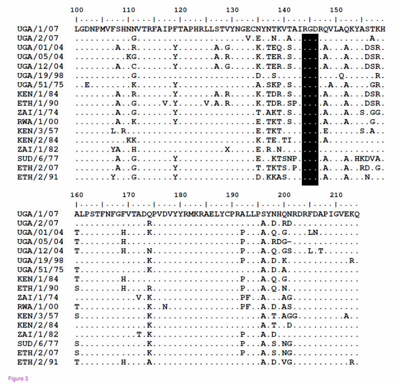
Figure 3
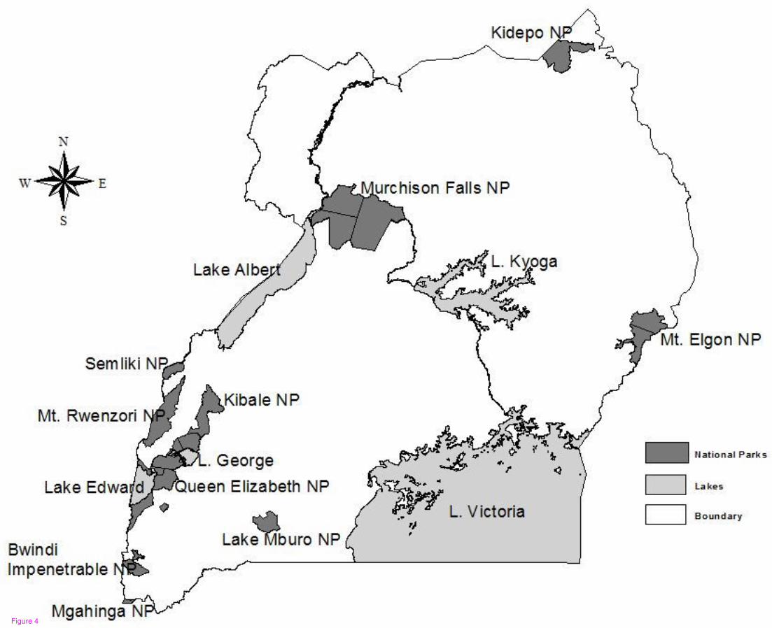
Figure 4


