JPET#145821
1
Title page
A novel peptide agonist of FPRL1 (ALX) displays anti-inflammatory
and cardioprotective effects
Iris Hecht, Jiang Rong, André LF Sampaio, Chen Hermesh, Caleb Rutledge, Ronen
Shemesh, Amir Toporik, Merav Beiman, Liat Dassa, Hagit Niv, Gady Cojocaru, Arie
Zauberman, Galit Rotman, Mauro Perretti, Jakob Vinten-Johansen, Yossi Cohen
Compugen Ltd., Tel Aviv 69512, Israel (IH, CC, RS, AT, MB, LD, HN, GC, AZ, GR,
YC); Carlyle Fraser Heart Center of Emory University and Emory Crawford Long
Hospital Atlanta, Georgia USA (JR, CR, JVJ); William Harvey Research Institute, Barts
and The London Medical School, London, United Kingdom (ALFS, MP)
JPET Fast Forward. Published on November 20, 2008 as DOI:10.1124/jpet.108.145821
Copyright 2008 by the American Society for Pharmacology and Experimental Therapeutics.
This article has not been copyedited and formatted. The final version may differ from this version.JPET Fast Forward. Published on November 20, 2008 as DOI: 10.1124/jpet.108.145821
at ASPE
T Journals on June 2, 2018
jpet.aspetjournals.orgD
ownloaded from
JPET#145821
2
Running title page
Running title: An anti-inflammatory and cardioprotective agonist of FPRL1
Corresponding author: Iris Hecht,
Compugen Ltd.
72 Pinchas Rosen St.
Tel Aviv, Israel 69512.
Phone: +972-3-765-8564;
Fax: +972-3-765-8555;
E-mail: [email protected]
Number of Text Pages: 30
Number of Tables: 0
Number of Figures: 6
Number of References: 40
Number of Words (Abstract): 196
Number of Words (Introduction): 584
Number of Words (Discussion): 814
Abbreviations:
GPCR: G-protein-coupled receptor; FPR: Formyl-peptide receptor; FPRL1: Formyl-peptide
receptor-like1; PBMCs: peripheral blood mononuclear cells; I/R: ischemia-reperfusion;
LXA4: lipoxin A4; PMNs: polymorphonuclear neutrophils; CI: Cell Index; LCA: left
coronary artery; AAR: Area at Risk; IS: Infarct Size; LV: left ventricular; AN: area of
necrosis; HPF: high power field
Recommended section assignment: Cardiovascular
This article has not been copyedited and formatted. The final version may differ from this version.JPET Fast Forward. Published on November 20, 2008 as DOI: 10.1124/jpet.108.145821
at ASPE
T Journals on June 2, 2018
jpet.aspetjournals.orgD
ownloaded from
JPET#145821
3
Abstract
Activation of the Formyl-peptide receptor-like1 (FPRL1) pathway has recently
gained high recognition for its significance in therapy of inflammatory diseases.
Agonism at FPRL1 affords a beneficial effect in animal models of acute inflammatory
conditions, as well as in chronic inflammatory diseases. CGEN-855A is a novel 21 amino
acid peptide agonist for FPRL1 and also activates FPRL2. CGEN-855A was discovered
using a computational platform designed to predict novel GPCR peptide agonists cleaved
from secreted proteins by convertase proteolysis. In vivo, CGEN-855A displays anti-
inflammatory activity manifested as 50% inhibition of PMN recruitment to inflamed air
pouch, and provides protection against ischemia-reperfusion mediated injury to the
myocardium in both murine and rat models (36 and 25% reduction in infarct size,
respectively). Both these activities are accompanied by inhibition of PMN recruitment to
the injured organ. The secretion of inflammatory cytokines, including IL-6, IL-1β, and
TNFα, was not affected upon incubation of human peripheral blood mononuclear cells
(PBMCs) with CGEN-855A, while IL-8 secretion was elevated up to 2 fold upon
treatment with highest CGEN-855A dose only. Collectively, these new data support a
potential role for CGEN-855A in the treatment of reperfusion-mediated injury and in
other acute and chronic inflammatory conditions.
This article has not been copyedited and formatted. The final version may differ from this version.JPET Fast Forward. Published on November 20, 2008 as DOI: 10.1124/jpet.108.145821
at ASPE
T Journals on June 2, 2018
jpet.aspetjournals.orgD
ownloaded from
JPET#145821
4
Introduction
Uncontrolled inflammation is a major component in the etiology of many diseases
and pathological conditions. Abundant evidence substantiates a critical role for
neutrophils in the myocardial ischemia-reperfusion (I/R)- mediated injury (Vinten-
Johansen, 2004). Neutrophils are recruited to the myocardial area at risk by pro-
inflammatory signals during the very early phase of reperfusion. These activated
neutrophils contribute to tissue damage by releasing proteolytic enzymes, cytokines and
reactive oxygen species. In accordance with these findings, several experimental
therapies targeting neutrophil activation and/or recruitment reduced myocardial I/R injury
in animal models. Among these, agonists of Formyl-peptide receptor-like 1 (FPRL1)
display cardioprotective effects in models of I/R, in part by negative regulation of PMN
activity (Leonard et al., 2002; Gavins et al., 2003; Bannenberg et al., 2004; Gavins et al.,
2005).
FPRL1, also known as ALXR (lipoxin A4 receptor) or CCR12, belongs to the
FPR (formyl-peptide receptor) related family of G-protein-coupled receptors (GPCRs)
that also includes FPR and FPRL2. It is expressed primarily on neutrophils and
monocytes, and is activated by a wide variety of endogenous and exogenous ligands,
most of which are non-specific (Le et al., 2002; Chiang et al., 2006). The prominent
endogenous FPRL1 ligands are derivates of lipoxin i.e., lipoxin A4 (LXA4) and the
aspirin-triggered lipoxins (ATLs) (Bannenberg et al., 2004), as well as the
glucocorticoid-regulated protein annexin 1 and its N-terminal derived peptide, Ac2-26
(Perretti et al., 1993). These ligands display anti-inflammatory properties via the FPRL1
pathway in various experimental animal models of acute and chronic inflammation,
This article has not been copyedited and formatted. The final version may differ from this version.JPET Fast Forward. Published on November 20, 2008 as DOI: 10.1124/jpet.108.145821
at ASPE
T Journals on June 2, 2018
jpet.aspetjournals.orgD
ownloaded from
JPET#145821
5
hence substantiating the therapeutic potential of FPRL1 agonists. Lipoxin- and annexin 1-
related molecules reduced inflammation induced by zymosan A in the air pouch (Perretti
et al., 2002) and peritonitis (Bannenberg et al., 2004) models, and afforded protection
against I/R-related damage in various organs including heart, lung, kidney, bowel,
cerebrum and mesentery (Cuzzocrea et al., 1999; La et al., 2001; Leonard et al., 2002;
Gavins et al., 2003; Bannenberg et al., 2004; Gavins et al., 2005). In addition, these
ligands are efficacious in models of asthma and pleurisy (Bandeira-Melo et al., 2000;
Bandeira-Melo et al., 2005) while lipoxin derivates also ameliorated colitis induced by
various agents including DSS, TNBS or aspirin (Fiorucci et al., 2002; Gewirtz et al.,
2002; Fiorucci et al., 2004). The mechanism underlying the anti-inflammatory activity
afforded upon FPRL1 activation by these ligands involves resolution of inflammation
through differential-regulation of leukocyte activity and life span. Activation of FPRL1
leads to inhibition of PMN migration, hence preventing neutrophil-mediated tissue injury
while promoting non–phlogistic monocytes emigration which is not accompanied by
degranulation; thus allowing clearance of apoptotic cells by macrophage phagocytosis
(Chiang et al., 2006).
The growing evidence supporting the anti-inflammatory and tissue-protective
effects of FPRL1 ligands prompted us to search for novel ligands for this receptor. This
was achieved using a computational biology discovery platform utilizing machine
learning algorithms designed to predict novel GPCR peptide ligands cleaved from
secreted proteins (extracted from the Swiss-Prot protein database) by convertase
proteolysis. Therefore, the ligands identified might also exist endogenously due to
naturally occurring proteolysis. The predicted ligands were synthesized and screened for
This article has not been copyedited and formatted. The final version may differ from this version.JPET Fast Forward. Published on November 20, 2008 as DOI: 10.1124/jpet.108.145821
at ASPE
T Journals on June 2, 2018
jpet.aspetjournals.orgD
ownloaded from
JPET#145821
6
activation of 152 GPCRs by calcium flux and cAMP assays. After intense screening
efforts, a novel peptide agonist of FPRL1 and FPRL2 was discovered and designated
CGEN-855A. CGEN-855A has no significant homology to known GPCR ligands, and is
highly specific to FPRL1 and FPRL2, out of the 152 GPCRs screened, that also included
the other member of the family, FPR (Shemesh et al., 2008). Herein, we investigated the
FPRL1-CGEN-855A interaction focusing on anti-inflammatory and cardioprotective
activities.
This article has not been copyedited and formatted. The final version may differ from this version.JPET Fast Forward. Published on November 20, 2008 as DOI: 10.1124/jpet.108.145821
at ASPE
T Journals on June 2, 2018
jpet.aspetjournals.orgD
ownloaded from
JPET#145821
7
Methods
Peptide synthesis: Peptide CGEN-855A (TIPMFVPESTSKLQKFTSWFM-
amide) was synthesized and purified in acetate salt by Sigma Aldrich, Israel.
Radioligand competition binding assay: The assay was performed by MDS
Pharma Services (Cat # 226200). Briefly, purified membranes of CHO cells transiently
transfected with FPRL1 were incubated at room temperature for 90 min, with 0.025 nM
[125I] WKYMVm (W peptide) in the absence or presence of increasing concentrations of
either CGEN-855A or CKβ8-1 (a.a 46-137). Unbound tracer was washed, and bound
label was counted using a TopCounts Microplate Scintillation and Luminescence Counter
(PerkinElmer Life Sciences).
Stable transfection of FPRL1 in CHO cells: Human FPRL1 cDNA was
amplified from a commercial cDNA clone in pcDNA3 (Forward primer:
5’CTAGCTAGCCACCATGGAAACCAACTTCTCC; Reverse primer: 5’
CGACCGGTTCACATTGCCTGTAACTCAGTC), inserting a NheI cloning site and an
AgeI site at the 5’ and 3’ end of the cDNA, respectively. The construct was verified by
sequencing. CHO-K1 cells (300,000/well) were transfected using 6 μl of FuGENE
(Roche Mannheim, Germany) and 2 μg of either FPRL1-encoding construct or mock
vector. Two days later, the medium was changed to selection medium (F12, 10% FBS,
10μg/ml puromycin) for 2 weeks. Pools of stably transfected cells were selected by
puromycin-resistance. Integration into the genome was verified by PCR using external
This article has not been copyedited and formatted. The final version may differ from this version.JPET Fast Forward. Published on November 20, 2008 as DOI: 10.1124/jpet.108.145821
at ASPE
T Journals on June 2, 2018
jpet.aspetjournals.orgD
ownloaded from
JPET#145821
8
primers resulting from the vector. Expression was validated by FACS analysis using anti-
FPRL1 antibodies (R&D Systems, Minneapolis, MN).
Cell Impedance Measurements: Real time electronic cell sensing was carried out
using RCD96 E-plate device (ACEA Biosciences Inc., CA). E-plates (ACEA Biosciences
Inc., San Diego, CA) were coated with 120μl of 1mg/mL gelatin (40min, 37ºC), washed,
and 0.1ml F12-HAM nutrient mixture (Biological Industries, Beit Haemek, Israel) was
added. After recording background levels, CHO-K1 cells stably transfected with the
FPRL1 were seeded in 5% FCS- complete F-12 medium, at 2-3x10⁴/well and incubated
for 22-26 hr at 37ºC, 5% CO2 in a humidified atmosphere. Cell Index (CI; arbitrary units
defined as the cell-electrode impedance of cells containing well subtracted of the
background impedance of the medium) was continuously recorded. At CI values ≥1, the
medium was replaced with 120μl serum-free F12-HAM nutrient mixture and CI readings
were allowed to stabilize for 5 min. The peptide (prepared in double distilled water +
0.1% BSA) was added at 5μl/well in triplicates and CI was measured in 71 s intervals. CI
was normalized to T0 (last recorded point prior to peptide addition) by integrated
software. Presented are ΔCI values, reflecting impedance changes (Solly et al., 2004).
Calcium mobilization assay: CHO-K1 cells were transiently co-transfected with
pcDNA3.1 constructs encoding Gα16 and either FPR1or FPRL1, using a lipid technique.
Five hours later, the cells were re-plated into 96-well plates (60,000 cells\well), grown
overnight, and loaded with Fluo4-NW (Invitrogen, Eugene, OR) according to the
manufacturer’s recommendations. Fluorescence was monitored by FlexStationTM plate
This article has not been copyedited and formatted. The final version may differ from this version.JPET Fast Forward. Published on November 20, 2008 as DOI: 10.1124/jpet.108.145821
at ASPE
T Journals on June 2, 2018
jpet.aspetjournals.orgD
ownloaded from
JPET#145821
9
reader (Molecular Devices). Seventeen seconds following initiation of reading, cells were
stimulated with the indicated agonist (prepared in PBS + 0.1% BSA) in triplicate.
Aequeorin assay: The assay was carried out by Euroscreen. CHO-K1 cells stably
expressing FPRL2, Gα16 and mitochondrial apoaequorin were plated at 106cells/ml in
assay medium (DMEM-F12 medium +0.1% BSA), and incubated with 5mM
Coelenterazine H (Molecular Probes, Burlington, Canada) overnight at room temperature.
Cells were then washed in assay medium, resuspended, and plated onto 96-well plate at
105cells/ml. The ligand was prepared in assay medium and added to the cells. Emission
was recorded over 60 s using a FDSS™ reader (Hamamatsu, Japan).
Neutrophil infiltration into murine air pouch: Male out-bred Swiss albino mice
(T.O. strain; Harlan UK, Oxon, England), weighing ~25g were used. Dorsal air pouches
were raised by subcutaneous injection of 2.5 ml of sterile air 6 and 3 days before
treatment. CGEN-855A and Ac2-26 (Perretti et al., 1993), were dissolved in sterile
pyrogen free PBS (Gibco, Grand Island, NY) and administered intravenously at 200μl
(n=8), followed immediately by an intra pouch challenge with 1 mg zymosan A (Sigma -
Aldrich, Steinheim, Germany). Alternatively, CGEN-855A or vehicle were administered
into the pouch (in situ) in the absence of zymosan A challenge. Four hours later, lavage
fluids were washed with 2 ml of ice cold PBS containing 3 mM EDTA and kept on ice.
An aliquot of the lavage fluid was stained for neutrophils with PE-conjugated anti-Gr-1
monoclonal antibody (BD Biosciences Pharmingen, San Jose, Calif) or isotype control
(rat IgG2b) and analyzed using FACScan analyser (Becton Dickinson, Cowley, UK).
This article has not been copyedited and formatted. The final version may differ from this version.JPET Fast Forward. Published on November 20, 2008 as DOI: 10.1124/jpet.108.145821
at ASPE
T Journals on June 2, 2018
jpet.aspetjournals.orgD
ownloaded from
JPET#145821
10
Myocardial I/R model in mice: Male Albino mice (Harlan UK, Oxon, England)
weighing ~30g were anesthetized and left coronary artery (LCA) ligation was performed
using a 7/0 silk suture (Ethicon, W593 7/0 BVl, Edinburgh, UK). After 25 min of
myocardial ischemia, the LCA was re-opened to allow reperfusion. Mice (n=6, each
group) were treated with CGEN-855A or with vehicle (PBS) at 200 µl per mouse. i.v.
immediately after reperfusion. To assess the Area at Risk (AAR), the LCA was re-
occluded two hours after reperfusion, and Evans blue dye (1 ml of 2% wv-1) was injected
i.v. The heart was cut into four to five horizontal slices. After removing the right
ventricular wall, the AAR (unstained) and non-ischaemic (blue) myocardium were
separated and weighed. The AAR is expressed as percent of the total left ventricular
(LV) weight. The Infarct Size (IS) was assessed by cutting the AAR into small pieces,
and incubating them with p-nitro-blue tetrazolium (NBT, 0.5 mgml-1, 20 min at 37°C),
and calculated as a percentage of necrotic tissue relative to the AAR mass.
Plasma Troponin I concentration: Plasma was collected at the end of the
reperfusion by centrifugation of whole blood at 4°C 3000 rpm for 10 minutes. Plasma
troponin I was quantified in duplicate by ELISA (Bio-Quant inc., San Diego, CA),
according to manufacturer’s instructions.
Myocardial I/R model in rats: Male Sprague-Dawley rats weighing 370-380 g
were used. The LCA was occluded with a 6-0 proline (Ethicon, NJ) ligature for 30 min
and reperfused for 3 h. The rats (n=9 or 5 for different experiments, as indicated) were
This article has not been copyedited and formatted. The final version may differ from this version.JPET Fast Forward. Published on November 20, 2008 as DOI: 10.1124/jpet.108.145821
at ASPE
T Journals on June 2, 2018
jpet.aspetjournals.orgD
ownloaded from
JPET#145821
11
treated intravenously with CGEN-855A or vehicle (saline) at 1 ml/kg, administered 5
minutes before reperfusion or postconditioning. Postconditioning was applied using an
algorithm of 10 seconds reperfusion interrupted by 10 seconds of reocclusion repeated
for three cycles prior to full reperfusion (Kin et al., 2005). The LCA was reoccluded and
the AAR was delineated by injecting 1.5 ml of 20% Unisperse blue dye via the external
jugular vein. The heart was excised and placed into 0.9% saline. The LV was separated
from the remaining cardiac tissue and thinly (2 mm) cross-sectioned before separating the
AAR (unstained) from the blue stained non-ischemic zone. The AAR was incubated for
10 min in a 1% solution of phosphate-buffered 2,3,5-triphenyltetrazolium chloride (TTC)
at 37◦C, enabling assessment of the area of necrosis (AN). The infarct size (IS) was
calculated as a percentage of the AAR (AN/AAR).
Detection of PMN by immunohistochemistry: After determination of AAR, the
left ventricular tissue samples from non-ischemic and ischemic zones were divided in
half transmurally, fixed in 4% paraformaldehyde 1 hour and transferred to 15% sucrose
overnight. The samples were embedded in optimal cutting temperature compound
(O.C.T., Sakura Finetek), and frozen in liquid nitrogen. Tissue samples (7 µm thick) were
cut using a Hacker-Bright cryostat and mounted onto coated Vectabond (Vector
Laboratories, Burlingame, CA) slides, refrozen and stored at -70°C. The cryostat sections
were incubated with monoclonal anti-rat CD18 antibody (BD Pharmingen, San Jose,
Calif), washed in PBS and incubated with a biotinylated horse anti-mouse IgG (Vector
Laboratories), stained using ABC-peroxidase (Vector Laboratories) and substrated with
3,3’-diaminobenzidine tetrahydrochloride (Sigma, St. Louis, MO). A non-immune IgG
This article has not been copyedited and formatted. The final version may differ from this version.JPET Fast Forward. Published on November 20, 2008 as DOI: 10.1124/jpet.108.145821
at ASPE
T Journals on June 2, 2018
jpet.aspetjournals.orgD
ownloaded from
JPET#145821
12
was used as a control. PMN accumulation is expressed as the number of CD18+
cells/mm2.
PBMCs preparation: Citrated blood was obtained from healthy donors.
Peripheral blood mononuclear cells (PBMCs) were isolated by centrifugation over equal
volume of Histopaque 1077 (Sigma, St. Louis, MO) at 800g, for 15 min, at 24ºC. PBMCs
were collected from the interface and washed with modified HBSS (10min, 250g, 24ºC)
before resuspension in RPMI 1640 supplemented with 10% FCS.
Cytokine assays: Freshly prepared PBMCs were suspended at 106 cells/ml in
RPMI1640 supplemented with 10% FCS, treated in duplicates with the indicated
concentration of CGEN-855A or IL-1β for 24 hr and incubated at 37ºC, 5% CO2, in a
humidified atmosphere. After 24 hours, samples were centrifuged, and supernatants were
collected and kept at -80ºC until analyzed. The content of IL-6, IL-8, IL-1β and TNF-α
in the supernatants was analyzed, in duplicate, using ELISA (Biosource, Camarillo, CA.).
Statistics: All data are expressed as means ± standard error of the mean (SEM).
All data were analyzed using SigmaStat 3.5 for Windows statistical software package
(SPSS, Chicago, IL). A one-way analysis of variance (ANOVA) (infarct size, area at
risk) was used, with post-hoc analysis between groups using the Student–Newman–Keuls
test correcting for multiple comparisons. Infarct size was analyzed for all groups together.
A p-value of less than 0.05 is considered significant.
This article has not been copyedited and formatted. The final version may differ from this version.JPET Fast Forward. Published on November 20, 2008 as DOI: 10.1124/jpet.108.145821
at ASPE
T Journals on June 2, 2018
jpet.aspetjournals.orgD
ownloaded from
JPET#145821
13
Results
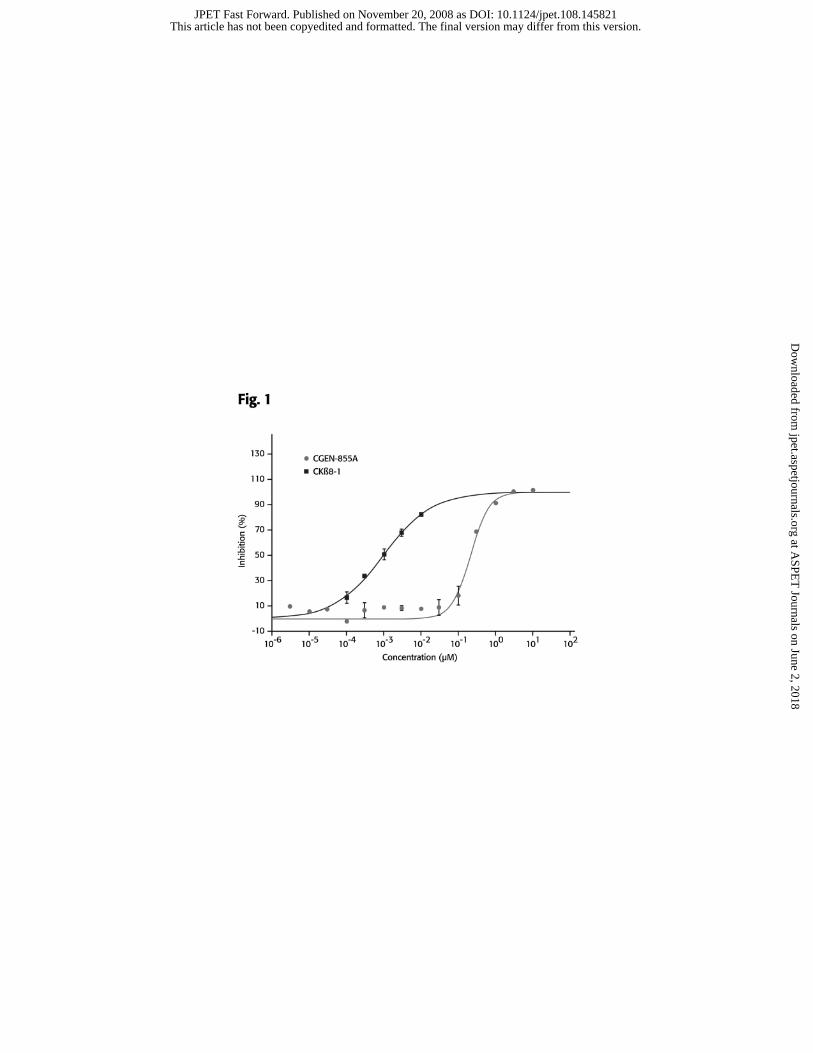
CGEN-855A competes with W peptide for binding to FPRL1
CGEN-855A was tested for its ability to compete with [125I] WKYMVm (W
peptide), a high-affinity ligand of FPRL1 (Christophe et al., 2001), on its binding to
membrane preparations from FPRL1 transiently transfected cells. CKβ8-1 (aa 46-137),
was used as a positive control (Elagoz et al., 2004). The value of 0% inhibition (i.e. 100%
binding of radioligand) was determined in the absence of either inhibitory peptide.
CGEN-855A displaced the radiolabeled-W peptide in a saturable manner with an IC50 of
189nM and a Ki of 54.1nM (Figure 1).
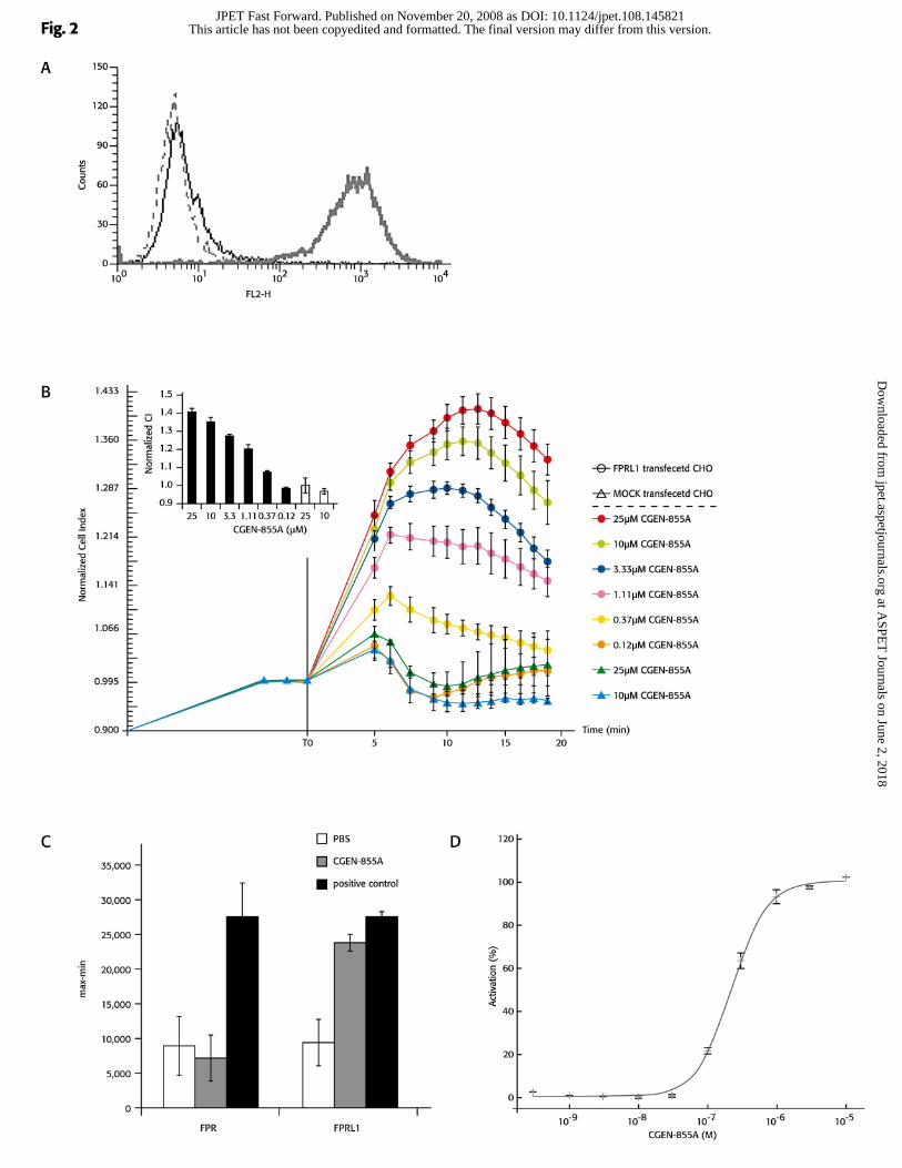
CGEN-855A activates FPRL1 and FPRL2 in a dose-dependent manner
A high and uniform expression of FPRL1 was detected in CHO cells that were
stably transfected with FPRL1 but not in mock transfected cells (Figure 2a). Activation of
these FPRL1-transfected cells with CGEN-855A resulted in an elevation of cell
impedance index in a dose-dependent manner, with an EC50 of 381nM (Figure 2b). This
activation was not observed after challenging mock transfected cells with CGEN-855A.
CGEN-855A elicited a cellular response in cells expressing either FPRL1 or
FPRL2 but not in FPR expressing cells (Figure 2c and 2d). fMLP and W peptide were
included as positive controls for FPRL1 and FPR, respectively. Furthermore, CGEN-
855A did not induce calcium flux in any of the other 149 GPCRs that were tested in the
original screen leading to its identification, although these receptors responded to relevant
positive controls (Shemesh et al, 2008).
This article has not been copyedited and formatted. The final version may differ from this version.JPET Fast Forward. Published on November 20, 2008 as DOI: 10.1124/jpet.108.145821
at ASPE
T Journals on June 2, 2018
jpet.aspetjournals.orgD
ownloaded from
JPET#145821
14
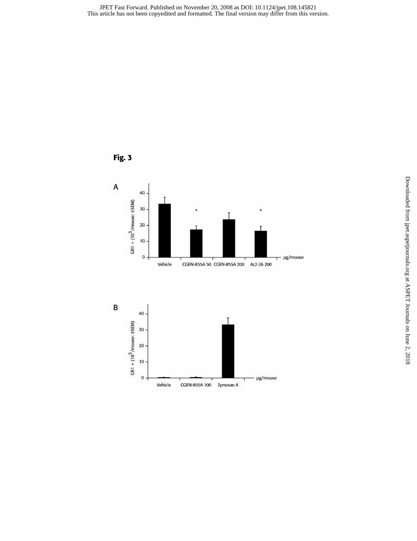
CGEN-855A exhibits anti-inflammatory activity in a model of acute inflammation
An intra-pouch challenge with zymosan A triggered a marked accumulation of
neutrophils in the air pouch, as determined by FACS analysis of Gr-1+ cells (Figure 3).
Administration of CGEN-855A at 50 and 200μg/mouse (corresponding to 2 and 8mg/kg),
reduced the accumulation of neutrophils triggered by zymosan A by 48.8% and 23.3%,
respectively (Figure 3a). Statistical significance was achieved only for the group treated
with 50μg/mouse but not with 200μg/mouse. Altogether, the extent of inhibition
achieved after treating the mice with 50μg/mouse CGEN-855A is comparable to that
obtained by administration of Ac2-26 at 200μg/mouse.
To validate that CGEN-855A does not elicit pro-inflammatory activity, we also
tested its direct effect upon administration into the air pouch in the absence of zymosan
A. As shown in Figure 3b, intra-pouch administration of 100μg CGEN-855A did not
induce neutrophil recruitment into the air pouch when used alone.
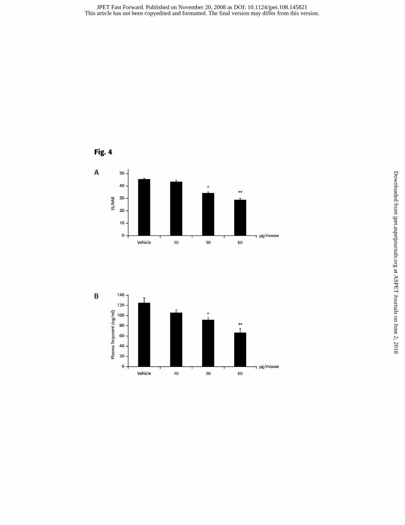
CGEN-855A displays cardioprotection in animal models of I/R-induced myocardial
infarction
The inhibitory activity on neutrophil migration demonstrated by CGEN-855A in
the air pouch model, prompted us to study its effect on I/R-induced myocardial injury.
When administered i.v. at 30 or 60 μg/mouse (corresponding to 1 or 2mg/kg,
respectively) immediately prior to reperfusion, CGEN-855A afforded significant and
dose-dependent cardioprotection, as illustrated by the reduction in infarct size (36%
reduction at the highest dose, Figure 4a). As expected, the AAR was similar in all groups,
with AAR/LV values ranging between 50-52% (data not shown). In addition, plasma
This article has not been copyedited and formatted. The final version may differ from this version.JPET Fast Forward. Published on November 20, 2008 as DOI: 10.1124/jpet.108.145821
at ASPE
T Journals on June 2, 2018
jpet.aspetjournals.orgD
ownloaded from
JPET#145821
15
levels of troponin I, an established marker of myocardial damage, were also reduced in a
dose-dependent manner (50% reduction at the highest dose, Figure 4b), with a pattern
mirroring that observed for reduction of infarct size.
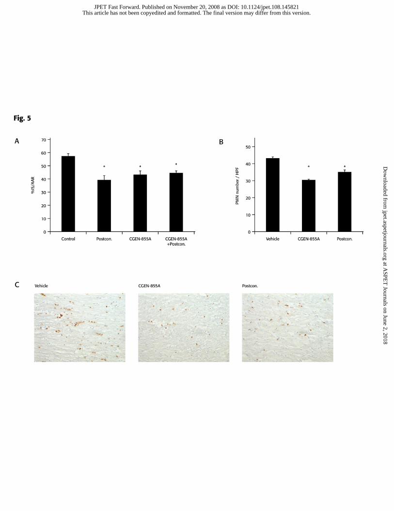
In addition, a rat model of I/R was used in order to compare the cardioprotective
effect of CGEN-855A to that of postconditioning- a mechanical maneuver defined as a
series of brief (i.e. seconds) interruptions of reperfusion following a specific prescribed
algorithm, applied at the very onset of reperfusion, that was shown to trigger
cardioprotective responses to reperfusion injury in animal models and in clinical studies
(Vinten-Johansen et al., 2007). Administration of CGEN-855A at 2 mg/kg reduced
infarct size to a similar extent as postconditioning (Figure 5a; 43.6±2.9 and 41.2±2.7%,
respectively, compared with 57.0±2.3% in the control group). Interestingly, the
combination of CGEN-855A with postconditioning did not further reduce infarct size
(44.6% ± 1.3%).
Finally, PMN accumulation in the AAR was analyzed to confirm that the
cardioprotective activity provided by CGEN-855A is due to inhibition of PMN
recruitment. CGEN-855A significantly attenuated PMN accumulation to the AAR
compared to vehicle (30.1 ± 0.6 vs. 43.2 ± 0.7 PMNs/HPF) (Figure 5 b, c). This
attenuation was comparable to that achieved by postconditioning (34.8±1.5).
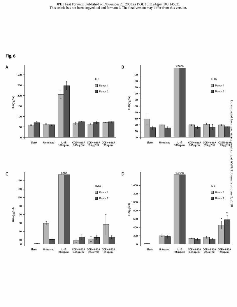
CGEN-855A does not affect cytokine secretion by human PBMCs
The human and murine families of FPRs are diverse, and might be differently
affected by certain compounds. This is of special importance due to the apparent
inconsistency in the effects mediated by FPRL1 agonists. Thus, we studied the effect of
This article has not been copyedited and formatted. The final version may differ from this version.JPET Fast Forward. Published on November 20, 2008 as DOI: 10.1124/jpet.108.145821
at ASPE
T Journals on June 2, 2018
jpet.aspetjournals.orgD
ownloaded from
JPET#145821
16
CGEN-855A on the secretion of inflammatory cytokines by human cells. Incubation of
PBMCs with CGEN-855A at 0.25, 2.5 or 25 µg/ml (corresponding to 0.1, 1 and 10 µM)
did not affect secretion of IL-6, IL-1β or TNFα (Figure 6 a-c). A moderate elevation in
IL-8 levels (up to 2 fold) was observed upon cells’ treatment with the highest dose
CGEN-855A (Figure 6d). IL-1β (100 ng/ml), which was used as positive control, induced
high levels of cytokine secretion.
This article has not been copyedited and formatted. The final version may differ from this version.JPET Fast Forward. Published on November 20, 2008 as DOI: 10.1124/jpet.108.145821
at ASPE
T Journals on June 2, 2018
jpet.aspetjournals.orgD
ownloaded from
JPET#145821
17
Discussion
FPRL1 is a promiscuous receptor, activated in vitro by a variety of ligands which
greatly vary in their biological features including origin, nature, size and specificity (Le
et al., 2002). The biological activities induced by interaction of an individual ligand with
FPRL1 are inconsistent- while some induce pro-inflammatory responses, others, namely
annexin 1 and Ac2-26 as well as LXA4, promote resolution of inflammation; the latter
findings have underpinned current clinical programs aiming at discovering novel FPRL1
agonists for treatment of acute and chronic inflammatory conditions. We demonstrated
here that CGEN-855A activates FPRL1 and display important anti-inflammatory
properties by reducing PMNs recruitment to inflamed sites. CGEN-855A produced a
similar efficacy to that of Ac2-26 in the air pouch model but a lower dose was required to
achieve similar degrees of inhibition (50 vs. 200 µg/mouse corresponding to 20 and 80
nmol, respectively). The smaller reduction in PMN accumulation obtained with
administration of higher doses of CGEN-855A might result from receptor desensitization,
and might indicate that even lower doses would be sufficient to exert important
checkpoint functions on the experimental inflammatory response. Nevertheless, these
results implied on the effective therapeutic range of CGEN-855A, and when administered
at lower doses in the disease-related model, the I/R-induced myocardial infarction (Fig. 4;
10, 30 and 60 µg/mouse), a clear dose-dependent effect was observed both in infarct size
and troponin levels in the plasma.
Due to the perplexing effect mediated via FPRL1, the possibility that CGEN-
885A might elicit pro-inflammatory responses was eliminated as PMNs were not
recruited to the air pouch upon intra-pouch administration of CGEN-855A alone.
This article has not been copyedited and formatted. The final version may differ from this version.JPET Fast Forward. Published on November 20, 2008 as DOI: 10.1124/jpet.108.145821
at ASPE
T Journals on June 2, 2018
jpet.aspetjournals.orgD
ownloaded from
JPET#145821
18
Furthermore, although a moderate elevation in IL-8 secretion was observed upon treating
human cells with the highest tested concentration of CGEN-855A, it did not induce any
prominent elevation in secretion of the other pro-inflammatory cytokines tested,
supporting a lack of pro-inflammatory, or activating downstream effects, upon human
FPRL1 agonism by this compound. In addition, the biological implications of FPRL2
activation by CGEN-855A are difficult to foresee since the biological role of FPRL2 is
unknown and an endogenous agonist for FPRL2 was only recently reported (Gao et al.,
2007).
Although controversial, previous findings substantiate a role for PMNs and
specifically neutrophils in the early stage of reperfusion-injury (reviewed by Vinten-
Johansen, 2004) and several studies describe experimental interventions aimed at
inhibiting PMN recruitment at the time of reperfusion or shortly before. PMN inhibition
was achieved by leukodepletion using neutrophil anti-serum (Kin et al., 2006), or by
antibodies targeting adhesion molecules such as P- and E- selectins (Lefer et al., 1994),
CD11/CD18 (Ma et al., 1991), ICAM-1 (Ma et al., 1992; Ioculano et al., 1994; Zhao et
al., 1997; Zhao et al., 2003) or PECAM-1 (Gumina et al., 1996). These strategies
provided up to ~50% reduction in infarct size. However, none of these anti-PMN
approaches have been shown to consistently be effective in the clinic (Vinten-Johansen,
2004; Frangogiannis, 2006; Yellon and Hausenloy, 2007). On the other hand,
postconditioning has shown significant protection when applied to patients (Tissier et al.,
2007; Thibault et al., 2008).
When tested in mouse and rat models of I/R, CGEN-855A afforded a significant
cardioprotective effect manifested by reduced infarct size (by 36% and 25% in the mouse
This article has not been copyedited and formatted. The final version may differ from this version.JPET Fast Forward. Published on November 20, 2008 as DOI: 10.1124/jpet.108.145821
at ASPE
T Journals on June 2, 2018
jpet.aspetjournals.orgD
ownloaded from
JPET#145821
19
and rat model, respectively), that was further confirmed by reduced levels of troponin I in
plasma (shown in mice). In accordance with the inhibitory effect of CGEN-855A on
PMN recruitment to the inflamed site attained by the air pouch model, the
cardioprotective activity of CGEN-855A in rats was accompanied by reduced recruitment
of PMN to the AAR. The extent of cardioprotection obtained by CGEN-855A treatment
is comparable to that observed by other groups using FPRL1 agonists in similar models
(Gavins et al., 2005) and comparable to that observed with postconditioning. Of note, no
additive protection was elicited upon combination of the two treatments. It is possible
that both FPRL1 and postconditioning exert their effect through similar pathways, i.e.
PMN-mediated injury. Indeed, studies report that postconditioning is associated with a
reduction in PMN accumulation, adherence to coronary vascular endothelium,
endothelial dysfunction, and cytokines relevant to the PMN recruitment process (Zhao et
al., 2003; Halkos et al., 2004). Overall, these data might suggest that the apparently
partial cardioprotective effect observed in these models of I/R is the maximal effect that
can be achieved via inhibition of PMNs recruitment and that processes mediated by other
cells, such as T cells (Varda-Bloom et al., 2000; Spagnoli et al., 2002; Lim et al., 2003)
and endothelial cells (Melo et al., 2004), also play a role in the myocardial damage
resulting from I/R.
Collectively, these data provide strong evidence that activation of the FPRL1
pathway could be beneficial for the treatment of acute and chronic inflammation. The
results presented herein support further development of CGEN-855A as a potential
candidate for therapeutic control of inflammatory diseases, in general, and for the
treatment of reperfusion-related cardiovascular damage, in particular.
This article has not been copyedited and formatted. The final version may differ from this version.JPET Fast Forward. Published on November 20, 2008 as DOI: 10.1124/jpet.108.145821
at ASPE
T Journals on June 2, 2018
jpet.aspetjournals.orgD
ownloaded from
JPET#145821
20
References
Bandeira-Melo C, Bonavita AG, Diaz BL, PM ES, Carvalho VF, Jose PJ, Flower RJ,
Perretti M and Martins MA (2005) A novel effect for annexin 1-derived peptide
ac2-26: reduction of allergic inflammation in the rat. J Pharmacol Exp Ther
313:1416-1422.
Bandeira-Melo C, Bozza PT, Diaz BL, Cordeiro RS, Jose PJ, Martins MA and Serhan
CN (2000) Cutting edge: lipoxin (LX) A4 and aspirin-triggered 15-epi-LXA4
block allergen-induced eosinophil trafficking. J Immunol 164:2267-2271.
Bannenberg G, Moussignac RL, Gronert K, Devchand PR, Schmidt BA, Guilford WJ,
Bauman JG, Subramanyam B, Perez HD, Parkinson JF and Serhan CN (2004)
Lipoxins and novel 15-epi-lipoxin analogs display potent anti-inflammatory
actions after oral administration. Br J Pharmacol 143:43-52.
Chiang N, Serhan CN, Dahlen SE, Drazen JM, Hay DW, Rovati GE, Shimizu T,
Yokomizo T and Brink C (2006) The lipoxin receptor ALX: potent ligand-
specific and stereoselective actions in vivo. Pharmacol Rev 58:463-487.
Christophe T, Karlsson A, Dugave C, Rabiet MJ, Boulay F and Dahlgren C (2001) The
synthetic peptide Trp-Lys-Tyr-Met-Val-Met-NH2 specifically activates
neutrophils through FPRL1/lipoxin A4 receptors and is an agonist for the orphan
monocyte-expressed chemoattractant receptor FPRL2. J Biol Chem 276:21585-
21593.
Cuzzocrea S, De Sarro G, Costantino G, Ciliberto G, Mazzon E, De Sarro A and Caputi
AP (1999) IL-6 knock-out mice exhibit resistance to splanchnic artery occlusion
shock. J Leukoc Biol 66:471-480.
This article has not been copyedited and formatted. The final version may differ from this version.JPET Fast Forward. Published on November 20, 2008 as DOI: 10.1124/jpet.108.145821
at ASPE
T Journals on June 2, 2018
jpet.aspetjournals.orgD
ownloaded from
JPET#145821
21
Elagoz A, Henderson D, Babu PS, Salter S, Grahames C, Bowers L, Roy MO, Laplante
P, Grazzini E, Ahmad S and Lembo PM (2004) A truncated form of CKbeta8-1 is
a potent agonist for human formyl peptide-receptor-like 1 receptor. Br J
Pharmacol 141:37-46.
Fiorucci S, de Lima OM, Jr., Mencarelli A, Palazzetti B, Distrutti E, McKnight W, Dicay
M, Ma L, Romano M, Morelli A and Wallace JL (2002) Cyclooxygenase-2-
derived lipoxin A4 increases gastric resistance to aspirin-induced damage.
Gastroenterology 123:1598-1606.
Fiorucci S, Wallace JL, Mencarelli A, Distrutti E, Rizzo G, Farneti S, Morelli A, Tseng
JL, Suramanyam B, Guilford WJ and Parkinson JF (2004) A beta-oxidation-
resistant lipoxin A4 analog treats hapten-induced colitis by attenuating
inflammation and immune dysfunction. Proc Natl Acad Sci U S A 101:15736-
15741.
Frangogiannis NG (2006) Targeting the inflammatory response in healing myocardial
infarcts. Curr Med Chem 13:1877-1893.
Gao JL, Guillabert A, Hu J, Le Y, Urizar E, Seligman E, Fang KJ, Yuan X, Imbault V,
Communi D, Wang JM, Parmentier M, Murphy PM and Migeotte I (2007) F2L, a
peptide derived from heme-binding protein, chemoattracts mouse neutrophils by
specifically activating Fpr2, the low-affinity N-formylpeptide receptor. J Immunol
178:1450-1456.
Gavins FN, Kamal AM, D'Amico M, Oliani SM and Perretti M (2005) Formyl-peptide
receptor is not involved in the protection afforded by annexin 1 in murine acute
myocardial infarct. Faseb J 19:100-102.
This article has not been copyedited and formatted. The final version may differ from this version.JPET Fast Forward. Published on November 20, 2008 as DOI: 10.1124/jpet.108.145821
at ASPE
T Journals on June 2, 2018
jpet.aspetjournals.orgD
ownloaded from
JPET#145821
22
Gavins FN, Yona S, Kamal AM, Flower RJ and Perretti M (2003) Leukocyte
antiadhesive actions of annexin 1: ALXR- and FPR-related anti-inflammatory
mechanisms. Blood 101:4140-4147.
Gewirtz AT, Collier-Hyams LS, Young AN, Kucharzik T, Guilford WJ, Parkinson JF,
Williams IR, Neish AS and Madara JL (2002) Lipoxin a4 analogs attenuate
induction of intestinal epithelial proinflammatory gene expression and reduce the
severity of dextran sodium sulfate-induced colitis. J Immunol 168:5260-5267.
Gumina RJ, el Schultz J, Yao Z, Kenny D, Warltier DC, Newman PJ and Gross GJ
(1996) Antibody to platelet/endothelial cell adhesion molecule-1 reduces
myocardial infarct size in a rat model of ischemia-reperfusion injury. Circulation
94:3327-3333.
Halkos ME, Kerendi F, Corvera JS, Wang NP, Kin H, Payne CS, Sun HY, Guyton RA,
Vinten-Johansen J and Zhao ZQ (2004) Myocardial protection with
postconditioning is not enhanced by ischemic preconditioning. Ann Thorac Surg
78:961-969; discussion 969.
Ioculano M, Squadrito F, Altavilla D, Canale P, Squadrito G, Campo GM, Saitta A and
Caputi AP (1994) Antibodies against intercellular adhesion molecule 1 protect
against myocardial ischaemia-reperfusion injury in rat. Eur J Pharmacol 264:143-
149.
Kin H, Wang NP, Halkos ME, Kerendi F, Guyton RA and Zhao ZQ (2006) Neutrophil
depletion reduces myocardial apoptosis and attenuates NFkappaB
activation/TNFalpha release after ischemia and reperfusion. J Surg Res 135:170-
178.
This article has not been copyedited and formatted. The final version may differ from this version.JPET Fast Forward. Published on November 20, 2008 as DOI: 10.1124/jpet.108.145821
at ASPE
T Journals on June 2, 2018
jpet.aspetjournals.orgD
ownloaded from
JPET#145821
23
Kin H, Zatta AJ, Lofye MT, Amerson BS, Halkos ME, Kerendi F, Zhao ZQ, Guyton RA,
Headrick JP and Vinten-Johansen J (2005) Postconditioning reduces infarct size
via adenosine receptor activation by endogenous adenosine. Cardiovasc Res
67:124-133.
La M, Tailor A, D'Amico M, Flower RJ and Perretti M (2001) Analysis of the protection
afforded by annexin 1 in ischaemia-reperfusion injury: focus on neutrophil
recruitment. Eur J Pharmacol 429:263-278.
Le Y, Murphy PM and Wang JM (2002) Formyl-peptide receptors revisited. Trends
Immunol 23:541-548.
Lefer DJ, Flynn DM, Phillips ML, Ratcliffe M and Buda AJ (1994) A novel sialyl
LewisX analog attenuates neutrophil accumulation and myocardial necrosis after
ischemia and reperfusion. Circulation 90:2390-2401.
Leonard MO, Hannan K, Burne MJ, Lappin DW, Doran P, Coleman P, Stenson C, Taylor
CT, Daniels F, Godson C, Petasis NA, Rabb H and Brady HR (2002) 15-Epi-16-
(para-fluorophenoxy)-lipoxin A(4)-methyl ester, a synthetic analogue of 15-epi-
lipoxin A(4), is protective in experimental ischemic acute renal failure. J Am Soc
Nephrol 13:1657-1662.
Lim YC, Garcia-Cardena G, Allport JR, Zervoglos M, Connolly AJ, Gimbrone MA, Jr.
and Luscinskas FW (2003) Heterogeneity of endothelial cells from different organ
sites in T-cell subset recruitment. Am J Pathol 162:1591-1601.
Ma XL, Lefer DJ, Lefer AM and Rothlein R (1992) Coronary endothelial and cardiac
protective effects of a monoclonal antibody to intercellular adhesion molecule-1
in myocardial ischemia and reperfusion. Circulation 86:937-946.
This article has not been copyedited and formatted. The final version may differ from this version.JPET Fast Forward. Published on November 20, 2008 as DOI: 10.1124/jpet.108.145821
at ASPE
T Journals on June 2, 2018
jpet.aspetjournals.orgD
ownloaded from
JPET#145821
24
Ma XL, Tsao PS and Lefer AM (1991) Antibody to CD-18 exerts endothelial and cardiac
protective effects in myocardial ischemia and reperfusion. J Clin Invest 88:1237-
1243.
Melo LG, Gnecchi M, Pachori AS, Kong D, Wang K, Liu X, Pratt RE and Dzau VJ
(2004) Endothelium-targeted gene and cell-based therapies for cardiovascular
disease. Arterioscler Thromb Vasc Biol 24:1761-1774.
Perretti M, Ahluwalia A, Harris JG, Goulding NJ and Flower RJ (1993) Lipocortin-1
fragments inhibit neutrophil accumulation and neutrophil-dependent edema in the
mouse. A qualitative comparison with an anti-CD11b monoclonal antibody. J
Immunol 151:4306-4314.
Perretti M, Chiang N, La M, Fierro IM, Marullo S, Getting SJ, Solito E and Serhan CN
(2002) Endogenous lipid- and peptide-derived anti-inflammatory pathways
generated with glucocorticoid and aspirin treatment activate the lipoxin A4
receptor. Nat Med 8:1296-1302.
Shemesh R, Toporik A, Levine Z, Hecht I, Rotman G, Wool A, Dahary D, Gofer E,
Kliger Y, Ayalon Soffer M, Rosenberg A, Eshel D and Cohen Y (2008)
Discovery and validation of novel peptide agonists for G-protein coupled
receptors. J Biol Chem.
Solly K, Wang X, Xu X, Strulovici B and Zheng W (2004) Application of real-time cell
electronic sensing (RT-CES) technology to cell-based assays. Assay Drug Dev
Technol 2:363-372.
This article has not been copyedited and formatted. The final version may differ from this version.JPET Fast Forward. Published on November 20, 2008 as DOI: 10.1124/jpet.108.145821
at ASPE
T Journals on June 2, 2018
jpet.aspetjournals.orgD
ownloaded from
JPET#145821
25
Spagnoli LG, Bonanno E, Mauriello A, Palmieri G, Partenzi A, Sangiorgi G and Crea F
(2002) Multicentric inflammation in epicardial coronary arteries of patients dying
of acute myocardial infarction. J Am Coll Cardiol 40:1579-1588.
Thibault H, Piot C, Staat P, Bontemps L, Sportouch C, Rioufol G, Cung TT, Bonnefoy E,
Angoulvant D, Aupetit JF, Finet G, Andre-Fouet X, Macia JC, Raczka F, Rossi R,
Itti R, Kirkorian G, Derumeaux G and Ovize M (2008) Long-term benefit of
postconditioning. Circulation 117:1037-1044.
Tissier R, Cohen MV and Downey JM (2007) Protecting the acutely ischemic
myocardium beyond reperfusion therapies: are we any closer to realizing the
dream of infarct size elimination? Arch Mal Coeur Vaiss 100:794-802.
Varda-Bloom N, Leor J, Ohad DG, Hasin Y, Amar M, Fixler R, Battler A, Eldar M and
Hasin D (2000) Cytotoxic T lymphocytes are activated following myocardial
infarction and can recognize and kill healthy myocytes in vitro. J Mol Cell
Cardiol 32:2141-2149.
Vinten-Johansen J (2004) Involvement of neutrophils in the pathogenesis of lethal
myocardial reperfusion injury. Cardiovasc Res 61:481-497.
Vinten-Johansen J, Zhao ZQ, Jiang R, Zatta AJ and Dobson GP (2007) Preconditioning
and postconditioning: innate cardioprotection from ischemia-reperfusion injury. J
Appl Physiol 103:1441-1448.
Yellon DM and Hausenloy DJ (2007) Myocardial reperfusion injury. N Engl J Med
357:1121-1135.
Zhao ZQ, Corvera JS, Halkos ME, Kerendi F, Wang NP, Guyton RA and Vinten-
Johansen J (2003) Inhibition of myocardial injury by ischemic postconditioning
This article has not been copyedited and formatted. The final version may differ from this version.JPET Fast Forward. Published on November 20, 2008 as DOI: 10.1124/jpet.108.145821
at ASPE
T Journals on June 2, 2018
jpet.aspetjournals.orgD
ownloaded from
JPET#145821
26
during reperfusion: comparison with ischemic preconditioning. Am J Physiol
Heart Circ Physiol 285:H579-588.
Zhao ZQ, Lefer DJ, Sato H, Hart KK, Jefforda PR and Vinten-Johansen J (1997)
Monoclonal antibody to ICAM-1 preserves postischemic blood flow and reduces
infarct size after ischemia-reperfusion in rabbit. J Leukoc Biol 62:292-300.
This article has not been copyedited and formatted. The final version may differ from this version.JPET Fast Forward. Published on November 20, 2008 as DOI: 10.1124/jpet.108.145821
at ASPE
T Journals on June 2, 2018
jpet.aspetjournals.orgD
ownloaded from
JPET#145821
27
Footnotes
Reprints requests should be addressed to:
Dr. Iris Hecht; Compugen Ltd.
72 Pinchas Rosen St.
Tel Aviv, Israel 69512.
Phone: +972-3-765-8564;
Fax: +972-3-765-8555;
E-mail: [email protected].
This article has not been copyedited and formatted. The final version may differ from this version.JPET Fast Forward. Published on November 20, 2008 as DOI: 10.1124/jpet.108.145821
at ASPE
T Journals on June 2, 2018
jpet.aspetjournals.orgD
ownloaded from
JPET#145821
28
Legends for Figures
Figure 1. CGEN-855A binds to FPRL1. Membranes from PFRL1 transfected
CHO cells were incubated with [125I] WKYMVm in the absence or presence of
increasing concentrations of either CGEN-855A (�) or CKβ8-1 (�). Results are
presented as mean ± SD of duplicates.
Figure 2. CGEN-855A specifically activates FPRL1 in a dose dependent
manner. A, CHO stably transfected with either FPRL1 (thick line) or mock vector (thin
line) were stained with PE- conjugated anti-human FPRL1 Ab or with IgG2b isotype
control Ab (dashed line) and surface expression of FPRL1 was analyzed by FACScan
(Becton Dickinson). B, Stable pools of FPRL1 were seeded on E-plates and stimulated
with CGEN-855A at 25, 10, 3.3, 1.1, 0.37, 0.12 µM. Mock transfected cells were
stimulated with 25 and 10 µM CGEN-855A. Cell impedance was recorded continuously
in intervals of 71 s and presented as normalized CI. Insert presents normalized CI of
FPRL1 (black bars) and mock (white bars) trasfected cells as mean ± SD of triplicates at
one time point (12.5 min). C, CHO-K1 cells transiently transfected with either FPRL1 or
FPR1 and Gα16 were loaded with Fluo4-NW. Calcium flux response was measured
using FlexStationTM (Molecular Devices), upon cells stimulation with CGEN-855A at
1μM. W peptide and fMLP (1μM each) were included as positive controls for FPR and
FPRL1, respectively. Assay was conducted in triplicates, mean ± SD is presented. D,
CHO cells stably expressing FPRL2, Gα16 and mitochondrial apoaequorin were
incubated with Coelenterazine H and activated with CGEN-855A at 0.3 1 3 10 30 100
This article has not been copyedited and formatted. The final version may differ from this version.JPET Fast Forward. Published on November 20, 2008 as DOI: 10.1124/jpet.108.145821
at ASPE
T Journals on June 2, 2018
jpet.aspetjournals.orgD
ownloaded from
JPET#145821
29
300 1000 3000 10000 nM. Results are expressed as percentage of activation compared to
the reference agonist.
Figure 3. CGEN-855A inhibits PMNs migration into mouse air-pouch
inflamed with Zymosan A. A, Zymosan A (1mg) was injected intra-pouch immediately
following i.v. treatment with either CGEN-855A, Ac2-26 or vehicle as indicated. Lavage
fluid was collected after 4 hr, stained with anti-Gr-1 antibody and analyzed by FACScan
(Becton Dickinson). Irrelevant rat IgG2b antibody was used as isotype control. Shown is
the number of Gr-1+ cells recovered in the lavage fluids (mean ± SEM of n=8). *P<0.05
vs. vehicle group. B, CGEN-855A (0.1mg), Zymosan A (1mg) or vehicle were injected
intra-pouch. Lavage fluids were collected and analyzed as described in panel A.
Figure 4. CGEN-855A reduces I/R- mediated myocardial injury in mice.
Mice were subjected to 25 min ischaemia followed by 120 min reperfusion, by LCA
occlusion. Vehicle (PBS) or CGEN-855A were administered at indicated doses
immediately after reperfusion. A, Myocardial infarct was determined as described under
materials and methods and expressed as percentage of AAR. Data presented as mean ±
SEM of n = 6. *P < 0.05 and ** P<0.01 vs. vehicle group. B, Plasma samples were tested
for troponin I using ELISA. Values were extrapolated from a calibration curve and
presented as mean ± SD of duplicates. *P<0.05 and ** P<0.01 vs. vehicle group.
Figure 5. CGEN-855A reduces I/R- mediated myocardial injury and PMN’s
recruitment in rats. Rats were subjected to 30 min ischaemia followed by 180 min
This article has not been copyedited and formatted. The final version may differ from this version.JPET Fast Forward. Published on November 20, 2008 as DOI: 10.1124/jpet.108.145821
at ASPE
T Journals on June 2, 2018
jpet.aspetjournals.orgD
ownloaded from
JPET#145821
30
reperfusion, by LADCA occlusion. Vehicle (Saline) or CGEN-855A were administered 5
minutes before reperfusion while postconditioning was applied immediately prior to
terminal reperfusion. A, Myocardial infarct was determined as described under materials
and methods and expressed as percentage of AAR. Data presented as mean ± SEM of n =
9. *P < 0.05 vs. vehicle group. B, PMN’S accumulation in the AAR tissue presented as
mean ± SEM of n=5 *P<0.05 vs. vehicle group. C, Representative sections of AAR
stained for PMN’S accumulation by immunohistochemistry using anti-CD11 and anti-
CD18 antibodies is presented for each study group as indicated. Magnification is x200.
Figure 6. CGEN-855A does not affect cytokine secretion by PBMCs. PBMCs
were incubated for 24hr with CGEN-855A at 0.25, 2.5 and 25µg/ml (corresponding to
0.1, 1 and 10µM). The levels of IL -6 (A), IL-1β (B), TNF-α (C), and IL-8 (D) in the
supernatants were evaluated by ELISA. Presented are means ± SEM of duplicate ELISA
from duplicate assay samples of two donors. *P<0.05 and ** P<0.01 vs. untreated cells.
This article has not been copyedited and formatted. The final version may differ from this version.JPET Fast Forward. Published on November 20, 2008 as DOI: 10.1124/jpet.108.145821
at ASPE
T Journals on June 2, 2018
jpet.aspetjournals.orgD
ownloaded from
This article has not been copyedited and formatted. The final version may differ from this version.JPET Fast Forward. Published on November 20, 2008 as DOI: 10.1124/jpet.108.145821
at ASPE
T Journals on June 2, 2018
jpet.aspetjournals.orgD
ownloaded from
This article has not been copyedited and formatted. The final version may differ from this version.JPET Fast Forward. Published on November 20, 2008 as DOI: 10.1124/jpet.108.145821
at ASPE
T Journals on June 2, 2018
jpet.aspetjournals.orgD
ownloaded from
This article has not been copyedited and formatted. The final version may differ from this version.JPET Fast Forward. Published on November 20, 2008 as DOI: 10.1124/jpet.108.145821
at ASPE
T Journals on June 2, 2018
jpet.aspetjournals.orgD
ownloaded from
This article has not been copyedited and formatted. The final version may differ from this version.JPET Fast Forward. Published on November 20, 2008 as DOI: 10.1124/jpet.108.145821
at ASPE
T Journals on June 2, 2018
jpet.aspetjournals.orgD
ownloaded from
This article has not been copyedited and formatted. The final version may differ from this version.JPET Fast Forward. Published on November 20, 2008 as DOI: 10.1124/jpet.108.145821
at ASPE
T Journals on June 2, 2018
jpet.aspetjournals.orgD
ownloaded from
This article has not been copyedited and formatted. The final version may differ from this version.JPET Fast Forward. Published on November 20, 2008 as DOI: 10.1124/jpet.108.145821
at ASPE
T Journals on June 2, 2018
jpet.aspetjournals.orgD
ownloaded from


![INDEX [jpet.aspetjournals.org]jpet.aspetjournals.org/content/jpet/230/3/local/back-matter.pdf · histrionicotoxin effects (frogs), 619 ... distribution kinetics ana- ... and myocardium](https://cdn.vdocument.in/doc/165x107/5b7ac0067f8b9ae1328d73ab/index-jpet-jpet-histrionicotoxin-effects-frogs-619-distribution-kinetics.jpg)