
TITLEVascular Tutorial (SET XXX)
INSERT DATE
Principles of UltrasoundVascular Tutorial (SET 1 and 2)
20 July 2020

Clinical Scenario 2• You are doing your ultrasound lab morning and scan a patient with
increasing claudication symptoms.
• The patient has had a right SFA angioplasty in the past.
• You obtain the following images...

Imaging

Learning Objectives 2• Interpret the ultrasound findings and describe the salient features
that you will include in the report. • Describe the concept of beam steering and why that is important in
this instance including methods to alter the beam (including arrays and electronic focusing).
• Discuss factors that influence image quality and accuracy with reference to principle image artefacts.
Ultrasound report preamble• An arterial duplex ultrasound was performed of the left lower limb in
a patient with increasing left leg claudication symptoms and a history of right SFA angioplasty.

SFA proximal
SFA mid

Lower extremity spectral waveforms
Normal
1-19% diameter reduction
20-49% diameter reduction
50-99% diameter reduction

Waveform features
University of Washington Duplex Criteria for Classification of Lower Extremity Arterial Stenosis

PSV Ratio PSV at stenotic segment / normal segment
Degree of stenosis
<150 cm/s <1.5 Normal
150-200 cm/s 1.5-2.0 30-49%
200-400 cm/s 2.0-4.0 50-75%
>400 cm/s >4.0 >75%
No colour flow - Occlusion
Grading via velocity shift
Cossman DV, Ellison JE, Wagner WH, et al. Comparison of contrast arteriography to arterial mapping with color-flow duplex imaging in the lower extremities. J Vasc Surg. 1989;10(5):522-529
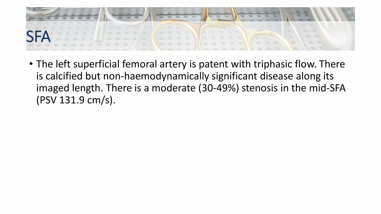
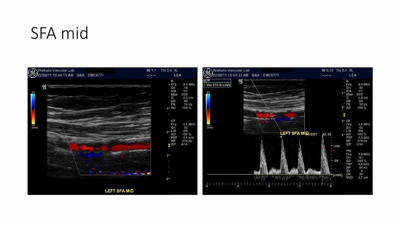
SFA• The left superficial femoral artery is patent with triphasic flow. There
is calcified but non-haemodynamically significant disease along its imaged length. There is a moderate (30-49%) stenosis in the mid-SFA (PSV 131.9 cm/s).


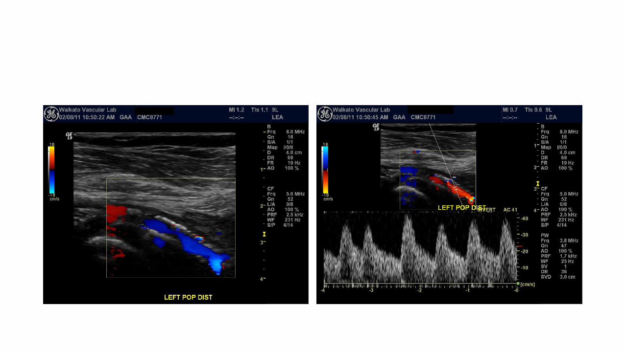

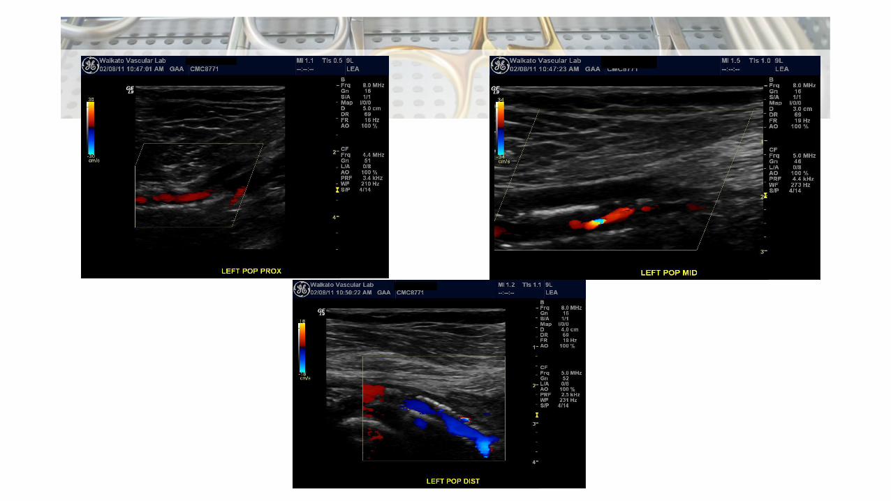
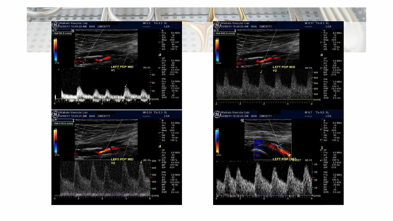
Popliteal• The popliteal artery appears patent with significant calcific disease.• In the proximal popliteal artery, there is dampened monophasic flow
with a delayed systolic upstroke suggestive of a high-grade proximal popliteal stenosis.
• The mid-popliteal artery is patent with a severe (>75%) stenosis (PSV 495 .5 cm/s).
• The distal popliteal artery is patent with dampened monophasic flow and reduced systolic velocity.

Linear array transducer
Courtesy of Dr Rachael Nightingale https://radiopaedia.org/cases/54644
From FW Kremkau. 2 - Principles and Instruments of Ultrasonography, Introduction to Vascular Ultrasonography (Seventh Edition), edited by JS Pellerito & JF Polak.
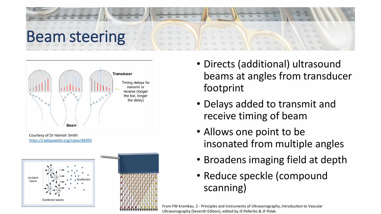
Beam steering• Directs (additional) ultrasound
beams at angles from transducer footprint
• Delays added to transmit and receive timing of beam
• Allows one point to be insonated from multiple angles
• Broadens imaging field at depth• Reduce speckle (compound
scanning)
Courtesy of Dr Hamish Smithhttps://radiopaedia.org/cases/68392
From FW Kremkau. 2 - Principles and Instruments of Ultrasonography, Introduction to Vascular Ultrasonography (Seventh Edition), edited by JS Pellerito & JF Polak.

Beam focusing• Similar principle• Time delays to “transmit focus”
• Fire outer elements first• Central element last
• Composite pulse converges at a focal point at depth
Courtesy of Dr Hamish Smithhttps://radiopaedia.org/cases/68386

Beam steering in Doppler
From FW Kremkau. 2 - Principles and Instruments of Ultrasonography, Introduction to Vascular Ultrasonography (Seventh Edition), edited by JS Pellerito & JF Polak.
• Use beam steering to indicate the Doppler angle

Optimising colour flow
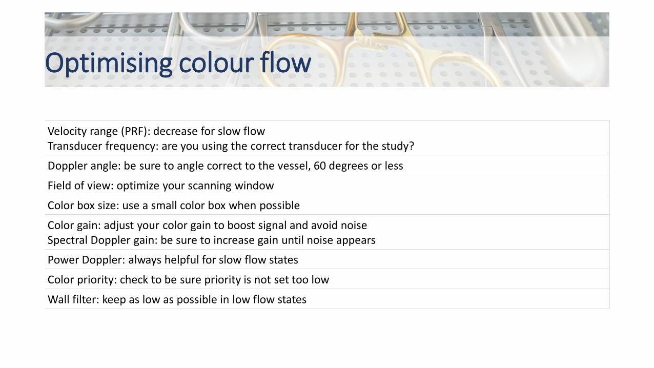
Velocity range (PRF): decrease for slow flowTransducer frequency: are you using the correct transducer for the study?
Doppler angle: be sure to angle correct to the vessel, 60 degrees or less
Field of view: optimize your scanning window
Color box size: use a small color box when possible
Color gain: adjust your color gain to boost signal and avoid noiseSpectral Doppler gain: be sure to increase gain until noise appears
Power Doppler: always helpful for slow flow states
Color priority: check to be sure priority is not set too low
Wall filter: keep as low as possible in low flow states
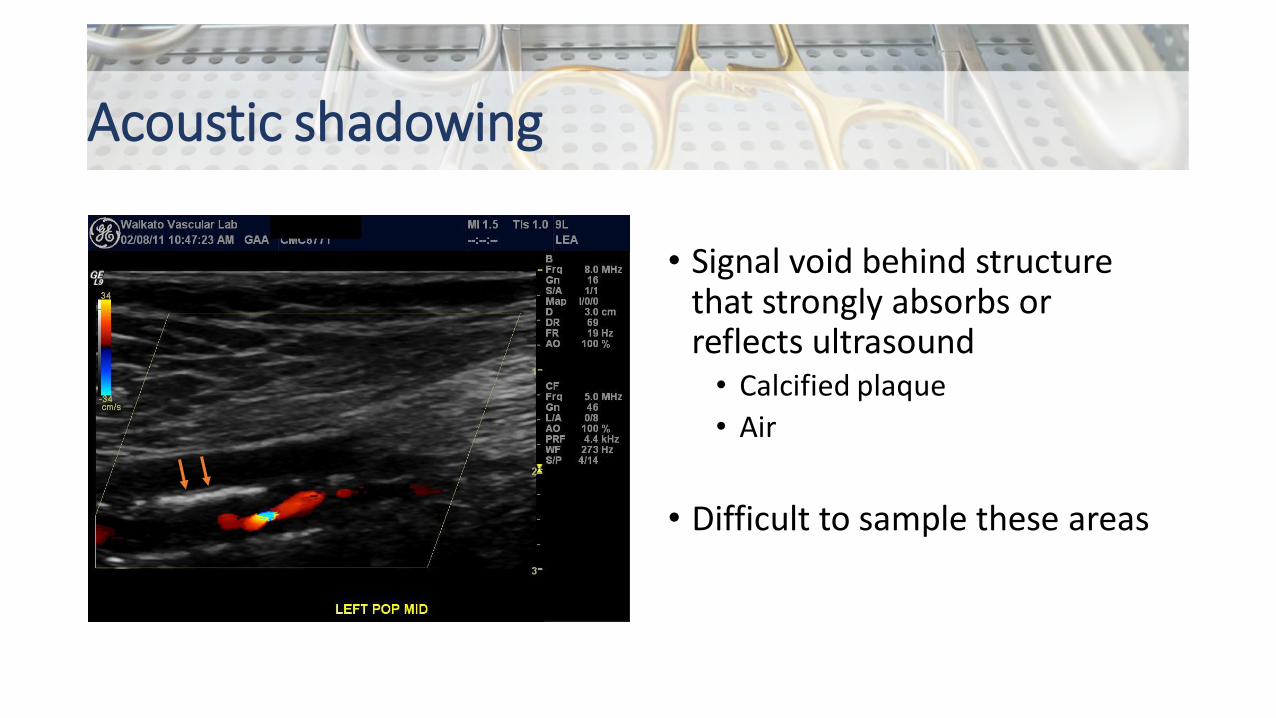
Acoustic shadowing
• Signal void behind structure that strongly absorbs or reflects ultrasound
• Calcified plaque• Air
• Difficult to sample these areas

Aliasing in pulsed Doppler
• Aliasing caused by intermittent Doppler sampling
• Cannot record direction and velocity accurately
• Nyquist limit: PRF needs to be x2 Doppler frequency
• Below this = aliasing• Incorrect spectral display
From FW Kremkau. 2 - Principles and Instruments of Ultrasonography, Introduction to Vascular Ultrasonography (Seventh Edition), edited by JS Pellerito & JF Polak.

Aliasing in pulsed Doppler
From FW Kremkau. 2 - Principles and Instruments of Ultrasonography, Introduction to Vascular Ultrasonography (Seventh Edition), edited by JS Pellerito & JF Polak.

Aliasing in colour Doppler
From FW Kremkau. 2 - Principles and Instruments of Ultrasonography, Introduction to Vascular Ultrasonography (Seventh Edition), edited by JS Pellerito & JF Polak.

Sample volume
• 3D volume• “Thickness” not displayed• Only get information from
positioned sample volume• Can sample flow from adjacent
branch!
From JS Pellerito & JF Polak, 3 - Doppler Flow Imaging and Spectral Analysis, Introduction to Vascular Ultrasonography (Seventh Edition), edited by JS Pellerito & JF Polak.

References• Introduction to Vascular Ultrasonography (Seventh Edition), edited by
JS Pellerito & JF Polak.• Radiopaedia• Cossman DV, Ellison JE, Wagner WH, et al. Comparison of contrast
arteriography to arterial mapping with color-flow duplex imaging in the lower extremities. J Vasc Surg. 1989;10(5):522-529


