
Program and Abstract Book
„Transglutaminases in Medicine”Debrecen University Symposium
2017
„Tra
nsgl
utam
inas
es i
n M
edic
ine”
Deb
rece
n U
nive
rsit
y S
ympo
sium
201
7

Cover illustration: Overlay of the structural models of the 'open' conformations of TG2 (blue) and FXIII-A (orange).
PDB: 2Q3Z and 4KTY. Design by Máté Demény
ISBN 978-963-473-965-4.
Map - walking directions from hotels.
DEBRECEN UNIVERSITY SYMPOSIUM
TRANSGLUTAMINASES IN MEDICINE
2017 August 3-5
Venue:
Hotel Divinus (August 3)
University of Debrecen Life Science Building (August 4-5)

2 Transglutaminases in Medicine, August 3-5 2017
Scientific Advisory Board:
László Fésüs (chair), László Muszbek, Ilma Korponay-Szabó, Zsuzsa Szondy
Local organizers
Nóra Elek, Róbert Király, Universitas GmbH (Timea Lilik and Erika Benke)
The symposium chair and the organizers are grateful to the university
choosing “Transglutaminases in Medicine” as the topic for the 2017
Debrecen University Symposium.
Generous supports from sponsors - DOTE Apoptosis Research Foundation,
the journal Cell Death and Differentiation, BioScience, Covalab, UD-
GenoMed GmbH, Zedira - are highly appreciated.
Practical information:
Registration will open on August 3, Thursday at 16:00 in Hotel Divinus
Buses will arrive from the airport to Hotel Divinus from where other hotels are in
walking distance or can be reached by tram (a few stops)
On August 4 (Friday) participants will be escorted from their hotel by young
colleagues of the Department of Biochemistry and Molecular Biology of the
University to the Life Science Building lecture hall which is approximately 15-20
minutes by walk. Meet them in the hotel lobbies at 8:30 am.
Evening program on August 4: Buses will leave at 19:30 from hotel Divinus
Farewell party on August 5 will take place in the Water Tower, located only a few
minutes from Hotel Divinus.

Transglutaminases in Medicine, August 3-5 2017 3
Dear Friends and Colleagues in Transglutaminase Research,
It is a great pleasure to welcome you at the Debrecen University Symposium
“Transglutaminases in Medicine” dedicated to four decades of transglutaminase
research in Debrecen and to mark overall progress in this exciting area of
biomedical sciences.
The interest of several research groups in various departments and clinics at our
university has turned to transglutaminases during the years attracting generations
of talented young researchers to join the field.
Worldwide transglutaminase research has reached a new phase of development
contributing to better understanding pathomechanism of a broad range of diseases
as well as leading to novel diagnostic possibilities and innovative therapeutic
strategies.
This is not the first transglutaminase meeting in Debrecen. The “Fourth
Transglutaminase Conference on Transglutaminases and Protein Cross-linking
Reactions” with 115 participants was held here in August 1994. We were
fortunate to have pioneers of the field among the speakers including Laszlo
Lorand, Ariel Loewy, Bob Graham, Peter Davies and Peter Steinert. The first
transglutaminase structure, of the FXIIIa zymogen, was introduced by Vivien Yee
during the conference. The meeting covered broad areas including novel
transglutaminases, inheritable transglutaminse diseases, transcriptional regulation
of TGase genes, apoptosis, involvement of TGase family members in disease
pathogenesis and significance of protein cross-linking in tissue homeostasis. I
strongly hope that the rich program of the current symposium offers similarly
memorable scientific exposures leading to highly valuable discussions.
The social programs coupled to the Symposium, the university campus and
cultural offerings of the city provide attractions to ensure you enjoy the days spent
with us as much as possible.
László Fésüs Symposium chair

4 Transglutaminases in Medicine, August 3-5 2017
PREVIOUS TRANSGLUTAMINASE CONFERENCES
Host/Chair Keynote address/Opening
lecture
MIAMI 1988 Peter Davies Russel Doolittle
CANNES 1990 Rainer Schmidt,
Uwe Reichert
Pierre Chambon
ARDMORE 1992 Paul Birckbichler Howard Green
DEBRECEN 1994 László Fésüs Laszlo Lorand, László Patthy
CHEJU 1996 Ron Chung Ron Chung (a tribute to Jack Folk)
LYON 2000 Said El Alaoui,
Gerard Quash
Peter Steinert
FERRARA 2002 Carlo Bergamini Laszlo Lorand
LÜBECK 2005 Rolf Hilgenfeld Chaitan Khosla,
Ursula Quitterer
MARRAKECH 2007 Said El Alaoui László Fésüs
GORDON CONFERENCES
2010 Richard Eckert László Fésüs, Martin Griffin
2012 Kapil Mehta Peter Davies
2014 Mauro Piacentini László Fésüs,
Peter Vandenabeele
2016 Tim Johnson Mauro Piacentini, Kapil Mehta,
Benjamin Drukarch

Transglutaminases in Medicine, August 3-5 2017 5
SCIENTIFIC PROGRAM

6 Transglutaminases in Medicine, August 3-5 2017
August 3 Thursday Venue: Hotel Divinus
OPENING 17:45-18:00
József Tőzsér vice rector, University of Debrecen
László Fésüs
WELCOME TO DEBRECEN TRANSGLUTAMINASE
RESEARCH AT FORTY YEARS
18:00-19:30
Chair: Mauro Piacentini
László Fésüs
University of Debrecen
MEDICINE ORIENTED TRANSGLUTAMINASE
RESEARCH – WHERE HAVE WE BEEN AND
WHERE ARE WE GOING?
18:00-18:20
1
Discussion 18:20-18:30
László Muszbek
University of Debrecen
COAGULATION FACTOR XIII: A
PROTRANSGLUTAMINASE WITH FUNCTIONS
OUTSIDE THE COAGULATION CASCADE
18:30-18:50
2
Discussion 18:50-19:00
László Nagy
University of Debrecen and Sanford Burnham Prebys
Medical Discovery Institute at Lake Nona
THE GENE REGULATION OF TISSUE
TRANSGLUTAMINASE AS A MODEL OF THE
INTERACTION OF SIGNAL AND LINEAGE
SPECIFIC TRANSCRIPTIONAL CONTROL
19:00-19:20
3
Discussion 19:20-19:30
WELCOME RECEPTION at Hotel Divinus 19:30

Transglutaminases in Medicine, August 3-5 2017 7
August 4 Friday Venue: Lecture Hall, Life Science Building University of Debrecen
HAEMOSTASIS – FACTOR XIII 9:00-11:00
Chair: László Muszbek
Robert Ariens
University of Leeds
FACTOR XIII AND FIBRIN STRUCTURE/FUNCTION
9:00-9:20
4
Discussion 9:20-9:30
Arijit Biswas
University Clinic Bonn
THE CLINICAL SPECTRUM OF INHERITED
FACTOR XIII DEFICIENCY IN THE CONTEXT OF
OTHER TRANSGLUTAMINASE RELATED
CLINICAL STATES
9:30-9:50
5
Discussion 9:50-10:00
Cedric Duval
University of Leeds
EFFECTS OF FIBRINOGEN α-CHAIN CROSS-
LINKING BY FXIII ON CLOT FORMATION AND
STABILITY
10:00-10:10
6
Discussion 10:10-10:15
Éva Katona
University of Debrecen
PLASMA FXIII LEVELS IN HEALTHY
INDIVIDUALS; REGULATION BY GENETIC AND
NON-GENETIC FACTORS
10:15-10:25
7
Discussion 10:25-10:30
Anna Bychkova
N. M. Emanuel Institute of Biochemical Physics, Russian
Academy of Sciences, Moscow
IDENTIFICATION OF THE OXIDATION SITES IN
PLASMA FIBRIN-STABILIZING FACTOR (pFXIII)
FOR UNDERSTANDING OF THE ALTERED
PROTEIN STRUCTURE AND FUNCTION UNDER
ROS ACTION
10:30-10:40
8
Discussion 10:40-10:45
8 Transglutaminases in Medicine, August 3-5 2017
Zsuzsa Orosz
University of Debrecen
BLOOD COAGULATION FACTOR XIII IN TEARS
AND ITS POSSIBLE ROLE IN CORNEAL WOUND
HEALING
10:45-10:55
9
Discussion 10:55-11:00
COFFEE BREAK 11:00-11:30
TISSUE REMODELLING AND FIBROSIS 11:30-13:00
Chair: Elisabetta Verderio
Siiri E Iismaa
Victor Chang Cardiac Research Institute
TG2 AND TISSUE REMODELLING
11:30-11:50
10
Discussion 11:50-12:00
Martin Griffin
Aston University
THE FUNCTIONAL RELATIONSHIP BETWEEN TG2
AND TGFβ1 AND ITS IMPORTANCE AS A
THERAPEUTIC TARGET IN HUMAN DISEASE
12:00-12:20
11
Discussion 12:20-12:30
Timothy Johnson
University of Sheffield
TG2 INHIBITORY THERAPY IN THE TREATMENT
OF FIBROTIC REMODELLING: A TRANSLATION
JOURNEY
12:30-12:50
12
Discussion 12:50-13:00
LUNCH AND POSTER VIEWING, DISCUSSION 13:00-14:30
All posters will be shown throughout the symposium. On the first day,
presenting authors of P1-P13 are expected to be present at their poster
between 13:30-14:15. There will brief (3 minutes) oral presentation of
posters P2, P6, P8, P10 and P12 by authors at poster boards.

Transglutaminases in Medicine, August 3-5 2017 9
SKIN DISEASES 14:30-16:00
Chair: Richard Eckert
Eleonora Candi
University of Rome “Tor Vergata”
TRANSGLUTAMINASE 3 PROTECTS AGAINST
PHOTODAMAGE
14:30-14:50
13
Discussion 14:50-15:00
Hitomi Kiyotaka
Nagoya University
ANALYSES OF TRANSGLUTAMINASES
ACTIVITIES AND THEIR SUBSTRATES USING
SPECIFIC SUBSTRATE PEPTIDES
15:00-15:20
14
Discussion 15:20-15:30
Dániel Törőcsik
University of Debrecen
TRANSGLUTAMINASE 2 IS A POTENTIAL
REGULATOR OF SEBOCYTE MATURATION
15:30-15:40
15
Discussion 15:40-15:45
Samuel Hjorth-Jensen
Queensland University of Technology
THE DYNAMIC NATURE OF
TRANSGLUTAMINASES
15:45-15:55
16
Discussion 15:55-16:00
COFFEE BREAK 16:00-16:30
CANCER 16:30-18:30
Chair: Carlo Bergamini
Richard Eckert
University of Maryland
TG2 DRIVES NOVEL MECHANISMS OF CANCER
STEM CELL SURVIVAL AND TUMOR FORMATION
16:30-16:50
17
Discussion 16:50-17:00

10 Transglutaminases in Medicine, August 3-5 2017
Kapil Mehta
The University of Texas MD Anderson Cancer Center and
Lifecare Innovations, Gurugaon
TARGETING TRANSGLUTAMINASE II (TG2) FOR
CANCER TREATMENT – HYPES AND HOPES
17:00-17:20
18
Discussion 17:20-17:30
János Kappelmayer
University of Debrecen
INTRACELLULAR FACTOR XIII IS A PROGNOSTIC
MARKER IN PEDIATRIC LYMPHOBLASTIC
LEUKEMIA
17:30-17:45
19
Discussion 17:45-17:50
Xian-Yang Qin
RIKEN Center for Life Science Technologies, Wako
ROLE OF TRANSGLUTAMINASE 2 IN SELECTIVE
DELETION OF MYCN+CD133+ LIVER CANCER
STEM CELLS BY ACYCLIC RETINOID
17:50-18:05
20
Discussion 18:05-18:10
Robert Wodtke
Helmholtz-Zentrum Dresden-Rossendorf
RADIOSYNTHESIS AND IN VITRO
CHARACTERISATION OF A POTENT 18F-
FLUORINATED Nε-ACRYLOYLLYSINE AS
ACTIVITY-BASED PROBE FOR
TRANSGLUTAMINASE 2
18:10-18:25
21
Discussion 18:25-18:30
COUNTRY SIDE FOLKLORE PROGRAM WITH
DINNER AT FRATER RANCH
19:30

Transglutaminases in Medicine, August 3-5 2017 11
August 5 Saturday Venue: Lecture Hall, Life Science Building University of Debrecen
NEURODEGENERATION 9:00-11:00
Chair: Martin Griffin
Gail VW Johnson
University of Rochester
DELETION OF ASTROCYTIC
TRANSGLUTAMINASE 2 IMPROVES OUTCOMES
SUBSEQUENT TO INJURY
9:00-9:20
22
Discussion 9:20-9:30
Philippe Djian
Université Paris Descartes
TRANSGLUTAMINASE AND NEUROLOGICAL
DISEASE: A STUDY BY FUNCTIONAL
PROTEOMICS
9:30-9:50
23
Discussion 9:50-10:00
Anne-Marie Van Dam
VU University Medical Center, Amsterdam
MONOCYTE-DERIVED TISSUE
TRANSGLUTAMINASE IN MULTIPLE SCLEROSIS
PATIENTS: REFLECTING AN ANTI-
INFLAMMATORY STATUS OF THE CELLS?
10:00-10:20
24
Discussion 10:20-10:30
Elisabetta Verderio
Nottingham Trent University
THE TRANSGLUTAMINASE-2 “TRANSAMIDOME”
IN PRIMARY HIPPOCAMPAL NEURONS EXPOSED
TO AMYLOID-β PEPTIDE (Aβ)
10:30-10:50
25
Discussion 10:50-11:00
COFFEE BREAK 11:00-11:30

12 Transglutaminases in Medicine, August 3-5 2017
GASTROINTESTINAL DISEASES – CELIAC
DISEASE AND HEPATITIS
11:30-13:00
Chair: Siiri Iismaa
Ludvig Sollid
University of Oslo
AUTOIMMUNITY TO TRANSGLUTAMINASE 2 IN
CELIAC DISEASE
11:30-11:50
26
Discussion 11:50-12:00
Ilma Korponay-Szabó
University of Debrecen and Heim Pál Children’s Hospital,
Budapest
ROLE OF TRANSGLUTAMINASE 2 EPITOPES IN
THE SELECTIVE DIAGNOSIS OF CELIAC DISEASE
12:00-12:20
27
Discussion 12:20-12:30
Soichi Kojima
RIKEN Center for Life Science Technologies, Wako
CONTROL OF NUCLEAR TG2 IN HEPATIC
DISEASE
12:30-12:50
28
Discussion 12:50-13:00
LUNCH AND POSTER VIEWING, DISCUSSION 13:00-14:30
All posters will be shown throughout the symposium. On the second day,
presenting authors of P14-P26 are expected to be present at their poster
between 13:30-14:15. There will brief (3 minutes) oral presentation of
selected posters by authors at poster boards.
STRESS, OBESITY AND INFLAMMATION 14:30-16:00
Chair: Ludvig Sollid
Mauro Piacentini
University of Rome "Tor Vergata"
TYPE 2 TRANSGLUTAMINASE: A KEY
REGULATOR OF PROTEOSTASIS UNDER
CELLULAR STRESSFUL CONDITIONS
14:30-14:50
29
Discussion 14:50-15:00

Transglutaminases in Medicine, August 3-5 2017 13
Zsuzsa Szondy
University of Debrecen
LOSS OF TRANSGLUTAMINASE 2 SENSITIZES FOR
THE DEVELOPMENT OF INFLAMMATION AND
INSULIN SENSITIVITY IN MICE KEPT ON HIGH
FAT DIET
15:00-15:20
30
Discussion 15:20-15:30
András Mádi
University of Debrecen
BROWNING DEFICIENCY AND LOW
MOBILIZATION OF TRIACYLGLYCEROLS IN
GONADAL WHITE ADIPOSE TISSUE LEADS TO
DECREASED COLD-TOLERANCE OF
TRANSGLUTAMINASE 2 KNOCK-OUT MICE
15:30-15:40
31
Discussion 15:40-15:45
Károly Jambrovics
University of Debrecen
PATHOBIOLOGICAL FUNCTION OF
TRANSGLUTAMINASE 2 IN DIFFERENTIATING
NB4 ACUTE PROMIELOCYTIC LEUKAEMIA
CELLS
15:45-15:55
32
Discussion 15:55-16:00
COFFEE BREAK 16:00-16:30
PERSPECTIVES 16:30-18:00
Chair: Timothy Johnson
Jeffrey Keillor
University of Ottawa
TARGETED COVALENT INHIBITION OF TISSUE
TRANSGLUTAMINASE IN CANCER STEM CELLS
16:30-16:50
33
Discussion 16:50-17:00
Said El Alaoui
Covalab, Lyon
COVISOLINKTM: NOVEL SITE-SPECIFIC ADCs
GENERATION USING mTG CROSSLINKING
17:00-17:20
34
Discussion 17:20-17:30

14 Transglutaminases in Medicine, August 3-5 2017
Daniel Aeschlimann
Cardiff University
UNCONVENTIONAL PROTEIN SECRETION IN
INNATE IMMUNITY, A NEW LINK BETWEEN
TRANSLGUTAMINASE 2 AND DISEASE
PROCESSES
17:30-17:50
35
Discussion 17:50-18:00
CONFERENCE CLOSING 18:00
SYMPOSIUM PHOTO in front of the main building of
the University
18:15
FAREWELL PATY WITH LIVE MUSIC AND DANCE
AT WATER TOWER.
19:30
POSTERS
TISSUE TRANSGLUTAMINASE CORRELATES WITH
DISEASE PROGRESSION AND EPITHELIAL-
MESENCHYMAL TRANSITION IN COLORECTAL CANCER
CELLS
Oluseyi Ayinde
Aston University
P1
RNA ANALYSIS INDICATES TIGHT CORRELATION
BETWEEN THE EXPRESSION OF A LONG NON CODING
RNA AND THE TG2 mRNA LEVELS
Nicoletta Bianchi
Universita’ di Ferrara Consorzio Futuro in Ricerca
P2
SMART COLLAGEN MATRICES INCORPORATING
BIOACTIVE GLASS FOR USE IN BONE HEALING AND
REGENERATION
Haris Choudhery
Aston University
P3

Transglutaminases in Medicine, August 3-5 2017 15
THE INTERACTION BETWEEN TG2 AND SYNDECAN-4 IS
CRITICAL FOR TG2 AND FIBRONECTIN MATRIX
DEPOSITION IN LUNG FIBROSIS
Shaun Alexander Fell
Aston University
P4
A FLUORESCENCE ANISOTROPY-BASED ASSAY TO
CHARACTERIZE THE GTP-BINDING SITE OF TISSUE
TRANSGLUTAMINASE
Christoph Hauser
University of Cologne
P5
DEVELOPMENT AND CHARACTERIZATION OF HUMAN
MELANOMA CELL LINES AND XENOGRAFT MODELS
EXHIBITING DIFFERENT LEVELS OF
TRANSGLUTAMINASE 2
Sandra Hauser
Helmholtz-Zentrum Dresden-Rossendorf
P6
A VALIDATED ALL ATOM MODEL FOR THE
COAGULATION FACTOR XIII A2B2 HETERO-
TETRAMERIC COMPLEX
Sneha Singh
University Clinic Bonn
P7
INCREASED ACTIVITY OF HUMAN TRANSGLUTAMINASE
2 IN THE ERYTHROCYTES OF PATIENTS WITH SICLE
CELL DISEASE
Sándor Sipka
University of Debrecen
P8
COVISOLINKTM: NEW BACTERIAL TRANSGLUTAMINASE
Q-TAG SUBSTRATE FOR THE DEVELOPMENT OF SITE
SPECIFIQUE ANTIBODY DRUG CONJUGATES
Éva Sivadó
Covalab Lyon
P9

16 Transglutaminases in Medicine, August 3-5 2017
TRANSGLUTAMINASE ACTIVITY REGULATES
DIFFERENTIATION, MIGRATION AND FUSION OF
OSTEOCLASTS VIA AFFECTING ACTIN DYNAMICS AND
RHOA ACTIVITY
Huifang Sun
McGill University
P10
CHARACTERISATION OF THE ISOPEPTIDASE ACTIVITY
OF HUMAN BLOOD COAGULATION FACTOR XIII-A
Zsuzsa Szabó
University of Debrecen
P11
DRUG-RESISTANCE OF TISSUE TRANSGLUTAMINASE
TRANSAMINASE AND GTP-BINDING FUNCTIONS IN
RENAL CELL CARCINOMA
Burge Ulukan
Yeditepe University
P12
THE FUNCTIONAL RELATIONSHIP BETWEEN TG2 AND
TGFβ1 AND ITS IMPORTANCE AS A THERAPEUTIC
TARGET IN HUMAN DISEASE
Zhuo Wang
Aston University
P13
TRANSGLUTAMINASE TYPE 2 INTERACTS WITH GRP75
AND INFLUENCES THE Ca2+ HOMEOSTASIS AT THE
ER/MITOCHONDRIA (MAM) CONTACT SITES
Manuela D'Eletto
University of Rome "Tor Vergata"
P14
ATTACHMENT OF SMALL PROTEIN MODIFIERS BY
TRANSGLUTAMINASES
Máté Demény
University of Debrecen
P15
UNEXPECTED ROLE OF N-TERMINAL β-SANDWICH AND
CORE DOMAINS’ INTERPLAY IN TG2 FUNCTIONS
Rita Elek
University of Debrecen
P16

Transglutaminases in Medicine, August 3-5 2017 17
THE ROLE OF P2X7R IN TRANSGLUTAMINASE 2 EXPORT
AND ACTIVATION: IMPLICATIONS FOR DISEASES
RELATED TO IMMUNITY
Rhiannon Griffiths
Cardiff University
P17
THE DYNAMIC NATURE OF TRANSGLUTAMINASES
Samuel Hjorth-Jensen
Queensland University of Technology P18
LIPOMA-PREFERRED PARTNER PROTEIN (LPP), A
GENETIC MARKER FOR COELIAC DISEASE
PREDISPOSITION IS INTERACTING WITH
TRANSGLUTAMINASE 2 (TG2)
Boglárka Kerekesné Tóth
University of Debrecen
P19
STUDYING BIOLOGICAL SIGNIFICANCE OF
TRANSGLUTAMINASE 2 BY COMPARING FUNCTIONS OF
HUMAN AND MOUSE RECOMBINANT ENZYMES
Róbert Király
University of Debrecen
P20
ADRENERGIC AGONIST FAILS TO INDUCE BROWNING IN
EPIDIDYMAL FAT OF TISSUE TRANSGLUTAMINASE
KNOCK-OUT MICE
Kinga Lénárt
University of Debrecen
P21
ANALYSIS OF THE PROTEIN CROSSLINK-PROFILE
CHANGES AND THE NEUTROPHIL EXTRACELLULAR
TRAP PATTERNS ELICITED BY DIFFERENT STIMULI
Bernadett Márkus
University of Debrecen
P22
STRUCTURE-ACTIVITY RELATIONSHIPS OF POTENT,
TARGETED COVALENT INHIBITORS THAT ABOLISH
BOTH THE TRANSAMIDATION AND GTP BINDING
ACTIVITIES OF HUMAN TISSUE TRANSGLUTAMINASE
Nicole McNeil
University of Ottawa
P23

18 Transglutaminases in Medicine, August 3-5 2017
UPDATE TO TRANSDAB – A COLLECTION OF
TRANSGLUTAMINASE SUBSTRATES AND INTERACTION
PARTNERS
Tímea Székely
University of Debrecen
P24
INVOLVEMENT OF TG2 IN CALCIUM HOMEOSTASIS IN
RAT PRIMARY HIPPOCAMPAL NEURONS
Elisa Tonoli
Nottingham Trent University
P25
UNCONVENTIONAL EXPORT OF TRANSGLUTAMINASE 2:
ROLE OF CO-FACTORS REGULATING ENZYME ACTIVITY
AND CONFORMATIONAL STATE
Shannon Turberville
Cardiff University
P26

Transglutaminases in Medicine, August 3-5 2017 19
ABSTRACTS –
ORAL PRESENTATIONS

20 Transglutaminases in Medicine, August 3-5 2017
1 MEDICINE ORIENTED TRANSGLUTAMINASE RESEARCH –
WHERE HAVE WE BEEN AND WHERE ARE WE GOING?
Fésüs L
University of Debrecen
Similarly to many other areas of biomedical research, findings in Mendelian
disorders started to drive transglutaminase research, particularly of FXIIIa
(hemorrhagic diatesis) and transglutaminase 1 (ichthyosis), leading to basic
biochemical findings, diagnostic tools and new therapies. Except TGM5
deficiency (acral peeling skin syndrome) and TG6 mutations (spinocerebellar
ataxia) no inherited disease has been linked, however, to other transglutaminases.
Loss of function variants in homozygous form in large human genomic data sets
still have not been found for TGM2, TGM3 and TGM7 which seem to be under
purifying selection in the human population.
Transglutaminase knock-out mice produced in an effort to model human diseases
(deletion of F13A1, TGM1), to find pathological consequences and to reveal
possible physiological functions, have supplied many useful information in recent
years. Notwithstanding, there are some controversies between results generated
in different laboratories and not all findings could be related to humans. In case
of TGM2 its deletion does not result in any apparent development or organ
specific abnormality in mice while population genomics data of humans point to
its possibly essential, albeit so far unknown functions. On the other hand, some
observations in TGM2 KO mice (e.g. increased tissue damage to cell death
inducing agents, development of autoimmune disease as a result of deficient
phagocytosis of apoptotic cells, resistance to septic shock) may have relevance to
human pathologies.
Regarding their physiological role in humans none of the transglutaminases have
been found essential for cell proliferation and maintaining cells in culture
implying that their functional importance can be interpreted only in contexts of in
vivo tissue functions as exemplified by specific tasks of FXIIIa in hemostasis and
TG1, TG3, TG5 in skin homeostasis. Nevertheless, each transglutaminase may
have differing functions at various sites in the organism as illustrated, for
example, by localization of FXIII-A in macrophages or TG1 in neutrophils where
it plays a significant role in the innate immune defense by stabilizing the
neutrophil extracellular trap. It remains a challenge to define cellular and tissue
functions of the biochemically multifunctional, in part intrinsically disordered and
broadly interacting TG2 in humans; developmental defects related to cell fates
(apoptosis, autophagy), its presence in high concentrations in some cell types or
recent RNA-Seq data may provide clues for future studies and clarifications.
Related or not to their physiological functions pathogenic circumstances may lead
to inappropriate induction and/or activation of a transglutaminases playing a
principle or compounding role in disease development. Most evidences have been
Transglutaminases in Medicine, August 3-5 2017 21
provided for TG2 pathologies including cataract formation, celiac disease,
fibrosis, cancer, inflammation and neurodegeneration. Retinoic acid treatment of
acute promyelocytic leukemia cells with therapeutic outcome often leads to
potentially fatal differentiation syndrome linked to induction of high intracellular
level of TG2 which boosts production and toxic release of cytokines. In
neurodegenerative diseases transglutaminase catalyzed reactions could be
localized to inclusions, such as Loewy bodies where cross-linked products of α-
synuclein were identified and increased concentration of free ε(γ-glutamyl)lysine
isodipeptide was detected in cerebrospinal fluid of patients.
Some of these findings have led to clinically relevant and valuable diagnostic
tools for the related disorders such as celiac disease. There are expectations that
chemical inhibitors or antibodies developed recently to target extra- or
intracellular TG2 may result in beneficial effects leading to novel therapies in
several disease modalities.

22 Transglutaminases in Medicine, August 3-5 2017
2 COAGULATION FACTOR XIII: A PROTRANSGLUTAMINASE
WITH FUNCTIONS OUTSIDE THE COAGULATION CASCADE
Muszbek L
University of Debrecen, Medical Faculty, Department of Laboratory
Medicine, Division of Clinical Laboratory Science
Blood coagulation factor XIII (FXIII) is a zymogen protransglutaminase
consisting of two catalytic A subunits (FXIII-A) and two carrier/inhibitory B
subunits (FXIII-B). It becomes transformed into an active transglutaminase
(FXIIIa) in the terminal phase of coagulation cascade by the proteolytic action of
thrombin and by Ca2+. FXIII is essential for hemostasis, it protects newly formed
fibrin from the shear stress of circulating blood and from the prompt fibrinolytic
degradation by cross-linking fibrin chains and α2 plasmin inhibitor to fibrin. FXIII
also exerts functions outside the hemostatic system. It is required for maintaining
pregnancy and is involved in wound healing. Platelets contain a huge amount of
FXIII-A dimer in their cytoplasm (3% of the total platelet protein content); its
intracellular activation does not need proteolysis. During platelet activation it is
not secreted, but in response to strong agonist it becomes externalized. Most
recently we have shown that microparticles formed during platelet activation also
expose FXIII-A. Plasma FXIII and FXIII delivered by platelets are available in
hemorrhagic atherosclerotic plaques and may affect cellular plaque components.
We demonstrated in scratch wound and EZ4U assays that the
proliferation/migration of human aortic smooth muscle cells (HAoSMCs) was
significantly enhanced by FXIIIa. FXIIIa also increased thrombospondin
secretion and matrix collagen production of HAoSMCs. These effects seem to be
important in the development/maturing of atherosclerotic plaques. We published
in 1985 that FXIII-A is also present in human monocytes and macrophages. Later
it was demonstrated that monocytes of FXIII-A deficient patients show impaired
phagocytic capacity. Reports concerning the bone marrow origin of CD34+
corneal stromal cells prompted us to look for FXIII-A containing cells in the
cornea. Our finding that the normally low FXIII concentration in tears became
40-50-fold elevated after penetrating keratoplasty also raised the possibility that
the source of part of this FXIII is the cornea. Indeed, a high number of stromal
keratocytes showed co-staining for CD34 and FXIII-A by immunofluorescent
technique. These cells were unequally distributed in the stroma, being particularly
abundant in the subepithelial region. As determined by quantitative Western
blotting human cornea contained 2.9 (2.1-3.7) ng FXIII-A/mg corneal protein.
Active FXIII-A synthesis by stromal cells was proven by the detection of FXIII-
A mRNA. Cells isolated from the stroma were characterized by flow cytometry.
Corneal topography was investigated in two patients FXIII-A deficiency. Its
alteration suggests that FXIII-A might play a role in keeping the structural
stability of the cornea.

Transglutaminases in Medicine, August 3-5 2017 23
3 THE GENE REGULATION OF TISSUE TRANSGLUTAMINASE AS A
MODEL OF THE INTERACTION OF SIGNAL AND LINEAGE
SPECIFIC TRANSCRIPTIONAL CONTROL
Nagy L
Department of Biochemistry and Molecular Biology, Faculty of Medicine,
University of Debrecen, Hungary
Sanford Burnham Prebys Medical Discovery Institute at Lake Nona, Orlando,
FL, USA
Macrophages are plastic cells, which respond to the changing tissue
microenvironment by reprograming their gene expression to alter their cellular
phenotype and effector functions. Macrophage gene expression is primarily
determined by linage specific transcription factors (LTFs) such as PU.1, CEBP or
subtype specific ones such as NFkB (classical polarization) and STAT6
(alternative polarization). In addition signal specific transcription factors (STFs)
downstream of cytokine and lipid signaling also contribute to transcriptional
regulation in this cell type. Our knowledge is limited regarding the hierarchy and
interaction between these factors in determining macrophage gene expression and
contributing to overall chromatin architecture and genome folding. In the last
several years we have systematically mapped the epigenomic state of the
chromatin (openness, histone marks) of non- and alternatively polarized murine
macrophages. We have also determined the genome-wide localization of LTFs
and STFs such as nuclear hormone receptors (RXR and PPARγ) in the same
contexts. Our analyses revealed unique interactions and hierarchical relationships
between these factors and unanticipated ligand independent roles for nuclear
receptors in genome folding and chromatin architecture. Tissue transglutaminase
appears to be one of the genes strictly and uniquely co-regulated by alternative
polarization and nuclear hormone receptors and subject to epigenomic
transcriptional memory and induced genome folding (looping) during repeated
cytokine exposure. The global analyses of macrophage gene expression and the
details of tissue transglutaminase regulation will be presented and discussed.

24 Transglutaminases in Medicine, August 3-5 2017
4 FACTOR XIII AND FIBRIN STRUCTURE/FUNCTION
Ariëns RAS and Duval C
Thrombosis and Tissue Repair Group, Leeds Institute of Cardiovascular and
Metabolic Medicine, School of Medicine, University of Leeds, UK
Factor XIII is a unique plasma based transglutaminase that requires activation by
thrombin and calcium. Once activated, its main substrate is fibrin, a biological
polymer with remarkable mechanical properties that supports the blood clot.
Fibrin clot structure has been linked to cardiovascular disease, with clots
composed of thin fibres and small pores increasing the risk of thrombosis through
resistance to fibrinolysis and increased stiffness. We have investigated the effects
of FXIII on clot structure and function by mutating the FXIII crosslinking sites in
fibrin, thereby producing fibrin variants with altered gamma and alpha chain
cross-linking. We have also investigated the effects of mutations in the FXIII
activation peptide, which alter FXIII activation rates, on fibrin cross-linking and
clot structure in vitro and in vivo. These studies highlight the role of fibrin cross-
linking by FXIII in clot structure and function, which help to elucidate novel
mechanisms that regulate thrombosis and cardiovascular disease.

Transglutaminases in Medicine, August 3-5 2017 25
5 5 THE CLINICAL SPECTRUM OF INHERITED FACTOR XIII
DEFICIENCY IN THE CONTEXT OF OTHER
TRANSGLUTAMINASE RELATED CLINICAL STATES
Biswas A, Singh S, Thomas A, Ivaskevicius V and Oldenburg J
Institute of Experimental Haematology and Transfusion Medicine, University
Clinic Bonn, Bonn, Germany
The first case of inherited Factor XIII deficiency was reported in a 16 year old
boy from Switzerland by Duckert et al in 1960 as a severe bleeding diathesis.
Since then more than 500 cases have been reported worldwide of inherited severe
Factor XIII deficiency. This deficiency can manifest itself into different forms of
bleeding i.e. umbilical stump bleeds, intracranial hemorrhage etc.. However, the
clinical expressivity of this disorder unlike hemophilia is consistently severe and
in fact more severe than other coagulative disorders. However, in the past decade
the concept of inherited mild Factor XIII deficiency has come to light that is
caused by a heterozygous mutation in either of the Factor XIII genes (A or B
subunit). Since this form of deficiency is by and large asymptomatic unless
exposed to physical trauma, its diagnosis and even clinical definition/relevance is
unclear. Our group for the past decade has been involved in epidemiologically
and functionally characterizing/defining this clinical state. In my talk I will
present to you our findings in this regard especially in context of other known
inherited Transglutaminase related disorders. I will discuss if the mild Factor XIII
deficiency is a phenomenon unique to Factor XIII protein in the Transglutaminase
family.
26 Transglutaminases in Medicine, August 3-5 2017
6 EFFECTS OF FIBRINOGEN α-CHAIN CROSS-LINKING BY FXIII
ON CLOT FORMATION AND STABILITY
Duval C1, Gold MJ1, McPherson HR1, Baker SR1, Casini A2, Connell SDA3
and Ariëns RAS1 1Thrombosis and Tissue Repair Group, Leeds Institute of Cardiovascular and
Metabolic Medicine, University of Leeds, Leeds, UK 2Division of Angiology and Haemostasis, Faculty of Medicine, Geneva
University Hospitals, Geneva, Switzerland 3Molecular and Nanoscale Physics Group, University of Leeds, Leeds, UK
BACKGROUND: FXIII plays a major role in stabilising clots by cross-linking
fibrin α- and γ-chains. Previous studies using a fibrinogen mutant (γ3X) that does
not support γ-chain cross-linking, indirectly revealed that α-chain cross-linking
may play a role in increasing clot stiffness and decreasing clot lysis rate. We have
produced a novel fibrinogen mutant (α4X) where all glutamine residues
previously identified as substrate for α-chain cross-linking have been mutated.
AIMS: The aims of this study were to directly investigate the relevance of fibrin
α-chain cross-linking in fibrin clot formation, structure and stability.
METHODS: Fibrinogen wild-type (WT), γ3X (γQ398N/Q399N/K406R) and
α4X (αQ221N/Q237N/Q328N/Q366N) were produced in CHO cells and purified
by affinity chromatography. Cross-linking of α- and γ-chains was analysed by
SDS-PAGE. Fibrin lysis was studied by turbidity. Microscale fibrin
viscoelasticity was analysed by magnetic tweezers.
RESULTS: Fibrinogen α- and γ-chains were cross-linked within 23 and 11
minutes (respectively) for WT, α-chain cross-linking was largely delayed
(120min) for α4X, whilst γ-chain cross-linking was abolished for γ3X. Addition
of FXIII increased the time to half-lysis for WT (x1.4**), but hardly for α4X
(x1.2ns) and γ3X (x1.2ns). Magnetic tweezers used at 0.1Hz (clot level) and 10.0Hz
(fibre level) showed that in the presence of FXIII, stiffness (G’) was significantly
increased for WT (x5.1****, x2.0**), but less for α4X (x2.1**, x1.4ns) and γ3X
(x1.6**, x2.5****). Non-elastic deformation (G’’) was unchanged for WT and
α4X, but significantly decreased for γ3X (x0.7*) at 0.1Hz. At 10.0Hz, G’’ was
significantly increased for WT (x14.9****) and γ3X (x39.6****) but not for α4X
(x1.2ns).
CONCLUSIONS: Our data show that fibrin α-chain cross-linking is a key player
in reducing lysis and contributes to stiffness at single fibre and whole clot levels.
This novel fibrinogen variant provides a tool for the study of (patho)physiological
role of fibrin α-chain cross-linking by FXIII in-vitro and in-vivo.

Transglutaminases in Medicine, August 3-5 2017 27
7 PLASMA FXIII LEVELS IN HEALTHY INDIVIDUALS;
REGULATION BY GENETIC AND NON GENETIC FACTORS
Katona É1, Mezei ZA1, Kállay J1, Bereczky Zs1, Kovács B1,2, Ajzner É3 and
Muszbek L1 1Division of Clinical Laboratory Science, Department of Laboratory
Medicine, University of Debrecen, Debrecen, Hungary; 2Borsod Abaúj
Zemplén County Hospital and University Teaching Hospital, Miskolc,
Hungary; 3András Jósa Szabolcs Szatmár Bereg County Hospital and
University Teaching Hospital, Nyíregyháza, Hungary
Background: Coagulation factor XIII (FXIII) is a tetramer of two catalytic FXIII-
A and two protective/inhibitory FXIII-B subunits (FXIII-A2B2). FXIII-A and
FXIII-B are synthesized at different locations, they forms 1:1 complex in the
plasma, where 99% of FXIII-A and 50% of FXIII-B are present in complex.
Complex formation significantly prolongs the half-life of FXIII-A in the
circulation and prevents its spontaneous activation. The regulation of plasma
factor XIII levels in healthy individuals has been only partially explored.
Objectives: To investigate the effect of age, smoking, BMI, fibrinogen and major
FXIII polymorphisms (FXIII-A p.Val34Leu, FXIII-B p.His95Arg, FXIII-B intron
K c.1952+144 C>G) on the level of FXIII activity, FXIII-A2B2, total and free
FXIII-B in the plasma.
Materials and methods: 268 apparently healthy Hungarians were enrolled in the
study. FXIII activity was measured by the ammonia release assay; FXIII-A2B2,
total FXIII-B (tFXIII-B) and free FXIII-B (fFXIII-B) concentrations were
measured by one-step sandwich ELISA methods; FXIII polymorphisms were
determined on LightCycler 480.
Results: All investigated FXIII parameters showed significant positive
correlation with fibrinogen level. Gender and BMI influenced only FXIII-B.
FXIII activity, FXIII-A2B2 and tFXIII-B showed positive correlation with age,
while there was no significant correlation between fFXIII-B and age. After
adjustment to age and fibrinogen level no significant effect of current smoking on
FXIII levels could be demonstrated. FXIII-A p.Val34Leu polymorphism had only
slight effect on FXIII levels. In carriers of FXIII-B p.His95Arg polymorphism
FXIII activity, FXIII-A2B2 and tFXIII-B levels were moderately elevated, while
fFXIII-B levels were not influenced. In carriers of FXIII-B intron K C>G
polymorphism all investigated FXIII parameters were significantly decreased as
compared to wild type individuals and it seemed to be in synergism with the
FXIII-A Leu34 allele.
Conclusions: Plasma FXIII levels are subjected to multifactorial regulation, in
which age, fibrinogen level and FXIII-B intron K polymorphism are major
determinants.

28 Transglutaminases in Medicine, August 3-5 2017
8 IDENTIFICATION OF THE OXIDATION SITES IN PLASMA
FIBRIN-STABILIZING FACTOR (pFXIII) FOR UNDERSTANDING
OF THE ALTERED PROTEIN STRUCTURE AND FUNCTION
UNDER ROS ACTION
Bychkova AV1, Vasilyeva AD1, Indeykina MI1,2, Bugrova AE1, Kononikhin
AS1,2,3, Nikolaev EN1,2,3 and Rosenfeld MA1
1 N. M. Emanuel Institute of Biochemical Physics, Russian Academy of
Sciences, Moscow, Russia 2 Moscow Institute of Physics and Technology, Russia, Dolgoprudny, Moscow
region 2 Institute for Energy Problems of Chemical Physics, Russian Academy of
Sciences, Moscow, Russia
By applying mass-spectrometry method, the oxidation-induced modification of
pFXIII has been explored. The amount of the oxidation sites revealed as well as
the protein coverage depend on ROS concentration. In the oxidation process of
the catalytic FXIII-A subunit among the most readily oxidizable residues are
sulfur-containing amino acid residues of cysteine (positions 152, 327, 409, 695)
and methionine (159, 247, 265, 350, 380, 406, 474, 475, 499, 512, 520, 595, 646,
676, and 709), cyclic amino acid residues of tyrosine (83, 103, 116, 441, 481, and
500), histidine (51, 450, 459, 605, and 666) and tryptophan (164, 292, 379, 664,
and 691) of the catalytic FXIII-A subunit. For the inhibitory/carrier FXIII-B
subunit there were also 6 cysteine, 4 methionine, 6 tyrosine, 5 histidine, and 3
tryptophan revealed with 1.5-2.0-fold decrease of sequence coverage compared
to FXIII-A. Apparently, the abundant content of oxidized Met residues and other
well-known highly-oxidized amino acid residues being ROS scavengers along
with possible additional protection by FXIII-B could provide an antioxidant
defense of the protein active sites that are in low-accessible areas of FXIII-A [1].
These data allow one to explain a relative functional stability of pFXIII to
oxidation established earlier [2] and low stability of cFXIII subjected to induced
oxidation [3]. Further studies aimed at identifying oxidation sites in pFXIII at
various stages of its activation during oxidation induced by different oxidants will
promote a more detailed insight into the functioning mechanism of the protein in
the blood plasma undergoing oxidative stress.
The study was supported by RFBR, research project No. 15-04-08188a. The part
of research related to peptides and PTM identification by high-resolution mass
spectrometry measurements was supported by the Russian Science Foundation
No. 16-14-00181.
[1] A. D. Vasilyeva, A. V. Bychkova, A. E. Bugrova, M. I. Indeykina, A. P. Chikunova, V. B.
Leonova, E. A. Kostanova, M. I. Biryukova, M. L Konstantinova,. A. S. Kononikhin, E. N.
Nikolaev, M. A Rosenfeld. Modification of the catalytic subunit of plasma fibrin-stabilizing
factor under induced oxidation. Dokl. Biochem. Biophys, 2017, Vol. 472, P. 40–43.

Transglutaminases in Medicine, August 3-5 2017 29
[2] M.A. Rosenfeld, A.V. Bychkova, A.N. Shchegolikhin, V.B. Leonova, M.I. Biryukova, E.A.
Kostanova. Ozone-Induced Oxidative Modification of Plasma Fibrin-Stabilizing Factor.
Biochimica Et Biophysica Acta (BBA) - Proteins and Proteomics. Vol. 1834, Issue 12.
December 2013, P. 2470-2479.
[3] M. A. Rosenfeld, A. N. Shchegolikhin, V. B. Leonova, E. A. Kostanova, M. I. Biryukova,
A. V. Bychkova, M. L. Konstantinova, and A. D. Vasilyeva. The Oxidative Modification of
Cellular Fibrin-Stabilizing Factor. Doklady Biochemistry and Biophysics, 2016, Vol. 467 (1),
P. 128-131.

30 Transglutaminases in Medicine, August 3-5 2017
9 BLOOD COAGULATION FACTOR XIII IN TEARS AND ITS
POSSIBLE ROLE IN CORNEAL WOUND HEALING Orosz ZZ1,2, Szöőr Á3, Veréb Z1, Hassan Z4, Katona É2, Vereb G3, Facskó A1
and Muszbek L2,5 1Department of Ophthalmology, University of Szeged, 2Division of Clinical
Laboratory Science, Department of Laboratory Medicine, University of
Debrecen, 3Department of Biophysics and Cell Biology, University of
Debrecen, 4Orbident Health and Lasercenter, Debrecen, 5Vascular Biology
and Thrombosis, Hemostasis Research Group of the Hungarian Academy of
Sciences, University of Debrecen
Purpose. Presence of blood coagulation factor XIII (FXIII) in tears of healthy
individuals was investigated and concentration changes were measured in tears of
patients following corneal surgeries with different types of wound:
phacoemulsification, penetrating keratoplasty (PKP) and photo-refractive
keratectomy (PRK). The effect of different FXIII concentrations was also
investigated on wound healing in vitro on corneal epithelial cells.
Methods. Using a hipersensitive chemiluminescent ELISA method, developed in
our laboratory, FXIII complex and subunits were detected in normal tears and in
tears of patients before and after different surgical interventions of the cornea, and
post surgical angiogenesis and re-epithelization was observed. Scratch-wound
assay, proliferation and migration assays were applied to detect the effect of FXIII
on wound healing of immortalized corneal epithelial cells.
Results. Comparing to normal FXIII ranges in tears, after corneal surgeries FXIII
complex and subunits concentrations increased in tears, then decreased reaching
the normal interval at different times after the surgical intervention. After cataract
surgery, FXIII concentrations correlated with the inflammation of the eye and the
corneal oedema. Slower re-epithelisation of the corneal surface after PRK
associated with lower FXIII concentrations. Extremely high FXIII concentrations
measured in a few cases after PKP was associated with neovascularization of the
normally avascular cornea. The addition of cellular FXIII (cFXIII) resulted in a
concentration dependent faster healing of the scratch wound. cFXIII promoted the
proliferation of corneal epithelial cells, but no effect on migration was observed.
Conclusion. According to our in vitro and in vivo investigations, FXIII present
in tear proteome has a beneficial effect on corneal re-epithelisation, which results
in decreased period of complaints caused by the corneal erosion. FXIII might be
considered as an additional therapy in the treatment of corneal erosions, but long
exposition to high FXIII concentrations in tears might induce undesired
angiogenesis of the cornea.
Transglutaminases in Medicine, August 3-5 2017 31
10 10 TG2 AND TISSUE REMODELLING
Iismaa SE
Victor Chang Cardiac Research Institute, University of NSW, Sydney,
Australia
TG2 modulates cell-matrix interactions and tissue stability. We have analysed the
role of TG2 in four tissue injury scenarios: normal wound healing of skin and
cornea, and pathological cardiac tissue remodelling as a result of chronic
hypertension or myocardial infarction. 1) Closure of skin punch biopsy wounds
was delayed in TG2 knockout (TG2-/-) mice compared to wild-type mice. This
delay was rescued with purified wild-type TG2 applied immediately post-
wounding. Cell adhesion, spreading and migration studies of TG2-/- murine
embryonic fibroblasts (MEFs) support both an intracellular and extracellular role
for TG2. 2) Corneal wound closure was also delayed in TG2-/- mice compared to
wild-type mice and knockdown of TG2 in corneal epithelial cells resulted in actin
cytoskeleton changes that affected cell adhesion, spreading and directional
migration through effects on key cell adhesion proteins in a pathway involving β-
3 integrin, paxillin, and Rac and Cdc42. 3) Hypertension was induced by
transverse aortic constriction of wild-type, TG2-/- and mutant TG2 knock-in mice
in which the wild-type Tgm2 gene was replaced by a mutation encoding a single
amino acid change that disinhibits TG2 enzyme activity, however, we observed
no difference between genotypes with respect to development of cardiac
hypertrophy, echocardiographic or hemodynamic parameters. 4) Myocardial
infarction, induced by ligation of the left anterior descending artery of wild-type
or TG2-/- hearts, on the other hand, resulted in a striking increase in incidence of
post-infarct ventricular free wall rupture in TG2-/- vs wild-type mice in both males
(100% vs 70.6%, p<0.044) and females (72.2% vs 28.6%, p<0.017), respectively,
indicating a role for TG2 in modulation of post-infarct tissue injury/repair. Further
characterisation of the role of TG2 in signalling pathways that promote tissue
injury/repair should provide mechanistic insights that may have significant
implications for the future development of safe and effective tissue repair
strategies.

32 Transglutaminases in Medicine, August 3-5 2017
11 THE FUNCTIONAL RELATIONSHIP BETWEEN TG2 AND TGFβ1
AND ITS IMPORTANCE AS A THERAPEUTIC TARGET IN HUMAN
DISEASE
Griffin M and Wang Z
School Life and Health Sciences, Aston University, Aston Triangle,
Birmingham, UK
This presentation will look at the functional relationship between TG2 and TGFβ1
in fibrosis and in cancer.It will demonstrate the importance of this relationship at
the in vitro and in vivo level in the transition of epithelial and endothelial cells to
mesenchymal cells and in the transition of fibroblasts to active myofibroblasts.
Using a highly selective small molecule inhibitor of TG2 the presentation will
also demonstrate the therapeutic effectiveness of targeting this relationship in in
vivo animal models of human disease.

Transglutaminases in Medicine, August 3-5 2017 33
12 TG2 INHIBITORY THERAPY IN THE TREATMENT OF FIBROTIC
REMODELLING: A TRANSLATION JOURNEY
Johnson TS1,2, Huang LH1, Watson PF2, Maamra M2, Holmes T2, Bon H1,
Brand H1, Cain K1, Atkinson JA1, Wang V3, Griffin M3 and Parr N1
1UCB Biopharma, Slough, UK 2Experimental Renal Medicine, Medical School, University of Sheffield, UK 3School of Life and Health Sciences, University of Aston, UK
Fibrotic remodelling is the largest unmet clinical need in the developed world.
According to the National Institutes of Health (USA) 45% of all deaths are
directly attributable to fibrosis, with significant implications in an additional 23%.
To date only 2 anti-fibrotic pharmaceuticals are licenced, only for the treatment
of IPF, with both having tolerability and efficacy issues. As such there is a
demand for anti-fibrotic agents with over 75 active clinical trials in 2016.
TG2 is known to be upregulated in numerous types of fibrotic disease, with TG2
modifying studies in kidney, lung, liver and heart fibrosis suggesting a significant
role in the pathobiology. It seems clear that this is an extracellular catalytic
function of TG2, with direct crosslinking of ECM proteins altering turnover and
ECM recruitment of the large latent TGFβ1 allowing activation and
enhancement/stabilisation of serotonin signalling by protein incorporation
appearing as central pathological roles.
To date, the lack of small molecule selective inhibitors of TG2 that have drug like
properties and appropriate pharmacokinetic and pharmacodynamics has restricted
clinical evaluation of TG2, although ZED1227 is reported to have appropriate
specificity and the recently completed Phase1b studies should advise as to its
potential. As an alternative we have developed a family of highly specific TG2
inhibitory antibodies with picomolar IC50 and Kd that target key epitopes in the
catalytic domain.
With such high specificity, a key issue is species cross reactivity. Here we report
on the generation of appropriate in vitro and in vivo translational models,
including application of these antibodies to generate data sets to facilitate clinical
studies. The development of suitable approaches to evaluate target engagement
with applicability to demonstrating inhibition of TG2 in human target organs will
be described.

34 Transglutaminases in Medicine, August 3-5 2017
13 TRANSGLUTAMINASE 3 PROTECTS AGAINST PHOTODAMAGE
Frezza V1, Terrinoni A2, Pitolli C1, Mauriello A3, Melino G1,4 and Candi E1,2
1 Dept Experimental Medicine and Surgery, University of Rome “Tor
Vergata”, Rome, Italy 2 Istututo Dermopatico dell’Immacolata, IDI-IRCCS, Rome, Italy 3 MRC Toxicology Unit, Leicester UK 4 Dept of Biomedicine and Prevention, University of Rome “Tor Vergata”,
Rome, Italy
Transglutaminase (TG) 3 belongs to an important family of protein cross-linking
enzymes that includes nine members, three of which (TG1, TG3 and TG5) are
expressed in the skin. TG3 expression is restricted almost exclusively to the
differentiated granular layer of the epidermis and to hair follicles. During
differentiation, TG3 undergoes proteolytic activation resulting in high cross-
linking enzyme activity, indicating that TG3 is crucial for terminal keratinocyte
differentiation and for the correct assembly and reinforcement of the cornified cell
envelope. Here, we show that the epidermis of novel TG3-deficient mice
presented mild defects in terms of skin formation, possibly due to the presence of
the other skin-specific family members. Nevertheless, the skin of TG3-deficient
mice demonstrated high sensitivity to the formation of cyclobutane pyrimidine
dimers after UVB irradiation of the skin of newborn mice, leading to increased
levels of UVB-induced cell death. The data indicated that TG3 strongly
contributed to the UVB-filtering capacity of the stratum corneum by reinforcing
the cornified cell envelopes adding specific cross-links. This novel finding could
explain the reason for including TG3 among candidate tumor suppressor genes in
human head and neck cancers.

Transglutaminases in Medicine, August 3-5 2017 35
14 ANALYSES OF TRANSGLUTAMINASES ACTIVITIES AND THEIR
SUBSTRATES USING SPECIFIC SUBSTRATE PEPTIDES
Hitomi K
Graduate School of Pharmaceutical Sciences, Nagoya University, Japan
For these years, we identified several preferred substrate sequences for Factor
XIII, TG1, TG2, TG3, TG6 and TG7. Based on these sequences, highly reactive
substrate peptides specifically recognized by each isozyme have been established.
Fluorescence-labeled peptides appeared efficient tool to detect in situ activity of
various tissue. Furthermore, these peptides are also available for identification of
possible substrate proteins using biotin-labeled peptides. In this presentation, I
would like to introduce the application of the peptides focusing on analyses for
the keratinocyte differentiation and progression of fibrosis.
(1) In differentiating keratinocytes, both TG1 and TG3 are induced to cross-link
several structural proteins, which contribute to cornified cell envelope formation
essential for barrier function of the epidermis. Using the culture system, the
activity and expression of TG1 and TG3 were investigated. Both enzymes were
detected as an active enzyme in a different pattern at differentiation stage.
Moreover, in the differentiating keratinocytes, possible TG1 substrate candidates
were identified.
(2) Mouse fibrosis models for liver and kidney were produced as surgical BDL
(bile duct ligation) and UUO (unilateral ureteral obstruction) procedure,
respectively. In situ activities in the tissue with fibrosis progression were
evaluated. Though the activities TG1 and TG2 were enhanced depending on the
progression of fibrosis, the area with higher the activity were different.
Furthermore, various isozyme-dependent substrate candidates in the tissue
extracts were identified.
These results indicated that our peptides are available as a tool for detection of the
active enzymes, and also for identification of substrates in an isozyme-specific
manner.
Ref. Yamane et al. BBRC (2016), Tatsukawa et al. Sci. Rep (2017)

36 Transglutaminases in Medicine, August 3-5 2017
15 TRANSGLUTAMINASE 2 IS A POTENTIAL REGULATOR OF
SEBOCYTE MATURATION
Kovács D1, Demény MÁ2, Lovászi M1, Zouboulis CC3, Fésüs L2 and
Törőcsik D1
1Department of Dermatology, Faculty of Medicine, University of Debrecen,
Debrecen, Hungary 2Department of Biochemistry and Molecular Biology,
Faculty of Medicine, University of Debrecen, Debrecen, Hungary 3Departments
of Dermatology, Venereology, Allergology and Immunology, Dessau Medical
Center, Dessau, Germany
Transglutaminases (TGMs) are Ca2+-dependent acyl-transferases catalysing the
posttranslational modification of various proteins. Of the eight enzymatically
active isoforms, TGM1-3 and 5 are highly expressed in the human epidermis and
hair follicles, with a crucial role in the terminal differentiation of keratinocytes
and in the formation of the cornified envelope. In this work, we characterized the
expression profiles of TGMs in the sebaceous glands of normal human skin.
Immunohistochemical stainings revealed a characteristic distribution: while
TGM2 was expressed most prominently in the basal cells, a diffuse expression of
TGM1 and 5 proteins were detected also in the upper layers formed by maturating
and dying sebocytes secreting the lipid rich sebum. Using the SZ95 sebocyte cell
line, our in vitro data showed that TGM2 was the most abundant isoform of the
TGMs (10 fold higher expression to TGM 1 and 5), which in line with the
immunostainings, had a significant decrease in its levels with the transition from
proliferation to lipid metabolism. To investigate the possible functions of TGM2
in sebocytes, we constructed TGM2 knock out SZ95 sebocytes using the
CRISPR/Cas9 technique. Our results revealed that TGM2 deficiency resulted in
a decreased proliferation and in an increased lipid accumulation of the sebocytes.
To further understand the involved pathways and identify the molecules behind
the prominent functional changes we are dissecting the differences in the gene
expression profiles of the wild type vs. knock out sebocytes. Our findings suggest
that TGM2 might have a key role in the differentiation of sebocytes and could
serve as a potential negative regulator in their lipid metabolism.

Transglutaminases in Medicine, August 3-5 2017 37
16 THE DYNAMIC NATURE OF TRANSGLUTAMINASES Hjorth-Jensen S1, Richard D1, Stephenson S1, Walsh T1 and Croll T2 1Institute of Health & Biomedical Innovation, Queensland University of
Technology, Brisbane, QLD, Australia 2Cambridge Institute for Medical Research, University of Cambridge,
Cambridge, CB2 0XY, UK
Introduction: Transglutaminases incorporate various primary amines into
specific glutamine residues of target proteins via a transamidation reaction. Nε(γ-
glutamyl)lysine crosslinks result should a lysine residue be incorporated, or
alternately, biogenic amines may be incorporated to form protein-amine
conjugates. While it has been suggested that transamidation may be reversible,
this has never been demonstrated and it is assumed (aside from hydrolysis of
products) transglutaminase activity is unidirectional. This project demonstrates
transamidation is reversible and proceeds towards a dynamic equilibrium
involving constant interconversion between each possible transamidated product.
This implies, in a biological context, fluctuations in amine concentrations effect
conversions between transamidated product varieties.
Methodology: Investigations utilised a fluorophore-labelled glutamine substrate,
a quencher-labelled lysine substrate, and their fluorescently-quenched Nε(γ-
glutamyl)lysine product. Transglutaminase reactions were run in the forward
(from free substrates) and reverse (from product) direction, under varying
concentrations and in the presence of varying amounts of competitive amines.
Results: Transglutaminase activity was shown to be freely reversible; under
equivalent conditions the forward and reverse reactions converged to similar FIs
at equilibrium. Furthermore, when the quenched product was incubated in the
presence of histamine, the incorporated lysine substrate was exchanged with the
unlabelled histamine.
Conclusions & Significance: Crosslinks may be reversibly formed or broken by
transglutaminase activity as incorporated lysines are interchanged with small
amines. Transglutaminases may therefore play a more dynamic role in biology
than currently believed, with reversibility key to understanding many of their
biological roles. This behaviour may explain why TG2-mediated serotonin
incorporation is necessary for its induction of vascular smooth muscle contraction.
Following an influx of serotonin, transglutaminase activity would serve to
reversibly exchange stabilising Nε(γ-glutamyl)lysine crosslinks for monoamine
adducts within structural/contractile proteins, resulting in destabilisation
facilitative of contraction.
In conclusion, the insight provided by this project will lead to a greater
understanding of transglutaminase biology and potentially lead to new avenues
for disrupting disease processes.

38 Transglutaminases in Medicine, August 3-5 2017
17 TG2 DRIVES NOVEL MECHANISMS OF CANCER STEM CELL
SURVIVAL AND TUMOR FORMATION
Eckert RL, Adhikary G, Fisher ML and Grun D
Department of Biochemistry and Molecular Biology, and Marlene and Stewart
Greenebaum Comprehensive Cancer Center, University of Maryland School
of Medicine, Baltimore, Maryland, 21201
We have identified an epidermal cancer stem cell (ECS cell) tumor subpopulation
that forms rapidly growing and highly invasive and vascularized tumors. TG2,
VEGF-A and neurophilin 1 (NRP-1) are markedly overexpressed in ECS cells,
and are required for ECS cell survival and tumor growth/vascularization. VEGF-
A/NRP-1 interacts with a TG2/α6/4-integrin complex to activate FAK/Src
signaling which leads to stabilization of YAP1/∆Np63α to drive ECS cell
survival, and tumor growth and angiogenesis. Loss of TG2 markedly reduces
ECS cell spheroid formation, invasion and migration, and pharmacologic
inactivation of TG2 results in dramatic reduction in tumor growth and
vascularization. These studies suggest that TG2 is a key component of a pro-
angiogenic complex and is required for tumor growth and angiogenesis.

Transglutaminases in Medicine, August 3-5 2017 39
18 TARGETING TRANSGLUTAMINASE II (TG2) FOR CANCER
TREATMENT – HYPES AND HOPES
Mehta K and Verma JN
The University of Texas MD Anderson Cancer Center, Houston, TX (USA)
and Lifecare Innovations, Gurugaon (India); Email - [email protected]
Cancer is a highly complex disease displaying marked inter- and intra-tumoral
heterogeneity. Although most cancers respond to therapies initially, in many
patients the diseases reappears in form of metastases (recurrent tumors).
Metastatic tumors are generally resistant to therapies (refractory cancer), as a
result the patients succumb to the disease due to uncontrolled growth of resistant
cancer. Recent discoveries have supported the presence of a small subpopulation
of self-renewing cancer stem cells (CSCs) in tumors that are inherently resistant
and survive toxic effect of therapy. Based on this conviction, it is now believed
that eradication of CSCs is critical for complete ablation of cancer.
Many reports have identified transglutaminase II (TG2) as among the most
abundant overexpressed proteins in multiple drug-resistant and metastatic tumors
and tumor cells lines. This is an important observation as increased expression of
TG2 in tumor samples from cancer patients is associated with shorter remissions
and poor survival rates. Chronic expression of TG2 in cancer cells led to the
activation of a developmental program of epithelial to mesenchymal transition
(EMT) and stem cell traits, the two important hallmarks of metastatic cancer.
At molecular level, TG2 expression constitutively activated the nuclear
transcription factory (NF)-kappa B via a non-canonical pathway. Hypoxia-
induced factor (HIF)-1alpha was identified as one of the downstream targets of
TG2-induced NF-kappaB. TG2-dependent activation of HIF-1alpha, resulted in
increased glucose uptake and altered metabolism from oxidative to aerobic
glycolysis. Conversely, experimental suppression of TG2 attenuated the invasion
and cell motility function and sensitized cancer cells to chemotherapeutic drugs.
Many recent studies have supported that ‘compact or closed’ conformation of
TG2 is essential for oncogenic signaling while ‘open or extended conformation’
acts as proapoptotic factor. Taken together, these results suggest that targeting of
TG2 could offer a promising therapeutic option for eradicating CSCs and treating
refractory/recurrent tumors. However, due to complex structure and multi-
functional nature of TG2, it is challenge to design small molecule inhibitors to
target it. To overcome this challenge, we used gene-specific small interfering
RNA (siRNA) for targeting TG2. Although siRNA is a validated method for
silencing gene expression, its delivery due to susceptibility to breakdown in vivo,
has been limited to site-specific injections, which is not always practical in the
clinic. As protective carriers of siRNA we developed liposomes, composed of
synthetic lipids to deliver siRNA to in vivo growing tumors. Liposomemediated
delivery of TG2-siRNA downregulated TG2 expression and inhibited the growth

40 Transglutaminases in Medicine, August 3-5 2017
and dissemination of ovarian and pancreatic tumors in a mouse model. We believe
that discovery of TG2 as a therapeutic target offers a great promise to address an
important unmet medical need of treating recurrent and refractory tumors.
Currently, we are working on preclinical development of a safe and affordable
nanosomal-TG2 siRNA formulation to harness TG2 expression in cancer patients.

Transglutaminases in Medicine, August 3-5 2017 41
19 INTRACELLULAR FACTOR XIII IS A PROGNOSTIC MARKER IN
PEDIATRIC LYMPHOBLASTIC LEUKEMIA
Kappelmayer J1, Hevessy Z1, Kárai B1, Szánthó E1, Katona É2, Gyurina K3,
Szegedi I3 and Kiss C3 1Department of Laboratory Medicine, 2Division of Clinical Laboratory
Science, 3Department of Pediatrics, Faculty of Medicine, University of
Debrecen, Hungary.
Background: The ‘A’ subunit of the plasma transglutaminase coagulation factor
XIII (FXIII-A) is present intracellularly in megakaryocytic and monocytic cell
lines. In earlier studies we have identified FXIII-A as a more sensitive marker of
acute myeloblastic leukemias (FAB M4, M5, M7) than conventionally used
surface markers.
Purpose of study: Since in the investigation of acute leukemias the identification
of a leukemia associated immunophenotype (LAIP) is considered important, we
investigated whether FXIII-A can be regarded as a LAIP in blasts of acute
lymphoblastic leukemias (ALL).
Methods: Magnetically separated normal B-cell precursors and bone marrow
samples from ALL cases, were investigated by 4-8 color immunophenotyping by
flow cytometry. FXIII-A was also detected by confocal laser scanning
microscopy, and lysates of normal and leukemic B-cells were analysed by
Western-blot and ELISA techniques.
Results: In lymphoid blasts derived from de novo cases suffering from ALL we
detected FXIII-A by a directly conjugated antibody in 67% of pediatric ALL
cases. The presence of FXIII-A was verified as an 82 kD band under reducing
conditions in lysates of ALL blasts but it was consistently negative in healthy B-
cells, normal B-cell precursors and in B-cells from chronic lymphoid leukemia
cases. The 10-year event-free survival (EFS) and overall survival (OS) rate of
FXIII-A-positive and FXIII-A-negative patients showed significant differences
(EFS: 84% vs. 61%, respectively; p = 0.031; OS: 89% vs. 61%; p = 0.008). In the
FXIII-A positive cases reduction of blast percentage was significantly higher in
the FXIII-A positive blast subclone than in the FXIII-A negative one both in day
15 (p=0.0003) and in day 33 (p<0.0001) bone marrow aspirate samples.
Conclusion: We found FXIII-A positivity as a LAIP in pediatric ALL that
provides an excellent correlation with survival. The results suggest a possible role
for FXIII-A expression in the prognostic grouping of childhood BCP-ALL
patients.

42 Transglutaminases in Medicine, August 3-5 2017
ROLE OF TRANSGLUTAMINASE 2 IN SELECTIVE DELETION OF
MYCN+CD133+ LIVER CANCER STEM CELLS BY ACYCLIC
RETINOID
Qin XY1, Shrestha R1, Sekihara S1, Wada A2 and Kojima S1 1Micro-Signaling Regulation Technology Unit, RIKEN Center for Life Science
Technologies, Wako, Japan 2Department of Organic Chemistry for Life Science, Kobe Pharmaceutical
University, Kobe, Japan
Hepatocellular carcinoma (HCC) is a highly lethal cancer that has a high rate of
recurrence in part because of cancer stem cell (CSC)-dependent “field
cancerization”. Acyclic retinoid (ACR) is a synthetic vitamin A–like compound
capable of preventing the recurrence of HCC. We previously reported that ACR
induced nuclear translocation of the crosslinking enzyme transglutaminase 2
(TG2) by accelerating the formation of a trimeric complex with importin-α/β.
Subsequently, accumulated TG2 in nucleus resulted in crosslinking and
inactivation of Sp1 transcription factor, thereby reducing expression of the Sp1-
responsive genes such as epidermal growth factor receptor (EGFR) thereby
apoptosis of hepatic cancer (stem) cells. In contrast, suppression of TG2 partially
restored these phenomena. Here, we performed a genome-wide transcriptome
screen and showed that ACR selectively suppressed the expression of MYCN in
HCC cell cultures but not normal hepatic cells. In addition, high-content single-
cell imaging analysis showed that MYCN+CD133+ CSCs in the heterogeneous
HCC cell cultures were selectively killed by ACR and cooperatively deleted
following MYCN knockdown. Further pathway analysis suggested a
TG2/Sp1/MYCN/caspase-8 signaling pathway underlying the ACR’s effect.
Binding of purified TG2 to an NH2 derivative of ACR immobilized FG beads was
observed and confirmed using competition binding assay with free ACR. Notably,
Sp1 has been reported to cooperate with E2F in activating the MYCN promoter.
Along with this line, a gene-selective Sp1 inhibitor, Mithramycin A, significantly
inhibited MYCN expression in HCC cell cultures. Upon knockdown of MYCN,
HCC cells showed suppressed cell growth and increased caspase-8 activity,
whereas a caspase-8 inhibitor z-IETD-FMK restored ACR-induced growth
suppression. In summary, our data suggested MYCN as a therapeutic target of
ACR in selective deletion of liver CSCs. Furthermore, ACR binds to TG2 and
prevents hepatic tumorigenesis through a TG2/Sp1/MYCN/caspase-8 dependent
signaling pathway.
20

Transglutaminases in Medicine, August 3-5 2017 43
21 RADIOSYNTHESIS AND IN VITRO CHARACTERISATION OF A
POTENT 18F-FLUORINATED Nε-ACRYLOYLLYSINE AS ACTIVITY-
BASED PROBE FOR TRANSGLUTAMINASE 2
Wodtke R1,3, Bauer D1, Pufe J1, Hauser S1, Hauser C2, Pietsch M2, Pietzsch J1,3
and Löser R1,3 1 Helmholtz-Zentrum Dresden-Rossendorf, Institute of Radiopharmaceutical
Cancer Research, Germany 2 University of Cologne, Center of Pharmacology, Germany 3 Technische Universität Dresden, Department of Chemistry and Food
Chemistry, Germany
Transglutaminase 2 (TGase 2) represents an interesting target for the development
of PET tracers for functional imaging of tumours. Among the TGase 2 inhibitors
described in the literature, Nα-phenylacetyl-Nε-acryloyl-L-lysine-4-(6-
methylpyridin-2-yl)piperazide (1) [1] seems to be most suitable for radiotracer
development as this compound exhibits both strong inhibitory potential and
selectivity towards human (h) TGase 2. Extensive structure-activity relationship
studies by our group revealed some potent fluorinated analogues of 1, of which
compound 3 (methyl group is substituted by fluorine) is a potential candidate for
PET tracer development due to its great inhibitory potency and promising
pharmacokinetic properties.
For the radiosyntheses of [18F]3, 6-nitro (2a) and 6-trimethylammonio-pyridines
(2b) were envisaged as precursors for direct 18F-fluorination. The fluorination
reactions using [18F]fluoride were performed under various conditions. Labelling
of 2a by [18F]fluoride resulted in only moderate radiochemical yields (≈20%)
accompanied by the formation of two 18F-labelled side products. In contrast to
this, 18F-fluorination of 2b yielded exclusively [18F]3 in high radiochemical yields
(≈70%). Therefore, precursor 2b was chosen for further radiosyntheses. In vitro
characterisation of [18F]3 with regards to its reactivity towards hTGase 2 as well
as its selectivity and specificity was done by radio-TLC and radio-SDS-PAGE.
Kinetic investigations by radio-TLC provided values for kinact/KI that are in good
agreement with values obtained by fluorimetric activity assays. Incubation of
whole cell lysates of different human tumour cell lines exhibiting a high
expression of TGase 2 with [18F]3, followed by SDS-PAGE and measurement of
the 18F activity revealed essentially a single band around the molecular mass of
hTGase 2 (≈77 kDa). Accordingly, no band was observed for those tumour cells
which do not express TGase 2. Further experiments with [18F]3 will include cell
uptake studies in living tumour cells as well as stability, biodistribution and PET
studies in mice.
[1] J. Wityak et al. ACS Med. Chem. Lett. 2012, 3, 1024-1028

44 Transglutaminases in Medicine, August 3-5 2017
22 DELETION OF ASTROCYTIC TRANSGLUTAMINASE 2 IMPROVES
OUTCOMES SUBSEQUENT TO INJURY
Monteagudo A1, Proschel C3, Feola J1, Gil de Rubio R1, Natola H3, Nadtochiy
S2 and Brookes PS1,2 and Munger J4 and Johnson GVW1,2
Departments of 1Pharmacology and Physiology, 2Anesthesiology, 3Molecular
Genetics, and 4Biochemistry, University of Rochester, Rochester, NY, USA
In response to central nervous system (CNS) injury, astrocytes take on what is
called a reactive phenotype, and play either beneficial or detrimental roles
depending on the type of CNS injury and the different molecular programs
initiated. Identifying and understanding the role modulatory proteins play in
directing the different molecular programs following injury is essential. It is
becoming clear that transglutaminase 2 (TG2) is one of the proteins that play key
roles in regulating astrocyte function and response to injury. Previously we have
shown that global deletion of TG2 results in positive outcomes following ischemic
brain injury in vivo. Studies in primary astrocyte cultures have also implicated
TG2 as a negative modulator of astrocyte viability subsequent to injury. We have
now extended these findings and show that knocking down TG2 in astrocytes
significantly increases their ability to protect neurons in a transwell model of
oxygen glucose deprivation/reperfusion (OGD/reperfusion) injury. Further, our
findings suggest that deletion of TG2 in astrocytes results in a more favorable
metabolic outcome subsequent to injury (OGD/reperfusion), which could
contribute to their resistance to ischemic-induced cell death and increased ability
to support neuronal cell health. We extended these findings into a mouse model
where we deleted TG2 just in astrocytes to determine how the absence of
astrocytic TG2 facilitates functional recovery after CNS injury. Our results show
that, subsequent to a spinal cord injury, mice with TG2 deleted just in astrocytes
recover mobility to a significantly greater extent than mice expressing normal
TG2 levels. These and other findings indicate that TG2 plays a key role in
mediating astrocyte function following CNS injury.

Transglutaminases in Medicine, August 3-5 2017 45
23 TRANSGLUTAMINASE AND NEUROLOGICAL DISEASE: A STUDY
BY FUNCTIONAL PROTEOMICS
André W1, Nondier I1, Valensi M1, Guillonneau F2,3, Federici C2,3, Hoffner G1
and Djian P1 1 Laboratoire de physiologie cérébrale, UMR 8118, Centre National de la
Recherche Scientifique (CNRS), université Paris Descartes, Paris, France 2 Institut Cochin, INSERM U1016/CNRS UMR8104, Paris, France 3 3P5 Plateforme de protéomique, université Paris Descartes.
Transglutaminase ispresent in the brain and has been thought to participate in the
protein aggregation characteristic of neurological diseases such as Huntington,
Alzheimer’s and Parkinson’s disease. We have developed a functional proteomics
strategy in which biotinylated amine-donor and amine-acceptor probes were used
to identify the transglutaminase substrates present in brain. Bioinformatics
analyses revealed that most of the 166 brain substrates identified interacted with
huntingtin, the amyloid precursor protein or α-synuclein and that neurological
disease was the most significant canonical pathway associated with the substrates.
The physiological relevance of the substrates identified by mass spectrometry was
confirmed by the fact that three of the substrates (actin, β-tubulin and a
neurofilament subunit) were polymerized in neuronal cells when cytosolic
calcium concentration was raised. We also showed by in-situ immunolabeling that
some of the substrates were part of the protein aggregates found in neurological
diseases. These results strongly support the idea that the crosslinking activity of
brain transglutaminase participates in the formation of the protein aggregates
found in diseases of the central nervous system.
46 Transglutaminases in Medicine, August 3-5 2017
24 MONOCYTE-DERIVED TISSUE TRANSGLUTAMINASE IN
MULTIPLE SCLEROSIS PATIENTS: REFLECTING AN ANTI-
INFLAMMATORY STATUS OF THE CELLS?
Sestito C1, Brevé JJP1, Killestein J2, Van den Elsen P3 and Van Dam A-M1
VU University Medical Center, Amsterdam Neuroscience, 1Department of
Anatomy and Neurosciences, 2Department of Neurology, Amsterdam, the
Netherlands; 3Leiden University Medical Center, Department of
Immunohematology and Blood Transfusion, Leiden, the Netherlands
Multiple Sclerosis (MS) is a chronic neuroinflammatory disease, clinically
resulting in sensory and motor impairment, and cognitive deficits.
Neuropathological characteristics of MS are alterations in blood-brain barrier
function, perivascular infiltration and migration of leukocytes, activation of local
glial cells, demyelination and axonal loss.
Tissue Transglutaminase (TG2) is a multifunctional enzyme, well known for its
role in cross-linking of proteins. In addition, TG2 acts as a co-receptor for β-
integrins to enhance fibronectin binding. In this way TG2 is considered to
contribute to cell adhesion and migration process. In a previous study we observed
the appearance of TG2 in MHCII positive cells in active MS lesions1. Moreover,
in an experimental model for MS, TG2 mediated the infiltration of monocytes into
the CNS and inhibition of TG2 activity dramatically reduced clinical symptoms1.
These data prompted us to study TG2 in monocytes from MS patients with
ongoing disease. Thus, in the present study we questioned whether TG2
expression is altered in monocytes from MS patients compared to control
subjects? Moreover, we determined the inflammatory status of the TG2
expression cells and whether TG2 can functionally contribute to human monocyte
adhesion/migration processes.
We observed that TG2 mRNA levels were significantly increased in monocytes
derived from MS patients. In addition, correlation analyses indicated that TG2-
expressing cells from MS patients display a more anti-inflammatory, migratory
profile. Using the human THP1 monocytic cell-line, we observed that interleukin-
4 (IL-4) was a major trigger of TG2 expression in this cell type. Furthermore,
knock-down of TG2 expression led to a pro-inflammatory profile and reduced
adhesion/migration of IL-4 treated monocytes. We now propose that IL-4 can be
an important enhancer of TG2 expression in monocytes of MS patients, driving
them into an anti-inflammatory status. Functionally, TG2 could, at least partly,
mediate the enhanced adhesion of anti-inflammatory monocytes to the CNS
endothelium of MS patients.
1Van Strien ME et al., Brain Behaviour and Immunity 50:141-154, 2015

Transglutaminases in Medicine, August 3-5 2017 47
25 THE TRANSGLUTAMINASE-2 “TRANSAMIDOME” IN PRIMARY
HIPPOCAMPAL NEURONS EXPOSED TO AMYLOID-β PEPTIDE
(Aβ)
Tonoli E1, Prada I2, Boocock D1, Coveney C1, Verderio C2 and Verderio EA1 1Biomedical Research Centre and John van Geest Cancer Research Centre,
College of Science & Technology, Nottingham Trent University, Nottingham,
United Kingdom, 2CNR Institute of Neuroscience, Milan, Italy.
Type 2 transglutaminase (TG2) is a calcium-dependent protein cross-linking
enzyme activated in mis-folding diseases (e.g. Alzheimer’s disease). In vitro TG2
has been implicated in the generation of toxic Aβ oligomers, and Aβ and tau are
substrates of TG2-mediated transamidation in brain. Although TG2 may be
directly involved in the progression of neurodegeneration, the underlying
mechanism remains unclear. Here we have characterised TG2 in primary rat
embryonic hippocampal neurons exposed to Aβ, to mimic a neurodegenerative
environment. Dual immunofluorescence staining of TG2, and either presynaptic
marker vGlut1 or postsynaptic marker Shank2 revealed that TG2 is mainly located
at neuronal pre-synapses. Stimulus with Aβ induced a 6-fold increase and spread
of TG2 activity in situ, when visualised by incorporation of fluorescent primary
amine substrate FITC-cadaverine into endogenous γ-glutamylsubstrates. To
identify potential substrates of TG2-transamidation (“TG2 transamidome”) we
employed a global quantitative proteomic approach involving incubation of
hippocampal neurons with FITC-cadaverine either in the presence of Aβ, or of
Aβ with a TG2 inhibitor. Proteins cross-linked to FITC-cadaverine were
immunoprecipitated (IP) from total cell lysates with an anti-FITC antibody,
proteolytically digested and analysed via SWATH™-MS/MS proteomics (n=3)
using a spectral library produced by classic shotgun-MS. Differences between Aβ
and Aβ-TG2 inhibitor (representing the background) were identified at a
confidence ≥70%. Furthermore, the proteome of hippocampal neurons with and
without Aβ treatment was also resolved, with a coverage of 1000+, and
interrogated with the “TG2 transamidome”. Six proteins were found to be specific
substrates of TG2-transamidation upon Aβ. An ionic channel protein was a highly
specific substrate of TG2 and its involvement in the modulation of calcium
homeostasis at neuronal synapses, and thus further TG2 activity, is currently being
investigated.
48 Transglutaminases in Medicine, August 3-5 2017
26 AUTOIMMUNITY TO TRANSGLUTAMINASE 2 IN CELIAC
DISEASE
Sollid LM1, 2, Blazevski J1, du Pré MF1, Iversen R1, Lundin KEA3, Neumann
RS2, Qiao SW1, 2, Roy B1, Sandve GK3, Snir O1 and Stamnaes J1 1Centre for Immune Regulation, Department of Immunology, University of
Oslo and Oslo University Hospital; 2KG Jebsen Coeliac Disease Research
Centre, Department of Immunology, University of Oslo, Oslo, Norway; 3KG
Jebsen Coeliac Disease Research Centre, Department of Gastroenterology,
University of Oslo and Oslo University Hospital, Oslo, Norway; 4KG Jebsen
Coeliac Disease Research Centre, Department of Informatics, University of
Oslo, Oslo, Norway.
Celiac disease is characterized by IgA and IgG autoantibodies directed to
transglutaminase 2 (TG2). Increased levels of anti-TG2 antibodies in serum with
the revised diagnostic criteria can be diagnostic in children. Anti-TG2
autoantibodies only develop in subjects who carry the HLA allotypes DQ2 or
DQ8 when they are consuming cereal gluten proteins. To understand why anti-
TG2 autoimmunity develops in response to an exogenous environmental factor,
we have characterized the immunoglobulin antigen receptors of thousands of
TG2-specific plasma cells of celiac disease lesions. Results from this endeavor
will be presented. We have also developed an immunoglobulin knock-in mouse
based on the rearranged immunoglobulin heavy and light chain genes of a celiac
lesion derived single plasma cell that encode an antibody recognizing both human
and mouse TG2. Preliminary results from the mouse model of anti-TG2
autoimmunity will also be presented.

Transglutaminases in Medicine, August 3-5 2017 49
27 ROLE OF TRANSGLUTAMINASE 2 EPITOPES IN THE SELECTIVE
DIAGNOSIS OF CELIAC DISEASE
Korponay-Szabó IR
Dept. of Pediatrics, Faculty of Medicine, University of Debrecen, Debrecen
and Heim Pál Children’s Hospital, Budapest, Hungary
Celiac disease is characterized by the development of small intestinal villous
atrophy and malabsorption in response to cereal gluten ingestion. Gluten-induced
autoantibodies against transglutaminase 2 (TG2) target specific surface parts of
TG2 and influence the enzymatic activities of the protein. It is currently, however,
unclear why the same TG2 epitopes are the preferential sites of the reaction in
different patients and why the two main epitope groups are found on opposite
surfaces of TG2. Further, little is known on the pathogenetic role of the
autoantibodies, although is it clear from the clinical experience that they are
invariably induced in all celiac cases and do bind to the tissues in vivo. It appears
that tissue binding is skewed according to TG2 epitope availability determined by
fibronectin and collagen interacting partners and during the remodelling of gut
mucosa it changes towards the compensatory expansion of tissue compartments
with less TG2. Interestingly, only vessel walls and specialized fibers (reticulin)
contain extracellularly accessible TG2 but the normal connective tissues do not.
Utilizing epitope-specific quantitation of patient antibodies it is possible to
investigate changes in antibody composition also in the blood and correlate the
findings with clinical prediction tools. A recent prospective multicenter European
study (Werkstetter, Gastroenterology in press) confirmed that high serum TG2
antibody levels by certain clinical tests reliably predict celiac disease and serum
antibody determination can replace duodenum biopsy in clinical practice.
However, for the correct measurement of antibody content and interchangeable
results, relative (arbitrary) units used in current clinical tests should be replaced
by exact concentration determinations. One option to circumvent analytical
challenges in measuring polyclonal antibodies is to construct epitope-specific
determination arrays based on selective TG2 mutants which at the same time will
distinguish celiac antibodies from anti-TG2 antibodies in other diseases and thus
ensure high clinical specificity.

50 Transglutaminases in Medicine, August 3-5 2017
28 CONTROL OF NUCLEAR TG2 IN HEPATIC DISEASE Kojima S
RIKEN Center for Life Science Technologies, Wako, Japan
Accumulation of TG2 in the nucleus induces apoptosis in both normal hepatic
cells treated with alcohol/free fatty acids (Gastroenterol 2009; JCP 2012) and
hepatic cancer cells treated with acyclic retinoid (ACR) (Mol Cancer 2011) via
crosslinking and silencing Sp1 transcription factor thus decreasing expression of
growth factor receptors. ACR directly binds to TG2 and enhances complex
formation with importins α/β, therefore getting localized in the nucleus (CDDIS
2015; Qin et al. this symposium). Here, we studied the impact of fungi/bacteria
on nuclear TG2 activation and the subsequent hepatic cell death (Sci Rep in press).
The liver acts as the first barrier to the spread of fungi and bacteria present in
intestine. In ASH and NASH patients, these fungi and bacteria invade
gastrointestinal mucosa to reach the liver. Co-incubation of either human hepatic
cells or mouse primary hepatocytes derived from wild-type but not TG2-/- mice
with pathogenic Candida species and E. coli, but not Saccharomyces cerevisiae,
induced cell death in host cells by enhancing nuclear TG activity. This
phenomenon was mediated partly by reactive oxygen species (ROS) such as
hydroxyl radicals, as detected by a fluorescent probe and ESR. Both N-acetyl
cysteine (a ROS scavenger) and phenosafranin (an inhibitor of nuclear
localization of TG2) suppressed nuclear TG activity and inhibited cell death,
while deletion of C. glabrata nox-1, which encodes a ROS-generating enzyme,
resulted in a strain that failed to induce the same phenomena. A similar induction
of hepatic ROS and TG activities was observed in C. albicans-infected mice.
These results address for the first time, an association of ROS-producing fungi
like C. albicans and glabrata as well as E. coli with enhanced nuclear TG2 activity
in hepatic cells leading to apoptosis, which may explain a molecular mechanism
of hepatic injury seen in ASH/NASH patients.

Transglutaminases in Medicine, August 3-5 2017 51
29 TYPE 2 TRANSGLUTAMINASE: A KEY REGULATOR OF
PROTEOSTASIS UNDER CELLULAR STRESSFUL CONDITIONS. Piacentini M
Department of Biology, University of Rome "Tor Vergata"
National Institute for Infectious Disease "Lazzaro Spallanzani"
The term Proteostasis was coined to represent the functional balance of the
proteome, which is permanently challenged by environmental stress and
alterations in physiology. A decline in proteostasis leads to progressive
aggregation of misfolded proteins, overwhelming the cellular protein quality
control networks. Proteostasis depends on constant protein degradation and
resynthesis, and is essential for proper homeostasis in systems from single cells
to whole organisms. Cells possess several mechanisms and processes to maintain
proteostasis. The heat shock proteins modulate protein folding and repair in case
this process fails the proteasome and autophagy as well as other lysosome-
dependent systems, function in the degradation of dysfunctional proteins. In the
last few years we have been studying how these systems interact to maintain
cellular proteostasis. We focused on Transglutaminase 2 (TG2), a multifunctional
enzyme, which is deregulated in several human diseases that are characterized by
unbalanced cellular adaptation to cell-autonomous or environmental stress
leading to deficient proteostasis. First we showed that TG2 plays a key role in the
regulation of the final steps of autophagy. More recently we demonstrated that the
trimerization and activation of heat shock factor 1 (HSF1), the main
transcriptional factor of the stress-responsive genes, is mediated by TG2. TG2
regulates adaptation to stress and proteostasis by acting as a protein disulphide
isomerase (PDI). We find that TG2 ablation is correlated to a defect in the nuclear
translocation of HSF1 and in the DNA binding ability to HSP70 promoter. In vivo
and in vitro heat shock treatments show that the expression of the stress inducible
chaperone HSP70 is dependent on TG2. Notably, we show that TG2 inhibition of
its PDI activity with cysteamine restores the unbalance in HSF1-HSP70 pathway
in Cystic fibrosis, a human disorder characterized by deregulation of proteostasis.
Altogether, these results indicate that TG2 plays a key role in the regulating of
cellular proteostasis through the modulation of the heat shock response.

52 Transglutaminases in Medicine, August 3-5 2017
30 LOSS OF TRANSGLUTAMINASE 2 SENSITIZES FOR THE
DEVELOPMENT OF INFLAMMATION AND INSULIN SENSITIVITY
IN MICE KEPT ON HIGH FAT DIET
Köröskényi K1, Sághy T1, Miklós A2, Hegedűs K2 and Szondy Z1
1Department of Biochemistry and Molecular Biology, and 2 Department of
Anatomy University of Debrecen, Debrecen, Hungary
Transglutaminase 2 (TG2) is a multifunctional enzyme with many biological
functions. One of its prominent functions is to ensure effective clearance of
apoptotic cells. Increasing amount of evidence indicate that defective clearance
of dying cells can lead to the development of chronic systemic inflammatory
diseases, and loss of TG2 seems to sensitize for various chronic inflammatory
diseases, such as atherosclerosis and autoimmunity. Obesity is also characterized
by chronic low- grade inflammation and by accumulation of apoptotic cells and
inflammatory macrophages in the adipose tissue. It is recognized that this chronic
inflammatory state is involved in the pathogenesis of obesity-related insulin
resistance, metabolic syndrome and type 2 diabetes.
In this study, we investigated the effects of the loss of TG2 and thus impaired
clearance of apoptotic cells on the development of obesity, insulin resistance and
obesity related inflammation. We performed a 17-week long feeding experiment
in which TG2 KO mice and their wild type counterparts were fed with either low
or high fat diet. Our preliminary results indicate that TG2 deficient mice - kept on
low or on high fat diet - are characterized by enhanced apoptotic cell accumulation
in the adipose tissue, and enhanced insulin, adipokine and macrophage derived
inflammatory cytokine production as well as prediabetic insulin resistance as
compared to their wild type counterparts. Our data describe a new phenotype of
transglutaminase 2 null mice.
This study was supported by grants from the National Research Fund (OTKA
T104228 and 105046) and by the GINOP-2.3.2-15-2016-00006 project (co-
financed by the European Union and the European Regional Development Fund).

Transglutaminases in Medicine, August 3-5 2017 53
31 BROWNING DEFICIENCY AND LOW MOBILIZATION OF
TRIACYLGLICEROLS IN GONADAL WHITE ADIPOSE TISSUE
LEADS TO DECREASED COLD-TOLERANCE OF
TRANSGLUTAMINASE 2 KNOCK-OUT MICE
Mádi A1,2, Monroy IC1, Lénárt K1, Pap A1, Kristóf E1, Oláh A3, Vámosi G4,
Bacso Z4, Bai P5 and Fésüs L1, 2 1Department of Biochemistry and Molecular Biology, University of Debrecen,
Debrecen, Hungary 2MTA-DE Stem Cells, Apoptosis and Genomics Research Group of the
Hungarian Academy of Sciences, Debrecen, Hungary 3Department of Laboratory Medicine, University of Debrecen, Debrecen,
Hungary 4Department of Biophysics and Cell Biology, University of Debrecen, Debrecen,
Hungary
5MTA-DE Lendület Laboratory of Cellular Metabolism Research Group,
Research Center for Molecular Medicine, Department of Medical Chemistry,
University of Debrecen, Debrecen, Hungary
When energy intake chronically exceeds, white adipose tissue (WAT) expands
leading to obesity and associated diseases. The less abundant thermogenic brown
AT (BAT) is responsible for the maintenance of body core temperature.
Interestingly, during cold-exposure ’beige’ adipocytes appear within WAT
supporting the tolerance of the organism. These cells, similarly to BAT, dissipate
stored chemical energy in the form of heat, thus, increasing energy expenditure.
Exploration of this browning process of WAT may lead to new strategies for
treatment of obesity and related disorders. We have studied the effect of TG2 on
AT formation in the mouse model. Although TG2+/+ and TG2-/- mice have the
same amount of AT, we have found that TG2+/+ animals can tolerate acute cold
exposure for 4h while TG2-/- mice only for 3h. TG2-/- and TG2+/+ animals have
used up half of their subcutaneous AT (SCAT) after 3h treatment; however, TG2-
/- mice still possess markedly whiter and higher amount of gonadal AT (GONAT)
compared to TG2+/+ animals (258+/-28 mg vs. 172+/-16 mg). Limited utilization
of GONAT in TG2-/- mice has been reflected in the significantly larger size of
adipocytes (479+/-51 µm2 vs. 296+/-14 µm2) and lower free fatty acid levels in
sera (1.28+/-0.12 mM vs. 2.12+/-0.1 mM). Moreover, we have measured
significantly lower expression of ’beige’ marker genes in TG2-/- GONAT, such
as UCP1 (0.236+/-0.11 FC), TBX1 (0.357+/-0.086 FC) and TNFRFS9 (0.416+/-
0.174 FC) compared to TG2+/+ samples after 3h cold exposure. We have
confirmed the significantly lower expression of UCP1 at protein level in cold-
treated TG2-/- GONAT using Western blot and immunohistochemistry. This
mitochondrial inner membrane carrier dissipates protonmotive force and
increases the rate of substrate oxidation to generate heat. Our data suggest that

54 Transglutaminases in Medicine, August 3-5 2017
TG2 has a crucial tissue-specific role in development of GONAT that comes into
apparent under stressful conditions like acute cold exposure.

Transglutaminases in Medicine, August 3-5 2017 55
32 PATHOBIOLOGICAL FUNCTION OF TRANSGLUTAMINASE 2 IN
DIFFERENTIATING NB4 ACUTE PROMIELOCYTIC LEUKAEMIA
CELLS
Jambrovics K, Keresztessy Z, Molnár O, Fésüs L and Balajthy Z
Department of Biochemistry and Molecular Biology, Medical and Health
Science Center, Faculty of Medicine, University of Debrecen, Hungary
Acute promielocytic leukaemia (APL) NB4 cells can be differentiated to
neutrophil granulocytes by all-trans-retinoic-acid (ATRA) treatment used as a
differentiation therapy in patient with APL to result in terminal differentiation of
promielocytic cells. This method results in up- and down regulation of several
genes to generate functional neutrophil granulocytes with abilities for quick
immune responses. One of the most up-regulated genes in ATRA induced
maturation of NB4 cells is transglutaminase 2 (TG2). Silencing of TG2 expression
in NB4 cells revealed functional contribution of TG2 to adhesion, migratory,
phagocytic capacity of neutrophils and superoxide (ROS) production (Balajthy Z,
et al. Blood. 2006; Csomós K, et al. Blood. 2010).
Retinoic acid syndrome or differentiation syndrome (DS) is a severe complication
associated with the treatment of APL with ATRA, in which induction and
secretion of CC chemokines (CCL2/MCP-1, CCL22/MDC, CCL23/MIP-3,
CCL24/MPIEF-2) and cytokines result in neutrophil infiltration into tissues
(lungs and heart). Knocking down TG2 expression in ATRA differentiated NB4
cells suppressed both chemokines/cytokines and ROS production suggesting a
role of TG2 in triggering DS (Csomós K, et al. Blood. 2010).
ATRA differentiated NB4 cells may act as inflammatory neutrophils. Several
genes like gpPhox91, CCL2/MCP-1 and others are regulated trough the NF-kB
pathway. To figure out the function of both TG2 and NF-kB in this pro-
inflammatory state of differentiated NB4 cells we knock out either the TG2 with
the technology of TALEn (Transcription activator like effector nucleases) to
estimate NF-kB transcription activity in differentiated NB4 TG2-KO cell lines
followed the generating a new NF-kB sensor NB4 TG2-KO sub-cell lines
expressing NK-kB transcriptional response element regulated reporter (luciferase)
genes.
Functional characterization of NB4 TG2-KO cells and the contribution of
transcriptional activity of NF-kB to the development of DS in differentiated NB4
cell lines will be presented.

56 Transglutaminases in Medicine, August 3-5 2017
33 TARGETED COVALENT INHIBITION OF TISSUE
TRANSGLUTAMINASE IN CANCER STEM CELLS
Keillor JW1, Akbar A1, McNeil NMR1, Kerr C2 and Eckert RL2 1University of Ottawa 2University of Maryland
Recent evidence has shown that tissue transglutaminase (TG2) is a key player in
the metastatic progression of cancer. TG2 is an enzyme that was first identified
for its ability to mediate the extracellular cross-linking of proteins through the
formation of isopeptide bonds; this transamidation reaction is catalysed by the
enzyme when it adopts an extended conformation. However, TG2 is also known
to play a role as a GTP-binding protein in intracellular signalling, when it adopts
a dramatically more compact conformation. Most recently, TG2 expression has
been implicated in inducing the epithelial-mesenchymal transition (EMT) in
ovarian, breast and epidermal cancer stem cells, contributing to their development
of drug resistance and metastatic competence.
Over the past ~20 years of research into TG2 mechanism and inhibition, we have
developed many reversible and irreversible inhibitors. Recent results will be
presented, where we have demonstrated that our targeted covalent inhibitors are
able to modulate the conformation of TG2 and abolish both transamidation and
GTP-binding activities, in vitro and in living human cells. Furthermore,
collaborative efforts have shown that several of our inhibitors are able to halt the
EMT of epidermal cancer stem cells (CSCs) and reduce tumour growth through a
mechanism of action that involves perturbation of a specific signalling pathway
critical to CSC survival.

Transglutaminases in Medicine, August 3-5 2017 57
34 CovIsoLinkTM: NOVEL SITE-SPECIFIC ADCs GENERATION USING
mTG CROSSLINKING
Sivado E1-2, Thomas V1, El Alaoui M2, Dyson MR3, Jones P3, McCafferty J3,
Valsesia-Wittmann S2 and El Alaoui S*1 1 Covalab, 11 Bd Einstein 69100 Villeurbanne, France, 2 Centre Léon Bérard, Immunotherapy lab-P3I, 28 rue Laennec 69008 LYON,
France, 3IONTAS Ltd, Unit 2, Iconix Park, Pampisford Cambridge UK
CovIsoLink™ (Covalently Isopeptide crosslinking) relates to methods for
enzymatic covalently coupling drugs and other compounds through
transglutaminase site specific generated in the targeted proteins including,
polypeptides, proteins and immunoglobulins (patent pending1).
Transglutaminases (TGases) catalyze covalent cross-linking of specific glutamine
residues to the primary amine of peptide-bound lysine residues or primary amines
of other compounds such as polyamines2. Using an in house peptide library, and
a transglutaminase colorometric assay3, we developed a screening system to
identify primary structures surrounding reactive glutamine residues that are
preferred by TGases. Screening was performed by selecting peptides that
incorporated biotin-labelled primary amine (biotin cadverine) by the catalytic
reactions of microbial and tissue transglutaminases (mTG, tTG). We identified
several small peptides that were recognized as glutamine donor substrates. The
optimum peptide sequences were selected and further confirmed that these
sequences have improved affinity compared with the known peptides (such as
ZQG, ...).
Monoclonal antibodies coupled to highly toxic agents or ADC (Antibody-Drug
Conjugate) are becoming a significant component of anticancer treatment. ADCs
are a new class of powerful drugs designed to target high-dose chemotherapy
directly to cancer cells. Trastuzumab/emtansine (T-DM1 Kadcyla R) is an ADC
that has been shown to cause significant. Despite their growing success,
commercial ADCs are still heterogeneous mixtures. They are largely
manufactured using chemical conjugation methods, in which the cytotoxic drug
is covalently attached to lysines or cysteines on the antibody. These methods
generally result various number of drugs (0 to 8) attached to different positions
on the antibody determining the DAR (drug-antibody ratio). At present, the DAR
distribution is not fully controlled, while it influences the pharmacokinetic-
pharmacodynamic profiles of ADCs: the naked antibody (DAR0) is a competitive
inhibitor, ADCs with a low DAR display poor efficiency, and those with a high
DAR are rapidly eliminated in plasma. Depending on the payload, DAR2 seems
to be the best compromises.

58 Transglutaminases in Medicine, August 3-5 2017
CovIsoLink™ is used to develop new ADCs since the major advantage of this
method is to obtain a homogenous immunoconjugate with uniform stoichiometry
by controlling: (a) the location of coupling sites on the antibody without affecting
its immunoreactivity and (b) the number of molecules coupled per molecule of
antibody by controlling the DAR and consequently the toxicity and efficacy of
therapeutic molecules.
We will discuss the results of our ADC in comparison with Trastuzumab-
emtansine (T-DM1) targeting Her2/neu, using series of in vitro and in vivo
models.
1) PCT/EP2014/0792278; 2) El Alaoui S et al, Int J Cancer, 1991; 3) Perez
AM et al, Anal Biochem, 2009

Transglutaminases in Medicine, August 3-5 2017 59
35 UNCONVENTIONAL PROTEIN SECRETION IN INNATE
IMMUNITY, A NEW LINK BETWEEN TRANSGLUTAMINASE 2
AND DISEASE PROCESSES
Aeschlimann A
Matrix Biology and Tissue Repair Research Unit, School of Dentistry, and
Arthritis Research UK Biomechanics and Bioengineering Centre of
Excellence, College of Biomedical and Life Sciences, Cardiff University,
Cardiff, UK
Transglutaminase (TG) 2 is an enzyme with a predominant role in the cell stress
response and in tissue repair. TG2 is released from cells via an unconventional
secretion pathway, and this mechanism controls extracellular activity. This
pathway is likely to be shared with other proteins undergoing alternative
secretion, many of which are potent biological signals, and hence understanding
the regulatory mechanism for this pathway is of central importance.
Purinergic (P2X) receptors are ion channels with important roles in innate
immunity. Extracellular ATP released from compromised tissue/cells triggers
their activation, and P2X7R activation can act as a danger signal amplification
system within the local milieu through paracrine signalling. We have shown that
rapid active export of TG2 is linked to activation of P2X7R, and that through co-
secretion of thioredoxin-1, this mechanism controls extracellular levels of active
enzyme (1). Our present aim is to gain an understanding, at the molecular level,
how P2X7R activation couples to downstream events including TG2
externalization and inflammatory signalling pathways. Our results identified
receptor membrane pore formation as essential but do not support packaging of
TG2 within vesicles for release. P2X7R is integral to the NLRP3 inflammasome
pathway that can also be triggered by caspase 4/5-mediated pyroptosis with
associated gasdermin D pore formation. However, TG2 externalisation occurs in
the absence of cell death (pyroptosis or apoptosis) and also inflammasome
assembly, although its impact on the export process identifies partial mechanistic
overlap. The conformational state of TG2 also critically affects export, identifying
nucleotide-binding as essential.
Our results show that P2X7R triggered membrane pore activity directly correlates
with TG2 export. Correspondingly, P2X7R polymorphisms affecting membrane
pore formation also affect extracellular levels of proteins secreted via this
pathway, impacting on TG2-mediated protein modifications and consequently the
inflammatory response in immune-mediated diseases.
1. J. Cell Sci. 128, 4615-4628 (2015).
2. Amino acids 49, 453-460 (2017).

60 Transglutaminases in Medicine, August 3-5 2017
ABSTRACTS –
POSTER PRESENTATIONS

Transglutaminases in Medicine, August 3-5 2017 61
P1 TISSUE TRANSGLUTAMINASE CORRELATES WITH DISEASE
PROGRESSION AND EPITHELIAL-MESENCHYMAL TRANSITION
IN COLORECTAL CANCER CELLS
Ayinde O, Wang Z and Griffin M
School of Life & Health Sciences, Aston University, Birmingham, B4 7ET.
Background: Colorectal cancer is the third largest cancer globally and the
metastatic disease presents a significantly poorer survival and clinical outcome.
Epithelial to mesenchymal transition (EMT), acquisition of cancer stem like
properties and drug resistance are cellular processes associated with tumour
progression. Tissue transglutaminase (TG2) a multifunctional protein has been
associated with these processes in certain cancer and fibrotic conditions. This
study employs three well characterised human colon cancer cell lines (CRCs)
RKO, SW480 and SW620 to investigate the involvement of TG2 and its
therapeutic potential in tumour advancement and aggression.
Methods: TG2 expression was manipulated in the different CRCs by shRNA or
TG2 transduction and the effect of TG2 small molecule inhibition was assessed
on cellular markers of EMT, and tumour-sphere formation.
Results: TG2 expression was found to correlate with the advancement of the
original tumour and with the presence of markers of EMT and drug resistance.
TG2 was highly expressed in the metastatic cell line SW620 when compared to
the primary cell lines SW480 and RKO and was the only cell line capable of
forming tumour spheroids under non-adherent conditions. Manipulation of TG2
in the different CRCs by shRNA or TG2 transduction confirmed the relationship
between TG2 and EMT. More so TG2 knockdown sensitised cells to 5
fluorouracil the conventional chemotherapeutic drug for CRCs, and the
expression of EMT markers and TG2 were also observed to be upregulated in
colorectal cancer cells with stem like properties. Interestingly application of TG2
site specific inhibitor attenuated EMT and the acquisition of stem like properties.
Conclusion: TG2 may play a role in tumour progression in human CRCs through
its involvement in EMT and the acquisition of cancer stem cell like properties,
and could hold both prognostic and therapeutic significance in colorectal cancer.

62 Transglutaminases in Medicine, August 3-5 2017
P2 RNA ANALYSIS INDICATES TIGHT CORRELATION BETWEEN
THE EXPRESSION OF A LONG NON CODING RNA AND THE TG2
mRNA LEVELS
Minotti L, Baldassari F, Volinia S, Bergamini CM and Bianchi N
University of Ferrara, Ferrara, Italy
The long non-coding RNAs (lncRNAs) are matter of intense investigation as
potential regulators of gene expression, but their mechanism of action, activity
and eventual therapeutic utility are not fully understood.
In the case of the tranglutaminase 2 gene (TGM2) the databases of genome
sequence indicate location of a lncRNA (LOC107987281) within the first intron.
This lncRNA is 588 nt long, arises from 2 exons and starts few nucleotides (nt) 3’
of the first splicing site of TGM2 gene.
In this study we looked for correlations between expression of LOC107987281
lncRNA and TGM2 mRNA by real time PCR in K562 cell line untreated or treated
with the anticancer drugs TPA (12-O-Tetradecanoylphorbol-13-acetate),
Docetaxel and Doxorubicin. According to our data the drugs increase the
transcript levels of both TGM2 mRNA and lncRNA.
To validate this finding we used HumanExon1_0ST Affymetrix; data were chip
background-adjusted, quantile-normalized and summarized using RMA robust
multiarray analysis implemented in the R package. The probesets recognize
sequences inside each exon, near intronic splicing sites and others located in the
untraslated regions of TGM2 gene The analysis of total RNA samples from K562,
HL60, THP-1 and U937 cell lines, untreated or treated with TPA in replicated
experiments confirmed our earlier results. Results demonstrate correlation
between LOC107987281 and TGM2 mRNA variant 1 in the cell lines (K562,
HL60 and THP-1) where increased levels of TGM mRNA are produced.
Additional array study on 358 samples of several normal and tumor tissues leads
to the same conclusions, indicating a correlation between TGM2 variant 1 mRNA
and LOC107987281 lncRNA in relation to development of several tumors.

Transglutaminases in Medicine, August 3-5 2017 63
P3 SMART COLLAGEN MATRICES INCORPORATING BIOACTIVE
GLASS FOR USE IN BONE HEALING AND REGENERATION
Choudhery H, Wang Z, Martin R, Collighan R and Griffin M.
Aston University, Birmingham, UK
In recent years the use of collagen as a biomaterial has become very
popular. Not only is it the most common type of protein found in the body, it also
has low immunogenic properties. Tissue transglutaminase (TG2) is a member of
a small family of enzymes with a reputation of being 'Nature's glue'. One of the
abilities that is being investigated in this investigation is how transglutaminases
are able to crosslink type I collagen via ε(γ-glutamyl) crosslinks. By cross-linking
collagen in this way it increases the mechanical strength of collagen as well as
making it more resistant to enzymatic degradation. Another much smaller non
mammalian transglutaminase from Streptomyces Mobaraensis, is microbial
Transglutaminase (mTG). Our previous work demonstrated that TG2 and mTG
can crosslink collagen I to generate a biomaterial which is able to support
osteoblasts differentiation and mineralization.
In this project, the effects of TG2 and mTG crosslinked collagen were
compared to native collagen to assess differences in biocompatibility, degradation
and its mineralisation by human osteoblasts (HOBs). It was found that both HOBs
seeded onto TG2 and mTG treated collagen proliferated much more than on native
collagen (where n=3 and p<0.05). Cell degradation of collagen was also found to
be reduced on TG2 and mTG crosslinked collagen and the mineralisation rate was
also when compared to native collagen (where n=3 and p<0.05). When
investigation crucial integrin expression on cells it was found that crosslinked
collagen was seen to cause an increased expression of 1, 3, 5 and V
integrins. These integrins are crucial to triggering the cell differentiation and
mineralisation process in cells (where n=2). The addition of nanoparticle glass to
the crosslinked collagen as means of increasing its mechanical strength was found
not to alter the biocompatibility (where n=3).

64 Transglutaminases in Medicine, August 3-5 2017
P4 THE INTERACTION BETWEEN TG2 AND SYNDECAN-4 IS
CRITICAL FOR TG2 AND FIBRONECTIN MATRIX DEPOSITION IN
LUNG FIBROSIS
Fell SA1, Wang Z1, Nanthakumar C2 and Griffin M1. 1 School of Life and Health Sciences, Aston University, Birmingham 2 Department of Fibrosis DPU, Respiratory TA, GlaxoSmithKline, Stevenage,
UK
Patients with idiopathic pulmonary fibrosis (IPF) have a median survival rate of
less than 3 years. The disease is characterised by excessive matrix deposition of
proteins, which includes proteoglycans. Proteoglycans (e.g. Syndecan-4) have
been shown to facilitate externalisation of the matrix crosslinking enzyme tissue
transglutaminase (TG2), thus increasing extracellular cross-linking of collagens
and fibronectin which contribute to IPF progression. This study identifies the
importance of the interaction between TG2 and syndecan-4 in the context of IPF,
along with characterisation of potentially therapeutic antibodies. Stimulation of
NHLF cells with TGFβ1 led to an increased interaction between TG2 and
syndecan-4 and also increased the matrix deposition of both TG2 and fibronectin.
The application of a TG2-specific inhibitor, which alters the conformation of
TG2, inhibited the TG2 to syndecan-4 interaction and the increased matrix
deposition of TG2 and fibronectin. IgM antibodies targeting the heparin-binding
site of TG2 prevented the interaction between TG2 and syndecan-4 in NIH3T3
(TG2) cells and reduced the deposition of TG2 and fibronectin into the matrix.
The interaction between TG2 and syndecan-4 is increased in response to pro-
fibrotic cytokines that drive the increased matrix deposition of TG2 and
fibronectin. The application of TG2-specific inhibitors and heparin-binding site
targeted antibodies demonstrate that preventing this interaction between TG2 and
syndecan-4 can markedly reduce TG2 and fibronectin matrix deposition. These
data suggest that targeting this interaction could serve as a therapeutic avenue in
fibrotic conditions that are characterised by excessive matrix deposition of
proteins, such as fibronectin.

Transglutaminases in Medicine, August 3-5 2017 65
P5 A FLUORESCENCE ANISOTROPY-BASED ASSAY TO
CHARACTERIZE THE GTP-BINDING SITE OF TISSUE
TRANSGLUTAMINASE
Hauser C1, Kasprzyk R², Wodtke R3, Kowalska J2, Jemielity J², Löser R3 and
Pietsch M1 1 Center of Pharmacology, University of Cologne, Germany
² Center of New Technologies, University of Warsaw, Poland 3 Helmholtz-Zentrum Dresden‑Rossendorf, Institute of Radiopharmaceutical
Cancer Research, Germany
High activity of tissue transglutaminase (TGase 2) in various tumors is associated
with both their increased metastatic and invasive potential and their resistance
towards chemotherapy and radiation. This renders TGase 2 an attractive target for
the development of agents that are capable of targeting the tumor-associated
TGase 2 for both imaging and therapeutic approaches [1]. Two different
conformations of TGase 2 are known, with the closed one being the major
intracellular form. It functions as a GTP-binding protein at low Ca2+ levels,
whereas transition to the open conformation by Ca2+ enables its transamidase
activity leading to protein-protein cross-linking [2].
Characterization of the GTP-binding activity of TGase 2 was done by means of a
fluorescence anisotropy assay using the literature known BODIPY FL-GTPγS [3].
This compound is internally quenched by an intramolecular stacking of the
BODIPY and the guanosine moieties. It is commonly used in fluorescence-based
assays that make use of the unfolding and thereby dequenching of the fluorophore
upon binding. [3] Such behavior, however, interferes with the analysis of
fluorescence anisotropy as the measured data have to be corrected [4].
To optimize this assay method, we investigated a small series of newly developed
GTP- and GDP-analogues labeled with fluorescein for TGase affinity and change
in fluorescence upon protein binding. All compounds show a significantly smaller
increase in fluorescence intensity compared to the BODIPY FL-labeled
nucleotide and – in some cases – an up to ten-fold superior binding affinity
towards TGase 2. The fluorescence anisotropy assay was then validated for
inhibition studies with GTP and GTPγS, which show IC50 values (64 nM and
109 nM, respectively) that are in agreement with literature data [5,6].
Further investigations included titration of the GTP/TGase 2 interaction with
CaCl2 and inhibition studies with GDP and ATP as well as compounds targeting
the acyltransferase domain of TGase 2.
References:
[1] Pietsch, M. et al. Bioorg. Med. Chem. Lett. 2013, 23, 6528.
[2] Kerr, C. et al. Oncogene 2016, doi: 10.1038/onc.2016.452.
[3] McEwen, D. P. et al. Methods Enzymol. 2002, 344, 403.

66 Transglutaminases in Medicine, August 3-5 2017
[4] Jameson, D. M. & Mocz, G. Methods Mol. Biol. 2005, 305, 301-322.
[5] Datta, S. et al. Biochemistry 2007, 46, 14819.
[6] Schaertl, S. et al. J. Biomol. Screen. 2010, 15, 478.

Transglutaminases in Medicine, August 3-5 2017 67
P6 DEVELOPMENT AND CHARACTERIZATION OF HUMAN
MELANOMA CELL LINES AND XENOGRAFT MODELS
EXHIBITING DIFFERENT LEVELS OF TRANSGLUTAMINASE 2
Hauser S1, Aepler J1,2, Pufe J1, Wodtke R1,2, Pietsch M3, Löser R1,2 and
Pietzsch J1,2 1 Helmholtz-Zentrum Dresden-Rossendorf, Institute of Radiopharmaceutical
Cancer Research, Dresden, Germany, [email protected] 2 Technische Universität Dresden, Department of Chemistry and Food
Chemistry, Dresden, Germany 3 Center of Pharmacology, University of Cologne, Cologne, Germany
Tissue transglutaminase (TGase2) is involved in the progression of many different
tumor entities, including malignant melanoma, via antiapoptotic processes and
mechanisms supporting cellular survival, adhesion, and epithelial-mesenchymal
transition [1]. Accordingly, it has been shown that TGase2 expression is higher in
metastatic and chemoresistant tumors compared to primary tumors, underlining
its role during tumor progression [2]. Therefore, TGase2 represents an interesting
target for the development of selective inhibitors for theranostics of progressive
malignant melanoma. In order to evaluate potent candidate compounds in vitro
and in vivo, suitable transgenic melanoma cell lines and xenograft models with
different TGase2 expression and activity were developed.
A375 and MeWo cells, two human malignant melanoma cell lines with high and
very low TGase2 expression, respectively, were stably transfected with a
lentiviral pHATtrick-mCherry vector (mCherry control cells) and a lentiviral
pHATtrick-TGase2 vector (TGase2 cells). The resulting cell lines differed in their
TGase2 expression and activity, as determined by Western Blotting and
fluorescence anisotropy assay [3]. Transfection and overexpression of TGase2 did
not influence cell proliferation behavior. 5×10⁶ cells of each cell line were injected
subcutaneously in athymic nude mice (NMRI-Foxn1nu) to form tumor xenografts
that differed in their growth characteristics as well as in their TGase2 expression
and activity. TGase2 activity in tumors was evaluated ex vivo by incorporation of
fluorescently labeled cadaverine derivatives, which could be inhibited by a
selective TGase2 inhibitor. These results indicate that the established tumor
xenograft models provide the opportunity to evaluate potent candidate substances
for diagnosis and therapy of melanoma on the one hand and to investigate
pathophysiological processes associated with TGase2 in detail on the other.
References:
[1] Huang, L et al. Am J Cancer Res. 2015, 5, 2756-2776
[2] Fok, JY et al. Mol Cancer Ther 2006, 5, 1493-1503
[3] Hauser, C et al. Amino Acids 2016, 49, 567–583
68 Transglutaminases in Medicine, August 3-5 2017
P7 A VALIDATED ALL ATOM MODEL FOR THE COAGULATION
FACTOR XIII A2B2 HETERO-TETRAMERIC COMPLEX.
Singh S1, Nazabal A2, Ivaskevicius V1, Oldenburg J1 and Biswas A1 1Institute of Experimental Haematology and Transfusion Medicine, University
Clinic Bonn, Bonn, Germany 2CovalX, Zürich, Switzerland
Background: The structure and interface chemistry of the FXIIIA2B2
heterotetramer complex is not known, and is critical for development of drug
delivery systems and diagnostics in regard to FXIII.
Aims: To use cross-linking chemistry with MS analysis to determine inter and
intra-subunit interface residues within the FXIIIA2B2 heterotetramer complex.
Furthermore, to use these residues as guiding constraints to generate an accurate
all-atom model of the complex. To determine putative antigenic and complex
disrupting sites on both subunits based on the all atom model of the FXIIIA2B2
complex.
Methods: The FXIIIA2B2 complex purified from the plasma concentrate
FibrogamminP was subjected to chemical cross-linking and High-Mass MALDI
MS analysis to determine the interlink peptides of the complex. The residues
identified were used as guiding constraints to model the FXIIIA2B2 complex.
Based on the model, residues disrupting heterotetramer assembly were predicted
based on pseudo-binding energies and validated in vitro.
Results: The High-Mass MALDI MS analysis allowed the detection of the intact
cross-linked protein complex, its molecular weight and its stoichiometry. Post
proteolysis more than 85% of the sequence of FXIIIA2B2 complex is covered by
the peptide mass fingerprint. The model generated from the interface residues
detected in the MS coverage is very stable as observed from a long MD
simulation. Majority of the predicted-interface residues tested in vitro showed
complex disrupting features post mutation. Similarly a number of previously
reported mutations in both A and B subunits were mapped to regions which would
disrupt the heterotetrameric complex. We could also identify key regions of
electrostatic interaction between A and B subunits which can potentially be
targeted by surface peptides for competitive inhibition.
Conclusion: We have been able to generate and validate an accurate all atom
model of the FXIIIA2B2 heterotetramer based on experimental data.

Transglutaminases in Medicine, August 3-5 2017 69
P8 „ This work is dedicated to Kálmán Laki’s memory”
INCREASED ACTIVITY OF HUMAN TRANSGLUTAMINASE 2 IN
THE ERYTHROCYTES OF PATIENTS WITH SICLE CELL DISEASE
Sipka S
The National Institute of Arthritis, Metabolism and Digestive Diseases, NIH.
U.S. A.
Background: The role of human transglutaminase 2 in the Ca2+ mediated
crosslinking of erythrocyte proteins was observed earlier (Lóránd L. et al, 1976).”
Sicle Cell Disease (SCD) as a disease of pain and suffering that affected black
Americans, became a political symbol for patients, doctors and politicians in the
U.S.A. (Mc Williams JM et al. 2017).”
Aim: During my research visit in 1978 in Kálmán Laki’s Lab (NIH, Bethesda) I
started a work on the measurement of human transglutaminase 2 in the
erythrocytes of patients with SCD. Now I am presenting those results what I could
achieve there during my visit with a limited time but strongly related to
Washington. Returning home I could not continue this work.
Methods: The 8 SCD patients (4-4 men and women) were collected from the
Howard University Hospital (Washington, D.C.) specialized for black people.
The healthy controls consisted of 2-2 black men/ women and 2-2 white men/
women with the ages of 20-40 years. The activity of transglutaminase was
measured by the uptake of 14C putrescine by pooled and washed erytrocytes in
Ca2+ milieu. The proteins of ghost cell membranes were analyzed by PAGE.
Results: The formation of high molecular weight, non-disulfide bonded, 14C
putrescine containg protein polymers (the products of transglutaminase) was
increased in the membranes of sickle cells.
Conclusion: In the sickle cells a higher activity of human transglutaminase 2 was
observed than in the healthy controls. This fact may explain the present good
therapeutic effects of hydroxyurea in SCD (McWilliams JM et al 2017). However,
the drugs of nitroso-urea like BCNU are inhibitors for human transglutaminase 2
(Laki K, Sipka S, 1978).

70 Transglutaminases in Medicine, August 3-5 2017
P9 COVISOLINKTM: NEW BACTERIAL TRANSGLUTAMINASE Q-TAG
SUBSTRATE FOR THE DEVELOPMENT OF SITE SPECIFIQUE
ANTIBODY DRUG CONJUGATES
Sivado E1-2, Thomas V1, El Alaoui M2, Dyson MR3, McCafferty J3 Valsesia-
Wittmann S2 and El Alaoui S1
1Covalab, 11 Bd Einstein 69100 Villeurbanne, France 2Centre Léon Bérard, Innovations in Immunotherapy Platform, 28 rue
Laennec 69008 LYON, France 3IONTAS Ltd, Babraham Research Campus, Babraham, Cambridge, CB22
3AT UK
CovIsoLink™ (Covalently Isopeptide crosslinking) relates to methods for
enzymatic covalently coupling drugs and other compounds through
transglutaminase site specific generated in the targeted proteins including,
polypeptides, proteins and immunoglobulins (patent pending1).
Transglutaminases (TGases) catalyze covalent cross- linking of specific
glutamine residues to the primary amine of peptide-bound lysine residues or
primary amines of other compounds such as polyamines2. Using an in house
peptide library and the transglutaminase colorometric assay3, we identified
several amino acid sequences that were recognized as glutamine donor substrates.
The optimum peptide sequences were selected and we further confirmed that these
sequences have improved affinity compared with the conventional small peptides
Z-QG. In different experiments we engineered Fc-containing polypeptide at the
C-terminal domain and showed that mTG incorporates with high efficiency
several types of amine donors to the engineered antibodies.
CovIsoLink™ is now used to develop new antibody drug conjugates (ADCs)
since the major advantage of this method is to obtain a homogenous
immunoconjugate with uniform stoichiometry.
We developed and characterized different recombinant anti Her2 IgG1 Mab
carrying optimized enzymatic substrates (tag) by genetic insertion in the coding
sequence of MAb. We then evaluated the best linkers and conformation to
incorporate different compounds through bacterial Transglutaminase (mTG)
enzymatic reaction. We set up experimental conditions, production, purification,
HPLC/MS analysis and control of the immunoreactivity of CovIsoLink™ Her2-
ADC. Using mTG, we obtained site specific conjugation of different modified
drugs with optimized linker on the anti Her2 IgG1 antibody. By HIC analysis, we
validated a specific and reproducible DAR reaching DAR2 depending on drugs
and experimental conditions. In vitro and In vivo characterization and dose
response studies of CovIsolink-ADC specificity and reactivity are currently
performed in Her2 positive models by comparison with Kadcyla (T-DM1).
1) PCT/EP2014/0792278, 2) El Alaoui S et al, Int J Cancer, 1991; 3) Perez AM et al, Anal
Biochem, 2009

Transglutaminases in Medicine, August 3-5 2017 71
P10 TRANSGLUTAMINASE ACTIVITY REGULATES
DIFFERENTIATION, MIGRATION AND FUSION OF
OSTEOCLASTS VIA AFFECTING ACTIN DYNAMICS AND RHOA
ACTIVITY
Sun H1 and Kaartinen MT1,2
1 Faculty of Dentistry, McGill University, Montreal, QC, Canada 2 Faculty of Medicine, Division of Experimental Medicine, McGill University,
Montreal, QC, Canada
Osteoclasts are multinucleated macrophage lineage cells capable of resorbing
mineralized bone. Increased osteoclast activity causes bone loss, i.e., osteopenia.
Transglutaminases (TG) are a family of structurally and functionally related
enzymes that catalyze the Ca2+-dependent crosslinking of proteins through
introducing an isopeptide bond between a lysine and a glutamine residue. TG
family is comprised of TG1-TG7 and Factor XIII-A (FXIII-A). TG2 and FXIII-
A are both expressed in monocyte/macrophage lineage cells; however, their
expression in osteoclasts and their potential role during osteoclastogenesis and
osteoclast resorption have not so far been explored. To address the role of TGs in
osteoclasts, we used murine bone marrow-derived macrophages (BMMs) which
were differentiated into osteoclasts with M-CSF and RANKL. We report here that
both macrophages and osteoclasts express mRNA of TG1, TG2 and FXIII-A.
Immunofluoresence microscopic analysis showed all the three enzymes co-
localized with podosomes in osteoclasts. To examine the role of TG activity in
osteoclastogenesis, BMMs were treated during the osteoclastogenesis with NC9 -
an irreversible TG inhibitor. Osteoclast size was decreased dramatically with low
concentrations of NC9 and osteoclast differentiation was blocked completely with
higher concentrations. When NC9 was added to the osteoclast precursors at
different stages, it inhibited the differentiation, migration and fusion of pre-
osteoclasts. Consistently, resorption pit assay showed that osteoclast resorption
activity was inhibited by NC9 treatment. In addition, osteoclast podosome belt
formation was found to decrease when treated with NC9 suggesting that TG
activity regulated actin dynamics in osteoclast precursors. Finally, the levels of
RhoA, regulator of actin dynamics and podosome belt/sealing zone formation,
was found significantly elevated in NC9 group compared to control group and the
inhibitory effect of NC9 on osteoclastogenesis was reversed by RhoA inhibitor.
Taken together, our data suggests that TG activity regulates osteoclastogenesis
via affecting cytoskeletal and actin dynamics and RhoA activity.

72 Transglutaminases in Medicine, August 3-5 2017
P11 CHARACTERISATION OF THE ISOPEPTIDASE ACTIVITY OF
HUMAN BLOOD COAGULATION FACTOR XIII-A
Szabó Z, Fésüs L and Király R
Department of Biochemistry and Molecular Biology, Faculty of Medicine,
University of Debrecen, Debrecen, Hungary
Human coagulation factor XIII-A (FXIII-A) is a member of the transglutaminase
enzyme family. Physiologically after thrombin activation it catalyses the
formation of γ-glutamyl-ε-lysine crosslinks within fibrin clots. This enzyme has
several other intra- and extra-cellular functions. Interestingly, transglutaminases
could cleave the previously formed isopeptide bond which activity is poorly
studied. Previous works demonstrated that FXIII-A is able to cleave out α2-
plasmin inhibitor from the fibrin clot potentially regulating both pro- and
anticoagulation processes (Mimuro et al. 1986. J Clin Invest).
In this study our goal was to characterise the isopeptidase activity of FXIII-A on
protein and peptide based substrates.
In our laboratory, a recently developed protein-peptide based TG2 isopeptidase
assay was successfully adopted for commercially available human FXIII-A
(expressed in insect cells, Zedira) replacing the TG2 specific FLpepT26 peptide
for α2-plasmin inhibitor derived FLpepPI2 peptide which earlier was found as a
good substrate for FXIII-A transamidase activity.
Then, the isopeptidase activity of FXIII-A was tested on cross-linked fibrin
degradation products using a recently developed commercial monoclonal
antibody (Zedira) which directly recognises the crosslinked fibrin neoepitope.
Applying various conditions we have not been able to observe cleavage of cross-
linked D-D fibrin dimer either on Coomassie Brilliant Blue stained SDS-
polyacrylamide gel or by Western blot.
Based on our results we intend to develop further methods to study and later
separate or modify isopeptidase activity of FXIII-A for potential therapeutic use.
This work was supported by Research University grant from the University of
Debrecen (RH/885/2013), OTKA NK 105046.

Transglutaminases in Medicine, August 3-5 2017 73
P12 DRUG-RESISTANCE OF TISSUE TRANSGLUTAMINASE
TRANSAMINASE AND GTP-BINDING FUNCTIONS IN RENAL
CELL CARCINOMA
Ulukan B1, Bihorac A1, İlhan H1, Kiraly R2, Fesus L2 and Telci D1
1Department of Genetics and Bioengineering, Yeditepe University, Istanbul,
Turkey 2Department of Biochemistry and Molecular Biology, University of
Debrecen, Debrecen, Hungary
Albeit the significant advances in the chemotherapy, cancer has continued to
increase. It is becoming obvious that the long-term survival is mainly dependent
on the early diagnosis and treatment of the disease yet there is no absolute
treatment that is 100% effective against disseminated cancer such as metastatic
renal cell carcinoma (mRCC). mRCC makes up to 3% of all malignant tumors in
adults and has first-line treatment with multikinase inhibitor Sorafenib followed
by mTOR inhibitor Everolimus. Tissue transglutaminase (TG2) was reported to
be involved in the drug resistance as the increased expression of TG2 in RCC
results in tumor metastasis with a significant decrease in disease- and cancer-
specific survival outcome. The aim of this study is to understand the role of
transamidating and GTP-binding activity of TG2 in response mechanism of
mouse epithelial kidney cancer cells, Renca to Sorafenib and Everolimus. For this
purpose, RenCa cells were transduced with lentiviral particles encoding wtTG2,
transaminase-null TG2-C277S form with low GTP-binding affinity, GTP-binding
deficient form TG2-R580A, and transaminase-inactive TG2-W241A. In order to
evaluate the drug-resistance phenotype of TG2 mutants, cell proliferation assay
(WST-1) and Annexin-V staining was performed following the treatment with
Sorafenib and Everolimus in time- and dose- dependent manner. Our results
showed that non-transduced control, wt-TG2, TG2-C277S and TG2-R580A
mutant RenCa cells demonstrated a similar cell viability pattern with a 30% and
15% cell survival against Everolimus and Sorafenib, respectively. On the other
hand, 2- and 3- fold increase in the cell survival was detected for the TG2-W241A
cells after Sorafenib and Everolimus treatment. Our data supports the notion that
GTP-binding activity of TG2 is closely related with the development of drug-
resistance phenotype in RenCa cells, suggesting that inhibitors targeting the GTP-
binding activity of TG2 may serve long-lasting advantage of anti-cancer
treatments in RCC.

74 Transglutaminases in Medicine, August 3-5 2017
P13 THE FUNCTIONAL RELATIONSHIP BETWEEN TG2 AND TGFβ1
AND ITS IMPORTANCE AS A THERAPEUTIC TARGET IN HUMAN
DISEASE
Griffin M and Wang Z
School Life and Health Sciences, Aston University, Aston Triangle,
Birmingham, UK
Transglutaminases in Medicine, August 3-5 2017 75
P14 TRANSGLUTAMINASE TYPE 2 INTERACTS WITH GRP75 AND
INFLUENCES THE Ca2+ HOMEOSTASIS AT THE
ER/MITOCHONDRIA (MAM) CONTACT SITES
D'Eletto M1, Rossin F1, Faccenda D2, Campanella M2 and Piacentini M3. 1Department of Biology, University of Rome ‘Tor Vergata’, Rome, 00133,
Italy 2 Department of Biology, University of Rome ‘Tor Vergata’, Rome, 00133,
Italy; Department of Comparative Biomedical Sciences, The Royal Veterinary
College London and UCL Consortium for Mitochondrial Research, Royal
College Street, NW1 0TU London, UK 3 Department of Biology, University of Rome ‘Tor Vergata’, Rome, 00133,
Italy; National Institute for Infectious Diseases, IRCCS "Lazzaro
Spallanzani", Rome, Italy
Transglutaminase type 2 (TG2) is a multifunctional enzyme with different
subcellular localizations. In the presence of high calcium levels, TG2 catalyzes a
vast array of protein post-translational modifications, including protein–protein
crosslinking, incorporation of primary amines into proteins. Recently, in order to
identify the TG2’s binding partners, we have carried out the enzyme interactome
analysis by utilizing Tandem Affinity Purification approach combined with
HPLC and MALDI TOF/TOF mass spectrometry. Our interactome analysis has
revealed that TG2 interacts with various proteins belonging to different functional
categories such as chaperones, mitochondria, metabolism and cytoskeleton.
Among these, particularly interesting is the GRP75 (glucose-regulated protein75),
a molecular chaperone belonging to the heat shock protein 70 family. GRP75 is
involved in facilitating the transport of Ca2+ from the endoplasmic reticulum to
mitochondria, acting as a bridge between the two organelles and assembling the
complex IP3R-GRP75-VDAC. These complexes are localized in the
mitochondria-associated membranes (MAM). The MAM constitute the site of
contact between the ER and mitochondria, providing Ca2+-rich microdomains
necessary for mitochondrial signaling.
We confirmed the interaction between TG2 and GRP75 and we demonstrated that
the interaction between the two proteins occurs primarily in the mitochondrial
fraction. Interestingly, we show the presence of TG2 in the MAM region.
Interestingly, the presence of TG2 influences the interaction between IP3R and
GRP75, resulting in an impaired ER-mitochondrial Ca2+ flux.

76 Transglutaminases in Medicine, August 3-5 2017
P15 ATTACHMENT OF SMALL PROTEIN MODIFIERS BY
TRANSGLUTAMINASES
Demeny MA and Fesus L
Stem cell, Apoptosis and Genomics Research Group of the Hungarian
Academy of Sciences, University of Debrecen, Hungary
The recognition of the significance of protein modification by covalent
conjugation to other proteins has deepened immensely in the last decade thanks
to the discovery of a bevy of ubiquitin-like (UBL) proteins and their roles on a
spectrum from transcription to immune signaling. In line with simpler
posttranslational attachments the presence of protein appendages is translated into
functional outcome through recognition by docking proteins, but unlike smaller
phosphate, methyl, etc. groups this binding does not involve motifs around the
site of attachment, but is limited to properly configured surfaces patches of the
attached proteins themselves.
Beyond the ubiquitousness of UBL systems covalent protein-protein conjugation
systems are rare in biology. Although different in mechanistic detail, in some
aspects transglutaminase (TG) catalyzed protein cross-linking is analogous to
primitive Ub-like systems. The TransDab database counts Ub among TG
substrates and our previous analysis of heavily ubiquitylated inclusions from
Alzheimer’s disease patients uncovered N--(glutamyl)-ε-lysine bonds between
Ub, α-synuclein and Hsp27. TG mediated protein cross-linking is ATP-
independent but calcium-dependent, consequently it is likely enhanced in stressed
cells. The analogy of the two processes and the laxity of the rationale of
recognition for the type of the actual attachment bond of UBLs prompted us to
test if under particular conditions TGs can also mediate attachment of small
protein modifiers with a functional consequence.
Here we report on testing the ability of purified recombinant members of the TG
family to append various UBL proteins to putative substrates, including the ones
identified in neurodegenerative disease, in in vitro reconstituted reactions.
Transglutaminases in Medicine, August 3-5 2017 77
P16 UNEXPECTED ROLE OF N-TERMINAL β-SANDWICH AND CORE
DOMAINS’ INTERPLAY IN TG2 FUNCTIONS
Elek R1, Nock M 1*, Király R.1, K. Tóth B1, Fésüs L1, Korponay-Szabó IR1,2
1 Department of Biochemistry and Molecular Biology, University of Debrecen; 2Department of Paediatrics, University of Debrecen
Transglutaminase 2 (TG2) is a multifunctional and ubiquitous protein that can
contribute to the development of various diseases such as celiac disease (CD).
Structurally, TG2 is composed of four distinct domains: an N-terminal β-
sandwich, a catalytic core and two C-terminal β-barrel domains. Catalysis of Ca2+-
dependent posttranslational modification of proteins is the major hallmark of
TG2. It is well-known that in the presence of calcium there is a significant
interdomain interaction between the catalytic core domain and domains 3 and 4,
which increases the accessibility of the active centre for the transamidase activity.
The question is whether the N-terminal domain itself or the N-terminal and core
domains’ interplay are implicated in the TG2 functions. To answer this we started
to manipulate mouse TG2 enzyme. In spite of the fact that the sequence homology
of mouse and human TG2 enzymes is about 86%, mouse TG2 has much higher
Ca2+-sensitivity and therefore works with higher activity at lower Ca2+
concentrations. In our study we compared structural features of the core domain
and the N-terminal domain by homology modelling and site-directed
mutagenesis. Structural analysis revealed subtle differences in the interface of
domains and in the Ca2+ binding sites. A point mutation of one positively charged
amino acid side chain related to the S4 Ca2+ binding site in the mouse TG2, into
a neutral side chain led to an increase in protein cross-linking activity, which can
potentially further increase the affinity for Ca2+. Also, changes in the N-terminal
domain influenced cross-linking activity, probably by influencing the
conformation of the protein. This work was supported by GINOP-2.3.2-15-2016-
00015 and NKFI 120392. *MN’s participation in this study was covered by
Erasmus Student Mobility Program between University of Surrey and University
of Debrecen.
78 Transglutaminases in Medicine, August 3-5 2017
P17 THE ROLE OF P2X7R IN TRANSGLUTAMINASE 2 EXPORT AND
ACTIVATION: IMPLICATIONS FOR DISEASES RELATED TO
IMMUNITY
Griffiths R1, Aeschlimann P1, Dewitt S1,2, Jones A3 and Aeschlimann D1,2 1Matrix Biology and Tissue Repair Research Unit, School of Dentistry, 2Arthritis Research UK Biomechanics and Bioengineering Centre of
Excellence, and 3School of Pharmacy and Pharmaceutical Sciences, College of
Biomedical and Life Sciences, Cardiff University, Cardiff, UK
Transglutaminase 2 (TG2) mediated stabilization of protein assemblies has a
pivotal function in tissue repair. However, aberrant TG2 activity has been linked
to fibrosis and autoimmunity. We have previously demonstrated that TG2 export
is linked to activation of purinergic receptor P2X7R, and that through co-secretion
of thioredoxin-1, this mechanism controls extracellular levels of active enzyme.
Objectives
Here, we investigate the relationship between inflammasome assembly and TG2
secretion in innate immune cells, as well as a potential role of thioredoxin-1
activity in TG2 secretion.
Experimental approach
Cell models included macrophages derived from human peripheral blood
monocytes, THP-1 cells and P2X7R-expressing HEK293 cells. TG2 and
thioredoxin-1 secretion was assessed by Western blotting.
Results
Gasdermin D activation by caspase-4/5 triggers pyroptosis and is part of the
NLRP3, AIM2 and NAIP-NLRC4 inflammasome pathways. Stimulation of
P2X7R activation alone or alongside gasdermin D activation in human
macrophages, demonstrated that TG2 externalisation occurs independently of
pore formation by cleaved gasdermin D, does not require inflammasome
assembly, and occurs in the absence of cell death, instead relating to P2X7R
membrane pore formation. TG2 secreted by macrophages in response to P2X7R
activation is a ~66kDa truncated form. We show that this is generated in a
processing event linked to NLRP3 inflammasome assembly and hence, this
processing does not occur in HEK293-P2X7R cells. We further demonstrate that
TG2 secretion does not require thioredoxin-1 activity.
Discussion
This work demonstrates that TG2 secretion on P2X7R activation is not related to
cell death but is linked to P2X7R membrane pore formation. Polymorphisms
affecting this function of P2X7R may therefore affect the extracellular levels of
TG2, which may impact TG2-mediated protein modifications, potentially
affecting the inflammatory response in pathology.

Transglutaminases in Medicine, August 3-5 2017 79
P18 THE DYNAMIC NATURE OF TRANSGLUTAMINASES Hjorth-Jensen S1, Richard D1, Stephenson S1, Walsh T1 and Croll, T2 1Institute of Health & Biomedical Innovation, Queensland University of
Technology, Brisbane, QLD, Australia 2Cambridge Institute for Medical Research, University of Cambridge,
Cambridge, CB2 0XY, UK
Introduction: Transglutaminases incorporate various primary amines into
specific glutamine residues of target proteins via a transamidation reaction. Nε(γ-
glutamyl)lysine crosslinks result should a lysine residue be incorporated, or
alternately, biogenic amines may be incorporated to form protein-amine
conjugates. While it has been suggested that transamidation may be reversible,
this has never been demonstrated and it is assumed (aside from hydrolysis of
products) transglutaminase activity is unidirectional. This project demonstrates
transamidation is reversible and proceeds towards a dynamic equilibrium
involving constant interconversion between each possible transamidated product.
This implies, in a biological context, fluctuations in amine concentrations effect
conversions between transamidated product varieties.
Methodology: Investigations utilised a fluorophore-labelled glutamine substrate,
a quencher-labelled lysine substrate, and their fluorescently-quenched Nε(γ-
glutamyl)lysine product. Transglutaminase reactions were run in the forward
(from free substrates) and reverse (from product) direction, under varying
concentrations and in the presence of varying amounts of competitive amines.
Results: Transglutaminase activity was shown to be freely reversible; under
equivalent conditions the forward and reverse reactions converged to similar FIs
at equilibrium. Furthermore, when the quenched product was incubated in the
presence of histamine, the incorporated lysine substrate was exchanged with the
unlabelled histamine.
Conclusions & Significance: Crosslinks may be reversibly formed or broken by
transglutaminase activity as incorporated lysines are interchanged with small
amines. Transglutaminases may therefore play a more dynamic role in biology
than currently believed, with reversibility key to understanding many of their
biological roles. This behaviour may explain why TG2-mediated serotonin
incorporation is necessary for its induction of vascular smooth muscle contraction.
Following an influx of serotonin, transglutaminase activity would serve to
reversibly exchange stabilising Nε(γ-glutamyl)lysine crosslinks for monoamine
adducts within structural/contractile proteins, resulting in destabilisation
facilitative of contraction.
In conclusion, the insight provided by this project will lead to a greater
understanding of transglutaminase biology and potentially lead to new avenues
for disrupting disease processes.

80 Transglutaminases in Medicine, August 3-5 2017
P19 LIPOMA-PREFERRED PARTNER PROTEIN (LPP), A GENETIC
MARKER FOR COELIAC DISEASE PREDISPOSITION IS
INTERACTING WITH TRANSGLUTAMINASE 2 (TG2)
Kerekesné Tóth B1, Bogáti R1, Kerekes T1, Hegymeginé Elek R1, Király R1,
Simon-Vecsei Zs1, Caja S2, Bartáné Tóth B1, Nadalutti C2, Mäki M2, Fésüs L1
and Korponay-Szabó IR3 1 Department of Biochemistry and Molecular Biology, University of Debrecen,
Debrecen, Hungary 2 Coeliac Disease Study Group Tampere Center for Child Health Research,
University of Tampere, Tampere, Finland 3 Department of Paediatrics, University of Debrecen and Heim Pál Children's
Hospital, Debrecen and Budapest, Hungary
Objectives and Study: Coeliac disease (CD) is an intestinal disorder triggered
by dietary gluten from wheat, rye and barley and is characterized by small-
intestinal lesion with villous atrophy, crypt hyperplasia and generation of
autoantibodies against TG2. It is a permanent gluten intolerance segregating in
families and is inheritable. The genetic risk for coeliac disease is determined by
the presence of HLA-DQ2 or DQ8 and other non-HLA genes with lower
contribution. The LPP gene encoding a structural protein important for cell
motility shows high association with CD in four consecutive intronic single
nucleotide polymorphisms. It is currently unknown how LPP in involved in the
disease pathogenesis. The aim of this study was to investigate whether LPP can
associate with TG2 or targeted by serum antibodies from coeliac disease patients.
Methods: Biopsy samples from 6 normal and 5 coeliac disease patients as well
as human umbilical cord endothelial vein (HUVEC) cells derived from normal
and coeliac subjects were analysed for LPP expression by real time PCR and
immunohistochemistry. Human recombinant LPP and TG2 (full length and
various domain/point-mutants) were expressed in E. coli and used for interaction
studies by enzyme-linked immunoassay (ELISA) and surface plasmon resonance
real-time binding analysis (Biacore).
Results: LPP was similarly expressed in normal and coeliac tissues. Both TG2
and LPP localised to focal adhesion complexes and associated with the
cytoskeleton of muscle cells maintained in culture. Recombinant LPP bound dose-
dependently to TG2 and the interaction involved multiple domains of TG2. Serum
IgA antibodies from coeliac disease patients showed a higher reaction with
recombinant LPP in ELISA than those from non-coeliac control.
Conclusion: LPP is directly binding to TG2, the main coeliac autoantigen.

Transglutaminases in Medicine, August 3-5 2017 81
P20 STUDYING BIOLOGICAL SIGNIFICANCE OF
TRANSGLUTAMINASE 2 BY COMPARING FUNCTIONS OF
HUMAN AND MOUSE RECOMBINANT ENZYMES
Bogár A1, Fésüs L1,2 and Király R1 1 Department of Biochemistry and Molecular Biology, Faculty of Medicine,
University of Debrecen, Debrecen, Hungary 2 MTA‑DE Stem Cell, Apoptosis and Genomics Research Group of Hungarian
Academy of Sciences, Faculty of Medicine, University of Debrecen, Debrecen,
Hungary
Transglutaminase 2 (TG2) is a multifunctional protein. Based on the analysis of
exome databases human TG2 protein is well conserved suggesting its essentiality
for humans, but TG2 KO mouse is viable and does not have disease manifestation
in spite of the high sequence homology. Recently, our in silico study (Thangaraju
et al. 2016. Amino Acids) revealed novel amino acid clusters in primates and
human TG2 compared to the mouse enzyme, raising the possibility of gain of
function during evolution.
We believe that systematic comparative study of human and mouse enzymes
could reveal evolutionary gained new function. In this study our goal was the
biochemical and functional comparison of human and mouse TG2.
First, both enzymes were expressed and purified from the same efficient bacterial
system with similar yield and purity. In case of various substrates human TG2
presented lower transamidase activity and Ca2+ sensitivity than mouse TG2. This
correlates with the predictions showing the stabilising effect of specific amino
acid clusters in the human enzyme. The human TG2 had also lower isopeptidase
activity on peptide substrate than mouse TG2 but their isopeptidase activities on
protein-peptide substrates were similar. There were no significant difference in
their BODIPY-GTPγS binding although the human enzyme was more sensitive
for GTPγS inhibition. Both exogenously added enzymes enhanced the adhesion
of HEK AD cells to fibronectin coated surface. Identification of potential species
specific TG2 substrates are in progress.
Based on our first set of results we may conclude that evolutional changes has
decreased the calcium sensitivity of human TG2 compared to the mouse enzyme.
Substrate specificity and the scaffolding function of TG2 could be also changed
which are the targets of our future studies.
This work was supported by Research University grant from the University of
Debrecen (RH/885/2013), OTKA NK 105046.

82 Transglutaminases in Medicine, August 3-5 2017
P21 ADRENERGIC AGONIST FAILS TO INDUCE BROWNING IN
EPIDIDYMAL FAT OF TISSUE TRANSGLUTAMINASE KNOCK-
OUT MICE
Lénárt K1, Sarang Z1, Fésüs L1, 2 and Mádi A1,2 1Department of Biochemistry and Molecular Biology, University of Debrecen,
Debrecen, Hungary 2MTA-DE Stem Cells, Apoptosis and Genomics Research Group of the
Hungarian Academy of Sciences, Debrecen, Hungary
Recently, it has been discovered that cold exposure or adrenergic stimuli can
provoke the appearance of clusters of UCP1-positive heat producing cells in white
adipose tissue (WAT) through the process of browning. These inducible cells
have been named ‘beige’ adipocytes and have an overlapping but distinct gene
expression pattern compared to classical brown adipocytes. Previously, we found
that TG2+/+ mice could not tolerate acute cold exposure as TG2-/- animals. Both
TG2-/- and TG2+/+ mice used up half of the triacylglycerol content of inguinal
WAT (IWAT) after 3h treatment; however, TG2-/- mice still possessed markedly
whiter and higher amount of epididymal WAT (EWAT) as reflected in the larger
size of adipocytes and lower free fatty acid levels in serum.
In order to investigate the underlying molecular mechanism, we isolated
preadipocytes from EWAT of animals and differentiated them to ‘beige’
direction. The differentiation process was successful as adipocytes with
multilocular lipid droplets appeared with significantly increased expression of
‘beige’ marker genes UCP1, TBX1, TNFRFS9 and TMEM26 compared to
preadipocytes. However, similarly to our previous results on whole EWAT
samples, we did not detect any differences in TG2-/- cells compared to TG2+/+
cells. These results together indicate that the differentiation process is probably
unaffected by TG2, and TG2-/- EWAT contains similar number of ‘beige’ cells
compared to TG2+/+. Next, we injected the mice intraperitoneally with 10µg/g
body weight arterenol 48 and 24 hours before the isolation of EWAT. We found
that the tissue of TG2-/- mice remained again lighter compared to TG2+/+
suggesting a difference in the activation process of existing ‘beige’ cells. Hence,
we currently investigate the characteristics of differentiated TG2-/- and TG2+/+
‘beige’ cells after 4h adrenergic stimuli. Our results suggest that transglutaminase
related regulatory processes will be possible targets for pharmacological
interventions in obesity and metabolic disorders in the future.

Transglutaminases in Medicine, August 3-5 2017 83
P22 ANALYSIS OF THE PROTEIN CROSSLINK-PROFILE CHANGES
AND THE NEUTROPHIL EXTRACELLULAR TRAP PATTERNS
ELICITED BY DIFFERENT STIMULI
Márkus B1,2,3, Kristóf E3, Csomós I4, Fésüs L3 and Csősz É1,2,3 1Biomarker Research group, 2Proteomic Core Facility, 3Department of
Biochemistry and Molecular Biology, 4Department of Biophysics and Cell
Biology, University of Debrecen
Mature neutrophils act as a first line of defence migrating to the site of infection
in response to microbial invasion. They are able to attack pathogen directly in
three different ways: phagocytosis, release of antimicrobial peptides and
formation of neutrophil extracellular traps (NETs). NET is the result of a unique
form of cell death in which neutrophils eject their mixture of nucleoplasm and
cytoplasm into the extracellular space forming a web-like structure. Hereby the
invaded pathogens are trapped, neutralized therefore their dissemination is
inhibited. Several antimicrobial proteins and proteases (such as neutrophil
elastase, myeloperoxidase, cathepsin G, azurocidin, lactoferrin) are essential
elements of the NET contributing to direct and indirect antimicrobial activities.
Based on MS/MS data of protein cross-links formed in the differently treated
NETs we could observe protein cross-linking by chlorinated polyamines and
transglutaminases and we could demonstrate that both myeloperoxidase and TG1
are required for the stabilization of NET (Csomos et al. Cell Death Dis. 2016).
Various bacteria, fungi and viruses can induce NET formation therefore we aimed
to investigate the changes in the protein crosslink-profile and in the complex web-
like structure during NET formation elicited by different pathogens.
84 Transglutaminases in Medicine, August 3-5 2017
P23 STRUCTURE-ACTIVITY RELATIONSHIPS OF POTENT,
TARGETED COVALENT INHIBITORS THAT ABOLISH BOTH THE
TRANSAMIDATION AND GTP BINDING ACTIVITIES OF HUMAN
TISSUE TRANSGLUTAMINASE
McNeil NMR, Akbar A, Albert MR and Keillor JW*
Department of Chemistry and Biomolecular Sciences, University of Ottawa,
Ottawa Ontario, Canada.
Human tissue transglutaminase (hTG2) is a multifunctional enzyme that is
ubiquitously expressed in tissues. It is primarily known for its calcium-
dependent transamidation activity that leads to formation of an isopeptide bond
between glutamine and lysine residues; however, hTG2 is also a GTP binding
protein. This GTP binding function distinguishes it from other members of the
transglutaminase family and recently, elevated hTG2 expression and GTP-
binding activity has been linked to cancer stem cell (CSC) survival and
metastatic phenotype in certain cancer cells. A powerful anticancer strategy
would involve the development of chemotherapeutics that target CSCs and
through collaborative efforts, we have identified hTG2 as a potential therapeutic
target. This work focuses on the development of potential therapeutic agents that
selectively inactivate human hTG2. We have prepared a series of targeted
covalent inhibitors (TCIs) based on our previously reported Cbz-Lys scaffold.
From this structure-activity relationship (SAR) study, novel irreversible
inhibitors were identified that block the transamidation activity of hTG2 and
allosterically abolish its GTP binding ability, with a high degree of selectivity
and efficiency. We will present and discuss our latest results in these directions.
Transglutaminases in Medicine, August 3-5 2017 85
P24 UPDATE TO TRANSDAB – A COLLECTION OF
TRANSGLUTAMINASE SUBSTRATES AND INTERACTION
PARTNERS
Székely T1, Markus B, Csősz É1 and Fésüs L2
1Proteomics Core Facility, 2Apoptosis Research Group, Department of
Biochemistry and Molecular Biology, Faculty of Medicine, University of
Debrecen
Transglutaminases are a family of enzymes able to covalently crosslink a broad
range of substrate proteins with available glutamine and lysine residues resulting
in protease-resistant isopeptide bonds between the substrate residues. Moreover,
transglutaminases can exert their functions also by interacting with various
interaction partners. TRANSDAB (www.genomics.dote/wiki), an interactive
database contains more than 500 transglutaminase substrate proteins and
interaction partners. This database was generated to provide user-friendly
information regarding substrate name, the type of the determination (in situ/in
vitro), the source organism, subcellular localization and Uniprot ID of the protein
and structural information, if available. The position of reactive glutamines and
lysines, and of interacting residues are indicated. Currently, TRANSDAB
contains 14 substrates for TG1, 173 substrates for TG2, 16 substrates for TG3, 3
substrates for TG4, 4 substrates for TG5, 10 substrates for microbial TG and 133
substrates for FXIIIa. Among the TG2 substrates there are 56 glutamine donor,
29 lysine donor and 88 glutamine and lysine donor substrates. The residues
serving as substrates for TG2 are known in case of 34 glutamine substrates, 13
lysine substrates and 30 proteins where both the glutamine and lysine residues
serve as substrates for the enzyme. TRANSDAB contains 116 glutamine donor,
3 lysine donor and 14 glutamine and lysine donor substrates for FXIIIa, and the
position of substrate residues were determined in case of 114 glutamine, 3 lysine
and 5 glutamine and lysine donor substrates. Additionally TRANSDAB contains
6 substrates for kinase and 2 for deamidase activities of TG2. Beside the
substrates, 80 interaction partners of TG2 are included. Our aim has been to
update this database by collecting the transglutaminase substrate proteins and
interaction partners which have been published in the scientific literature by
creating an easily accessible and user friendly website which can be edited by the
scientific community.

86 Transglutaminases in Medicine, August 3-5 2017
P25 INVOLVEMENT OF TG2 IN CALCIUM HOMEOSTASIS IN RAT
PRIMARY HIPPOCAMPAL NEURONS
Tonoli E1, Prada I2, Verderio C2 and Verderio EA1
1School of Science and Technology, Interdisciplinary Biomedical Research
Centre, Nottingham Trent University, Nottingham, United Kingdom. 2CNR Institute of Neuroscience, Milan, Italy
Type 2 transglutaminase (TG2) is a calcium-dependent cross-linking enzyme
known to be implicated in multiple diseases linked to calcium dysregulation, e.g.
oxidative-stress, cell-death and neurodegeneration. Here we investigated TG2
activity in rat hippocampal neurons and its impact on neuronal calcium
homeostasis. Neurons treatment with tetrodotoxin (TTX), an inhibitor of synaptic
transmission, led to a 2.4-fold increase in TG2 transamidating activity. To verify
if this effect could represent a compensatory response aimed at rescuing neuronal
firing we analysed how manipulation of TG2 activity impacted synchronous
calcium oscillations, a form of intense synaptic activity driven by bursts of
neuronal firing. TG2 inhibition decreased calcium oscillations’ frequency and the
interspike calcium concentrations in fura-2-loaded neurons, suggesting a role for
TG2 in sustained excitatory transmission. Conversely, TG2 promoted the onset of
calcium transients followed by sustained plateaus and reversible block of calcium
oscillations. Overexpression of human TG2 by transient transfection induced a
significant rise in basal calcium concentration, evaluated in the presence of TTX.
Basal calcium increase mediated by TG2 did not occur in calcium-free medium,
revealing that the protein induces influx of extracellular calcium ions.
Nimodipine, a blocker of L-type voltage-gated calcium channels (VOOCs),
caused a partial recovery of calcium concentration towards resting levels when
applied during the TG2-induced plateau phase, suggesting that calcium influx
partially occurs through L-type VOCCs. Consistently, VOCCs-mediated calcium
influx was enhanced by TG2. However, other channels/transporters may
contribute to this process. Few putative TG2 targets identified by a proteomic
approach are now under investigation. Collectively these data indicate a possible
role of TG2 in the enhancement of basal calcium levels and in the facilitation of
calcium transmission through VOCCs in neurons.

Transglutaminases in Medicine, August 3-5 2017 87
P26 UNCONVENTIONAL EXPORT OF TRANSGLUTAMINASE 2: ROLE
OF CO-FACTORS REGULATING ENZYME ACTIVITY AND
CONFORMATIONAL STATE.
Turberville S, Griffiths R, Beck K, Aeschlimann P, and Aeschlimann D
Matrix Biology and Tissue Repair Research Unit, School of Dentistry, and
Arthritis Research UK Biomechanics and Bioengineering Centre of
Excellence, College of Biomedical and Life Sciences, Cardiff University,
Cardiff, UK
Cells secrete transglutaminase-2 (TG2) via an unconventional protein export
mechanism. Our group has shown that purinergic signalling controls TG2
export, and has implicated P2X7 receptor activation. Intra- and extracellular
TG2 functions depend on mutually exclusive conformations induced by GTP
and Ca2+ binding, respectively. This allosteric regulation enables an extended
conformation with transamidase activity extracellulary, driven by Ca2+-binding,
and a compact conformation stabilized by nucleotide binding intracellulary.
Here, we investigate how mutations affecting the conformational state of TG2
affect its externalisation, with a focus on the nucleotide-binding site.
Objective
We aim to assess whether conformational changes upon GTP binding or G-
protein functionality of TG2 are part of the mechanism enabling externalisation.
Methods
TG2 mutant cDNAs were generated by site-directed mutagenesis. Secretion of
TG2 mutants in response to P2X7R activation was assessed in HEK293-P2X7R
cells following cDNA transfection. TG2 mutant proteins were generated in
E.coli for biochemical studies investigating GTP binding, GTPase activity, and
transamidase activity. Structural analysis was performed using circular
dichorism spectroscopy (CD).
Results
Cells failed to secrete TG2K173N/F174D and R580A following stimulation with
P2X7R agonist, and secretion of TG2K173L but not TG2K173N was impaired in
comparison to wild-type TG2 (wtTG2). CD revealed no overt structural changes
between wtTG2 and the GTP binding site mutants. Mutants with impaired
export displayed reduced binding affinity for GTP as detected in direct binding
assay as well as by inhibition of isopeptidase activity using GTP, with a
correlation between GTP binding affinity and protein export. No GTP binding
was observed for TG2K173N/F174D and R580A. Similarly, wtTG2, TG2K173N and
TG2K173L displayed GTPase activity, whilst no activity was measurable for
TG2K173N/F174D and R580A.
Conclusion
The work presented here demonstrates that the ability of TG2 to bind GTP is
essential to its active export from cells via the pathway regulated by purinergic
signalling.

88 Transglutaminases in Medicine, August 3-5 2017
LIST OF PARTICIPANTS
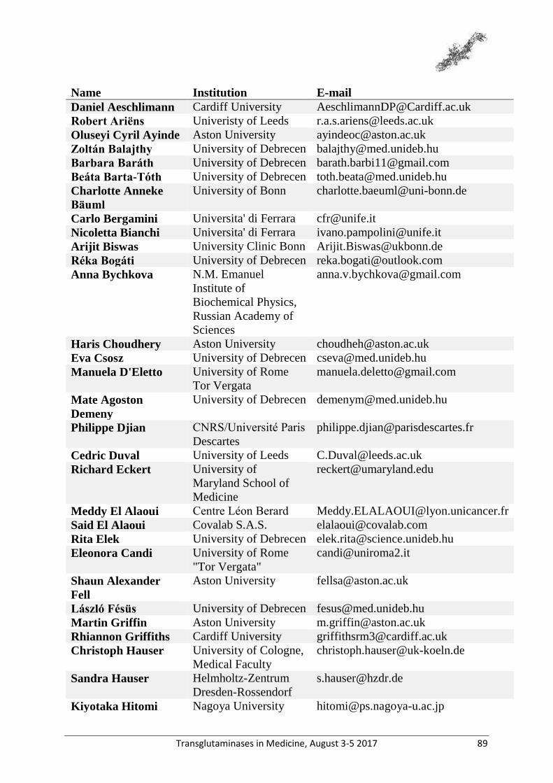
Transglutaminases in Medicine, August 3-5 2017 89
Name Institution E-mail
Daniel Aeschlimann Cardiff University [email protected]
Robert Ariëns Univeristy of Leeds [email protected]
Oluseyi Cyril Ayinde Aston University [email protected]
Zoltán Balajthy University of Debrecen [email protected]
Barbara Baráth University of Debrecen [email protected]
Beáta Barta-Tóth University of Debrecen [email protected]
Charlotte Anneke
Bäuml
University of Bonn [email protected]
Carlo Bergamini Universita' di Ferrara [email protected]
Nicoletta Bianchi Universita' di Ferrara [email protected]
Arijit Biswas University Clinic Bonn [email protected]
Réka Bogáti University of Debrecen [email protected]
Anna Bychkova N.M. Emanuel
Institute of
Biochemical Physics,
Russian Academy of
Sciences
Haris Choudhery Aston University [email protected]
Eva Csosz University of Debrecen [email protected]
Manuela D'Eletto University of Rome
Tor Vergata
Mate Agoston
Demeny
University of Debrecen [email protected]
Philippe Djian CNRS/Université Paris
Descartes
Cedric Duval University of Leeds [email protected]
Richard Eckert University of
Maryland School of
Medicine
Meddy El Alaoui Centre Léon Berard [email protected]
Said El Alaoui Covalab S.A.S. [email protected]
Rita Elek University of Debrecen [email protected]
Eleonora Candi University of Rome
"Tor Vergata"
Shaun Alexander
Fell
Aston University [email protected]
László Fésüs University of Debrecen [email protected]
Martin Griffin Aston University [email protected]
Rhiannon Griffiths Cardiff University [email protected]
Christoph Hauser University of Cologne,
Medical Faculty
Sandra Hauser Helmholtz-Zentrum
Dresden-Rossendorf
Kiyotaka Hitomi Nagoya University [email protected]
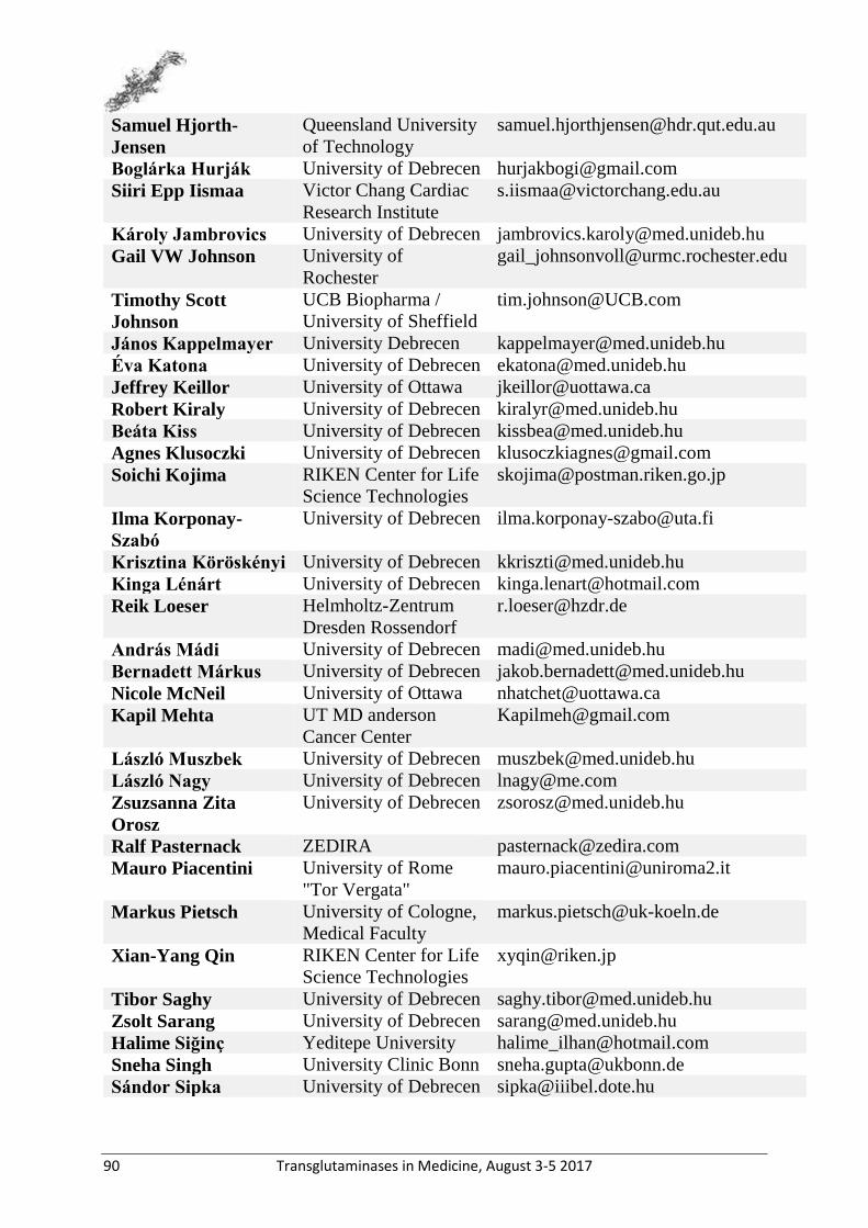
90 Transglutaminases in Medicine, August 3-5 2017
Samuel Hjorth-
Jensen
Queensland University
of Technology
Boglárka Hurják University of Debrecen [email protected]
Siiri Epp Iismaa Victor Chang Cardiac
Research Institute
Károly Jambrovics University of Debrecen [email protected]
Gail VW Johnson University of
Rochester
Timothy Scott
Johnson
UCB Biopharma /
University of Sheffield
János Kappelmayer University Debrecen [email protected]
Éva Katona University of Debrecen [email protected]
Jeffrey Keillor University of Ottawa [email protected]
Robert Kiraly University of Debrecen [email protected]
Beáta Kiss University of Debrecen [email protected]
Agnes Klusoczki University of Debrecen [email protected]
Soichi Kojima RIKEN Center for Life
Science Technologies
Ilma Korponay-
Szabó
University of Debrecen [email protected]
Krisztina Köröskényi University of Debrecen [email protected]
Kinga Lénárt University of Debrecen [email protected]
Reik Loeser Helmholtz-Zentrum
Dresden Rossendorf
András Mádi University of Debrecen [email protected]
Bernadett Márkus University of Debrecen [email protected]
Nicole McNeil University of Ottawa [email protected]
Kapil Mehta UT MD anderson
Cancer Center
László Muszbek University of Debrecen [email protected]
László Nagy University of Debrecen [email protected]
Zsuzsanna Zita
Orosz
University of Debrecen [email protected]
Ralf Pasternack ZEDIRA [email protected]
Mauro Piacentini University of Rome
"Tor Vergata"
Markus Pietsch University of Cologne,
Medical Faculty
Xian-Yang Qin RIKEN Center for Life
Science Technologies
Tibor Saghy University of Debrecen [email protected]
Zsolt Sarang University of Debrecen [email protected]
Halime Siğinç Yeditepe University [email protected]
Sneha Singh University Clinic Bonn [email protected]
Sándor Sipka University of Debrecen [email protected]

Transglutaminases in Medicine, August 3-5 2017 91
Éva Sivadó Covalab, Centre Léon-
Bérard
Ludvig M Sollid University of Oslo [email protected]
Laura Somodi University of Debrecen [email protected]
Huifang Sun McGill University [email protected]
Zsuzsa Szabo University of Debrecen [email protected]
Timea Szekely University of Debrecen [email protected]
Zsuzsanna Szondy University of Debrecen [email protected]
Elisa Tonoli Nottingham Trent
University
Daniel Torocsik University of Debrecen [email protected]
Boglárka Tóth University of Debrecen [email protected]
Shannon Turberville Cardiff University [email protected]
Bürge Ulukan Yeditepe University [email protected]
Anne-Marie van
Dam
VU University
Medical Center
Eli Verdeio NTU [email protected]
Elisabetta AM
Verderio
Nottingham Trent
University
Alexandra D.
Vasilyeva
N.M. Emanuel
Institute of
Biochemical Physics,
Russian Academy of
Sciences
Zhuo Wang Aston University [email protected]
Robert Wodtke Helmholtz-Zentrum
Dresden-Rossendorf
92 Transglutaminases in Medicine, August 3-5 2017
Notes:

Transglutaminases in Medicine, August 3-5 2017 93
Notes:

94 Transglutaminases in Medicine, August 3-5 2017
Notes:

Transglutaminases in Medicine, August 3-5 2017 95
Notes:

96 Transglutaminases in Medicine, August 3-5 2017
Notes:

Transglutaminases in Medicine, August 3-5 2017 97
Notes:

98 Transglutaminases in Medicine, August 3-5 2017
Notes:
Transglutaminases in Medicine, August 3-5 2017 99
Notes:


