
D. Schardt / GSI-DLMarch 2012
Tumor Tumor therapytherapy withwith ionion beamsbeams Dieter Dieter SchardtSchardt
GSI DarmstadtGSI Darmstadt
1.
Introduction
(Radiation
therapy and particle
therapy)
2. Physical characteristics of ion beams in tumor therapy
3.
Technical
developments Accelerators, Beam
scanning
4.
Radiobiological
aspects
5.
Dose verification
techniques
6.
Clinical
results
Today
only
protons and light ions (4He,12C,20Ne) are
of practical
importance(neutrons, pions, antiprotons)XXX
???

D. Schardt / GSI-DLMarch 2012
Historical development of radiation therapyHistorical development of radiation therapy
1895 Discovery of X-rays
(Roentgen) 1896 Production
of X-ray
tubes
by
RGS Erlangen (later
Siemens)
1896 Discovery of Radioactivity
(Bequerel)1898 Radium (Curie) 226Ra, α
and γ
186 keV
(Brachytherapy)
Conventional radiotherapy (photons and electrons)1950 60Co ( γ
1 MeV) 137Cs ( γ
661 keV)
Betatron Electrons
up to 20 MeV + conv.target
(Photons)
1970 Linear accelerators
Electrons
6-25 MeV (Photons)
1985 IMRT (Intensity-Modulated
Radiation
Therapy)
→ about
1950: Development
of Particle
Therapy

D. Schardt / GSI-DLMarch 2012
ElektronElektron--LinacLinac

D. Schardt / GSI-DLMarch 2012
DepthDepth--dose profiledose profile
Photon-Irradiation
Rotation & Intensity Modulation
IMRTInverse Planning
Multifield-Irradiation
100%

D. Schardt / GSI-DLMarch 2012
IMRT TechniquesIMRT Techniques
Intensity-Modulated Radiation Therapy
IMRT
Treatment headVARIAN Systems

D. Schardt / GSI-DLMarch 2012
DepthDepth--dose profile dose profile –– charged particlescharged particles
Heavy charged particlesProtons / Light Ions
100%

D. Schardt / GSI-DLMarch 2012
Ion Ion BeamsBeams in in RadiotherapyRadiotherapy
1946 R.R. Wilson, Radiology 47,487
"…potential benefits
of heavy
chargedparticles
in radiotherapy"
R.R. Wilson at Harvard mid
1940s†2000
1954 First
proton
treatments
at 184“
cyclotronLBL BerkeleyJohn and Ernest Lawrence, C. Tobias, J. Castro
"…
for
a given
range, the
straggling
and the
angular
spread
of alpha particles willbe
one-half
as much
as for
protons.Heavier
nuclei, such as very
energeticcarbon atoms , may
eventually
becometherapeutically
practical."
Today: 32 clinical
proton
facilities
83,000 patients
treated6 carbon
ion
9,200

D. Schardt / GSI-DLMarch 2012
Ion Ion BeamsBeams in in RadiotherapyRadiotherapy
New projects planned or under construction …see PTCOG Newsletter http://ptcog.web.psi.ch/
Beam scanning
First clinical HI-facility
patients
1957 -
92 4He 184-inch SC Berkeley / USA 2054
1975 -
92 20Ne BEVALAC Berkeley / USA 433
1997 -
2008 12C SIS-18 Darmstadt / Germany 440 G. Kraft
Beam scanning
1994 12C HIMAC Chiba / Japan 5497
2003 12C,p HIBMC Hyogo
/ Japan 638
2009 12C IMPCAS Lanzhou / China 126
2010 12C,p HIT Heidelberg /Germany 400
2010
12C GHMC Gunma
/ Japan 454
2011 12C,p CNAO Pavia / Italy
5

D. Schardt / GSI-DLMarch 2012
Proton and Ion-Therapy in Europe
Protons
Light Ions
Centers in Operation
Under construction
Under discussion
?
x Marburg / Kiel(stopped)
Berlin
HIT
PSI
CPO
CNAO
ITEP
Gatchina
Uppsala Dubna
Krakow
Vienna
Prague
?
?
?
Clatterbridgex
x
Catania
RPTCMunich
Kiel
Berlin
Marburg

D. Schardt / GSI-DLMarch 2012
Cancer situation in EuropeCancer situation in Europe
Lokalisierte Tumore: 58% Metastasierende Tumore: 42%
Operation: 22%
Radiotherapie: 12%
Chemotherapie: 5%
Palliative Behandlung: 37%
Operation+Radiotherapie: 6%
Versagen bei der lokalen Kontrolle: 18%
(Tubiana
1992)
> 10.000 Patients/year could be treated better with particle therapy
In Germany about
480,000 new
cancer
incidences
per year
Localized
Tumors 58% Metastatic
Tumors 42%
Surgery
22%Chemotherapy
5%
Palliative treatment
37%
Surgery
22%
Radiotherapy
12%
Surgery
+ RT 6 %
Failure
of local
control
18%

D. Schardt / GSI-DLMarch 2012
HeavyHeavy--ionion therapytherapy isis an an interdiscplinaryinterdiscplinary fieldfield
Physics
Radiobiology
Engineering
Energy deposition, depth-dose
profile,particle
field, physical
model
Biological
effect
(RBE)cell
killing
& repair
mechanismsbiological
model
Accelerator, beam
delivery, scanning, Control
system, safety
aspectsIn-vivo
range
verification
(PET)
Clinical application
Indications, treatment
planning, fractionation
scheme, clinical
studies
...
Treatment planning

D. Schardt / GSI-DLMarch 2012
PhysicalPhysical characterizationcharacterization of of ionion beamsbeams
Energy deposition
Depth-dose distribution (Bragg curve)
Nuclear fragmentation
Lateral scattering

D. Schardt / GSI-DLMarch 2012
Definition
J/kg] 1 Gy [1
mED
AbsorbedAbsorbed DoseDose
Lethal
Dose:LD50/30 = 3-4 Gy (Human)
1000 Gy (Wasp)
Radation
therapy: ~ 40-60 Gy in target
volume
1 Gy is
a very
small
amount
of energy
(0,0002388 kcal/kg) (1 Gy heats 1 Liter Water by 0.0002 deg)
Radiation
effects
are
not
due
to heat
!
D. Schardt / GSI-DLMarch 2012
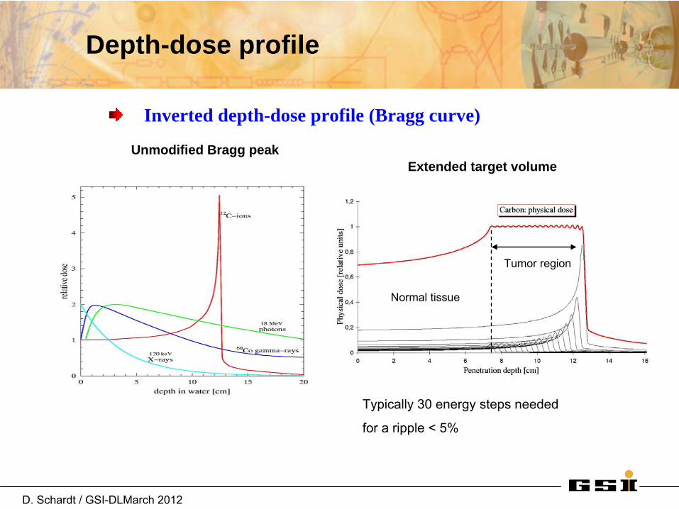
Inverted depth-dose profile (Bragg curve)
Unmodified Bragg peak
Typically
30 energy
steps
needed
for
a ripple
< 5%
Extended target volume
Tumor region
Normal tissue
Depth-dose profile
D. Schardt / GSI-DLMarch 2012
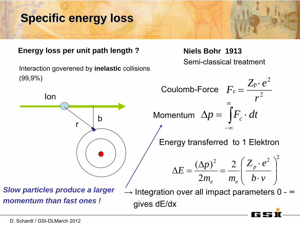
SpecificSpecific energyenergy lossloss
2
2p
cr
eZF
dtFp c
Energy loss per unit path length ? Niels Bohr 1913Semi-classical treatment
Ion
br
Momentum
Energy transferred
to 1 Elektron
Interaction goverened
by
inelastic collisions(99,9%)
222 22
)(
vbeZ
mmpE p
ee
Coulomb-Force
→ Integration over
all impact
parameters
0 -
∞gives
dE/dx
Slow particles produce a larger momentum than fast ones !
D. Schardt / GSI-DLMarch 2012
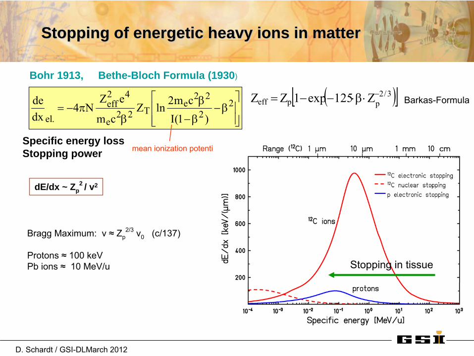
StoppingStopping of of energeticenergetic heavyheavy ionsions in matterin matter
Specific energy lossStopping power
2
2
22e
T22e
42eff
.el )1(Icm2lnZ
cm
eZN4
dxde 3/2
ppeff Z125exp1ZZ Barkas-Formula
mean
ionization
potential
dE/dx ~ Zp2 / v2
Bragg
Maximum: v ≈
Zp2/3
v0
(c/137)
Protons ≈
100 keVPb ions
≈
10 MeV/u
Bohr 1913, Bethe-Bloch Formula (1930)
Stopping
in tissue

D. Schardt / GSI-DLMarch 2012
BraggBragg curvescurves of of 1212C in C in waterwater
peak-width and height are affected by–
straggling –
fragmentation
increasing tail dose

D. Schardt / GSI-DLMarch 2012
Mechanical accuracy:10 μm for
relative thickness0.2 mm absolute
BraggBragg curvecurve measurementsmeasurements
IC2/IC1

D. Schardt / GSI-DLMarch 2012
particle flux fluence F =
tAbsorbed dose
erg/g] 100 Gy [1 mED
1
dxdEF106.1D 9
[Gy] [cm-2
keV/m g/cm3]Specific energy loss
m][keV/ xElim
dxdE
0x
Example:
12C ions
300 MeV/u de/dx=13 keV/mF=108
/cm2
Dose in water: 2 Gy
LET (linear energy
transfer)
ParticleParticle dosedose

D. Schardt / GSI-DLMarch 2012
Significance of nuclear fragmentation Significance of nuclear fragmentation in RT with light ionsin RT with light ions
Carbon
ion
therapy100-400 MeV/u
I. Pshenichnov
High-energy carbon beam stopping in water
Nuclear
fragmentation Loss
of primary
ions
depth-dose, RBE
Total reaction
cross section
1-2 b
Buildup
of secondary
fragments
dose-tail, lateral dose
Exp. Investigations (physical characterization)–
LBL Berkeley Ne 670 MeV/u
1970’s W. Schimmerling–
NIRS/HIMAC Chiba C, light ions 1990’s T. Kanai –
GSI Biophysics C, light ions 1990’s

D. Schardt / GSI-DLMarch 2012
NuclearNuclear fragmentationfragmentation reactionsreactions
Peripheral
collisions
at high energies
Fragment spectrum
„Geometrical“
reaction
cross section23/1
t3/1
p20tot )bAA(r
Brad-Peters (1950)
xne)0()x( ; ndxd
x

D. Schardt / GSI-DLMarch 2012
Loss of carbon Ions bynuclear reactions
E. Haettner
et al., GSI 2005
Buildup
of secondary fragments
Surviving fraction
Attenuation of primary flux and Attenuation of primary flux and buildupbuildup of of secondary fragmentssecondary fragments

D. Schardt / GSI-DLMarch 2012
HowHow nuclearnuclear fragmentationfragmentation affectsaffects thethe BraggBragg curvescurves
Model calculations (HIBRAC-code)Lembit
Sihver
GSI, 1997semi-empirical
cross section
formula
Depth in water [cm]
Range 2.6 cm
Range 36 cm
Today
powerful
Monte-Carlo codes
likeFLUKA, GEANT4, PHITS etc.are
able
to simulate
all atomic
and nuclearInteractions
and reproduce
the
Bragg
curves
D. Schardt / GSI-DLMarch 2012
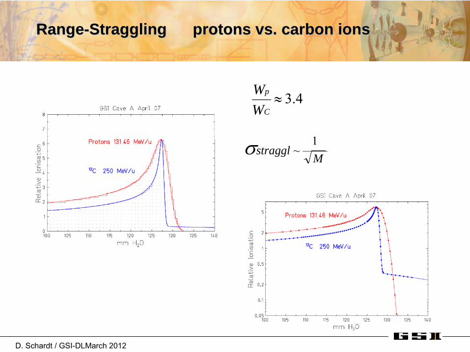
RangeRange--Straggling protons vs. carbon ionsStraggling protons vs. carbon ions
Mstraggl
1 ~
3.4 C
p
WW
D. Schardt / GSI-DLMarch 2012
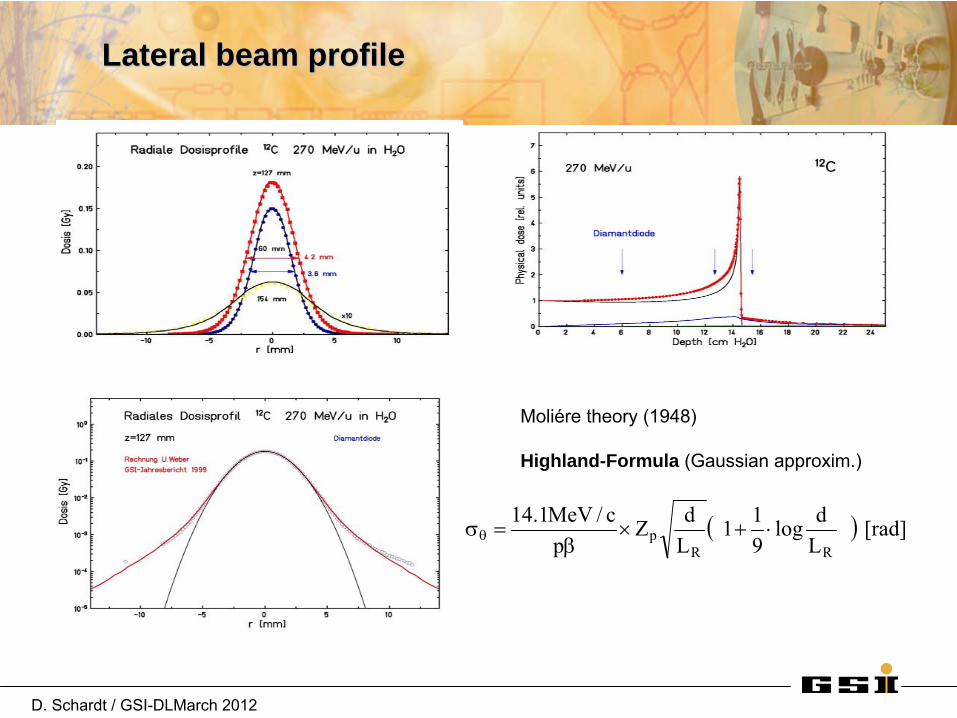
Lateral Lateral beambeam profileprofile
Moliére
theory
(1948)
Highland-Formula (Gaussian
approxim.)
[rad] Ldlog
911
LdZ
pc/MeV1.14
RRp

D. Schardt / GSI-DLMarch 2012
ComparisonComparison of lateral of lateral beambeam spreadspread
Protons
12C ions0 cm 15 cm
Film dosimetry, LBL Berkeley
Ion beam

D. Schardt / GSI-DLMarch 2012
Beam spread Beam spread -- protons vs. carbon ionsprotons vs. carbon ions
Bea
m w
idth
FWH
M
[mm
]
0
5
10
15
20
25
30
Distance from exit window [cm]
0 20 40 60 80 100 120 140 160
nozzle
50 MeV
80
100 150
200
250
491 386
92
285
148 186
protons12C ions
waterair
MeV/u
≈
1mPatient
Multiple scattering–
nozzle
elements–
patient
tissue
Molière theory

D. Schardt / GSI-DLMarch 2012
Comparison of Carbon Ions vs. Protons
C-12 (GSI)
Protons (Capetown/SA)
Heavy ions offer higher precision close to organs at risk

D. Schardt / GSI-DLMarch 2012
PhysicalPhysical characteristicscharacteristics of of ionion beamsbeams relevant relevant forfor tumortumor therapytherapy
„Inverted“
depth-dose
profile
(Bragg
curve)major
advantage
for
treating
deep-seated
tumors
Nuclear
fragmentation
is
a significant
effectfor
heavy
ions
(tail
dose)
Lateral scattering
significant
for
protons


