
Unedited Book of Contributed Papers from the International Conference on
Radiation Protection in Medicine: Achieving Change in Practice
Vienna, 11-15 December 2017
The material in this book has been supplied by the authors and has not been edited. The views expressed remain the
responsibility of the named authors and do not necessarily reflect those of the government of the designating Member
State(s). The IAEA cannot be held responsible for any material reproduced in this book.
Session 1
Justification in the use of radiation in
medical imaging
REVIEW OF RADIATION SAFETY ISSUES DURING
INVENTORY OF RADIATION SAFETY ISSUES
DURING INVENTORY OF RADIATION
EMANATING EQUIPMENT
K.P. ADHIKARI
National Academy of Medical Sciences, Bir Hospital
Mahabouddha/Kathmandu, Nepal
Abstract
Nepal, one of the least developed countries with population of 26.6 million people is the most populated country
without a regulatory body [1]. Newer modalities and latest radiological equipment are being introduced, but lack of control and
inventory system is a serious problem. The aim of this study was to start inventory of radiation emanating equipment used in
medical field and also to find out radiation safety issues and challenges. This study was done for first the time in Nepal to start
inventory system. Questionnaire was designed to find out actual number of equipment among with the date of installation,
source number, number of staff and their qualifications. Questionnaire was also designed to find out radiation safety issues like
personnel radiation dose monitoring, commissioning and quality control tests. Altogether, 296 institutions in Kathmandu were
inspected. Inventory was made for diagnostic radiology, nuclear medicine, radiotherapy and dental X-rays. Commissioning and
quality control program have not been practiced in most hospitals, and few only have a maintenance contract with vendor. Sixty
percent of workers have never been monitored for radiation exposure. There is an urgent need to establish regulatory authority to
regulate radiation used in medicine.
1. INTRODUCTION
As we all know that radiation is a fact of life because in every aspect of our life we have to encounter some
form of radiation. Nepal, one of the least developed countries with population of 26.6 million people is the most
populated country without a regulatory body [1]. Due to lack of laws and regulations in Nepal radiation protection
survey and quality control (QC) of radiation emitting equipment are not even "recommended" and only some
institutions have voluntarily established QC systems [3]. In Nepal, radiation-emanating equipment is mainly used in
diagnostic radiology, radiotherapy and nuclear medicine [2]. We are using ionizing radiation in the form of X -rays
and gamma rays in the diagnosis and treatment of many diseases. Though we are using radioisotopes in medicine,
but still, we do not have any formal regulations to record import and use of these radioisotopes in the country [2].
So, it is sought that an inventory or record of radioisotopes and equipment should be made in Kathmandu Valley
which consists of three different districts, Kathmandu, Lalitpur and Bhakatapur.. This record is very important in a
country like ours, where there is no institution or organization to look after radiation safety and the safety of
radioisotopes itself [3][4].
This study contains the first hand information provided by different institutions on current status and
radiation safety infrastructure.
The main objective of this study is to start inventory process and to find out present status of radiation
emanating equipment being used at different hospitals in Kathmandu Valley. The other objectives are to find out
radiation safety issues and challenges.
2. METHODS
To implement this program, questionnaire was prepared. Survey team was constituted and prepared them to
visit the site to inspect the room and to complete the questionnaire. The questionnaire consists of questions seeking
information regarding professional responsibility, qualifications, safety and security, personnel dose monitoring,
commissioning and quality control (QC) tests. Background radiation was only measured at the centers, which have
radioactive materials and radiopharmaceuticals.
200.00
150.00
100.00
50.00
Kathmandu
Lalitpur
Bhakatpur
0.00
Diagnos=c Radiotherapy Nuclear Radiology Medicine
3. RESULT & DISCUSSION
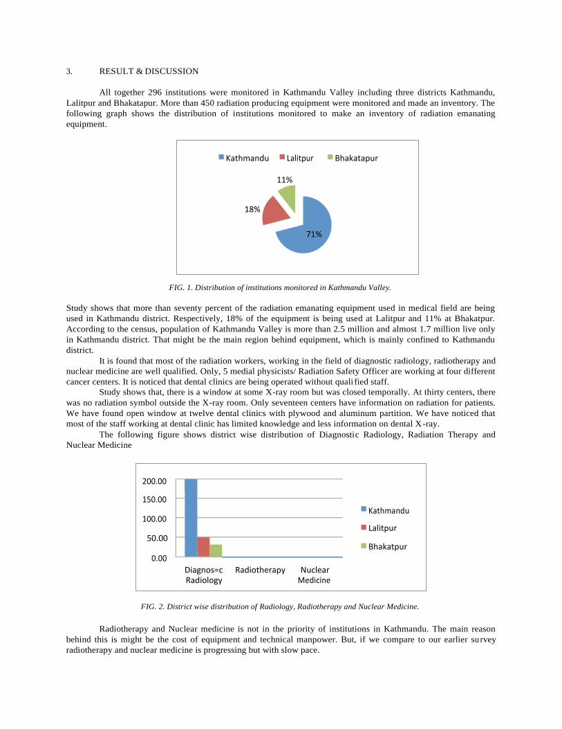
All together 296 institutions were monitored in Kathmandu Valley including three districts Kathmandu,
Lalitpur and Bhakatapur. More than 450 radiation producing equipment were monitored and made an inventory. The
following graph shows the distribution of institutions monitored to make an inventory of radiation emanating
equipment.
FIG. 1. Distribution of institutions monitored in Kathmandu Valley.
Study shows that more than seventy percent of the radiation emanating equipment used in medical field are being
used in Kathmandu district. Respectively, 18% of the equipment is being used at Lalitpur and 11% at Bhakatpur.
According to the census, population of Kathmandu Valley is more than 2.5 million and almost 1.7 million live only
in Kathmandu district. That might be the main region behind equipment, which is mainly confined to Kathmandu
district.
It is found that most of the radiation workers, working in the field of diagnostic radiology, radiotherapy and
nuclear medicine are well qualified. Only, 5 medial physicists/ Radiation Safety Officer are working at four different
cancer centers. It is noticed that dental clinics are being operated without quali fied staff.
Study shows that, there is a window at some X-ray room but was closed temporally. At thirty centers, there
was no radiation symbol outside the X-ray room. Only seventeen centers have information on radiation for patients.
We have found open window at twelve dental clinics with plywood and aluminum partition. We have noticed that
most of the staff working at dental clinic has limited knowledge and less information on dental X-ray.
The following figure shows district wise distribution of Diagnostic Radiology, Radiation Therapy and
Nuclear Medicine
FIG. 2. District wise distribution of Radiology, Radiotherapy and Nuclear Medicine.
Radiotherapy and Nuclear medicine is not in the priority of institutions in Kathmandu. The main reason
behind this is might be the cost of equipment and technical manpower. But, if we compare to our earlier survey
radiotherapy and nuclear medicine is progressing but with slow pace.
Kathmandu Lalitpur Bhakatapur
11%
18%
71%

The following figure shows distribution of equipment used in diagnostic radiology at Kathmandu. Valley
FIG. 3. Distribution of Equipment used in Diagnostic Radiology and Dental Clinic.
Regarding personal radiation dose monitoring, more than ninety seven percent of the institutions are not
monitoring their radiation workers. Most of the institutions, using personal radiation belong to the institutions
having either radiotherapy or nuclear medicine facility. The main reason behind this is because of the medical
physicist / Radiation Safety Officer. Recently, busy institutions has just started personnel radiation dose monitoring
through National Academy of Science & Technology (NAST), which has started its service with the help from
IAEA, Technical Cooperation (TC) project. But, according to them, personnel radiation service from NAST is still
not smooth and continuous.
Study shows that more than ninety five percent of institutes have never done radiation survey before
starting radiation treatment expect radiotherapy facility and few centers with diagnostic radiology. There is no
Quality Control (QC) program in diagnostic radiology but there is a QC program in Radiotherapy facility centers.
Regarding further information on QC in diagnostic radiology, there are some maintenance contracts with the
supplier company at few institutes.
Background radiation was also measured around at three cancer centers. Well calibrated survey meter was
used to measure Cobalt-60 area and HDR Brachytherapy area and was found within limit as background level.
Background radiation was also measured in three Nuclear Medicine facilities and found within safe limit.
Study also shows that there are few unqualified radiation personnel working in this field. We have noticed, some
workers are over-conscious on radiation.
4. CONCLUSION
Altogether, 296 institutions in Kathmandu were monitored. Inventory was made for diagnostic radiology,
nuclear medicine, radiotherapy and dental X-rays. This study has initiated to start inventory system in Nepal.
Through proper education and training and regularly organized seminars people are becoming more and more aware
of the benefits of radiation and its uses in medicine [5]. The provision of such services at the national level does not
detract from the ultimate responsibility for radiation protection and safety borne by the legal persons authorized to
conduct the practices. Those, especially the radiation workers, are very much careful and conscious about the safe
handling and use of radiation sources to protect public and environment. By establishing basic safety standard and
Radiation Regulatory Authority, Rules and Regulations can be enforced in the country effectively and efficiently
[3].
ACKNOWLEDGEMENTS
Author is grateful to Ministry of Science & Technology, National Academy of Medical Sciences, Bir Hospital,
working committee members and surveyed hospitals/institutions for their assistance, encouragement and guidance
throughout the entire period of study.
250
200
150
100
50
0
Kathmanu
Lalitpur
Bhakatpur

REFERENCES
[1] The World Bank website: http://www.worldbank.org/en/country/nepal/overview [Accessed 30.08.2017].
[2] ADHIKARI, K.P., et al. Status of radiation protection at different Hospitals in Nepal. Journal of Medical Physics. 2012;
37(4): 240-244.
[3] INTERNATIONAL ATOMIC ENERGY AGENCY, Radiation Protection and Safety of Radioactive Sources: International
Basic Safety Standard, GSR No. 3, Vienna, 2014.
[4] Radiological Protection and Safety in Medicine. ICRP Publication 73. Ann. ICRP 26 (2);1996
[5] ADHIKARI, K.P., Radiation Regulation in Nepal: A Challenging Journey. Radiation Regulator. 2012; 1(1): 6-10.

E.G. FRIBERG
HERCA EUROPEAN ACTION WEEK – RESULTS OF A
COORDINATED INSPECTION INITIATIVE ASSESSING
JUSTIFICATION IN RADIOLOGY
E.G. FRIBERG Heads of the European Radiological Protection Competent Authorities (HERCA), Working Group on
Medical Applications (WGMA)
Email: [email protected] and [email protected]
Abstract
Justification is a fundamental principle in radiation protection and has to be carried out before any individual
exposure, according to the European Basic Safety Standards Directive. However, there are strong indications that up to 20-
30% of medical imaging exposures are unjustified in many economically developed countries. Heads of the European
Radiological Protection Competent Authorities (HERCA) has recognized that the regulatory bodies have an important role in
promoting and ensuring that the principle of justification is properly implemented at medical imaging facilities. HERCA
performed a coordinated European Action Week on inspection of justification in radiology in November 2016. The aim was
to identify the main challenges in the justification process. 17 European countries participated in the Action Week, and 148
inspections were carried out. All inspections were performed according to a common inspection template. Main weaknesses
identified were: 1) lack of written procedures describing the justification process, 2) lack of availability, awareness and use of
referral guidelines, 3) lack of national or local procedures for performing clinical audits and 4) incomplete referrals from
referring practitioners. HERCA has identified a need to increase the awareness of justification among health professionals
and facility management in follow-up actions in 2017 and 2018.
1. INTRODUCTION
Justification is one of the fundamental principles in the international radiation protection framework
established by the International Commission on Radiological Protection [1]. The intention behind the act of
justification is to ensure that the benefit of the exposure outweighs the associated potential radiation detriment.
To ensure the appropriate use of medical imaging, justification has to be carried out at an individual level before
the exposure takes place. The necessity for individual justification is reinforced in the new European Basic Safety
Standards Directive and the International Basic Safety Standards [2, 3]. However, there are strong indications that
up to 20-30% of medical imaging exposures are unjustified in many economically developed countries [4].
Therefore, the International Atomic Energy Agency (IAEA) have introduced the “Triple A” initiative, promoting
Awareness about radiation risks; Appropriateness to ensure that those referred for radiological examinations really
need them; and Audit to check the effectiveness of the referral and related processes [5]. The need to enhance the
implementation of the principle of justification is also addressed in the joint position statement “Bonn Call for
Action” by the IAEA and World Health Organization (WHO) and in the council conclusions on justification by
the European Commission [6, 7].
Heads of the European Radiological protection Competent Authorities (HERCA) has recognized that the
regulatory bodies have an important role in promoting and ensuring that the principle of justification is properly
implemented at medical imaging facilities. Consequently, HERCA has published a position paper on justification
of individual medical exposures for diagnosis to provide clarity on the regulatory framework for justification [8].
HERCA highlighted that justification is not just one action, but a process that includes a number of events from
initial presentation of the patient to the radiology department to the final authorization for an exposure to take
place. During the European Inspection Workshop organized by HERCA in 2015, it was revealed that very few
radiation protection authorities actually inspected the justification process in depth [9]. HERCA identified an
urgent need to improve the implementation of justification in medical exposure situations and decided to support
this through a coordinated European Action Week on the inspection of justification, focussing on radiology
departments. [10].
2. METHODS
1

IAEA-CN-255/106
A European Action Week, with the scope of performing coordinated inspections of justification in
radiological medical imaging facilities across Europe, was undertaken by HERCA in November 2016 [11]. The
aim was to assess whether justification takes place in the facilities and to identify the main challenges in the
justification process. All HERCA countries were invited to take part in this Action Week and to perform
inspections in a representative number and type of imaging facilities. All inspections were notified in advance and
performed according to a common inspection template provided by HERCA Working Group on Medical
Applications (WGMA). Requested documentation was, in most countries, submitted to the competent authorities
prior to the inspections. The inspection template collected information about the regulatory framework and the
competent authority and inspection teams of the participating countries in addition to the results from each
inspected medical imaging facility. Questions addressed during the inspections were aimed to identify if and how
justification was implemented in the daily workflow. Availability of written procedures for the justification
process, assignment of tasks and responsibilities, daily processes for assessment of justification and
appropriateness of referrals, general practice to handle incomplete or unjustified referrals, availability and use of
referral guidelines together with performance of clinical audits were among the examined topics. The overall
quality of 10 referrals (5 for CT and 5 for conventional X-ray) per inspected facility was also reviewed to check
if there was sufficient information for the radiological practitioner to assess if the referred examination was
justified and appropriate.
3. RESULTS
In total, 17 countries participated in the Action Week and 148 inspections were carried out. The
participating countries and number of inspections performed per country are shown in FIG. 1. The mean number
of inspections per country was 9 (range: 1-19). 44% of the inspected facilities were public and 56% were private.
The inspections were carried out by one, two or three competent authorities in 76%, 18% and 6% of participating
countries, respectively. Radiation protection competent authorities were mainly the responsible authority for
medical exposures. The inspection teams generally consisted of medical physicists and radiographers and/or
engineers, but in some countries physicians or radiologists were also part of the inspection teams. Key personnel
to be interviewed were typically the facility management, radiological practitioners and radiographers. Additional
staff such as medical physicists, radiation protection officers and responsible persons for the quality assurance
system, were also interviewed in some countries.
20
18
16
14
12
10
8
6
4
2
0
BE CZ DK EE FI FR GR IS LV LT LU NO RO SI SE CH UK
Countries
FIG. 1. Overview of participating countries and number of inspections performed per country. BE: Belgium, CH:
Switzerland, CZ: Czech Republic, DK: Denmark, EE: Estonia, FI: Finland, FR: France, GR: Greece, IS: Iceland, LT:
Lithuania, LU: Luxembourg, LV: Latvia, NO: Norway, RO: Romania, SI: Slovenia, SE: Sweden, UK: United Kingdom.
Results regarding the availability, knowledge and content of procedures for the justification process are
shown in FIG. 2.A. Written procedures were only available at 55% of the inspected facilities. Even though written
procedures were not available in almost half of inspected facilities, many had established processes for
justification. About 10% of facilities had no procedures or routines for justification at all. Where procedures and
processes were available, the staff involved implemented these in daily practice. They were frequently revised
and updated in about 70% of facilities. Most topics identified by HERCA as important to ensure proper
justification process were covered by 60% to 80% of facilities, while information to patients about risk and benefit,
No. o
f in
spec
tio
ns

E.G. FRIBERG
a requirement of the latest Euratom Basic Safety Standards Directive, was only addressed by 34% of facilities.
Allocation of tasks and assignment of responsibilities were clearly defined and documented for referring
physicians, radiological practitioner, radiographers and the receptionist in 52%, 65%, 71% and 62% of facilities,
respectively. Allocated tasks and responsibilities were known by the staff in 76% of facilities where these were
defined, while delegation of tasks was only documented in 52% of facilities.
Results relating to the availability and use of referral guidelines and performance of clinical audits are
summarized in FIG. 2.B. Referral guidelines for medical imaging were available in 70% of inspected facilities.
The sources of referral guidelines were national (58%), regional (15%) and/or local (28%). These guidelines were
made available to the referrers in almost all facilities, but assumed to be implemented in daily use by only 31%
of the referrers and 48% of the radiological practitioners. Only 20% of the inspected facilities had local procedures
for clinical audits and clinical audits were seldom performed. Less than half of the facilities performed any other
type of audit or review (internal or external) covering the justification process.
Written procedures available
Procedures known by staff
Procedures implemented in work
Procedures revised/updated
Minimun content of referral
Justification/appropriateness
Contact: practitioner/referrer
Identification of pregnancy
Information: risk/benefit to patient
0 % 10 % 20 % 30 % 40 % 50 % 60 % 70 % 80 % 90 % 100 %
Yes No Unsure
FIG. 2.A: Summary of the results regarding the availability of procedures for justification and their coverage of
important topics to ensure for a proper justification process.
RG available at facility
RG available to referrer
RG in use by referrer
RG in use by practitioner
Procedure for CA
Performance of CA
Other audits/revisions
0 % 10 % 20 % 30 % 40 % 50 % 60 % 70 % 80 % 90 % 100 %
Yes No Unsure
FIG.2.B Summary of the results regarding referral guidelines (RG) and clinical audits (CA).
Many of the inspected facilities had established good practice for evaluation of the referrals before the
examinations were performed, as shown in FIG. 3.A. However, as many as 26% of facilities did not perform a
satisfactory evaluation of the referral before the examinations were performed and even more did not reject
unjustified examinations (31%) or fully prove that the examinations were authorized by the radiological
practitioner (35%). The presence and overall quality of the referrals are summarized in FIG. 3.B. Referrals were
available for almost all examinations (99%). Information about the patient, referrer, date and signature of the
referral was satisfactory in over 90% of referrals. Clinical information was sufficient and contained the clinical
question to be answered in 86% and 81% of referrals, respectively. Information about previous examinations and
identification of pregnancy was only included in 54% and 63% of referrals, respectively. Education and training,
covering the justification process, was documented in only 60% of the inspected facilities.
3

IAEA-CN-255/106
Justification evaluated
Appropriateness evaluated
Insufficient referral: referrer contacted
Examination changed, if inappropriate
Examination rejected, if unjustified
Examination authorized
0 % 10 % 20 % 30 % 40 % 50 % 60 % 70 % 80 % 90 % 100 %
Yes No Partial
FIG.3.A: Summary of the results regarding the daily practice on evaluation of referrals prior to the performance of the
examination.
Referral available
Patient identification
Referring practitioner identified
Contact information, referrer
Date of referral
Signature of referral
Clinical information
Clinical question
Info, previous examinations
Referred examination
Identification of pregnancy
0 % 10 % 20 % 30 % 40 % 50 % 60 % 70 % 80 % 90 % 100 %
Yes No
FIG. 3.B: Summary of the results regarding the presence and overall quality of the referrals. In total, 1367 referrals (for X-
ray and CT) were evaluated.
4. DISCUSSION AND CONCLUSIONS
The European Action Week was supported by all HERCA countries and 53% of countries participated.
Half of the countries (53%) performed less than 10 inspections and the results may not be representative of the
real situation in some of these countries. Results from the inspections were mainly based on interviews and
reviews of documentation. Differences in the interpretation of some questions in the inspection template by
different inspectors were observed, and areas for improvements identified. Even though this Action Week must
be considered as a pilot study, the results obtained provide strong indications of the weak links in the justification
process.
Implementation of the principle of justification varied among countries. Often, justification was only
covered in general terms by the quality system and the justification process was not formally described and
documented in procedures. However, established routines covered, to some extent, important steps in the
justification process in most countries. Radiologists are mainly involved in the evaluation of referrals for CT, MR
and “high-dose” or complex examinations, while radiographers are often allocated this task for many conventional
X-ray examinations. In such cases it is usual to find examination appointments are arranged before referrals are
evaluated. Radiographers almost always evaluate the referrals and check for pregnancy and other
contraindications at the time of appointment, and immediately before the examination is performed. In this respect,
radiographers carry out the key aspects of the justification process whether formally or informally, as a
responsibility or as a delegated task. It is important that the different steps and associated tasks and responsibilities

E.G. FRIBERG
are recognized by management, formalized in procedures and that involved staff receive proper training to take
on the assigned tasks and responsibilities. Despite the presence of procedures and routines, steps in the justification
process are sometimes not followed in daily practice. Lack of time, payment per procedure/examination,
reimbursement systems and loyalty towards the referrers were given as reasons for this situation.
Reviews of the referrals indicated a need for improved quality, more structured referrals and harmonized
guidance on minimum information to be included. Generally, the quality of referrals was worse among general
practitioners, but large variations were observed among participating countries. Many inspection teams found it
difficult to evaluate the quality of the referrals due to lack of expertise and the involvement of a radiologist in the
team is highly recommended. Use of referral guidelines is modest and in many countries the only referral
guidelines available are those covering standardized pathways for cancer and information about the radiation dose
or risk is seldom included. The inspections revealed that the concept of clinical audit is not fully understood and
rarely performed within medical imaging. Review of national regulatory frameworks among the participating
countries also indicated that referral guidelines and clinical audits were not fully implemented at a national level.
Conclusions from the European Action Week: There is still a need to increase the awareness and to reiterate
the importance of the justification process. Inspection is a good tool to address justification and HERCA will
follow-up the identified weak links by providing targeted key messages to involved stakeholders on how they can
take responsibility to act on the different aspects of the justification process.
ACKNOWLEDGEMENTS
HERCA WG MA would like to thank all participating countries, involved competent authorities and inspectors
for performing the inspections and Annette Andersen at the Norwegian Radiation Protection Authority for
analysing the results of the inspections.
REFERENCES
[1] ICRP, The 2007 recommendations of the International Commission on Radiological Protection. ICRP publication 103.
Ann. ICRP, 37(2–4), Elsevier (2007)
[2] European Commission, Council Directive 2013/59/Euratom of 5 December 2013 laying down basic safety standards for
protection against the dangers arising from exposure to ionizing radiation. Official Journal L 13 (2014)
[3] IAEA, Radiation protection and safety of radiation sources: international basic safety standards, GSR part 3, International
Atomic Energy Agency, Vienna (2014)
[4] IAEA, Justification of medical exposure in diagnostic imaging: Proceedings of an International workshop, held in
Brussels in 2009, Vienna (2011)
[5] IAEA, Triple-A investment in patients’ health (2010), https://www.iaea.org/newscenter/news/triple-investment-patients-
health
[6] IAEA and WHO, Bonn call for action. 10 Actions to improve radiation protection in medicine in the next decade. Joint
position statement by IAEA and WHO (2013)
[7] European Commission, Council conclusions on justification of medical imaging involving exposure to ionizing radiation.
Council of the European Union, Brussels (2015)
[8] Heads of the European Radiological Protection Competent Authorities, HERCA position paper. Justification of individual
medical exposure for diagnosis. (2014), http://www.herca.org/herca_news.asp?newsID=40
[9] Heads of the European Radiological Protection Competent Authorities, HERCA MedInspector 2015 Workshop (2015),
http://www.herca.org/highlight_item.asp?itemID=3
[10] Heads of the European Radiological Protection Competent Authorities, European Action Week on inspection of
justification in medical imaging: A HERCA Initiative. EuroSafe Imaging 2016/ESI-0049 (2016),
http://dx.doi.org/10.1594/esi2016/ESI-0049
[11] Heads of the European Radiological Protection Competent Authorities, HERCA European Action Week (2016),
http://www.herca.org/highlight_item.asp?itemID=16
5
KAROUSSOU-SCHREINER and BOUËTTÉ
NATIONAL AUDIT ON THE ADEQUATE
COMPLETION OF MEDICAL IMAGING
REQUEST FORMS IN LUXEMBOURG
A. KAROUSSOU-SCHREINER
Ministry of Health, Radiation Protection Department
Luxembourg, Luxembourg
Email: [email protected]
A. BOUËTTÉ
Ministry of Health, Radiation Protection Department
Luxembourg, Luxembourg
Abstract
The high frequency of radiological procedures in Luxembourg results in an overall total collective effective dose per
caput that is among the highest compared to other European countries. For this reason the Ministry of Health and the
Ministry of Social Security decided in 2015 to put in place an action plan in order to reduce the number of unjustified
radiological procedures. The first part of this action plan was a national audit on the adequate completion of medical imaging
request forms. The aim of the audit was to evaluate the quality of the requests and the compliance of the requests with
Luxembourgish legislation. For this audit the adequate completion of 200 requests was evaluated per radiology department.
The audit was carried out in all 10 radiology departments of Luxembourg.The results of the audit clearly show that the
compliance rate of requests for medical imaging in Luxembourg is overall unsatisfactory. Of the 2000 requests audited only
one single request included all mandatory information. 42% of the requests were in conformity for the presence of the items
"clinical backround" and "question to be asked", while 39% had only one of these two items and 19% had none of these
items. From the results of this audit it is clear that there is a need for improvement of the quality of the requests in
Luxembourg. This can be achieved mainly through the education and training of the referrers.
1. INTRODUCTION
Two fundamental principles of radiation protection according to the International Commission on
Radiological Protection system are the justification of medical radiological procedures and the optimization of
their dose [1]. According to the principle of Justification as described in the International Basic Safety Standards
[2] and the European Basic Safety Standards Directive [3], the medical exposure from a radiological
examination shall show a sufficient net benefit weighing the total potential diagnostic or therape utic benefits it
produces against the individual detriment that the exposure might cause. These principles are anchored in the
Luxembourgish legislation [4]. The principle of Justification is difficult to implement in the medical field, in
particular because of the multiplicity of different stakeholders involved in the process of justification: the
patient, the referrer, the practitioner and the undertaking.
In 2000 and 2009, the European Commission carried out a project with the objective to collect data on
doses received by patients following radiological procedures in the European Community [5]. The results of
these projects showed that the frequency of medical radiological procedures in Luxembourg is among the
highest in Europe and is continually increasing. This results in a high overall total collective effective dose per
caput due to radiological procedures in Luxembourg. In principle, any medical radiological examination should
be justified by the expected medical benefit for the patient, but studies in other European countries have shown
that the proportion of unjustified examinations can reach 20-30% or more [6–8].
In this context the Minister of Health and the Minister of Social Security adopted on 18 December 2015
an action plan to promote the use of referral guidelines in medical imaging and in particular to reduce the
number of unjustified radiological procedures [9]. The first phase of this action plan consisted of a national audit
on the conformity of medical imaging requests. It was considered that the long delays for obtaining an
appointment for imaging modalities such as MRI or ultrasound could be a contributing factor to the high
frequency of CT imaging. For this reason it was decided to carry out the audit on all types of medical imaging
requests received by radiology departments, and not only on requests for medical imaging procedures using
ionizing radiation. This paper describes the results and conclusions of the audit, the purpose of which was to
1

IAEA-CN-255/243
verify whether the requests for medical imaging examinations included all the information according to current
legislation and standards of good practice.
2. METHODS
The method used for this audit was based on that proposed in the publication “ Clinical Audit in
Radiology100 + recipes” [10]. According to this method a standard has to be established and local practice is
evaluated using collected data items and an indicator. The findings are then compared with the standard.
The standard used for this audit was based on the Luxembourgish legislation. According to this
legislation all requests for medical imaging examinations using ionising radiation must include the follo wing
mandatory items: surname and name of patient, patient identification number, age or date of birth of the patient,
sex of the patient, name of the referrer, contact details of the referrer, date of the request, type of examination
requested, question to be answered, clinical background, information on previous examinations, information on
a possible pregnancy, validation signature of the referrer and validation signature of the practitioner. The
adapted standard for this audit was defined as the presence of each of these items on all types of medical
imaging requests. The indicator was the percentage of request forms with adequate items. The data collected
were for each request the presence or absence of the mandatory items in the standard. The audit was carried out
in 2016 on 200 requests for medical imaging examinations per radiology department. All 10 radiology
departments of the Luxembourg hospitals participated in the audit. This audit was carried out by two auditors
from the Ministry of Health who verified the presence of the mandatory items according to the standard. In
order to further refine the results of the audit by sub-group, the following additional information was also
retrieved: the identification of the radiology department, the type of specialty of the referrer, the type of imaging
modality for the requested examination.
The data collected was analysed and the following global statistics were extracted: percentage of
presence for each type of mandatory items and percentage of requests for which all mandatory items were
present. These statistics were also refined according to the following subgroups: radiology department, type of
referrer specialty and type of imaging modality for the requested examination. The auditors prepared a national
summary report of the results, which included these statistics. The report was transmitted to each of the
participating radiology departments. The radiology departments held meetings to discuss the findings of the
audit and to propose changes in order to improve the completion of the request forms.
3. RESULTS
A sufficient number of requests were audited in each of the 10 radiology departments. Anonymised data
for a total of 1998 requests was collected (two requests were unacceptable). The percentage of presence for each
of the 14 mandatory items on the requests, starting with the most present item to the less present item, is shown
below in Fig. 1.
The items “surname and name of patient”, “patient identification number”, “age or date of birth”, “sex”,
FIG. 1. Percentage of presence of each item on the request forms. “Not applicable” means that a request does not concern
a female patient with age between 15 and 55 years old.
present not present unreadable not applicable
Surname and name of the pa4ent Age or date of birth of the pa4ent
Date of the request
Pa4ent iden4fica4on number
Type of medical imaging examina4on Sex of the pa4ent
Name of the referrer Valida4on signature of the referrer
Contact details of the referrer
Clinical background
Ques4on to by answered
Informa4on onprevious examina4ons Valida4on signature of the prac44oner
Informa4on on a possible pregnancy
0% 10% 20% 30% 40% 50% 60% 70% 80% 90% 100%

KAROUSSOU-SCHREINER and BOUËTTÉ
FIG. 4. Percentage of presence for the items “clinical background” and “question to be answered” according to the
imaging modality. “other” includes mammography, dental imaging, and undetermined modality.
“name of the referrer”, “contact details of the referrer”, “date of the request”, “type of examination requested”
and “validation signature of the referrer” were present on more than 95% of the requests. The items
“information on previous examinations” and “validation signature of the practitioner” were present on less than
5% of the requests. The item “information on a possible pregnancy” was present on only 6% of the requests
where the sex was female and the age was between 15 and 55 years old. The item “clinical background” was
present on 69.5% of the requests and the item “question to be answered” was present on 54.5% of the requests.
Of the total of 1998 requests 42% had both of these items, 39% had only one of these items and 19% had none.
The percentage of the presence of the two items “clinical background” and “question to be answered” per
radiology department is shown in Fig. 2. It can be seen that there is a difference in the percentage of these items
present on the requests for the different radiology departments with three times more requests having both items
present for the radiology department ‘h’ compared to the department ‘j’.
All requests were classified according to the medical specialty of the referrer: general practitioners (643;
32%), specialists (1155; 58%), dentists (35; 2%), undetermined specialties (165; 8%). Fig. 3 shows the
percentage of presence for the two data items “clinical background” and “question to be answered” according to
the specialty of the referrer. It can be seen that there is a difference on the presence of the two data items
depending on the specialty of the referrer.
All requests were classified according to the imaging modality: radiography (924; 46%), CT (438; 22%),
echography (378; 19%), MRI (191; 10%), other (67; 3%). Fig. 4 shows the percentage of presence of the two
items “clinical background” and “question to be answered” according to the imaging modality. It can be seen
that the presence of the two items is greater for the requests for CT examinations than it is for the requests for
conventional radiological examinations.
3
FIG. 2. Percentage of presence on the request forms for the items “ clinical background” and “the question to be
answered”, for each radiology department. Each radiology department is identified by a letter. “2 items present” means
both items are present.
2 items present 1 item present no item present
h
c
e
b
i
f
g
d
a
j
0% 10% 20% 30% 40% 50% 60% 70% 80% 90% 100%
FIG. 3. Percentage of presence on the request forms for the items “clinical background” and “question to be answered”
according to the specialty of the referrer.
2 items present 1 item present no items present
General prac66oner
Specialists
Den6sts
Undetermined
0% 10% 20% 30% 40% 50% 60% 70% 80% 90% 100%
2 items present 1 item present no items
Radiography
CT
Echography
MRI
Other
0% 10% 20% 30% 40% 50% 60% 70% 80% 90% 100%

IAEA-CN-255/243
4. DISCUSSION AND CONCLUSIONS
This national audit was conducted in the 10 radiology departments without any constraint. The results of
the audit clearly show that the compliance rate of requests for medical imaging examinations in Luxembourg is
overall unsatisfactory. Information concerning “clinical background” and “ question to be answered” is
necessary for the justification of medical imaging examinations using ionising radiation. This audit showed that
this information was not present on a number of requests concerning this type of imaging examinations.
The request form used for medical imaging examinations is not adapted to the needs of today. It is an old
style order form. From the audit it is clear that there is a need to develop a new request form specific to medical
imaging. The referrers need to be trained and educated on the correct completion of request forms, the use of
referral guidelines and on current legislation. The practitioners need to be trained and educated on current
legislation. Communication between referrers and practitioners needs to be promoted. Easy access to previous
medical imaging examinations should be made available to both referrers and practitioners. Internal clinical
audits should be carried out by the hospitals themselves. This audit had as a result to make the radiology
departments and the hospital managements aware of the fact that a large percentage of request forms for medical
imaging examinations are not compliant with current legislation and has made them take actions to improve this
situation. The main action being to send back individual request forms which are incomplete.
ACKNOWLEDGEMENTS
The authors would like to thank the staff of the Luxembourg radiology departments for their excellent
collaboration, and Jean-Christophe Bouëtté, statistician, for his very useful contribution to the analysis of the
results.
REFERENCES
[1] INTERNATIONAL COMMISSION ON RADIOLOGICAL PROTECTION, The 2007 Recommendations of the
International Commission on Radiological Protection, Publication 103, Elsevier (2007).
[2] INTERNATIONAL ATOMIC ENERGY AGENCY, Radiation Protection and Safety of Radiation Sources:
International Basic Safety Standards : General Safety Requirements, (2014).
[3] EUROPEAN COMMISSION, Council Directive 2013/59/Euratom of 5 December 2013 laying down basic safety
standards for protection against the dangers arising from exposure to ionising radiation. Official Journal L 13
(2014).
[4] LUXEMBOURG GOVERNMENT, Règlement grand-ducal du 16 mars 2001 relatif à la protection sanitaire des
personnes contre les dangers des rayonnements ionisants lors d’expositions à des fins médicales. Journal Officiel
du Grand-Duché de Luxembourg (2001).
[5] EUROPEAN COMMISSION, DIRECTORATE-GENERAL FOR ENERGY, Medical Radiation Exposure of the
European Population. Publications Office, Luxembourg (2015).
[6] INTERNATIONAL ATOMIC ENERGY AGENCY, INTERNATIONAL WORKSHOP (Eds), Justification of
Medical Exposure in Diagnostic Imaging: Proceedings of an International Workshop, Brussels, 2-4 September
2009, IAEA, Vienna (2011).
[7] TAHVONEN, P. et al., Justification of CT examinations in young adults and children can be improved by
education, guideline implementation and increased MRI capacity, Br. J. Radiol. 86 1029 (2013) 20130337.
[8] OIKARINEN, H. et al., Unjustified CT examinations in young patients, Eur. Radiol. 19 5 (2009) 1161.
[9] LUXEMBOURG GOVERNMENT, Plan d’action : Mise en place des recommandations de prescriptions en
imagerie médicale (2015), http://www.sante.public.lu/fr/publications/p/plan-action-prescriptions-imagerie-
medicale/index.html
[10] GODWIN, R., DE LACEY, G., MANHIRE, A., Clinical Audit in Radiology: 100+ Recipes, Royal College of
Radiologists, London (1996).
Shahed Khan
JUSTIFICATION IN PRACTICE, IS IT WORKING?
A MEDICAL PHYSICISTS VIEW
SHAHED KHAN
RCS Ltd.
Twickenham, Middlesex, England, UK Email: [email protected]
Abstract
There have been a substantial number of recommendations from the ICRP to the Bonn Call for Action,
all suggesting what should be done and what the benefits would be in applying the Principles of Justification.
This paper examines the practical implementation of the recommendations and the difficulties faced by
Referrers, practitioners, managers and physicists and discusses issues raised for all users involved in the
implementation of the justification process. It appears that the difficulties are numerous from providing 'best
practice' imaging within finite budgetary resources available to the differing interpretations of how the
recommendations are to be implemented. The process of developing and improving awareness and
demonstrating its effectiveness, auditing current practice and implementing change where necessary to alter the
mind set of referrers with regards to modality and examination of choice for a given clinical need, in the rapidly
evolving modern healthcare environment, and a market shift towards cross-sectional imaging which is costly in
terms of capital investment, staffing and limited capacity, and the difficulty in justifying such examinations is
more complex than would appear on paper and in theory.
1. INTRODUCTION
There has been a continued desire and attempt at justifying the referral of examinations using
ionizing radiation; how effective and easy that has been is subject to question. IAEA
workshop in 2011 [1] identified the difficulties, identifying the 3 A’s (Awareness,
Appropriateness and Audit). There have been recommendations from the ICRP since 1960
with updates including the publication 103 in 2007 [2] and the joint position statement by
IAEA and WHO ‘Bonn call for action’ in 2012. The need for justification in the ethical
context has been discussed by Malone [3], along with regulatory requirements EURATOM
[4], IRMER2000 [5] as well as recommendations from professional societies including RCR
[6]. These are great and motivational but how easy are they to implement in practice?
2. REFERRERAL POLICY
In the UK the Royal Collage of Radiologists (RCR) have a software application package
called iRefer. The American Collage or Radiology (ACR) [7] have a similar grading system.
These provide guidance to the referrer in the selection and justification of examinations, in
line with the requirements of the Ionising Radiation (Medical Exposure) Regulations 2000
(IR(ME)R) 2000. Under these regulations, the referrer must supply sufficient relevant
information pertaining to clinical need given the patient history, to enable the practitioner to
justify the exposure and follow the recognized pathway for referral and investigations. The
referrer should request imaging to confirm their diagnosis, not make it, however this is
debatable in practice.
1
As part of the referral policy there will be a statement giving the radiographer/operator the
right to question and refusal of the exam due to any contraindications. Radiographer
awareness of the national and local dose reference levels (NDRLs, LDRLs), using them as a
guide in keeping doses as low as reasonably achievable (ALARA), this has the potential to
become a controlling factor in both justification and optimisation and the individual
radiographers’ experience and level of authority has a significant impact on the application of
this safeguard.
3. DIFFICULTIES
Referrers, or practitioners who are able to referrer examinations are defined by the hospital
and can vary from GP’s, to other doctors, dentists and other health care professionals, they
are generally registered and experienced individuals. They possess a core of knowledge in
radiation protection; this knowledge may have been acquired a long time ago and the
frequency of updates is questionable. A bigger concern is that the quality of such training is
difficult to assess as there is no standardization. As the referral is pathology guided,
knowledge of the modalities capability is integral to that process and without updates and
constant refreshers on what the technology is capable of this becomes a limiting factor in the
justification process. Keeping up to date with the functionalities available on all modalities is
not only time consuming, but costly and in a limited financial climate can be one of the first
options to be dropped. The financial cuts required to maintain patient services, along with the
constant addition of new system capabilities by the manufacturers to get ahead in the market
mean that physics support is stretched to its limit and sacrifices are made. Such sacrifices can
include time and effort spent on optimisation and improving the justification process where
showing the benefit is not easily proven.
Making changes to formatting, policies and training needs are extremely difficult due, not
only to the bureaucracy involved, but inevitably the idiom of ‘if it’s not broke, don’t fix it’.
The important factor in meeting the requirements becomes ‘Are we within the NDRLs, do we
need to report it to the Care Quality Commission (CQC) or Health and safety executive
(HSE)? In the national health service (NHS) maintaining the approval of CQC is a
determining factor in the ability to provide a service, in the private sector the fear of
investigation sometimes has a positive impact on implementing the justification process.
4. WHAT HAPPENS WHEN IT GOES WRONG?
An internal audit of the implantation of the OTTAWA rules in the referral of CT ankle
examinations, it was discovered that even though the instructions were present and criteria
clearly laid down, only 5% (2/42) of the referrals were justified under the rules. However,
radiographers were unable to question and reverse the decision due to hierarchy and seniority
and in all 95% cases not following the rules, the remote possibility that it could have shown a
fracture was enough to justify the exam. At a different site an audit of the confirmation of the
area of examination with the patient showed a 72% (364/505) compliance. Recent publication
by Faggioni et al [8] concluded that radiology residents and radiography students have
limited awareness about radiation protection and understanding of real radiation doses of
examinations. Which leads to the question of where does justification start and finish? The
answer is complex and requires investment of time, money and manpower therefore unlikely
to be a high priority.
Shahed Khan
When an examination is unwarranted but the dose and risk does not meet the criteria for
reporting due to not being outside the multiplication factor according to the CQC, are these to
be ignored and the justification process remain unchanged? The answer goes back to ‘if it’s
not broke, don’t fix it’. The increasing use of electronic requesting may lead to improvements
in the referral process but it does increase the concern over authorization and responsibility.
In law the justification process is descriptive and not prescriptive enough to make prosecution
almost impossible. If there is no potential threat of the law, why spend time and effort on it?
5. CONCLUSIONS
The principles of justification are well accepted and seen as a great idea. There have been
great improvements in the guides to referrers in the justification process but it is still far too
easy to justify any request whether needed or not. In the practical world the implementation
is seen as both difficult and costly. The key issues include: training and education of doctors
and future referrers, the newly qualified doctors may have had little training in appropriate
radiological requesting and image interpretation; the perceived hierarchy can prevent
radiographers from vetoing examinations when contraindications are present; the need for all
involved to be up to date with the advancing technology and the potential benefits; the
communication of risk in a simple and easily understood format remains a concern; the lack
of clarity of auditing and the role of medical physicists needs improving. Some practical
guidance on auditing, methodology of implementation and more enforcement powers for
relevant authorities for poor implementation may improve the justification process.
6. ACKNOWLEDGEMENT
V. DEAN Reporting Radiographer.
7. REFERENENCES
[1] Justification of Medical Exposure in Diagnostic Imaging (Proc. Int. Workshop Brussels, 2009), IAEA, Vienna 2011.
[2] INTERNATIONAL COMMISSION ON RADIOLOGICAL PROTECTION, The 2007 Recommendations of the ICRP,
Publication 103, Elsevier 2008.
[3] MALONE, J., “Ethical issues in clinical radiology”, Social and Ethical Aspects of Radiation Risk Management (OUGHTON, D., HANSSON, S.O., Eds), Elsevier, Oxford and Amsterdam 2013.
[4] EUROPEAN COMMISSION, Council Directive 97/43/EURATOM of 30 June 1997: “On Health Protection of
Individuals against the Dangers of Ionizing Radiation in Relation to Medical Exposure, and Repealing Directive
84/466/Euratom”, Official Journal of the European Communities No. L 180/22-27, 9.7.1997.
[5] Ionising Radiation (Medical Exposure) Regulations (IRMER2000) 2012.
[6] The Royal Collage of Radiologists (RCR), “iRefer guidelines: making the best use of clinical radiology, 2013.
[7] American College of Radiology (ACR), “Appropriateness criteria ®, 2015.
[8] Lorenzo Faggioni, Fabio Paolicchi, Luca Bastiani, Davide Guido, Davide Caramella. “Awareness of radiation protection
and dose levels of imaging procedures among medical students, radiography students, and radiology residents at an
academic hospital: Results of a comprehensive survey”. European Journal of Radiology 86, 135-142, 2017.
3

MEDICAL IMAGING AND RADIATION PROTECTION IN ALGERIA
CHALLENGES AND PERSPECTIVES
Boudjema MANSOURI M.D
Ministry of Health
Algiers, ALGERIA
Email : [email protected]
Nadia KHELASSI-TOUTAOUI PhD
CRNA/COMENA
Algiers, ALGERIA
Abstract
In Algeria, largest country in Africa, the investment in the health system by introducing new technologies in
medical imaging transformed how health care is delivered, giving more people in remote areas access to better
care.
Even with these strides, however, the healthcare system faces big challenges to match the safety practice due to
an insufficient awareness of radiation protection by the professionals.
So the access to the international requirements and recommendations as outlined by the Bonn Call for Action
helps both health professionals and policymakers make better-informed decisions about how to continue to
improve and perform the system, to provide an efficient response to a good medical practice, ensuring that the
benefits outweigh risks in all radiological medical procedures.
Progress in Algeria is well illustrated by several actions already taken as revising the sanitary law, the establishment and implementation of regulations to standardize with the development of policies, guidelines, and
a launch of a cancer plan (2015-2019).
The topic of radiation protection is ongoing concerning the knowledge, the skills and also the chapter of culture
related to the safe use of radiation.
1-INTRODUCTION
Since its independence (1962), Algeria has made enormous efforts to promote and defend health in the last
fifteen years in the field of the non-communicable diseases as cancer.
In terms of resources, Algeria, largest country in Africa (2381741 km²), with a total population of 41,6 M
(2017), a life expectancy at birth M/F of 74/78 , a total expenditure on health per capita in $ of 932 , a total expenditure on health as % of GPD of 7.2 (WHO, 2014 ) has invested large resources in financing,
infrastructure, equipment and human resources .
2-MATERIAL and METHODS
-Participating institutions:
Ministry of Health: Ministry of health (MSPRH, Algeria), Atomic Energy Commission and Nuclear Research
Centre of Algiers (Regulatory body COMENA and CRNA, Algeria)
- Evolution of the health system
Linked to several factors (demographic and epidemiological transition, economic and social factors) four
critical periods are defined, ranging from 1962 to 1972 marked by a dramatic shortage of health workers; 1973
to 1986 important measures as the free care, reform of university, education in medicine and the sanitary law 85-
05; 1987 to 2003 the reforms of the health system devoted to a concept of hospital-technical platform ; and since
2004 the new approach for the development of the health organization system.
1

-Infrastructures and human resources
In terms of infrastructure, Algeria has a very wide coverage of the territory provided with a high number of
establishments ranging from the care room to the university hospital.
Algeria currently has in the public sector15 university hospitals,481 regional hospitals, 75 hospitals specialized,
1659 polyclinics, and 5077 treatment rooms;01hospital and 299 clinics in the private sector.
Algeria has a medical density of 01 M.D for less than 600 inhabitants. Medical and paramedical staff have
adequate operating standards.
Medical imaging is covered by more than 1400 radiologists, 2500 paramedics, using more than 574 CT, 150 MRI, 2800 X rays, 281 mammographs,120 fluoroscopic devices, 2165 ultrasounds, with an introduction of a
large scale of digital modalities.
Since 2011, the medical physicist is recognized as a health professional. A potential of approximately 100
medical physicists works in different radiation medicine departments and in research sectors. Presently, 59
medical physicists having received an adequate training practising in radiotherapy departments, 12 in nuclear
medicine departments and only 04 in medical imaging. 12 medical physicists are in the research sector.
Recently, in 2015/2016, a training of 16 of radiation protection officers (RPO) has been organized by the
Ministry of health and the regulatory body with a project to extend the number will cover the major departments.
-Legislative, statutory and regulatory framework of radiation protection
The health sector is governed by Act No. 85-05 of 16 February 1985 on the protection and promotion of
health, amended and supplemented, and by a number of regulatory instruments organizing public institutions
under tutorship, public and private institutions, as well as prevention and care activities
The Presidential Decree 05-117 fixes the general rules of protection against the risks of the ionizing
radiations. It fixes also exclusions and exemptions, regulates the professional exposures, the potential exposures,
the medical exposures, the exposures of the public and the emergencysituation.
To strengthen the capacities of radiation protection two new decrees was published on 2015 related to the
medical supervision of workers exposed to ionizing radiation, the optimization and the DRL’s. The decrees
include several sections related to justification, control quality, and assurance quality.
The Atomic Energy Commission (COMENA) created by decree 96-436 of December 1996, is the authority as
regards protection against ionizing radiation.
The Nuclear Research Center of Algiers (CRNA), created by decree 99-86 of April 1999 and placed under
supervision of the COMENA, provides the technical support related to radiation protection
3-RESULTS
Today, profound changes have affected all the health system due to the demographic and epidemiologic
situation and impose the draft of a new law introducing reforms as the strengthening the rights of citizens, the
development of the health organization scheme, promoting good practices in medical activities, modern
management tools and new technologies.
It obviously integrates institutional, social and economic changes at the level of all sectors. It also takes
into account the emerging issues within international health institutions.
The organization of care in Algeria is based on the principle of a pyramidal hierarchy allowing a continuum
of the care of patients from community structures to referral centres.
The pyramidal organization of care include the private sector, without forgetting the role of loco-regional
structures whose coordination should be organized, which is foreseen in the future health law.
The decision by the authorities to set up a "National Cancer Plan" for the period 2015-2019 aims at gathering
and organizing, in the face of this scourge that is cancer; where medical imaging plays an important role in all
phases of cancer management. this is dependent on technological means which are costly and in constant
progress; which requires standardization of equipment, adapted and scalable training of staff to ensure quality
care , by the reinforcement of equipment for medical imaging services in sufficient quality and quantity, putting
in place a policy of regular maintenance of these equipment; the development of biomedical engineering; the
improvement of the safety for the radiation protection of the practionners and the professionals of radiation
departments including medical imaging services and nuclear medicine services the institutionalization of medical
physics in medical imaging to strengthen radiation protection, quality assurance and safety programs; the
compliance with quality assurance and safety standards.
Nuclear medicine is part of the group called Imaging Medical because it is subject to the same rules of practice
and procedures with a more prescriptive and regulatory, in particular due to the use of radioelements.
2

Today the new technology is transforming how health care is delivered in Algeria. Contemporary
discussions in radiation protection entail a systematic articulation of the health system as well as the explanation
of how the professionals apprehend reality and interpret their experiences. The emergent notion of justification
and optimization in the context of radiation protection, need to be strengthened taking in consideration the
weakness in the field and related to the culture and the knowledge of the professionnals.
At the international level, the technical cooperation program 2016-2017 covers all the field of human
health.
The technical cooperation activities of Algeria with the IAEA recorded in the National Framework Program
(NCP) 2012-2017 are illustrated by the activities implemented illustrated as follows:
- As part of the national cancer control program and with the support of the IAEA, new diagnostic modalities in
nuclear medicine such as Positron Emission Tomography(PET) as well as new treatment techniques and in
radiotherapy such as tomotherapy.
- The integration of quality assurance programs in medical services has been privileged, with the training of
qualified medical physicists.
-The recommendations of phase II of the mission Im PACT carried out in Algeria were the subject of an action
plan allowing the setting up of all the new anti-cancer centres being received in the different regions of the
country, in accordance with standards and good practices related to the use of ionizing radiation.
In December 2015, the NCP achieved a completion rate of 92%.
In addition, Algeria has contributed with the IAEA (AFRA , RAF project) to the organization of a large number of regional courses and workshops in the field of medical physics, radiotherapy, nuclear medicine and medical
imaging.
The involvement of the Ministry of Health, the Ministry of Higher Education and the Atomic Energy
commission (COMENA) is ongoing for the implementation of a program of radiation protection, which will be
part of the implementation of the ten actions identified in the Bonn Call for Action (IAEA/WHO 2012) and a
support is given to the radiation protection awareness campaign, such as Arabsafe or Afrosafe . As part of this
campaign, Algeria was selected by the IAEA to pilot a project to adapt the use of the clinical imaging
guidelines
The cooperation and the assistance of the IAEA in strengthening member state technical capabilities in
medical radiation protection in compliance with requirements of the new International Basic Safety Standards
(BSS) are expressed through several projects (RAF 9033,RAF 9044, RAF 9057,RAF 9059,RAF 6048), and
actions as local DRL’s in radiology and cardiology (published in radiation protection dosimetry,168 (1), 55-
60,2016), andin mammography and CT(work ongoing)
4-DISCUSSIONS
Today, profound changes have affected all the activities of the political, economic and social life of the
country
Algeria remains confronted with both the health priorities of developing countries and those of developed
countries, including an increase in non-communicable diseases.
In its successive constitutions, it has enshrined the right of citizens to the protection of the health, realized by a
gigantic effort with the development of national health programs, massive training in the medical and
paramedical fields, infrastructure and equipment.
Health planning involves an equitable distribution of human, material and financial resources, as well as the
organization of activities and the development of human resources; while specifying the complementarity of
prevention, care and rehabilitation activities; the most efficient diagnostic and therapeutic approaches,
technological innovation; and the research.
Medical imaging occupies a prominent place with involvement in three outstanding health system issues.
First, the hospital reform launched in 2002 to respond to the changes brought about by the various demographic,
epidemiological transitions, which defined the concept of hospital - technical platform; followed by the
implementation of the Cancer Plan 2015-2019, with the main objective to provide access to an adequate and
appropriate care; the draft of the sanitary law which will be adopted deals with all aspects of an effective health
care system .
Thus, the national health policy and system base the principles of health protection by involving all
institutions, and the society in the promotion and the prevention of medical conditions considering the growing
advances linked to the technological progress and the development of science and medicine in order to ensure
the best access to diagnosis, care and quality services including the safety chapter taking into account the
benefits and the risks in the use of ionizing radiation.
Unfortunately, until now weaknesses for a safe use of ionizing radiation are reported.
3

Medical imaging growth quickly with the availability high-tech medical imaging equipment such as multi-
slice helical computed tomography (CT), single photon emission tomography (SPECT) and positron emission
tomography (PET), as well as hybrid systems that allow the fusion of morphologic and functional information.
The number of medical physicists is sharply insufficient for all the structures considering several important
centres of medical imaging with the strong technological overhang which requires medical physicist presence.
. The radiation dose delivered to the patients during those medical procedures must be closely monitored by
dosimetric tests performed by medical physicists.It is necessary to have a sufficient number of clinically
qualified medical physicists in Medical imaging to ensure patient security and for the establishment of QA/QC
programs. Professional accreditation or registration with a competent body such as Ministry of Health or
professional body is strongly needed to ensure and permit medical physics services.
The lack of the awareness and the poor knowledge of the professionals concerning the radiation protection is due
to the curricula, radiation protection is not included in the education program on graduate or post graduate.
The establishment and implementation of regulations to standardize the development of policies, as
clinical decision support and guidelines for the safe use of radiation is required.
The implementation of the BSS, and the Bonn Call for Action guided by the principles of justification and
optimization must be driven for a good and safe medical practice.
The objectives of the future sanitary law are to adapt our health care system to the country's socio-economic
environment and the advances of medicine in the world. The law introduces the right to health protection by
establishing the principle that health actors must use all available means to implement it and for the benefit of all
persons.
The health policy will cover the whole field of health, such as protecting the population against risks,
whether related to the environment, food, toxic products or the health care system, the continuity of care and
health security.
The implementation of a profound dynamics involving a revision of the laws and the health policy with the
national plan of cancer (2015/2019) inscribed in a multi-sectorial approach take in consideration all the aspects
related to the security and makes it possible to prejudge a certain future for the development and the perennity of
radiation protection in Algeria.
The perspectives consists to strength the traditional programs; the launch of new programs for early
diagnosis of non transmitted diseases; the worth, qualification and promotion of personnel; logical use of human
potentialities; the rational and safe use and availability of equipment taking, the efficient chain of cares and the
financial aspects (resources, management and best health cost control ).
The reforms of the programs of education for the professionals and the implementation of a culture of safety for
the public could be directed in the future.
5-CONCLUSIONS
The international and national context is the source of social, economic and cultural changes imposing
constraints specific with the aggravation of dysfunctions in the quality of care due to the demographic and
epidemiological transition.
In order to guarantee equity in access to care for the population, an important program to increase and
improve care capacities with a comprehensive development plan, strengthening and upgrading the system is
embarked.
With the emergence of new techniques, the medical use of ionizing radiation continues to intensify. The number of facilities continues to grow and applications are constantly diversifying, and the extent of radiation exposure
has increased.
The cancer plan 2015/2019 and the new sanitary law will ensure the success of a safe and secure policy,
with a constant concern for the safety of the professionals and the patients.
The goal is to enhance the capabilities related to the prevention, diagnosis and treatment of diseases using
medical imaging.
Radiation used must be delivered in the most effective and safest way.
Considerable efforts have been made by the IAEA through the AFRA program to support the efforts in the
development of infrastructures and the training of human resources, to ensure adapted solutions for the safety as
in medical imaging.
6-REFERENCES
- www.sante.gov.dz
- Ministère de la santé, de la population, et de la reforme hospitalière, direction des études et de la
planification, santé en chiffres 2015, Algérie, édition juillet 2016
- Plan Cancer 2015/2019, nouvelle vision stratégique centrée sur le malade, Algérie, octobre 2014
4

- WHO, Algeria, country information, 2014
- hhttp://www.who.int/ionizing_radiation/about/14-2649_bonncallforaction.pdf?ua=1
ttps://rpop.iaea.org/RPOP/RPoP/Content/News/bonn-call-for-action-joint-position-statement.htm
5
OLERUD H.M. et.al
THE ROLE OF RADIOGRAPHERS AS GATEKEEPERS IN THE
JUSTIFICATION PROCESS
Project initiative and possible impact
OLERUD H.M. (corresponding author), LYSDAHL K.B., MYKLEBUST A.M.
University College of Southeast Norway
Email: [email protected]
ALMÉN A.
Skåne University Hospital, Sweden
KATSIFARAKIS D.
International Society of Radiographers and Radiological Technologists, ISRRT
Abstract
The contribution of radiographers in the process of justification can improve the quality of care, and facilitate
radiation protection as well as the resource utilisation in radiology. The paper present a project aimed to develop the skills of
the radiographers in assessment of medical imaging referrals. It involves survey of the expectations on roles from a
professional and management point of view, identifying knowledge gaps, and the design of new courses on bachelor and
master level, implementation on selected university radiographer colleges in Europe and Australia, and research to evaluate
the outcome. The project ideas are shared as input to the next IAEA action plan for radiation protection in medical sector.
1. INTRODUCTION
Diagnostic imaging is a core element in modern medicine; most other medical disciplines would be almost
unrecognisable in the absence of these services. Radiological services is needed to exclude and detect diseases,
and to assess responses to therapy. It also expands beyond diagnostic purposes by supporting or replacing
traditional treatment technologies. The increased demands for services combined with a lack of radiologists, are
reasons why the radiology department can become a bottleneck in health care [1]. We are challenged by
inappropriate and unjustified imaging, i.e. examinations that are not medically useful, necessary or indicated (i.e.
overutilization). The proportion of unjustified CT examinations is estimated to be 20 - 30% [2, 3]. Inadequate
referrals is a substantial problem causing unjustified imaging, and the quality of the information in the imaging
referral is the centre of this problem [4, 5]. The negative consequence in shape of ineffective use of health care
resources is obvious. The other main problem involved is the potential hazards from exposure to ionising radiation.
The principle of justification applies to three levels [6], of which justification of a procedure for an individual
patient is of special interest here. Excessive utilization and unnecessary examinations also represents a practical
and moral challenge for radiologists and radiographers [7], partly due to radiation protection considerations.
Measures to ensure appropriate investigations for each patient delivered in a timely manner will therefore
be beneficial for many reasons. The referral is the key source of information that enable radiographers and
radiologists to provide good quality services i.e. to conduct appropriate examinations (using proper modalities
and techniques) and provide appropriate radiology reports [8]. This means that vetting and justification of referrals
need to be a team work including radiographers, radiologists and referring clinicians. Radiologists play a critical
role in justifying and accepting examination requests as, by virtue of their medical training, to ensure the clinical
question is answered. However, increasingly [in UK] these roles are shared with radiographers and delegated to
other team members that have undertaken appropriate training [9]. A study of Norwegian radiologists show that
they act upon inadequate referrals regularly, mainly by searching for more information [4]. Nevertheless, the
radiographers’ contribution to vetting and justification of referrals is largely on unknown. As they are the first and
often only health care professional interacting with the patient in the radiology department, they are in a good
position to recognise cases of duplicate examinations, questionably indicated examination, and patients
undergoing multiple similar examinations [10]. Radiographers are responsible for notifying the radiologist in
cases suspected unjustified referrals, and their role can be to discuss imaging requests with the referring clinicians
[11]. The referral process is illustrated in FIG. 1 showing the various actors involved. – The question is if the tasks
and responsibilities for vetting and justification of referrals can be shared between health professions in a more
efficient manner, and what preparation, precondition and premises this would require.
1
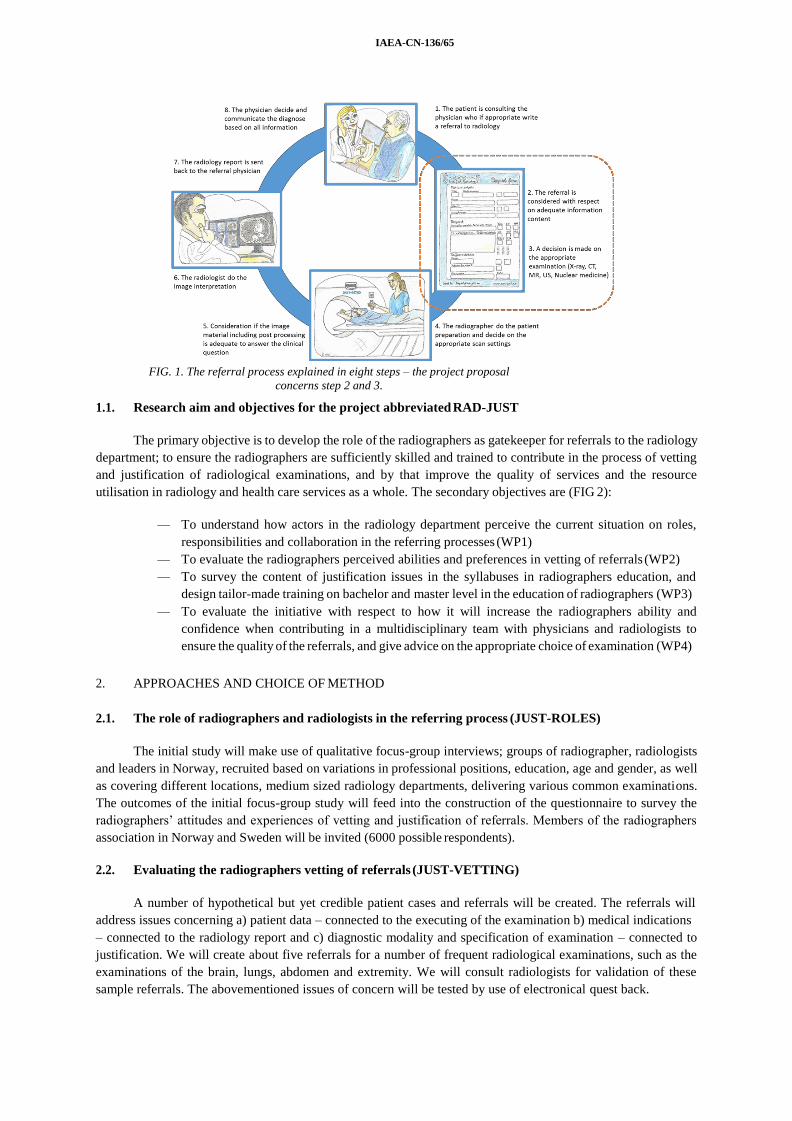
IAEA-CN-136/65
FIG. 1. The referral process explained in eight steps – the project proposal
concerns step 2 and 3.
1.1. Research aim and objectives for the project abbreviated RAD-JUST
The primary objective is to develop the role of the radiographers as gatekeeper for referrals to the radiology
department; to ensure the radiographers are sufficiently skilled and trained to contribute in the process of vetting
and justification of radiological examinations, and by that improve the quality of services and the resource
utilisation in radiology and health care services as a whole. The secondary objectives are (FIG 2):
— To understand how actors in the radiology department perceive the current situation on roles,
responsibilities and collaboration in the referring processes (WP1)
— To evaluate the radiographers perceived abilities and preferences in vetting of referrals (WP2)
— To survey the content of justification issues in the syllabuses in radiographers education, and
design tailor-made training on bachelor and master level in the education of radiographers (WP3)
— To evaluate the initiative with respect to how it will increase the radiographers ability and
confidence when contributing in a multidisciplinary team with physicians and radiologists to
ensure the quality of the referrals, and give advice on the appropriate choice of examination (WP4)
2. APPROACHES AND CHOICE OF METHOD
2.1. The role of radiographers and radiologists in the referring process (JUST-ROLES)
The initial study will make use of qualitative focus-group interviews; groups of radiographer, radiologists
and leaders in Norway, recruited based on variations in professional positions, education, age and gender, as well
as covering different locations, medium sized radiology departments, delivering various common examinations.
The outcomes of the initial focus-group study will feed into the construction of the questionnaire to survey the
radiographers’ attitudes and experiences of vetting and justification of referrals. Members of the radiographers
association in Norway and Sweden will be invited (6000 possible respondents).
2.2. Evaluating the radiographers vetting of referrals (JUST-VETTING)
A number of hypothetical but yet credible patient cases and referrals will be created. The referrals will
address issues concerning a) patient data – connected to the executing of the examination b) medical indications
– connected to the radiology report and c) diagnostic modality and specification of examination – connected to
justification. We will create about five referrals for a number of frequent radiological examinations, such as the
examinations of the brain, lungs, abdomen and extremity. We will consult radiologists for validation of these
sample referrals. The abovementioned issues of concern will be tested by use of electronical quest back.

OLERUD H.M. et.al
2.3. Learning objectives, curriculum and teaching material (JUST-SYLLABUS)
The content of current bachelor curriculums in western countries will be studied as input to the design of
a tailor-made syllabus on justification to inspire both bachelor and master level radiographer educations. One
master level course will be designed for a digital learning environment to recruit students internationally.
2.4. Implementation at selected radiography schools (JUST-IMPLEMENT)
W4 aims to implement the new syllabus and training programme created in WP3 to selected university
radiography colleges and evaluate the initiative. The colleges will be selected in collaboration with the ISRRT; as
planned currently this will involve educational institutions in Australia, Norway, Sweden, Turkey and UK.
Evaluation from the students’ perspective will be done based on focus group interviews in the start, middle and
end of the course. An assessment tool organized for the university radiography colleges will be created.
FIG. 2. How the proposed project RAD-JUST are planned in work-packages chaired by experts
representing both the University College- and University Hospital sector.
3. PRELIMINARY RESULTS
There are recognized differences in syllabuses between radiographer educations in Europe in the content
of justification. The level and length of the education is different, from high school level to bachelor, four - year
education to master level. WP3 will therefore provide a long list of topics that other universities can supplement
from. Even though the scope will vary between bachelor and master level, the following topics are relevant:
— The normative foundation of justification of medical exposures as addressed by the international
organizations in regulations and recommendations
— Typical work flow from doctors office to appropriate investigated patients – health professionals
involved in various steps in the flow chart
— Radiological equipment and modalities with pro- and contras on what sort of clinical questions
they can answer
— Update technology knowledge: Planar X-ray radiographs and fluoroscopy, angio/intervention,
Flat detector CT, Computed Tomography (CT), Magnetic Resonance Imaging (MR), Ultrasound
— Improve anatomy and pathology skills for radiographers
— Referral criteria and guidelines. What are the clinicians’ needs, about what are their concerns?
— A good referral – what kind of information should be included. Appropriateness criteria [12]
— The radiographer as gatekeeper for the justification process; how to work in a multidisciplinary
team
— Radiation risks and risk communication in a person-centred perspective
3

IAEA-CN-136/65
4. DISCUSSION
There is a comprehensive use of radiology in western part of the world. When a patient suffers from certain
clinical symptoms, the physician may need answers from radiology to decide on diagnosis and further treatment.
Several modalities are used in imaging, that is conventional planar X-ray, computed tomography (CT), nuclear
medicine, magnetic resonance imaging (MR) and Ultrasound (US); the two latter do not involve ionizing radiation
but have other pro and contras. It is obviously of major importance to select the appropriate modality and
procedure for the clinical question. Projects like the presented should be part of the next action plan for radiation
protection in the medical sector, since it will have impact in all the following three perspectives:
(a) Insure that the patient are referred to the most appropriate examination and thereby can get the right
diagnose
(b) Reduce the number of unnecessary examinations that will reduce negative health consequences for
patients and costs for the public healthcare system, and
(c) Reduce the radiation dose to individual patients and collective dose to the population.
Despite the many useful applications of ionizing radiation in society there are harmful effects addressed
by bodies like the World Health Organization (WHO), the International Atomic Energy Agency (IAEA) and the
International Commission on Radiological Protection (ICRP). One issue of concern is the increasing use of
radiology and the collective dose burden to the population in western parts of the world [13]. The pillars of
radiation protection in medical exposures are justification, optimization and dose limitation. The principle of
justification applies to three levels: Justification of a practice, generic justification of a defined procedure, and
justification of a procedure for an individual patient [6]. The latter is exactly what this research application
address: how to improve the process of justification of the individual patient procedure in radiology.
REFERENCES
[1] Lekve, K., Olsen, D., Fevolden, A. Gradual transition. Bottlenecks and task sharing in diagnostic imaging. NIFU
Report 46/2013. Nordic Institute for Studies in Innovation Research and Education, Oslo (2013)
[2] Almén, A., Leitz, W. & Richter, S. National Survey on Justification of CT-examination in Sweden. Report 2009:03.
Swedish Radiation Safety Authority, Stockholm (2009).
[3] Oikarinen, H. et al.. Unjustified CT examinations in young patients. European Radiology 5 (2009) 1161-1165.
[4] Lysdahl, K. B., Hofmann, B. M., Espeland, A. Radiologists' responses to inadequate referrals. European Radiology
20(5) (2010) 1227-1233.
[5] Oswal, D., Sapherson, D., Rehman, A. A study of adequacy of completion of radiology request forms. Radiography
15(3) (2009) 209-213.
[6] Malone J. et.al. Justification of diagnostic medical exposures: some practical issues. Report of an International
Atomic Energy Agency Consultation. Br J Radiol. 85(1013) (2012) 523–538. doi:10.1259/bjr/42893576
[7] Vom, J. and I. Williams, Justification of radiographic examinations: What are the key issues? Journal of Medical
Radiation Sciences, 2017. 11: p. 11
[8] Kruse, J. et al. Scrutinized with inadequate control and support: Interns' experiences communicating with and writing
referrals to hospital radiology departments - A qualitative study. Radiography 22(4) (2016) 313-318.
[9] The society of radiographers (2012). Team working in clinical imaging services. Available from:
http://www.sor.org/learning/ document-library/team-working-clinical-imaging/3-team-working-clinical-imaging-
services
[10] Amis, E. S. et al. American College of Radiology white paper on radiation dose in medicine. J Am Coll Radiol 4(5)
(2007) 272-284.
[11] Strudwick, R. M., Day, J. Interprofessional working in diagnostic radiography. Radiography 20(3) (2014) 235-240.
[12] ACR Appropriateness criteria, American College of Radiology, ACR (2016)
[13] European Commission (2014) Medical Radiation Exposure of the European Population Radiation Protection Report
RP180 Part 1/2. https://ec.europa.eu/energy/sites/ener/files/documents/RP180.pdf

DAVIT NADAREISHVILI et al.
WAYS OF IMPLEMENTATION OF SCIENTIFIC AND
EDUCATIONAL ISSUS OF PATIENT’S AND
PERSONNEL’S RADIATION PROTECTION IN
GEORGIA IN THE LIGHT OF BONN CALL FOR
ACTION
DAVIT NADAREISHVILI
Ivane Beritashvili Center of Experimental Biomedicine
Tbilisi, Georgia
FRIDON TODUA
Research Institute of Clinical Medicine
Tbilisi, Georgia
GIORGI ORMOTSADZE
Ivane Beritashvili Center of Experimental Biomedicine
Tbilisi, Georgia
TAMAR SANIKIDZE
Tbilisi State Medical University
Tbilisi, Georgia
SALOME KIPAROIDZE New Vision University
Tbilisi, Georgia
Abstract
An evaluation of the results of the study of modern post-graduate program of the continuous medical education
program “Radiation Protection and Safety” for medical workers were carried out. The study included 200 radiologists from
Tbilisi and different regions of Georgia, among them 35% of dentists, 40% of conventional radiology, 15% of CT and 10%
of specialists in nuclear medicine. The level of knowledge and skills was checked in the field of Physical principles of
medical imaging, Basic radiobiology and radiation risk, Principles of radiation protection, including the competence of
“justification” of radiological procedures and National Radiation safety infrastructure. The results of the tests showed the
necessity for improvement of the educational curriculum at the level of a bachelor in medical profile universities. The
changes required are: the inclusion of a basic radiobiological course in the curriculum of the faculty of medicine and
expansion of the medical imaging methods. In addition, in the residency courses we consider to be appropriate to introduce
“risk management” elements.
1. INTRODUCTION
In January 2013, on the Conference of the National Academy of Sciences of Georgia "Medical radiation
protection issues, the challenges, opportunities, development perspectives", the scientific and educational issues
of implementation of the new International Basic Standards in nuclear and radiation safety were introduced. The
resolutions of the conference in the form of Recommendations were sent to the relevant government agencies.
On the basis of the above mentioned resolutions in the Laboratory of Problem of Radiation Safety of
Beritashvili Center of Experimental Biomedicine the following priority directions for research was developed:
1. Experimental and clinical trials for the development of complex (cyto-genetic, and physiosological)
criteria and elaboration of the test-methods of individual radiosensitivity [1].
1

IAEA-CN-123/45
2. Preparation of methodological basis of assessment of radiation dose and risk in the medical exposition
for Georgian population.
3. Preparation of educational programs on radiobiology and radiation protection, corresponding to the new
International and European standards for undergraduate Bachelor's, and Master's level in Medical
Universities and post graduated training courses for medical professionals [2].
To support this process, Tbilisi State Medical University (TSMU) in collaboration with Georgian National
Association of Radiology and Beritashvili Centre of Experimental Biomedicine initiated several activities:
postgraduate continuing medical education program “Radiation protection” for medical professionals; syllabus
“Medical and biological physics” as a basic course in medical physics for the faculty of medicine (first-year
students), syllabus of elective course “Radiobiology and radiogenic health risk” as an elective course in the
faculty of medicine (fifth-year students), school-seminar programs for medical students from different regions
of Georgia (1–2 year students) and conferences with participation of leading International experts and Georgian
specialists, program “Biomedical engineering educational initiative in Eastern neighboring area” (Tempus
project) and ongoing preparatory work on Master degree Program in Medical Physics.
The present article describes the analysis of the results of the work which was performed for the purpose
of the assessment of learning outcomes of the continuous Medical Education program “Radiation protection and
safety in Medical Radiology” for Medical Professionals and determining the ways for its further development.
MATERIALS AND METHODS
The study included 200 radiologists from Tbilisi and various regions of Georgia, including 35% of
dentists, 40% of conventional radiology, 15% of CT and 10% of specialists in nuclear medicine. The
questionnaire (block of 500 multiple choice questions), (40 questions per listener) was compiled on the Basis of
“Guidelines on Radiation Protection Education and Training of Medical Professionals in the European Union”
[3], which included five main topics:
a) Radiation Hazard – the health effects and mechanisms of low and high dose of radiation.
b) Medical Imaging Physics – characteristics of different types of radiation and mechanisms of their
interaction with matter, physical principles of medical imaging, the image quality, techniques
constructive elements characteristics, the factors influencing the image quality and its indicators.
c) Radiation Protection - basic principles, methods and ways of radiation protection, elements of
operational radiation protection.
d) Radiation Risk Management - health risk assessment, ‘benifit–risk balance assessment and
competence of justification, understanding of quality assurance program.
e) Elements of National Infrastructure for Radiation Safety.
For testing the “Appropriateness Criteria” of American College of Radiology was used.
Basic knowledge and skills was assessed by the number of correct answers, according 5-point scale
system. The block of 40 multiple choice questions included 8 questions from each above mentioned 5 topics (6
questions of basic knowledge - 0.1 point each, and 2 questions revealing skills (quantitative evaluation skills) -
1.2 points each). Listeners were tested before and after training courses with the same tests questionnaire.
The results were processed by parametric and nonparametric statistics methods (Wilkinson, Kruskal-
Wallis H Test, factorial ANOVA).

DAVIT NADAREISHVILI et al.
RESULTS
FIG 1. Results of the assessment of medical specialists in the Continuous Medical Education Program "Radiation Protection
and Safety in Medical Radiology" (before and after training course). A - Radiation Hazard; B – Medical Imaging Physics; C
– Radiation Protection; D - Radiation Risk Management: E - Radiation Safety. Boxes represent 95 % confidence interval of
scores
The results of the study represent statistically significant difference between pre- and post-testing results
for each group of listeners (Fig 1). Different direction and diffrent level of initial knowledge, skills and
competencies was revealed. This differences were also reflected in the high variability of the initial level of
knowledge.
Above mentioned clearly indicates the necessity of further improvement of educational programs in
Medical Radiology. First of all, it concerns the sphere of medical imaging physics and radiation risk
management. It is obvious that the realization of this problem is less likely to be performed within the training
courses.
DISCUSSION
The education of medical professionals that complies with the modern requirements of
radioprotection and safety is a systemic problem and requires a system solution. Taking into the account the
academic curricula in the Higher Medical Schools of Georgia it will be advisable to make the following
changes:
a) Basic course of Medical Physics for Medical Universities must be strengthened by the module of
Medical Visualization Physics (3d year);
b) It is expedient to development of integrated module in Radiobiology and Radiogenic and Health
Risk for Bachelor's programs in Medical Radiology, Radiation Oncology and Radiation Hygiene
courses.
c) Radiation Risk Management (justification) module should be included in residency course of
Medical Radiology.
3

IAEA-CN-123/45
REFERENCES
[1] Alla Zedginidze, Ema Namchevadze, George Ormocadze, Archil Kapanadze, Tamara Nikuradze, and Darejan
Lomidze. “Biodosimetry of Persons Chronically Exposed to Low and Therapeutic Doses of Ionizing Radiation”.
Genome Integr. 7: 12.2016.
[2] F. Todua, D. Nadareishvili, G. Ormotsadze and T. Sanikidze. “BASIC RADIATION PROTECTION
EDUCATION AND TRAINING FOR MEDICAL PROFESSIONALS; GEORGIAN EXPERIENCE AND
FUTURE PERSPECTIVE”. Radiation Protection Dosimetry (2016), pp. 1–3
[3] European Commission Guidelines on radiation protection. Education and training of medical professionals in the
European Union. Radiation Protection No. 175; European Union (2014).
.

PAPÍRNÍK and MÍRKA
AUDITS OF JUSTIFICATION IN
RADIODIAGNOSTICS
Experience from HERCA Campaign on inspection of justification in radio
diagnostics in The Czech Republic.
P. PAPÍRNÍK
State Office for Nuclear Safety
Prague, Czech Republic
Email: [email protected]
H. MÍRKA
University Hospital of Pilsen, Czech Society of Radiology
Pilsen, Czech Republic
Abstract
The Czech Republic participated at the HERCA European inspection Campaign focused on justification in
radiodiagnostics. The Campaign in The Czech Republic was very successful and worthy. In every Czech inspection team a
radiologist evaluated the content of the referrals and the justification of the performed examinations. This highlighted the
essential role of the radiologist in the audit teams that are evaluating the justification in radiology and showed that a team
without a radiologist isn’t able to audit the justification. The Czech Campaign also showed that it is essential not to evaluate
only the content of the referral, but whole the justification of the examinations.
1

IAEA-CN-123/45
1. INTRODUCTION
During the HERCA (Heads of the European Radiological protection Competent Authorities) inspector
workshop on November 2015 an initiative to launch the European inspection Campaign focused on justification
in radiodiagnostics was created. The HERCA Board of Heads then agreed with the intention and HERCA
Working Group for Medical Applications created the common template for the inspection and organized the
Campaign.
The Campaign started on the International Day of Radiology on 8 November 2016 and 18 European
countries including the Czech Republic participated on it.
10 inspections have been done in our country during the Campaign starting on 16 November 2016 and
ending on 31 January 2017.
2. METHODS
The HERCA template for the Campaign included an overview of the regulations and practise of the
country in relation to the justification in radiodiagnostics, a template for the inspection at the hospitals about the
general overview of the hospital concerning if it follows the rules and good practise in the justification, the
statistics of the examinations from the previous week and number of denied and changed referrals, and a
template of number of questions that evaluated the content of 10 referrals randomly chosen from the previous
week.
Because the Czech radiation protection inspectors aren’t allowed to look at the clinical data of the
patients (including the referrals), the Czech radiation protection competent authority SÚJB asked the Czech
Radiological Society to participate at the Campaign. The Radiological Society then sent one radiologist to every
inspection team. The radiologists in the team were evaluating the content of the referrals and they judged the
justification practise of the hospital.
We used the advantage of having a radiologist in every team and in 4 hospitals we were not evaluating
only the content of the referrals, but whole the process of justification for each and every of the 10 checked
referrals and examinations. In these 4 hospitals we added a question in the template: “Was the examination
justified (concerning all the information at the referral and all the other information about the examination that
the practitioners received from the referrer and patient)?” And the radiologist in the team answered this question
for every evaluated examination during every inspection.
We were checking mainly the referrals for general radiography and CT – 5 CT and 5 general radiography
referrals in each hospital.
3. RESULTS
The formal columns of the referrals (e.g. patient’s identification, age, sex, referrer’s identification, his or
her signature and contact information, date of the referral) were present in 99 % of cases.
Identification of pregnancy was missing only in 2 % of the relevant cases.
The clinical information (sufficient clinical information / medical history available, information about
previous examinations, clinical question asked, type of examination given in referral) were missing at 13 % of
cases. 39 % of the referrals didn’t contain some of the clinical information. 25 % of the referrals missed one or
more of this information: sufficient clinical information / medical history available, clinical question asked, type
of examination given in referral. 40 % of the referrals didn’t contain the information about previous
examinations.
During the inspections when we found that something clinically important was missing in the referral the
practitioners explained how they handled with that particular referral. In almost all of the cases they followed
the legal requirements: they contacted the referrer and asked him about the patient’s clinical data that were
missing, insufficient, inadequate or inappropriate. In some cases were the additional data available in the clinical
information system. The practitioners of the controlled hospital that were present at the inspection described
these procedures in very details, so it was obvious that they truthfully described what they had been really doing
in these cases. This was the advantage of auditing the examinations performed during the last week - the
practitioners remembered most of them very well and when they were describing them it was easy to recognize
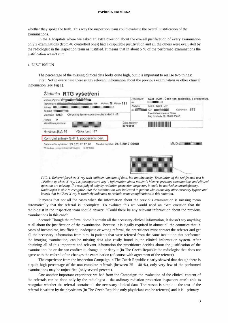
PAPÍRNÍK and MÍRKA
whether they spoke the truth. This way the inspection team could evaluate the overall justification of the
examinations.
In the 4 hospitals where we asked an extra question about the overall justification of every examination
only 2 examinations (from 40 controlled ones) had a disputable justification and all the others were evaluated by
the radiologist in the inspection team as justified. It means that in about 5 % of the performed examinations the
justification wasn’t sure.
4. DISCUSSION
The percentage of the missing clinical data looks quite high, but it is important to realise two things:
First: Not in every case there is any relevant information about the previous examination or other clinical
information (see Fig 1).
FIG. 1. Referral for chest X-ray with sufficient amount of data, but not obviously. Translation of the red framed text is „Follow-up chest X-ray, 1st. postoperative day“. Information about patient’s history, previous examinations and clinical
question are missing. If it was judged only by radiation protection inspector, it could be marked as unsatisfactory.
Radiologist is able to recognize, that the examination was indicated in patient who is one day after coronary bypass and knows that in Chest X-ray is routinely indicated to exclude acute complications in this situation.
It means that not all the cases when the information about the previous examination is missing mean
automatically that the referral is incomplete. To evaluate this we would need an extra question that the
radiologist in the inspection team should answer: “Could there be any relevant information about the previous
examinations in this case?”
Second: Though the referral doesn’t contain all the necessary clinical information, it doesn’t say anything
at all about the justification of the examination. Because it is legally required in almost all the countries that in
cases of incomplete, insufficient, inadequate or wrong referral, the practitioner must contact the referrer and get
all the necessary information from him. In patients that were referred from the same institution that performed
the imaging examination, can be missing data also easily found in the clinical information system. After
obtaining all of this important and relevant information the practitioner decides about the justification of the
examination: he or she can confirm it, change it, or deny it (in The Czech Republic the radiologist that does not
agree with the referral often changes the examination (of course with agreement of the referrer).
The experience from the inspection Campaign in The Czech Republic clearly showed that though there is
a quite high percentage of the non-complete referrals (between 25 – 40 %), only very few of the performed
examinations may be unjustified (only several percent).
One another important experience we had from the Campaign: the evaluation of the clinical content of
the referrals can be done only by the radiologist – the ordinary radiation protection inspectors aren’t able to
recognize whether the referral contains all the necessary clinical data. The reason is simple – the text of the
referral is written by the physicians (in The Czech Republic only physicians can be referrers) and it is primary
3
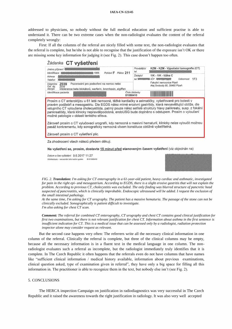
IAEA-CN-123/45
addressed to physicians, so nobody without the full medical education and sufficient practise is able to
understand it. There can be two extreme cases when the non-radiologist evaluates the content of the referral
completely wrongly:
First: If all the columns of the referral are nicely filled with some text, the non-radiologist evaluates that
the referral is complete, but he/she is not able to recognise that the justification of the exposure isn’t OK or there
are missing some key information for judging it (see Fig. 2). This case doesn’t happen too often.
FIG. 2. Translation: I'm asking for CT enterography in a 61-year-old patient, heavy cardiac and asthmatic, investigated
for pain in the right epi- and mesogastrium. According to EGDS, there is a slight erosive gastritis that will not explain the
problem. According to previous CT, cholecystitis was excluded. The only finding was blurred structure of pancretic head
suspected of pancreatitis, which is clinically improbable. Endoscopic ultrasound will be added. I request the exclusion of
the small intestinal pathology.
At the same time, I'm asking for CT urography. The patient has a massive hematuria. The passage of the stone can not be
clinically excluded. Sonographically is patient difficult to investigate.
I'm also asking for chest CT scan.
Comment: The referral for combined CT enterography, CT urography and chest CT contains good clinical justification for
first two examinations, but there is not relevant justification for chest CT. Information about asthma in the first sentence is
insufficient indication for CT. This is a medical issue that can be assessed only by a radiologist, radiation protection
inspector alone may consider request as relevant.
But the second case happens very often: The referrers write all the necessary clinical information in one
column of the referral. Clinically the referral is complete, but three of the clinical columns may be empty,
because all the necessary information is in a fluent text in the medical language in one column. The non-
radiologist evaluates such a referral as incomplete, but the radiologist immediately truly identifies that it is
complete. In The Czech Republic it often happens that the referrals even do not have columns that have names
like “sufficient clinical information / medical history available, information about previous examinations,
clinical question asked, type of examination given in referral”, they have only a big space for filling all this
information in. The practitioner is able to recognize them in the text, but nobody else isn’t (see Fig. 2).
5. CONCLUSIONS
The HERCA inspection Campaign on justification in radiodiagnostics was very successful in The Czech
Republic and it raised the awareness towards the right justification in radiology. It was also very well accepted

PAPÍRNÍK and MÍRKA
by the controlled / audited hospitals and the practitioners. The Czech Radiological Society appreciated the
Campaign and the questions from the European template and the Campaign helped to create a very good
relationship between the Czech radiologists and the radiation protection competent authority. During the
Campaign a number of specific professional questions and issues, that should be solved in cooperation of the
Radiological Society, the Radiation protection competent authority, Ministry of Health and professional bodies,
was identified. The experience from the Campaign is already being used widely during the ongoing upgrade of
the Czech National Radiological Standards, Czech Referral Guidelines and the rules for the clinical audits.
The experience from the Campaign showed that evaluation or audit of justification in radiodiagnostics
essentially needs following:
— The radiologist must be the main person in the team of the auditors of the justification;
— No-one else than the radiologist is able to evaluate the completeness of thereferral;
— In some cases there is no relevant clinical history that could be present at the referral – the auditor-
radiologist is able to recognize these cases;
— If the referral isn’t complete, the evaluation must continue by asking the practitioners what they did
with this case (therefore only referrals from few days back should be evaluated, so they remember
them), because the completeness or incompleteness of the referral doesn’t indicate anything about the
justification or non-justification of the performed examination;
— After evaluating of the content of the referral and after the oral information from the practitioners about
the case, the auditor-radiologist should judge if the examination was or wasn’t justified (concerning all
the information at the referral and all the other information about the examination that the practitioners
received from the referrer and patient);
— In the 10 Czech hospitals controlled / audited during the Campaign only few percent of the examination
were identified as possibly unjustified.
ACKNOWLEDGEMENTS
We would like to thank HERCA Working Group on Medical Applications for the initiative of the
inspection Campaign and for organizing it and for preparing the European template for it – the Campaign helped
a lot in creating a good relationship between the Czech radiologists and radiation protection competent authority
and helped to identify many of the issues that need to be solved in the radio diagnostics (not only in context of
justification).
We would like to thank the Czech Radiological Society for very positive reaction on the request for their
participation at the Campaign. And we would like to thank Daniel Bartušek, Aleš Bílek, Miroslav Heřman,
Marek Mechl, Hynek Mírka and Jiří Weichet the participating radiologists for their essential role in the
inspections and for all the knowledge that they kindly shared during it.
We would like to thank also the radiation protection inspectors that agreed to contribute on the Campaign
and didn’t mind the complications of their standard inspections. Especially we would like to thank Jitka
Nožičková who was the main organizer of the Campaign in the Czech Republic and who participated at 60 % of
the inspections of the Campaign.
5
CILLIERS and PITCHER
THE IMPACT OF REAL-TIME ELECTRONIC JUSTIFICATION OF
DIGITAL IMAGING REQUESTS
WILLIE G. CILLIERS
Division of Radiodiagnosis, Department of Medical Imaging and Clinical Oncology, Faculty of Medicine
and Health Sciences, Stellenbosch University, Cape Town, South Africa.
RICHARD D. PITCHER
Division of Radiodiagnosis, Department of Medical Imaging and Clinical Oncology, Faculty of Medicine
and Health Sciences, Stellenbosch University, Cape Town, South Africa.
Email: [email protected] (corresponding author)
Abstract
Introduction: Although real-time, electronic justification of digital imaging requests can be seamlessly incorporated
into modern radiological workflow, there have been few reports of its implementation and impact. Aim: To assess the impact
of real-time justification on imaging equipment utilization in a resource-limited setting. Method: The justification decisions
on non-emergency special radiological requests at a tertiary-level South African teaching hospital for the period 1 December
2012 through 31 December 2013 were reviewed. Cancelled requests were analysed by modality and the reason for
cancellation. The impact of cancelled examinations on departmental workload was computed. Results: Of 55589 requests,
2704(4.9%) were cancelled. Duplication accounted for more than two-thirds of cancellations (1831/2704; 67.7%),
inappropriate requests for almost one-third (846/2704; 31.2%) and medical contra-indications for less than one percent
(37/2704; 0.06%), with similar proportions across modalities. Computed tomography (CT) requests accounted for more than
half the cancellations (1361/2704; 50.3%). Real-time justification contributed to saving 111 routine modality work-days (CT
30, fluoroscopy 30, sonography 18, MR 17, intervention 16). Conclusion: Real-time electronic justification of imaging
requests is feasible and has the potential to effect substantial resource savings, limit unnecessary radiation exposure and
decrease patient waiting times for elective imaging investigations.
1. INTRODUCTION
Spectacular technological advances in radiology in the past forty years have stimulated the burgeoning
global demand for diagnostic imaging.[1] From 1988 to 2008, the number of imaging studies performed
worldwide increased from 1.38 to 3.14 billion annually, while the global per capita effective medical radiation
dose increased from 0.4mSv to 0.62mSv between 1991-1996 and 1997-2007.[2] However, increases in imaging
utilization are not necessarily associated with incremental patient benefit.[3,4] There are thus concerns that
current global imaging practices are unsustainable, being costly and potentially hazardous.[5]
There is evidence that unnecessary radiological investigations exacerbate worldwide imaging service
pressures.[6] Physician referral patterns, unrealistic patient expectations, defensive medicine, self-referral and
duplication contribute to unnecessary examinations.[5] Efforts to minimize inappropriate imaging include the
drafting of evidence-based imaging appropriateness criteria, physician and patient education initiatives, imaging
facility accreditation and revised imaging funding models.[7-12] Although there have been attempts to quantify
the monetary cost of inappropriate imaging [13], there have been only limited analyses of the impact of
unnecessary imaging on equipment utilization and patient waiting times.
Developments in information technology have paralleled advances in diagnostic imaging over the past
four decades. Digital radiology departments utilizing electronic workflow are now commonplace in well-
resourced environments and are increasingly being introduced into resource-limited settings, where the capacity
for remote teleradiology reporting represents a particular benefit.[14,15] The digital imaging workflow is driven
by the radiology information system (RIS), which has a number of efficiency-enhancing features. These include
electronic clinician requests and the capacity for requests to be informed by real-time, embedded, evidence-
based imaging algorithms. Such clinical decision-support (CDS) systems for special radiological investigations,
are increasingly being implemented [14,16], but to date have shown only modest clinical impact.[10,17,18] The
low impact has been attributed to the radiologist not being integral to the workflow.[19]
Justification is an optional step in the modern RIS workflow, allowing radiologist-driven, real-time evaluation of the appropriateness of electronic imaging requests. Digital justification allows radiologists to
evaluate new imaging requests while reviewing the patient’s complete imaging history, and to approve, modify
or cancel studies. Cancellation is accompanied by an electronic explanation to the referring clinician. Data and
1

IAEA-CN-255
timelines relating to cancelled requests are archived on the RIS, facilitating analysis of institutional referral and
imaging patterns.
In September 2012, the radiology department of Tygerberg Hospital (TBH), a 1386-bed tertiary–level
public-sector teaching hospital in Cape Town, South Africa (SA), converted to a fully digital workflow and
introduced radiologist-driven real-time electronic justification of all non-emergency requests for computed
tomography (CT), magnetic resonance (MR), fluoroscopy (RF), ultrasound (US) and interventional procedures
(IR). The commissioning was the culmination of more than two years of meticulous electronic workflow
planning and an extensive institutional change-management program. Justification is performed on any RIS
workstation invoking existing departmental protocols and published international guidelines to assess the
appropriateness of imaging requests.[7,8] The TBH Radiology Department operates under the prevailing
resource constraints of SA’s public health sector [20], performing approximately 190,000 examinations
annually. It has a single 1.5 Tesla MRI unit, three CT scanners, five sonar machines, and dedicated single suites
for mammography, fluoroscopy machine and vascular-interventional procedures. There are eight Consultant
Radiologists, twenty-five Radiology Residents and three Sonographers. The institutional brief was to introduce
real-time justification without any change to the radiology staff complement or working hours, and to complete
the justification process within an hour of receipt of requests for non-emergencyimaging.
To the best of our knowledge there has been only one study of the impact of electronic justification on
imaging utilization.[21] Ingraham and co-workers recently reported that electronic justification of outpatient
CT and MRI requests by a large US-based healthcare provider resulted in modification of 9% and rejection of
4% of requests. They concluded that justification, through inclusion of the radiologist in patient care,
contributed to safer and more cost-effective imaging methods.
The aim of this study was to assess the impact of real-time cancellation of unnecessary imaging
procedures through electronic justification of non-emergency requests for special radiological investigations on
imaging equipment utilization in a large tertiary hospital in a resource-constrained environment.
2. METHODS
This was a retrospective analysis of electronic radiological justifications at TBH from 1 December 2012
through 31 December 2013. A customised search of the institutional RIS was conducted for all non-emergency
requests for CT, MRI, US, RF and IR procedures in the review period. Included data were sorted by modality
and stratified by justification outcome, into “approved/modified” or “cancelled”. Cancelled requests served as
the primary measure of the impact of justification on imaging equipment utilization and were further analysed
by the reason for cancellation. Data were summarized in overall and modality-specific descriptive statistics. The
average clinical output in each of the modalities for a normal, 8-hour week-day was calculated for the review
period and used to compute the impact of request cancellation on equipment utilization. This study was
approved by the Health Research Ethics Committee of Stellenbosch University.
3. RESULTS
Of 55589 requests, 2704 (4.9%) were cancelled. Duplication accounted for more than two-thirds of
cancellations (1831/2704; 67.7%), inappropriate requests for almost one-third (846/2704; 31.2%) and medical
contra-indications for less than 1% (37/2704; 0.06%), with similar proportions across modalities. (Table 1) CT
requests represented more than half the cancellations (1361/2704; 50.3%). Fluoroscopic requests had the highest
proportion of cancelled examinations (244/2779; 8.8%) and MR the lowest (192/5543; 3.5%). Typical reasons
for requests being deemed inappropriate were inability of further imaging to augment existing radiological
findings or influence clinical management, and other examinations outside the radiology department, such as
endoscopy, scintigraphy or echocardiography being considered more appropriate. Real-time justification
contributed to a total saving of 111 modality work-days. (Table 2)
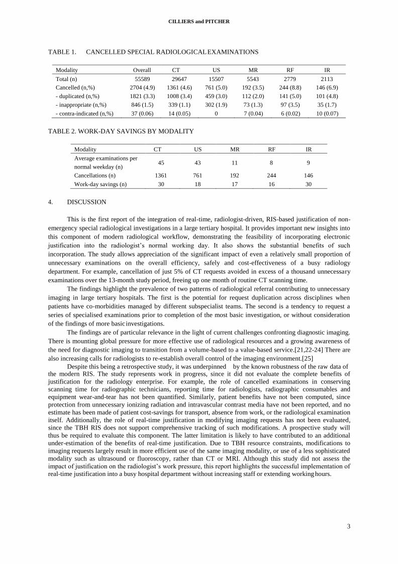
CILLIERS and PITCHER
TABLE 1. CANCELLED SPECIAL RADIOLOGICAL EXAMINATIONS
Modality Overall CT US MR RF IR
Total (n) 55589 29647 15507 5543 2779 2113
Cancelled (n,%) 2704 (4.9) 1361 (4.6) 761 (5.0) 192 (3.5) 244 (8.8) 146 (6.9)
- duplicated (n,%) 1821 (3.3) 1008 (3.4) 459 (3.0) 112 (2.0) 141 (5.0) 101 (4.8)
- inappropriate (n,%) 846 (1.5) 339 (1.1) 302 (1.9) 73 (1.3) 97 (3.5) 35 (1.7)
- contra-indicated (n,%) 37 (0.06) 14 (0.05) 0 7 (0.04) 6 (0.02) 10 (0.07)
TABLE 2. WORK-DAY SAVINGS BY MODALITY
Modality CT US MR RF IR
Average examinations per
normal weekday (n)
45
43
11
8
9
Cancellations (n) 1361 761 192 244 146
Work-day savings (n) 30 18 17 16 30
4. DISCUSSION
This is the first report of the integration of real-time, radiologist-driven, RIS-based justification of non-
emergency special radiological investigations in a large tertiary hospital. It provides important new insights into
this component of modern radiological workflow, demonstrating the feasibility of incorporating electronic
justification into the radiologist’s normal working day. It also shows the substantial benefits of such
incorporation. The study allows appreciation of the significant impact of even a relatively small proportion of
unnecessary examinations on the overall efficiency, safely and cost-effectiveness of a busy radiology
department. For example, cancellation of just 5% of CT requests avoided in excess of a thousand unnecessary
examinations over the 13-month study period, freeing up one month of routine CT scanning time.
The findings highlight the prevalence of two patterns of radiological referral contributing to unnecessary
imaging in large tertiary hospitals. The first is the potential for request duplication across disciplines when
patients have co-morbidities managed by different subspecialist teams. The second is a tendency to request a
series of specialised examinations prior to completion of the most basic investigation, or without consideration
of the findings of more basic investigations.
The findings are of particular relevance in the light of current challenges confronting diagnostic imaging.
There is mounting global pressure for more effective use of radiological resources and a growing awareness of
the need for diagnostic imaging to transition from a volume-based to a value-based service.[21,22-24] There are
also increasing calls for radiologists to re-establish overall control of the imaging environment.[25]
Despite this being a retrospective study, it was underpinned by the known robustness of the raw data of the modern RIS. The study represents work in progress, since it did not evaluate the complete benefits of
justification for the radiology enterprise. For example, the role of cancelled examinations in conserving
scanning time for radiographic technicians, reporting time for radiologists, radiographic consumables and
equipment wear-and-tear has not been quantified. Similarly, patient benefits have not been computed, since
protection from unnecessary ionizing radiation and intravascular contrast media have not been reported, and no
estimate has been made of patient cost-savings for transport, absence from work, or the radiological examination
itself. Additionally, the role of real-time justification in modifying imaging requests has not been evaluated,
since the TBH RIS does not support comprehensive tracking of such modifications. A prospective study will
thus be required to evaluate this component. The latter limitation is likely to have contributed to an additional
under-estimation of the benefits of real-time justification. Due to TBH resource constraints, modifications to
imaging requests largely result in more efficient use of the same imaging modality, or use of a less sophisticated
modality such as ultrasound or fluoroscopy, rather than CT or MRI. Although this study did not assess the
impact of justification on the radiologist’s work pressure, this report highlights the successful implementation of
real-time justification into a busy hospital department without increasing staff or extending working hours.
3

IAEA-CN-255
5. CONCLUSION
Real-time electronic justification of imaging requests is feasible and has the potential to effect substantial
resource savings, limit unnecessary radiation exposure and decrease patient waiting times for elective imaging
investigations.
6. REFERENCES
1. Smith-Bindman, R., Miglioretti, D., Johnson, E., et al. Use of diagnostic imaging studies and associated radiation
exposure for patients enrolled in large integrated health care systems, 1996-2010. JAMA. 2012;307(22):2400-9.
2. United Nations Scientific Committee on the Effects of Atomic Radiation (UNSCEAR) 2008 Report. Volume I.
Annex A. http://www.unscear.org/docs/publications/2008/UNSCEAR_2008_Report_Vol.I.pdf
3. Baker, L. Acquisition of MRI equipment by doctors drives up imaging use and spending. Health Aff (Millwood)
2010;29(12):2252-9.
4. Hughes, D., Bhargavan, M., Sunshine, J. Imaging self-referral associated with higher costs and limited impact on
duration of illness. Health Aff (Millwood) 2010;29:2244-51.
5. Hendee, W. Addressing overutilization in medical imaging. Radiology. 2010;257(1):240-5.
6. Gonzales, J. Imaging professionals' beliefs on overutilization of CT and MRI exams. Radiol Manage.
2011;33(6):41-6.
7. American College of Radiology. ACR appropriateness criteria. www.acr.org/Quality-Safety/Appropriateness-
Criteria.pdf.
8. The Royal College of Radiologists. Imaging referral guidelines. iRefer: Making the best use of clinical radiology.
http://www.irefer.org.uk
9. Durand, D., Kohli, M. Advanced Practice Quality Improvement Project: how to influence physician radiologic
imaging ordering behavior. J Am Coll Radiol. 2014;11(12):1155-9.
10. Sheng, A., Castro, A., Lewiss, R. Awareness, Utilization, and Education of the ACR Appropriateness Criteria: A
Review and Future Directions. J Am Coll Radiol. 2016;13(2):131-6.
11. Harvey, H., Chow, D., Boston, M., et al. Initial outcomes from federally mandated accreditation site surveys of
advanced diagnostic imaging facilities performed by the ACR. J Am Coll Radiol. 2014;11(7):686-91.
12. Dhanoa, D., Burton, K.R., de Tilly, L., et al. Effect of a preauthorization policy on the utilization rate of after-
hours emergency department neuroradiology computed tomography. Can J Emerg Med. 2013;15(3):161-6.
13. Friedman, D., Smith, N., Bree, R., et al. Experience of an academic neuroradiology division participating in a
utilization management program. J Am Coll Radiol. 2009;6(2):119-24
14. Nance, J. The Future of the Radiology Information System. AJR Am J Roentgenol. 2013; 200:1064–70
15. Saliba, V., Legido-Quigley, H., Hallik, R., et al. Telemedicine across borders: a systematic review of factors that
hinder or support implementation. Int J Med Inform. 2012;81(12):793-809.
16. Sistrom, C. The ACR appropriateness criteria: translation to practice and research. J Am Coll Radiol. 2005;2(1):
61–7.
17. Moriarity, A., Klochko, C., O'Brien, M., et al. The effect of clinical decision support for advanced inpatient
imaging. J Am Coll Radiol. 2015;12(4):358-63.
18. Ip, I., Schneider, L., Marchello, D., et al. Adoption and meaningful use of computerized physician order entry with
an integrated clinical decision support system for radiology: ten-year analysis in an urban teaching hospital. J Am
Coll Radiol. 2012;9(2):129-36.
19. Duszak, R., Berlin, J. Utilization management in radiology, part 1: rationale, history, and current status. J Am Coll
Radiol. 2012;9(10):694-9.
20. Kabongo, J., Nel, S., Pitcher, R. An analysis of licenced South African diagnostic imaging equipment. Pan Afr
Med J. 2015; 22:57.
21. Ingraham, B., Miller, K., Iaia, A., et al. Reductions in High-End Imaging Utilization With Radiology Review and
Consultation. J Am Coll Radiol. 2016;13(9):1079-82.
22. Lau, L. Leadership and management in quality radiology. Biomed Imaging Interv J. 2007;3(3):e21.
23. Remfry, A., Abrams, H., Dudzinski, D., et al. Assessment of inpatient multimodal cardiac imaging appropriateness
at large academic medical centers. Cardiovasc Ultrasound. 2015;13(1):44.
24. Sarwar, A., Boland, G., Monks, A., et al. Metrics for Radiologists in the Era of Value-based Health Care Delivery.
Radiographics. 2015;35(3):866-76.
25. Levin DC. The 2014 RSNA Annual Oration in Diagnostic Radiology: Transitioning from volume-based to value-
based practice—a meaningful goal for all radiologists or a meaningless platitude? Radiology 2015;275:314-20.

CLINICAL IMAGING REFERRAL GUIDELINES:
WHERE ARE WE NOW?
M.H. Reed
Department of Diagnostic Imaging, Children’s Hospital
Winnipeg, Canada
Abstract
In order to enhance the implementation of the principle of justification the Bonn Call-for-Action
recommended that clinical imaging referral guidelines (CIRGs) should be implemented globally. The purpose of this
study was to assess the current state of CIRGs. Methods: This report is based on searches of PubMed and of
relevant websites on the internet and on information gained from attendance at professional meetings and
discussions with many colleagues, national and international. Results: Several organizations continue to update and
produce new CIRGs. There is increasing activity worldwide in the development, standardization and
implementation of medical guidelines which is relevant to the development of CIRGs. Radiology is leading the
way in the development of computerized decision support (CDS) by integrating CIRGs into computerized order
entry systems (CPOEs). The development of CDS has raised new issues, including the number of CIRGs which are
needed and the potential effect of other clinical practice guidelines on CIRGs. Conclusions: CIRGs continue to be
upgraded and developed but this must be done in the wider context of all medical guidelines. CDS is an important
advance for medical guidelines but it also raises new issues.
1.INTRODUCTION
In order to enhance the implementation of the principle of justification, the Bonn Call-for-Action in 2013
recommended that clinical imaging referral guidelines (CIRG s) should be implemented globally. During the
following four years CIRGs continue to be revised and produced. There have also been a number of developments in
health care guidelines generally, and computerized decision support (CDS) has become a reality. The paper will
review these developments and assess the current state of CIRGs.
2.METHODS
The material for the paper is based on searches of PubMed and of relevant websites on the internet and on
information gained from attendance at professional meetings and discussions with many colleagues both national
and international.
3.RESULTS
A number of organizations were producing CIRGs at the time of the Bonn Call-for-Action, including the Royal
College of Radiologists (RCR), the American College of Radiology (ACR), la Société Française de Radiologie
(SFR), Diagnostic Imaging Pathways (DIP), and the Canadian Association of Radiologists (CAR). The RCR has
recently released the 8th edition of its CIRGs, iRefer [1]. The ACR also continues to revise its Appropriateness
Criteria and to add new CIRGs [2], as does DIP [3]. The CAR recently completed a revision of all its CIRGs and
made the revised version available on its web site in 2014 [4]. It has recently set up a working group to advise the
Board on how it should maintain and enhance the effectiveness of its CIRGs.
Since the Bonn Call-for-Action there have also been a number of developments in health care guidelines at large.
Guidelines International Network (GIN) [5] is an organization which represents and promotes consultation and
cooperation among many organizations and individuals internationally who are involved in the development of

health care guidelines. Among other activities GIN endorses and supports other organizations which have developed
standards for various aspects of guideline development. These include AGREE II and GRADE. AGREE II has
developed standards for assessing the quality of medical guidelines. These can also be used to assist guideline
developers in producing high quality guidelines. AGREE II defines six domains of quality: scope and purpose,
stakeholder involvement including users and patients, rigour of development, clarity and presentation, applicability
and editorial independence [6]. The AGREE II instrument was assessed at two technical meetings hosted by the
International Atomic Energy Agency and was evaluated for its relevance to CIRGs. The consensus was that the
majority of items in AGREE II should apply uniformly to CIRGs But some should allow for regional differences
[7]. The Grading of Recommendations Assessment, Development and Evaluation working group (GRADE) has
developed standards for evaluating the quality of evidence and the strength of recommendations in guidelines [8].
GRADE states, among other criteria, that recommendations in guidelines should be categorized as strong or weak,
and that a strong recommendation should be based on high quality evidence.
The most important advance in the last few years for improving the effectiveness of CIRGs is the development of
robust software which allows CIRGs to be integrated into computerized order entry systems thus providing
computerized decision support (CDS) to physicians as part of their daily workflow. Diagnostic imaging CDS has
been shown to be effective in decreasing inappropriate requests for diagnostic imaging [9]. However, the
development of CDS has raised important new issues in the area of CIRGs. One of these issues is the question of
how many CIRGs should be integrated into CDS systems and in turn how many CIRGs do we actually need.
Another important issue is the challenge of clinical care pathways (CCPs).
4. DISCUSSION
The interest in CIRGs extends well beyond the countries discussed above which are producing their own CIRGs.
The RCR’s iRefer has been adopted by a number of countries which do not have the resources to develop their own
CIRGs, and the iRefer app is used widely throughout Europe and in many other countries worldwide [10 ]. The
International Society of Radiology Quality and Safety Alliance [ISRQSA] is also interested in developing an
international set of CIRGs [11].
The methodologies of RCR’s iRefer and of DIP have been accredited by the National institute for Health and Care
excellence (NICE) [1, 12 ], and the methodology of the ACR Appropriateness Criteria has been endorsed by the
Agency for Healthcare Research and Quality (AHRQ) [13 ]. However, some of the international standards for
guideline development may not always be appropriate for CIRGs. For instance, GRADE emphasizes evidence of
accuracy as being very important in determining the strength of recommendations for diagnostic tests [14].
However, accuracy may not always be the most important evidence to consider in developing recommendations for
diagnostic imaging [15, 16].
There is growing interest in CDS internationally. ACR has developed a CDS system which incorporates the
Appropriateness Criteria, and it is now available commercially [17]. The European Society of radiology is
collaborating with the ACR to develop a European version of CDS [18]. The RCR has also incorporated iRefer into
a CDS system which it is now testing in pilot projects [19 ].
These CDS systems incorporate all the organizations CIRGS, but this may in fact not be the best approach to the
development of CDS systems. The experience with drug alerts has identified a phenomenon called alert fatigue
which happens when users get too many alerts and start to ignore all the alerts [20 ]. It is possible that this
phenomenon may also be true for diagnostic imaging CDS systems which incorporate a large number of guidelines.
The Centers for Medicare and Medicaid Services (CMS) in the United States considered the options of requiring
CDS systems which they would approve to have a large number of CIRGs or only a core group of CIRGs. They
analyzed their own data and determined that 40% of the requests for advanced imaging were covered by eight
clinical conditions. Therefore, they have decided that they will require only CIRGs for those eight clinical priority
areas in any CDS systems which they approve [ ].
CCPs are algorithms designed to map out the most appropriate care for patients in a given clinical conditions.
Diagnostic imaging is frequently part of CCPs. Software is now becoming available that allows the integration of
CCPs into electronic medical records. As CCPs become more widely integrated into electronic medical records
there will probably be less need for dedicated diagnostic imaging CDS and radiologists will have to start working
more closely with other clinicians to ensure that the recommendations in the CCPs are appropriate.
5. CONCLUSIONS
There is growing international interest in CIRGs. However, groups who develop CIRGs need to be aware of the
international standards for medical guidelines. CDS is widely considered to be the most effective way of getting
CIRGs used and there is growing international interest that the in this technology. However CDS is raising new
issues ofits own.
6.REFERENCES
[1] Remedios D, France B, Alexander M. Clin Radiol 72 (2017) 705-707
[2] https://www.acr.org/Quality-Safety/Appropriateness-Criteria
[3] https://www.healthdirect.gov.au/partners/diagnostic-imaging-pathways?_escaped_fragment_=
[4] http://www.car.ca/en/standards-guidelines/guidelines.aspx
[5] http://www.g-i-n.net/
[6] http://www.agreetrust.org/wp-content/uploads/2013/10/AGREE-II-Users-Manual-and-23-item-
Instrument_2009_UPDATE_2013.pdf
[7] Remedios D, Brink J, Holmberg O, Kawooya M, Mendelson R, Naidoo A, Reed M, Bettmann M, Clinical imaging guidelines
part1: a proposal for uniform methodology. J Am Coll Radiol 12 (2015) 45-50
[8] http://www.gradeworkinggroup.org/
[9] Hentel K, Menard A, Khorasani R. New CMS clinical decision support recommendations: a potential opportunity with major
challenges. Radiology 283 (2017) 10-13
[10] Remedios D., RCR, personal communication, 2017
[11] Frija, G, ISRQSA, personal communication, 2017
[12] http://imagingpathways.health.wa.gov.au/index.php/endorsements
[13] https://www.acr.org/Quality-Safety/Appropriateness-Criteria/About-AC
[14] Schünemann HJ, Oxman AD, Brozek J, Glasziou P, Jaeschke R, Vist GE, Williams JW Jr, Kunz R, Craig J. GRADE:
grading quality of evidence and strength of recommendations for diagnostic tests and strategies. BMJ 336 (2008) 1106-1110
[15] Reed MH. Evidence in diagnostic imaging: going beyond accuracy. J Am Coll Radiol 9 (2012) 90 -92

[16] Reed MH. Evidence and the role of diagnostic imaging. J Am Coll Radiol 11 (2014) 4-5
[17] https://www.acr.org/quality-safety/appropriateness-criteria/acr-select
[18] http://www.nationaldecisionsupport.com/esriguide/index.html
[19] https://rcr.ac.uk/posts/introducing-irefer8-updated-diagnostic-imaging-guidelines-referring-clinicians
[20] Lacson R, Ip I, Hentel KD, Malhotra S, Balthazar P, Langlotz CP, Raja AS, Khorasaani R. Medicare imaging
demonstration: assessing attributes of appropriate use criteria and their influence on ordering behavior. AJR 208 (2017) 1051-
1057
[21] https://www.cms.gov/medicare/quality-initiatives-patient-assessment-instruments/appropriate-use-criteria-program/index.html


