Faculty of Sciences
UNRAVELING THE MECHANISM OF THE LIGHTSTRUCK FLAVOR OF BEER
Arne Heyerick
Thesis submitted to the Faculty of Sciences in order to obtain the degree of
Doctor in Sciences: Biochemistry
Promoter
Prof. dr. D. De Keukeleire
Academic year 2000-2001

i
Acknowledgements
Herewith, I wish to express my sincere gratitude and appreciation to all individuals who have
made contributions to the accomplishment of this Ph.D. project.
Above all, I am indebted to the promoter of my doctoral thesis, Prof. Dr. Denis De Keukeleire, for
his versed guidance, for sharing his authoritative knowledge of both the phytochemistry of hops and
photochemistry, for his continuing confidence, and for his scrutinous revision of the manuscript.
I deeply acknowledge the close contacts with Prof. Dr. Malcolm D.E. Forbes, who gave me the
opportunity to spend a fruitful stay in his laboratory and contributed invaluably both to my scientific
insights and to the content of this doctoral work. Furthermore, Dr. Colin S. Burns is profoundly
thanked for sharing his expertise on time-resolved EPR with respect to direct irradiation of hop-
derived compounds, while I am grateful to Dr. Haruhiko Yashiro, Ms. Vanessa McCaffrey, and Ms.
Chrystal Bruce for their highly appreciated assistance with EPR experiments and for their friendship
during my stay at the University of North Carolina at Chapel Hill, USA. The ‘Fonds voor
Wetenschappelijk Onderzoek – Vlaanderen’,Brussels, Belgium, is thanked for providing a research
grant to support my stay abroad.
Special thanks are due to Prof. Dr. Frans De Schryver and Prof. Dr. Lucien Viaene for
familiarizing me with the technique of nanosecond laser-flash photolysis transient absorption
spectroscopy, for allowing me to use their equipment, and for the assistance and advices. Prof. Dr.
Pat Sandra and Dr. Yining Zhao are gratefully thanked for their particularly successful collaboration
with the LC-MS experiments.
The Interbrew-Baillet Latour Foundation is respectfully thanked for the generous financial
support throughout my Ph.D. study, while fruitful interactions and encouragements at the occasion of
the yearly meetings on reporting the research progress remain deeply acknowledged. I express my
sincere gratitude to Cobrew N.V, Leuven, Belgium, for generously covering the financial needs
regarding my Ph.D. work. I particularly wish to highlight the personal support and the simulating
interest of Dr. Erik Van Den Eynde and Dr. Stéphane Dupire from Interbrew N.V., Leuven, Belgium.
I owe a great appreciation to Dr. Luc De Cooman for many highly competent scientific
discussions and for his continuing association with the hop research program conducted at the Ghent
University by Prof. Dr. D. De Keukeleire. Prof. Dr. Serge Van Calenbergh, Dr. Inge Van Parijs, Dr.
Wim Saeyens, Dr. Ilse Van Overmeire, Dr. Haojing Rong, Dr. Steven De Jonghe, Kevin, Julie, Ulrik,
Gerd, Frederik, An, Kristien, Steven, Veerle, Philippe, and Inge are thanked for creating a stimulating
and friendly scientific environment and for helping at various occasions in the laboratory.
I am very grateful to all my friends for the many good times. I thank my family for continuing
mindful and financial assistance during my studies. Finally, I want to thank Nancy for her true support
and endless caring as my closest friend.

iii
Conventions
For the sake of clarity, compounds are labelled per chapter. The first digit indicates the
chapter number, the second digit refers to the order of appearance in the chapter. Labels a-
c are for the isomers/homologs of the hop-derived substances (a: R = i-butyl; b: R = i-
propyl; c: R = sec-butyl). References are also arranged per chapter.
Abbreviations
A/E absorptive/emissive
amu atomic mass units
API atmospheric pressure ionization
CID collision induced dissociation
CIDEP chemically induced dynamic electron polarization
CW continuous wave
D zero field splitting parameter
DAD diode array detector
DCHA dicyclohexylamine
DNA deoxyribonucleic acids
E/A emissive/absorptive
EDTA ethylenediaminetetraacetic acid
EPR electron paramagnetic resonance
ESI electrospray ionization
ESR electron spin resonance
FAD flavin adenine dinucleotide
FMN flavin mononucleotide
FMW field modulation width
FPD flame photometric detection
GC gas chromatography
HPLC high performance (or presssure) liquid chromatography
ISC intersystem crossing
ISO isohumulones
LF lumiflavin
LSF lightstruck flavor
MBT 3-methylbut-2-enyl-1-thiol
MS mass spectroscopy
MW molecular weight
NMR nuclear magnetic resonance
OMA optical multichannel analyzer
PM photomultiplier
RF riboflavin
iv
RP reversed phase
RPM radical pair mechanism
RTPM radical triplet pair mechanism
S/N signal to noise ratio
SCRP spin correlated radical pair mechanism
SSEPR steady-state electron paramagnetic resonance
TEMPO 2,2,6,6-tetramethylpiperidine-N-oxyl
TM triplet mechanism
tR retention time
TREPR time-resolved electron paramagnetic resonance
UV ultraviolet
UV/Vis ultraviolet/visible
YAG yttrium aluminium garnet
v
Contents
Acknowledgements i
Conventions iii
Abbreviations iii
Contents v
BACKGROUND INFORMATION AND LITERATURE REVIEW 1
Chapter 1: Beer and lightstruck flavor: historical perspective and generalities 3
1.1 Lightstruck flavor in beer: an age of investigations 3
1.1.1 Early reports 3
1.1.2 Kuroiwa formalism 4
1.1.3 Further mechanistic investigations 4
1.1.4 Quantitative analysis of 3-methylbut-2-ene-1-thiol 9
1.2 Role of hops 10
1.2.1 Botanical description 10
1.2.2 Chemical composition of the hop cones 10
1.2.2.1 Hop acids 11
1.2.2.2 Hop essential oil 12
1.2.2.3 Hop polyphenols 14
1.2.3 Isohumulones 16
1.2.3.1 Formation of isohumulones 16
1.2.3.2 Properties of isohumulones in beer 18
1.2.4 Reduced derivatives of isohumulones 19
1.2.4.1 Dihydroisohumulones 20
1.2.4.2 Tetrahydroisohumulones 21
1.2.4.3 Hexahydroisohumulones 23
1.3 Role of flavins 23
1.3.1 Introduction 23
1.3.2 Biological role of flavins 24
1.3.3 Chemical properties of flavins 24
vi
1.3.4 Photophysical and photochemical properties of flavins 26
1.3.5 Flavins in beer 29
1.4 Role of the sulfur source 30
1.5 Flavor stability of beer 30
1.6 References 33
OBJECTIVES 45
DIRECT IRRADIATION WITH UV LIGHT 47
Chapter 2: Time-resolved electron paramagnetic resonance of isohumulones and
reduced derivatives under direct irradiation with UV light 47
2.1 Electron paramagnetic resonance 47
2.1.1 General introduction 47
2.1.2 Time-resolved electron paramagnetic resonance (TREPR) 49
2.2 TREPR of isohumulones and reduced derivatives 52
2.2.1 Observation of TREPR signals 52
2.2.2 Simulation of TREPR signals 54
2.2.3 Further mechanistic investigations 59
2.3 Mechanism for photodegradation of isohumulones and
tetrahydroisohumulones 62
2.4 Experimental part 64
2.5 References 66
Chapter 3: Photoreactivity of isohumulones and reduced derivatives under direct
irradiation with UV light 69
3.1 Introduction 69
3.2 Method Development 70
3.2.1 HPLC and HPLC-MS 70
3.2.2 Photostationary irradiation 71
3.3 Direct irradiation of isohumulones 72
3.3.1 Photoreaction products originating from radical couplings 73
3.3.2 Photoreaction products formed by dimeric-type radical couplings 75
vii
3.3.3 Photoreaction products arising from addition to alloisohumulone 76
3.3.4 Isomeric photoreaction products 78
3.3.5 Other photoreaction products 79
3.3.6 Conclusion 80
3.4 Direct irradiation of tetrahydroisohumulones 80
3.4.1 Photoreaction products originating from radical couplings 82
3.4.2 Photoreaction products formed by dimeric-type radical couplings 82
3.4.3 Minor photoreaction products 83
3.4.4 Isomeric photoreaction products 84
3.4.5 Other photoreaction products 85
3.4.6 Relevant differences with the photolysis of isohumulones 86
3.5 Direct irradiation of dihydroisohumulones 87
3.6 Experimental part 87
3.7 References 90
SENSITIZED IRRADIATION 91
Chapter 4: Laser-flash photolysis transient absorption spectroscopy and
photoreactivity of isohumulones and reduced derivatives under
sensitized irradiation conditions 93
4.1 Introduction 93
4.2 Transient signals from irradiation of riboflavin 95
4.2.1 Transient emission of riboflavin 96
4.2.2 Transient absorption of riboflavin 97
4.3 Transient absorption of riboflavin in the presence of isohumulones
and reduced derivatives 99
4.3.1 Transient absorption of riboflavin in the presence of isohumulones 99
4.3.2 Transient absorption of riboflavin in the presence of
tetrahydroisohumulones and dihydroisohumulones 102
4.4 Experimental part 104
4.5 References 105
viii
Chapter 5: Steady-state and time-resolved electron paramagnetic resonance of
isohumulones and reduced derivatives under sensitized irradiation
conditions 107
5.1 Introduction 107
5.2 Steady-state Electron paramagnetic resonance (SSEPR) 107
5.3 Time-resolved Electron paramagnetic resonance (TREPR) 110
5.3.1 Investigations in model systems 111
5.3.2 Investigations on isohumulones and reduced derivatives 112
5.3.3 Investigations in the presence of cysteine 114
5.4 Experimental part 115
5.5 References 116
Chapter 6: Photoreactivity of isohumulones and reduced derivatives under
sensitized irradiation conditions 117
6.1 Introduction 117
6.2 Photoreactivity of isohumulones under sensitized irradiation conditions 118
6.2.1 Photoreaction products derived from riboflavin 119
6.2.2 Photoreaction products derived from trans-isohumulone 121
6.3 Photoreactivity of tetrahydroisohumulones under sensitized irradiation
conditions 124
6.3.1 Photoreaction products derived from riboflavin 126
6.3.2 Photoreaction products derived from trans-tetrahydroisohumulones 127
6.4 Photoreactivity of dihydroisohumulones under sensitized irradiation
conditions 128
6.5 Experimental part 130
6.6 References 131
ANNEX 133
Summary 135
Mechanisms of formation of lightstruck flavor derived from trans-isohumulone 139
Samenvatting 141

BACKGROUND INFORMATION AND
LITERATURE REVIEW

3
CHAPTER 1
BEER AND LIGHTSTRUCK FLAVOR:
HISTORICAL PERSPECTIVE AND GENERALITIES
Light exposure has a detrimental effect on the quality of a variety of drinks. Next to milk
and fruit juices, all fermented non-distilled beverages suffer from light-induced development
of off-flavors and deterioration of the product. Such beverages contain vitamin B2
(riboflavin), a yellow-colored compound known for its versatile (photo)chemistry, as a key
component in the photodegradation of ingredients. Irradiation of milk leads to a decrease in
vitamin B2 (riboflavin) content, along with color changes1 and production of methional.
2 The
appearance of off-flavors in champagne and wine, often described as ‘cooked cabbage’ and
‘onion-garlic’, is linked to photogeneration of thiols (methylthiol and dihydrogen sulfide) and
dimethyldisulfide.3 Beer, upon exposure to light, stands out due to the development of a very
specific, obnoxious skunky off-flavor. The compound responsible for the ‘skunkiness’ was
identified as 3-methylbut-2-ene-1-thiol.4
1.1 LIGHTSTRUCK FLAVOR IN BEER: AN AGE OF INVESTIGATIONS
1.1.1 Early reports
Lintner highlighted the lightstruck flavor (LSF) of beer already in 1875.5 This finding was
the onset of more than an age of investigations. In 1908, Brand showed that sunlight had no
effect on the flavor of hopped wort and concluded that some of the compounds involved in
the development of the LSF are produced during fermentation.6 Furthermore, dark-brown
colored bottles proved most effective in inhibiting the formation of the LSF. In 1934, De
Clerck indicated that development of a lightstruck character coincided with a decrease in
redox potential.7 Gray et al. confirmed this finding in 1941 by showing that copper ions, as
well as molecular oxygen reduced the tendency of beer to develop the LSF.8 They were also
the first to show that low-molecular-weight thiols were present in lightstruck beer. Jacobssen
and Högberg (1947) used thiol-binding agents to highlight the importance of thiols.9
4
1.1.2 Kuroiwa formalism
In the early sixties, Kuroiwa et al. established much of the basic chemistry underlying the
formation of LSF in beer. First, they found that unhopped beer did not produce the typical
skunky flavor. Addition of the hop-derived beer-bittering isohumulones reinstalled the
potential for formation of LSF.10
Further studies using model systems showed that LSF was
produced in a non-enzymic light-induced reaction involving a flavin (as a sensitizer),
isohumulones, and a suitable sulfur-containing compound.11-13
The typical skunky flavor was
attributed to the formation of 3-methylbut-2-ene-1-thiol (MBT). The effective wavelength
range for formation of the LSF deduced from sensory analysis was found to be 350-500 nm.14
On the other hand, oxygen and other oxidizing agents such as hydrogen peroxide and
potassium permanganate were found to suppress formation of MBT in model systems.15
Independently, Obata et al. observed the typical LSF of beer on addition of trace amounts of
synthetic MBT to a fermented solution of sucrose.16
In 1978, Gunst and Verzele confirmed
unambigously that the content of MBT increased on illumination of beer. They used GC
analysis with flame photometric detection after concentrating the sample on Porapak Q,4
while MBT was identified by the retention time, by co-chromatography with synthetic MBT
and by mass spectroscopy. However, LSF does not arise solely by formation of MBT.
Although MBT is governing the overall off-flavor due to its specific flavor and low threshold,
formation of other sulfur-containing compounds on exposure of beer to light including
dihydrogen sulfide and methylthiol, as found by Kattein et al., suggests a contribution to the
overall off-flavor.17
1.1.3 Further mechanistic investigations
In 1987, Blondeel et al. reported that isohumulones showed photoreactivity when
irradiated in the lower UV-wavelength range.18
While irradiation of trans-isohumulone (1.1a)
in deaerated methanol at 254 nm led to a very complex mixture containing no major
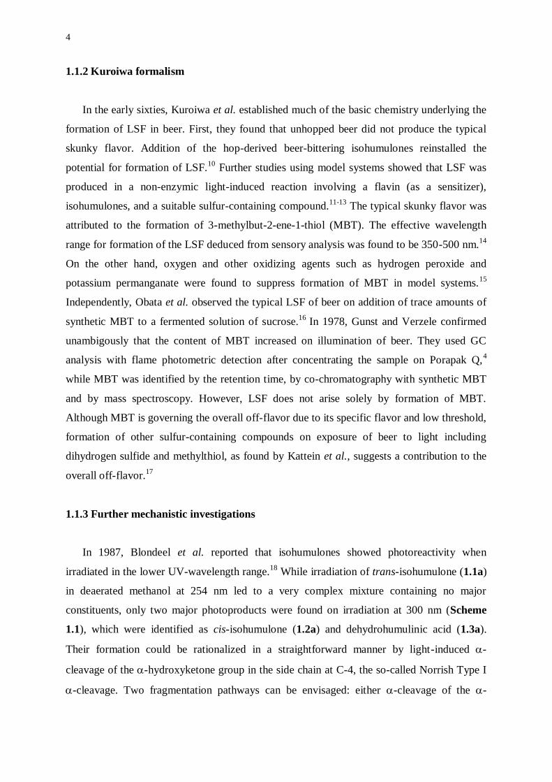
constituents, only two major photoproducts were found on irradiation at 300 nm (Scheme
1.1), which were identified as cis-isohumulone (1.2a) and dehydrohumulinic acid (1.3a).
Their formation could be rationalized in a straightforward manner by light-induced -
cleavage of the -hydroxyketone group in the side chain at C-4, the so-called Norrish Type I
-cleavage. Two fragmentation pathways can be envisaged: either -cleavage of the -
5
hydroxyketone (a) or -cleavage in the acyclic ,-enone part (b) (Scheme 1.1). In both
instances, a stabilized radical pair is formed. Ready recombination accounts for the low
quantum yield, while cis-isohumulone results from epimerization of the ketyl radical center.
Dehydrohumulinic acid is formed from the ketyl radical, derived either directly via route (a)
or after decarbonylation of the acyl radical from route (b) by -hydrogen elimination. It was
suggested that Norrish Type I -cleavage arose from the (n,*)-triplet manifold. Formation of
dehydrohumulinic acid provided the first direct proof for photodegradation of trans-
isohumulone (and, also, of isohumulones in general) with concurrent release of a precursor of
MBT. It is, therefore, the key route to the development of LSF on UV irradiation. No other
hop components seem to be involved in the process.
OO
OHO
OH
OO
OHO
OO
OHO
OH(a)
(b)
+h (300 nm)
1.1a 1.2a 1.3a
MeOH
Scheme 1.1
Major photoreaction products formed on irradiation (300 nm) of trans-isohumulone.
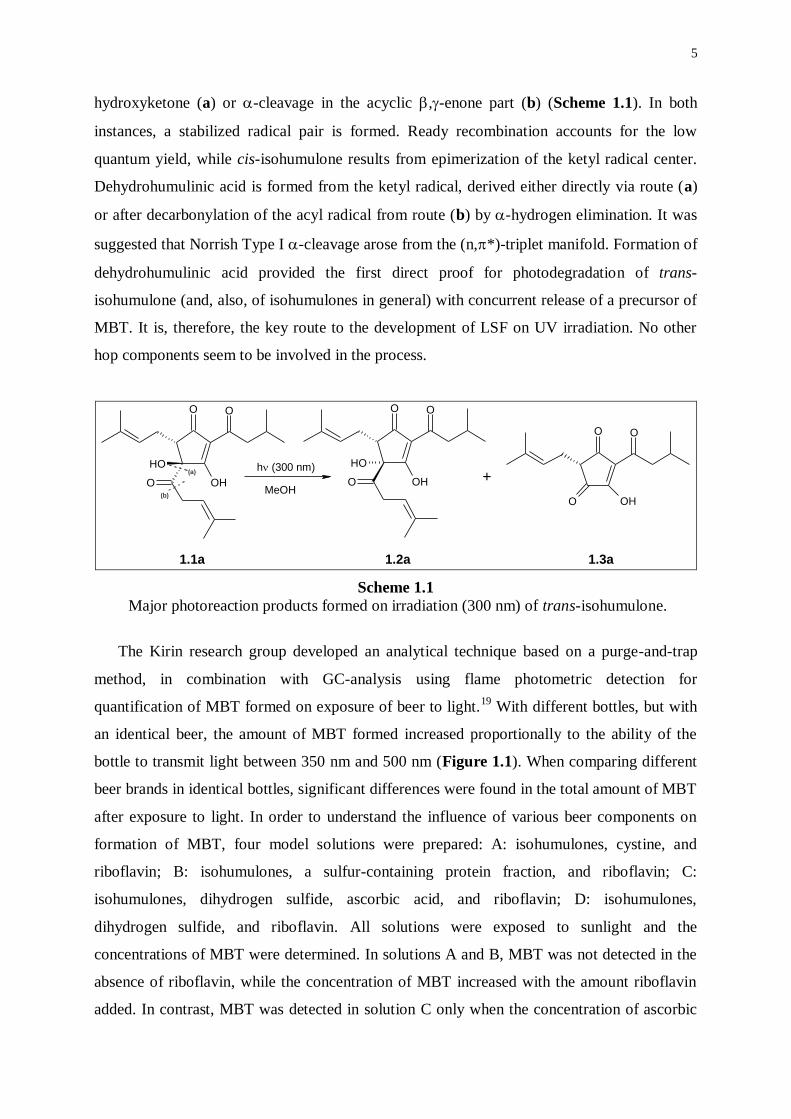
The Kirin research group developed an analytical technique based on a purge-and-trap
method, in combination with GC-analysis using flame photometric detection for
quantification of MBT formed on exposure of beer to light.19
With different bottles, but with
an identical beer, the amount of MBT formed increased proportionally to the ability of the
bottle to transmit light between 350 nm and 500 nm (Figure 1.1). When comparing different
beer brands in identical bottles, significant differences were found in the total amount of MBT
after exposure to light. In order to understand the influence of various beer components on
formation of MBT, four model solutions were prepared: A: isohumulones, cystine, and
riboflavin; B: isohumulones, a sulfur-containing protein fraction, and riboflavin; C:
isohumulones, dihydrogen sulfide, ascorbic acid, and riboflavin; D: isohumulones,
dihydrogen sulfide, and riboflavin. All solutions were exposed to sunlight and the
concentrations of MBT were determined. In solutions A and B, MBT was not detected in the
absence of riboflavin, while the concentration of MBT increased with the amount riboflavin
added. In contrast, MBT was detected in solution C only when the concentration of ascorbic
6
acid reached 50 mg/L. In solution D, MBT was absent. The results of these experiments
clearly show that photochemical sensitization by riboflavin was indispensable for formation
of MBT from isohumulones and from sulfur-containing amino acids or proteins, and that a
high level of ascorbic acid was necessary for formation of MBT from isohumulones and
dihydrogen sulfide.
Figure 1.1
A: Light transmittance patterns of various beer bottles.
B: Formation of 3-methylbut-2-ene-1-thiol (MBT) in a lager beer exposed to sunlight in
different bottles: A: clear bottle; B, C, D: green bottles; E: brown bottle.
To further clarify the contribution of beer components to the formation of MBT,
compounds implicated in the development of LSF were added to a beer in green-colored
bottles and the beers were exposed to sunlight. Formation of MBT was greatly accelerated by
addition of both isohumulones and riboflavin. Sulfur-containing amino acids and ascorbic
acid also led to an increase in the contents of MBT, dihydrogen sulfide had no effect, and
sulfite suppressed its formation. Furthermore, intensifying the beer color gradually eliminated
formation of MBT. From these results, Sakuma et al. concluded that the main route to MBT
on exposure to sunlight involves decomposition of isohumulones decompose to a 3-
methylbut-2-enyl radical, while sulfur-containing amino acids and proteins are a source of
thiol radicals in riboflavin-photosensitized reactions. Both radicals then combine to form
MBT. Also, the amount of riboflavin in beer is an important factor resulting in differences in
beer brands regarding formation of LSF. They proposed that, if riboflavin could be removed
from beer, a light-stable beer might be obtained.

7
A formal mechanism for formation of LSF in beer was commonly accepted (Schemes 1.2,
Scheme 1.3). According to route (a), riboflavin is excited to the singlet state by light in the
wavelength range of 350-500 nm. The excited singlet state gives rise to an excited triplet state
via intersystem crossing (isc). Subsequent triplet energy transfer to isohumulones in the
ground state leads to triplet-excited isohumulones. From the excited triplet state (also formed
on direct irradiation, route (b)), Norrish Type I -cleavage of the -hydroxyketone group
furnishes a 4-methylpent-3-enoyl radical, which undergoes decarbonylation to a 3-methylbut-
2-enyl radical. Trapping of this stabilized allyl radical by a suitable sulfur source (e.g.
cysteine) leads to formation of 3-methylbut-2-ene-1-thiol (MBT).
RF0
1RF*
3RF* 3ISO*
1ISO*
ISO0
+ RF0
ISO0
isc isc
h h
Route (a) Route (b)
Norrish I
radical precursors of MBT
Scheme 1.2
Formal mechanism for direct and photosensitized (RF: riboflavin) irradiation of
isohumulones (ISO) furnishing radical precursors for formation of 3-methylbut-2-ene-1-thiol
(MBT).
This proposal still holds on, although it was proven incorrect by Hastings et al. in 1992.20
From the absorption and phosphorescence spectra of both riboflavin and trans-isohumulone,
they were able to conclude that the wavelengths separating the excited triplet state from the
ground state of riboflavin and trans-isohumulone corresponded to ca. 560 nm and 395 nm,
respectively. These wavelengths are associated to triplet energies of 210 kJ/mol for riboflavin
and 300 kJ/mol for trans-isohumulone. Direct uphill triplet energy transfer by 90 kJ/mol can
be ruled out on the basis of thermodynamic considerations.
8
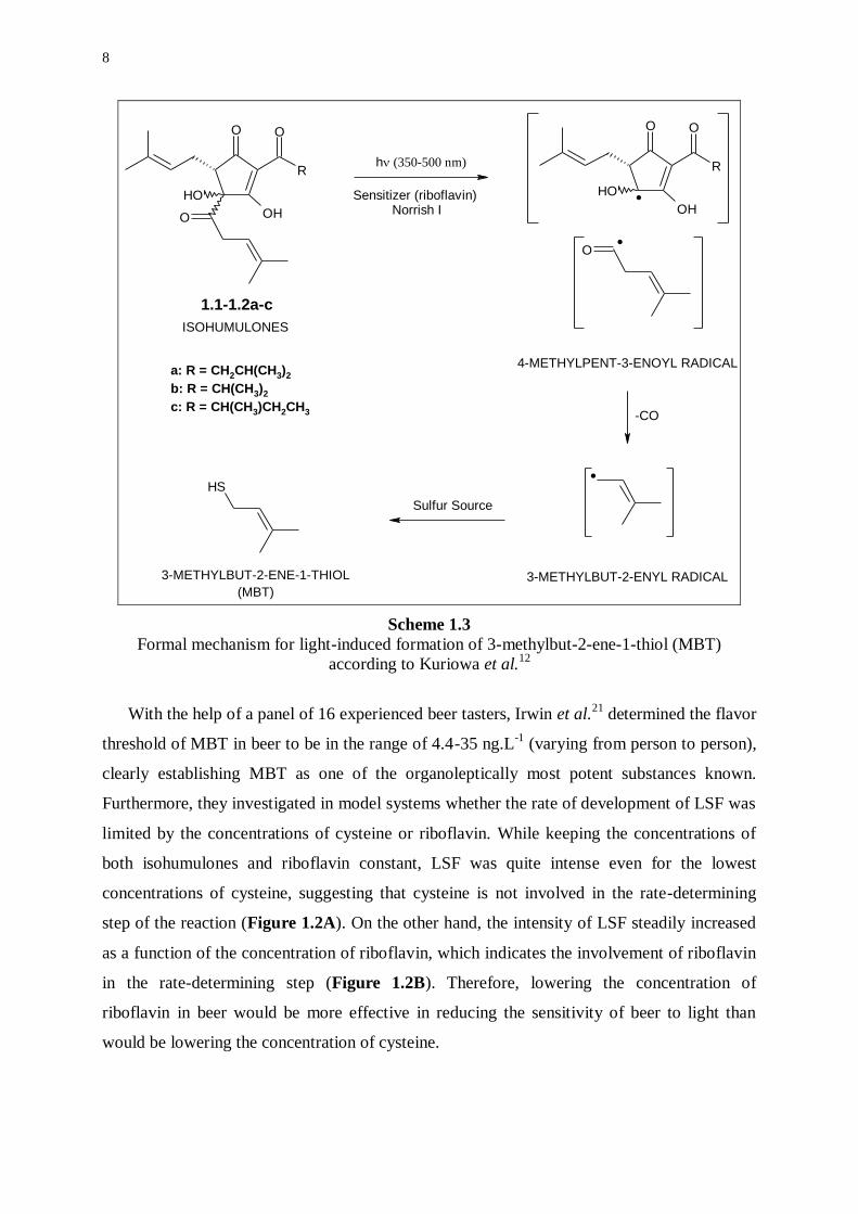
R
OO
OHO
OH
SH
3-METHYLBUT-2-ENE-1-THIOL
(MBT)
.
3-METHYLBUT-2-ENYL RADICAL
R
OO
OH
OH
O
h(350-500 nm)
Sensitizer (riboflavin) Norrish I
ISOHUMULONES
-CO
4-METHYLPENT-3-ENOYL RADICAL
Sulfur Source
..
1.1-1.2a-c
a: R = CH2CH(CH3)2
b: R = CH(CH3)2
c: R = CH(CH3)CH2CH3
Scheme 1.3
Formal mechanism for light-induced formation of 3-methylbut-2-ene-1-thiol (MBT)
according to Kuriowa et al.12
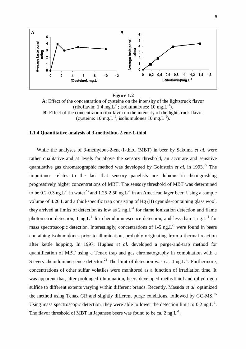
With the help of a panel of 16 experienced beer tasters, Irwin et al.21
determined the flavor
threshold of MBT in beer to be in the range of 4.4-35 ng.L-1
(varying from person to person),
clearly establishing MBT as one of the organoleptically most potent substances known.
Furthermore, they investigated in model systems whether the rate of development of LSF was
limited by the concentrations of cysteine or riboflavin. While keeping the concentrations of
both isohumulones and riboflavin constant, LSF was quite intense even for the lowest
concentrations of cysteine, suggesting that cysteine is not involved in the rate-determining
step of the reaction (Figure 1.2A). On the other hand, the intensity of LSF steadily increased
as a function of the concentration of riboflavin, which indicates the involvement of riboflavin
in the rate-determining step (Figure 1.2B). Therefore, lowering the concentration of
riboflavin in beer would be more effective in reducing the sensitivity of beer to light than
would be lowering the concentration of cysteine.
9
Figure 1.2
A: Effect of the concentration of cysteine on the intensity of the lightstruck flavor
(riboflavin: 1.4 mg.L-1
; isohumulones: 10 mg.L-1
).
B: Effect of the concentration riboflavin on the intensity of the lightstruck flavor
(cysteine: 10 mg.L-1
; isohumulones 10 mg.L-1
).
1.1.4 Quantitative analysis of 3-methylbut-2-ene-1-thiol
While the analyses of 3-methylbut-2-ene-1-thiol (MBT) in beer by Sakuma et al. were
rather qualitative and at levels far above the sensory threshold, an accurate and sensitive
quantitative gas chromatographic method was developed by Goldstein et al. in 1993.22
The
importance relates to the fact that sensory panelists are dubious in distinguishing
progressively higher concentrations of MBT. The sensory threshold of MBT was determined
to be 0.2-0.3 ng.L-1
in water23
and 1.25-2.50 ng.L-1
in an American lager beer. Using a sample
volume of 4.26 L and a thiol-specific trap consisting of Hg (II) cyanide-containing glass wool,
they arrived at limits of detection as low as 2 ng.L-1
for flame ionization detection and flame
photometric detection, 1 ng.L-1
for chemiluminescence detection, and less than 1 ng.L-1
for
mass spectroscopic detection. Interestingly, concentrations of 1-5 ng.L-1
were found in beers
containing isohumulones prior to illumination, probably originating from a thermal reaction
after kettle hopping. In 1997, Hughes et al. developed a purge-and-trap method for
quantification of MBT using a Tenax trap and gas chromatography in combination with a
Sievers chemiluminescence detector.24
The limit of detection was ca. 4 ng.L-1
. Furthermore,
concentrations of other sulfur volatiles were monitored as a function of irradiation time. It
was apparent that, after prolonged illumination, beers developed methylthiol and dihydrogen
sulfide to different extents varying within different brands. Recently, Masuda et al. optimized
the method using Tenax GR and slightly different purge conditions, followed by GC-MS.25
Using mass spectroscopic detection, they were able to lower the detection limit to 0.2 ng.L-1
.
The flavor threshold of MBT in Japanese beers was found to be ca. 2 ng.L-1
.
10
1.2 ROLE OF HOPS
1.2.1 Botanical description
The hop plant belongs to the family of the Cannabaceae,26
consisting of two genera:
Humulus and Cannabis. Humulus exists as two species, Humulus lupulus L., which has a high
economical value because of its use in beer brewing, and Humulus japonicus Sieb. & Zucc.,
which is only grown as an ornamental plant. The hop plant is a fast growing vine (up to 6-7 m
per year) with unisexual male and female flowers growing on separate plants. Only the female
flowers are important for commercial purposes, while they contain all compounds of interest
for brewing. Day-length requirements restrict cultivation of hops to latitudes between 35° and
55° in both hemispheres. Further requirements such as fertile soil, relatively high summer
temperatures, and access to water via rain, ground water or irrigation are met in the larger hop
growing areas situated in Europe (Germany, Czech Republic, England), the United States of
America (mainly in the States of Washington and Oregon), Australia, and New-Zealand. In
the northern hemisphere, the female flowers start to grow during July and the hop cones can
be harvested from the end of August to the end of September, depending on hop variety and
weather conditions.
1.2.2 Chemical composition of the hop cones
The female hop flowers contain, next to the primary metabolites, hundreds of secondary
metabolitesa comprising many different groups of organic compounds. Of particular interest
are: a) hop resins, containing the hop acids;
b) hop essential oil;
c) hop polyphenols.
These three classes are not only important because of their profound impact on various
beer characteristics, but they are also useful as significant biochemical markers in varietal
a Secondary metabolites are distinguished from primary metabolites by the following criteria: they have a
restricted distribution being found mostly in plants and micro-organisms, and are often characteristic of
individual genera, species, or strains; they are formed along specialized pathways from primary metabolites.
Secondary metabolites are non-essential to life, although they are important to the organism that produces them
in correlation to environmental factors or stress.
11
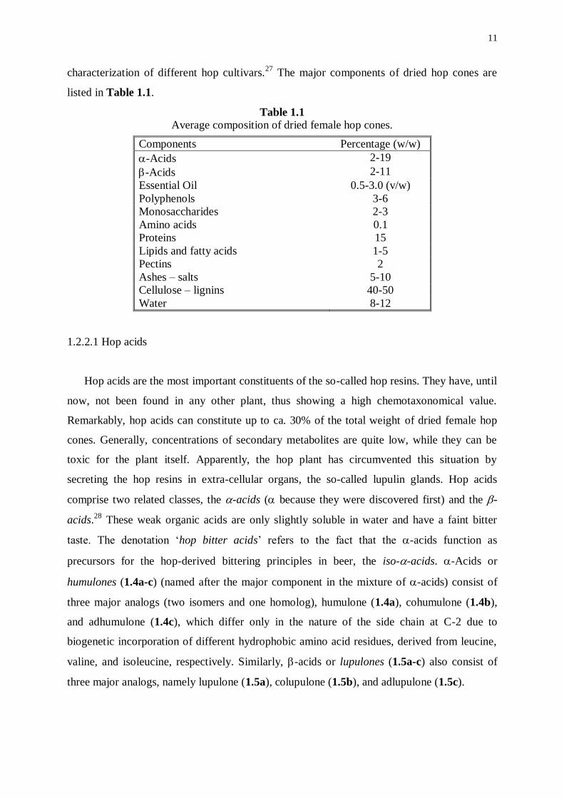
characterization of different hop cultivars.27
The major components of dried hop cones are
listed in Table 1.1.
Table 1.1
Average composition of dried female hop cones.
Components Percentage (w/w)
-Acids 2-19
-Acids 2-11
Essential Oil 0.5-3.0 (v/w)
Polyphenols 3-6
Monosaccharides 2-3
Amino acids 0.1
Proteins 15
Lipids and fatty acids 1-5
Pectins 2
Ashes – salts 5-10
Cellulose – lignins 40-50
Water 8-12
1.2.2.1 Hop acids
Hop acids are the most important constituents of the so-called hop resins. They have, until
now, not been found in any other plant, thus showing a high chemotaxonomical value.
Remarkably, hop acids can constitute up to ca. 30% of the total weight of dried female hop
cones. Generally, concentrations of secondary metabolites are quite low, while they can be
toxic for the plant itself. Apparently, the hop plant has circumvented this situation by
secreting the hop resins in extra-cellular organs, the so-called lupulin glands. Hop acids
comprise two related classes, the -acids ( because they were discovered first) and the -
acids.28
These weak organic acids are only slightly soluble in water and have a faint bitter
taste. The denotation ‘hop bitter acids’ refers to the fact that the -acids function as
precursors for the hop-derived bittering principles in beer, the iso--acids. -Acids or
humulones (1.4a-c) (named after the major component in the mixture of -acids) consist of
three major analogs (two isomers and one homolog), humulone (1.4a), cohumulone (1.4b),
and adhumulone (1.4c), which differ only in the nature of the side chain at C-2 due to
biogenetic incorporation of different hydrophobic amino acid residues, derived from leucine,
valine, and isoleucine, respectively. Similarly, -acids or lupulones (1.5a-c) also consist of
three major analogs, namely lupulone (1.5a), colupulone (1.5b), and adlupulone (1.5c).
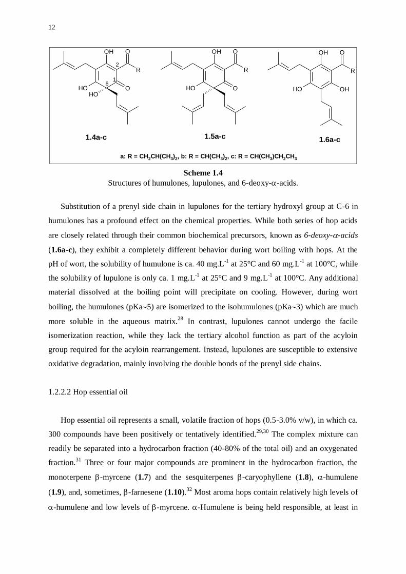
12
OH
OOH
R
O
OH
OH
OOH
R
O OH
OHOH
R
O
a: R = CH2CH(CH3)2, b: R = CH(CH3)2, c: R = CH(CH3)CH2CH3
1.4a-c 1.5a-c 1.6a-c
2
61
Scheme 1.4
Structures of humulones, lupulones, and 6-deoxy--acids.
Substitution of a prenyl side chain in lupulones for the tertiary hydroxyl group at C-6 in
humulones has a profound effect on the chemical properties. While both series of hop acids
are closely related through their common biochemical precursors, known as 6-deoxy--acids
(1.6a-c), they exhibit a completely different behavior during wort boiling with hops. At the
pH of wort, the solubility of humulone is ca. 40 mg.L-1
at 25°C and 60 mg.L-1
at 100°C, while
the solubility of lupulone is only ca. 1 mg.L-1
at 25°C and 9 mg.L-1
at 100°C. Any additional
material dissolved at the boiling point will precipitate on cooling. However, during wort
boiling, the humulones (pKa5) are isomerized to the isohumulones (pKa3) which are much
more soluble in the aqueous matrix.28
In contrast, lupulones cannot undergo the facile
isomerization reaction, while they lack the tertiary alcohol function as part of the acyloin
group required for the acyloin rearrangement. Instead, lupulones are susceptible to extensive
oxidative degradation, mainly involving the double bonds of the prenyl side chains.
1.2.2.2 Hop essential oil
Hop essential oil represents a small, volatile fraction of hops (0.5-3.0% v/w), in which ca.
300 compounds have been positively or tentatively identified.29,30
The complex mixture can
readily be separated into a hydrocarbon fraction (40-80% of the total oil) and an oxygenated
fraction.31
Three or four major compounds are prominent in the hydrocarbon fraction, the
monoterpene -myrcene (1.7) and the sesquiterpenes -caryophyllene (1.8), -humulene
(1.9), and, sometimes, -farnesene (1.10).32
Most aroma hops contain relatively high levels of
-humulene and low levels of -myrcene. -Humulene is being held responsible, at least in
13
part and in combination with a number of other constituents of the essential oil, for the
pleasant smell of hops. -Myrcene and the sesquiterpenes are notoriously reactive, and, thus,
a great variety of oxidized terpenes can be found in the oxygenated fraction.
H H
S S
H H
OHH
OHO
O O
O
OH
O
OH
O
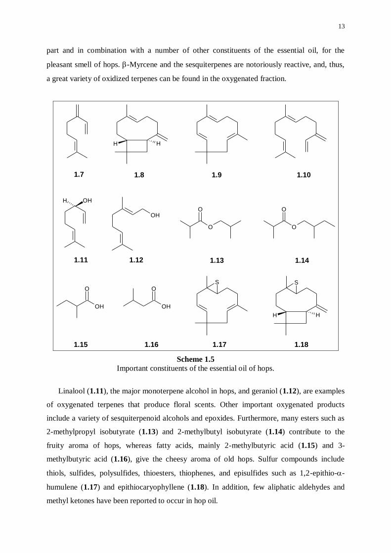
1.7 1.8 1.9 1.10
1.11 1.12 1.13 1.14
1.15 1.17 1.181.16
Scheme 1.5
Important constituents of the essential oil of hops.
Linalool (1.11), the major monoterpene alcohol in hops, and geraniol (1.12), are examples
of oxygenated terpenes that produce floral scents. Other important oxygenated products
include a variety of sesquiterpenoid alcohols and epoxides. Furthermore, many esters such as
2-methylpropyl isobutyrate (1.13) and 2-methylbutyl isobutyrate (1.14) contribute to the
fruity aroma of hops, whereas fatty acids, mainly 2-methylbutyric acid (1.15) and 3-
methylbutyric acid (1.16), give the cheesy aroma of old hops. Sulfur compounds include
thiols, sulfides, polysulfides, thioesters, thiophenes, and episulfides such as 1,2-epithio--
humulene (1.17) and epithiocaryophyllene (1.18). In addition, few aliphatic aldehydes and
methyl ketones have been reported to occur in hop oil.
14
O OH
OHOH OH
OH
O
H H
O
O
OOH
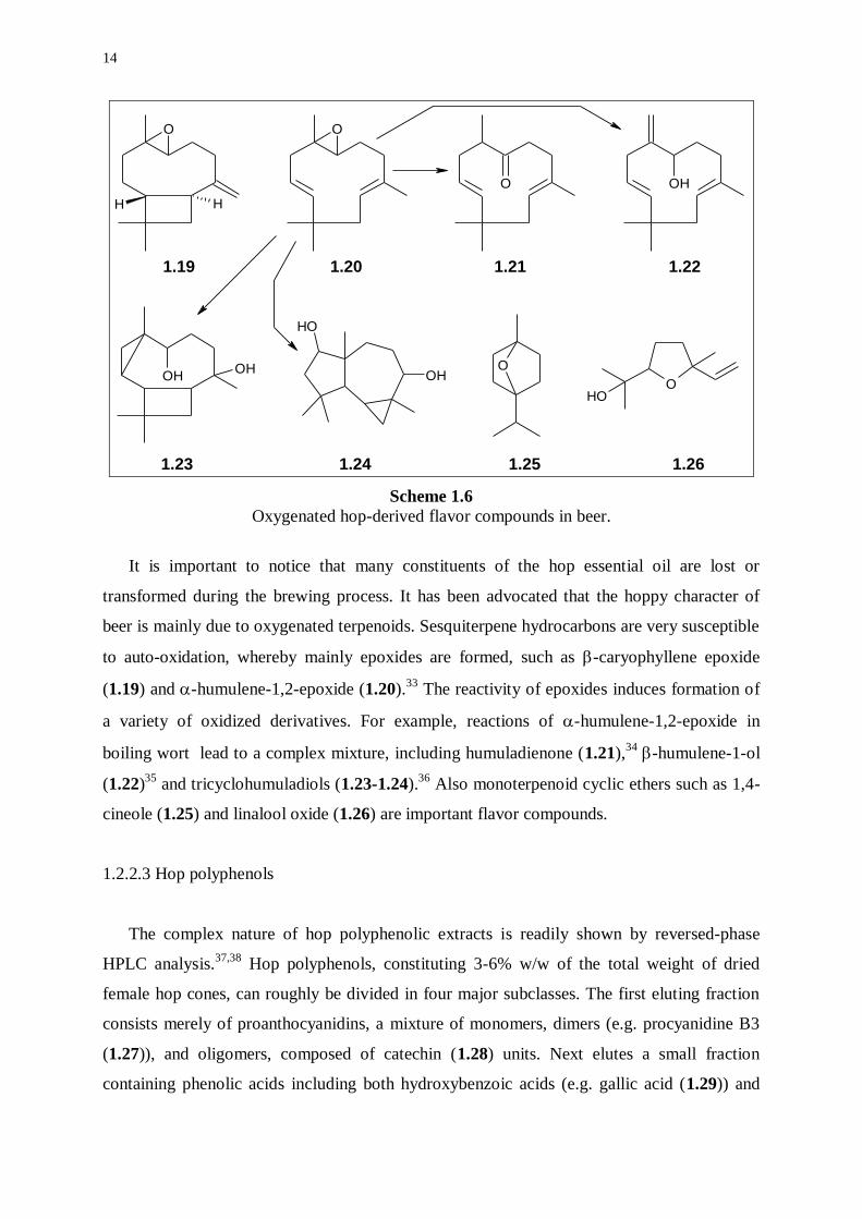
1.19 1.20 1.21 1.22
1.23 1.24 1.25 1.26
Scheme 1.6
Oxygenated hop-derived flavor compounds in beer.
It is important to notice that many constituents of the hop essential oil are lost or
transformed during the brewing process. It has been advocated that the hoppy character of
beer is mainly due to oxygenated terpenoids. Sesquiterpene hydrocarbons are very susceptible
to auto-oxidation, whereby mainly epoxides are formed, such as -caryophyllene epoxide
(1.19) and -humulene-1,2-epoxide (1.20).33
The reactivity of epoxides induces formation of
a variety of oxidized derivatives. For example, reactions of -humulene-1,2-epoxide in
boiling wort lead to a complex mixture, including humuladienone (1.21),34
-humulene-1-ol
(1.22)35
and tricyclohumuladiols (1.23-1.24).36
Also monoterpenoid cyclic ethers such as 1,4-
cineole (1.25) and linalool oxide (1.26) are important flavor compounds.
1.2.2.3 Hop polyphenols
The complex nature of hop polyphenolic extracts is readily shown by reversed-phase
HPLC analysis.37,38
Hop polyphenols, constituting 3-6% w/w of the total weight of dried
female hop cones, can roughly be divided in four major subclasses. The first eluting fraction
consists merely of proanthocyanidins, a mixture of monomers, dimers (e.g. procyanidine B3
(1.27)), and oligomers, composed of catechin (1.28) units. Next elutes a small fraction
containing phenolic acids including both hydroxybenzoic acids (e.g. gallic acid (1.29)) and
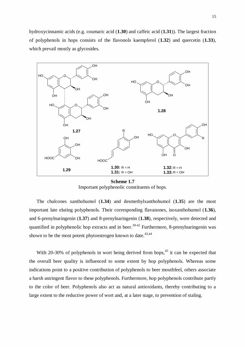
15
hydroxycinnamic acids (e.g. coumaric acid (1.30) and caffeic acid (1.31)). The largest fraction
of polyphenols in hops consists of the flavonols kaempferol (1.32) and quercetin (1.33),
which prevail mostly as glycosides.
OOH
OH
OH
OH
OH
OOH
OH
OH
OH
OH
OOH
OH
OH
OH
OH
O
O
OH
OH
OH
OH
ROH
HOOC
R
OH
OH
OH
HOOC
1.27
1.28
1.291.30:
1.31:1.32:
1.33:
R = H
R = OH
R = H
R = OH
Scheme 1.7
Important polyphenolic constituents of hops.
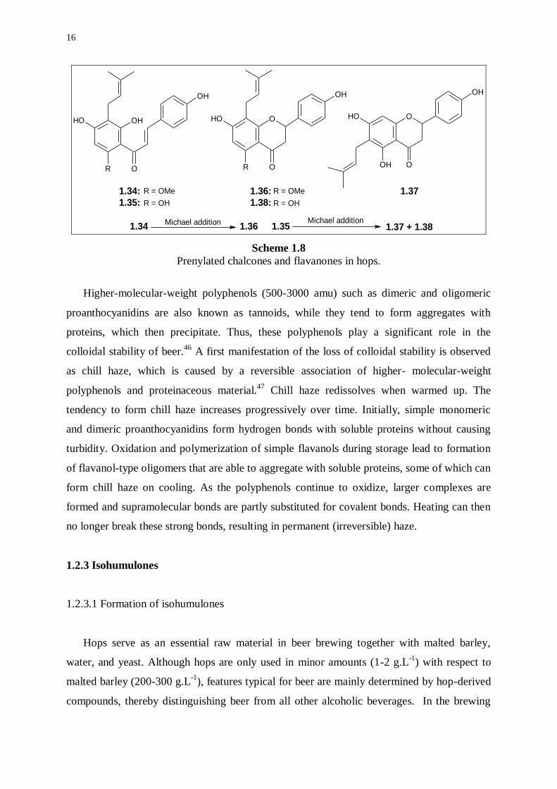
The chalcones xanthohumol (1.34) and desmethylxanthohumol (1.35) are the most
important late eluting polyphenols. Their corresponding flavanones, isoxanthohumol (1.36),
and 6-prenylnaringenin (1.37) and 8-prenylnaringenin (1.38), respectively, were detected and
quantified in polyphenolic hop extracts and in beer.39-42
Furthermore, 8-prenylnaringenin was
shown to be the most potent phytoestrogen known to date.43,44
With 20-30% of polyphenols in wort being derived from hops,45
it can be expected that
the overall beer quality is influenced to some extent by hop polyphenols. Whereas some
indications point to a positive contribution of polyphenols to beer mouthfeel, others associate
a harsh astringent flavor to these polyphenols. Furthermore, hop polyphenols contribute partly
to the color of beer. Polyphenols also act as natural antioxidants, thereby contributing to a
large extent to the reductive power of wort and, at a later stage, to prevention of staling.
16
O
OH O
OH
R O
OH O
OH
OHO
OH
OH
OH
R
1.34:
1.35:
1.36:
1.38:
1.37R = OMe
R = OH
R = OMe
R = OH
1.34 1.351.36 1.37 + 1.38Michael addition Michael addition
Scheme 1.8
Prenylated chalcones and flavanones in hops.
Higher-molecular-weight polyphenols (500-3000 amu) such as dimeric and oligomeric
proanthocyanidins are also known as tannoids, while they tend to form aggregates with
proteins, which then precipitate. Thus, these polyphenols play a significant role in the
colloidal stability of beer.46
A first manifestation of the loss of colloidal stability is observed
as chill haze, which is caused by a reversible association of higher- molecular-weight
polyphenols and proteinaceous material.47
Chill haze redissolves when warmed up. The
tendency to form chill haze increases progressively over time. Initially, simple monomeric
and dimeric proanthocyanidins form hydrogen bonds with soluble proteins without causing
turbidity. Oxidation and polymerization of simple flavanols during storage lead to formation
of flavanol-type oligomers that are able to aggregate with soluble proteins, some of which can
form chill haze on cooling. As the polyphenols continue to oxidize, larger complexes are
formed and supramolecular bonds are partly substituted for covalent bonds. Heating can then
no longer break these strong bonds, resulting in permanent (irreversible) haze.
1.2.3 Isohumulones
1.2.3.1 Formation of isohumulones
Hops serve as an essential raw material in beer brewing together with malted barley,
water, and yeast. Although hops are only used in minor amounts (1-2 g.L-1
) with respect to
malted barley (200-300 g.L-1
), features typical for beer are mainly determined by hop-derived
compounds, thereby distinguishing beer from all other alcoholic beverages. In the brewing
17
process, hops are boiled for about 1.5 h with wort, a sweet tasting solution resulting from
enzymic degradation of starch and proteins contained in malted barly. During boiling in the
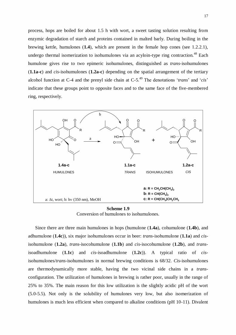
brewing kettle, humulones (1.4), which are present in the female hop cones (see 1.2.2.1),
undergo thermal isomerization to isohumulones via an acyloin-type ring contraction.48
Each
humulone gives rise to two epimeric isohumulones, distinguished as trans-isohumulones
(1.1a-c) and cis-isohumulones (1.2a-c) depending on the spatial arrangement of the tertiary
alcohol function at C-4 and the prenyl side chain at C-5.49
The denotations ‘trans’ and ‘cis’
indicate that these groups point to opposite faces and to the same face of the five-membered
ring, respectively.
R
OH
OOH
O
OH
R
OO
OH
OH
O
R
OO
OH
OH
O+a
b
1.4a-c 1.1a-c 1.2a-c
a: R = CH2CH(CH3)2
b: R = CH(CH3)2
c: R = CH(CH3)CH2CH3a: t, wort; b: h (350 nm), MeOH
HUMULONES TRANS CISISOHUMULONES
Scheme 1.9 Conversion of humulones to isohumulones.
Since there are three main humulones in hops (humulone (1.4a), cohumulone (1.4b), and
adhumulone (1.4c)), six major isohumulones occur in beer: trans-isohumulone (1.1a) and cis-
isohumulone (1.2a), trans-isocohumulone (1.1b) and cis-isocohumulone (1.2b), and trans-
isoadhumulone (1.1c) and cis-isoadhumulone (1.2c)). A typical ratio of cis-
isohumulones/trans-isohumulones in normal brewing conditions is 68/32. Cis-isohumulones
are thermodynamically more stable, having the two vicinal side chains in a trans-
configuration. The utilization of humulones in brewing is rather poor, usually in the range of
25% to 35%. The main reason for this low utilization is the slightly acidic pH of the wort
(5.0-5.5). Not only is the solubility of humulones very low, but also isomerization of
humulones is much less efficient when compared to alkaline conditions (pH 10-11). Divalent
18
cations such as magnesium(II) ions also increase the isomerization rate, while the ratio of cis-
isohumulones/trans-isohumulones may vary according to the experimental conditions.
Thermal isomerization of the humulones can also be carried out in the solid state. The
isomerization process proceeds most efficiently on heating of the solid metal salts of
humulones, which are first precipitated from an aqueous alkaline solution. The best results are
obtained with the calcium(II) and magnesium(II) salts of the humulones, leading to a ratio of
cis-isohumulones/trans-isohumulones of ca. 85/15. Such process is used in industrial
production of isomerized hop extracts.
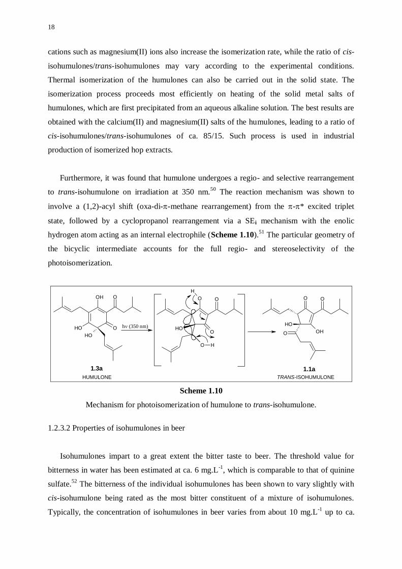
Furthermore, it was found that humulone undergoes a regio- and selective rearrangement
to trans-isohumulone on irradiation at 350 nm.50
The reaction mechanism was shown to
involve a (1,2)-acyl shift (oxa-di--methane rearrangement) from the -* excited triplet
state, followed by a cyclopropanol rearrangement via a SEi mechanism with the enolic
hydrogen atom acting as an internal electrophile (Scheme 1.10).51
The particular geometry of
the bicyclic intermediate accounts for the full regio- and stereoselectivity of the
photoisomerization.
OH
OOH
O
OH
OO
OH
OH
O
O
O
O
H
O H
OH
1.3a
a1.1a
h (350 nm)
HUMULONE TRANS-ISOHUMULONE
Scheme 1.10
Mechanism for photoisomerization of humulone to trans-isohumulone.
1.2.3.2 Properties of isohumulones in beer
Isohumulones impart to a great extent the bitter taste to beer. The threshold value for
bitterness in water has been estimated at ca. 6 mg.L-1
, which is comparable to that of quinine
sulfate.52
The bitterness of the individual isohumulones has been shown to vary slightly with
cis-isohumulone being rated as the most bitter constituent of a mixture of isohumulones.
Typically, the concentration of isohumulones in beer varies from about 10 mg.L-1
up to ca.
19
100 mg.L-1
. The perceived bitterness in beer is much less astringent than in water possibly
due to the masking effect of other beer compounds including carbohydrates and proteins.
Isohumulones have been implicated in both an increase in beer foam cling and lacing to the
beer glass and an improvement in the overall stability of the beer head.53,54
According to
current knowledge, isohumulones form supramolecular complexes, on the one hand with
foam-active barley proteins involving both hydrogen bonding and hydrophobic interactions,
and on the other hand with divalent cations (e.g. Cu(II)) via formation of stable chelates. This
amphiphilic behavior creates favorable conditions for crosslinking of surface-adsorbed
proteins, thus fortifying the film around foam bubbles. As a consequence, a strong foam head
characterizes a well-hopped beer. Isohumulones inhibit the growth of Gram-positive bacteria
and protect beer against spoilage by lactic acid bacteria (Lactobacillus spp. and Pediococcus
spp.).55
Undissociated isohumulones act as mobile carriers of ions, thereby dissipating the
transmembrane pH gradient. A lower pH leads to a more efficient inhibition of microbial
growth. Some strains of lactic acid bacteria were found resistant to isohumulones, but the
mechanism remains elusive. On the other hand, isohumulones are quite vulnerable to light.
Either direct or sensitized irradiation affords precursors for 3-methylbut-2-ene-1-thiol (MBT),
the compound mainly responsible for the typical lightstruck flavor (LSF) of beer.
1.2.4 Reduced derivatives of isohumulones
The use of whole hops in brewing has a number of disadvantages. Whole hops are very
bulky, heterogeneous, prone to oxidative degradation, and the utilization of humulones is
usually very poor. Solutions to these problems were provided by the development of a variety
of hop products (a hop product is any form of hops in which the vegetative material cannot be
recognized) including hop pellets, hop extracts, and isomerized hop products.56
Currently, hop
pellets have a market share of ca. 40% regarding the application of hops in brewing.
Advantages are reflected in a much higher bulk density and an improved stability when
protected from oxygen, while also an improvement in utilization of humulones is notable.
Hop extracts, with a current market share of ca. 30%, are prepared by extraction of whole
hops with hexane, ethanol, or carbon dioxide. Especially, liquid and supercritical carbon
dioxide extracts are preferred while carbon dioxide is both environmentally benign and a
natural by-product of brewing. Hop extracts are characterized by a further increase in bulk
density, a significantly increased stability of hop substances, and a substantial improvement in
utilization. In addition, the homogeneity results in a more consistent bitterness. To date,
20
isomerized hop products (or advanced hop products) account for ca. 10% of the use of hops
and the utilization is improved by two- to threefold. The market share of isomerized hop
products is expected to grow rapidly, while brewers become increasingly aware of the
benefits. Furthermore, three types of reduced isomerized hop products, called
dihydroisohumulones, tetrahydroisohumulones, and hexahydroisohumulones, are gaining
widespread acceptance. The prefixes of the reduced isohumulones refer to the number of
hydrogen atoms being incorporated during reduction. At first, these advanced hop products
were produced in order to protect beer bottled in clear or green glass from developing a
lightstruck character. But, other properties including foam enhancement and differences in
relative bitterness were gradually exploited in brewing.
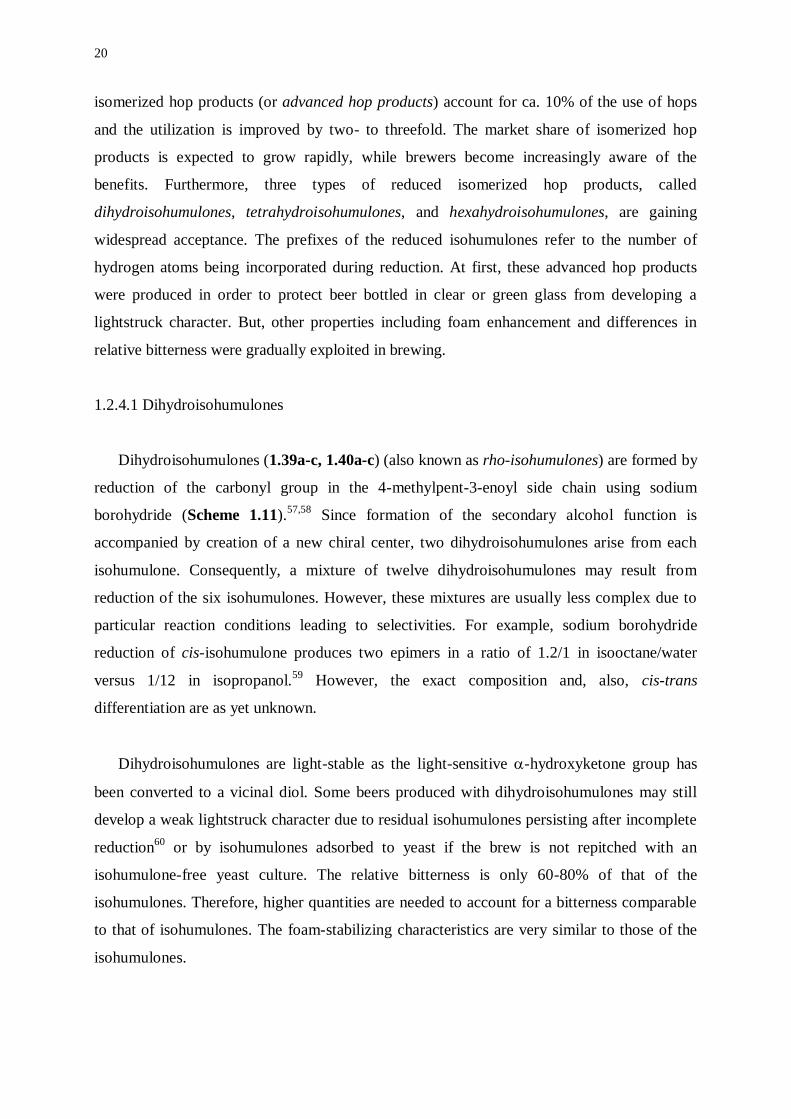
1.2.4.1 Dihydroisohumulones
Dihydroisohumulones (1.39a-c, 1.40a-c) (also known as rho-isohumulones) are formed by
reduction of the carbonyl group in the 4-methylpent-3-enoyl side chain using sodium
borohydride (Scheme 1.11).57,58
Since formation of the secondary alcohol function is
accompanied by creation of a new chiral center, two dihydroisohumulones arise from each
isohumulone. Consequently, a mixture of twelve dihydroisohumulones may result from
reduction of the six isohumulones. However, these mixtures are usually less complex due to
particular reaction conditions leading to selectivities. For example, sodium borohydride
reduction of cis-isohumulone produces two epimers in a ratio of 1.2/1 in isooctane/water
versus 1/12 in isopropanol.59
However, the exact composition and, also, cis-trans
differentiation are as yet unknown.
Dihydroisohumulones are light-stable as the light-sensitive -hydroxyketone group has
been converted to a vicinal diol. Some beers produced with dihydroisohumulones may still
develop a weak lightstruck character due to residual isohumulones persisting after incomplete
reduction60
or by isohumulones adsorbed to yeast if the brew is not repitched with an
isohumulone-free yeast culture. The relative bitterness is only 60-80% of that of the
isohumulones. Therefore, higher quantities are needed to account for a bitterness comparable
to that of isohumulones. The foam-stabilizing characteristics are very similar to those of the
isohumulones.
21
b
R
OO
OHO
OH
R
OO
OH
OH
OH H
R
OO
OH
OH
H OH
R
OO
OH
OH
OH H
R
OO
OH
OH
H OH
R
OO
OHO
OH
ISOHUMULONES DIHYDROISOHUMULONES
TETRAHYDROISOHUMULONES HEXAHYDROISOHUMULONES
1.1-1.2a-c 1.39a-c 1.40a-c
1.41-1.42a-c 1.45a-c 1.46a-c
a: NaBH4, MeOH; b: 2H2, Pd/C
+a
+a
a,ba: R = CH2CH(CH3)2
b: R = CH(CH3)2
c: R = CH(CH3)CH2CH3
Scheme 1.11
Formation of reduced isohumulones.
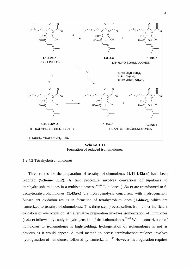
1.2.4.2 Tetrahydroisohumulones
Three routes for the preparation of tetrahydroisohumulones (1.41-1.42a-c) have been
reported (Scheme 1.12). A first procedure involves conversion of lupulones to
tetrahydroisohumulones in a multistep process.61,62
Lupulones (1.5a-c) are transformed to 6-
deoxytetrahydrohumulones (1.43a-c) via hydrogenolysis concurrent with hydrogenation.
Subsequent oxidation results in formation of tetrahydrohumulones (1.44a-c), which are
isomerized to tetrahydroisohumulones. This three-step process suffers from either inefficient
oxidation or overoxidation. An alternative preparation involves isomerization of humulones
(1.4a-c) followed by catalytic hydrogenation of the isohumulones.63-65
While isomerization of
humulones to isohumulones is high-yielding, hydrogenation of isohumulones is not as
obvious as it would appear. A third method to access tetrahydroisohumulones involves
hydrogenation of humulones, followed by isomerization.66
However, hydrogenation requires
22
close monitoring of the hydrogen uptake, as both overreduction and incomplete reduction
may occur. On the other hand, Hay and Homiski found that humulones could be
simultaneously isomerized and hydrogenated in alkaline conditions resulting in
tetrahydroisohumulones in high yields (ca. 92%).67
The reaction time was found not critical
and the pH value could be easily maintained with a carbonate buffer. Consequently, this
efficient one-step preparation of tetrahydroisohumulones proved suitable for large-scale
production.
OH
R
O
OOH
OH
OH
R
O
OOH
OH
OH
R
O
OOH
OH
R
O
OOH
R
OO
OHO
OHa b
c
d
a: hydrogenation; b: isomerization; c: hydrogenation/hydrogenolysis;
d: oxidation; e: simultaneous hydrogenation and isomerization
e
1.4a-c
1.5a-c1.43a-c
1.44a-c 1.41-1.42a-c
a: R = CH2CH(CH3)2
b: R = CH(CH3)2
c: R = CH(CH3)CH2CH3
HUMULONES
LUPULONES
TETRAHYDROISOHUMULONES
Scheme 1.12
Preparation of tetrahydroisohumulones from humulones and lupulones.
Saturation of the double bonds leads to enhanced hydrophobicity and diminished
reactivity. Photochemical degradation may still occur, since the -hydroxyketone group is
still present, but a 3-methylbut-2-enyl radical, intervening in the formation of 3-methylbut-2-
ene-1-thiol (MBT), can no longer be formed. Increased hydrophobicity as compared to
23
isohumulones results on the one hand in improved foam stabilizing and lacing characteristics,
on the other hand in an increased relative bitterness (a factor of 1.6-1.8).68
Advanced hop
products based on tetrahydroisohumulones can be used at low levels (2-5 mg.L-1
) in normal
beers that do not require protection against light in order to affect significantly the foam
quality. While full bittering with tetrahydroisohumulones results in the appearance of an
unattractive foam, a suitable combination of desired bitterness and foam stability can be found
using well-balanced mixtures of dihydroisohumulones and tetrahydroisohumulones.69
1.2.4.3 Hexahydroisohumulones
Hexahydroisohumulones (1.45a-c, 1.46a-c) are accessible by a combination of processes
used in the preparation of dihydroisohumulones and tetrahydroisohumulones (Scheme 1.11).
Simultaneous or successive reduction of the carbonyl group in the side chain at C-4 and
catalytic hydrogenation of the double bonds in the prenyl side chains of the isohumulones
afford hexahydroisohumulones. The mixture of hexahydroisohumulones may consist of
twelve isomers and homologs, but real mixtures are usually less complex due to selectivities
exerted during the reactions. Consequently, the exact composition and, also, cis-trans
differentiation are as yet unknown.
Hexahydroisohumulones are light-stable and they combine the features associated to
dihydroisohumulones and tetrahydroisohumulones. The relative bitterness is weakly increased
with respect to isohumulones, while foam-enhancing characteristics are even further
improved relative to those of tetrahydroisohumulones. Low solubility and high cost hamper
applications in the brewing practice.
1.3 ROLE OF FLAVINS
1.3.1 Introduction
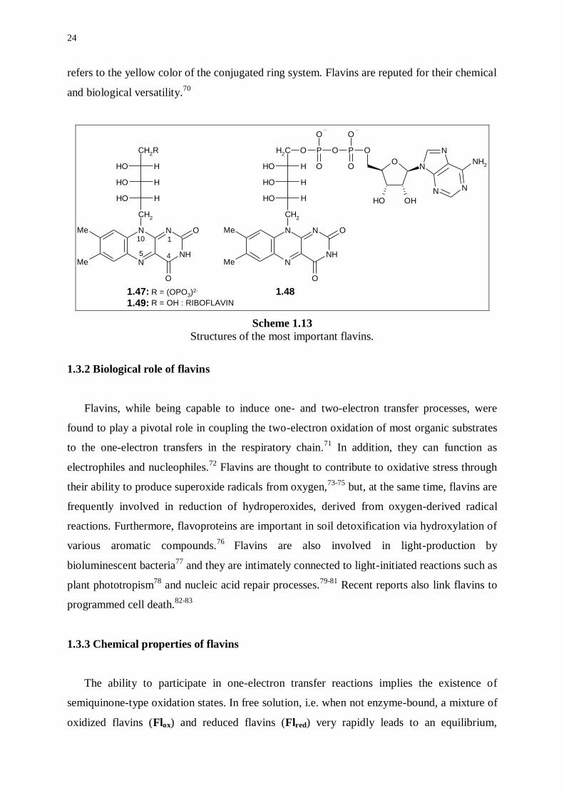
Flavins are based on a nitrogen-containing heterocyclic system, called 7,8-
dimethylisoalloxazine or 7,8-dimethylbenzo[g]pteridine-2,4-(3H,10H)-dione. The most
common natural flavins are flavin mononucleotide (FMN) (1.47), flavin adenine dinucleotide
(FAD) (1.48), and riboflavin (RF, vitamin B2) (1.49) (Scheme 1.13). The denotation ‘flavin’
24
refers to the yellow color of the conjugated ring system. Flavins are reputed for their chemical
and biological versatility.70
NH
N
N
NMe
Me
CH2
CH2R
O
O
OH H
OH H
OH H NO
N
N
OHOH
N
NH2
O
NH
N
N
NMe
Me
CH2
O
O
OH H
OH H
OH H
CH2
O P O P
O
O O
O
1
45
10
R = (OPO3)2-
R = OH : RIBOFLAVIN
1.47:
1.49:
1.48
Scheme 1.13
Structures of the most important flavins.
1.3.2 Biological role of flavins
Flavins, while being capable to induce one- and two-electron transfer processes, were
found to play a pivotal role in coupling the two-electron oxidation of most organic substrates
to the one-electron transfers in the respiratory chain.71
In addition, they can function as
electrophiles and nucleophiles.72
Flavins are thought to contribute to oxidative stress through
their ability to produce superoxide radicals from oxygen,73-75
but, at the same time, flavins are
frequently involved in reduction of hydroperoxides, derived from oxygen-derived radical
reactions. Furthermore, flavoproteins are important in soil detoxification via hydroxylation of
various aromatic compounds.76
Flavins are also involved in light-production by
bioluminescent bacteria77
and they are intimately connected to light-initiated reactions such as
plant phototropism78
and nucleic acid repair processes.79-81
Recent reports also link flavins to
programmed cell death.82-83
1.3.3 Chemical properties of flavins
The ability to participate in one-electron transfer reactions implies the existence of
semiquinone-type oxidation states. In free solution, i.e. when not enzyme-bound, a mixture of
oxidized flavins (Flox) and reduced flavins (Flred) very rapidly leads to an equilibrium,
25
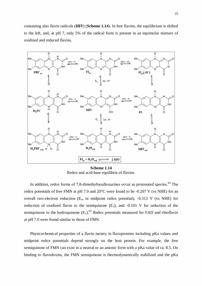
containing also flavin radicals (HFl.) (Scheme 1.14). In free flavins, the equilibrium is shifted
to the left, and, at pH 7, only 5% of the radical form is present in an equimolar mixture of
oxidized and reduced flavins.
NH
N
N
N+
Me
Me
O
O
R
NH
N
N
NMe
Me
O
O
R
N
N
N
NMe
Me
O
O
R
NH
N
N
NMe
Me
O
OH
R
NH
N
N
NMe
Me
O
O
R
NH
N
N+
NMe
Me
O
O
R
H H
NH
N
N
NMe
Me
O
O
R
NH
N
N
NMe
Me
O
O
R
N+ NH
NNMe
Me
O
O
R
H
H H
HH
H
H
Flox + H2Flred 2 HFl.
. . .
pKa ~ 0 pKa ~ 10
pKa ~ 8pKa ~ 2
pKa ~ 0 pKa ~ 6
E2
E1
1e-, H+
1e-, H+
Flox
HFl.
H2Flred
FlH+ox Flox(-H
+)
H2Fl+.Fl-.
H2FlH+red HFl-red
Scheme 1.14 Redox and acid-base equilibria of flavins.
In addition, redox forms of 7,8-dimethylisoalloxazines occur as protonated species.84
The
redox potentials of free FMN at pH 7.0 and 20°C were found to be -0.207 V (vs NHE) for an
overall two-electron reduction (Em or midpoint redox potential), -0.313 V (vs NHE) for
reduction of oxidized flavin to the semiquinone (E2), and -0.101 V for reduction of the
semiquinone to the hydroquinone (E1).85
Redox potentials measured for FAD and riboflavin
at pH 7.0 were found similar to those of FMN.
Physicochemical properties of a flavin moiety in flavoproteins including pKa values and
midpoint redox potentials depend strongly on the host protein. For example, the free
semiquinone of FMN can exist in a neutral or an anionic form with a pKa value of ca. 8.5. On
binding to flavodoxins, the FMN semiquinone is thermodynamically stabilized and the pKa
26
value is shifted by ca. four units to a higher value with accompanying large changes in the
redox potentials of one-electron reductions.86
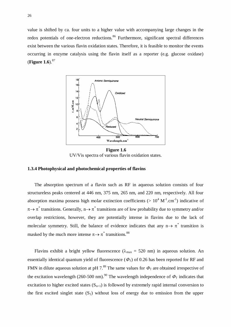
Furthermore, significant spectral differences
exist between the various flavin oxidation states. Therefore, it is feasible to monitor the events
occurring in enzyme catalysis using the flavin itself as a reporter (e.g. glucose oxidase)
(Figure 1.6).87
Figure 1.6
UV/Vis spectra of various flavin oxidation states.
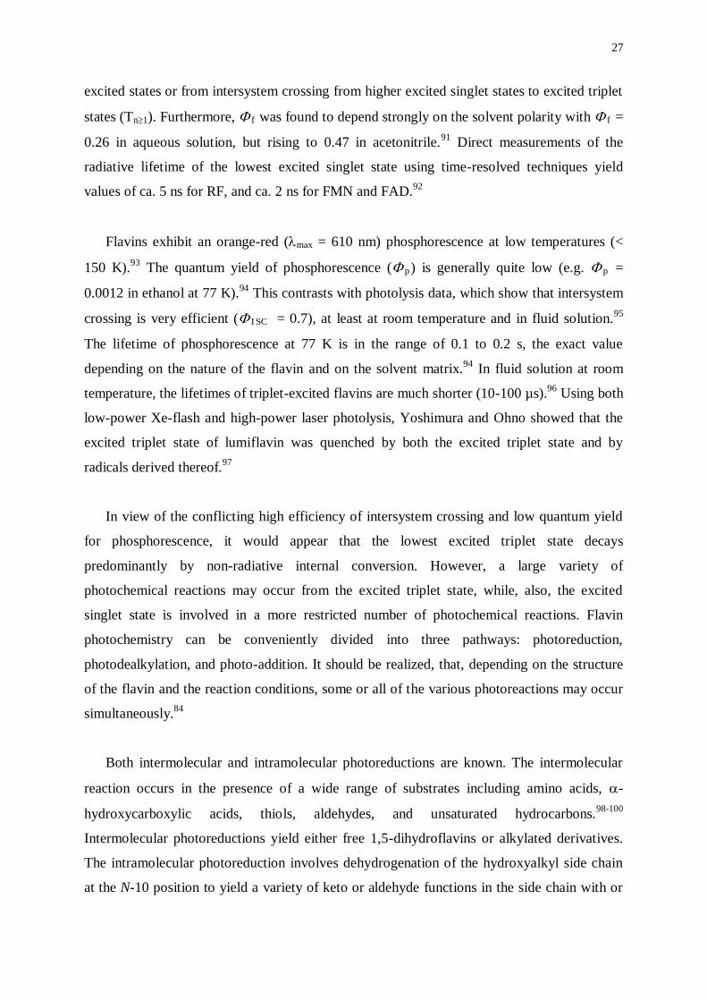
1.3.4 Photophysical and photochemical properties of flavins
The absorption spectrum of a flavin such as RF in aqueous solution consists of four
structureless peaks centered at 446 nm, 375 nm, 265 nm, and 220 nm, respectively. All four
absorption maxima possess high molar extinction coefficients (> 104
M-1
.cm-1
) indicative of
* transitions. Generally, n
* transitions are of low probability due to symmetry and/or
overlap restrictions, however, they are potentially intense in flavins due to the lack of
molecular symmetry. Still, the balance of evidence indicates that any n * transition is
masked by the much more intense * transitions.
88
Flavins exhibit a bright yellow fluorescence (max = 520 nm) in aqueous solution. An
essentially identical quantum yield of fluorescence ( f) of 0.26 has been reported for RF and
FMN in dilute aqueous solution at pH 7.89
The same values for f are obtained irrespective of
the excitation wavelength (260-500 nm).90
The wavelength independence of f indicates that
excitation to higher excited states (Sn>1) is followed by extremely rapid internal conversion to
the first excited singlet state (S1) without loss of energy due to emission from the upper
27
excited states or from intersystem crossing from higher excited singlet states to excited triplet
states (Tn1). Furthermore, f was found to depend strongly on the solvent polarity with f =
0.26 in aqueous solution, but rising to 0.47 in acetonitrile.91
Direct measurements of the
radiative lifetime of the lowest excited singlet state using time-resolved techniques yield
values of ca. 5 ns for RF, and ca. 2 ns for FMN and FAD.92
Flavins exhibit an orange-red (max = 610 nm) phosphorescence at low temperatures (<
150 K).93
The quantum yield of phosphorescence ( p) is generally quite low (e.g. p =
0.0012 in ethanol at 77 K).94
This contrasts with photolysis data, which show that intersystem
crossing is very efficient ( I SC = 0.7), at least at room temperature and in fluid solution.95
The lifetime of phosphorescence at 77 K is in the range of 0.1 to 0.2 s, the exact value
depending on the nature of the flavin and on the solvent matrix.94
In fluid solution at room
temperature, the lifetimes of triplet-excited flavins are much shorter (10-100 µs).96
Using both
low-power Xe-flash and high-power laser photolysis, Yoshimura and Ohno showed that the
excited triplet state of lumiflavin was quenched by both the excited triplet state and by
radicals derived thereof.97
In view of the conflicting high efficiency of intersystem crossing and low quantum yield
for phosphorescence, it would appear that the lowest excited triplet state decays
predominantly by non-radiative internal conversion. However, a large variety of
photochemical reactions may occur from the excited triplet state, while, also, the excited
singlet state is involved in a more restricted number of photochemical reactions. Flavin
photochemistry can be conveniently divided into three pathways: photoreduction,
photodealkylation, and photo-addition. It should be realized, that, depending on the structure
of the flavin and the reaction conditions, some or all of the various photoreactions may occur
simultaneously.84
Both intermolecular and intramolecular photoreductions are known. The intermolecular
reaction occurs in the presence of a wide range of substrates including amino acids, -
hydroxycarboxylic acids, thiols, aldehydes, and unsaturated hydrocarbons.98-100
Intermolecular photoreductions yield either free 1,5-dihydroflavins or alkylated derivatives.
The intramolecular photoreduction involves dehydrogenation of the hydroxyalkyl side chain
at the N-10 position to yield a variety of keto or aldehyde functions in the side chain with or
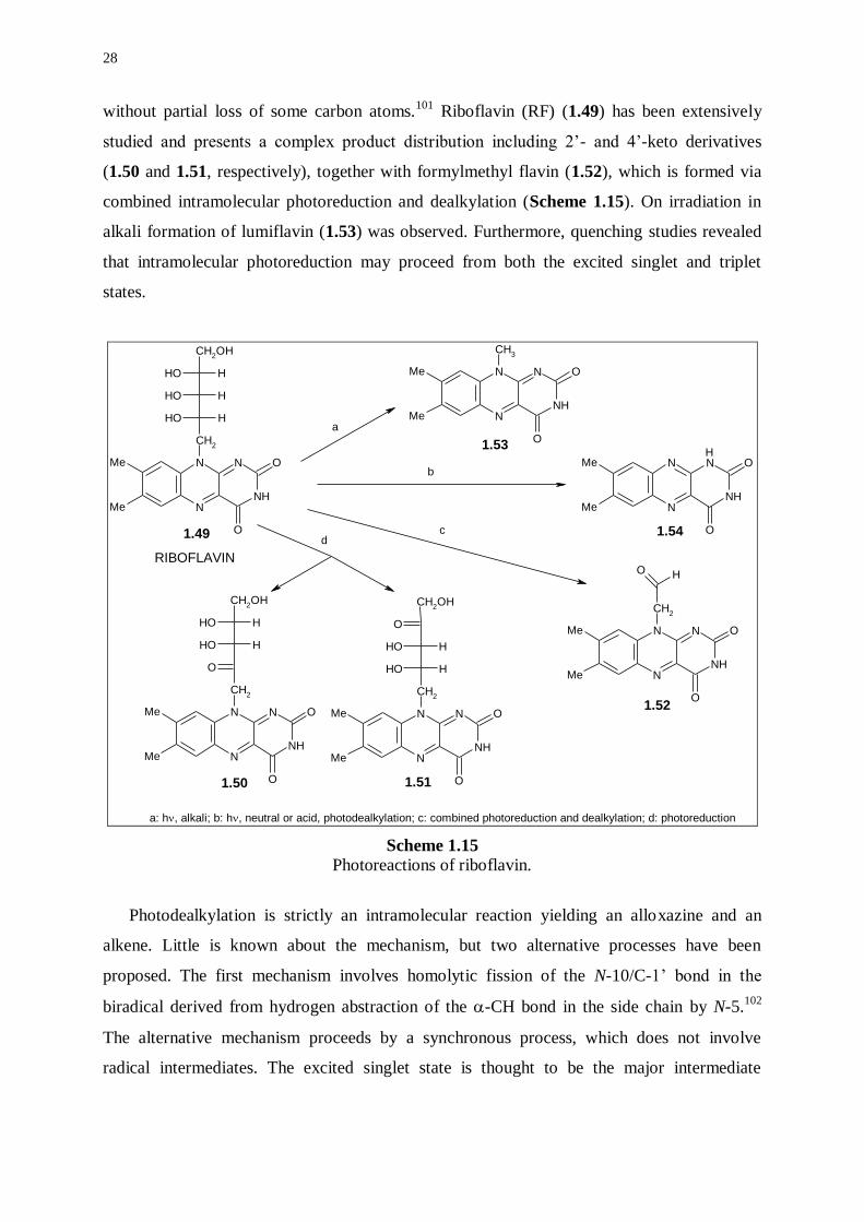
28
without partial loss of some carbon atoms.101
Riboflavin (RF) (1.49) has been extensively
studied and presents a complex product distribution including 2’- and 4’-keto derivatives
(1.50 and 1.51, respectively), together with formylmethyl flavin (1.52), which is formed via
combined intramolecular photoreduction and dealkylation (Scheme 1.15). On irradiation in
alkali formation of lumiflavin (1.53) was observed. Furthermore, quenching studies revealed
that intramolecular photoreduction may proceed from both the excited singlet and triplet
states.
NH
N
N
NMe
Me
CH2
CH2OH
O
O
OH H
OH H
OH H
NH
N
N
NMe
Me
CH2
CH2OH
O
O
O
OH H
OH H
NH
N
N
NMe
Me
CH2
O
O
O H
NH
N
N
NMe
Me
CH3
O
O
NH
N
N
NMe
Me
O
O
H
NH
N
N
NMe
Me
CH2
O
O
OH H
OH H
O
CH2OH
a
b
cd
a: h, alkali; b: h, neutral or acid, photodealkylation; c: combined photoreduction and dealkylation; d: photoreduction
1.49
1.50 1.51
1.52
1.53
1.54
RIBOFLAVIN
Scheme 1.15 Photoreactions of riboflavin.
Photodealkylation is strictly an intramolecular reaction yielding an alloxazine and an
alkene. Little is known about the mechanism, but two alternative processes have been
proposed. The first mechanism involves homolytic fission of the N-10/C-1’ bond in the
biradical derived from hydrogen abstraction of the -CH bond in the side chain by N-5.102
The alternative mechanism proceeds by a synchronous process, which does not involve
radical intermediates. The excited singlet state is thought to be the major intermediate

29
involved in photodealkylation of riboflavin, since triplet-state quenchers do not inhibit
formation of lumichrome (1.54) (Scheme 1.15).101
Intermolecular photoaddition has been reported where ROH is the solvent (R = H or alkyl)
and the solvent residue ‘RO’ adds to the C-6 or C-9 positions of the benzenoid subnucleus.103
Intramolecular photoaddition to the C-9 position occurs if a free hydroxy group is present at
the C-2’ position.104
Furthermore, a number of other photophysical phenomena and
photochemical reactions have been reported including photoelectron ejection,105
energy
transfer,106
cyclization of flavins possessing a phenyl substituent at N-10,107
photohydrolysis
and ring cleavage reactions,108
and photodehalogenation.109
In the presence of oxygen, flavins sensitize or catalyze the oxidation of various substrates
including amino acids,110
proteins,111
DNA and nucleotides,112
and lipids.113
Oxidation of
substrates may proceed via a radical (or Type I) mechanism involving electron abstraction
from the substrate, or via a non-radical (or Type II) mechanism involving energy transfer
from triplet-excited flavin to oxygen in the ground state thereby converting triplet oxygen to
the highly reactive singlet oxygen.114
Furthermore, photooxidations may also proceed by
direct triplet-triplet exergonic energy transfer from triplet-excited flavin to a substrate. Such
mechanism may operate, for example, in the flavin-sensitized photooxidation of retinal,115
bilirubin,116
and stilbenes.117
1.3.5 Flavins in beer
Quantitative analyses using HPLC with fluorescence detection have shown that riboflavin
(RF) is by far the most important flavin in beer with concentrations ranging from 100 to 600
µg.L-1
.118
Flavin adenine dinucleotide (FAD) was detected in all beers at concentrations of 10-
70 µg.L-1
, while flavin mononucleotide (FMN) was found in trace amounts in few beers.
Barley contains RF (1.2-1.5 mg.kg-1
) and the concentration increases during malting,
especially at higher germination temperatures.119
RF is extracted quantitatively into wort and
survives wort boiling. Furthermore, RF is also produced by yeast with the production rate
being proportional to yeast growth. On the other hand, part of RF is destroyed during the
stationary phase of the yeast as a result of enzymic breakdown.
30
1.4 ROLE OF THE SULFUR SOURCE
The source of the sulfur atom in 3-methylbut-2-ene-1-thiol (MBT), has not yet been
identified. Experiments, in which beer was dialyzed against water in cellophane membranes,
indicated that the donor molecules are of both high and low molecular weight. Thus, proteins,
polypeptides, as well as free amino acids may take part in the lightstruck reaction. Cysteine
and cystine have been implicated by sensory analysis in the formation of lightstruck flavor
(LSF) in model systems.21
In contrast, Blockmans et al. detected no MBT after irradiation of a
solution containing cysteine, isohumulones and riboflavin.120
It is conceivable that differences
in the redox potentials of the model systems contributed to the discrepancy of the results.
Previous work implicated dihydrogen sulfide as a possible sulfur source, but MBT is only
formed from dihydrogen sulfide in a highly reducing medium. Sakuma et al. showed that
dihydrogen sulfide could contribute to the formation of MBT, when high concentrations (> 50
mg.L-1
) of ascorbic acid were present in addition to isohumulones and RF.19
In commercial
beers, cleavage of dihydrogen sulfide to a thiol radical may be less significant than
decomposition of sulfur-containing compounds. It has been suggested that sulfur-containing
amino acids and proteins decompose to a thiol radical in RF-photosensitized reactions.19
On
the other hand, reactive oxygen species such as hydroxyl and hydroxyethyl radicals have been
shown to generate thiyl radicals from thiol-containing compounds including cysteine.21
1.5 FLAVOR STABILITY OF BEER
Beer flavor is not only subject to deterioration by the influence of light. Next to light-
induced formation of MBT as a ‘skunky’ thiol, significant flavor changes can be observed
during ageing of beer. The quality of bitterness decreases122
and chemical reactions of
compounds in beer produce an oxidized flavor.123
A variety of radical reactions may lead to
formation of unsaturated aliphatic aldehydes containing six to ten carbon atoms such as trans-
2-nonenal, which is largely responsible for the cardboard flavor of aged beers,124
whereas
formation of vicinal diketones and furan derivatives results in ‘buttery’ off-flavors.
Formation of aldehydes in beer is complex.125
During malting and mashing, aldehydes can
be formed via oxidation of fatty acids, either through combined actions of lipoxygenase and
31
lipase or via auto-oxidation to form hydroperoxides, which can be converted to staling
aldehydes. Aldehydes could also be formed via Strecker degradation of amino acids and these
could combine to form long-chain aldehydes via aldol condensations. During fermentation,
the reducing power of yeast converts most aldehydes remaining in the wort post-boiling to
their corresponding alcohols.126
During storage, aldehydes may be formed via melanoidin-
mediated oxidation of higher alcohols, oxidation of isohumulones, Strecker degradation of
amino acids, auto-oxidation of fatty acids or aldol condensations of short-chain aldehydes.
It is clear, that maintaining a low temperature during storage of beer and minimization of
oxygen during brewing result in improvements in flavor stability. These facts form the basis
of flavor stability tests using aerobic forced ageing at elevated temperatures. The increase in
the concentration of carbonyls during beer ageing has been linked to the presence of oxygen
and metal ions.127
Free radicals have been identified by electron spin resonance spectroscopy
as reaction intermediates in oxidation processes occurring in beer during aerobic forced
ageing. It was demonstrated that addition of iron salts and hydrogen peroxide accelerated
formation of radicals in beer. Moreover, addition of catalase, which destroys hydrogen
peroxide, or addition of chelating agents that bind metal ions proved to slow down production
of radicals.127
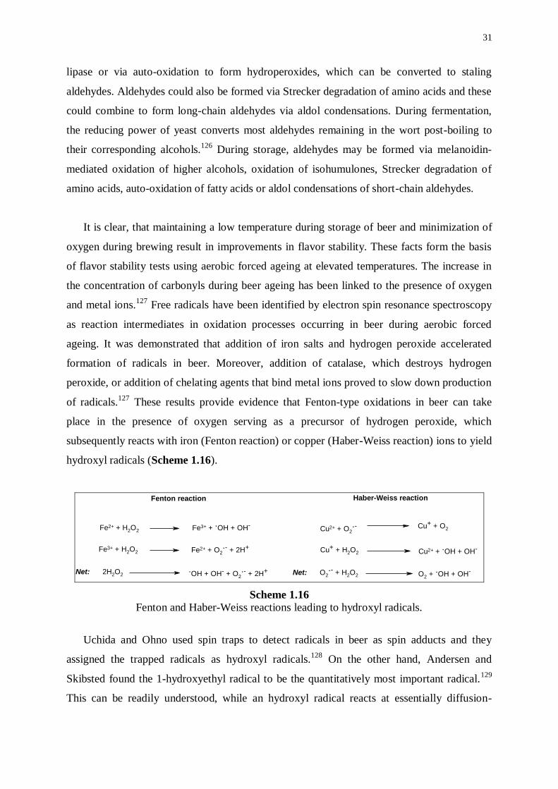
These results provide evidence that Fenton-type oxidations in beer can take
place in the presence of oxygen serving as a precursor of hydrogen peroxide, which
subsequently reacts with iron (Fenton reaction) or copper (Haber-Weiss reaction) ions to yield
hydroxyl radicals (Scheme 1.16).
Fenton reaction Haber-Weiss reaction
Cu2+ + O2
.- Cu+ + O2
Cu+ + H2O2 Cu2+ + .OH + OH-
Net: O2
.- + H2O2 O2 + .OH + OH-Net: 2H2O2
.OH + OH- + O2
.- + 2H+
Fe3+ + H2O2 Fe2+ + O2
.- + 2H+
Fe2+ + H2O2 Fe3+ + .OH + OH-
Scheme 1.16 Fenton and Haber-Weiss reactions leading to hydroxyl radicals.
Uchida and Ohno used spin traps to detect radicals in beer as spin adducts and they
assigned the trapped radicals as hydroxyl radicals.128
On the other hand, Andersen and
Skibsted found the 1-hydroxyethyl radical to be the quantitatively most important radical.129
This can be readily understood, while an hydroxyl radical reacts at essentially diffusion-
32
controlled reaction rates with most organic compounds and while ethanol is the beer
component found in the highest concentration besides water. Furthermore, by monitoring the
concentration of the spin adducts in function of time during aerobic forced ageing, it was
observed that the spin adducts were not immediately formed on starting the forcing test.
Initially, only a negligible amount of spin adducts was detected, while, after a certain period
of time, called the lag phase, the amount of spin adducts began to increase linearly with time.
The length of the lag phase of fresh beer has been shown to correlate with the flavor stability
of beer and the ESR technique is quite appropriate to predict the stability of beer.130
The
length of the lag phase has also been referred to as the endogenous antioxidant activity of
beer, while it is the result of competition between the actions of prooxidative and
antioxidative components of beer. The method, therefore, provides an excellent way to
examine potential antioxidants in beer.131
Furthermore, this assay observes both the time it
takes before the natural antioxidants are exhausted (the lag phase) and the rate at which
radicals are formed in the absence of antioxidants. Until now, only sulfite was able to delay
formation of radicals, whereas phenolic compounds such as phenolic acids, catechin,
epicatechin, and proanthocyanidin dimers had no effect on the formation of radicals.
Ascorbate, cysteine, and cysteamine, on the other hand, were found to be prooxidants. In
order to be effective under aerobic forced ageing tests, antioxidants must be able to either
scavenge peroxides or trap metal ions. The effectiveness of sulfite is probably a consequence
of its two-electron non-radical producing reaction with peroxides.132
33
1.6 REFERENCES
(1) Lee, K.H., Jung, M.Y., Kim, S.Y.; Effects of Ascorbic Acid on the Light-Induced Riboflavin
Degradation and Color Changes in Milks. J. Agric. Food Chem., 1998, 46, 407-410.
(2) Bosset J.O., Gallmann, P.U., Sieber, R.; Influence of Light Transmittance of Packing
Materials on the Shelf-life on Milk and Dairy Products – A Review. Lait, 1993, 73, 3-49.
(3) Maujean, A., Seguin, N.; Sunlight Flavors in Champagne Wines. 3. Photochemical Reactions
Responsible for Sunlight Flavors in Champagne Wine. Sci. Aliment., 1983, 3, 589-601.
(4) Gunst, F., Verzele, M. (1978); On the Sunstruck Flavor of Beer. J. Inst. Brew., 84, 291-292.
(5) Lintner, C.; In: Lehrbuch der Bierbrauerei, Verlag Vieweg und Sohn, Braunschweig, 1875, p.
343.
(6) Brand, J.; On the Detection of Arsenic in Sulphur. Zeit. ges. Brauwesen, 1908, 31, 333.
(7) De Clerck, J.; RH and its Applications in Brewing. J. Inst. Brew., 1934, 40, 407-419.
(8) Gray, P., Stone, I., Rothschild, H.; The Action of Sunlight on Beer. Wallerstein Lab.
Commun., 1941, 4, 29-40.
(9) Jacobssen, B., Högberg, B.; The Sensitivity of Beer to Light. Protection Afforded By Glass
Bottles. Wallerstein Lab. Commun., 1947, 10, 5-16.
(10) Kuroiwa, Y., Hashimoto, H.; Studies on Hops with Reference to their Role in the Evolution of
Sunstruck Flavor of Beer. Reports Res. Lab. Kirin Brew. Co., 1961, 4, 35-40.
(11) Kuroiwa, Y., Hashimoto, H.; Composition of Sunstruck Flavour Substance and Mechanism of
its Evolution. Proc. Am. Soc. Brew. Chem., 1961, 28-36.
(12) Kuroiwa, Y., Hashimoto, N., Hashimoto, H., Kobuko, E., Nakagawa, K.; Factors Essential for
the Evolution of Sunstruck Flavor. Proc. Am. Soc. Brew. Chem., 1963, 181-193.
(13) Kuroiwa, Y., Hashimoto, H., Hashimoto, N.; Factors Essential for the Evolution of Sunstruck
Flavor. Reports Res. Lab. Kirin Brew. Co., 1965, 8, 13-26.
(14) Kuroiwa, Y., Hashimoto, H., Nakagawa, K.; Quantitative Evaluation of the Sunstruck Flavor
of Beer. Reports Res. Lab. Kirin Brew. Co., 1960, 3, 17-23.
(15) Kuroiwa, Y., Nakagawa, K.; Conditions Necessary for the Evolution of Sunstruck Flavor,
Comparing with Those for Oxidation Flavor. Reports Res. Lab. Kirin Brew. Co., 1962, 5, 33-
40.
(16) Obata, Y., Koshika, M., Tanaka, H.; Studies on the Sunlight Flavor of Beer. Part VIII.
Mechanism of the Formation of the Sunlight Flavor. Agr. Biol. Chem., 1961, 25, 588-593.
(17) Kattein, U., Miedaner, H., Narzi, L.; Zur Problematik des Lichtgeschmacks im Bier.
Monatsschr. Brauwiss., 1988, 41, 205-208.
(18) Blondeel, G.M.A., De Keukeleire, D., Verzele, M.; The Photolysis of Isohumulone to
Dehydrohumulinic Acid, a Key Route to the Development of Sunstruck Flavour in Beer. J.
Chem. Soc. Perkin Trans.1, 1987, 1, 2715-2717.
34
(19) Sakuma, S., Rikimaru, Y., Kobayashi, K., Kowaka, M.; Sunstruck Flavor Formation in Beer.
J. Am. Soc. Brew. Chem., 1991, 49, 162-165.
(20) Hastings, D.J., McGarrity, M.J., Bordeleau, L., Thompson, D.J.; Results presented at the
Brewing Congress of the Americas, St. Louis, MO, 1992, Sept. 20-24.
(21) Irwin, A.J., Bordeleau, L., Barker, R.L.; Model Studies and Flavor Threshold Determination
of 3-Methyl-2-Butene-1-Thiol in Beer. J. Am. Soc. Brew. Chem., 1993, 51, 1-3.
(22) Goldstein, H., Rader, S., Murakami, A.A.; Determination of 3-Methyl-2-Butene-1-Thiol in
Beer. J. Am. Soc. Brew. Chem., 1993, 51, 70-74.
(23) Holscher, W., Vitzthum, O.G., Steinhart, H.; Prenyl Alcohol – Source for Odorants in Coffee.
J. Agric. Food Chem., 1992, 40, 655-658.
(24) Hughes, P.; The Lightstruck Flavour Problem. Cerevisia, 1999, 24(2), 21-25.
(25) Masuda, S., Kikuchi, K., Harayama, K.; Determination of Lightstruck Character in Beer by
Gas Chromatography-Mass Spectroscopy. J. Am. Soc. Brew. Chem., 2000, 58, 152-154.
(26) Lowson, J.M.; Textbook of Botany. University Tutorial Press, London, 1962.
(27) De Cooman, L., Everaert, E., De Keukeleire, D.; Quantitative Analysis of Hop Acids,
Essential Oils and Flavonoids as a Clue to the Identification of Hop Varieties. Phytochem.
Anal., 1998, 9, 145-150.
(28) Verzele, M., De Keukeleire, D.; Chemistry and Analysis of Hop and Beer Bitter Acids.
Elsevier, Amsterdam, 1991.
(29) Sharpe, F.R., Laws, D.R.J.; The Essential Oil of Hops. A Review. J. Inst. Brew., 1981, 87, 96-
107.
(30) Eri, S., Khoo, B.K., Lech, J., Hartman, T.G.; Direct Thermal Desorption-Gas
Chromatography and Gas Chromatography-Mass Spectrometry Profiling of Hop (Humulus
lupulus L.) Essential Oils in Support of Varietal Characterization. J. Agric. Food Chem.,
2000, 48, 1140-1149.
(31) Howard, G.A.; Evaluation of Hops. The Essential Oil of Hops. J. Inst. Brew., 1956, 62, 158-
159.
(32) Moir, M.; Hop Aromatic Compounds. In: European Brewery Convention Monograph XXII -
EBC-Symposium on Hops, Zoeterwoude, The Netherlands, 1994, May-June. Verlag Hans
Carl, Nürnberg, 1994, pp. 165-180.
(33) Peacock, V.E., Deinzer, M.L.; Chemistry of Hop Aroma in Beer. J. Am. Soc. Brew. Chem.,
1981, 39, 136-141.
(34) Yang, X., Lederer, C., McDaniel, M., Deinzer, M.L.; Chemical Analysis and Sensory
Evaluation of Hydrolysis Products of Humulene Epoxides II and III. J. Agric. Food Chem.,
1993, 41, 1300-1304.
(35) Damodaran, N.P., Dev, S.; Studies in Sesquiterpenes. 39. Structure of Humulenols.
Tetrahedron, 1968, 24, 4133-4142.
35
(36) Naya, Y., Kotake, M.; Natural Occurrence of Humuladienone, Humulenone-II, and -
Corocalene: New Constituents of Hop Oil. Bull. Chem. Soc. Jp., 1969, 42, 2405.
(37) Forster, A., Beck, B., Schmidt, R.; Untersuchungen zu Hopfenpolyphenolen. In: Proc. 25th
European Brewery Convention Congress, Brussels, Belgium, 1995. Oxford University Press,
Oxford, 1995, pp. 143-150.
(38) Stevens, J.R., Miranda, C.L., Buhler, D.L., Deinzer, M.L.; Chemistry and Biology of Hop
Flavonoids. J. Am. Soc. Brew. Chem., 1998, 56, 136-145.
(39) De Keukeleire, D., Milligan, S.R., De Cooman, L., Heyerick, A.; Hop-derived Phytoestrogens
in Beer? In: Proc. 26th
European Brewery Convention Congress, Maastricht, The
Netherlands, 1997. Oxford University Press, Oxford, 1997, pp. 239-246.
(40) Tekel’, J., De Keukeleire, D., Rong, H., Daeseleire, E., Van Peteghem, C.; Determination of
the Hop-Derived Phytoestrogen, 8-Prenylnaringenin, in Beer by Gas Chromatography-Mass
Spectrometry. J. Agric. Food Chem., 1999, 47, 5059-5063.
(41) Rong, H., Zhao, Y., Lazou, K., De Keukeleire, D., Milligan, S.R., Sandra, P.; Quantitation of
8-Prenylnaringenin, a Novel Phytoestrogen in Hops (Humulus lupulus L.), Hop Products, and
Beers, by Benchtop HPLC-MS Using Electrospray Ionization. Chromatographia, 2000, 51,
545-552.
(42) Vanhoenacker, G., Rong, H., De Keukeleire, D., Baeyens, W., Van Der Weken, G., Sandra,
P.; Simultaneous Analysis of Hop Acids and Prenylated Flavonoids by Microemulsion
Electrokinetic Chromatography with Diode Array Detection. Biomed. Chromatogr., 2000, 14,
34-36.
(43) Milligan, S.R., Kalita, J.C., Heyerick, A., Rong, H., De Cooman, L., De Keukeleire, D.;
Identification of a Potent Phytoestrogen in Hops (Humulus lupulus L.) and Beer. J. Clin.
Endocrinol. Metab., 1999, 83, 2249-2252.
(44) Milligan, S.R., Kalita, J.C., Pocock, V., Van de Kauter, V., Rong, H., De Keukeleire, D.,
Stevens, J.F., Deinzer, M.; The Endocrine Activities of 8-Prenylnaringenin and Related Hop
(Humulus lupulus L.) Flavonoids. J. Clin. Endocrinol. Metab., 2000, 85, 4916-4920.
(45) Moll, M.; Colloidal Stability of Beer. In: Brewing Science, Vol. 3, J.R.A. Pollock, Ed.
Academic Press, London, 1987.
(46) McMurrough, I., Madigan, D., Kelly, R.J.; The Role of Flavanoid Polyphenols in Beer
Stability. J. Am. Soc. Brew. Chem., 1996, 54, 141-148.
(47) Bamforth, C.W.; Beer Haze. J. Am. Soc. Brew. Chem., 1999, 57, 81-90.
(48) Van Boven, M., Verzele, M.; The Isomerization Mechanism of Humulone. Bull. Soc. Chim.
Belges, 1971, 80, 677-682.
(49) De Keukeleire, D., Verzele, M., The Absolute Configuration of the Isohumulones and the
Humulinic Acids. Tetrahedron, 1971, 27, 4939-4945.
36
(50) Clarke, B.J., Hildebrand, R.P.; The Isomerization of Humulone I - Isolation of
Photoisohumulone. J. Inst. Brew., 1965, 71, 26-36.
(51) De Keukeleire, D., Blondeel, G.M.; The Mechanism of the Regio- and Stereospecific
Photorearrangement of Humulone to the Beer Bitter Component trans-Isohumulone.
Tetrahedron Lett., 1979, 15, 1343-1346.
(52) Hughes, P.S., Simpson, W.J.; Bitterness of Congeners and Stereoisomers of Hop-Derived
Bitter Acids Found in Beer. J. Am. Soc. Brew. Chem., 1996, 54, 234-237.
(53) Clark, D.C., Wilde, P.J., Wilson, D.R.; The Effect of Pre-Isomerized Hop Extract on the
Properties of Model Protein Stabilized Foams. J. Inst. Brew., 1991, 97, 169-172.
(54) Asano, K., Hashimoto, N.; Contribution of Hop Bitter Substances to Head Formation of Beer.
Reports Res. Lab. Kirin Brew. Co., 1976, 19, 9-16.
(55) Simpson, W.J.; Studies on the Sensitivity of Lactic Acid Bacteria to Hop Bitter Acids. J. Inst.
Brew., 1993, 99, 405-411.
(56) Benitez, J.L., Forster, A., De Keukeleire, D., Moir, M., Sharpe, F.R., Verhagen, L.C.,
Westwood, K.T.; Hops and Hop Products. Fachverlag Hans Carl, Nürnberg, 1997.
(57) Hougen, O.A.; Catalytic Reduction of Hop Extracts for Use in Light-Stable Beer. U.S. Patent
3,079,262, 1963.
(58) Khokher, A., Anteunis, M., Verzele, M.; -Isohumulones. Bull. Soc. Chim. Belges, 1967, 76,
101-106.
(59) Ting, P.L., Goldstein, H.; Preparation and Purification of Hop Acids and Their Derivatives. J.
Am. Soc. Brew. Chem., 1996, 54, 103-109.
(60) Todd, P.H., Johnson, P.A., Worden, L.R.; Evaluation of the Relative Bitterness and Light
Stability of Reduced Iso-Alpha Acids. Techn. Quart. Master Brew. Assoc. Am., 1971, 9 (1),
31-35.
(61) Worden, L.R.; Production of Hoplike Beverage Bittering Materials. U.S. Patent 3,923,897,
1975.
(62) Cowles, J.M., Goldstein, H., Chicoye, E., Ting, P.L.; Preparation of Tetrahydroisohumulones.
U.S. Patent 4,644,084, 1987.
(63) Verzele, M., Govaert, F.; Transformation of Humulone. Congr. Int. Ind. Ferment. Conf.
Commun., 1947, 297-301.
(64) Brown, P.M., Howard, G.A., Tatchell, A.R.; Chemistry of Hop Constituents. Part XIII. The
Hydrogenation of Isohumulone. J. Chem. Soc., 1959, 545-551.
(65) Byrne, E., Shaw, S.J.; Reduction Products from Cis- and Trans-Isocohumulone. J. Chem.
Soc., 1971, 2810-2813.
(66) Verzele, M., Anteunis, M.; On the Hydrogenation of Humulone. Part I. The Preparation of
Tetrahydrohumulone. Bull. Soc. Chim. Belges, 1959, 68, 315-324.
37
(67) Hay, B.A., Homiski, J.W.; Efficient One-Step Preparation of the Beer Additive Tetrahydroiso-
-Acids. J. Agric. Food. Chem., 1991, 39, 1732-1734.
(68) Moir, M., Smith, R.J.; Foam-Enhancing and Bitter, Reduced Iso--Acids Derived from Hops.
In: Proc. 25th European Brewery Convention Congress, Brussels, Belgium, 1995. Oxford
University Press, Oxford, 1995, pp. 125-134.
(69) Combes, R.P.; Hop Products. The Brewer, 1998, January, 29-35.
(70) Massey, V.; The Chemical and Biological Versatility of Riboflavin. Biochem. Soc. Trans.,
2000, 28, 283-296.
(71) Voet, D., Voet, J.G.; Biochemistry., John Wiley & Sons, New York, 1990.
(72) Clerin, D., Bruice, T.C.; Kinetic Study of Fate of a Covalent Intermediate of Type Proposed to
be Involved in Flavin Catalysis. J. Am. Chem. Soc., 1974, 96, 5571-5573.
(73) Stroes, E., Hijmering, M., Van Zandvoort, M., Wever, R., Rabelink, T.J., Van Faassen, E.E.;
Origin of Superoxide Production by Endothelial Nitric Oxide Synthase. FEBS Lett., 1998,
438, 161-164.
(74) Harris, C.M., Massey, V.; The Reaction of Reduced Xanthine Dehydrogenase with Molecular
Oxygen - Reaction Kinetics and Measurement of Superoxide Radical. J. Biol. Chem., 1997,
272, 8370-8379.
(75) Imlay, J.A.; A Metabolic Enzyme that Rapidly Produces Superoxide, Fumarate Reductase of
Escherichia Coli. J. Biol. Chem., 1995, 270, 19767-19777.
(76) Dagley, S.; Lessons from Biodegradation. Annu. Rev. Microbiol., 1987, 41, 1-23.
(77) Tu, S.C., Mager, H.I.X.; Biochemistry of Bacterial Bioluminescence. Photochem. Photobiol.,
1995, 62, 615-624.
(78) Christie, J.M., Reymond, P., Powell, G.K., Bernasconi, P., Raibekas, A.A., Liscum, E.,
Briggs, W.R.; Arabidopsis NPH1: A Flavoprotein with the Properties of a Photoreceptor for
Phototropism. Science, 1998, 282, 1698-1701.
(79) Heelis, P.F., Kim, S., Okamura, T., Sancar, A.; The Photo Repair of Pyrimidine Dimers by
DNA Photolyase and Model Systems. J. Photochem. Photobiol. B: Biol., 1993, 17, 219-228.
(80) Kim, S.-T., Sancar, A., Essenmacher, C., Babcock, G.T.; Time-Resolved EPR Studies with
DNA Photolyase: Excited-state FADH0 Abstracts an Electron from Trp-306 to Generate
FADH-, the Catalytically Active Form of the Cofactor. Proc. Natl. Acad. Sci. USA, 1993, 90,
8023-8027.
(81) Aubert, C., Vos, M.H., Mathis, P., Eker, A.P.M., Brettel, K.; Intraprotein Radical Transfer
during Photoactivation of DNA Photolyase. Nature, 2000, 405, 586-590.
(82) Susin, S.A., Lorenzo, H.K., Zamzami, N., Marzo, I., Snow, B.E., Brothers, G.M., Mangion, J.,
Jacotot, E., Costantini, P., Loeffler, M., Larochette, N., Goodlett, D.R., Aebersold, R.,
Siderovski, D.P., Penninger, J.M., Kroemer, G.; Molecular Characterization of Mitochondrial
Apoptosis-inducing Factor. Nature, 1999, 397, 441-446.
38
(83) Daugas, E., Nochy, D., Ravagnan, L., Loeffler, M., Susin, S.A., Zamzami, N., Kroemer, G.;
Apoptosis-inducing Factor (AIF): an Ubiquitous Mitochondrial Oxidoreductase Involved in
Apoptosis. FEBS Lett., 2000, 476, 118-123.
(84) Heelis, P.F.; The Photophysical and Photochemical Properties or Flavins (Isoalloxazines).
Chem. Soc. Rev., 1982, 11, 15-39.
(85) Mayhew, S.G.; The Effects of pH and Semiquinone Formation on the Oxidation-Reduction
Potentials of Flavin Mononucleotide. Eur. J. Biochem., 1999, 265, 698-702.
(86) Ludwig, M.L., Schopfer, L.M., Metzger, A.L., Pattridge, K.A., Massey, V.; Structure and
Oxidation-reduction Behavior of 1-Deaza-FMN Flavodoxin: Modulation of Redox Potentials
in Flavodoxins. Biochemistry, 1990, 29, 10364-10375.
(87) Massey, V., Palmer, G.; On the Existence of Spectrally Distinct Classes of Flavoprotein
Semiquinones. A New Method for the Quantitative Production of Flavoprotein Semiquinones.
Biochemistry, 1966, 3181-3189.
(88) Sun, M., Song, P.S.; Excited-states and Reactivity of 5-Deazaflavine. Comparative Studies
with Flavine. Biochemistry, 1973, 12, 4663-4669.
(89) Sun, M., Song, P.S., Moore, T.A.; Molecular Luminescence Studies of Flavins. I. Excited-
states of Flavins. J. Am. Chem. Soc., 1972, 94, 1730-1740.
(90) Weber, G., Teale, W.J.; Determination of the Absolute Quantum Yield of Fluorescent
Solutions. Trans. Faraday Soc., 1957, 53, 646-655.
(91) Eweg, J.K., Müller, F., Visser, A.J.W.G., Veeger, C., Bebelaar, D., Vanvoorst, J.D.W.;
Molecular Luminescence of Some Isoalloxazines in Apolar Solvents at Various Temperatures.
Photochem. Photobiol., 1979, 30, 463-471.
(92) Wahl, P., Auchet, J.C., Visser, A.J.W.G., Müller, F.; Time-resolved Fluorescence of Flavin
Adenine-dinucleotide. FEBS Lett., 1974, 44, 67-70.
(93) Bowd, A., Byrom, P., Hudson, J.B., Turnbull, J.H.; Excited States of Flavine Coenzymes – III.
Fluorescence and Phosphorescence Emissions. Photochem. Photobiol., 1968, 8, 1-10.
(94) Grodowski, M.S., Veyret, B., Weiss, K.; Photochemistry of Flavins. II. Photophysical
Properties of Alloxazines and Isoalloxazines. Photochem. Photobiol., 1977, 26, 341-352.
(95) Moore, W.M., McDaniels, J.C., Hen, J.A.; The Photochemistry of Riboflavin - VI. The
Photophysical Properties or Isoalloxazines. Photochem. Photobiol., 1977, 25, 505-512.
(96) Song, P.S., Kurtin, W.E.; The Nature of the Triplet States of Flavins: A Further Study.
Photochem. Photobiol., 1969, 10, 211-214.
(97) Yoshimura, A., Ohno, T.; Quenching of Excited Triplet Lumiflavin by Lumiflavin Radicals
Formed in Its T-T Reaction. Photochem. Photobiol., 1991, 53, 175-179.
(98) Silva, E., Godoy, J.; Riboflavin-Sensitized Photooxidation of Tyrosine. Int. J. Nutr. Res., 1994,
64, 253-256.
39
(99) Silva, E., Ugarte, R., Andrade, A., Edwards, A.M.; Riboflavin-Sensitized Photoprocesses of
Tryptophan. J. Photochem. Photobiol. B: Biol., 1994, 23, 43-48.
(100) Jung, M.Y., Kim, S.K., Kim, S.Y.; Riboflavin-Sensitized Photooxidation of Ascorbic Acid:
Kinetics and Amino Acid Effects. Food Chem., 1995, 53, 397-403.
(101) Cairns, W.L., Metzler, D.E.; Photochemical Degradation of Flavins. VI. A New Photoproduct
and Its Use in Studying the Photolytic Mechanism. J. Am. Chem. Soc., 1971, 93, 2772-2777.
(102) Moore, W.M., Ireton, R.C.; The Photochemistry of Riboflavin - V. The Photodegradation of
Isoalloxazines in Alcoholic Solvents. Photochem. Photobiol., 1977, 25, 347-356.
(103) Schöllhammer, G., Hemmerich, P.; Nucleophilic Addition at Photoexcited Flavin Cation –
Synthesis and Properties of 6-Hydroxy-flavocoenzyme and 9-Hydroxy-flavocoenzyme
Chromophores. Eur. J. Biochem., 1974, 44, 561-577.
(104) Schumanjorns, M., Schöllhammer, G., Hemmerich, P.; Intramolecular Addition of Riboflavin
Side Chain – Anion-catalyzed Neutral Photochemistry. Eur. J. Biochem., 1975, 57, 35-48.
(105) Getoff, N., Solar, S., McCormick, D.B.; Photoejection of Electrons from Flavins in Polar
Media. Science, 1978, 201, 616-618.
(106) Naman, N.; Energy Conversion in the Decay of Triplet Lumiflavin in the Presence of Ferri-
and Ferrocyanide. Photochem. Photobiol., 1988, 47, 43-48.
(107) Knappe, W.R.; In: Flavins and Flavoproteins, Elsevier, Amsterdam, 1976, p. 788.
(108) Yoneda, F., Sakuma, Y., Hemmerich, P.; Dehydrogenation of Alcohols by Pyrimido[4,5-
B]quinoline-2(3H),4(10H)-dione (5-Deazaflavin). J. Chem. Soc. Chem. Commun., 1977, 22,
825-826.
(109) Massey, V., Husain, M., Hemmerich, P.; Photodehalogenation of 7-Halogen-substituted and
8-Halogen-substituted Flavins. Photochemistry of the Reduced Flavin Chromophore. J. Biol.
Chem., 1980, 255, 1393-1398.
(110) Garcia, J., Silva, E.; Flavin-Sensitized Photooxidation of Amino Acids Present in a Parenteral
Nutrition Infusate: Protection by Ascorbic Acid. J. Nutr. Biochem., 1997, 8, 341-345.
(111) Roldan, J.M., Calero, F., Aparicio, P.J.; Photoreactivation of Spinach Nitrate Reductase –
Role of Flavins. Zeitschr. f. Pflanzenphysiologie, 1978, 90, 467-474.
(112) Saito, I., Takayama, M., Sugiyama, H., Nakatami, K.; Photoinduced DNA Cleavage via
Electron Transfer: Demonstration That Guanine Residues Located 5' to Guanine Are the Most
Electron-Donating Sites. J. Am. Chem. Soc., 1995, 117, 6406-6407.
(113) Silva, E., Gonzalez, T., Edwards, A.M., Zuloaga, F.; Visible Light Induced Lipoperoxidation
of a Parenteral Nutrition Fat Emulsion Sensitized by Flavins. J. Nutr. Biochem., 1998, 9, 149-
154.
(114) Smith, K.C.; The Science of Photobiology, Plenum Press, New York, 1989, pp. 82-84.
(115) Futterman, S., Rollins, M.H.; Catalytic Isomerization of All-trans-retinal to 9-Cis-retinal and
13-Cis-retinal. J. Biol. Chem., 1973, 248, 7773-7779.
40
(116) Sanvordeker, D.R., Kostenbouder, H.B.; Mechanism for Riboflavin Enhancement of Bilirubin
Photodecomposition in-vitro. J. Pharm. Sci., 1974, 63, 404-408.
(117) Gordon-Walker, A., Radda, G.K.; Flavin-Photosensitized Reactions of Retinol and Stilbene.
Biochem. J., 1970, 120, 673-681.
(118) Andres-Lacueva, C., Mattivi, F., Tonon, D.; Determination of Riboflavin, Flavin
Mononucleotide and Flavin Adenine Dinucleotide in Wine and Other Beverages by High-
Performance Liquid Chromatography with Fluorescence Detection. J. Chromatogr. A, 1998,
823, 355-363.
(119) Koksel, H., Edney, M.J., Ozkaya, B.; Barley Bulgur: Effect of Processing and Cooking on
Chemical Composition. J. Cereal Sci., 1999, 29, 185-190.
(120) Blockmans, C., Van De Meersche J., Masschelein, C.A., Devreux, A.; In: Proc. 18th European
Brewery Convention Congress, Copenhagen, 1981. IRL Press, London, 1981, p. 347.
(121) Asmus, K.D., Chatgillialoglu, C.; In: Sulfur-centered Reactive Intermediates in Chemistry and
Biology. Plenum Press, New York, 1991, p. 359.
(122) King, B.M., Duineveld, C.A.A.; Changes in Bitterness as Beer Ages Naturally. Food Quality
Preference, 1999, 10, 315-324.
(123) Bamforth, C.W.; The Science and Understanding of the Flavour Stability of Beer: a Critical
Assessment. Brauwelt Int., 1999, 11, 98-110.
(124) Meilgaard, M., Elizondo, A., Moya, E.; A Study of Carbonyl Compounds in Beer; II. Flavor
and Flavor Thresholds of Aldehydes and Ketones Added to Beer. Techn. Quart. Master Brew.
Assoc. Am., 1970, 7, 143-149.
(125) Walters, M.T.; Natural Antioxidants and Flavour Stability. Ferment, 1997, 10 (2), 111-119.
(126) Peppard, T.L., Halsey, S.A.; Malt Flavour – Transformation of Carbonyl Compounds by Yeast
During Fermentation. J. Inst. Brew., 1981, 87, 386-390.
(127) Kaneda, H., Kano, Y., Koshino, S., Ohya-Nishiguchi, H.; Behavior and Role of Iron Ions in
Beer Deterioration. J. Agric. Food Chem., 1992, 40, 2102-2107.
(128) Uchida, M., Ono, M.; Improvement for Oxidative Flavor Stability of Beer – Role of OH-
Radical in Beer Oxidation. J. Am. Soc. Brew. Chem., 1996, 54, 198-204.
(129) Andersen, M.L., Skibsted, L.H.; Electron Spin Resonance Spin Trapping - Identification of
Radicals Formed during Aerobic Forced Aging of Beer. J. Agric. Food Chem., 1998, 46,
1272-1275.
(130) Uchida, M., Suga, S., Ono, M.; Improvement for Oxidative Flavor Stability of Beer – Rapid
Prediction Method for Beer Flavor Stability by Electron Spin Resonance Spectroscopy. J. Am.
Soc. Brew. Chem., 1996, 54, 205-211.
(131) Andersen, M.L., Outtrup, H., Skibsted, L.H.; Potential Antioxidants in Beer Assessed by ESR
Spin Trapping. J. Agric. Food Chem., 2000, 48, 3106-3111.

41
(132) Hoffmann, M.R., Edwards, J.O.; Kinetics of the Oxidation of Sulfite by Hydrogen Peroxide in
Acidic Solution. J. Phys. Chem., 1975, 79, 2096-2098.
************************


43
OBJECTIVES
Our current understanding of formation of the lightstruck flavor in beer dates from the
original reports by Kuroiwa et al. in the early sixties. A formal mechanism involves
photosensitized degradation of isohumulones by riboflavin and subsequent trapping of a 3-
methylbut-2-enyl radical by a thiol radical derived from suitable sulfur source (e.g. cysteine)
to give 3-methylbut-2-ene-1-thiol. All relevant data collected to date do not contradict the
Kuroiwa premise. Although the key components are known for more than three decades, very
few investigations have reported on the mechanisms of interaction between these components.
This Ph.D. project aimed at obtaining a comprehensive understanding of the mechanism
of formation of the lightstruck flavor in beer. The issue was approached by an in-depth
investigation of the interacting components on a molecular level using simple model systems,
consisting of isohumulones, dihydroisohumulones or tetrahydroisohumulones in the presence
or absence of riboflavin or flavin mononucleotide as a sensitizer.
Unraveling a photochemical mechanism implies studies in various time domains. Thus,
model systems must be subjected to both photostationary irradiation and photodynamic flash-
initiated experiments. Information on the nature of intervening photo-excited states and
intermediates, along with mechanistic features, can be retrieved from time-resolved electron
paramagnetic resonance and laser-flash photolysis transient absorption spectroscopy.
Characterization and identification of prevalent photorection products by spectroscopic
techniques should allow to elaborate relevant reaction schemes to account for formation of
key compounds.
The objective was, in a first phase, to undertake a detailed study of the photolability of
isohumulones, dihydroisohumulones, and tetrahydroisohumulones. In a second phase, the
purpose was to investigate the interaction between isohumulones, dihydroisohumulones or
tetrahydroisohumulones with riboflavin or flavin mononucleotide on sensitized irradiation.
It was advanced that interpretation of photostationary and photodynamic experiments
would prove that the events leading to formation of the lightstruck flavor differ according to
the irradiation conditions. Thus, the principal target was to elucidate the (photo)reaction
mechanisms operating either under direct UV irradiation or under visible light irradiation in
the presence of a sensitizer. Knowledge of intricate mechanistic details should then allow to
find innovative and proprietary ways to prevent formation of the lightstruck flavor in beer.

DIRECT IRRADIATION
WITH UV LIGHT

47
CHAPTER 2
TIME-RESOLVED ELECTRON PARAMAGNETIC RESONANCE OF
ISOHUMULONES AND REDUCED DERIVATIVES UNDER DIRECT
IRRADIATION WITH UV LIGHT
2.1 ELECTRON PARAMAGNETIC RESONANCE
2.1.1 General introduction
The technique of electron paramagnetic resonance (EPR) spectroscopy is very similar to
the more familiar nuclear magnetic resonance (NMR) technique. While NMR involves the
interaction of electromagnetic radiation in the radiofrequency range with the magnetic
moments of nuclei, EPR is based on the interaction of microwave radiation with the magnetic
moments of electrons.b Each electron possesses an intrinsic magnetic dipole moment that
arises from its spin. In most systems, electrons occur in pairs such that the net moment is zero.
Hence, only species that contain one or more unpaired electrons possess the net spin moment
necessary for suitable interaction with an electromagnetic field. Typical systems that are
studied by EPR include free radicals (containing one unpaired electron), triplet-state systems
and biradicals (containing two unpaired electrons), transition metal ions (including actinide
ions), and point defects in solids.1
In order to characterize free radicals in liquid solution, two magnetic parameters are
normally measured. These are the g-factor (chemical shift) and the electron-nuclear hyperfine
interaction, which for protons is written as aH. The g-factor for a free electron (ge), determined
to be 2.002319304386, is one of the most accurately known physical constants. Most organic
radicals centered on C, N, O, S or P atoms have a g-factor, which varies only some +/- 0.003
units from the value for a free electron. This follows from the fact that an electron in free
radicals can move more or less freely over an orbital encompassing the whole molecule and
the electron is not confined to a localized orbital between two atoms in a molecule. Therefore,
its behavior is very much the same as an electron in free space. For the abovementioned
b EPR is also frequently referred to as electron spin resonance (ESR), while, in most cases, the absorption is
linked primarily to the electron-spin angular momentum. The denotation ‘EPR’ takes into account contributions
from an electron orbital as well as from a spin angular momentum.
48
radicals, the g-factor on itself is not very informative and further information can be obtained
from the electron-nuclear hyperfine interaction, which arises through coupling of the unpaired
electron with neighbouring nuclear spins in much the same way as the coupling between
nuclear spins in NMR. In general, a nucleus with spin I will split the resonance line of an
electron into a multiplet with 2I +1 lines of equal intensity. Hyperfine interactions between
carbon-centered radicals and protons are on the order of 1-40 Gauss, giving spectral widths
(in frequency units) that range from 10 MHz to 300 MHz for typical organic radicals. These
couplings are larger by a factor of ca. 106 with respect to nucleus-nucleus coupling, which can
be accounted for by the fact that an electron can approach a nucleus more closely than another
nucleus and will, therefore, interact more strongly with the nucleus. The most important factor
influencing the magnitude of electron-nucleus coupling is the length of time, which the
electron spends in the vicinity of the coupled nucleus or, in other words, the electron density
at the nucleus (more correctly, the spin density rather than the physical density). Therefore,
EPR can be used to build up a qualitative picture of electron distribution within a radical.
The resonant frequency of most organic radicals in the microwave region of the
electromagnetic spectrum is equal to ca. 9.5 GHz with an applied external field of 3.4 kGauss.
The 9.5 GHz, called the X-band, is the standard frequency for most commercial EPR
spectrometers due to the availability of inexpensive X-band microwave components used in
radar. The EPR signals (either absorption or emission) are detected using a microwave bridge
circuit coupled to a resonant cavity with the sample in the center, positioned such that the
microwave magnetic field is at a maximum for EPR sensitivity and, concurrently, that the
microwave electric field is at a minimum. In a conventional EPR experiment, which is
referred to as steady-state EPR (SSEPR), the resonant cavity is constantly irradiated
(continuous wave or CW) during the entire data collection process and a stationary
concentration of radicals of 10-8
M is readily detectable. In SSEPR, the transitions are affected
by sweeping an external magnetic field through each resonance at constant frequency.
Resonance occurs when the energy difference for allowed EPR transitions (between energy
levels with a corresponding inverted electron spin, but not an inverted nuclear spin) matches
the frequency. Furthermore, the combination of a modulated external field (usually at a
frequency of 100 kHz) and phase-sensitive detection are used to increase the signal-to-noise
(S/N) ratio and the spectral resolution. The resulting spectra, therefore, have typical first-
derivative line shapes.
49
As a consequence of the field modulation, the time response of the EPR spectrometer
becomes limited and, therefore, species with chemical lifetimes less than 40 µs are difficult to
detect. Organic free radicals are, with few exceptions, short-lived species at room temperature
with chemical lifetimes ranging from ns to ms. Thus, many paramagnetic organic compounds
cannot be detected by SSEPR. Possibilities to overcome this problem include detection at
lower temperatures (below –50°C) with inherent longer lifetimes or by continuous, intense
irradiation in order to generate a detectable steady-state concentration of radicals.
2.1.2 Time-Resolved Electron Paramagnetic Resonance (TREPR)
A more elegant method to achieve the time resolution needed for detection of radicals
with short lifetimes is based on pulsed production of radicals and synchronized gated
detection. While radicals are often produced using light, pulsed production of radicals can
readily be accomplished with a pulsed laser source. Connecting the EPR signal from the
microwave bridge to a boxcar-gated integrator allows the signal to be trapped and stored on
the sub-s time scale. This method is referred to as time-resolved electron paramagnetic
resonance (TREPR), but it is not a pulsed technique in the same sense as modern NMR
spectroscopy. While the microwave excitation of the spin levels is continuous during the
TREPR experiment, it is more appropriate to refer to this technique as time-resolved
continuous-wave electron paramagnetic resonance (TREPR (CW)).2 A schematic overview of
the apparatus is presented in Figure 2.1. It should be noted that pulsed EPR can be used also
(e.g. Fourier-transform EPR), but such technique is considerably more expensive and
complex, and suffers from limitations imposed by the pulsing technology.3,4
The timing sequence for a TREPR (CW) experiment is shown in Figure 2.2. After the
trigger, the first gate or boxcar samples the dark EPR signal directly from the output of the
microwave bridge to provide a baseline. The second gate or boxcar opens at a set delay time
after the flash and samples the altered EPR signal. The light-induced EPR signal is then given
as the difference between the two gates. The time resolution of the experiment is determined
by the width of the gates (typically 100 ns) and common delay times () are 0.1-10 µs.

50
Figure 2.1
Experimental set-up for the TREPR (CW) technique.
Figure 2.2 Timing sequence for a TREPR (CW) experiment.
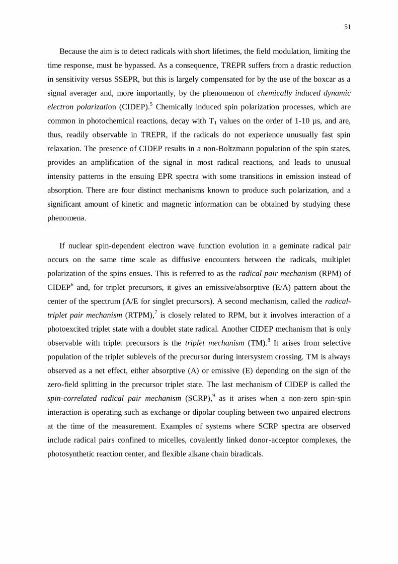
51
Because the aim is to detect radicals with short lifetimes, the field modulation, limiting the
time response, must be bypassed. As a consequence, TREPR suffers from a drastic reduction
in sensitivity versus SSEPR, but this is largely compensated for by the use of the boxcar as a
signal averager and, more importantly, by the phenomenon of chemically induced dynamic
electron polarization (CIDEP).5 Chemically induced spin polarization processes, which are
common in photochemical reactions, decay with T1 values on the order of 1-10 µs, and are,
thus, readily observable in TREPR, if the radicals do not experience unusually fast spin
relaxation. The presence of CIDEP results in a non-Boltzmann population of the spin states,
provides an amplification of the signal in most radical reactions, and leads to unusual
intensity patterns in the ensuing EPR spectra with some transitions in emission instead of
absorption. There are four distinct mechanisms known to produce such polarization, and a
significant amount of kinetic and magnetic information can be obtained by studying these
phenomena.
If nuclear spin-dependent electron wave function evolution in a geminate radical pair
occurs on the same time scale as diffusive encounters between the radicals, multiplet
polarization of the spins ensues. This is referred to as the radical pair mechanism (RPM) of
CIDEP6 and, for triplet precursors, it gives an emissive/absorptive (E/A) pattern about the
center of the spectrum (A/E for singlet precursors). A second mechanism, called the radical-
triplet pair mechanism (RTPM),7 is closely related to RPM, but it involves interaction of a
photoexcited triplet state with a doublet state radical. Another CIDEP mechanism that is only
observable with triplet precursors is the triplet mechanism (TM).8 It arises from selective
population of the triplet sublevels of the precursor during intersystem crossing. TM is always
observed as a net effect, either absorptive (A) or emissive (E) depending on the sign of the
zero-field splitting in the precursor triplet state. The last mechanism of CIDEP is called the
spin-correlated radical pair mechanism (SCRP),9 as it arises when a non-zero spin-spin
interaction is operating such as exchange or dipolar coupling between two unpaired electrons
at the time of the measurement. Examples of systems where SCRP spectra are observed
include radical pairs confined to micelles, covalently linked donor-acceptor complexes, the
photosynthetic reaction center, and flexible alkane chain biradicals.
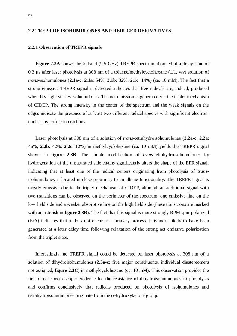
52
2.2 TREPR OF ISOHUMULONES AND REDUCED DERIVATIVES
2.2.1 Observation of TREPR signals
Figure 2.3A shows the X-band (9.5 GHz) TREPR spectrum obtained at a delay time of
0.3 µs after laser photolysis at 308 nm of a toluene/methylcyclohexane (1/1, v/v) solution of
trans-isohumulones (2.1a-c; 2.1a: 54%, 2.1b: 32%, 2.1c: 14%) (ca. 10 mM). The fact that a
strong emissive TREPR signal is detected indicates that free radicals are, indeed, produced
when UV light strikes isohumulones. The net emission is generated via the triplet mechanism
of CIDEP. The strong intensity in the center of the spectrum and the weak signals on the
edges indicate the presence of at least two different radical species with significant electron-
nuclear hyperfine interactions.
Laser photolysis at 308 nm of a solution of trans-tetrahydroisohumulones (2.2a-c; 2.2a:
46%, 2.2b: 42%, 2.2c: 12%) in methylcyclohexane (ca. 10 mM) yields the TREPR signal
shown in figure 2.3B. The simple modification of trans-tetrahydroisohumulones by
hydrogenation of the unsaturated side chains significantly alters the shape of the EPR signal,
indicating that at least one of the radical centers originating from photolysis of trans-
isohumulones is located in close proximity to an alkene functionality. The TREPR signal is
mostly emissive due to the triplet mechanism of CIDEP, although an additional signal with
two transitions can be observed on the perimeter of the spectrum: one emissive line on the
low field side and a weaker absorptive line on the high field side (these transitions are marked
with an asterisk in figure 2.3B). The fact that this signal is more strongly RPM spin-polarized
(E/A) indicates that it does not occur as a primary process. It is more likely to have been
generated at a later delay time following relaxation of the strong net emissive polarization
from the triplet state.
Interestingly, no TREPR signal could be detected on laser photolysis at 308 nm of a
solution of dihydroisohumulones (2.3a-c; five major constituents, individual diastereomers
not assigned, figure 2.3C) in methylcyclohexane (ca. 10 mM). This observation provides the
first direct spectroscopic evidence for the resistance of dihydroisohumulones to photolysis
and confirms conclusively that radicals produced on photolysis of isohumulones and
tetrahydroisohumulones originate from the -hydroxyketone group.
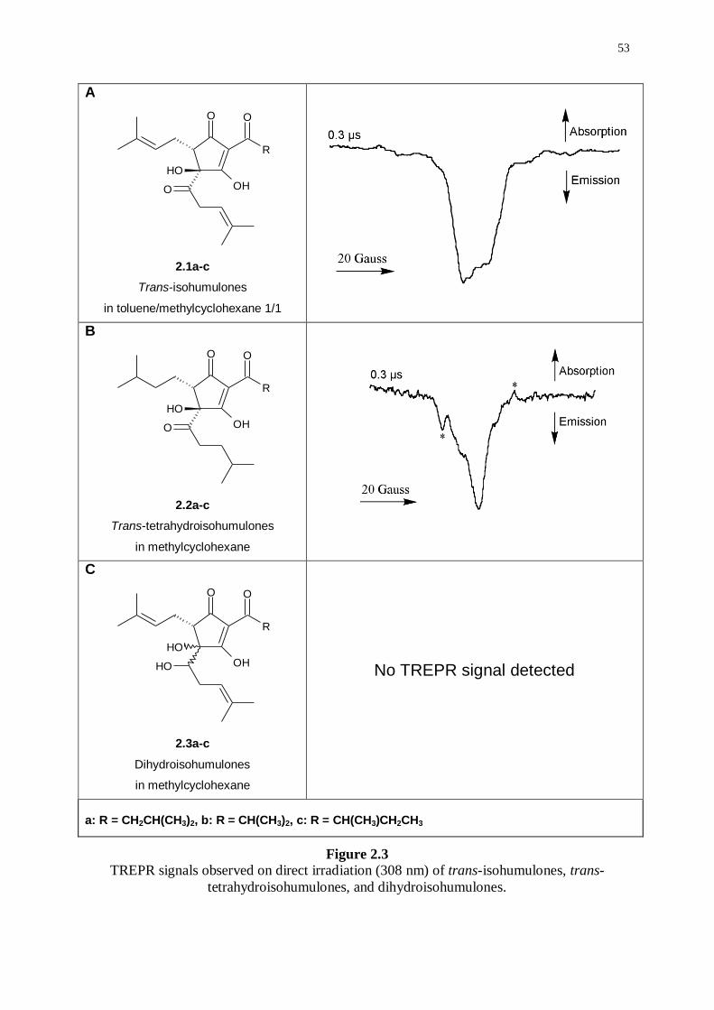
53
A
O
R
O
OHO
OH
2.1a-c
Trans-isohumulones
in toluene/methylcyclohexane 1/1
B
O
R
O
OHO
OH
2.2a-c
Trans-tetrahydroisohumulones
in methylcyclohexane
C
O
R
O
OHOH
OH
2.3a-c
Dihydroisohumulones
in methylcyclohexane
No TREPR signal detected
a: R = CH2CH(CH3)2, b: R = CH(CH3)2, c: R = CH(CH3)CH2CH3
Figure 2.3 TREPR signals observed on direct irradiation (308 nm) of trans-isohumulones, trans-
tetrahydroisohumulones, and dihydroisohumulones.
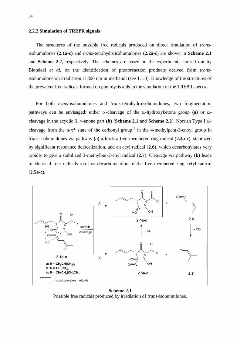
54
2.2.2 Simulation of TREPR signals
The structures of the possible free radicals produced on direct irradiation of trans-
isohumulones (2.1a-c) and trans-tetrahydroisohumulones (2.2a-c) are shown in Scheme 2.1
and Scheme 2.2, respectively. The schemes are based on the experiments carried out by
Blondeel et al. on the identification of photoreaction products derived from trans-
isohumulone on irradiation at 300 nm in methanol (see 1.1.3). Knowledge of the structures of
the prevalent free radicals formed on photolysis aids in the simulation of the TREPR spectra.
For both trans-isohumulones and trans-tetrahydroisohumulones, two fragmentation
pathways can be envisaged: either -cleavage of the -hydroxyketone group (a) or -
cleavage in the acyclic , -enone part (b) (Scheme 2.1 and Scheme 2.2). Norrish Type I -
cleavage from the n-* state of the carbonyl group10
in the 4-methylpent-3-enoyl group in
trans-isohumulones via pathway (a) affords a five-membered ring radical (2.4a-c), stabilized
by significant resonance delocalization, and an acyl radical (2.6), which decarbonylates very
rapidly to give a stabilized 3-methylbut-2-enyl radical (2.7). Cleavage via pathway (b) leads
to identical free radicals via fast decarbonylation of the five-membered ring ketyl radical
(2.5a-c).
O
R
O
OHO
OH
O .
O
R
O
OH
OH
O .
O
R
O
OHOH
3 *
(a)
(b)
+
+
- CO- CO
(a)
(b)
.
cleavage
Norrish I
2.1a-c
2.4a-c
2.5a-c
2.6
2.7
a: R = CH2CH(CH3)2
b: R = CH(CH3)2
c: R = CH(CH3)CH2CH3
.
= most prevalent radicals
Scheme 2.1 Possible free radicals produced by irradiation of trans-isohumulones.
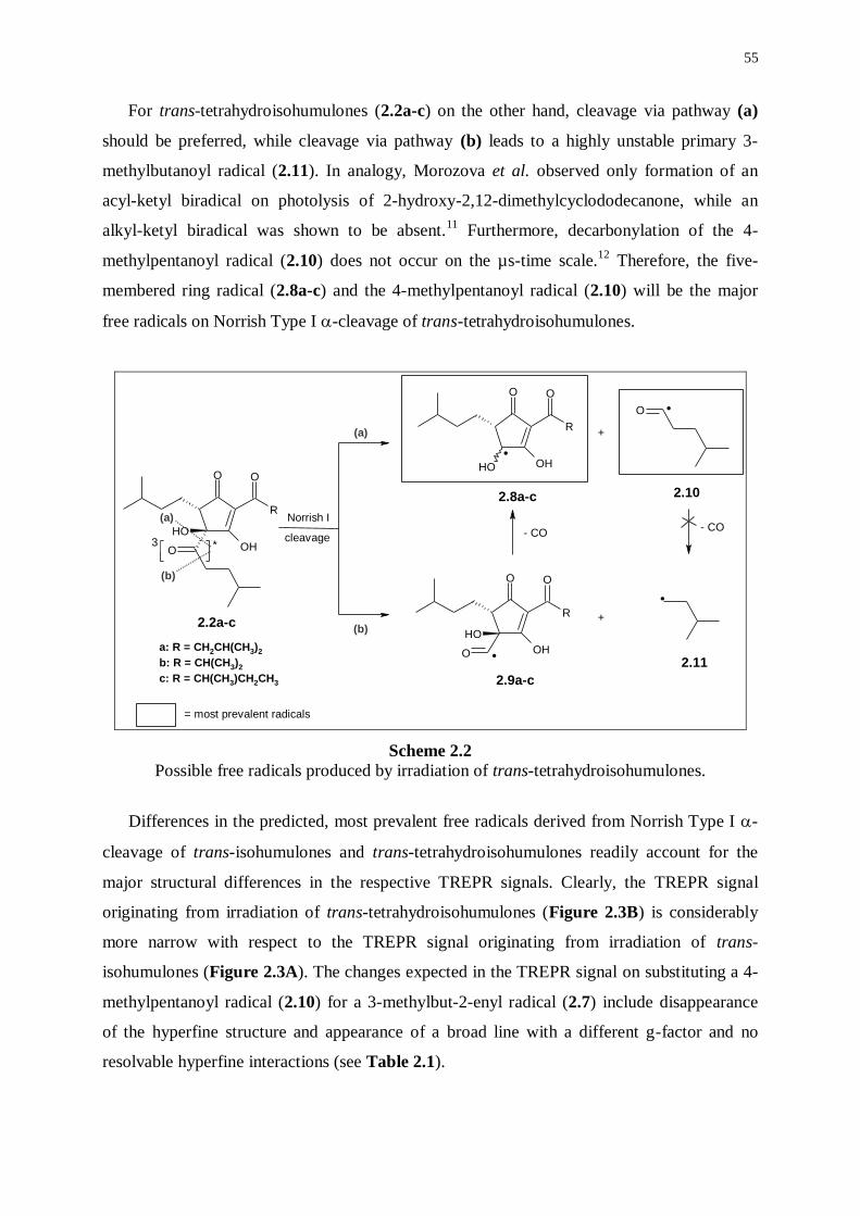
55
For trans-tetrahydroisohumulones (2.2a-c) on the other hand, cleavage via pathway (a)
should be preferred, while cleavage via pathway (b) leads to a highly unstable primary 3-
methylbutanoyl radical (2.11). In analogy, Morozova et al. observed only formation of an
acyl-ketyl biradical on photolysis of 2-hydroxy-2,12-dimethylcyclododecanone, while an
alkyl-ketyl biradical was shown to be absent.11
Furthermore, decarbonylation of the 4-
methylpentanoyl radical (2.10) does not occur on the µs-time scale.12
Therefore, the five-
membered ring radical (2.8a-c) and the 4-methylpentanoyl radical (2.10) will be the major
free radicals on Norrish Type I -cleavage of trans-tetrahydroisohumulones.
O
R
O
OHO
OH
O
R
O
OHOH
O
O
R
O
OH
OH
O
3 *
(a)
(b)
+
+
- CO- CO
(a)
(b)
.
.
.
.
Norrish I
cleavage
2.2a-c
2.8a-c
2.9a-c
2.10
2.11
a: R = CH2CH(CH3)2
b: R = CH(CH3)2
c: R = CH(CH3)CH2CH3
= most prevalent radicals
Scheme 2.2
Possible free radicals produced by irradiation of trans-tetrahydroisohumulones.
Differences in the predicted, most prevalent free radicals derived from Norrish Type I -
cleavage of trans-isohumulones and trans-tetrahydroisohumulones readily account for the
major structural differences in the respective TREPR signals. Clearly, the TREPR signal
originating from irradiation of trans-tetrahydroisohumulones (Figure 2.3B) is considerably
more narrow with respect to the TREPR signal originating from irradiation of trans-
isohumulones (Figure 2.3A). The changes expected in the TREPR signal on substituting a 4-
methylpentanoyl radical (2.10) for a 3-methylbut-2-enyl radical (2.7) include disappearance
of the hyperfine structure and appearance of a broad line with a different g-factor and no
resolvable hyperfine interactions (see Table 2.1).
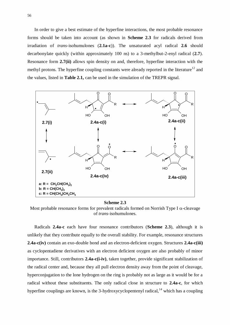
56
In order to give a best estimate of the hyperfine interactions, the most probable resonance
forms should be taken into account (as shown in Scheme 2.3 for radicals derived from
irradiation of trans-isohumulones (2.1a-c)). The unsaturated acyl radical 2.6 should
decarbonylate quickly (within approximately 100 ns) to a 3-methylbut-2-enyl radical (2.7).
Resonance form 2.7(ii) allows spin density on and, therefore, hyperfine interaction with the
methyl protons. The hyperfine coupling constants were already reported in the literature12
and
the values, listed in Table 2.1, can be used in the simulation of the TREPR signal.
.
.
R
OO
OHOH
HR
OO
OHOH
H
R
OO
OHOH
HR
OO
OHOH
H
..
..
2.7(i) 2.4a-c(i) 2.4a-c(ii)
2.4a-c(iv) 2.4a-c(iii)
2.7(ii)
a: R = CH2CH(CH3)2
b: R = CH(CH3)2
c: R = CH(CH3)CH2CH3
Scheme 2.3
Most probable resonance forms for prevalent radicals formed on Norrish Type I -cleavage
of trans-isohumulones.
Radicals 2.4a-c each have four resonance contributors (Scheme 2.3), although it is
unlikely that they contribute equally to the overall stability. For example, resonance structures
2.4a-c(iv) contain an exo-double bond and an electron-deficient oxygen. Structures 2.4a-c(iii)
as cyclopentadiene derivatives with an electron deficient oxygen are also probably of minor
importance. Still, contributors 2.4a-c(i-iv), taken together, provide significant stabilization of
the radical center and, because they all pull electron density away from the point of cleavage,
hyperconjugation to the lone hydrogen on the ring is probably not as large as it would be for a
radical without these substituents. The only radical close in structure to 2.4a-c, for which
hyperfine couplings are known, is the 3-hydroxycyclopentenyl radical,14
which has a coupling
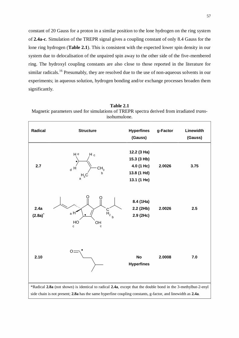
57
constant of 20 Gauss for a proton in a similar position to the lone hydrogen on the ring system
of 2.4a-c. Simulation of the TREPR signal gives a coupling constant of only 8.4 Gauss for the
lone ring hydrogen (Table 2.1). This is consistent with the expected lower spin density in our
system due to delocalisation of the unpaired spin away to the other side of the five-membered
ring. The hydroxyl coupling constants are also close to those reported in the literature for
similar radicals.16
Presumably, they are resolved due to the use of non-aqueous solvents in our
experiments; in aqueous solution, hydrogen bonding and/or exchange processes broaden them
significantly.
Table 2.1 Magnetic parameters used for simulations of TREPR spectra derived from irradiated trans-
isohumulone.
Radical
Structure
Hyperfines
(Gauss)
g-Factor
Linewidth
(Gauss)
2.7
H
CH3
CH3
H
H
.
a
b
c
d
e
12.2 (3 Ha)
15.3 (3 Hb)
4.0 (1 Hc)
13.8 (1 Hd)
13.1 (1 He)
2.0026
3.75
2.4a
(2.8a)*
CH
2
OO
OH OH
Hab
c c
.
8.4 (1Ha)
2.2 (2Hb)
2.9 (2Hc)
2.0026
2.5
2.10
O.
No
Hyperfines
2.0008
7.0
*Radical 2.8a (not shown) is identical to radical 2.4a, except that the double bond in the 3-methylbut-2-enyl
side chain is not present; 2.8a has the same hyperfine coupling constants, g-factor, and linewidth as 2.4a.
58
It is perhaps somewhat surprising to require hyperfine interaction on the alkyl group on
the other side of the -triketone (position b for 2.4a in Table 2.1) in order to fit the spectrum.
However, with delocalised radicals such as 2.4a-c, there is certainly the possibility for
hyperconjugation to the -system leading to these small couplings. Although there are two
such couplings for radical 2.4a and only one for 2.4b and 2.4c, respectively, we obtain a best
fit with two. This is consistent with our samples having a considerably higher mole fraction of
precursors 2.1a and 2.2a. The possible existence of a superposition of radicals with one or
two couplings at this position is most likely to be responsible for the small discrepancies in
the overall line shape of the simulations, especially at the perimeter of the transitions from
radicals 2.4a-c. The possibility of hyperfine interaction with the methylene hydrogens of the
3-methylbut-2-enyl side chain can be dismissed, while the carbon atom bearing them is not
directly bonded to the delocalised electrons; therefore, hyperconjugation would be very weak.
Furthermore, the relative intensities of the radicals 2.4a-c and 2.7 appear to be quite
different, but this is easily understood. The 3-methylbut-2-enyl radical has its intensity
distributed over a very large number of hyperfine lines (128) that cover more than 100 Gauss.
Therefore, even its most intense lines are diluted over much of the sweep width and, in fact,
the least intense lines on the perimeter are undetectable and left off the total sweep in the
figure. Radicals 2.4a-c, on the other hand, have EPR signals distributed over just 18 closely
spaced lines, hence this signal appears to be much more intense. In fact, the integrated
intensity of each radical type is nearly the same. Small differences in the electron spin lattice
relaxation time of the radicals or the decarbonylation rates of 2.5a-c and 2.6 may be
responsible for the integrations being not precisely equal. In regard to the simulation in
Figure 2.4, it should be noted that a small amount of multiplet spin polarization
(emission/absorption or E/A) has been added to this spectrum for best fit. This contribution is
from the radical pair mechanism of CIDEP and we note that it is of the correct phase for a
triplet precursor to the radical pair.15
For radical 2.8a-c, the only difference being the saturation of the 3-methylbut-2-enyl side
chain, identical values for the hyperfines, g-factor and linewidth can be used for the
simulation. An overview of the values of the parameters used for the simulations of the
TREPR spectra, together with the radicals, is given in Table 2.1. The simulations of the
TREPR spectra, which are displayed in Figures 2.4a and 2.4b, conclusively support the
above findings.
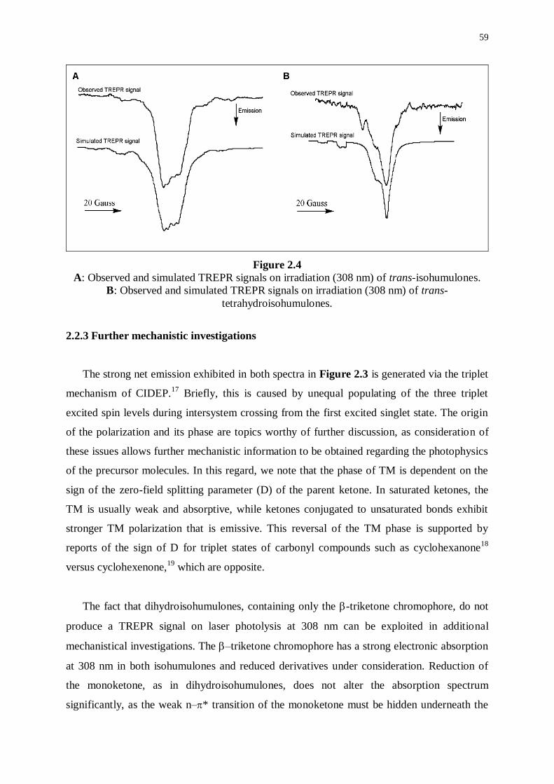
59
Figure 2.4
A: Observed and simulated TREPR signals on irradiation (308 nm) of trans-isohumulones.
B: Observed and simulated TREPR signals on irradiation (308 nm) of trans-
tetrahydroisohumulones.
2.2.3 Further mechanistic investigations
The strong net emission exhibited in both spectra in Figure 2.3 is generated via the triplet
mechanism of CIDEP.17
Briefly, this is caused by unequal populating of the three triplet
excited spin levels during intersystem crossing from the first excited singlet state. The origin
of the polarization and its phase are topics worthy of further discussion, as consideration of
these issues allows further mechanistic information to be obtained regarding the photophysics
of the precursor molecules. In this regard, we note that the phase of TM is dependent on the
sign of the zero-field splitting parameter (D) of the parent ketone. In saturated ketones, the
TM is usually weak and absorptive, while ketones conjugated to unsaturated bonds exhibit
stronger TM polarization that is emissive. This reversal of the TM phase is supported by
reports of the sign of D for triplet states of carbonyl compounds such as cyclohexanone18
versus cyclohexenone,19
which are opposite.
The fact that dihydroisohumulones, containing only the -triketone chromophore, do not
produce a TREPR signal on laser photolysis at 308 nm can be exploited in additional
mechanistical investigations. The –triketone chromophore has a strong electronic absorption
at 308 nm in both isohumulones and reduced derivatives under consideration. Reduction of
the monoketone, as in dihydroisohumulones, does not alter the absorption spectrum
significantly, as the weak n–* transition of the monoketone must be hidden underneath the
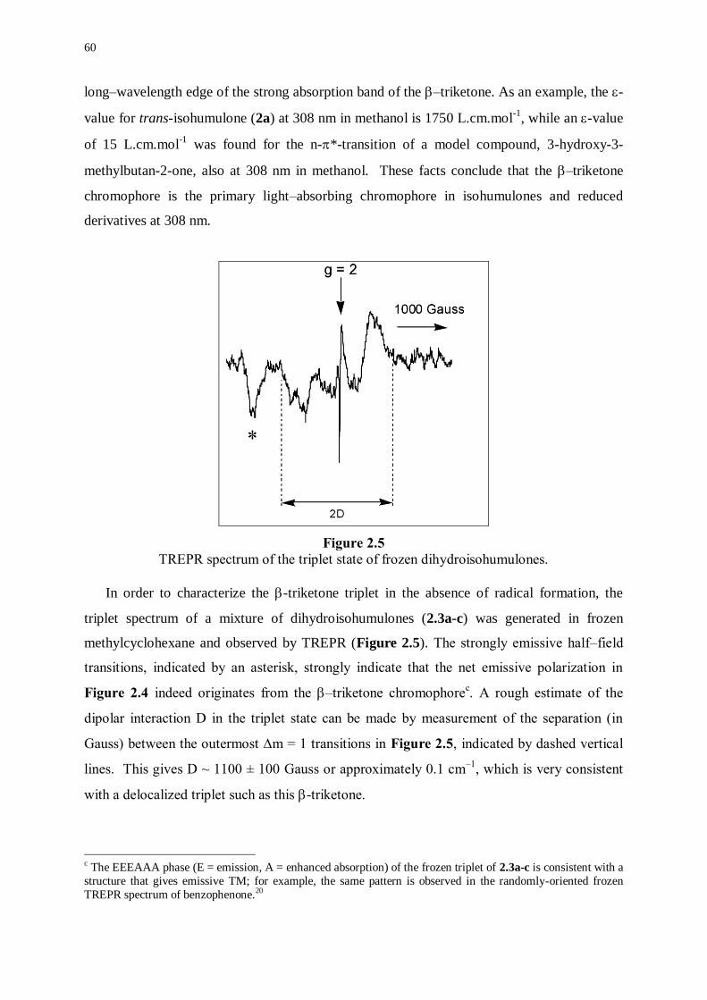
60
long–wavelength edge of the strong absorption band of the –triketone. As an example, the -
value for trans-isohumulone (2a) at 308 nm in methanol is 1750 L.cm.mol-1
, while an -value
of 15 L.cm.mol-1
was found for the n-*-transition of a model compound, 3-hydroxy-3-
methylbutan-2-one, also at 308 nm in methanol. These facts conclude that the –triketone
chromophore is the primary light–absorbing chromophore in isohumulones and reduced
derivatives at 308 nm.
Figure 2.5
TREPR spectrum of the triplet state of frozen dihydroisohumulones.
In order to characterize the -triketone triplet in the absence of radical formation, the
triplet spectrum of a mixture of dihydroisohumulones (2.3a-c) was generated in frozen
methylcyclohexane and observed by TREPR (Figure 2.5). The strongly emissive half–field
transitions, indicated by an asterisk, strongly indicate that the net emissive polarization in
Figure 2.4 indeed originates from the –triketone chromophorec. A rough estimate of the
dipolar interaction D in the triplet state can be made by measurement of the separation (in
Gauss) between the outermost m = 1 transitions in Figure 2.5, indicated by dashed vertical
lines. This gives D ~ 1100 ± 100 Gauss or approximately 0.1 cm–1
, which is very consistent
with a delocalized triplet such as this -triketone.
c The EEEAAA phase (E = emission, A = enhanced absorption) of the frozen triplet of 2.3a-c is consistent with a
structure that gives emissive TM; for example, the same pattern is observed in the randomly-oriented frozen
TREPR spectrum of benzophenone.20
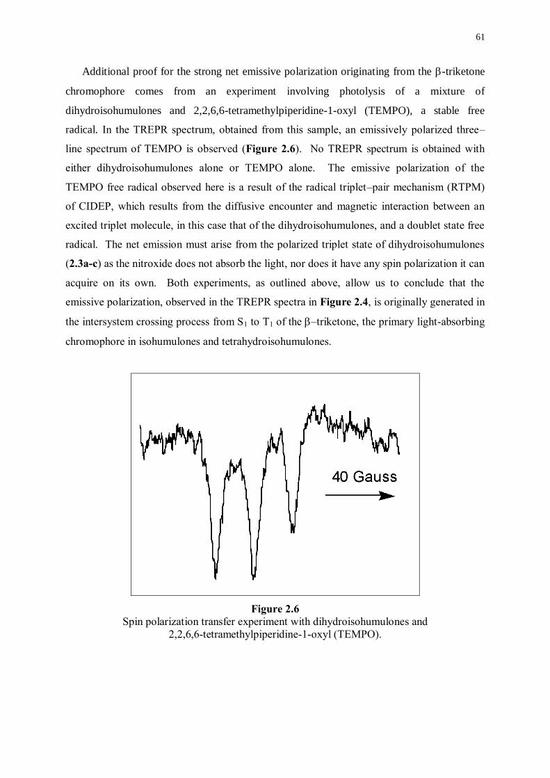
61
Additional proof for the strong net emissive polarization originating from the -triketone
chromophore comes from an experiment involving photolysis of a mixture of
dihydroisohumulones and 2,2,6,6-tetramethylpiperidine-1-oxyl (TEMPO), a stable free
radical. In the TREPR spectrum, obtained from this sample, an emissively polarized three–
line spectrum of TEMPO is observed (Figure 2.6). No TREPR spectrum is obtained with
either dihydroisohumulones alone or TEMPO alone. The emissive polarization of the
TEMPO free radical observed here is a result of the radical triplet–pair mechanism (RTPM)
of CIDEP, which results from the diffusive encounter and magnetic interaction between an
excited triplet molecule, in this case that of the dihydroisohumulones, and a doublet state free
radical. The net emission must arise from the polarized triplet state of dihydroisohumulones
(2.3a-c) as the nitroxide does not absorb the light, nor does it have any spin polarization it can
acquire on its own. Both experiments, as outlined above, allow us to conclude that the
emissive polarization, observed in the TREPR spectra in Figure 2.4, is originally generated in
the intersystem crossing process from S1 to T1 of the –triketone, the primary light-absorbing
chromophore in isohumulones and tetrahydroisohumulones.
Figure 2.6
Spin polarization transfer experiment with dihydroisohumulones and
2,2,6,6-tetramethylpiperidine-1-oxyl (TEMPO).
62
2.3 MECHANISM FOR PHOTODEGRADATION OF ISOHUMULONES AND
TETRAHYDROISOHUMULONES
Combining the information collected from the TREPR experiments and simulated spectra
allowed to propose a mechanism for photodegradation of isohumulones and
tetrahydroisohumulones (shown for trans-isohumulones in Scheme 2.4). Absorption of UV
radiation by the -triketone chromophore leads to its first excited singlet state. After
intersystem crossing (isc) to the excited triplet state, triplet energy transfer occurs to the
isolated -hydroxyketone, which in the excited state undergoes Norrish Type I -cleavage,
thereby furnishing radicals shown in Scheme 2.1 and Scheme 2.2. There is good evidence
supporting the assumption that spin polarization transfer accompanies triplet energy
transfer,21,22
hence, we expect the isolated monocarbonyl triplet to show emission from the
original absorbing chromophore rather than enhanced absorption from TM, which is expected
if the monocarbonyl function underwent direct excitation.
Intramolecular triplet energy transfer from the –triketone to the isolated –
hydroxyketone is noteworthy as it is expected that it should be uphill in energy by ~25 kJ
mol–1
.23
Even so, the radicals are formed from the –hydroxyketone within a few hundred ns
(the TREPR signals from 2.1a–c, for example, show maximum intensity at approximately 400
ns after the laser-flash). For two chromophores diffusing in free solution, collisional triplet
energy transfer is relatively slow (~105.s
–1) for this large an energy difference between the
states.23
However, when the two chromophores are connected, as in this case, the triplet
energy transfer rate can be much faster. Closs et al. measured triplet energy transfer from
naphthalene to biphenyl at greater than diffusion-controlled rates (7.7x109.s
–1), when the two
chromophores were connected to an alkane spacer and separated by only two C–C -bonds.24
Although that result was for a downhill reaction (~17 kJ.mol–1
), it demonstrated that
covalently linking the triplet donor and acceptor can increase the intrinsic triplet energy
transfer rate. It is possible that the two chromophores are, in fact, very strongly coupled in our
system. Because the hydroxyl group and the acyl side chain at C–4 are oriented above and
under the five-membered ring containing the –triketone, the two chromophores are in a fair
geometry for direct through-space interaction, while through–bond coupling is also expected
to be very strong. Therefore, it is not unreasonable to observe this uphill energy transfer
process taking place on the sub-s time scale.
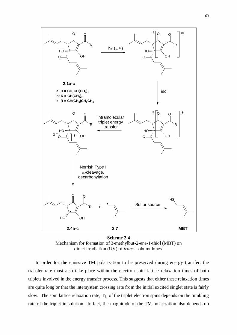
63
R
OO
OHO
OH
R
OO
OHO
OH
R
OO
OHO
OH
R
OO
OHO
OH
SH.
R
OO
OHOH
1
3
3
*
*
*
2.1a-c
h(UV)
isc
Intramoleculartriplet energy
transfer
Norrish Type I
-cleavage,decarbonylation
+Sulfur source
2.4a-c 2.7 MBT
a: R = CH2CH(CH3)2
b: R = CH(CH3)2
c: R = CH(CH3)CH2CH3
.
Scheme 2.4 Mechanism for formation of 3-methylbut-2-ene-1-thiol (MBT) on
direct irradiation (UV) of trans-isohumulones.
In order for the emissive TM polarization to be preserved during energy transfer, the
transfer rate must also take place within the electron spin–lattice relaxation times of both
triplets involved in the energy transfer process. This suggests that either these relaxation times
are quite long or that the intersystem crossing rate from the initial excited singlet state is fairly
slow. The spin lattice relaxation rate, T1, of the triplet electron spins depends on the tumbling
rate of the triplet in solution. In fact, the magnitude of the TM-polarization also depends on
64
this tumbling rate. With so many alkyl appendages, tumbling will be slower than for less-
substituted and more symmetric triplet states, which have electron T1 values on the order of
1–10 ns. Therefore, a longer triplet electron spin–lattice relaxation rate than this for the triplet
states of 2.1a–c and 2.2a–c is reasonable.
2.4 EXPERIMENTAL PART
Sample Preparation
Isohumulones are commercially available as an aqueous solution (ca. 30% w/v) of the
potassium salts of trans–isohumulones and cis–isohumulones (Wigan Co., Eardiston, Near
Tenbury Wells, Worcestershire, England). The isohumulones (100 mL, ca. 32 % w/v) were
freed from the salts by tenfold dilution with H2O, acidification to pH 1 with 1 N HCl,
extraction with isooctane (2 x 250 mL), and removal of the solvent (yield: 30.6 g, 96%). The
trans–isohumulones (2.1a: 45%; 2.1b: 39%; 2.1c: 16%) were accessed via selective
precipitation using dicyclohexylamine (DCHA) (15.5 g) in ethyl acetate (50 mL), recovery of
the DCHA–salts, recrystallization from methanol and water, redissolution in warm ethyl
acetate, addition of acidified water (pH 1), phase separation and evaporation of the organic
solvent (yield: 2.9 g, 9.4%).6
Tetrahydroisohumulones are commercially available as an aqueous solution (ca. 10% w/v)
of the potassium salts (Wigan Co., Eardiston, Near Tenbury Wells, Worcestershire, England).
Trans-tetrahydroisohumulone (2.2a: 42%), trans-tetrahydroisocohumulone (2.2b: 46%), and
trans-tetrahydroisoadhumulone (2.2c: 12%) were separated from the cis–epimers according to
the procedure described above for the isolation of the trans–isohumulones (yield: 0.4 g,
1.5%).6
Dihydroisohumulones (five major constituents, diastereomers not assigned individually)
were a gift from Kalsec Corp., Kalamazoo, Michigan, USA.
TREPR-experiments
All spectra were recorded on a JEOL, USA, Inc. RE-1X X-Band (9.5 GHz) CW EPR
spectrometer. The experimental setup is outlined above (see 2.1.2). The microwave power
was varied from 0.1-20 mW. Laser excitation at 308 nm was produced using a Lambda-

65
Physik LPX 100i excimer laser running at a repetition rate of 60 Hz. A rectangular cavity was
used with a Suprasil flat cell, 0.4 mm optical path length. The samples were bubbled with N2
and continuously flowed to prevent sample depletion and overheating.
66
2.5 REFERENCES
(1) Weil, J.A., Bolton, J.R., Wertz, J.E.; Electron Paramagnetic Resonance: Elementary Theory
and Applications. Wiley-Interscience, New-York, 1994.
(2) Forbes, M.D.E.; Time-Resolved (CW) Electron Paramagnetic Resonance Spectroscopy: An
Overview of the Technique and Its Use in Organic Photochemistry. Photochem. Photobiol.,
1997, 65, 73-81.
(3) Van Willigen, H., Levstein, P.R., Ebersole, M.H.; Application of Fourier Transform Electron
Paramagnetic Resonance in the Study of Photochemical Reactions. Chem. Rev., 1993, 93,
173-198.
(4) Stehlik, D., Möbius, K.; New EPR Methods for Investigating Photoprocesses with
Paramagnetic Intermediates. Annu. Rev. Phys. Chem., 1997, 48, 745-784.
(5) Salikhov, K.M., Molin, Y.N., Sagdeev, R.Z., Buchachenko, A.L.; Spin Polarization and
Magnetic Effects in Radical Reactions. Molin, Y.N, Ed., Elsevier, Amsterdam, 1984.
(6) Salikhov, K.M., Molin, Y.N., Sagdeev, R.Z., Buchachenko, A.L.; Spin Polarization and
Magnetic Effects in Radical Reactions. Molin, Y.N, Ed., Elsevier, Amsterdam, 1984, pp. 57-
93.
(7) Blättler, C., Jent, F., Paul, H.; A Novel Radical-Triplet Pair Mechanism for Chemically
Induced Electron Polarization (CIDEP) of Free Radicals in Solution. Chem. Phys. Lett.,
1990, 166, 375-380.
(8) Wong, S.K., Hutchinson, D.A., Wan, J.K.S.; Chemically Induced Dynamic Electron
Polarization. II. A General Theory for Radicals Produced by Photochemical Reactions of
Excited Triplet Carbonyl Compounds. J. Chem. Phys., 1973, 58, 985-989.
(9) Closs, G.L., Forbes, M.D.E., Norris, J.R.; Spin-Polarized Electron Paramagnetic Resonance
Spectra of Radical Pairs at the Solid/Solution Interface. J. Am. Chem. Soc., 1987, 91, 3592-
3599.
(10) Wagner, P.J., Hammond, G.S.; Properties and Reactions of Organic Molecules in their Triplet
States. Adv. Photochem., 1968, 5, 21-156.
(11) Morozova, O.B., Yurkovskaya, A.V., Tsentalovich, Y.P., Sagdeev, R.Z., Wu, T., Forbes,
M.D.E.; Study of Consecutive Biradicals from 2-Hydroxy-2,12-Dimethylcyclododecanone by
TR-CIDNP, TREPR, and Laser-flash Photolysis. J. Phys. Chem., 1997, 101, 8803-8808.
(12) Vollenweider, J.-K., Paul, H.; On the Rates of Decarbonylation of Hydroxyacetyl and Other
Acyl Radicals. Int. J. Chem. Kinet., 1986, 18, 791-800.
(13) Krusic, P.J., Meakin, P., Smart, B.E.; An Electron Spin Resonance Study of the Steric Rigidity
in the Allyl and 1,1-Disubstituted Allyl Radicals. J. Am. Chem. Soc., 1974, 96, 6211-6213.
67
(14) Gilbert, B.C., Holmes, R.G.G., Norman, R.O.C.; Electron Spin Resonance Studies. Part 52.
Reactions of Secondary Alkoxyl Radicals. J. Chem. Res., 1977, 1,1.
(15) Wilson, R.J.; Electron Spin Resonance Study of Radicals Photolytically Generated from
Aromatic Carbonyl Compounds. The Diphenylhydroxymethyl and -Hydroxybenzyl Radicals.
J. Chem. Soc. B, 1968, 84-90.
(16) McLauchlan, K.A.; In: Advanced EPR: Applications in Biology and Biochemistry. A.J. Hoff,
Ed., Elsevier, New York, 1989, 350-371.
(17) Wan, J.K.S., Depew, M.C.; Time-Resolved ESR Spectroscopy (CIDEP) of Ketones. Res.
Chem. Intermed., 1992, 18, 227-292.
(18) Tominaga, K., Yamauchi, S., Hirota, N.; Time-Resolved Electron-Paramagnetic Resonance
Studies on the Lowest Excited Triplet-States of Aliphatic Carbonyl Compounds. J. Phys.
Chem., 1990, 94, 4425-4431.
(19) Yamauchi, S., Hirota, N., Higuchi, J.; Direct Observation of Twisted Excited Triplet-States of
Monocyclic Conjugated Enones by Time-Resolved Electron Paramagnetic Resonance. J. Phys.
Chem., 1988, 92, 2129-2133.
(20) Murai, H., Imamura, T., Obi, K.; Time-Resolved ESR Detection of Benzophenone n-* Triplet
State in Glassy Matrices at 77 K. Chem. Phys. Lett., 1982, 87, 295-298.
(21) Akiyama, K., Tero-Kubota, S., Ikoma, T., Ikegami, Y.; Spin Polarization Conservation during
Intramolecular Triplet-Triplet Energy Transfer Studied by Time-Resolved EPR Spectroscopy.
J. Am. Chem. Soc., 1994, 116, 5324-5327.
(22) Akiyama, K., Tero-Kubota, S.; Spin Polarization Transfer During Intramolecular Triplet-
Triplet Engergy Transfer as Studied by Time Resolved EPR Spectroscopy. Mol. Phys., 1994,
83, 1091-1097.
(23) Murov, S.L.; Handbook of Photochemistry, Marcel Dekker, New York, 1993.
(24) Closs, G.L., Johnson, M.D., Miller, J.R., Piotrowiak, P.; A Connection Between
Intramolecular Long-Range Electron, Hole, and Triplet Engergy Transfers. J. Am. Chem.
Soc., 1989, 111, 3751-3753.
************************

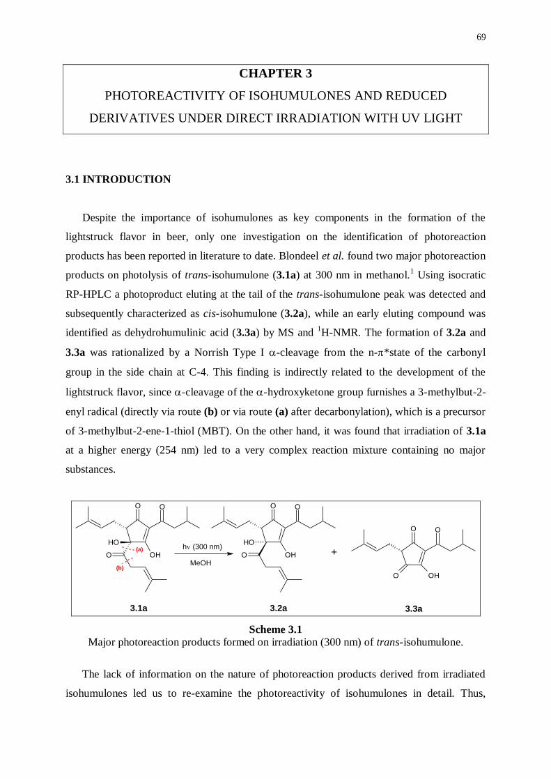
69
CHAPTER 3
PHOTOREACTIVITY OF ISOHUMULONES AND REDUCED
DERIVATIVES UNDER DIRECT IRRADIATION WITH UV LIGHT
3.1 INTRODUCTION
Despite the importance of isohumulones as key components in the formation of the
lightstruck flavor in beer, only one investigation on the identification of photoreaction
products has been reported in literature to date. Blondeel et al. found two major photoreaction
products on photolysis of trans-isohumulone (3.1a) at 300 nm in methanol.1 Using isocratic
RP-HPLC a photoproduct eluting at the tail of the trans-isohumulone peak was detected and
subsequently characterized as cis-isohumulone (3.2a), while an early eluting compound was
identified as dehydrohumulinic acid (3.3a) by MS and 1H-NMR. The formation of 3.2a and
3.3a was rationalized by a Norrish Type I -cleavage from the n-*state of the carbonyl
group in the side chain at C-4. This finding is indirectly related to the development of the
lightstruck flavor, since -cleavage of the -hydroxyketone group furnishes a 3-methylbut-2-
enyl radical (directly via route (b) or via route (a) after decarbonylation), which is a precursor
of 3-methylbut-2-ene-1-thiol (MBT). On the other hand, it was found that irradiation of 3.1a
at a higher energy (254 nm) led to a very complex reaction mixture containing no major
substances.
OO
OHO
OH
OO
OHO
OO
OHO
OH(a)
(b)
+h (300 nm)
3.1a 3.2a 3.3a
MeOH
Scheme 3.1
Major photoreaction products formed on irradiation (300 nm) of trans-isohumulone.
The lack of information on the nature of photoreaction products derived from irradiated
isohumulones led us to re-examine the photoreactivity of isohumulones in detail. Thus,
70
mixtures of photoreaction products derived from photostationary irradiation of isohumulones,
dihydroisohumulones, and tetrahydroisohumulones, were analyzed using gradient reversed-
phase HPLC-MS. Comparison of the findings should be most revealing, as hydrogenation of
salient functionalities in the acyl side chain at C-4, i.e. a carbonyl group and a double bond,
should correspond to clear differences in the distribution of photoreaction products, as well as
in mass differences of comparable compounds.
3.2 METHOD DEVELOPMENT
3.2.1 HPLC and HPLC-MS
The HPLC separation method was adapted from a previously reported isocratic system for
the separation of tetrahydroisohumulones. Using a mobile phase consisting of a mixture of
acetonitrile and an aqueous buffer (48/52, v/v), Hay et al. were able to separate all six
tetrahydroisohumulones.2 The buffer solution was prepared by adjusting the pH to 2.8 with
85% phosphoric acid. Using this procedure with slightly different concentrations of organic
solvent in the mobile phase, we were able to separate all six isohumulones and all six
tetrahydroisohumulones, respectively, while separation of dihydroisohumulones resulted in
five major peaks. We found that the nature of the octadecylsilica stationary phase was very
critical for optimized peak shape and resolution. A most efficient separation was obtained
with high-purity demineralized C18-derivatized silica having a high carbon load and provided
with efficient endcapping, such as Alltima (Alltech) and Omnisphere (Varian). Moreover,
columns should be handled with extreme care, while acidic conditions (< pH 2) lead to
irreversible damage of the derivatized silica, and, consequently, to loss of resolving power
along with pronounced tailing. It should be noted that peak broadening is sometimes
observed, especially for the later eluting adisohumulones, but the resolution was only
marginally affected. Presumably, peak broadening is correlated to the existence of two or
more tautomeric forms for each of the isohumulones. This isocratic method was applied for
qualitative and quantitative analyses of commercially available preparations of isohumulones,
dihydroisohumulones, and tetrahydroisohumulones.
For the analysis of mixtures of photoreaction products, a gradient using the same mobile
phase was optimized to allow efficient separations. On switching from HPLC-DAD to HPLC-
MS, some incompatibilities needed to be taken into consideration. In the aqueous part of the
71
mobile phase, formic acid was substituted for the non-volatile phosphoric acid, while a
mixture of acetonitrile/methanol 90/10 (v/v) was substituted for acetonitrile. These
modifications resulted in a drastic increase in sensitivity of the hyphenated HPLC-MS
method, while the resolution was only affected to a minor extent. Electrospray ionization
(ESI) both in the positive and the negative ionization modes proved necessary, while
photoreaction products derived from irradiation of isohumulones and reduced derivatives
showed substantial variations in ionization behavior.
3.2.2 Photostationary irradiation
In order to detect primary photoreaction products, short irradiation times - preferably in
accordance with less than 5% consumption of substrate - should be used to avoid formation of
secondary reaction products. While the -tricarbonyl chromophore inherently resists
photolysis, primary photoreaction products compete effectively with the original
photosubstrates for radiant energy, thereby leading to formation of secondary photoreaction
products and, thus, complex photoreaction mixtures. However, even at very short exposure
times, a distinct number of photoreaction products was observed and the complexity of the
chromatographic profiles was apparent. Therefore, it appeared reasonable to terminate the
irradiation after a given time period for which the yield of photoreaction products was
maximized for complete consumption of the substrates. Further exposure led to extremely
complex mixtures, most likely as a result of photoinduced degradation of primary and, also,
of secondary photoreaction products. Direct irradiation of the photosubstrates was performed
both at 254 nm and 300 nm. The chromatographic profiles of the mixtures of photoreaction
products at relatively short exposure times did not differ significantly, although, at 254 nm,
extensive photodecomposition became apparent within much shorter time than observed at
300 nm. Thus, in order to decrease the complexity, irradiation at 300 nm was preferred.
Irradiations were carried out in buffered aqueous systems, methanol, and acetonitrile.
Only minor differences were noticed in the distribution of photoreaction products. Thus,
methanol and acetonitrile were selected as most suitable solvents, because of the high
solubilities of isohumulones and reduced derivatives. Also, sample preparation and solvent
removal (evaporation under N2 or under reduced pressure) were facilitated.
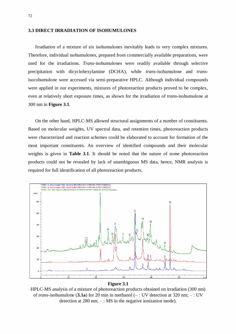
72
3.3 DIRECT IRRADIATION OF ISOHUMULONES
Irradiation of a mixture of six isohumulones inevitably leads to very complex mixtures.
Therefore, individual isohumulones, prepared from commercially available preparations, were
used for the irradiations. Trans-isohumulones were readily available through selective
precipitation with dicyclohexylamine (DCHA), while trans-isohumulone and trans-
isocohumulone were accessed via semi-preparative HPLC. Although individual compounds
were applied in our experiments, mixtures of photoreaction products proved to be complex,
even at relatively short exposure times, as shown for the irradiation of trans-isohumulone at
300 nm in Figure 3.1.
On the other hand, HPLC-MS allowed structural assignments of a number of constituents.
Based on molecular weights, UV spectral data, and retention times, photoreaction products
were characterized and reaction schemes could be elaborated to account for formation of the
most important constituents. An overview of identified compounds and their molecular
weights is given in Table 3.1. It should be noted that the nature of some photoreaction
products could not be revealed by lack of unambiguous MS data, hence, NMR analysis is
required for full identification of all photoreaction products.
Figure 3.1
HPLC-MS analysis of a mixture of photoreaction products obtained on irradiation (300 nm)
of trans-isohumulone (3.1a) for 20 min in methanol ( : UV detection at 320 nm; : UV
detection at 280 nm; : MS in the negative ionization mode).
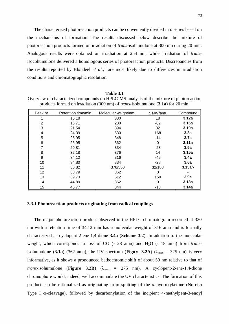
73
The characterized photoreaction products can be conveniently divided into series based on
the mechanisms of formation. The results discussed below describe the mixture of
photoreaction products formed on irradiation of trans-isohumulone at 300 nm during 20 min.
Analogous results were obtained on irradiation at 254 nm, while irradiation of trans-
isocohumulone delivered a homologous series of photoreaction products. Discrepancies from
the results reported by Blondeel et al.,1 are most likely due to differences in irradiation
conditions and chromatographic resolution.
Table 3.1
Overview of characterized compounds on HPLC-MS-analysis of the mixture of photoreaction
products formed on irradiation (300 nm) of trans-isohumulone (3.1a) for 20 min.
Peak nr. Retention time/min Molecular weight/amu MW/amu Compound
1 16.18 380 18 3.12a
2 16.71 280 -82 3.16a
3 21.54 394 32 3.10a
4 24.39 530 168 3.8a
5 25.95 348 -14 3.7a
6 26.95 362 0 3.11a
7 29.81 334 -28 3.5a
8 32.18 376 14 3.15a
9 34.12 316 -46 3.4a
10 34.80 334 -28 3.6a
11 36.82 376/550 32/188 3.15a/-
12 38.79 362 0 -
13 39.73 512 150 3.9a
14 44.89 362 0 3.13a
15 46.77 344 -18 3.14a
3.3.1 Photoreaction products originating from radical couplings
The major photoreaction product observed in the HPLC chromatogram recorded at 320
nm with a retention time of 34.12 min has a molecular weight of 316 amu and is formally
characterized as cyclopent-2-ene-1,4-dione 3.4a (Scheme 3.2). In addition to the molecular
weight, which corresponds to loss of CO (- 28 amu) and H2O (- 18 amu) from trans-
isohumulone (3.1a) (362 amu), the UV spectrum (Figure 3.2A) (max = 325 nm) is very
informative, as it shows a pronounced bathochromic shift of about 50 nm relative to that of
trans-isohumulone (Figure 3.2B) (max = 275 nm). A cyclopent-2-ene-1,4-dione
chromophore would, indeed, well accommodate the UV characteristics. The formation of this
product can be rationalized as originating from splitting of the -hydroxyketone (Norrish
Type I -cleavage), followed by decarbonylation of the incipient 4-methylpent-3-enoyl
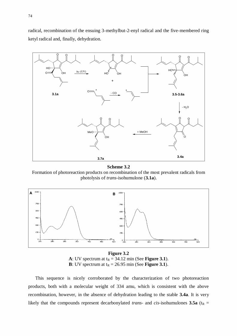
74
radical, recombination of the ensuing 3-methylbut-2-enyl radical and the five-membered ring
ketyl radical and, finally, dehydration.
O O
OH
OH
O
O O
O
O O
OH
MeO
O O
OH
OH
O O
OHOH
.
O. .
+
- H2O
+ MeOH
3.5-3.6a
3.4a3.7a
h
3.1a
(UV)
- CO
Scheme 3.2
Formation of photoreaction products on recombination of the most prevalent radicals from
photolysis of trans-isohumulone (3.1a).
Figure 3.2
A: UV spectrum at tR = 34.12 min (See Figure 3.1).
B: UV spectrum at tR = 26.95 min (See Figure 3.1).
This sequence is nicely corroborated by the characterization of two photoreaction
products, both with a molecular weight of 334 amu, which is consistent with the above
recombination, however, in the absence of dehydration leading to the stable 3.4a. It is very
likely that the compounds represent decarbonylated trans- and cis-isohumulones 3.5a (tR =
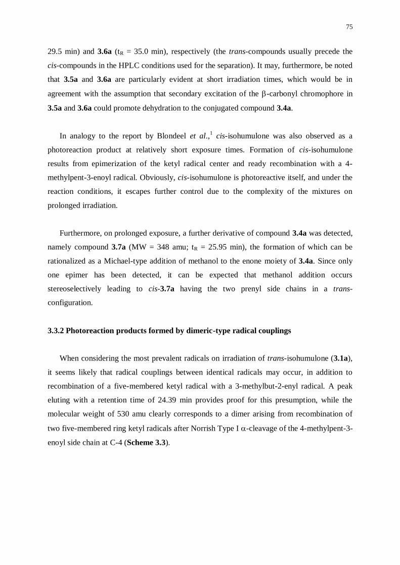
75
29.5 min) and 3.6a (tR = 35.0 min), respectively (the trans-compounds usually precede the
cis-compounds in the HPLC conditions used for the separation). It may, furthermore, be noted
that 3.5a and 3.6a are particularly evident at short irradiation times, which would be in
agreement with the assumption that secondary excitation of the -carbonyl chromophore in
3.5a and 3.6a could promote dehydration to the conjugated compound 3.4a.
In analogy to the report by Blondeel et al.,1 cis-isohumulone was also observed as a
photoreaction product at relatively short exposure times. Formation of cis-isohumulone
results from epimerization of the ketyl radical center and ready recombination with a 4-
methylpent-3-enoyl radical. Obviously, cis-isohumulone is photoreactive itself, and under the
reaction conditions, it escapes further control due to the complexity of the mixtures on
prolonged irradiation.
Furthermore, on prolonged exposure, a further derivative of compound 3.4a was detected,
namely compound 3.7a (MW = 348 amu; tR = 25.95 min), the formation of which can be
rationalized as a Michael-type addition of methanol to the enone moiety of 3.4a. Since only
one epimer has been detected, it can be expected that methanol addition occurs
stereoselectively leading to cis-3.7a having the two prenyl side chains in a trans-
configuration.
3.3.2 Photoreaction products formed by dimeric-type radical couplings
When considering the most prevalent radicals on irradiation of trans-isohumulone (3.1a),
it seems likely that radical couplings between identical radicals may occur, in addition to
recombination of a five-membered ketyl radical with a 3-methylbut-2-enyl radical. A peak
eluting with a retention time of 24.39 min provides proof for this presumption, while the
molecular weight of 530 amu clearly corresponds to a dimer arising from recombination of
two five-membered ring ketyl radicals after Norrish Type I -cleavage of the 4-methylpent-3-
enoyl side chain at C-4 (Scheme 3.3).
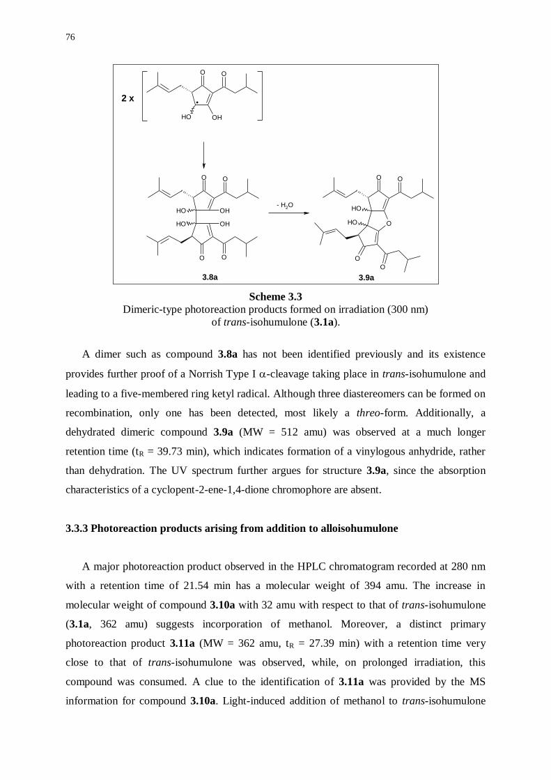
76
O O
OHOH
.
OO
O
OH
O
OH
OH
OH
O
OO
O
O
OH
OH
2 x
- H2O
3.8a 3.9a
Scheme 3.3
Dimeric-type photoreaction products formed on irradiation (300 nm)
of trans-isohumulone (3.1a).
A dimer such as compound 3.8a has not been identified previously and its existence
provides further proof of a Norrish Type I -cleavage taking place in trans-isohumulone and
leading to a five-membered ring ketyl radical. Although three diastereomers can be formed on
recombination, only one has been detected, most likely a threo-form. Additionally, a
dehydrated dimeric compound 3.9a (MW = 512 amu) was observed at a much longer
retention time (tR = 39.73 min), which indicates formation of a vinylogous anhydride, rather
than dehydration. The UV spectrum further argues for structure 3.9a, since the absorption
characteristics of a cyclopent-2-ene-1,4-dione chromophore are absent.
3.3.3 Photoreaction products arising from addition to alloisohumulone
A major photoreaction product observed in the HPLC chromatogram recorded at 280 nm
with a retention time of 21.54 min has a molecular weight of 394 amu. The increase in
molecular weight of compound 3.10a with 32 amu with respect to that of trans-isohumulone
(3.1a, 362 amu) suggests incorporation of methanol. Moreover, a distinct primary
photoreaction product 3.11a (MW = 362 amu, tR = 27.39 min) with a retention time very
close to that of trans-isohumulone was observed, while, on prolonged irradiation, this
compound was consumed. A clue to the identification of 3.11a was provided by the MS
information for compound 3.10a. Light-induced addition of methanol to trans-isohumulone
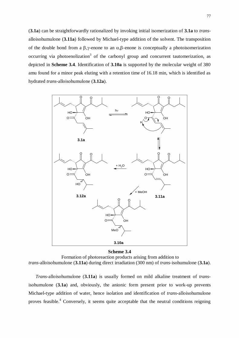
77
(3.1a) can be straightforwardly rationalized by invoking initial isomerization of 3.1a to trans-
alloisohumulone (3.11a) followed by Michael-type addition of the solvent. The transposition
of the double bond from a ,-enone to an ,-enone is conceptually a photoisomerization
occurring via photoenolization3 of the carbonyl group and concurrent tautomerization, as
depicted in Scheme 3.4. Identification of 3.10a is supported by the molecular weight of 380
amu found for a minor peak eluting with a retention time of 16.18 min, which is identified as
hydrated trans-alloisohumulone (3.12a).
O O
O
OH
OH
O O
O
OH
OH
H
O O
O
OH
OH
O O
O
OH
OH
OH
O O
O
OH
OH
MeO
3.1a
3.11a
3.10a
3.12a
h
+ MeOH
+ H2O
Scheme 3.4
Formation of photoreaction products arising from addition to
trans-alloisohumulone (3.11a) during direct irradiation (300 nm) of trans-isohumulone (3.1a).
Trans-alloisohumulone (3.11a) is usually formed on mild alkaline treatment of trans-
isohumulone (3.1a) and, obviously, the anionic form present prior to work-up prevents
Michael-type addition of water, hence isolation and identification of trans-alloisohumulone
proves feasible.4 Conversely, it seems quite acceptable that the neutral conditions reigning
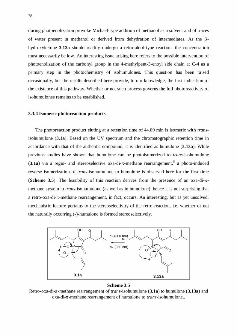
78
during photoenolization provoke Michael-type addition of methanol as a solvent and of traces
of water present in methanol or derived from dehydration of intermediates. As the -
hydroxyketone 3.12a should readily undergo a retro-aldol-type reaction, the concentration
must necessarily be low. An interesting issue arising here refers to the possible intervention of
photoenolization of the carbonyl group in the 4-methylpent-3-enoyl side chain at C-4 as a
primary step in the photochemistry of isohumulones. This question has been raised
occasionally, but the results described here provide, to our knowledge, the first indication of
the existence of this pathway. Whether or not such process governs the full photoreactivity of
isohumulones remains to be established.
3.3.4 Isomeric photoreaction products
The photoreaction product eluting at a retention time of 44.89 min is isomeric with trans-
isohumulone (3.1a). Based on the UV spectrum and the chromatographic retention time in
accordance with that of the authentic compound, it is identified as humulone (3.13a). While
previous studies have shown that humulone can be photoisomerized to trans-isohumulone
(3.1a) via a regio- and stereoselective oxa-di--methane rearrangement,5 a photo-induced
reverse isomerization of trans-isohumulone to humulone is observed here for the first time
(Scheme 3.5). The feasibility of this reaction derives from the presence of an oxa-di--
methane system in trans-isohumulone (as well as in humulone), hence it is not surprising that
a retro-oxa-di--methane rearrangement, in fact, occurs. An interesting, but as yet unsolved,
mechanistic feature pertains to the stereoselectivity of the retro-reaction, i.e. whether or not
the naturally occurring (-)-humulone is formed stereoselectively.
OH O
O
O
O
H
OH O
OO
OH
h (300 nm)
h (350 nm)
3.1a 3.13a
Scheme 3.5
Retro-oxa-di--methane rearrangement of trans-isohumulone (3.1a) to humulone (3.13a) and
oxa-di--methane rearrangement of humulone to trans-isohumulone..
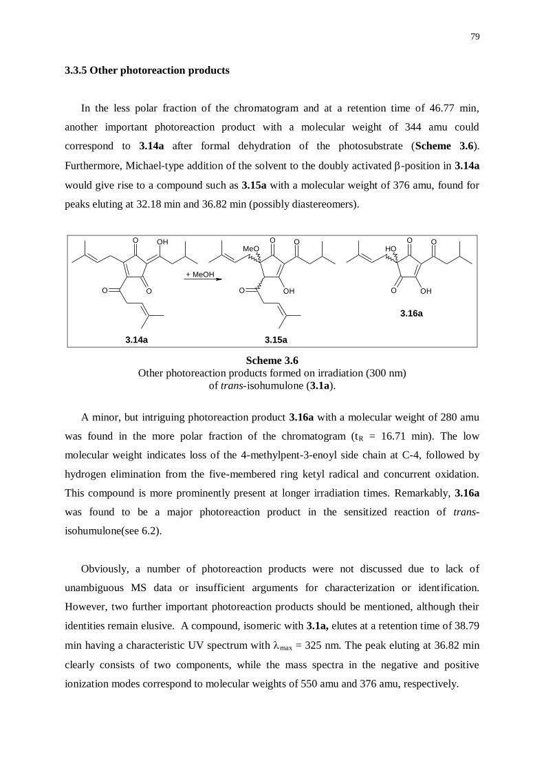
79
3.3.5 Other photoreaction products
In the less polar fraction of the chromatogram and at a retention time of 46.77 min,
another important photoreaction product with a molecular weight of 344 amu could
correspond to 3.14a after formal dehydration of the photosubstrate (Scheme 3.6).
Furthermore, Michael-type addition of the solvent to the doubly activated -position in 3.14a
would give rise to a compound such as 3.15a with a molecular weight of 376 amu, found for
peaks eluting at 32.18 min and 36.82 min (possibly diastereomers).
O OH
OO
O O
OHO
MeOO O
O OH
OH
+ MeOH
3.14a 3.15a
3.16a
Scheme 3.6
Other photoreaction products formed on irradiation (300 nm)
of trans-isohumulone (3.1a).
A minor, but intriguing photoreaction product 3.16a with a molecular weight of 280 amu
was found in the more polar fraction of the chromatogram (tR = 16.71 min). The low
molecular weight indicates loss of the 4-methylpent-3-enoyl side chain at C-4, followed by
hydrogen elimination from the five-membered ring ketyl radical and concurrent oxidation.
This compound is more prominently present at longer irradiation times. Remarkably, 3.16a
was found to be a major photoreaction product in the sensitized reaction of trans-
isohumulone(see 6.2).
Obviously, a number of photoreaction products were not discussed due to lack of
unambiguous MS data or insufficient arguments for characterization or identification.
However, two further important photoreaction products should be mentioned, although their
identities remain elusive. A compound, isomeric with 3.1a, elutes at a retention time of 38.79
min having a characteristic UV spectrum with max = 325 nm. The peak eluting at 36.82 min
clearly consists of two components, while the mass spectra in the negative and positive
ionization modes correspond to molecular weights of 550 amu and 376 amu, respectively.
80
3.3.6 Conclusion
Although some photoreaction products indicate the existence of interesting minor
pathways, it is clear that the majority of photoreaction products are derived from a Norrish
Type I -cleavage of the -hydroxyketone group of the 4-methylpent-3-enoyl side chain at C-
4. Moreover, the most prevalent radicals as identified by TREPR (see 2.2) readily account for
the major photoreaction products via recombination of key radicals. Combination of the
TREPR investigations and the detailed study on the nature of photoreaction products provides
solid proof for the primary mechanism of formation of the lightstruck flavor in beer, while
Norrish Type I -cleavage of the side chain at C-4 affords precursors for 3-methylbut-2-ene-
1-thiol (MBT).
3.4 DIRECT IRRADIATION OF TETRAHYDROISOHUMULONES
Preparation of trans-tetrahydroisohumulones (3.17a-c) proved much more difficult than
the preparation of trans-isohumulones. The yield of trans-tetrahydroisohumulones was too
low to obtain sufficient quantities of individual constituents by semi-preparative HPLC.
Therefore, we preferred to carry out irradiation experiments on a mixture of trans-
tetrahydroisohumulones, consisting of trans-tetrahydroisohumulone (3.17a, 42%), trans-
tetrahydroisocohumulone (3.17b, 46%), and trans-tetrahydroisoadhumulone (3.17c, 12%).
HPLC-MS analysis of the mixture of photoreaction products, obtained on irradiation of
the mixture of trans-tetrahydroisohumulones (3.17a-c) at 300 nm in methanol after 25 min, is
shown in Figure 3.3. An overview of characterized compounds and their molecular weights is
given in Table 3.2.
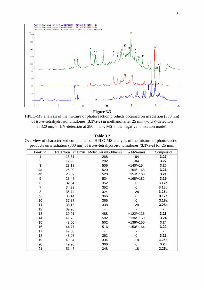
81
Figure 3.3
HPLC-MS analysis of the mixture of photoreaction products obtained on irradiation (300 nm)
of trans-tetrahydroisohumulones (3.17a-c) in methanol after 25 min (: UV-detection
at 320 nm; : UV-detection at 280 nm; : MS in the negative ionization mode).
Table 3.2
Overview of characterized compounds on HPLC-MS-analysis of the mixture of photoreaction
products on irradiation (300 nm) of trans-tetrahydroisohumulones (3.17a-c) for 25 min.
Peak nr. Retention Time/min Molecular weight/amu MW/amu Compound
1 16.51 268 -84 3.27
2 17.93 282 -84 3.27
3 23.18 506 +140/+154 3.20
4a 25.00 520 +154/+168 3.21
4b 25.39 520 +154/+168 3.21
5 28.48 534 +168/+182 3.19
6 32.64 352 0 3.17b
7 34.33 352 0 3.18b
8 35.74 324 -28 3.25b
9 36.14 366 0 3.17a
10 37.37 366 0 3.18a
11 38.15 338 -28 3.25a
12 39.20 - - -
13 39.91 488 +122/+136 3.23
14 41.75 502 +136/+150 3.24
15 43.06 502 +136/+150 3.24
16 44.77 516 +150/+164 3.22
17 47.08 - - -
18 48.08 352 0 3.28
19 49.34 334 -18 3.25b
20 49.96 366 0 3.28
21 51.45 348 -18 3.25a
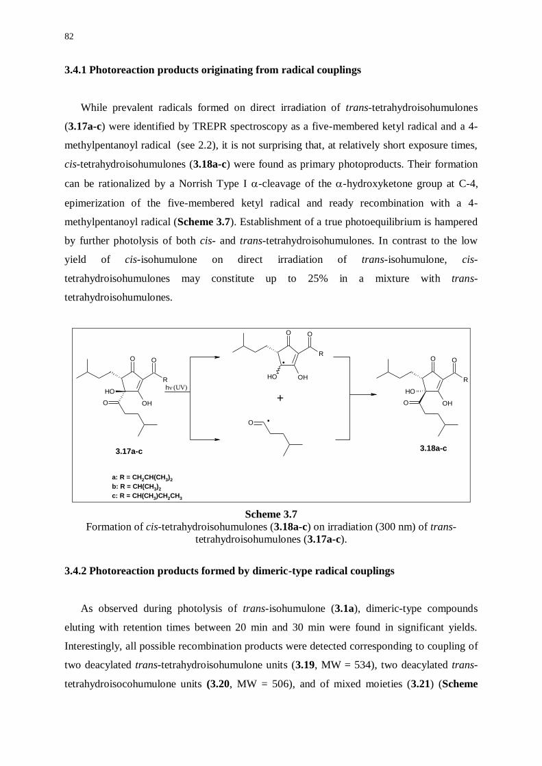
82
3.4.1 Photoreaction products originating from radical couplings
While prevalent radicals formed on direct irradiation of trans-tetrahydroisohumulones
(3.17a-c) were identified by TREPR spectroscopy as a five-membered ketyl radical and a 4-
methylpentanoyl radical (see 2.2), it is not surprising that, at relatively short exposure times,
cis-tetrahydroisohumulones (3.18a-c) were found as primary photoproducts. Their formation
can be rationalized by a Norrish Type I -cleavage of the -hydroxyketone group at C-4,
epimerization of the five-membered ketyl radical and ready recombination with a 4-
methylpentanoyl radical (Scheme 3.7). Establishment of a true photoequilibrium is hampered
by further photolysis of both cis- and trans-tetrahydroisohumulones. In contrast to the low
yield of cis-isohumulone on direct irradiation of trans-isohumulone, cis-
tetrahydroisohumulones may constitute up to 25% in a mixture with trans-
tetrahydroisohumulones.
O
OH
OH
R
OO
O
OH
OH
R
OO
O
R
O
OHOH
O
+
3.17a-c 3.18a-c
h
.
.
(UV)
a: R = CH2CH(CH3)2
b: R = CH(CH3)2
c: R = CH(CH3)CH2CH3
Scheme 3.7
Formation of cis-tetrahydroisohumulones (3.18a-c) on irradiation (300 nm) of trans-
tetrahydroisohumulones (3.17a-c).
3.4.2 Photoreaction products formed by dimeric-type radical couplings
As observed during photolysis of trans-isohumulone (3.1a), dimeric-type compounds
eluting with retention times between 20 min and 30 min were found in significant yields.
Interestingly, all possible recombination products were detected corresponding to coupling of
two deacylated trans-tetrahydroisohumulone units (3.19, MW = 534), two deacylated trans-
tetrahydroisocohumulone units (3.20, MW = 506), and of mixed moieties (3.21) (Scheme
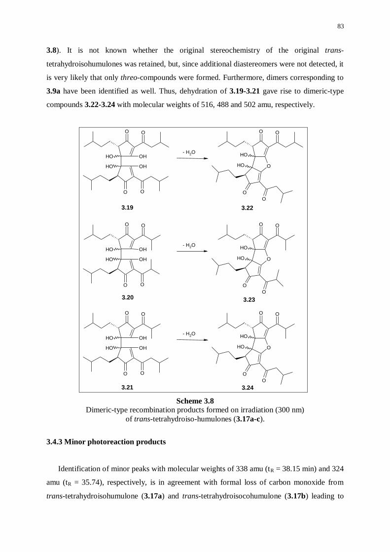
83
3.8). It is not known whether the original stereochemistry of the original trans-
tetrahydroisohumulones was retained, but, since additional diastereomers were not detected, it
is very likely that only threo-compounds were formed. Furthermore, dimers corresponding to
3.9a have been identified as well. Thus, dehydration of 3.19-3.21 gave rise to dimeric-type
compounds 3.22-3.24 with molecular weights of 516, 488 and 502 amu, respectively.
OO
O
OH
O
OH
OH
OH
O
OO
O
O
OH
OH
- H2O
OO
O
OH
O
OH
OH
OH
O
OO
O
O
OH
OH
- H2O
OO
O
OH
O
OH
OH
OH
O
OO
O
O
OH
OH
- H2O
3.20 3.23
3.19 3.22
3.21 3.24
Scheme 3.8
Dimeric-type recombination products formed on irradiation (300 nm)
of trans-tetrahydroiso-humulones (3.17a-c).
3.4.3 Minor photoreaction products
Identification of minor peaks with molecular weights of 338 amu (tR = 38.15 min) and 324
amu (tR = 35.74), respectively, is in agreement with formal loss of carbon monoxide from
trans-tetrahydroisohumulone (3.17a) and trans-tetrahydroisocohumulone (3.17b) leading to
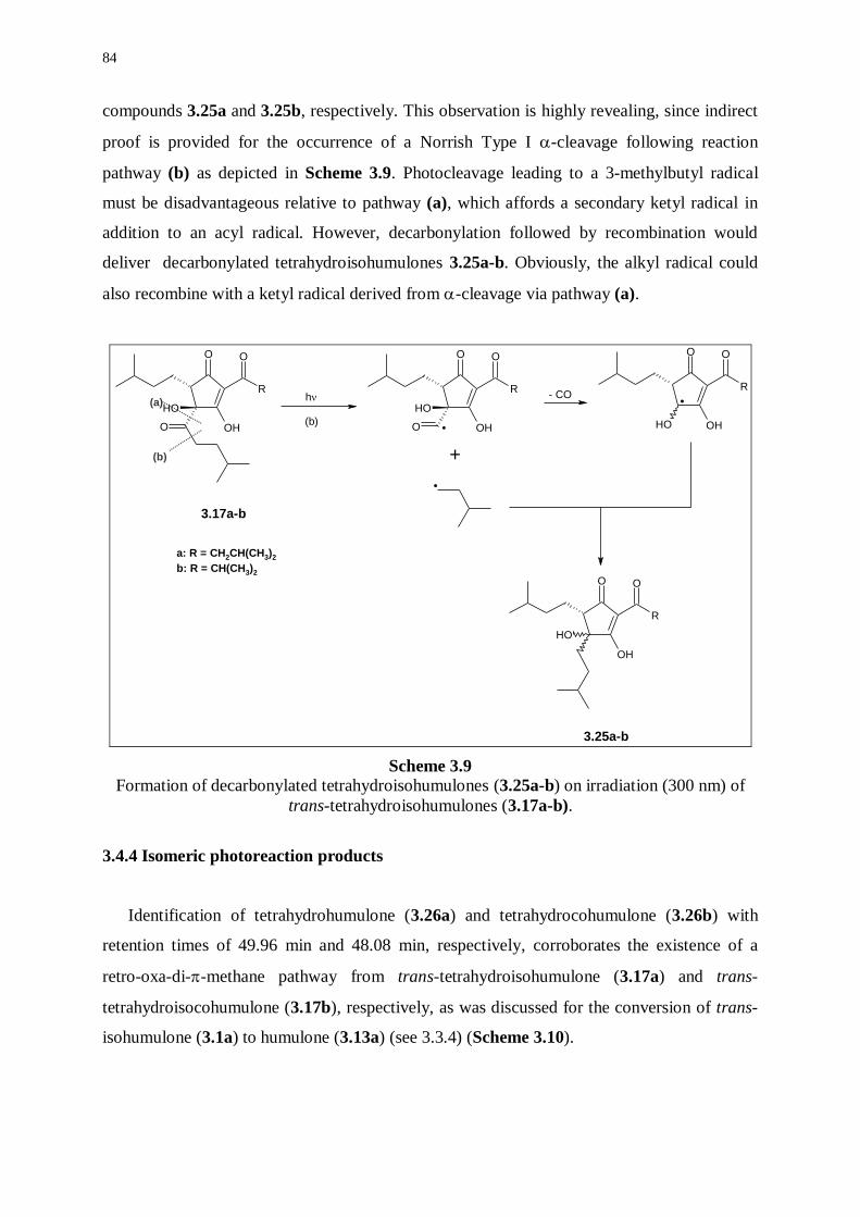
84
compounds 3.25a and 3.25b, respectively. This observation is highly revealing, since indirect
proof is provided for the occurrence of a Norrish Type I -cleavage following reaction
pathway (b) as depicted in Scheme 3.9. Photocleavage leading to a 3-methylbutyl radical
must be disadvantageous relative to pathway (a), which affords a secondary ketyl radical in
addition to an acyl radical. However, decarbonylation followed by recombination would
deliver decarbonylated tetrahydroisohumulones 3.25a-b. Obviously, the alkyl radical could
also recombine with a ketyl radical derived from -cleavage via pathway (a).
O
R
O
OHO
OH
O
R
O
OHO
OH
R
OO
OH
OH
O
R
O
OHOH
(a)
(b)
h
.
.
+
3.17a-b
3.25a-b
(b)
- CO
.
a: R = CH2CH(CH3)2
b: R = CH(CH3)2
Scheme 3.9
Formation of decarbonylated tetrahydroisohumulones (3.25a-b) on irradiation (300 nm) of
trans-tetrahydroisohumulones (3.17a-b).
3.4.4 Isomeric photoreaction products
Identification of tetrahydrohumulone (3.26a) and tetrahydrocohumulone (3.26b) with
retention times of 49.96 min and 48.08 min, respectively, corroborates the existence of a
retro-oxa-di--methane pathway from trans-tetrahydroisohumulone (3.17a) and trans-
tetrahydroisocohumulone (3.17b), respectively, as was discussed for the conversion of trans-
isohumulone (3.1a) to humulone (3.13a) (see 3.3.4) (Scheme 3.10).
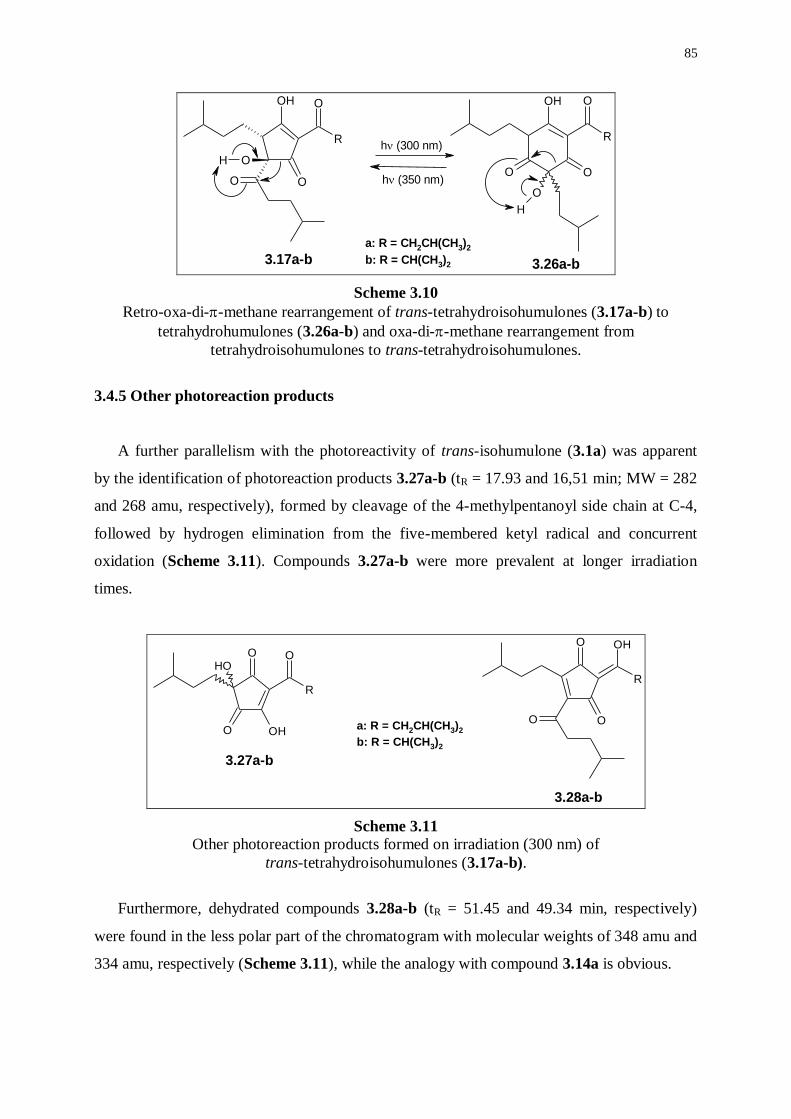
85
OH
R
O
OO
O
H
OH
R
O
O
O
O
H
h (300 nm)
3.17a-b 3.26a-b
h (350 nm)
a: R = CH2CH(CH3)2
b: R = CH(CH3)2
Scheme 3.10
Retro-oxa-di--methane rearrangement of trans-tetrahydroisohumulones (3.17a-b) to
tetrahydrohumulones (3.26a-b) and oxa-di--methane rearrangement from
tetrahydroisohumulones to trans-tetrahydroisohumulones.
3.4.5 Other photoreaction products
A further parallelism with the photoreactivity of trans-isohumulone (3.1a) was apparent
by the identification of photoreaction products 3.27a-b (tR = 17.93 and 16,51 min; MW = 282
and 268 amu, respectively), formed by cleavage of the 4-methylpentanoyl side chain at C-4,
followed by hydrogen elimination from the five-membered ketyl radical and concurrent
oxidation (Scheme 3.11). Compounds 3.27a-b were more prevalent at longer irradiation
times.
O
R
OH
OO
O
R
O
O OH
OH
3.28a-b
3.27a-b
a: R = CH2CH(CH3)2
b: R = CH(CH3)2
Scheme 3.11
Other photoreaction products formed on irradiation (300 nm) of
trans-tetrahydroisohumulones (3.17a-b).
Furthermore, dehydrated compounds 3.28a-b (tR = 51.45 and 49.34 min, respectively)
were found in the less polar part of the chromatogram with molecular weights of 348 amu and
334 amu, respectively (Scheme 3.11), while the analogy with compound 3.14a is obvious.
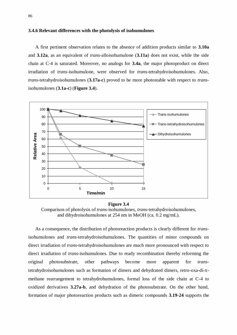
86
3.4.6 Relevant differences with the photolysis of isohumulones
A first pertinent observation relates to the absence of addition products similar to 3.10a
and 3.12a, as an equivalent of trans-alloisohumulone (3.11a) does not exist, while the side
chain at C-4 is saturated. Moreover, no analogs for 3.4a, the major photoproduct on direct
irradiation of trans-isohumulone, were observed for trans-tetrahydroisohumulones. Also,
trans-tetrahydroisohumulones (3.17a-c) proved to be more photostable with respect to trans-
isohumulones (3.1a-c) (Figure 3.4).
0
10
20
30
40
50
60
70
80
90
100
0 5 10 15
Time/min
Re
lati
ve
Are
a
Trans-isohumulones
Trans-tetrahydroisohumulones
Dihydroisohumulones
Figure 3.4 Comparison of photolysis of trans-isohumulones, trans-tetrahydroisohumulones,
and dihydroisohumulones at 254 nm in MeOH (ca. 0.2 mg/mL).
As a consequence, the distribution of photoreaction products is clearly different for trans-
isohumulones and trans-tetrahydroisohumulones. The quantities of minor compounds on
direct irradiation of trans-tetrahydroisohumulones are much more pronounced with respect to
direct irradiation of trans-isohumulones. Due to ready recombination thereby reforming the
original photosubstrate, other pathways become more apparent for trans-
tetrahydroisohumulones such as formation of dimers and dehydrated dimers, retro-oxa-di--
methane rearrangement to tetrahydrohumulones, formal loss of the side chain at C-4 to
oxidized derivatives 3.27a-b, and dehydration of the photosubstrate. On the other hand,
formation of major photoreaction products such as dimeric compounds 3.19-24 supports the
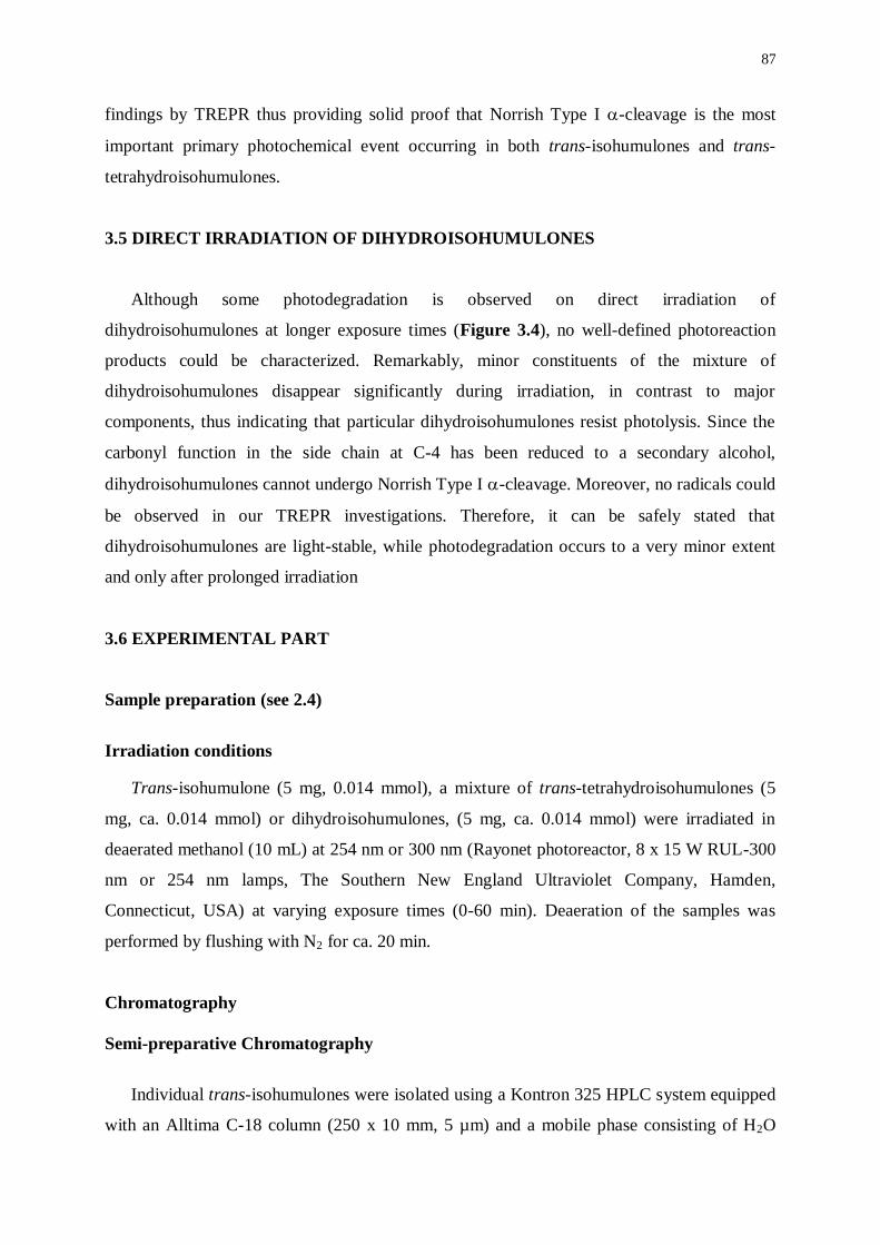
87
findings by TREPR thus providing solid proof that Norrish Type I -cleavage is the most
important primary photochemical event occurring in both trans-isohumulones and trans-
tetrahydroisohumulones.
3.5 DIRECT IRRADIATION OF DIHYDROISOHUMULONES
Although some photodegradation is observed on direct irradiation of
dihydroisohumulones at longer exposure times (Figure 3.4), no well-defined photoreaction
products could be characterized. Remarkably, minor constituents of the mixture of
dihydroisohumulones disappear significantly during irradiation, in contrast to major
components, thus indicating that particular dihydroisohumulones resist photolysis. Since the
carbonyl function in the side chain at C-4 has been reduced to a secondary alcohol,
dihydroisohumulones cannot undergo Norrish Type I -cleavage. Moreover, no radicals could
be observed in our TREPR investigations. Therefore, it can be safely stated that
dihydroisohumulones are light-stable, while photodegradation occurs to a very minor extent
and only after prolonged irradiation
3.6 EXPERIMENTAL PART
Sample preparation (see 2.4)
Irradiation conditions
Trans-isohumulone (5 mg, 0.014 mmol), a mixture of trans-tetrahydroisohumulones (5
mg, ca. 0.014 mmol) or dihydroisohumulones, (5 mg, ca. 0.014 mmol) were irradiated in
deaerated methanol (10 mL) at 254 nm or 300 nm (Rayonet photoreactor, 8 x 15 W RUL-300
nm or 254 nm lamps, The Southern New England Ultraviolet Company, Hamden,
Connecticut, USA) at varying exposure times (0-60 min). Deaeration of the samples was
performed by flushing with N2 for ca. 20 min.
Chromatography
Semi-preparative Chromatography
Individual trans-isohumulones were isolated using a Kontron 325 HPLC system equipped
with an Alltima C-18 column (250 x 10 mm, 5 µm) and a mobile phase consisting of H2O
88
(acidified to pH 2.8 with H3PO4) and acetonitrile in a ratio of 52/48 (v/v). UV-detection was
performed at 280 nm using an ISCO single wavelength detector at a flow rate of 5 mL.min-1
.
Fractions containing the compounds of interest were collected, the organic solvent was
removed under reduced pressure, and the remaining aqueous fraction was applied to a solid
phase extraction cartridge (Varian C-18 HLB 500 mg; Varian, Harbor City, California, USA)
after acidification to pH 1 (activation: 1 mL MeOH, conditioning: 1 mL H2O (pH = 1)).
Subsequent elution with MeOH (1 mL) gave the desired compounds as a residue after
removal of the solvent under a gentle stream of nitrogen.
Analytical Chromatography
Isocratic analyises of trans-isohumulones, trans-tetrahydroisohumulones, and dihydro-
isohumulones, and gradient analyses of mixtures of photoreaction products were carried out
on a Kontron system consisting of a Kontron 325 Pump and a Kontron DAD-440 Series.
Samples were concentrated to ca. 2 mg.mL-1
prior to analysis. Separations (loop injections of
20 l) were achieved on an Alltima C-18 column (250 x 4.6 mm, 5 µm) at a flow rate of 1.8
mL.min–1
(isocratic) or 1.0 mL.min-1
(gradient) using solvent A (water and formic acid, pH
2.8) and solvent B (acetonitrile/methanol 9/1 v/v). Composition of the mobile phase for
isocratic analysis of trans-isohumulones: 52% B in A, trans-tetrahydroisohumulones: 62 % B
in A, dihydroisohumulones: 57% B in A. Gradient profile for the analysis of mixtures of
photoreaction products: 0-3 min: 28% B in A, 3-13 min: from 28 % to 55% B in A, 13-18
min: 55% B in A, 18-20 min: from 55% to 63% B in A, 20-25 min: 63% B in A, 25-31 min:
from 63 % to 72% B in A, 31-48 min: from 72 % to 95% B in A, 48-50 min: 95% B in A, 50-
57 min: from 95 % to 28% B in A, 57-60 min: 28% B in A.
HPLC-ESI-MS
Photoreaction mixtures were analyzed with HPLC-ESI-MS using the chromatographic
conditions described above for HPLC-DAD. Samples were concentrated to 5 mg.mL-1
. The
chromatographic apparatus was a Hewlett Packard 1100 Series HPLC-MSD. MS conditions:
electrospray ionization (ESI) both in the positive and the negative ionization modes; mass
analysis with a quadrupole mass-spectrometer (API-ES: atmospheric pressure ionization -
electrospray), gas temperature: 340°C, nebulizing gas pressure: 40 psi (2.75x10⁵ Pa), drying
gas flow rate of 11 L.min-1
, capillary voltage: 4000 V in the positive ionization mode and

89
3500 V in the negative ionization mode, quadrupole temperature: 100°C, scan range: 105-600
amu and fragmentor (CID) set at 100 or 160 V in the positive ionization mode and 100 V in
the negative ionization mode.
90
3.7 REFERENCES
(1) Blondeel, G.M.A., De Keukeleire, D., Verzele, M.; The Photolysis of Trans-Isohumulone to
Dehydrohumulinic Acid, a Key Route to the Development of Sunstruck Flavour in Beer. J.
Chem. Soc. Perkin Trans. I, 1987, 2715-2717.
(2) Hay, B.A., Homiski, J.W., Howie, J.L.; Differences in Ultraviolet Absorbance of
Tetrahydroiso--acid Components. J. Am. Soc. Brew. Chem., 1994, 52, 54-56.
(3) Wagner, P.J., Hammond, G.S.; Properties and Reactions of Organic Molecules in their Triplet
States. Adv. Photochem., 1968, 5, 21-156.
(4) Verzele, M., De Keukeleire, D.; Chemistry and Analysis of Hop and Beer Bitter Acids.
Elsevier, Amsterdam, 1991.
(5) De Keukeleire, D., Blondeel, G.M.; The Mechanism of the Regio- and Stereospecific
Photorearrangement of Humulone to the Beer Bitter Component trans Isohumulone.
Tetrahedron Letters, 1979, 15, 1343-1346.
(6) Thornton, H.A., Kulandai, J., Bond, M., Jontef, M.P., Hawthorne, D.B., Kavanagh, T.E.;
Preparation of Trans-Iso-Alpha-Acids and Use of their Dicyclohexylamine Salts as a Standard
for Iso-Alpha-Acids Analysis. J. Inst. Brew., 1993, 99, 473-477.
************************
SENSITIZED IRRADIATION

93
CHAPTER 4
LASER-FLASH PHOTOLYSIS TRANSIENT ABSORPTION
SPECTROSCOPY OF ISOHUMULONES AND REDUCED DERIVATIVES
UNDER SENSITIZED IRRADIATION CONDITIONS
4.1 INTRODUCTION
Laser-flash photolysis coupled to emission or absorption spectroscopy is a more
conventional technique - with respect to time-resolved electron paramagnetic resonance - for
characterization of short-lived intermediates in photochemical processes. Flash photolysis
involves production of high ‘instantaneous’ concentrations [M*]0 of short-lived intermediates
using a very high-intensity pulse of radiation with very short duration. Decay of fluorescence
(or phosphorescence) can be observed by monitoring the emission from the sample at right
angles to the direction of the incident radiation with a spectrophotometer coupled to an
oscilloscope. If only one emitting state is populated in the experiment, the fluorescence
should decay exponentially according to (4.1) and the lifetime for fluorescence (f) may be
determined directly from the decay rate.1
[M*] = [M
*]0 e
-t/f (4.1)
Similarly, decay and formation of transients can be monitored by measuring the
absorbance (at a particular wavelength) using an analyzing beam passing through the reaction
cell at right angles with respect to the incident radiation. Alternatively, absorption spectra of
transients can be measured as difference absorption spectra using gated detection techniques
at a predetermined time interval after the initial flash. The decay of an intermediate can be
monitored by recording a series of spectra at different, successive time intervals after the
excitation flash. A complete spectrum of the intermediate can be constructed point by point
by changing the setting of the monitoring monochromator in a series of readings at a fixed
time interval after the initial flash or directly with a diode array detector (DAD).2
Obviously, the time resolution of the method is limited by the duration of the initial flash.
While excited states of most organic radicals decay very rapidly (subnanoseconds to
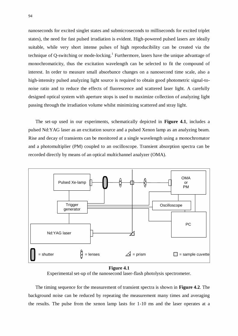
94
nanoseconds for excited singlet states and submicroseconds to milliseconds for excited triplet
states), the need for fast pulsed irradiation is evident. High-powered pulsed lasers are ideally
suitable, while very short intense pulses of high reproducibility can be created via the
technique of Q-switching or mode-locking.1 Furthermore, lasers have the unique advantage of
monochromaticity, thus the excitation wavelength can be selected to fit the compound of
interest. In order to measure small absorbance changes on a nanosecond time scale, also a
high-intensity pulsed analyzing light source is required to obtain good photometric signal-to-
noise ratio and to reduce the effects of fluorescence and scattered laser light. A carefully
designed optical system with aperture stops is used to maximize collection of analyzing light
passing through the irradiation volume whilst minimizing scattered and stray light.
The set-up used in our experiments, schematically depicted in Figure 4.1, includes a
pulsed Nd:YAG laser as an excitation source and a pulsed Xenon lamp as an analyzing beam.
Rise and decay of transients can be monitored at a single wavelength using a monochromator
and a photomultiplier (PM) coupled to an oscilloscope. Transient absorption spectra can be
recorded directly by means of an optical multichannel analyzer (OMA).
Pulsed Xe-lamp
Trigger generator
Nd:YAG laser
Oscilloscope
OMAor
PM
PC
= shutter = lenses = prism = sample cuvette
Figure 4.1 Experimental set-up of the nanosecond laser-flash photolysis spectrometer.
The timing sequence for the measurement of transient spectra is shown in Figure 4.2. The
background noise can be reduced by repeating the measurement many times and averaging
the results. The pulse from the xenon lamp lasts for 1-10 ms and the laser operates at a
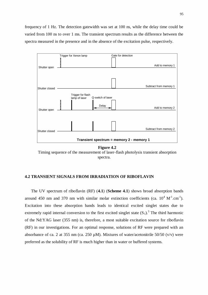
95
frequency of 1 Hz. The detection gatewidth was set at 100 ns, while the delay time could be
varied from 100 ns to over 1 ms. The transient spectrum results as the difference between the
spectra measured in the presence and in the absence of the excitation pulse, respectively.
Trigger for Xenon lamp Gate for detection
Trigger for flashlamp of laser Q-switch of laser
Shutter open
Shutter open
Shutter closed
Shutter closed
Add to memory 1
Subtract from memory 1
Add to memory 2
Subtract from memory 2
Transient spectrum = memory 2 - memory 1
Delay
Figure 4.2 Timing sequence of the measurement of laser-flash photolysis transient absorption
spectra.
4.2 TRANSIENT SIGNALS FROM IRRADIATION OF RIBOFLAVIN
The UV spectrum of riboflavin (RF) (4.1) (Scheme 4.1) shows broad absorption bands
around 450 nm and 370 nm with similar molar extinction coefficients (ca. 104
M-1
.cm-1
).
Excitation into these absorption bands leads to identical excited singlet states due to
extremely rapid internal conversion to the first excited singlet state (S1).3 The third harmonic
of the Nd:YAG laser (355 nm) is, therefore, a most suitable excitation source for riboflavin
(RF) in our investigations. For an optimal response, solutions of RF were prepared with an
absorbance of ca. 2 at 355 nm (ca. 250 µM). Mixtures of water/acetonitrile 50/50 (v/v) were
preferred as the solubility of RF is much higher than in water or buffered systems.
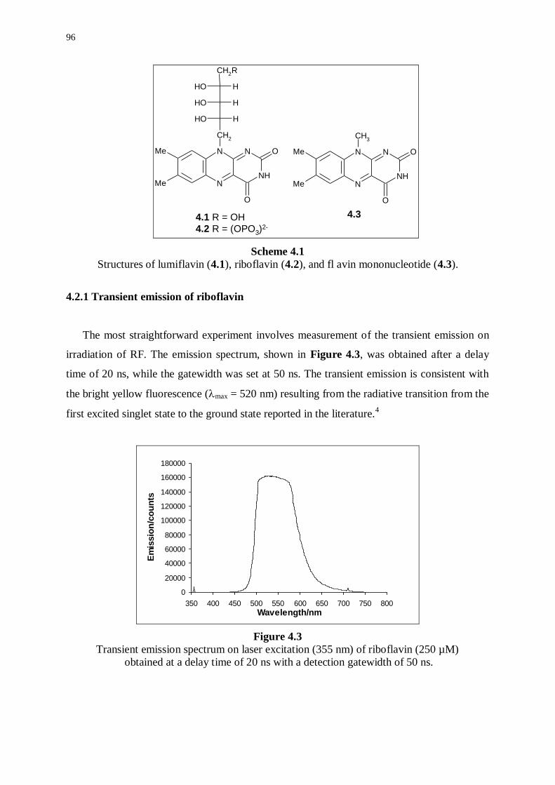
96
NH
N
N
NMe
Me
CH2
CH2R
O
O
OH H
OH H
OH H
NH
N
N
NMe
Me
CH3
O
O
4.34.1 R = OH
4.2 R = (OPO3)2-
Scheme 4.1
Structures of lumiflavin (4.1), riboflavin (4.2), and fl avin mononucleotide (4.3).
4.2.1 Transient emission of riboflavin
The most straightforward experiment involves measurement of the transient emission on
irradiation of RF. The emission spectrum, shown in Figure 4.3, was obtained after a delay
time of 20 ns, while the gatewidth was set at 50 ns. The transient emission is consistent with
the bright yellow fluorescence (max = 520 nm) resulting from the radiative transition from the
first excited singlet state to the ground state reported in the literature.4
0
20000
40000
60000
80000
100000
120000
140000
160000
180000
350 400 450 500 550 600 650 700 750 800
Wavelength/nm
Em
issio
n/c
ou
nts
Figure 4.3
Transient emission spectrum on laser excitation (355 nm) of riboflavin (250 µM)
obtained at a delay time of 20 ns with a detection gatewidth of 50 ns.
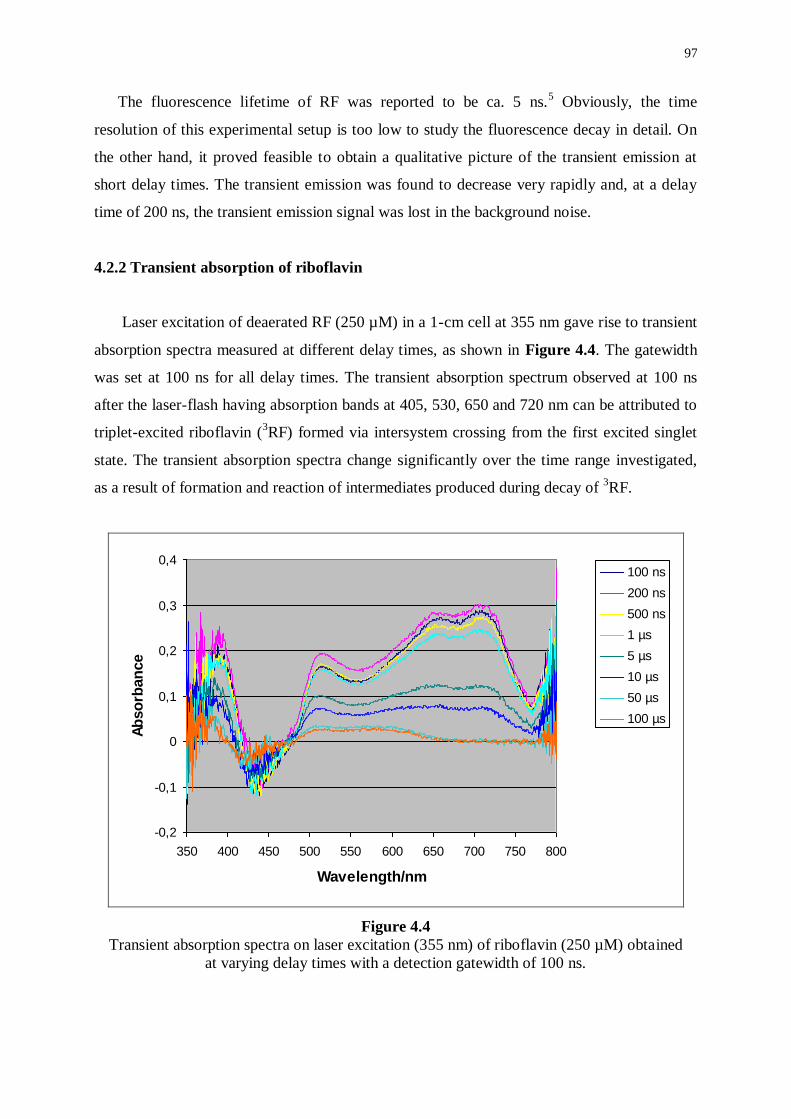
97
The fluorescence lifetime of RF was reported to be ca. 5 ns.5 Obviously, the time
resolution of this experimental setup is too low to study the fluorescence decay in detail. On
the other hand, it proved feasible to obtain a qualitative picture of the transient emission at
short delay times. The transient emission was found to decrease very rapidly and, at a delay
time of 200 ns, the transient emission signal was lost in the background noise.
4.2.2 Transient absorption of riboflavin
Laser excitation of deaerated RF (250 µM) in a 1-cm cell at 355 nm gave rise to transient
absorption spectra measured at different delay times, as shown in Figure 4.4. The gatewidth
was set at 100 ns for all delay times. The transient absorption spectrum observed at 100 ns
after the laser-flash having absorption bands at 405, 530, 650 and 720 nm can be attributed to
triplet-excited riboflavin (3RF) formed via intersystem crossing from the first excited singlet
state. The transient absorption spectra change significantly over the time range investigated,
as a result of formation and reaction of intermediates produced during decay of 3RF.
-0,2
-0,1
0
0,1
0,2
0,3
0,4
350 400 450 500 550 600 650 700 750 800
Wavelength/nm
Ab
so
rban
ce
100 ns
200 ns
500 ns
1 µs
5 µs
10 µs
50 µs
100 µs
Figure 4.4
Transient absorption spectra on laser excitation (355 nm) of riboflavin (250 µM) obtained
at varying delay times with a detection gatewidth of 100 ns.
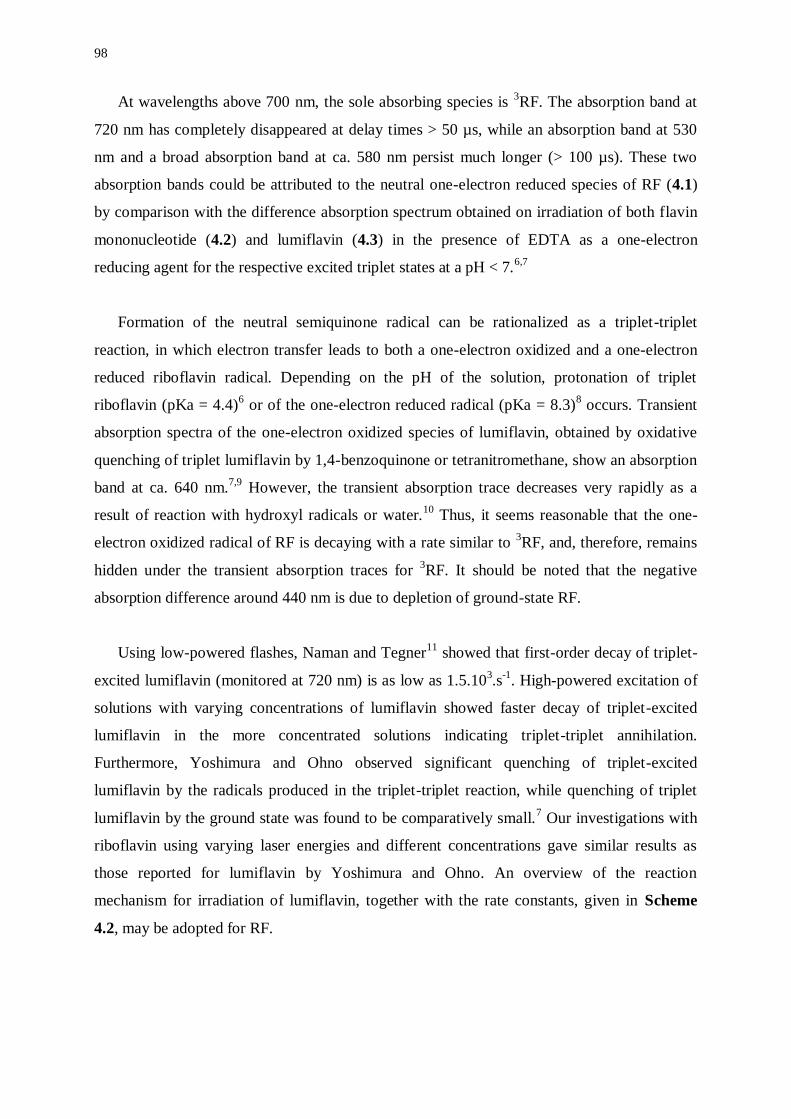
98
At wavelengths above 700 nm, the sole absorbing species is 3RF. The absorption band at
720 nm has completely disappeared at delay times > 50 µs, while an absorption band at 530
nm and a broad absorption band at ca. 580 nm persist much longer (> 100 µs). These two
absorption bands could be attributed to the neutral one-electron reduced species of RF (4.1)
by comparison with the difference absorption spectrum obtained on irradiation of both flavin
mononucleotide (4.2) and lumiflavin (4.3) in the presence of EDTA as a one-electron
reducing agent for the respective excited triplet states at a pH < 7.6,7
Formation of the neutral semiquinone radical can be rationalized as a triplet-triplet
reaction, in which electron transfer leads to both a one-electron oxidized and a one-electron
reduced riboflavin radical. Depending on the pH of the solution, protonation of triplet
riboflavin (pKa = 4.4)6 or of the one-electron reduced radical (pKa = 8.3)
8 occurs. Transient
absorption spectra of the one-electron oxidized species of lumiflavin, obtained by oxidative
quenching of triplet lumiflavin by 1,4-benzoquinone or tetranitromethane, show an absorption
band at ca. 640 nm.7,9
However, the transient absorption trace decreases very rapidly as a
result of reaction with hydroxyl radicals or water.10
Thus, it seems reasonable that the one-
electron oxidized radical of RF is decaying with a rate similar to 3RF, and, therefore, remains
hidden under the transient absorption traces for 3RF. It should be noted that the negative
absorption difference around 440 nm is due to depletion of ground-state RF.
Using low-powered flashes, Naman and Tegner11
showed that first-order decay of triplet-
excited lumiflavin (monitored at 720 nm) is as low as 1.5.103.s
-1. High-powered excitation of
solutions with varying concentrations of lumiflavin showed faster decay of triplet-excited
lumiflavin in the more concentrated solutions indicating triplet-triplet annihilation.
Furthermore, Yoshimura and Ohno observed significant quenching of triplet-excited
lumiflavin by the radicals produced in the triplet-triplet reaction, while quenching of triplet
lumiflavin by the ground state was found to be comparatively small.7 Our investigations with
riboflavin using varying laser energies and different concentrations gave similar results as
those reported for lumiflavin by Yoshimura and Ohno. An overview of the reaction
mechanism for irradiation of lumiflavin, together with the rate constants, given in Scheme
4.2, may be adopted for RF.
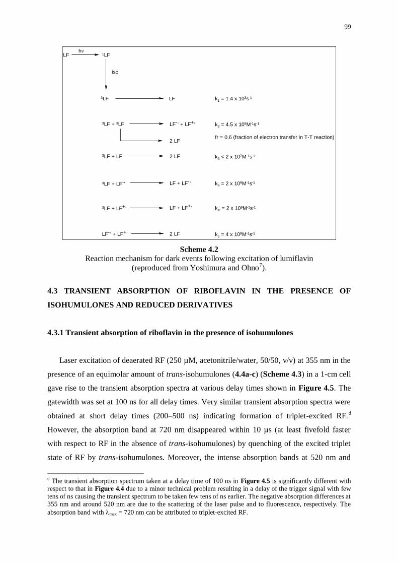
99
LF 1LF
3LF + 3LF LF3LF
3LF + LF
3LF + LF-.
2 LF
3LF + LF+.
LF-. + LF+.
LF-. + LF+.
2 LF
LF + LF-.
LF + LF+.
2 LF
3LF LF
h
isc
k1 = 1.4 x 10³s-1
k2 = 4.5 x 10⁹M-1s-1
fr = 0.6 (fraction of electron transfer in T-T reaction)
k3 < 2 x 10⁷M-1s-1
k4 = 2 x 10⁹M-1s-1
k4' = 2 x 10⁹M-1s-1
k5 = 4 x 10⁹M-1s-1
Scheme 4.2
Reaction mechanism for dark events following excitation of lumiflavin
(reproduced from Yoshimura and Ohno7).
4.3 TRANSIENT ABSORPTION OF RIBOFLAVIN IN THE PRESENCE OF
ISOHUMULONES AND REDUCED DERIVATIVES
4.3.1 Transient absorption of riboflavin in the presence of isohumulones
Laser excitation of deaerated RF (250 µM, acetonitrile/water, 50/50, v/v) at 355 nm in the
presence of an equimolar amount of trans-isohumulones (4.4a-c) (Scheme 4.3) in a 1-cm cell
gave rise to the transient absorption spectra at various delay times shown in Figure 4.5. The
gatewidth was set at 100 ns for all delay times. Very similar transient absorption spectra were
obtained at short delay times (200–500 ns) indicating formation of triplet-excited RF.d
However, the absorption band at 720 nm disappeared within 10 µs (at least fivefold faster
with respect to RF in the absence of trans-isohumulones) by quenching of the excited triplet
state of RF by trans-isohumulones. Moreover, the intense absorption bands at 520 nm and
d The transient absorption spectrum taken at a delay time of 100 ns in Figure 4.5 is significantly different with
respect to that in Figure 4.4 due to a minor technical problem resulting in a delay of the trigger signal with few
tens of ns causing the transient spectrum to be taken few tens of ns earlier. The negative absorption differences at
355 nm and around 520 nm are due to the scattering of the laser pulse and to fluorescence, respectively. The
absorption band with max = 720 nm can be attributed to triplet-excited RF.
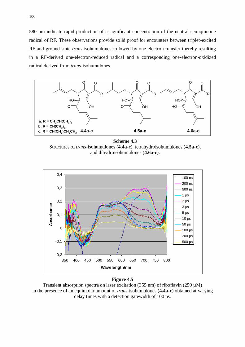
100
580 nm indicate rapid production of a significant concentration of the neutral semiquinone
radical of RF. These observations provide solid proof for encounters between triplet-excited
RF and ground-state trans-isohumulones followed by one-electron transfer thereby resulting
in a RF-derived one-electron-reduced radical and a corresponding one-electron-oxidized
radical derived from trans-isohumulones.
O
R
O
OH
OH
O
O
R
O
OH
OH
O
O
R
O
OH
OH
OH
a: R = CH2CH(CH3)2
b: R = CH(CH3)2
c: R = CH(CH3)CH2CH3
4.4a-c 4.5a-c 4.6a-c
Scheme 4.3
Structures of trans-isohumulones (4.4a-c), tetrahydroisohumulones (4.5a-c),
and dihydroisohumulones (4.6a-c).
-0,2
-0,1
0
0,1
0,2
0,3
0,4
350 400 450 500 550 600 650 700 750 800
Wavelength/nm
Ab
so
rban
ce
100 ns
200 ns
500 ns
1 µs
2 µs
3 µs
5 µs
10 µs
50 µs
100 µs
200 µs
500 µs
Figure 4.5
Transient absorption spectra on laser excitation (355 nm) of riboflavin (250 µM)
in the presence of an equimolar amount of trans-isohumulones (4.4a-c) obtained at varying
delay times with a detection gatewidth of 100 ns.
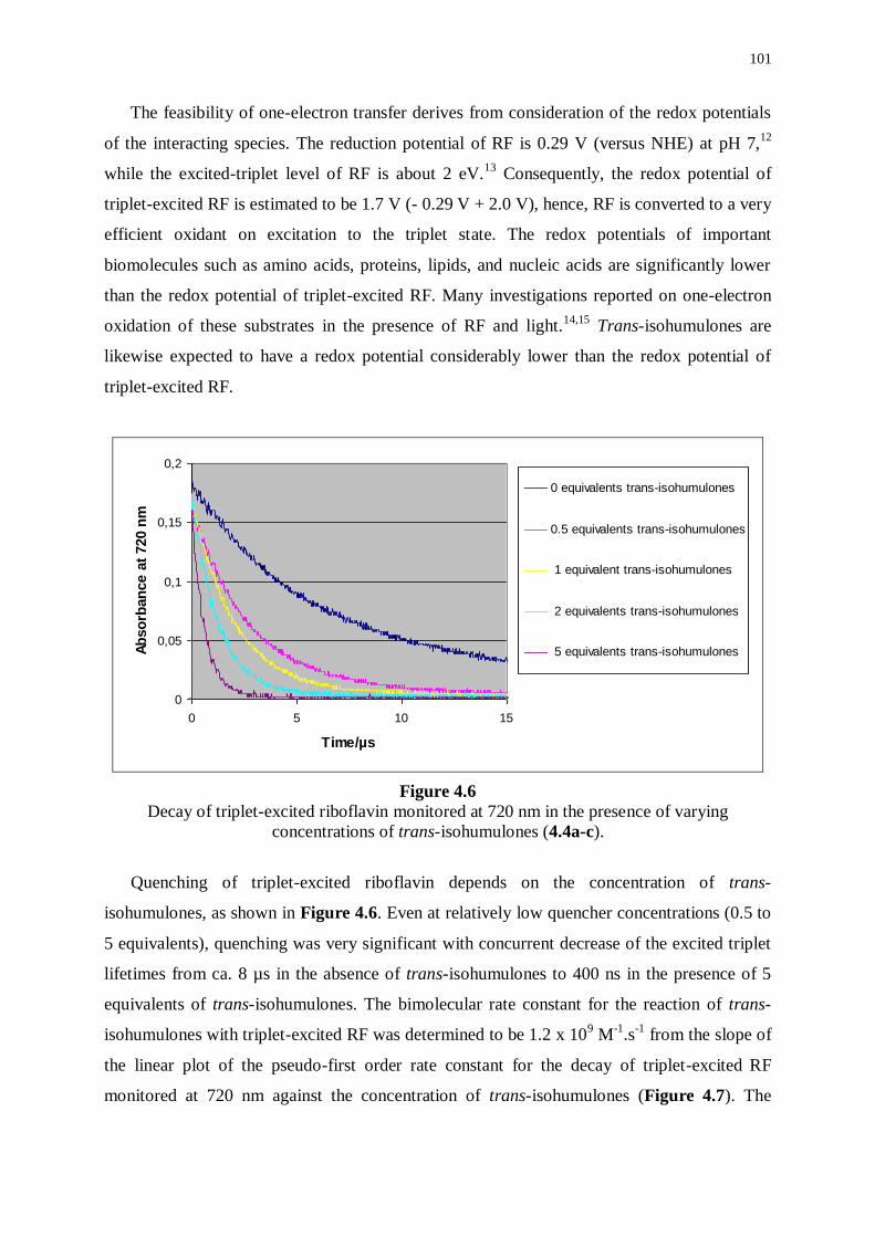
101
The feasibility of one-electron transfer derives from consideration of the redox potentials
of the interacting species. The reduction potential of RF is 0.29 V (versus NHE) at pH 7,12
while the excited-triplet level of RF is about 2 eV.13
Consequently, the redox potential of
triplet-excited RF is estimated to be 1.7 V (- 0.29 V + 2.0 V), hence, RF is converted to a very
efficient oxidant on excitation to the triplet state. The redox potentials of important
biomolecules such as amino acids, proteins, lipids, and nucleic acids are significantly lower
than the redox potential of triplet-excited RF. Many investigations reported on one-electron
oxidation of these substrates in the presence of RF and light.14,15
Trans-isohumulones are
likewise expected to have a redox potential considerably lower than the redox potential of
triplet-excited RF.
0
0,05
0,1
0,15
0,2
0 5 10 15
Time/µs
Ab
so
rban
ce
at
720 n
m
0 equivalents trans-isohumulones
0.5 equivalents trans-isohumulones
1 equivalent trans-isohumulones
2 equivalents trans-isohumulones
5 equivalents trans-isohumulones
Figure 4.6
Decay of triplet-excited riboflavin monitored at 720 nm in the presence of varying
concentrations of trans-isohumulones (4.4a-c).
Quenching of triplet-excited riboflavin depends on the concentration of trans-
isohumulones, as shown in Figure 4.6. Even at relatively low quencher concentrations (0.5 to
5 equivalents), quenching was very significant with concurrent decrease of the excited triplet
lifetimes from ca. 8 µs in the absence of trans-isohumulones to 400 ns in the presence of 5
equivalents of trans-isohumulones. The bimolecular rate constant for the reaction of trans-
isohumulones with triplet-excited RF was determined to be 1.2 x 10⁹ M
-1.s
-1 from the slope of
the linear plot of the pseudo-first order rate constant for the decay of triplet-excited RF
monitored at 720 nm against the concentration of trans-isohumulones (Figure 4.7). The
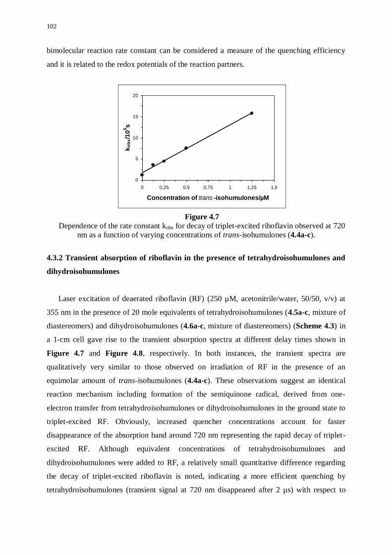
102
bimolecular reaction rate constant can be considered a measure of the quenching efficiency
and it is related to the redox potentials of the reaction partners.
0
5
10
15
20
0 0,25 0,5 0,75 1 1,25 1,5
Concentration of trans -isohumulones/µM
ko
bs/1
05s
Figure 4.7
Dependence of the rate constant kobs for decay of triplet-excited riboflavin observed at 720
nm as a function of varying concentrations of trans-isohumulones (4.4a-c).
4.3.2 Transient absorption of riboflavin in the presence of tetrahydroisohumulones and
dihydroisohumulones
Laser excitation of deaerated riboflavin (RF) (250 µM, acetonitrile/water, 50/50, v/v) at
355 nm in the presence of 20 mole equivalents of tetrahydroisohumulones (4.5a-c, mixture of
diastereomers) and dihydroisohumulones (4.6a-c, mixture of diastereomers) (Scheme 4.3) in
a 1-cm cell gave rise to the transient absorption spectra at different delay times shown in
Figure 4.7 and Figure 4.8, respectively. In both instances, the transient spectra are
qualitatively very similar to those observed on irradiation of RF in the presence of an
equimolar amount of trans-isohumulones (4.4a-c). These observations suggest an identical
reaction mechanism including formation of the semiquinone radical, derived from one-
electron transfer from tetrahydroisohumulones or dihydroisohumulones in the ground state to
triplet-excited RF. Obviously, increased quencher concentrations account for faster
disappearance of the absorption band around 720 nm representing the rapid decay of triplet-
excited RF. Although equivalent concentrations of tetrahydroisohumulones and
dihydroisohumulones were added to RF, a relatively small quantitative difference regarding
the decay of triplet-excited riboflavin is noted, indicating a more efficient quenching by
tetrahydroisohumulones (transient signal at 720 nm disappeared after 2 µs) with respect to
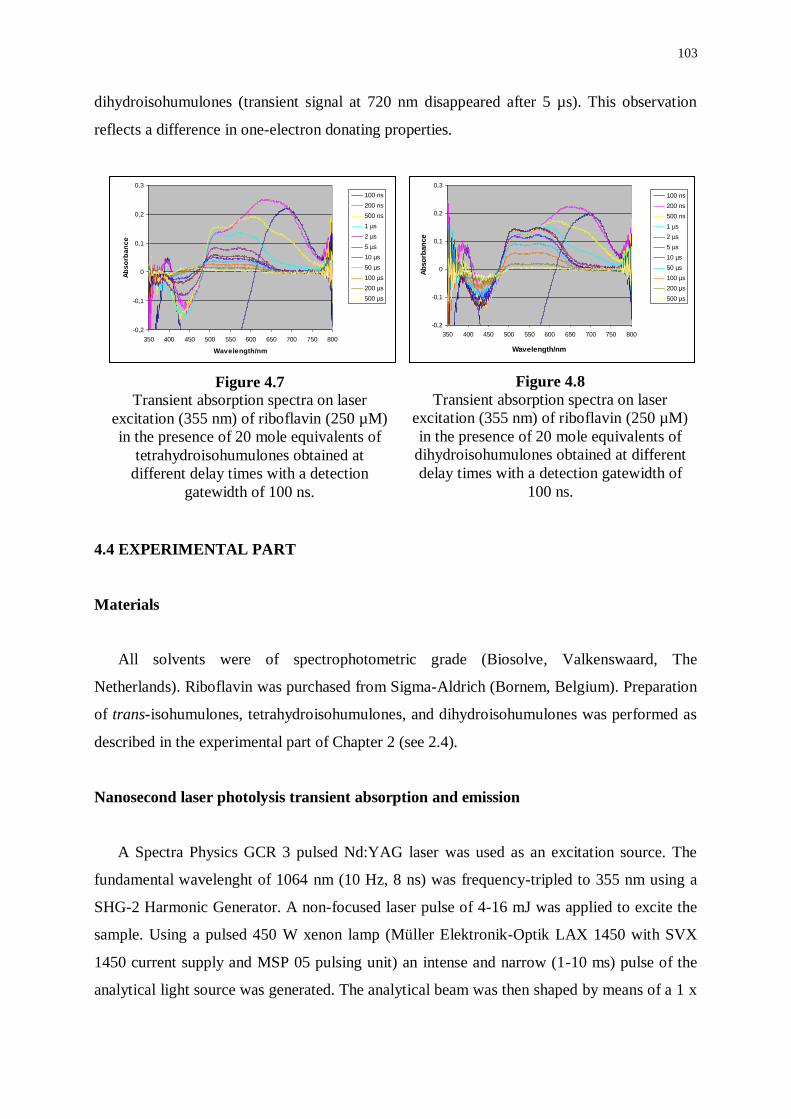
103
dihydroisohumulones (transient signal at 720 nm disappeared after 5 µs). This observation
reflects a difference in one-electron donating properties.
4.4 EXPERIMENTAL PART
Materials
All solvents were of spectrophotometric grade (Biosolve, Valkenswaard, The
Netherlands). Riboflavin was purchased from Sigma-Aldrich (Bornem, Belgium). Preparation
of trans-isohumulones, tetrahydroisohumulones, and dihydroisohumulones was performed as
described in the experimental part of Chapter 2 (see 2.4).
Nanosecond laser photolysis transient absorption and emission
A Spectra Physics GCR 3 pulsed Nd:YAG laser was used as an excitation source. The
fundamental wavelenght of 1064 nm (10 Hz, 8 ns) was frequency-tripled to 355 nm using a
SHG-2 Harmonic Generator. A non-focused laser pulse of 4-16 mJ was applied to excite the
sample. Using a pulsed 450 W xenon lamp (Müller Elektronik-Optik LAX 1450 with SVX
1450 current supply and MSP 05 pulsing unit) an intense and narrow (1-10 ms) pulse of the
analytical light source was generated. The analytical beam was then shaped by means of a 1 x
Figure 4.7 Transient absorption spectra on laser
excitation (355 nm) of riboflavin (250 µM)
in the presence of 20 mole equivalents of
tetrahydroisohumulones obtained at
different delay times with a detection
gatewidth of 100 ns.
Figure 4.8
Transient absorption spectra on laser
excitation (355 nm) of riboflavin (250 µM)
in the presence of 20 mole equivalents of
dihydroisohumulones obtained at different
delay times with a detection gatewidth of
100 ns.
-0,2
-0,1
0
0,1
0,2
0,3
350 400 450 500 550 600 650 700 750 800
Wavelength/nm
Ab
so
rban
ce
100 ns
200 ns
500 ns
1 µs
2 µs
5 µs
10 µs
50 µs
100 µs
200 µs
500 µs
-0,2
-0,1
0
0,1
0,2
0,3
350 400 450 500 550 600 650 700 750 800
Wavelength/nm
Ab
so
rba
nc
e
100 ns
200 ns
500 ns
1 µs
2 µs
5 µs
10 µs
50 µs
100 µs
200 µs
500 µs
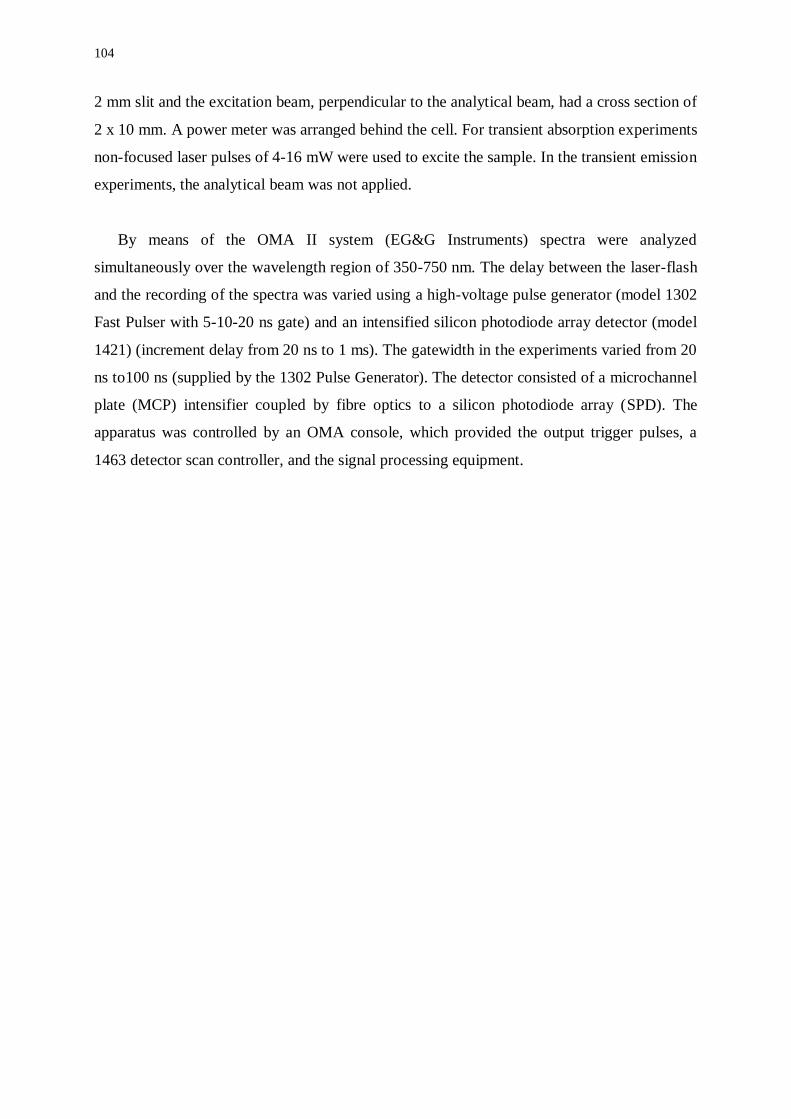
104
2 mm slit and the excitation beam, perpendicular to the analytical beam, had a cross section of
2 x 10 mm. A power meter was arranged behind the cell. For transient absorption experiments
non-focused laser pulses of 4-16 mW were used to excite the sample. In the transient emission
experiments, the analytical beam was not applied.
By means of the OMA II system (EG&G Instruments) spectra were analyzed
simultaneously over the wavelength region of 350-750 nm. The delay between the laser-flash
and the recording of the spectra was varied using a high-voltage pulse generator (model 1302
Fast Pulser with 5-10-20 ns gate) and an intensified silicon photodiode array detector (model
1421) (increment delay from 20 ns to 1 ms). The gatewidth in the experiments varied from 20
ns to100 ns (supplied by the 1302 Pulse Generator). The detector consisted of a microchannel
plate (MCP) intensifier coupled by fibre optics to a silicon photodiode array (SPD). The
apparatus was controlled by an OMA console, which provided the output trigger pulses, a
1463 detector scan controller, and the signal processing equipment.
105
4.5 REFERENCES
(1) Gilbert A., Baggot, J.; Essentials of Molecular Photochemistry. Blackwell Scientific
Publications, Oxford, 1991.
(2) Barltrop, J.A., Coyle, J.D.; Excited States in Organic Chemistry. John Wiley & Sons, London,
1975.
(3) Eweg, J.K., Müller, F., Visser, A.J.W.G., Veeger, C., Bebelaar, D., Vanvoorst, J.D.W.;
Molecular Luminescence of Some Isoalloxazines in Apolar Solvents at Various Temperatures.
Photochem. Photobiol., 1979, 30, 463-471.
(4) Heelis, P.F.; The Photophysical and Photochemical Properties of Flavins (Isoalloxazines).
Chem. Soc. Rev., 1982, 11, 15-39.
(5) Fugate, R.D., Song, P.S.; Lifetime Study of Phototautomerism of Alloxazine and Lumichromes.
Photochem. Photobiol., 1976, 24, 479-481.
(6) Sakai, M., Takahashi, H.; One-Electron Photoreduction of Flavin Mononucleotide: Time-
Resolved Resonance Raman and Absorption Study. J. Mol. Struct., 1996, 379, 9-18.
(7) Yoshimura, A., Ohno, T.; Quenching of Excited Triplet Lumiflavin Radicals by Lumiflavin
Radicals Formed in Its T-T Reaction. Photochem. Photobiol., 1991, 53, 175-179.
(8) Land, E.J., Swallow, A.; One-Electron Reactions in Biochemical Systems as Studied by Pulse
Radiolysis. II. Riboflavin. Biochemistry, 1969, 8, 2117-2125.
(9) Heelis, P.F., Parsons, B.J., Thomas, B., Phillips, G.O.; One-Electron Oxidation of the Flavin
Triplet State as Studied by Laser-Flash Photolysis. J. Chem. Soc. Chem. Commun., 1985,
954-955.
(10) Heelis, P.F., Parsons, B.J., Phillips, G.O., Swallow, A.J.; One-Electron Oxidation of Flavins.
A Flash Photolysis and Pulse Radiolysis Study. J. Phys. Chem., 1986, 90, 6833-6836.
(11) Naman, S.A., Tegnèr, L.; Decay Kinetics of the Triplet Excited State of Lumiflavin.
Photochem. Photobiol., 1986, 43, 331-333.
(12) Meisel, D., Neta, P.; One-Electron Reduction Potential of Riboflavin Studied by Pulse
Radiolysis. J. Phys. Chem., 1975, 17, 2459-2461.
(13) Sun, M.T., Moore, T.A., Song, P.S.; Molecular Luminescence Studies of Flavins. I. The
Excited States of Flavins. J. Am. Chem. Soc., 1972, 94, 1730-1740.
(14) Lu, C., Lin, W., Wang, W., Han, Z., Yao, S., Lin., N.; Riboflavin (RB2) Photosensitized
Oxidation of 2’-Deoxyguanosine-5’-monophosphate (dGMP) in Aqueous Solution: a
Transient Intermediates Study. Phys. Chem. Chem. Phys., 2000, 2, 329-334.
(15) Garcia, J., Silva, E.; Flavin-Sensitized Photooxidation of Amino Acids Present in a Parenteral
Nutrition Infusate: Protection by Ascorbic Acid. J. Nutr. Biochem., 1997, 8, 341-345.
************************

107
CHAPTER 5
STEADY-STATE AND TIME-RESOLVED ELECTRON PARAMAGNETIC
RESONANCE OF ISOHUMULONES AND REDUCED DERIVATIVES
UNDER SENSITIZED IRRADIATION CONDITIONS
5.1 INTRODUCTION
Time-resolved electron paramagnetic resonance (TREPR) has been proven successful in
elucidating intricate details of the primary mechanism of formation of the lightstruck flavor in
beer (Chapter 2). It was, therefore, straightforward to apply the powerful technique to
investigate the role of isohumulones along with reduced derivatives in the formation of the
lightstruck flavor in beer on sensitized irradiation. In analogy with the experiments described
previously (Chapter 4), the third harmonic of the Nd:YAG laser (at 355 nm) was applied as a
most suitable excitation source for riboflavin (RF) with respect to its stability, high power,
convenience, and ready availability. Although experiments using well-known model systems
produced a very good signal-to-noise response, no TREPR signals could be observed on
irradiation at 355 nm of solutions containing RF (0.24 mM) on a time scale between 100 ns
and 8 µs. As this appeared to be due to lack of sensitivity, application of various solvents and
buffers led to an optimized concentration of 0.8 mM RF in acetonitrile/water 1/1 (v/v) (ca. 15
mg in 50 mL). On the other hand, the concentration of the radical anion of RF can be
increased, when EDTA is added as a one-electron reducing agent of triplet-excited RF. Even,
taking into account these additional precautions, no TREPR signal was apparent. Conversely,
the higher sensitivity of steady-state experiments could be exploited to detect a steady-state
concentration of RF-derived radicals under continuous irradiation, although time resolution
and mechanistical information are lost.
5.2 STEADY-STATE ELECTRON PARAMAGNETIC RESONANCE (SSEPR)
A 1-kW high-pressure mercury arc, exhibiting a broad emission in the UV and the visible
wavelength regions, was preferred over a laser as the excitation source (set at 400 W) in the
steady-state electron paramagnetic resonance (SSEPR) experiments. Also, the resonator was
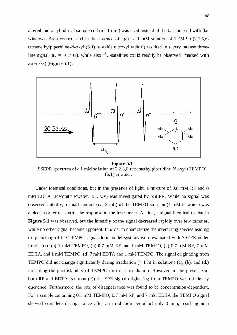
108
altered and a cylindrical sample cell (id: 1 mm) was used instead of the 0.4 mm cell with flat
windows. As a control, and in the absence of light, a 1 mM solution of TEMPO (2,2,6,6-
tetramethylpiperidine-N-oxyl (5.1), a stable nitroxyl radical) resulted in a very intense three-
line signal (aN = 16.7 G), while also 13
C-satellites could readily be observed (marked with
asterisks) (Figure 5.1).
Figure 5.1
SSEPR-spectrum of a 1 mM solution of 2,2,6,6-tetramethylpiperidine-N-oxyl (TEMPO)
(5.1) in water.
Under identical conditions, but in the presence of light, a mixture of 0.8 mM RF and 8
mM EDTA (acetonitrile/water, 1/1, v/v) was investigated by SSEPR. While no signal was
observed initially, a small amount (ca. 2 mL) of the TEMPO solution (1 mM in water) was
added in order to control the response of the instrument. At first, a signal identical to that in
Figure 5.1 was observed, but the intensity of the signal decreased rapidly over few minutes,
while no other signal became apparent. In order to characterize the interacting species leading
to quenching of the TEMPO signal, four model systems were evaluated with SSEPR under
irradiation: (a) 1 mM TEMPO, (b) 0.7 mM RF and 1 mM TEMPO, (c) 0.7 mM RF, 7 mM
EDTA, and 1 mM TEMPO, (d) 7 mM EDTA and 1 mM TEMPO. The signal originating from
TEMPO did not change significantly during irradiation (> 1 h) in solutions (a), (b), and (d,)
indicating the photostability of TEMPO on direct irradiation. However, in the presence of
both RF and EDTA (solution (c)) the EPR signal originating from TEMPO was efficiently
quenched. Furthermore, the rate of disappearance was found to be concentration-dependent.
For a sample containing 0.1 mM TEMPO, 0.7 mM RF, and 7 mM EDTA the TEMPO signal
showed complete disappearance after an irradiation period of only 3 min, resulting in a
N MeMe
Me Me
O
.
5.1
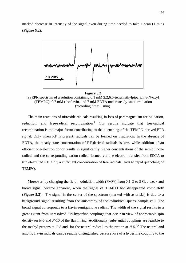
109
marked decrease in intensity of the signal even during time needed to take 1 scan (1 min)
(Figure 5.2).
Figure 5.2
SSEPR spectrum of a solution containing 0.1 mM 2,2,6,6-tetramethylpiperidine-N-oxyl
(TEMPO), 0.7 mM riboflavin, and 7 mM EDTA under steady-state irradiation
(recording time: 1 min).
The main reactions of nitroxide radicals resulting in loss of paramagnetism are oxidation,
reduction, and free-radical recombination.1 Our results indicate that free-radical
recombination is the major factor contributing to the quenching of the TEMPO-derived EPR
signal. Only when RF is present, radicals can be formed on irradiation. In the absence of
EDTA, the steady-state concentration of RF-derived radicals is low, while addition of an
efficient one-electron donor results in significantly higher concentrations of the semiquinone
radical and the corresponding cation radical formed via one-electron transfer from EDTA to
triplet-excited RF. Only a sufficient concentration of free radicals leads to rapid quenching of
TEMPO.
Moreover, by changing the field modulation width (FMW) from 0.1 G to 5 G, a weak and
broad signal became apparent, when the signal of TEMPO had disappeared completely
(Figure 5.3). The signal in the center of the spectrum (marked with asterisks) is due to a
background signal resulting from the anisotropy of the cylindrical quartz sample cell. The
broad signal corresponds to a flavin semiquinone radical. The width of the signal results to a
great extent from unresolved 14
N-hyperfine couplings that occur in view of appreciable spin
density on N-5 and N-10 of the flavin ring. Additionally, substantial couplings are feasible to
the methyl protons at C-8 and, for the neutral radical, to the proton at N-5.2,3
The neutral and
anionic flavin radicals can be readily distinguished because loss of a hyperfine coupling to the
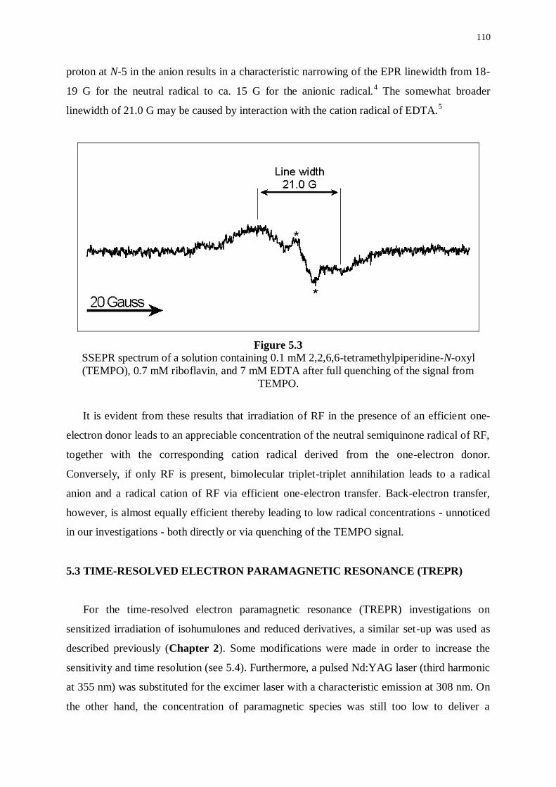
110
proton at N-5 in the anion results in a characteristic narrowing of the EPR linewidth from 18-
19 G for the neutral radical to ca. 15 G for the anionic radical.4 The somewhat broader
linewidth of 21.0 G may be caused by interaction with the cation radical of EDTA.5
Figure 5.3
SSEPR spectrum of a solution containing 0.1 mM 2,2,6,6-tetramethylpiperidine-N-oxyl
(TEMPO), 0.7 mM riboflavin, and 7 mM EDTA after full quenching of the signal from
TEMPO.
It is evident from these results that irradiation of RF in the presence of an efficient one-
electron donor leads to an appreciable concentration of the neutral semiquinone radical of RF,
together with the corresponding cation radical derived from the one-electron donor.
Conversely, if only RF is present, bimolecular triplet-triplet annihilation leads to a radical
anion and a radical cation of RF via efficient one-electron transfer. Back-electron transfer,
however, is almost equally efficient thereby leading to low radical concentrations - unnoticed
in our investigations - both directly or via quenching of the TEMPO signal.
5.3 TIME-RESOLVED ELECTRON PARAMAGNETIC RESONANCE (TREPR)
For the time-resolved electron paramagnetic resonance (TREPR) investigations on
sensitized irradiation of isohumulones and reduced derivatives, a similar set-up was used as
described previously (Chapter 2). Some modifications were made in order to increase the
sensitivity and time resolution (see 5.4). Furthermore, a pulsed Nd:YAG laser (third harmonic
at 355 nm) was substituted for the excimer laser with a characteristic emission at 308 nm. On
the other hand, the concentration of paramagnetic species was still too low to deliver a

111
significant TREPR signal. Therefore, flavin mononucleotide (FMN) was substituted for RF as
a sensitizer, because of the significantly increased solubility due to incorporation of a
phosphate group at the 5’-position. While carrying an identical chromophore, the
photochemical characteristics of both compounds are very similar.
5.3.1 Investigations in model systems
As successfully applied in SSEPR experiments, TEMPO could also be used as a handle
for indirect observation of paramagnetic species via the radical-triplet pair mechanism of
CIDEP (Chapter 2). A mixture containing 10 mM flavin mononucleotide (FMN) and 1 mM
TEMPO resulted in an expected intense three-line spectrum with a maximal intensity at a
delay time of ca 750 ns, originating from polarization of the triplet energy levels of TEMPO
in the collision with triplet-excited FMN (Figure 5.4).
Figure 5.4
TREPR spectrum of a mixture containing 10 mM flavin mononucleotide (FMN) and 1 mM
2,2,6,6-tetramethylpiperidine-N-oxyl (TEMPO) recorded at a delay time of 750 ns
On irradiation of a 10 mM FMN-solution, a very weak, predominantly emissive, EPR
signal was observed, while the intensity of the signal appeared enhanced at longer delay times
(3-4 µs). On the other hand, irradiation of a mixture containing 5 mM FMN and 50 mM
EDTA resulted in a significant emissive/absorptive signal with a predominant emissive
component, while a maximal amplitude was observed after a delay time of ca. 400 ns (Figure
5.5). The overall shape of the signal did not change over a time interval of 100 ns to 3 µs.
The linewidth of 19.0 G is consistent with formation of the neutral semiquinone radical. The
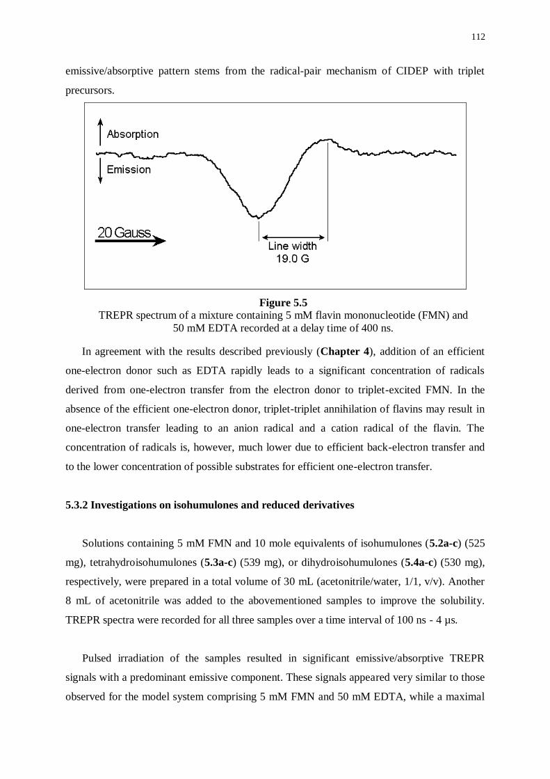
112
emissive/absorptive pattern stems from the radical-pair mechanism of CIDEP with triplet
precursors.
Figure 5.5
TREPR spectrum of a mixture containing 5 mM flavin mononucleotide (FMN) and
50 mM EDTA recorded at a delay time of 400 ns.
In agreement with the results described previously (Chapter 4), addition of an efficient
one-electron donor such as EDTA rapidly leads to a significant concentration of radicals
derived from one-electron transfer from the electron donor to triplet-excited FMN. In the
absence of the efficient one-electron donor, triplet-triplet annihilation of flavins may result in
one-electron transfer leading to an anion radical and a cation radical of the flavin. The
concentration of radicals is, however, much lower due to efficient back-electron transfer and
to the lower concentration of possible substrates for efficient one-electron transfer.
5.3.2 Investigations on isohumulones and reduced derivatives
Solutions containing 5 mM FMN and 10 mole equivalents of isohumulones (5.2a-c) (525
mg), tetrahydroisohumulones (5.3a-c) (539 mg), or dihydroisohumulones (5.4a-c) (530 mg),
respectively, were prepared in a total volume of 30 mL (acetonitrile/water, 1/1, v/v). Another
8 mL of acetonitrile was added to the abovementioned samples to improve the solubility.
TREPR spectra were recorded for all three samples over a time interval of 100 ns - 4 µs.
Pulsed irradiation of the samples resulted in significant emissive/absorptive TREPR
signals with a predominant emissive component. These signals appeared very similar to those
observed for the model system comprising 5 mM FMN and 50 mM EDTA, while a maximal
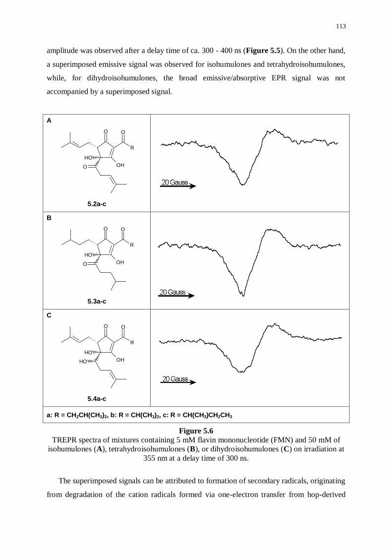
113
amplitude was observed after a delay time of ca. 300 - 400 ns (Figure 5.5). On the other hand,
a superimposed emissive signal was observed for isohumulones and tetrahydroisohumulones,
while, for dihydroisohumulones, the broad emissive/absorptive EPR signal was not
accompanied by a superimposed signal.
A
O
R
O
OHO
OH
5.2a-c
B
O
R
O
OHO
OH
5.3a-c
C
O
R
O
OHOH
OH
5.4a-c
a: R = CH2CH(CH3)2, b: R = CH(CH3)2, c: R = CH(CH3)CH2CH3
Figure 5.6
TREPR spectra of mixtures containing 5 mM flavin mononucleotide (FMN) and 50 mM of
isohumulones (A), tetrahydroisohumulones (B), or dihydroisohumulones (C) on irradiation at
355 nm at a delay time of 300 ns.
The superimposed signals can be attributed to formation of secondary radicals, originating
from degradation of the cation radicals formed via one-electron transfer from hop-derived
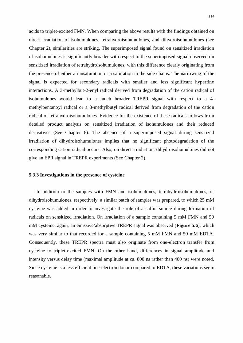
114
acids to triplet-excited FMN. When comparing the above results with the findings obtained on
direct irradiation of isohumulones, tetrahydroisohumulones, and dihydroisohumulones (see
Chapter 2), similarities are striking. The superimposed signal found on sensitized irradiation
of isohumulones is significantly broader with respect to the superimposed signal observed on
sensitized irradiation of tetrahydroisohumulones, with this difference clearly originating from
the presence of either an insaturation or a saturation in the side chains. The narrowing of the
signal is expected for secondary radicals with smaller and less significant hyperfine
interactions. A 3-methylbut-2-enyl radical derived from degradation of the cation radical of
isohumulones would lead to a much broader TREPR signal with respect to a 4-
methylpentanoyl radical or a 3-methylbutyl radical derived from degradation of the cation
radical of tetrahydroisohumulones. Evidence for the existence of these radicals follows from
detailed product analysis on sensitized irradiation of isohumulones and their reduced
derivatives (See Chapter 6). The absence of a superimposed signal during sensitized
irradiation of dihydroisohumulones implies that no significant photodegradation of the
corresponding cation radical occurs. Also, on direct irradiation, dihydroisohumulones did not
give an EPR signal in TREPR experiments (See Chapter 2).
5.3.3 Investigations in the presence of cysteine
In addition to the samples with FMN and isohumulones, tetrahydroisohumulones, or
dihydroisohumulones, respectively, a similar batch of samples was prepared, to which 25 mM
cysteine was added in order to investigate the role of a sulfur source during formation of
radicals on sensitized irradiation. On irradiation of a sample containing 5 mM FMN and 50
mM cysteine, again, an emissive/absorptive TREPR signal was observed (Figure 5.6), which
was very similar to that recorded for a sample containing 5 mM FMN and 50 mM EDTA.
Consequently, these TREPR spectra must also originate from one-electron transfer from
cysteine to triplet-excited FMN. On the other hand, differences in signal amplitude and
intensity versus delay time (maximal amplitude at ca. 800 ns rather than 400 ns) were noted.
Since cysteine is a less efficient one-electron donor compared to EDTA, these variations seem
reasonable.
115
Figure 5.7
TREPR spectrum of a mixture containing 5 mM flavin mononucleotide (FMN) and 50 mM
cysteine under pulsed irradiation at 355 nm recorded at a delay time of 800 ns.
Irradiation of samples containing 5 mM FMN, 25 mM cysteine, and 50 mM of the
respective hop-derived acids resulted in TREPR spectra, which were very similar to those,
found for the samples without addition of cysteine. These observations indicate that the sulfur
source does not interfere significantly with primary photochemical events.
5.4 EXPERIMENTAL PART
Steady-State Electron Paramagnetic Resonance
Steady-state EPR experiments were carried out on a JEOL, USA, Inc. RE-1X EPR
instrument operating at X-band frequency (9.5 GHz) interfaced with a personal computer for
data acquisition. A cylindrical sample cell (id: 1 mm) was used to accommodate the polar
solvents.
Time-Resolved Electron Paramagnetic Resonance
The experimental set-up for time-resolved electron paramagnetic resonance was already
described in detail in Chapter 2. Some modifications were made for the experiments under
sensitized irradiation conditions. A microwave amplifier (GaAs FET (noise figure 0.9 dB))
was positioned in front of the detector to increase the sensitivity and a wideband preamplifier
(0.5-50 MHz) was arranged at the back of the detector to increase time resolution.
Furthermore, a Double Balanced Mixer (DBM) was substituted for the Magic Tee with
Crystal Microwave detector. The YAG-laser (Continuum Laser Powerlite Series) could
deliver ca. 30 mJ/pulse at 355 nm.
116
5.5 REFERENCES
(1) Kocherginsky, N., Swartz, H.M.; Nitroxide Spin Labels: Reactions and Chemistry.
CRC Press, Boca Raton, New York, 1995.
(2) Palmer, G., Muller, F., Massey, V.; Flavins and Flavoproteins. University Park Press,
Baltimore, 1971, 123-137.
(3) Edmondson, D.E.; Electron-Spin-Resonance Studies on Flavoenzymes. Biochem. Soc.
Trans., 1985, 13, 593-600.
(4) Yue, K.T., Bhattacharyya, A.K., Zhelyaskov, V.R., Edmondson, D.E.; Resonance
Raman-Spectroscopic Evidence for an Anionic Flavin Semiquinone in Bovine Liver.
Arch. Biochem. Biophys., 1993, 300, 178-185.
(5) Rustandi, R.R., Jorns, M.S.; Photoinduced Spin-Polarized Radical Pair Formation in
a DNA Photolyase.Substrate Complex at Low Temperature. Biochemistry, 1995, 34,
2284-2288.
************************

117
CHAPTER 6
PHOTOREACTIVITY OF ISOHUMULONES AND REDUCED
DERIVATIVES UNDER SENSITIZED IRRADIATION CONDITIONS
6.1 INTRODUCTION
Detailed product analyses of mixtures of photoreaction products derived from irradiation
(at 300 nm) of isohumulones, tetrahydroisohumulones, and dihydroisohumulones,
respectively, resulted in profound understanding of the primary photochemistry of these hop-
derived acids (see Chapter 3). However, lightstruck flavor formation has been studied almost
exclusively in model systems and real beer samples on exposure to visible light, with 350-500
nm being the most effective wavelength range. While isohumulones and their reduced
derivatives are transparent at these wavelengths, the intermediacy of a sensitizer is required.
Studies using model systems have shown unequivocally that riboflavin (RF; Vitamin B2) is
able to sensitize the formation of lightstruck flavor in the presence of isohumulones and a
suitable sulfur source. Moreover, several hundreds of ppm’s of RF are present in beer,1 hence
it can safely be accepted that RF is a key element - although possibly not the unique factor -
in the skunking of beer.
All previous investigations related to the formation of lightstruck flavor have invested on
detection and quantification of 3-methylbut-2-ene-1-thiol (MBT),2-5
the major contributor to
the off-flavor, while the fate of RF (as a sensitizer) or of isohumulones (as photosubstrates) on
exposure to visible light has escaped attention. Comprehensive product analyses of mixtures
of photoreaction products, derived from sensitized irradiation of solutions containing RF and
isohumulones, tetrahydroisohumulones, or dihydroisohumulones, respectively, should give
valuable information on the corresponding photoreactivities (in accordance with procedures
described in Chapter 3). Combining the results from such photostationary experiments with
those from photodynamic endeavors should lead to elaboration of relevant reaction schemes.
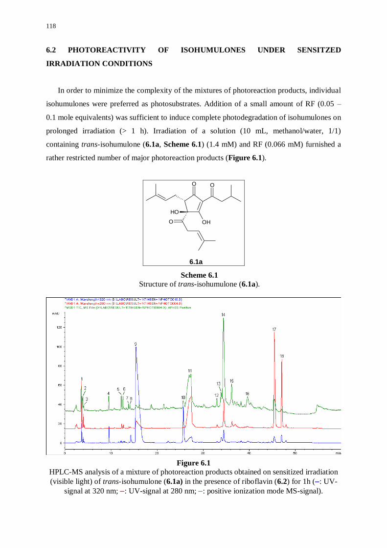
118
6.2 PHOTOREACTIVITY OF ISOHUMULONES UNDER SENSITZED
IRRADIATION CONDITIONS
In order to minimize the complexity of the mixtures of photoreaction products, individual
isohumulones were preferred as photosubstrates. Addition of a small amount of RF (0.05 –
0.1 mole equivalents) was sufficient to induce complete photodegradation of isohumulones on
prolonged irradiation (> 1 h). Irradiation of a solution (10 mL, methanol/water, 1/1)
containing trans-isohumulone (6.1a, Scheme 6.1) (1.4 mM) and RF (0.066 mM) furnished a
rather restricted number of major photoreaction products (Figure 6.1).
O O
OH
O OH
6.1a
Scheme 6.1
Structure of trans-isohumulone (6.1a).
Figure 6.1
HPLC-MS analysis of a mixture of photoreaction products obtained on sensitized irradiation
(visible light) of trans-isohumulone (6.1a) in the presence of riboflavin (6.2) for 1h (: UV-
signal at 320 nm; : UV-signal at 280 nm; : positive ionization mode MS-signal).
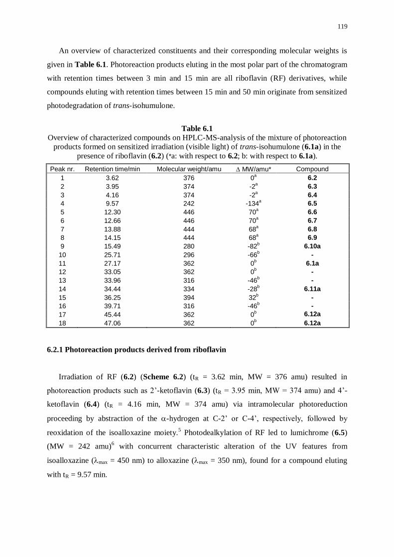
119
An overview of characterized constituents and their corresponding molecular weights is
given in Table 6.1. Photoreaction products eluting in the most polar part of the chromatogram
with retention times between 3 min and 15 min are all riboflavin (RF) derivatives, while
compounds eluting with retention times between 15 min and 50 min originate from sensitized
photodegradation of trans-isohumulone.
Table 6.1
Overview of characterized compounds on HPLC-MS-analysis of the mixture of photoreaction
products formed on sensitized irradiation (visible light) of trans-isohumulone (6.1a) in the
presence of riboflavin (6.2) (*a: with respect to 6.2; b: with respect to 6.1a).
Peak nr. Retention time/min Molecular weight/amu MW/amu* Compound
1 3.62 376 0a 6.2
2 3.95 374 -2a 6.3
3 4.16 374 -2a 6.4
4 9.57 242 -134a 6.5
5 12.30 446 70a 6.6
6 12.66 446 70a 6.7
7 13.88 444 68a 6.8
8 14.15 444 68a 6.9
9 15.49 280 -82b 6.10a
10 25.71 296 -66b -
11 27.17 362 0b 6.1a
12 33.05 362 0b -
13 33.96 316 -46b -
14 34.44 334 -28b 6.11a
15 36.25 394 32b -
16 39.71 316 -46b -
17 45.44 362 0b 6.12a
18 47.06 362 0b 6.12a
6.2.1 Photoreaction products derived from riboflavin
Irradiation of RF (6.2) (Scheme 6.2) (tR = 3.62 min, MW = 376 amu) resulted in
photoreaction products such as 2’-ketoflavin (6.3) (tR = 3.95 min, MW = 374 amu) and 4’-
ketoflavin (6.4) (tR = 4.16 min, MW = 374 amu) via intramolecular photoreduction
proceeding by abstraction of the -hydrogen at C-2’ or C-4’, respectively, followed by
reoxidation of the isoalloxazine moiety.5 Photodealkylation of RF led to lumichrome (6.5)
(MW = 242 amu)6 with concurrent characteristic alteration of the UV features from
isoalloxazine (max = 450 nm) to alloxazine (max = 350 nm), found for a compound eluting
with tR = 9.57 min.
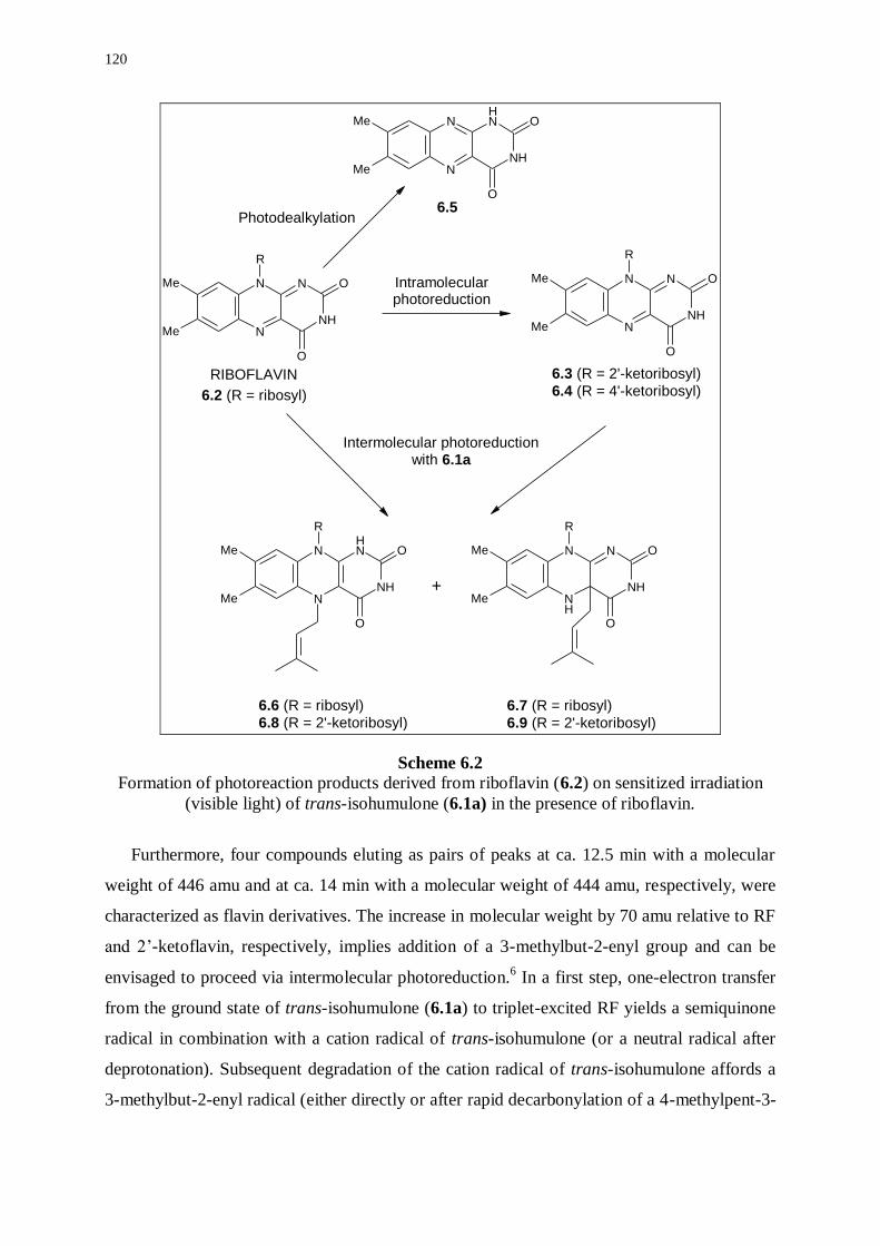
120
NH
N
N
NMe
Me
O
O
R
NH
NH
N
NMe
Me
O
O
NH
N
N
NMe
Me
O
O
R
NH
NH
N
NMe
Me
O
O
R
NH
N
NH
NMe
Me
O
O
R
6.2 (R = ribosyl)
6.3 (R = 2'-ketoribosyl)
6.4 (R = 4'-ketoribosyl)
6.5
Intramolecularphotoreduction
Photodealkylation
Intermolecular photoreductionwith 6.1a
6.6 (R = ribosyl)
6.8 (R = 2'-ketoribosyl)
6.7 (R = ribosyl)
6.9 (R = 2'-ketoribosyl)
+
RIBOFLAVIN
Scheme 6.2 Formation of photoreaction products derived from riboflavin (6.2) on sensitized irradiation
(visible light) of trans-isohumulone (6.1a) in the presence of riboflavin.
Furthermore, four compounds eluting as pairs of peaks at ca. 12.5 min with a molecular
weight of 446 amu and at ca. 14 min with a molecular weight of 444 amu, respectively, were
characterized as flavin derivatives. The increase in molecular weight by 70 amu relative to RF
and 2’-ketoflavin, respectively, implies addition of a 3-methylbut-2-enyl group and can be
envisaged to proceed via intermolecular photoreduction.6 In a first step, one-electron transfer
from the ground state of trans-isohumulone (6.1a) to triplet-excited RF yields a semiquinone
radical in combination with a cation radical of trans-isohumulone (or a neutral radical after
deprotonation). Subsequent degradation of the cation radical of trans-isohumulone affords a
3-methylbut-2-enyl radical (either directly or after rapid decarbonylation of a 4-methylpent-3-
121
enoyl radical) via formal -cleavage of the side chain at C-4 (see 6.2.2). Finally,
recombination of the 3-methylbut-2-enyl radical with a semiquinone radical either from
riboflavin or 2’-ketoflavin gives the observed addition products 6.6/6.7 and 6.8/6.9,
respectively. The pairs of isomeric compounds observed in our experiments are most likely
due to regiodifferentiated addition of a 3-methylbut-2-enyl radical at N-5 or C-4a. It has been
reported that, depending on substrate, temperature, solvent, and pH, C-4a-, N-5-, or C-8
adducts may be formed on intermolecular photoreduction of flavins.6,7
6.2.2 Photoreaction products derived from trans-isohumulone
A bathochromic shift of ca. 50 nm with respect to the UV spectrum of trans-isohumulone
(max = 280 nm) was observed for a major photoreaction product eluting with tR = 15.49 min
(max = 330 nm), suggesting extended conjugation with respect to trans-isohumulone (6.1a).
The molecular weight of 280 amu indicates formal loss of the 4-methylpent-3-enoyl side
chain at C-4 resulting in cyclopent-2-ene-1,4-dione derivative 6.10a. This photoreaction
product was also formed under direct irradiation conditions, albeit in very low yield (see
3.3.5).
Moreover, a major photoreaction product eluting with tR = 34.44 min was found to be
identical with a minor photoreaction product derived from direct irradiation of trans-
isohumulone (6.1) (see 3.3.1). The molecular weight of 334 amu is consistent with compound
6.11a (Scheme 6.3), formed by formal loss of CO (28 amu with respect to 6.1a). On
ionization in the negative MS-mode, two complementary daughter ions were observed at 125
amu [C7H9O2]- and 207 amu [C13H19O2]
-, respectively. These ions are indicative of
fragmentation of the molecular ion [M-H]- (formed on deprotonation of 6.11a) to a fragment
ion containing the saturated side chain [D1-H]- and a fragment ion containing both unsaturated
side chains [D2-H]- (Figure 6.2).
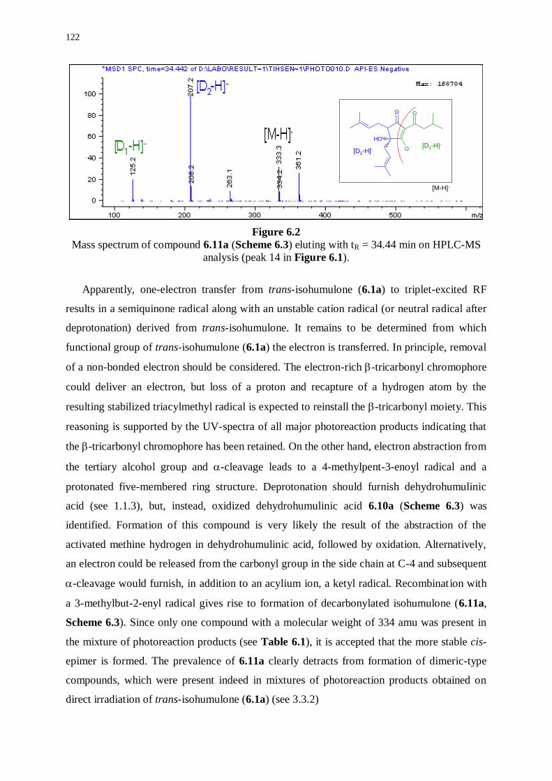
122
Figure 6.2
Mass spectrum of compound 6.11a (Scheme 6.3) eluting with tR = 34.44 min on HPLC-MS
analysis (peak 14 in Figure 6.1).
Apparently, one-electron transfer from trans-isohumulone (6.1a) to triplet-excited RF
results in a semiquinone radical along with an unstable cation radical (or neutral radical after
deprotonation) derived from trans-isohumulone. It remains to be determined from which
functional group of trans-isohumulone (6.1a) the electron is transferred. In principle, removal
of a non-bonded electron should be considered. The electron-rich -tricarbonyl chromophore
could deliver an electron, but loss of a proton and recapture of a hydrogen atom by the
resulting stabilized triacylmethyl radical is expected to reinstall the -tricarbonyl moiety. This
reasoning is supported by the UV-spectra of all major photoreaction products indicating that
the -tricarbonyl chromophore has been retained. On the other hand, electron abstraction from
the tertiary alcohol group and -cleavage leads to a 4-methylpent-3-enoyl radical and a
protonated five-membered ring structure. Deprotonation should furnish dehydrohumulinic
acid (see 1.1.3), but, instead, oxidized dehydrohumulinic acid 6.10a (Scheme 6.3) was
identified. Formation of this compound is very likely the result of the abstraction of the
activated methine hydrogen in dehydrohumulinic acid, followed by oxidation. Alternatively,
an electron could be released from the carbonyl group in the side chain at C-4 and subsequent
-cleavage would furnish, in addition to an acylium ion, a ketyl radical. Recombination with
a 3-methylbut-2-enyl radical gives rise to formation of decarbonylated isohumulone (6.11a,
Scheme 6.3). Since only one compound with a molecular weight of 334 amu was present in
the mixture of photoreaction products (see Table 6.1), it is accepted that the more stable cis-
epimer is formed. The prevalence of 6.11a clearly detracts from formation of dimeric-type
compounds, which were present indeed in mixtures of photoreaction products obtained on
direct irradiation of trans-isohumulone (6.1a) (see 3.3.2)
O O
O
OH
[M-H]-
[D1-H]-[D2-H]-
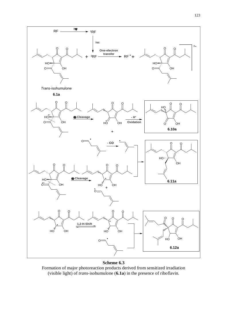
123
O O
OHO
OH
O O
OHO
OH
O O
OHO
OH
O O
OHHO
.
O O
OHOH
O
O.
O O
OHOH
O O
OHOH
O.
O O
OHO
OH
6.10a
O O
OHO
OH
O O
OH
OH
O O
OHOH
O
RF 1RF
3RF+ RF-.+
+.
h
isc
One-electron
transfer
6.1a
- CO
Trans-isohumulone
+.
+.
+
- H+
Oxidation
-Cleavage
+
.
-Cleavage
+ +
.
1,2-H-Shift.
6.11a
6.12a
Scheme 6.3
Formation of major photoreaction products derived from sensitized irradiation
(visible light) of trans-isohumulone (6.1a) in the presence of riboflavin.
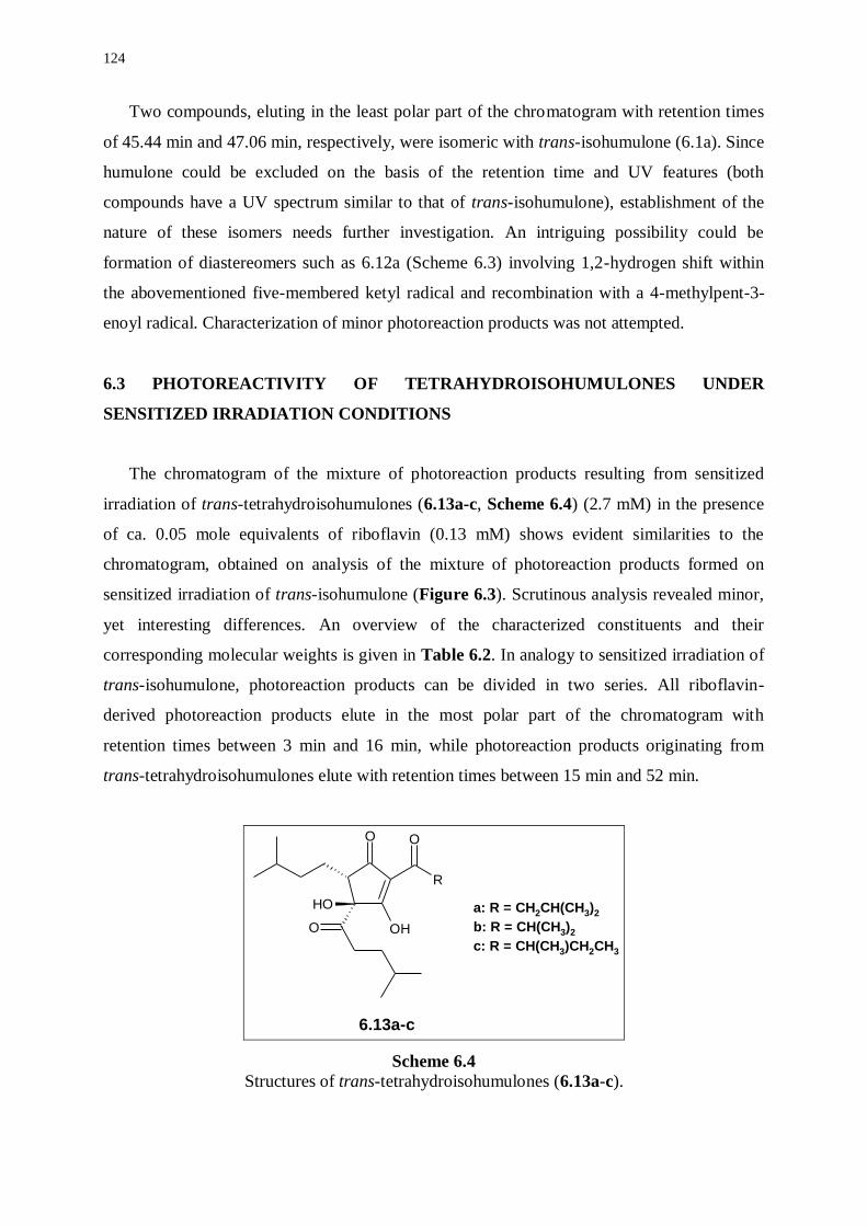
124
Two compounds, eluting in the least polar part of the chromatogram with retention times
of 45.44 min and 47.06 min, respectively, were isomeric with trans-isohumulone (6.1a). Since
humulone could be excluded on the basis of the retention time and UV features (both
compounds have a UV spectrum similar to that of trans-isohumulone), establishment of the
nature of these isomers needs further investigation. An intriguing possibility could be
formation of diastereomers such as 6.12a (Scheme 6.3) involving 1,2-hydrogen shift within
the abovementioned five-membered ketyl radical and recombination with a 4-methylpent-3-
enoyl radical. Characterization of minor photoreaction products was not attempted.
6.3 PHOTOREACTIVITY OF TETRAHYDROISOHUMULONES UNDER
SENSITIZED IRRADIATION CONDITIONS
The chromatogram of the mixture of photoreaction products resulting from sensitized
irradiation of trans-tetrahydroisohumulones (6.13a-c, Scheme 6.4) (2.7 mM) in the presence
of ca. 0.05 mole equivalents of riboflavin (0.13 mM) shows evident similarities to the
chromatogram, obtained on analysis of the mixture of photoreaction products formed on
sensitized irradiation of trans-isohumulone (Figure 6.3). Scrutinous analysis revealed minor,
yet interesting differences. An overview of the characterized constituents and their
corresponding molecular weights is given in Table 6.2. In analogy to sensitized irradiation of
trans-isohumulone, photoreaction products can be divided in two series. All riboflavin-
derived photoreaction products elute in the most polar part of the chromatogram with
retention times between 3 min and 16 min, while photoreaction products originating from
trans-tetrahydroisohumulones elute with retention times between 15 min and 52 min.
O
R
O
OH
O OH
6.13a-c
a: R = CH2CH(CH3)2
b: R = CH(CH3)2
c: R = CH(CH3)CH2CH3
Scheme 6.4
Structures of trans-tetrahydroisohumulones (6.13a-c).
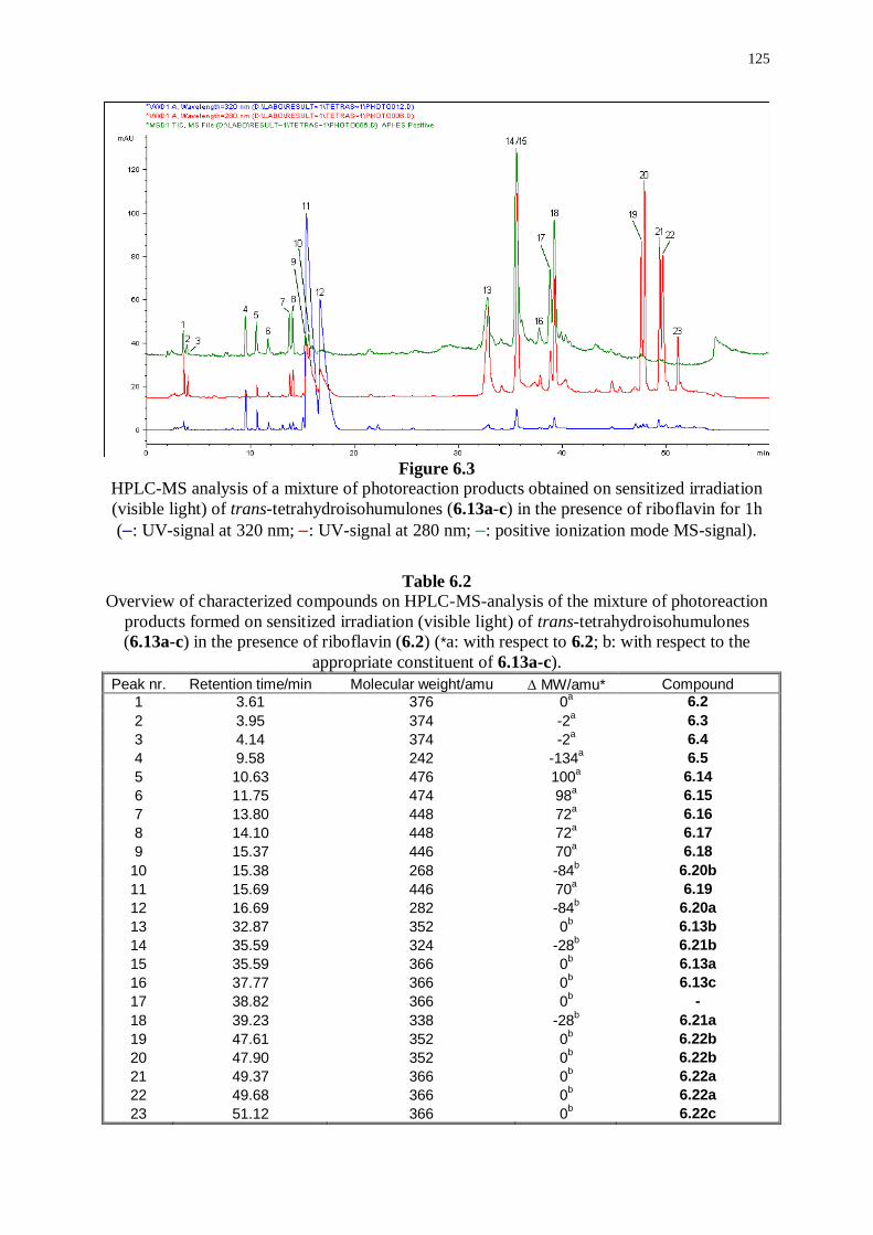
125
Figure 6.3
HPLC-MS analysis of a mixture of photoreaction products obtained on sensitized irradiation
(visible light) of trans-tetrahydroisohumulones (6.13a-c) in the presence of riboflavin for 1h
(: UV-signal at 320 nm; : UV-signal at 280 nm; : positive ionization mode MS-signal).
Table 6.2 Overview of characterized compounds on HPLC-MS-analysis of the mixture of photoreaction
products formed on sensitized irradiation (visible light) of trans-tetrahydroisohumulones
(6.13a-c) in the presence of riboflavin (6.2) (*a: with respect to 6.2; b: with respect to the
appropriate constituent of 6.13a-c).
Peak nr. Retention time/min Molecular weight/amu MW/amu* Compound
1 3.61 376 0a 6.2
2 3.95 374 -2a 6.3
3 4.14 374 -2a 6.4
4 9.58 242 -134a 6.5
5 10.63 476 100a 6.14
6 11.75 474 98a 6.15
7 13.80 448 72a 6.16
8 14.10 448 72a 6.17
9 15.37 446 70a 6.18
10 15.38 268 -84b 6.20b
11 15.69 446 70a 6.19
12 16.69 282 -84b 6.20a
13 32.87 352 0b 6.13b
14 35.59 324 -28b 6.21b
15 35.59 366 0b 6.13a
16 37.77 366 0b 6.13c
17 38.82 366 0b -
18 39.23 338 -28b 6.21a
19 47.61 352 0b 6.22b
20 47.90 352 0b 6.22b
21 49.37 366 0b 6.22a
22 49.68 366 0b 6.22a
23 51.12 366 0b 6.22c
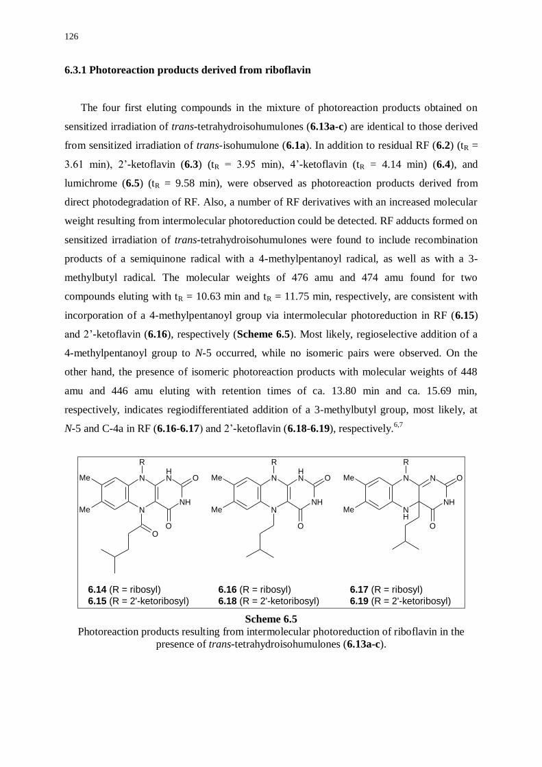
126
6.3.1 Photoreaction products derived from riboflavin
The four first eluting compounds in the mixture of photoreaction products obtained on
sensitized irradiation of trans-tetrahydroisohumulones (6.13a-c) are identical to those derived
from sensitized irradiation of trans-isohumulone (6.1a). In addition to residual RF (6.2) (tR =
3.61 min), 2’-ketoflavin (6.3) (tR = 3.95 min), 4’-ketoflavin (tR = 4.14 min) (6.4), and
lumichrome (6.5) (tR = 9.58 min), were observed as photoreaction products derived from
direct photodegradation of RF. Also, a number of RF derivatives with an increased molecular
weight resulting from intermolecular photoreduction could be detected. RF adducts formed on
sensitized irradiation of trans-tetrahydroisohumulones were found to include recombination
products of a semiquinone radical with a 4-methylpentanoyl radical, as well as with a 3-
methylbutyl radical. The molecular weights of 476 amu and 474 amu found for two
compounds eluting with tR = 10.63 min and tR = 11.75 min, respectively, are consistent with
incorporation of a 4-methylpentanoyl group via intermolecular photoreduction in RF (6.15)
and 2’-ketoflavin (6.16), respectively (Scheme 6.5). Most likely, regioselective addition of a
4-methylpentanoyl group to N-5 occurred, while no isomeric pairs were observed. On the
other hand, the presence of isomeric photoreaction products with molecular weights of 448
amu and 446 amu eluting with retention times of ca. 13.80 min and ca. 15.69 min,
respectively, indicates regiodifferentiated addition of a 3-methylbutyl group, most likely, at
N-5 and C-4a in RF (6.16-6.17) and 2’-ketoflavin (6.18-6.19), respectively.6,7
NH
NH
N
NMe
Me
O
O
R
NH
N
NH
NMe
Me
O
O
R
NH
NH
N
NMe
Me
O
O
R
O
6.14 (R = ribosyl)
6.15 (R = 2'-ketoribosyl)
6.16 (R = ribosyl)
6.18 (R = 2'-ketoribosyl)
6.17 (R = ribosyl)
6.19 (R = 2'-ketoribosyl)
Scheme 6.5
Photoreaction products resulting from intermolecular photoreduction of riboflavin in the
presence of trans-tetrahydroisohumulones (6.13a-c).
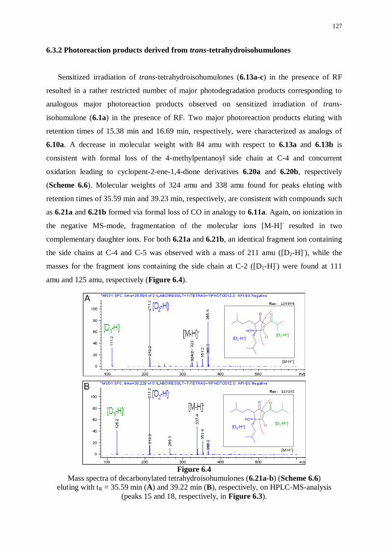
127
6.3.2 Photoreaction products derived from trans-tetrahydroisohumulones
Sensitized irradiation of trans-tetrahydroisohumulones (6.13a-c) in the presence of RF
resulted in a rather restricted number of major photodegradation products corresponding to
analogous major photoreaction products observed on sensitized irradiation of trans-
isohumulone (6.1a) in the presence of RF. Two major photoreaction products eluting with
retention times of 15.38 min and 16.69 min, respectively, were characterized as analogs of
6.10a. A decrease in molecular weight with 84 amu with respect to 6.13a and 6.13b is
consistent with formal loss of the 4-methylpentanoyl side chain at C-4 and concurrent
oxidation leading to cyclopent-2-ene-1,4-dione derivatives 6.20a and 6.20b, respectively
(Scheme 6.6). Molecular weights of 324 amu and 338 amu found for peaks eluting with
retention times of 35.59 min and 39.23 min, respectively, are consistent with compounds such
as 6.21a and 6.21b formed via formal loss of CO in analogy to 6.11a. Again, on ionization in
the negative MS-mode, fragmentation of the molecular ions [M-H]- resulted in two
complementary daughter ions. For both 6.21a and 6.21b, an identical fragment ion containing
the side chains at C-4 and C-5 was observed with a mass of 211 amu ([D2-H]-), while the
masses for the fragment ions containing the side chain at C-2 ([D1-H]-) were found at 111
amu and 125 amu, respectively (Figure 6.4).
Figure 6.4
Mass spectra of decarbonylated tetrahydroisohumulones (6.21a-b) (Scheme 6.6)
eluting with tR = 35.59 min (A) and 39.22 min (B), respectively, on HPLC-MS-analysis
(peaks 15 and 18, respectively, in Figure 6.3).
O O
O
OH
[M-H+]-
[D1-H+]-
[D2-H+]-
O O
O
OH
[M-H+]-
[D1-H+]-
[D2-H+]-
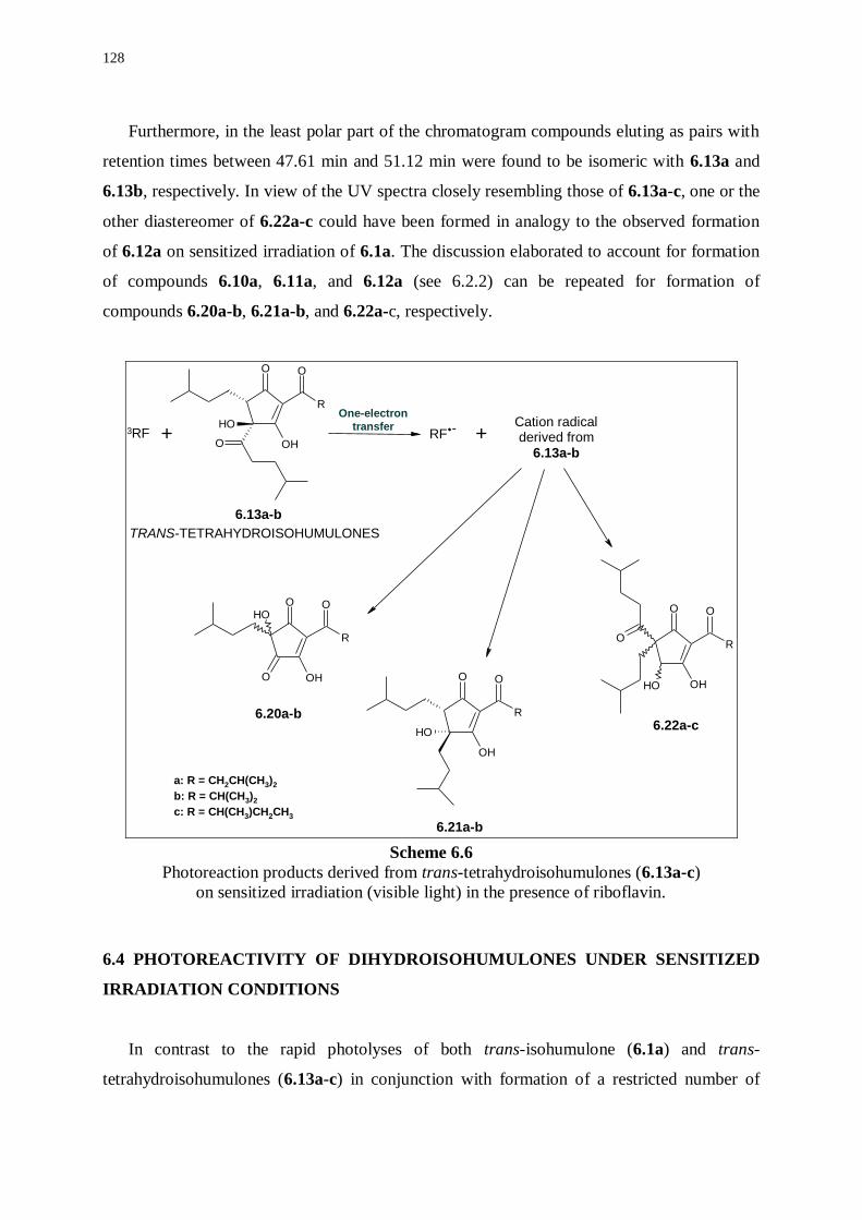
128
Furthermore, in the least polar part of the chromatogram compounds eluting as pairs with
retention times between 47.61 min and 51.12 min were found to be isomeric with 6.13a and
6.13b, respectively. In view of the UV spectra closely resembling those of 6.13a-c, one or the
other diastereomer of 6.22a-c could have been formed in analogy to the observed formation
of 6.12a on sensitized irradiation of 6.1a. The discussion elaborated to account for formation
of compounds 6.10a, 6.11a, and 6.12a (see 6.2.2) can be repeated for formation of
compounds 6.20a-b, 6.21a-b, and 6.22a-c, respectively.
O
R
O
OHO
OH
O
R
O
OH
OH
O O
R
O
OH
OH
O
R
O
OH
O
OH
+ +Cation radicalderived from
6.13a-b
3RF RF.-
6.13a-b
6.20a-b
6.21a-b
6.22a-c
One-electron
transfer
TRANS-TETRAHYDROISOHUMULONES
a: R = CH2CH(CH3)2
b: R = CH(CH3)2
c: R = CH(CH3)CH2CH3
Scheme 6.6
Photoreaction products derived from trans-tetrahydroisohumulones (6.13a-c)
on sensitized irradiation (visible light) in the presence of riboflavin.
6.4 PHOTOREACTIVITY OF DIHYDROISOHUMULONES UNDER SENSITIZED
IRRADIATION CONDITIONS
In contrast to the rapid photolyses of both trans-isohumulone (6.1a) and trans-
tetrahydroisohumulones (6.13a-c) in conjunction with formation of a restricted number of
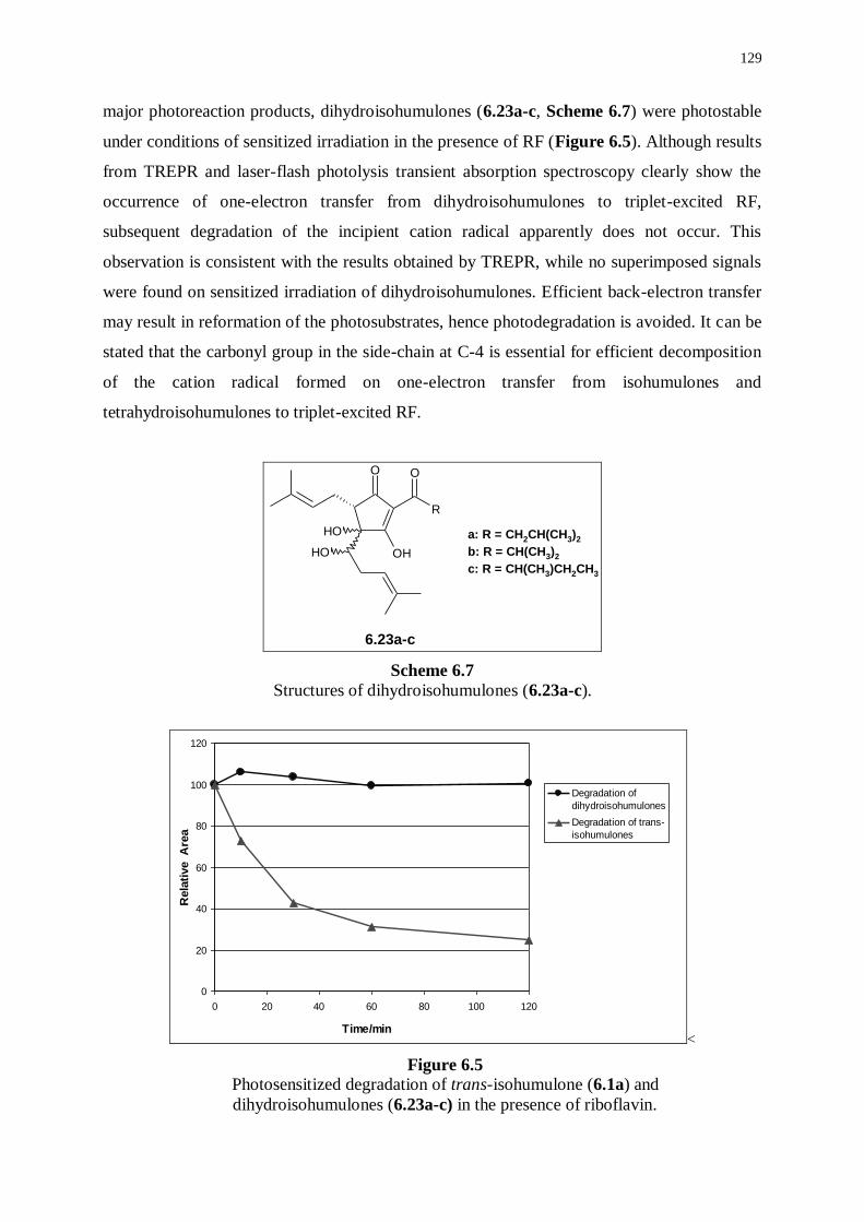
129
major photoreaction products, dihydroisohumulones (6.23a-c, Scheme 6.7) were photostable
under conditions of sensitized irradiation in the presence of RF (Figure 6.5). Although results
from TREPR and laser-flash photolysis transient absorption spectroscopy clearly show the
occurrence of one-electron transfer from dihydroisohumulones to triplet-excited RF,
subsequent degradation of the incipient cation radical apparently does not occur. This
observation is consistent with the results obtained by TREPR, while no superimposed signals
were found on sensitized irradiation of dihydroisohumulones. Efficient back-electron transfer
may result in reformation of the photosubstrates, hence photodegradation is avoided. It can be
stated that the carbonyl group in the side-chain at C-4 is essential for efficient decomposition
of the cation radical formed on one-electron transfer from isohumulones and
tetrahydroisohumulones to triplet-excited RF.
O
R
O
OH
OH OH
6.23a-c
a: R = CH2CH(CH3)2
b: R = CH(CH3)2
c: R = CH(CH3)CH2CH3
Scheme 6.7
Structures of dihydroisohumulones (6.23a-c).
0
20
40
60
80
100
120
0 20 40 60 80 100 120
Time/min
Rela
tive A
rea
Degradation of
dihydroisohumulones
Degradation of trans-
isohumulones
<
Figure 6.5 Photosensitized degradation of trans-isohumulone (6.1a) and
dihydroisohumulones (6.23a-c) in the presence of riboflavin.
130
6.5 EXPERIMENTAL PART
Irradiation conditions
Trans-isohumulone (1.4 mM), a mixture of trans-tetrahydroisohumulones (2.7 mM), or
dihydroisohumulones (2.7 mM) were irradiated in a deaerated mixture of methanol/water
(1/1, v/v, 10 mL) containing 0.05 mole equivalents of riboflavin with visible light
(photoreactor containing 4 Philips Cool White Lamps of 8 W) at varying exposure times (up
to 8 h). Deaeration of the samples was performed by flushing with solvent saturated N2 for ca.
20 min.
Remark: A detailed description of sample preparation, chromatographic conditions, and mass
spectral settings can be found in the experimental part of Chapter 3 (see 3.6).
131
6.6 REFERENCES
(1) Andres-Lacueva, C., Mattivi, F., Tonon, D.; Determination of Riboflavin, Flavin
Mononucleotide and Flavin Adenine Dinucleotide in Wine and Other Beverages by High-
Performance Liquid Chromatography with Fluorescence Detection. J. Chromatogr. A, 1998,
823, 355-363.
(2) Gunst, F., Verzele, M.; On the Sunstruck Flavor of Beer. J. Inst. Brew., 1978, 84, 291-292.
(3) Sakuma, S., Rikimaru, Y., Kobayashi, K., Kowaka, M.; Sunstruck Flavor Formation in Beer.
J. Am. Soc. Brew. Chem., 1991, 49, 162-165.
(4) Goldstein, H., Rader, S., Murakami, A.A.; Determination of 3-Methyl-2-Butene-1-Thiol in
Beer. J. Am. Soc. Brew. Chem., 1993, 51, 70-74.
(5) Masuda, S., Kikuchi, K., Harayama, K.; Determination of Lightstruck Character in Beer by
Gas Chromatography-Mass Spectroscopy. J. Am. Soc. Brew. Chem., 2000, 58, 152-154.
(6) Cairns, W.L., Metzler, D.E.; Photochemical Degradation of Flavins. VI. A New Photoproduct
and Its Use in Studying the Photolytic Mechanism. J. Am. Chem. Soc., 1971, 93, 2772-2777.
(7) Heelis, P.F.; The Photophysical and Photochemical Properties of Flavins (Isoalloxazines).
Chem. Soc. Rev., 1982, 11, 15-39.
(8) Knappe, W.R., Hemmerich, P.; Reductive Photoalkylation of Flavin Nucleus – Structure and
Reactivity of Photoproducts. Liebigs Ann. Chem., 1976, 11, 2037-2057.
***********************

133
ANNEX
In a final chapter, a fully innovative method is presented to inhibit formation
of the lightstruck flavor in beer based on the results of this Ph.D. project. The
research contract d.d. 1st of March 1997 between Cobrew N.V., Leuven, and
Ghent University stipulates (page 4, item 7 “Proprietary rights”; page 5, item
7a “Confidential information”) that any and all inventions with potential
industrial applications must not be made available to the public before a
patent is filed. As patent application is in preparation, no results can be
disclosed at this time. However, if any member of the reading and examination
commissions should wish to consult the chapter at his discretion, he may do so
by personal inspection with the author and the promoter after signing a
confidentiality agreement.
Ghent, 14 May 2001
Arne Heyerick Prof. Dr. D. De Keukeleire

135
Summary
An objectionable off-flavor is produced on exposure of beer to light. Current
understanding regarding formation of the lightstruck flavor (LSF) in beer dates from the
original reports by Kuroiwa et al. in the early sixties. Using model systems, they showed that
LSF was produced in a light-induced reaction involving riboflavin as a sensitizer (riboflavin
is the main flavin in beer), isohumulones (hop-derived, beer-bittering compounds), and a
sulfur source (e.g. cysteine or sulfur-rich proteins). The off-flavor was attributed to formation
of 3-methylbut-2-ene-1-thiol (MBT). This mechanism has been frequently referred to in
articles dealing with light-induced off-flavors in beer and all relevant data collected to date do
not contradict the Kuroiwa-premise. Although the key components are known for more than
three decades, details on the mechanism of interaction between these components are lacking.
This Ph.D. project aimed at obtaining a thorough and detailed understanding of the
mechanism of formation of LSF in beer. The issue was approached by an in-depth
investigation of the interacting components on a molecular level using model systems,
consisting of isohumulones, dihydroisohumulones, or tetrahydroisohumulones in the presence
or absence of riboflavin or flavin mononucleotide as a sensitizer.
Unraveling a photochemical mechanism implies studies in various time domains. Thus,
model systems were subjected to both photostationary irradiation and photodynamic flash-
initiated experiments. Information on the nature of intervening photo-excited states and
intermediates, along with mechanistic features, were retrieved from time-resolved electron
paramagnetic resonance (TREPR) and flash photolysis transient absorption spectroscopy
(TAS). On characterization and identification of prevalent photoreaction products by
comprehensive product analysis, relevant reaction schemes were elaborated to account for
formation of key compounds.
In a first section, a detailed study of the photolability of isohumulones,
dihydroisohumulones, and tetrahydroisohumulones, is described. On laser-flash photolysis at
308 nm, isohumulones and tetrahydroisohumulones yielded strong emissive TREPR signals
(originating from the triplet mechanism of chemically induced dynamic electron polarization),
while, under identical conditions, no TREPR signal was observed for dihydroisohumulones.
This finding provides the first direct spectroscopic evidence for the resistance of
dihydroisohumulones to photolysis and confirms conclusively that radicals produced on

136
photolysis of isohumulones and tetrahydroisohumulones originate from the -hydroxyketone
chromophore as present in isohumulones and tetrahydroisohumulones.
Using well-substantiated simulations, the most prevalent radicals on photolysis of
isohumulones were characterized as a five-membered ring ketyl radical in conjunction with a
3-methylbut-2-enyl radical, while photolysis of tetrahydroisohumulones led to production of a
similar five-membered ring ketyl radical and a 4-methylpentanoyl radical. Combined TREPR
data allowed to propose a mechanism for photodegradation of isohumulones and
tetrahydroisohumulones on direct irradiation. Absorption of UV light by the -tricarbonyl
chromophore leads to the excited singlet state. After intersystem crossing to the excited triplet
state, intramolecular triplet energy transfer occurs to the isolated -hydroxyketone, which in
the excited state ultimately undergoes Norrish Type I -cleavage, thereby furnishing the
radicals as detailed above. Comprehensive product analyses of complex mixtures of
photoreaction products derived from isohumulones and tetrahydroisohumulones showed that
recombination of the most prevalent radicals as characterized by TREPR accounts for
formation of the majority of photoreaction products, while minor photoreaction products were
found to originate from other pathways. The main reaction products on direct irradiation of
isohumulones were characterized as decarbonylated dehydrated isohumulones, while the
predominant reaction products on direct irradiation of tetrahydroisohumulones were identified
as dimeric-type compounds formed on recombination of two five-membered ring ketyl
radicals.
In a second section, the interaction between isohumulones, dihydroisohumulones, and
tetrahydroisohumulones with riboflavin or flavin mononucleotide on sensitized irradiation
was investigated in detail. Laser-flash photolysis at 355 nm of mixtures containing either
isohumulones, dihydroisohumulones or tetrahydroisohumulones, and riboflavin resulted in
transient absorption spectra (350-800 nm) dominated by the absorption of a neutral flavin
semiquinone radical, while the absorption of triplet-excited riboflavin decreased very rapidly.
These observations are consistent with a one-electron transfer from the ground state of either
isohumulones, dihydroisohumulones or tetrahydroisohumulones to the excited triplet state of
riboflavin. The feasibility of one-electron transfer derives from consideration of the redox
potentials of the interacting species. The redox potential of riboflavin shifts from -0.3 V vs.
NHE in the ground state to +1.7 V vs. NHE in the excited triplet state. Furthermore, TREPR
signals on laser-flash photolysis of mixtures containing isohumulones or
tetrahydroisohumulones, in addition to flavin mononucleotide, showed a superimposed signal

137
with respect to the TREPR signal derived from laser-flash photolysis of a mixture containing
dihydroisohumulones and flavin mononucleotide, or of a model system consisting of EDTA
and flavin mononucleotide. The superimposed signal is derived from further degradation of
the cation radicals resulting from one-electron transfer of isohumulones or
tetrahydroisohumulones to triplet-excited flavin mononucleotide.
Although dihydroisohumulones underwent one-electron transfer in the presence of a
triplet-excited flavin, no photolysis of dihydroisohumulones was apparent, even at prolonged
exposure times, suggesting occurrence of back-electron transfer. Important photoreaction
products found on sensitized irradiation of isohumulones include riboflavin adducts
containing a 3-methylbut-2-enyl side chain, oxidized five-membered ring fragments, and
decarbonylated isohumulones. The distribution of photoreaction products for
tetrahydroisohumulones is very similar, except for riboflavin adducts which were found to
include addition of both 3-methylbutyl and 4-methylpentanoyl side chains. The identified
photoreaction products are consistent with a reaction mechanism involving one-electron
transfer from isohumulones or tetrahydroisohumulones in the ground state to triplet-excited
riboflavin. Further degradation of the resulting cation radical leads to a 3-methylbut-2-enyl
radical in conjunction with a five-membered ring fragment for isohumulones, or to a 3-
methylbutyl or a 4-methylpentanoyl radical and a five-membered ring fragment for
tetrahydroisohumulones.
A fully innovative method to inhibit formation of the lightstruck flavor in beer results from
this Ph.D. project. Filing of a patent is in preparation.
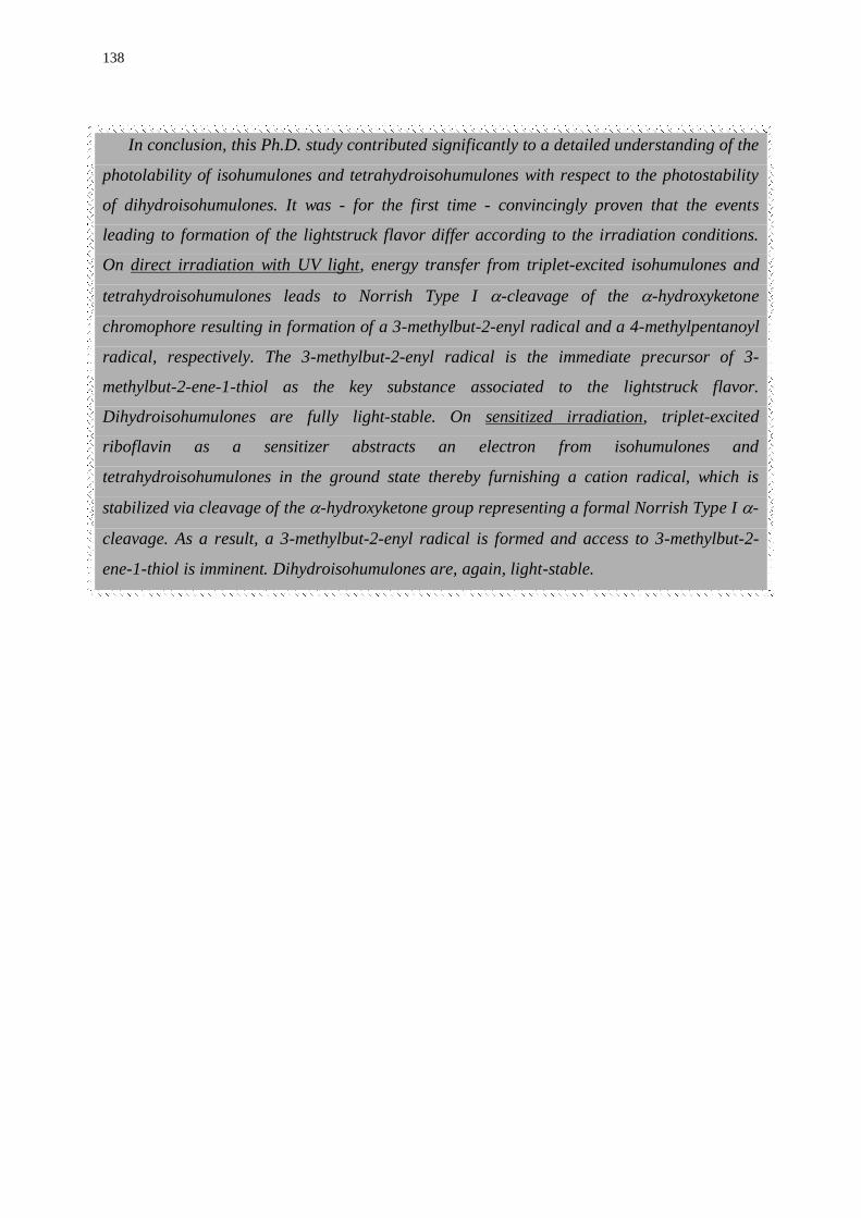
138
In conclusion, this Ph.D. study contributed significantly to a detailed understanding of the
photolability of isohumulones and tetrahydroisohumulones with respect to the photostability
of dihydroisohumulones. It was - for the first time - convincingly proven that the events
leading to formation of the lightstruck flavor differ according to the irradiation conditions.
On direct irradiation with UV light, energy transfer from triplet-excited isohumulones and
tetrahydroisohumulones leads to Norrish Type I -cleavage of the -hydroxyketone
chromophore resulting in formation of a 3-methylbut-2-enyl radical and a 4-methylpentanoyl
radical, respectively. The 3-methylbut-2-enyl radical is the immediate precursor of 3-
methylbut-2-ene-1-thiol as the key substance associated to the lightstruck flavor.
Dihydroisohumulones are fully light-stable. On sensitized irradiation, triplet-excited
riboflavin as a sensitizer abstracts an electron from isohumulones and
tetrahydroisohumulones in the ground state thereby furnishing a cation radical, which is
stabilized via cleavage of the -hydroxyketone group representing a formal Norrish Type I -
cleavage. As a result, a 3-methylbut-2-enyl radical is formed and access to 3-methylbut-2-
ene-1-thiol is imminent. Dihydroisohumulones are, again, light-stable.
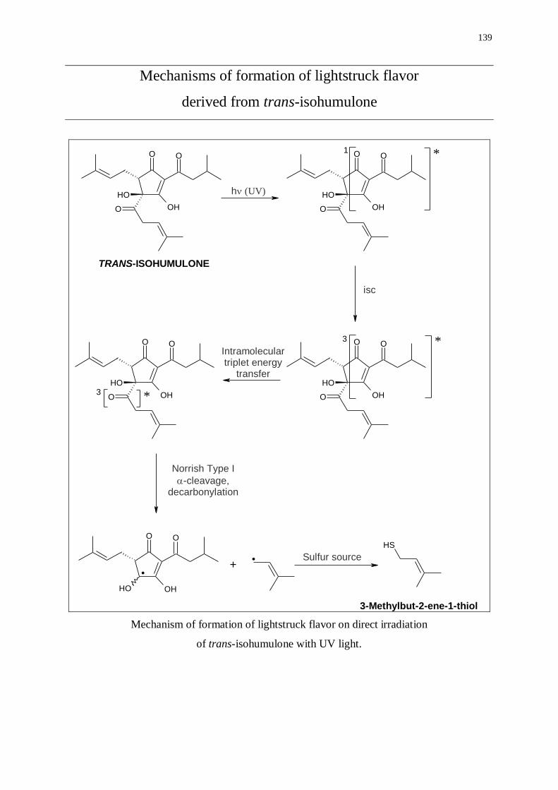
139
Mechanisms of formation of lightstruck flavor
derived from trans-isohumulone
OO
OHO
OH
OO
OHO
OH
OO
OHO
OH
OO
OHO
OH
SH
.
OO
OHOH
1
3
3
*
*
*
TRANS-ISOHUMULONE
h(UV)
isc
Intramoleculartriplet energy
transfer
Norrish Type I
-cleavage,decarbonylation
+Sulfur source
3-Methylbut-2-ene-1-thiol
.
Mechanism of formation of lightstruck flavor on direct irradiation
of trans-isohumulone with UV light.

140
O O
OHO
OH
O O
OHO
OH
SH.
O.
RF 1RF
3RF+ RF-. +
+.
h
isc
One-electrontransfer
TRANS-ISOHUMULONE
- CO
Sulfur source
-Cleavage
3-Methylbut-2-ene-1-thiol
Mechanism of formation of lightstruck flavor on sensitized irradiation of trans-isohumulone
in the presence of ribfoflavin (RF) with visible light.
141
Samenvatting
Een onaangename geur en smaak worden geproduceerd bij het blootstellen van bier aan
licht. De huidige kennis over de vorming van de lichtsmaak in bier dateert van de
oorspronkelijke publicaties door Kuroiwa et al. in het begin van de jaren 60. Door gebruik
van modelsystemen toonden zij aan dat de lichtsmaak ontstaat via een licht-geïnduceerde
reactie, waarbij riboflavine als sensibilisator (riboflavine is het belangrijkste flavine in bier),
isohumulonen (hop-afgeleide bitterstoffen in bier) en een zwavelbron (bijvoorbeeld cysteïne
of zwavelhoudende eiwitten) betrokken zijn. De onaangename geur en smaak werden
toegeschreven aan de vorming van 3-methylbut-2-een-1-thiol (MBT). Naar dit mechanisme
wordt vaak verwezen in de literatuur over de lichtsmaak in bier en alle relevante gegevens,
die tot op heden verzameld werden, zijn niet in tegenspraak met de Kuroiwa-premisse.
Hoewel de sleutelverbindingen sinds meer dan drie decennia bekend zijn, ontbreken details
over het interactiemechanisme tussen deze componenten.
Dit doctoraatsproject beoogt een grondige en gedetailleerde kennis te verwerven over het
mechanisme van de vorming van lichtsmaak in bier. Deze problematiek werd benaderd via
een diepgaand onderzoek van de interagerende verbindingen op moleculair niveau door
modelsystemen toe te passen, bestaande uit isohumulonen, dihydro-isohumulonen of
tetrahydro-isohumulonen in aan- of afwezigheid van riboflavine of flavine mononucleotide
als sensibilisator.
Opheldering van een fotochemisch mechanisme vergt studies in verschillende
tijdsdomeinen. Aldus werden modelsystemen onderworpen aan fotostationaire bestraling én
aan fotodynamische flash-geïnduceerde experimenten. Informatie over de aard van
geëxciteerde toestanden en intermediairen werd, naast mechanistische kenmerken, geleverd
door tijdsgeresolveerde elektron paramagnetische resonantie (TREPR) en flash-fotolyse
transiënte absorptiespectroscopie (TAS). Door karakterisering en identificatie van belangrijke
fotoreactieproducten via grondige structuuranalysen werden relevante reactieschema’s
uitgewerkt om de vorming van sleutelverbindingen te verklaren.
In een eerste fase werd een gedetailleerde studie ondernomen over de fotochemische
instabiliteit van isohumulonen, dihydro-isohumulonen en tetrahydro-isohumulones. Na laser-
flash fotolyse bij 308 nm gaven isohumulonen en tetrahydro-isohumulonen intense TREPR-
emissiesignalen (veroorzaakt door het triplet-mechanisme van chemisch-geïnduceerde
dynamische elektronpolarisatie), terwijl, in identische omstandigheden, geen TREPR-signaal
142
waargenomen werd voor dihydro-isohumulonen. Deze bevinding levert de eerste directe
spectroscopische evidentie voor de stabiliteit van dihydro-isohumulonen tegen fotolyse en
bewijst éénduidig dat radicalen, geproduceerd bij fotolyse van isohumulonen en tetrahydro-
isohumulonen, afkomstig zijn van de -hydroxyketo-chromofoor, aanwezig in isohumulonen
en tetrahydro-isohumulonen.
Door gefundeerde simuleringen toe te passen werden de meest voorkomende radicalen bij
fotolyse van isohumulonen gekarakteriseerd als een vijfring-ketylradicaal en een 3-methylbut-
2-enylradicaal, terwijl fotolyse van tetrahydro-isohumulonen leidde tot productie van een
analoog vijfring-ketylradicaal en een 4-methylpentanoylradicaal. Combinatie van TREPR-
gegevens liet toe een mechanisme voor te stellen voor de fotochemische afbraak van
isohumulonen en tetrahydro-isohumulonen bij directe bestraling. Absorptie van UV-licht
door de -tricarbonylchromofoor leidt tot de geëxciteerde singulet-toestand. Na
intersysteemkruising tot de geëxciteerde triplet-toestand, gebeurt intramoleculaire triplet-
energietransfer naar het geïsoleerde -hydroxyketon, dat, in de geëxciteerde toestand,
tenslotte Norrish Type I -splitsing ondergaat, waarbij de hoger beschreven radicalen
ontstaan. Uitgebreid onderzoek van de complexe mengsels fotoreactieproducten, afgeleid van
isohumulonen en tetrahydro-isohumulonen, toonde aan dat recombinatie van de belangrijkste
radicalen, gekarakteriseerd via TREPR, de vorming van de voornaamste fotoreactieproducten
kan verklaren, terwijl minder belangrijke fotoreactieproducten ontstaan via andere
reactiewegen. De hoofdproducten bij directe bestraling van isohumulonen werden
gekarakteriseerd als gedecarbonyleerde gedehydrateerde isohumulonen, terwijl de
overwegende reactieproducten bij directe bestraling van tetrahydro-isohumulonen
geïdentificeerd werden als dimeer-type verbindingen, gevormd door recombinatie van twee
vijfring-ketylradicalen.
In een tweede fase werd de interactie tussen isohumulonen, dihydro-isohumulonen en
tetrahydro-isohumulonen met riboflavine of flavine mononucleotide bij gesensibiliseerde
bestraling in detail bestudeerd Laser-flash fotolyse bij 355 nm van mengsels, bestaande uit
isohumulonen, dihydro-isohumulonen of tetrahydro-isohumulonen, en riboflavine
resulteerden in transiënte absorptiespectra (350-800 nm), gedomineerd door de absorptie van
een neutraal flavine semichinonradicaal, terwijl de absorptie van triplet-geëxciteerd
riboflavine zeer snel afnam. Deze waarnemingen zijn in overeenstemming met een één-
elektrontransfer uit de grondtoestand van isohumulonen, dihydro-isohumulonen of tetrahydro-
isohumulonen naar de geëxciteerde triplet-toestand van riboflavine. De één-elektrontransfer
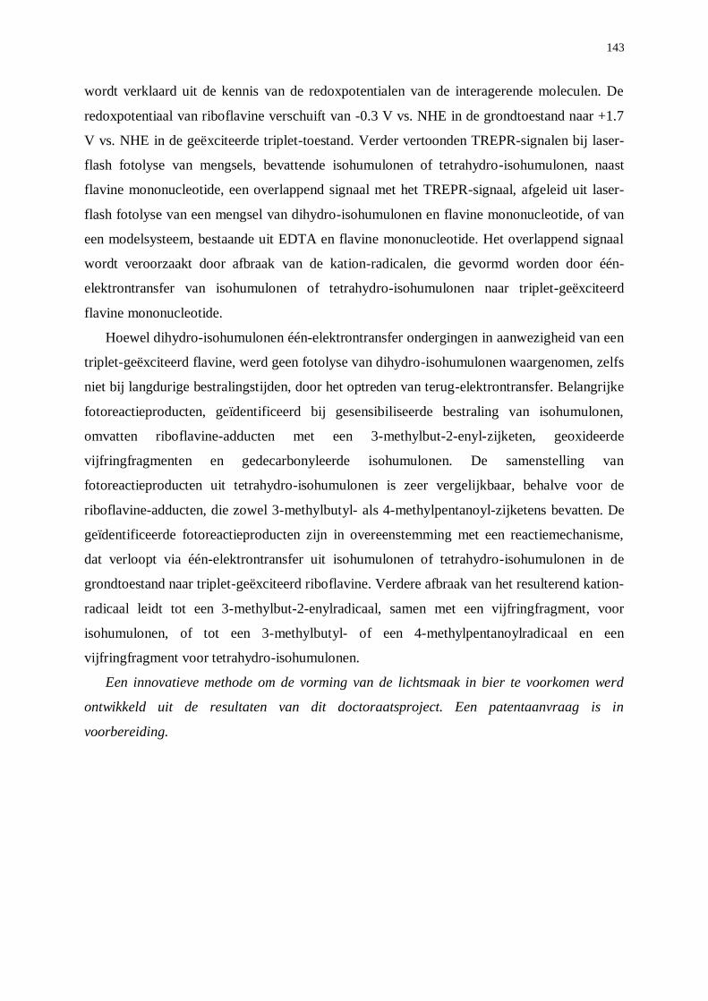
143
wordt verklaard uit de kennis van de redoxpotentialen van de interagerende moleculen. De
redoxpotentiaal van riboflavine verschuift van -0.3 V vs. NHE in de grondtoestand naar +1.7
V vs. NHE in de geëxciteerde triplet-toestand. Verder vertoonden TREPR-signalen bij laser-
flash fotolyse van mengsels, bevattende isohumulonen of tetrahydro-isohumulonen, naast
flavine mononucleotide, een overlappend signaal met het TREPR-signaal, afgeleid uit laser-
flash fotolyse van een mengsel van dihydro-isohumulonen en flavine mononucleotide, of van
een modelsysteem, bestaande uit EDTA en flavine mononucleotide. Het overlappend signaal
wordt veroorzaakt door afbraak van de kation-radicalen, die gevormd worden door één-
elektrontransfer van isohumulonen of tetrahydro-isohumulonen naar triplet-geëxciteerd
flavine mononucleotide.
Hoewel dihydro-isohumulonen één-elektrontransfer ondergingen in aanwezigheid van een
triplet-geëxciteerd flavine, werd geen fotolyse van dihydro-isohumulonen waargenomen, zelfs
niet bij langdurige bestralingstijden, door het optreden van terug-elektrontransfer. Belangrijke
fotoreactieproducten, geïdentificeerd bij gesensibiliseerde bestraling van isohumulonen,
omvatten riboflavine-adducten met een 3-methylbut-2-enyl-zijketen, geoxideerde
vijfringfragmenten en gedecarbonyleerde isohumulonen. De samenstelling van
fotoreactieproducten uit tetrahydro-isohumulonen is zeer vergelijkbaar, behalve voor de
riboflavine-adducten, die zowel 3-methylbutyl- als 4-methylpentanoyl-zijketens bevatten. De
geïdentificeerde fotoreactieproducten zijn in overeenstemming met een reactiemechanisme,
dat verloopt via één-elektrontransfer uit isohumulonen of tetrahydro-isohumulonen in de
grondtoestand naar triplet-geëxciteerd riboflavine. Verdere afbraak van het resulterend kation-
radicaal leidt tot een 3-methylbut-2-enylradicaal, samen met een vijfringfragment, voor
isohumulonen, of tot een 3-methylbutyl- of een 4-methylpentanoylradicaal en een
vijfringfragment voor tetrahydro-isohumulonen.
Een innovatieve methode om de vorming van de lichtsmaak in bier te voorkomen werd
ontwikkeld uit de resultaten van dit doctoraatsproject. Een patentaanvraag is in
voorbereiding.
144
Samenvattend heeft dit doctoraatsonderzoek aanzienlijk bijgedragen tot een
gedetailleerde kennis van de fotochemische instabiliteit van isohumulonen en tetrahydro-
isohumulonen met betrekking tot de fotostabiliteit van dihydro-isohumulonen. Voor de eerste
maal werd overtuigend bewezen dat de fenomenen, die leiden tot vorming van de lichtsmaak,
verschillen naargelang de bestralingsomstandigheden. Bij directe bestraling met UV-licht
leidt energietransfer van triplet-geëxciteerde isohumulonen en tetrahydro-isohumulonen tot
een Norrish Type I fotosplitsing van de -hydroxyketo-chromofoor, resulterend in de vorming
van, respectievelijk, een 3-methylbut-2-enylradicaal en een 4-methylpentanoyl radical. Het 3-
methylbut-2-enylradicaal is de directe precursor van 3-methylbut-2-een-1-thiol als de
sleutelverbinding, geassocieerd aan de lichtsmaak. Dihydro-isohumulonen zijn geheel
lichtstabiel. Bij gesensibiliseerde bestraling met zichtbaar licht onttrekt triplet-geëxciteerd
riboflavine als sensibilisator een elektron uit isohumulonen en tetrahydro-isohumulonen in de
grondtoestand met vorming van een kation-radicaal, dat gestabiliseerd wordt via splitsing
van de -hydroxyketogroup volgens een formele Norrish Type I reactie. Bijgevolg ontstaat
een 3-methylbut-2-enylradicaal, dat direct leidt tot 3-methylbut-2-een-1-thiol. Dihydro-
isohumulonen zijn opnieuw lichtstabiel.


