
Wednesday, April 1st
Scientific Session VI – Complex Aneurysm
Presentation
Number: 18
Publishing
Title: Staged Hybrid Repair of Extensive Thoracoabdominal Aortic Aneurysms Secondary to Aortic Dissections: Mid-Term Outcomes
Author Block:
Amit Jain, MD1, William F. Johnston, MD2, Tanya F. Flohr, MD2, Margaret C. Tracci, MD2, Kenneth J. Cherry, MD2, Gorav
Ailawadi, MD2, Gilbert R. Upchurch, Jr., MD2, John A. Kern, MD2, Ravi K. Ghanta, MD2. 1University of Cincinnati, Cincinnati, OH, USA, 2University of Virginia, Charlottesville, VA
Abstract
Body:
OBJECTIVES: Open repair of Crawford Extent I/II thoracoabdominal aortic aneurysms(TAAA) are associated with a high rate
of major adverse complications. Staged hybrid repair of these extensive TAAAs may reduce this operative risk. In the present
study, we review the mid-term outcomes of a previously described technique that combines proximal thoracic endovascular
aneurysm repair (TEVAR) followed by staged distal open thoracoabdominal repair for patients with Crawford Extent I/II TAAAs.
METHODS: From July 2007 to June 2014, 19 patients with Crawford Extent I(n=1) or Extent II(n=18) TAAAs secondary to
chronic aortic dissections underwent a staged hybrid repair. All patients had TEVAR as Stage 1 and open repair as Stage 2, with
partial cardio-pulmonary bypass via left femoral arterial and venous cannulation for visceral and lower body perfusion. The open
thoracoabdominal graft was anastomosed proximally in end to end fashion with the endograft.
RESULTS: Average patient age was 54 ±17.6 years (14 male). Nine patients had prior open proximal aortic surgery for Type 1
aortic dissections. TEVAR was performed via percutaneous (n=8), femoral cutdown (n=8) or iliac exposure (n=3). The left
subclavian artery was covered in 9 patients and re-vascularized in 8 patients by carotid-subclavian bypass (n=7) or laser
fenestration (n=1). There were no incidents of death, stroke, or paralysis in this cohort. Following TEVAR, three patients required
repeat intervention for endoleak (Type 1A, n=1; Type1B, n =1; Type 2, n=1) prior to open repair. Following open repair, there
was a single delayed permanent paralysis. Hospital length of stay was 7±4 days after TEVAR and 9±5 days after open repair. No
deaths or neurologic events occurred in the remaining 18 patients over a median 85 week follow up (range 4 weeks to 6.2 years).
Importantly, all patients have stable aortic size and remain free of reintervention over the follow-up period.
CONCLUSIONS: Staged hybrid repair, combining proximal TEVAR with open distal repair, for extensive TAAAs secondary to
chronic dissection is an effective, durable and safe alternative to traditional open repair. This mid-term follow up data suggests
that staged repair may reduce perioperative morbidity and mortality in patients with extensive TAAAs.
Outcome of Staged Hybrid Repair of Extent I/II TAAAs
Outcome Staged Hybrid Repairs
(n=19)
Stage 1: TEVAR
Death
Stroke / Paraplegia
Acute Kidney Injury
Type 1 Endoleak
Type 2 Endoleak
0 (0%)
0 (0%)
1 (5.2%)
2 (10.5%)
1 (5.2%)
Stage 2: Open Distal Repair
Death
Stroke / Paraplegia
Acute Kidney Injury (serum Cr > 2)
Chronic Renal Failure
0 (0%)
1 (5.2%)
5 (26.3%)
0 (0%)

Presentation
Number: 19
Publishing
Title: Aortobifemoral Graft Infection: Is Unilateral Limb Excision Definitive?
Author Block:
Jeffrey D. Crawford, MD, Amir F. Azarbal, MD, Timothy K. Liem, MD, Gregory J. Landry, MD, Gregory L. Moneta, MD,
Erica L. Mitchell, MD.
Oregon Health and Sciences University, Portland, OR
Abstract
Body:
OBJECTIVES: Aortobifemoral graft (ABFG) infections presenting with single limb involvement can be managed with
unilateral limb excision or complete graft removal. This study aims to identify factors predictive of subsequent contralateral limb
infection in patients initially undergoing unilateral limb excision.
METHODS: A retrospective review of patients treated for infected ABFGs from 2001-July 2014 was performed. Endovascular
and aortic tube graft infections were excluded. Primary outcomes were freedom from contralateral graft limb excision, overall
survival and factors potentially predictive of subsequent contralateral limb infection.
RESULTS: Fifteen patients underwent unilateral graft limb excision with retroperitoneal exploration of the affected ABFG limb
and revascularization for unilateral graft limb infection. Original indication for placement of the ABFG was aortoiliac occlusive
disease in 11 patients and aneurysm in 4. All patients had no clinical or radiographic evidence for contralateral limb infection at
initial presentation. Seven patients, all of whom underwent initial operation for aortoiliac occlusive disease developed
contralateral limb infection at a median follow up of 23.2 months. The remaining 8 patients had no evidence of contralateral limb
infection at median follow up of 38.8 months. Patient demographics were similar between the two groups. Five of the seven
patients who developed contralateral limb infection had imaging evidence of ipsilateral graft infection above the inguinal
ligament at the time of initial graft infection. Operative exploration during unilateral excision in this group revealed a well-
incorporated graft without extension to the bifurcation. There was no dominant organism cultured in either group and duration of
targeted antibiotic therapy was similar in both groups (≥ 6 weeks). For the series, there were no amputations and overall mortality
was 40% with median follow-up of 44.7 months.
CONCLUSIONS: Unilateral infection of an ABFG can be managed with single limb excision, however, 50% of patients will
return with contralateral limb infection at a median of two years. Clinical assessment of graft incorporation lacks specificity and
does not indicate freedom from contralateral limb infection. Factors predicting contralateral involvement include initial operation
for aortoiliac occlusive disease and initial imaging or operative findings suspicious for infection above the inguinal ligament of
the unilateral limb.
Presentation
Number: 20
Publishing
Title:
Percutaneous Endovascular Aortic Repair (PEVAR) of Complex Aneurysms using Large-Diameter Sheaths for Thoracic,
Fenestrated and Branched Endografts
Author Block:
Leonardo Reis de Souza, MD, Gustavo S. Oderich, MD, Peter Banga, MD, Jan Hofer, MD, Stephen Cha, MS, Peter Gloviczki,
MD.
Mayo Clinic, Rochester, MN
Abstract
Body:
OBJECTIVES: To review the outcomes of PEVAR of complex aortic aneurysms using large-diameter sheaths for thoracic,
fenestrated and branched stent-grafts.
METHODS:: We reviewed the outcomes of all consecutive patients who underwent PEVAR of descending thoracic (DTA),
thoracoabdominal (TAAA), pararenal (PRA) or aortoiliac aneurysms (AIAs) using large-diameter sheaths for placement of
thoracic, fenestrated or branched stent-grafts. Patients treated by fenestrated and branched stent-grafts were enrolled in
prospective physician-sponsored investigational device exemption protocols. A percutaneous approach was selected in patients
with <50% posterior, minimal anterior or no calcification in the common femoral artery using standardized pre-closure technique
with two Perclose® devices (Abbott, CA) in each femoral puncture site. End-points were technical success, conversion to open
femoral artery repair, 30-day mortality and major adverse events, and freedom from femoral access-site complications.
RESULTS:: There were 102 patients treated for 48 PRA, 27 TAAA, 19 DTA and 8 AIAs. A total of 171 femoral arteries were
closed using pre-closure technique. Trans-femoral sheath size was 18Fr in 4 vessels (3%), 20Fr in 120 (70%) and ≥22Fr in 47
(27%). 83 patients (81%) had visceral branch incorporation, which required brachial artery access using small incision in 48.
Technical success for percutaneous trans-femoral closure was 95% (162/171). Nine intraoperative failures were managed by open
femoral conversion using primary repair in 6, interposition graft in 2, and patch angioplasty in 1. Mean estimated blood loss was
444±569 ml. There were no patients with had uncontrolled puncture-related hemorrhage, retroperitoneal hematoma or intra-
operative hypotension. 30-day mortality was 0.9% (1/101) and 30-day rate of major adverse events was 15% (16/102). Spinal
cord injury occurred in 1 patient (0.9%). There were 5 (3%) access-related complications, including femoral artery occlusion in 3
and hematoma or pseudoaneurysm in 1 each. Wound-related complications occurred in 1 patient (0.5%) who required open
femoral artery conversion for exposure and repair. After a mean follow up of 1-year, freedom from femoral access-site
complication was 97±2%.
CONCLUSIONS: PEVAR using pre-closure technique is safe and effective in select patients with complex aortic aneurysms
who have minimal or no femoral calcifications and require large-diameter sheaths for thoracic, fenestrated and branched stent-
grafts. Rate of puncture (3%) and wound-related complications (0.5%) is low, and uncontrolled puncture-related hemorrhage,
retroperitoneal hematoma and systemic hypotension has not occurred in this series.
Presentation
Number: 21
Publishing
Title: Left Subclavian Artery Occlusion During TEVAR in the Elderly is Associated with Significant Morbidity
Author Block: Khanjan H. Nagarsheth, MD, Jonathan Schor, MD, Matthew D'Alessandro, DO, Kuldeep Singh, MD, Jonathan Deitch, MD.
Staten Island University Hospital, Staten Island, NY
Abstract
Body:
OBJECTIVES: Covering the left subclavian artery (LSA) during thoracic endovascular aortic repair (TEVAR) for proximal seal
is generally safe and well tolerated. The purpose of this study is to determine if this practice is safe in elderly patients.
METHODS: The National Surgical Quality Improvement Program (NSQIP) database was queried, from the years 2005 to 2011,
to identify patients who underwent TEVAR. Octogenarians were separated into two groups, one where the LSA was covered (C-
SA) and another where it was not covered (U-SA). Patient demographics, comorbidities, perioperative data, and outcomes were
compared.
RESULTS: There were a total of 392 patients over age 80 who underwent TEVAR. There were 128 patients in the C-SA group
and 264 in the U-SA group. There was no significant difference in demographics or baseline cardiovascular or pulmonary
comorbidities between groups. There was also no difference in emergency procedures between C-SA and U-SA groups (27% v.
21%, p=0.18). It was found that the C-SA group had significantly more intra-operative cardiac arrest (4% v. 1%, p=0.03) and
significantly more received intra-operative blood transfusions (32% v. 21%, p=0.02). There was also a higher post-operative rate
of stroke (9% v. 3%, p=0.03) and sepsis (9% v. 3%, <0.01) in the C-SA group compared to the U-SA group.
CONCLUSIONS: Covering the LSA in octogenarians is associated with significantly increased peri-operative morbidity. We
recommend caution when considering coverage of the LCA during TEVAR. These patients may benefit from elective
revascularization when possible.
Presentation
Number: 22
Publishing
Title: Development of New Acute Dissection in the Ascending Aorta after Type B Dissection: Intramural Hematoma is not Benign
Author
Block:
Samuel S. Leake, BS, Harleen K. Sandhu, MD, Charles C. Miller, III, PhD, Rana O. Afifi, MD, Ali Azizzadeh, MD, Anthony L.
Estrera, MD, Hazim J. Safi, MD, Kristofer M. Charlton-Ouw, MD.
The University of Texas at houston Medical School, Houston, TX
Abstract
Body:
OBJECTIVES: Aortic dissection is a dynamic process that can extend distal to the entry tear or proximally in a retrograde
fashion. We sought to determine associations for development of new acute type A aortic dissection (ATAD) after type B
dissection (TBAD).
METHODS: We reviewed our aortic dissection database for cases of ATAD from 2002-2013 that had known TBAD. Imaging and
intraoperative reports were used to determine presence of entry tear with dissection flap vs. intramural hematoma (IMH).
Demographic and disease-related variables were analyzed.
RESULTS: Among 419 new cases of ATAD, we identified 16 (3.8%) patients with previous known TBAD. Presence of flap vs.
IMH could be determined in 403/419 cases (96%). IMH was more common in patients with previous TBAD (56% vs. 13%,
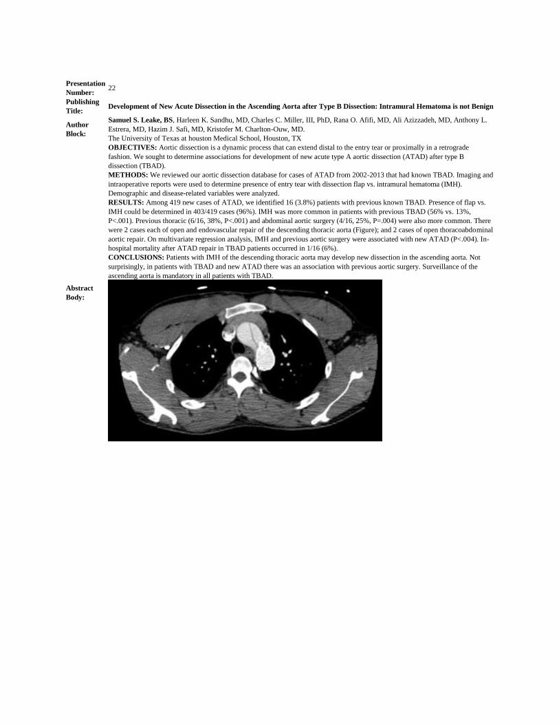
P<.001). Previous thoracic (6/16, 38%, P<.001) and abdominal aortic surgery (4/16, 25%, P=.004) were also more common. There
were 2 cases each of open and endovascular repair of the descending thoracic aorta (Figure); and 2 cases of open thoracoabdominal
aortic repair. On multivariate regression analysis, IMH and previous aortic surgery were associated with new ATAD (P<.004). In-
hospital mortality after ATAD repair in TBAD patients occurred in 1/16 (6%).
CONCLUSIONS: Patients with IMH of the descending thoracic aorta may develop new dissection in the ascending aorta. Not
surprisingly, in patients with TBAD and new ATAD there was an association with previous aortic surgery. Surveillance of the
ascending aorta is mandatory in all patients with TBAD.

Presentation
Number: MP24
Publishing
Title:
Evolution from Physician-Modified to Manufactured Fenestrated and Branched Endografts to Treat Pararenal and
Thoracoabdominal Aortic Aneurysms
Author Block:
Gustavo S. Oderich, MD, Leonardo Reis de Souza, MD, Jan Hofer, RN, Peter Banga, MD, Sharon Mueller, RN, Stephen Cha,
MS, Thanila Macedo, MD, Peter Gloviczki, MD
Mayo Clinic, Rochester, MN
Abstract
Body:
PURPOSE: To review outcomes of patients treated for pararenal (PRA) or thoracoabdominal aortic aneurysms (TAAAs) using
physician-modified (PMSGs) or manufactured fenestrated and branched stent-grafts (MSGs).
METHODS: We reviewed clinical data of 207 consecutive patients (164 male, mean age 76±9 years old) treated for PRA/
TAAAs using fenestrated and branched stent-grafts. Choice of device evolved from PMSGs (2007-2013) to MSGs (2012-2014)
in patients enrolled in prospective physician-sponsored investigational device exemption protocols (PS-IDE). End-points were
30-day mortality, major adverse events (MAEs), patient survival, freedom from type I/III endoleak, sac growth (>5mm), primary
target vessel patency and re-intervention.
RESULTS: 138 patients were treated by PMSGs, 69 had MSGs. 131 patients had PRAs (82 PMSGs, 49 MSGs), 76 had TAAAs
(56 PMSGs, 20 MSGs). PMSGs patients had larger aneurysms, more cardiac, pulmonary and kidney disease, and higher
comorbidity scores (P<0.05). A total of 670 visceral arteries were targeted by fenestrations and branches. Technical success was
98% for PMSGs and 99.6% for MSGs (P=0.9). 30-day mortality was 1% for PRAs (PMSGs 1%, MSGs 0%, P=0.44) and 7% for
TAAAs (PMSGs 9%, MSGs 0%; P=0.17). There were more (P<0.05) MAEs among PMSG patients treated for PRAs (44%,
24%) and TAAAs (58%, 25%). Mean follow up was longer in PMSG patients (31±21, 12±7 months; P<0.0001). At 1-year,
PMSGs and MSGs had similar freedom from type I/III endoleaks (PRAs: 95±4%, 99±1%; TAAAs: 100%, 100%), sac growth
(PRAs: 99±1%, 100%; TAAAs: 97±3%, 100%), primary target vessel patency (PRAs: 97±1%, 98±1%, TAAAs: 98±1, 97±2%)
and re-intervention (PRAs: 85±5%, 93±4%; TAAAs: 82±5%, 100%). Survival was lower (P<0.05) in PMSGs patients treated for
PRAs (84±3%, 98±2%) and TAAAs (78±5%, 100%). At 5-years, 20 PMSG patients (14%) developed type I/III endoleak and 10
(7%) had sac growth. In the PMSG group, patient survival, freedom from type I/III endoleak, sac growth, primary target vessel
patency and re-intervention at 3- and 5-years was 71±4%/63±5%, 87±3%/76±7%, 96±2%/80±6%, 96±1%/96±1%, and
71±5%/57±7%, respectively.
CONCLUSION: Patients treated by PMSGs had higher clinical risk and larger aneurysms, reflecting the more liberal indication
of MSGs under PS-IDE protocols. Despite differences, both devices were implanted with similar technical success, mortality,
endoleak, sac growth, vessel patency and re-intervention rates. At late follow up, PMSGs were associated with significant rate
(14%) of type I/III endoleaks and sac growth (7%).

Presentation
Number: MP25
Publishing
Title:
Clinical Application of Continuous Intraoperative Neuromonitoring And Selective Use of Temporary Femoral Conduits
to Minimize Lower Extremity Ischemia and Risk of Spinal Cord Injury During Complex Endovascular Thoracic and
Thoracoabdominal Aortic Repair
Author Block:
Peter Banga, MD, Gustavo S. Oderich, MD, Eric Sorenson, MD, Leonardo Reis de Souza, MD, Josh Netcott, R. EEG. T, Jan
Hofer, RN, Stephen Cha, MS, Peter Gloviczki MD, MD.
Mayo Clinic, Rochester, MN
Abstract
Body:
PURPOSE: To review the clinical utility of intra-operative motor-evoked (MEP) and somatosensory-evoked potential (SSEP)
monitoring with selective use of temporary femoral conduits (TFCs) in patients undergoing endovascular repair of descending
thoracic (DTA) and thoracoabdominal aortic aneurysms (TAAAs).
METHODS: We reviewed the clinical data of 49 patients (38 male; mean age of 75±8 years old) who underwent endovascular
repair of DTA and TAAAs (2007-2014). Patients treated by fenestrated and branched endografts were enrolled in prospective
physician-sponsored investigational device exemption protocols. Patients requiring extensive aortic coverage had cerebrospinal
fluid (CSF) drainage, permissive hypertension and MEP/SSEP monitoring. TFCs were used in patients with difficult visceral
artery anatomy, allowing withdrawal of the device sheath into the conduit while performing visceral stenting. Changes in
MEP/SSEPs prompted maneuvers to optimize spinal cord perfusion and restore lower extremity (LE) blood flow whenever
possible. End-points were spinal cord injury (SCI) and LE ischemic complications.
RESULTS: 45 patients (92%) had TAAAs, 4 (8%) had DTAs. 163 visceral arteries were targeted by fenestrations and branches
(mean, 3.7±0.1 vessels/patient). Temporary conduits were used in 12 patients/14 limbs (11 TFCs, 3 iliac). A stable MEP/SSEP
was achieved in all patients. 29 patients (59%) had >75% decrease in MEP amplitude in 40 limbs. MEP changes started 76±29
minutes after vascular access and were more prominent in the side of the larger sheath. Patients with temporary conduits less
often had MEP changes compared to those without conduits (21% vs 47% of limbs, P<0.01). MEP amplitude partially improved
in 7 patients/ 9 limbs (23%) with intra-operative maneuvers to increase mean arterial pressure and lower CSF pressure. With
restoration to LE blood flow, MEP/SSEPs returned to baseline in all except for one patient who developed immediate permanent
paraplegia. A second patient developed delayed paraplegia after open repair of retrograde type A dissection 2 days after the initial
procedure. There were no other SCIs or LE ischemic complications.
CONCLUSION: Neuromonitoring is a reliable technique to assess LE ischemia and spinal cord function and predicts immediate
SCI during endovascular repair of complex aortic aneurysms. Temporary iliofemoral conduits allow immediate restoration of LE
blood flow in difficult cases where prolonged LE ischemia is anticipated. Identification of MEP/SSEP changes allows institution
of a standardized protocol to optimize spinal cord perfusion and restore LE blood flow.

Presentation
Number: MP26
Publishing
Title:
Anatomic Severity Grading Score for Primary Descending Thoracic Aneurysms Predicts Reinterventions and Mortality
After Thoracic Endovascular Aortic Repair
Author Block:
Chad Ammar, MD, Sebastian Larion, MS, Sadaf S. Ahanchi, MD, Kedar S. Lavingia, MD, David J. Dexter, MD, Jean M.
Panneton, MD
Eastern Virginia Medical School, Norfolk, VA
Abstract
Body:
OBJECTIVES: An anatomic severity grading (ASG) score for primary descending thoracic aortic aneurysms (DTAs) has recently
been developed by our group. The objective of this study is to determine if an ASG score cutoff value for DTAs is predictive of
reinterventions and mortality in patients undergoing thoracic endovascular aortic repair (TEVAR).
METHODS: A retrospective review from 2008 to 2013 of patient records was conducted of all consecutive patients who
underwent a TEVAR for a primary DTA. A comprehensive scoring system of preoperative DTA morphology based on computed
tomography angiography (CTA) images has been established to identify and classify anatomic features that may influence
outcome after TEVAR. ASG score calculations were achieved using 3D reconstructions of preoperative CTA images (TeraRecon
Aquarius iNtuition Workstation, Foster City, CA). Primary outcomes included aneurysm-related death, 30-day mortality, all-
cause mortality, aneurysm rupture, primary technical success, and aneurysm-related reintervention rates. Secondary outcomes
included device migration, endoleak formation, endoleak requiring reintervention, hospital readmission within 30 days, stroke,
paraplegia, and conversion to open repair.
RESULTS: Of 469 patients with an ICD-9 diagnosis of a thoracic aortic aneurysm, 62 (13%) patients underwent TEVAR and had
adequate preoperative imaging (mean age, 71 years). Applying the ASG score, we identified 30 (48%) patients with a score ≥28
(high score group) and 32 (52%) patients with a score < 28 (low score group). Mean follow-up was 32.8 months post-TEVAR for
both groups. Technical success was 100% in both groups. Aneurysm-related and all-cause mortality was significantly higher in
the high score versus the low score group (13% vs. 0%, P=0.049 and 27% vs. 3.1%, P=0.011, respectively). A significantly
higher reintervention rate (43% vs. 13% P=0.015) was present in the high score versus low score group. Endoleak formation and
endoleak requiring reintervention was significantly higher in the high versus the low score group (53% vs 9%, P= <0.001 and
40% vs 6%, P=0.004, respectively). No significant difference in aneurysm rupture (4%), device migration (6%), 30-day hospital
readmission (13%), stroke or paraplegia (6%) was present between groups, and no patient had a conversion to open repair during
the follow-up period.
CONCLUSIONS: Preoperative ASG score for primary DTAs predicts reinterventions for endoleaks, aneurysm-related and all-
cause mortality after TEVAR.
Presentation
Number: MP27
Publishing
Title: Preventing Spinal Cord Ischemia in Complex Endovascular Aortic Aneurysm Repair
Author Block:
Stephen C. Hanley1, Oren K. Steinmetz2, Kent MacKenzie2, Daniel Obrand3, Marc Corriveau2, Heather L. Gill2, Cherrie Z.
Abraham3 1McGill University, Montreal, QC, Canada, 2McGill University Health Centre, Montreal, QC, Canada, 3Jewish General Hospital,
Montreal, QC, Canada
Abstract
Body:
OBJECTIVES: Complex endovascular aortic aneurysm repair (cEVAR) with branched and fenestrated aortic stent grafts has
revolutionized the treatment of complex aortic aneurysms. While the risks of cEVAR appear to be less than those of open repair,
devastating complications may still arise, particularly with repair of aneurysms of the thoracoabdominal aorta. One of the most
severe complications of cEVAR is spinal cord ischemia (SCI), with a reported risk in the range of 10-15%. While multiple
strategies have been proposed to reduce the risk of SCI, the use of 'paraplegia-prevention branches' (PPBs) to maintain temporary
perfusion of the aneurysm sac allows for a staged approach to endovascular repair of complex aneurysms.
METHODS: Patients undergoing cEVAR who were deemed high risk for SCI, based mainly on extensive aortic coverage or
previous aortic intervention, were treated with custom-made stent grafts with PPBs, consisting of one or two upward-pointing
6mm branches, ideally in proximity to large patent intercostal arteries. These branches were left open during the initial
intervention, maintaining perfusion to the aneurysm sac. Routine institutional SCI prevention strategies were employed. During a
planned secondary procedure, PPBs were then occluded using Amplatzer occluder devices, under local anesthesia. Demographic
and peri-operative data were collected retrospectively for patients identified from a prospectively maintained database of all
patients undergoing EVAR at our institution, and compared for patients undergoing cEVAR with and without PPBs.
RESULTS: Between 2010 and 2014, 71 patients underwent cEVAR for complex abdominal or thoracoabdominal aneurysms.
Seven patients underwent cEVAR using PPBs, with a time-to-occlusion of 19±8 days. The overall rate of SCI was 12.7%,
however none of the patients treated with PPBs developed SCI. Likewise, peri-operative mortality was 9.0%, consistent with
reports in the literature. None of the patients treated with PPBs died in the peri-operative period.
CONCLUSIONS: Staged endovascular repair of complex aortic aneurysms using PPBs may represent a means of reducing the
risk of SCI. Our experience adds to the growing body of literature supporting the notion of staged endovascular repair of complex
aortic aneurysms. Further study is necessary to determine whether PPBs significantly reduce the risk of SCI.

Presentation
Number: MP28
Publishing
Title:
A Novel Anatomic Severity Grading Score for Acute Type B Aortic Dissections and Correlation to Aortic Reinterventions after
Thoracic Endovascular Aortic Repair
Author
Block:
Shirui Chen, BA, Sebastian Larion, MS, S. Sadie Ahanchi, MD, Chad P. Ammar, MD, Colin T. Brandt, MD, Jean M. Panneton,
MD.
Eastern Virginia Medical School, Norfolk, VA
Abstract
Body:
OBJECTIVES: We introduce a novel preoperative anatomic severity grading (ASG) system for acute type B aortic dissections and
test the system with a cohort of patients who underwent thoracic endovascular aortic repair (TEVAR).
METHODS: A cohort of patients who received TEVAR for acute type B aortic dissection between 2008-2014 was identified. We
developed an ASG score to measure aspects of aortic anatomy that we hypothesized may affect difficulty of repair (shown below).
We also calculated a false lumen volume index (FLVI=false lumen volume divided by the sum of the true and false lumen
volumes). Measurements were made using reconstructed CT angiography images and categorized using scores of 0 to 3 based on
hypothesized severity, giving a potential range of 0-38.
RESULTS: We analyzed the CTA images on a cohort of 30 patients with acute (within 2 weeks of symptoms onset) type B aortic
dissection who underwent TEVAR. The average age was 63 years (range 28-89 years) with 70% male and 50% Caucasian. The
mean ASG score was 19 (range 10-29). We created an area under the receiver operating characteristic curve (AUROC) using ASG
to predict aortic reinterventions. The area was 0.72 (95% CI 0.39 to 1.1). Guided by the AUROC, we divided patients into two
groups, one with ASG scores <22 (n=20) and the other with scores >22 (n=10). With this cutoff, ASG exhibited 80% sensitivity
and 76% specificity in predicting aortic reinterventions, with reintervention in 40% of high-score patients and 5% of low-score
patients (P=0.031). In addition, patients in the high-score group were significantly younger (53.8 vs 66.9 years, P=0.006).
The mean thoracic aorta FLVI was 0.59 (range 0.19-0.94). Patients with high ASG score exhibited significantly larger thoracic
aortic FLVI (0.73 vs 0.52, P=0.001). The correlation coefficient between thoracic aorta FLVI and ASG was 0.74 (P<0.001).
CONCLUSIONS: A preoperative ASG score of acute type B aortic dissections consists of analysis of the proximal landing zone,
curvature and tortuosity of the aorta, dissection anatomy, aortic branch vessel anatomy, and supraceliac aorta anatomy. ASG score
>22 correlated with larger false lumen to total aortic volume ratio and was an excellent predictor of aortic reinterventions.
Karmody Competition Topic: Aneurysm
Presentation
Number: KC1
Publishing
Title: Predictors of Perioperative Outcomes after Fenestrated EVAR
Author Block:
Tarik Z. Ali, MD, David E. Timaran, MD, Kimberly Borges, Luis Gomez, MD, Martyn Knowles, MD, Mirza Shadman Baig,
MD, Carlos H. Timaran, MD.
University of Texas Southwestern Medical Center, Dallas, TX
Abstract
Body:
OBJECTIVES: Fenestrated EVAR is a novel treatment modality for complex AAAs that could be applied to a greater number of
patients compared to open repair. The safety and efficacy of fenestrated EVAR in high risk and frail patients has not been
established. The aim of this study was to assess the risk factors, including a simplified frailty index (SFI), for postoperative
complications and major adverse events after fenestrated EVAR.
METHODS: Over a 22-month period, 49 patients with complex AAAs underwent fenestrated EVAR with a custom made device,
the Zenith Fenestrated AAA Endovascular Graft (Cook Inc, Bloomington, Ind). Perioperative data was collected into a
prospectively maintained database. A SFI was calculated according to the presence of 11 items, including diabetes; functional
status (not independent); chronic obstructive pulmonary disease or pneumonia; congestive heart failure; history of myocardial
infarction; either prior percutaneous coronary intervention, previous coronary surgery, or history of angina; hypertension
requiring medication; peripheral vascular disease or rest pain; impaired sensorium; history of either transient ischemic attack or
cerebrovascular accident; or history of cerebrovascular accident with neurologic deficit. Perioperative major adverse events were
defined as death, hospital re-admission, disability or life threating adverse events at 30 days. Univariate analysis, non-parametric
analysis and simple logistic analysis were used.
RESULTS: The Median age was 73 years (interquartile [IQR] 68-81). Median SFI was 0.27 (IQR, 0.18-0.36). The median
number of fenestrations was 2 (IQR, 2-3). Univariate analysis and simple logistic analysis revealed that SFI was not a predictor of
post-operative complications and major adverse events at 30 days (p>0.1). BMI, increasing age and cardiac history of arrhythmias
or valvular disease were found to be statistically significant risk factors for major adverse events (P<0.05). Patients with lower
BMI (25.5[IQR, 20.9-29.6] vs 28 [IQR, 25-31]) (p=0.03), increasing age (78 [IQR 69-84] vs 70 [IQR, 65-75]) (p<0.01) and a
history of arrhythmias or valvular disease (47 % vs 4%) (p=0.03) had a higher frequency of major adverse events and
postoperative complications.
CONCLUSION: Fenestrated EVAR is a safe and effective for the treatment of complex AAAs, even in high-risk and frail
patients. Increasing age, BMI and cardiac problems (other than MI and CHF) are predictors of perioperative majors adverts after
fenestrated EVAR.

Presentation
Number: KC2
Publishing
Title: Midterm Results of Parallel Grafts Used in the Endovascular Treatment of Complex Abdominal Aortic Aneurysms
Author Block: Andrew Cha, DO, Michael Lieb, DO, Naiem Nassiri, MD, Randy Shafritz, MD, Saum Rahimi, MD.
Rutgers-Robert Wood Johnson Medical School, New Brunswick, NJ
Abstract
Body:
OBJECTIVE: The use of endovascular stent grafts to repair abdominal and thoracic aortic aneurysms has become the mainstay of
treatment. The technique of parallel stents grafts has emerged as a modality to provide an endovascular approach to aneurysm
repair in high risk patients with challenging aneurysm neck anatomy. Here we evaluate our results using parallel self expanding
covered stent grafts to treat complex aneurysms.
METHODS: We evaluated all patients undergoing endovascular aneurysm repair (EVAR) utilizing covered self expanding stents
to extend either the proximal or distal seals zones from November 2010 to December 2013. Only patients with a post-operative
CT angiogram were included for analysis. We evaluated the patients’ most recent CTA to determine graft position and patency.
The 30-Day and overall mortality, parallel graft patency rate, change in aneurysm size, and rate of endoleaks were calculated.
RESULTS: There were 13 patients ( 11 male, 2 female) an average of 75±7.5 years old included in analysis. Two thoracic
aneurysms, 3 para-renal aneurysms, and 8 infra-renal aneurysms were treated. A total of 20 vessels were treated with 22 parallel
grafts ( 14 Renal, 2 SMA, 1 Polar renal, 1 Celiac, 2 Hypogastric). There was an average of 1.7 parallel grafts per patient. The
average length of parallel stent graft used varied by location (Renal 10.5cm, SMA 10 cm, Celiac 10cm, Hypogastric 12.5
cm). The average follow up was 13.6 months (Range 3-45 months). There was one early death resulting in a 7.7% 30-day
mortality. The average maximum pre-operative aneurysm diameter was 57 ± 11 mm (Range 40-84mm). The average maximum
post-operative aneurysm diameter was 54 ± 7 mm (Range 41-66mm). Post-operatively, there was an average decrease in
aneurysm size of 4mm, representing a 4.4% decrease in aneurysm size. There were no patients with Type I or Type III endoleaks.
There are currently 3 patients with Type II endoleaks (23%)
CONCLUSION: The use of parallel stent grafts has expanded the use of EVAR to patients who previously were deemed not to be
candidates. We have observed excellent patency of the stent grafts in our patient population. These mid term results illustrate the
utility of parallel grafts to treat complex aneurysm in high risk patients when fenestrated grafts are not an option.
Presentation
Number: KC3
Publishing
Title: Outcomes of Endovascular Interventions in Patients with Connective Tissue Diseases
Author Block:
J Westley Ohman, MD, Chandu Vemuri, MD, Patrick J. Geraghty, MD, Luis A. Sanchez, MD, Alan Braverman, MD, Jeffrey
Jim, MD.
WashU STL, Saint Louis, MO
Abstract
Body:
OBJECTIVES:Patients with connective tissue disorders (CTD) requiring aortic repair are traditionally treated with open surgery
as endovascular repair has been contraindicated due to concern about tissue integrity and outcomes with TEVAR. Our objective is
to review a single center experience in endovascular treatment in CTD patients.
METHODS: A retrospective chart review was performed to identify patients with a diagnosis of a CTD who underwent an
endovascular intervention between 12/1/2004 and 12/1/2013. Preoperative demographic information and postoperative outcomes
were collected.
RESULTS: During the study period, a total of 6 patients met selection criteria, and were followed for 42.5 +/- 13.9 months. 4
carried a diagnosis of Marfan syndrome, 1 had Loeys-Dietz syndrome, and 1 was unspecified. 4 were treated for aneurysmal
disease: 1 thoracic aorta, 2 abdominal aortoiliac, and 1 popliteal; and 2 were treated for acute complications from Type B aortic
dissection. 1 patient had a recent thoracotomy with proximal suture line anastomosis dehiscence whom was not felt to be able to
tolerate an open procedure following a prolonged code and was treated with proximal thoracic endovascular aortic aneurysm
repair (TEVAR) on an emergent basis, the remainder were considered to be at prohibitive risk due to comorbidities and prior
procedures. There was 1 periprocedural complications, the emergent TEVAR patient developed asymptomatic retrograde
dissection
In follow-up, secondary interventions were required in two patients including embolization of a type II endoleak at 7 months and
stent graft placement for a type IB endoleak at 1 month. The patient treated for popliteal aneurysm required open popliteal
aneurysmorrhaphy at 15 months. 5 patients are alive with 1 death at 9.2 years from a gastrointestinal bleed unrelated to the
endovascular repair.
CONCLUSIONS:Endovascular treatment for patients with CTD are associated with high rates of complication and reintervention.
The use of this treatment modality should be reserved for patients where traditional open reconstruction is at prohibitive risk.
Presentation
Number: KC4
Publishing
Title: Evaluation of the current management guideline for popliteal artery aneurysms
Author Block:
Inkyong K. Parrack, M.D., Andrew J. Meltzer, M.D., Jeffrey J. Siracuse, M.D., Heather L. Gill, M.D., Darren B. Schneider,
M.D., Peter H. Connolly, M.D..
NY Presbyterian, Cornell University, New York, NY
Abstract
Body:
OBJECTIVES: Popliteal artery aneurysms (PAAs) represent a challenging treatment paradigm with a variety of presentations and
management options. The purpose of this study is to better delineate the management algorithm and to evaluate the outcomes of
the current management guideline.
METHODS: A retrospective review was performed to identify all PAAs between Aug 2007 to Dec 2013.
RESULTS: 74 limbs with PAAs in 61 patients (mean age 74.6±11, 98% male) were identified. 51 PAAs underwent repair, 27 by
endovascular means (ENDO) and 24 repaired via open bypass graft (BPG). 23 PAAs were followed via routine duplex imaging
(OBS) q6-12months for asymptomatic size <2.0cm. Average PAA size was 2.8cm±1.0 ENDO, 3.6±0.5 BPG, 2.0±0.6 OBS.
30% (9 patients) ENDO cohort had symptoms prior to repair, compared with 50% (12 patients) in BPG group. 4/27(15%) PAA in
ENDO group presented with acute thrombosis and underwent thrombolysis followed by covered stent placement. In the BPG
cohort, there was acute ischemia in 6, rest pain in 2, tissue loss in 1, and swelling in 1.
4 (17%) patients crossed over from OBS to treatment for aneurysm growth, 2 ENDO and 2 BPG (mean follow-up 14.6±10mo and
24mo, respectively and average increase growth 0.8cm across both groups).
Primary patency for ENDO and BPG was 93.3%±6.4 and 71.3±15.2 at 1yr, respectively. The 1/27 ENDO failure case was
initially reintervened via endovascular means, however, eventually required a bypass. Of 3/24 BPG failure cases, 2 were
successfully opened via endovascular means, and one required excision of the graft for infection. Secondary patency was 93% for
both groups at 1yr. No limbs underwent major amputation.
CONCLUSIONS: The results suggest that the observation threshold of <2.0cm is safe in monitoring asymptomatic PAAs without
any incidence of rupture. Further, even though surgical bypass was more commonly used for symptomatic and larger diameter
PAAs, there are similar patency outcomes between the ENDO and BPG groups. Therefore, determination of ENDO vs BPG
method should be as indicated by anatomic parameters such as landing zone, symptomatology such as the need for relieving mass
effect, and specific adjunct need such as thrombolysis for either acute thrombosis or for distal target.

Presentation
Number: KC5
Publishing
Title:
One-Year Results of the Multilayer Flow Modulator
Stent in the Management of Thoracoabdominal Aortic Aneurysms
Author Block: Sherif Sultan, MD FRCS FACS, Niamh Hynes.
Western Vascular Institute, Galway, Ireland.
Abstract
Body:
OBJECTIVES: We report early results from the Global Independent MFM Registry using a uni-modular multi-layer flow
modulator stent technology.
METHODS: We present the first 55 thoracoabdominal aortic cases that were implanted under indication for use. All were done
on compassionate basis, in 11 countries, and were fully analyzed through the MFM registry. Mean age of 64.5 years +/- 18years;
mean aneurysm diameter was 6.04cm+/-1.66cm (Median 5.76cm) Primary Endpoints are Freedom from Rupture and Aneurysm-
related Death. They were 31 Crawford Thoraco-abdominal aortic aneurysms (8 Type I, 3 Type II, 9 Type III, and 11 Type IV), 7
arch aneurysms, 3 abdominal aortic aneurysms, 8 suprarenal aortic aneurysms and 6 type B dissections.The mean number of side
branches covered was 3.7 per case (SD 1.3, SE 0.18, median 4, range 0-6) for a total number of 202branches. The Total numbers
of stent used were 108 with mean of 1.96 MFM stents per case.(SD1.09, SE 0.15, Median 2, range 1-5)
RESULTS: Aneurysm-related survival 93.7% (SE+/-4.44%) at one year. No rupture occurred. Four cases of consumptive
coagulopathy were observed, two of which resulted in death from hemorrhagic cerebrovascular stroke and one of which resulted
in death from a gastrointestinal bleed. Technical success was 98.2%. One-year all-cause survival was 84.8% (SE+/-6.25%). There
was no paraplegia No peri-operative visceral or renal insult occurred. At 12 months all of the 202 side branches were patent.
There were no stent fractures. One-year intervention free survival was 92.4% (SE+/-5.09%) At six months the mean rate of sac
volume increase was 0.36% per month, resulting in a mean volume increase of 2.14%. At twelve months the rate of increase had
slowed to 0.28% per month, resulting in a total average increase in sac volume of 3.26%. The ratio of thrombus to total volume
stayed almost constant over the 12 months at 0.48, while the ratio of flow to total volume fell from 0.21 to 0.12 at 12 months.
CONCLUSIONS: Increasing sac size did not herald rupture. MFM implantation instigates a process of aortic remodeling
involving initial thrombus deposition, which slowed between six and twelve months. With physiological modulation of the
aneurysm, MFM offers immense promise for resolution of complex thoraco-abdominal pathology with off-the-shelf availability.
Presentation
Number: KC6
Publishing
Title: Outcome of Secondary Aortic Endovascular Interventions for Type Ia Endoleaks following EVAR
Author
Block:
Areck A. Ucuzian, MD, PhD, Eric Hager, MD, Luke Marone, MD, Rabih Chaer, MD, Steven Leers, MD, Michel Makaroun, MD,
Michael J. Singh, MD.
UPMC, Pittsburgh, PA
Abstract
Body:
Outcome of Secondary Aortic Endovascular Interventions for Type Ia Endoleaks following EVAR
OBJECTIVE: The snorkel technique for endovascular aneurysm (sn-EVAR) repair has been described in the literature as a safe and
viable alternative for treating juxtarenal aortic aneurysms. The purpose of this study was to determine the outcome of secondary
aortic interventions (renal artery snorkel technique combined with proximal aortic cuff extension) for Type Ia endoleaks following
EVAR.
METHODS: A retrospective chart review from January 2012 to April 2014 identified patients that underwent secondary aortic
interventions for Type Ia endoleaks. Demographics, endoleak characteristics, surgical details and outcomes were collected. Patients
with radiographically detectable Type Ia endoleaks (CT angio or completion aortography at the time of EVAR) were included.
RESULTS: A total of 8 patients underwent secondary aortic interventions for type Ia endoleaks. Five patients underwent single
renal artery snorkel combined with aortic cuff and 3 patients underwent bilateral renal artery snorkels combined with aortic cuff.
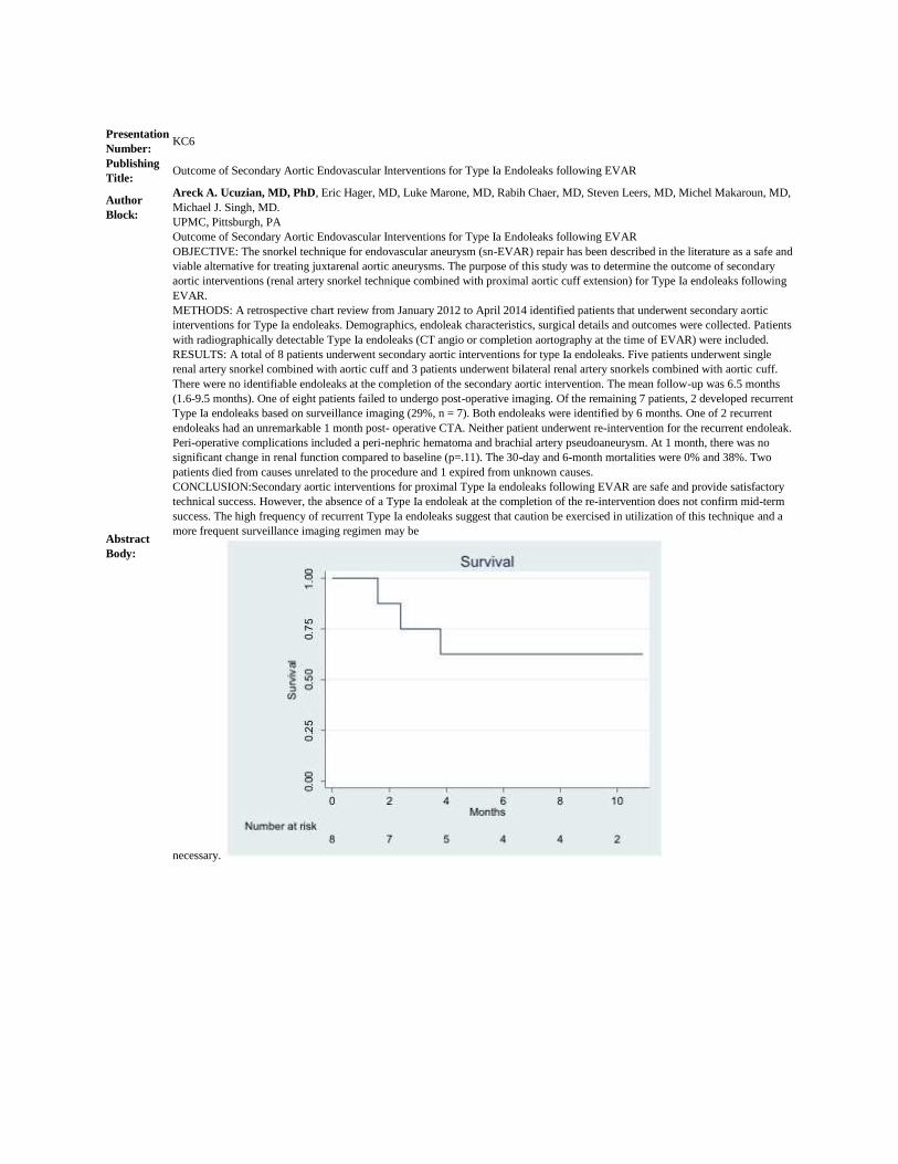
There were no identifiable endoleaks at the completion of the secondary aortic intervention. The mean follow-up was 6.5 months
(1.6-9.5 months). One of eight patients failed to undergo post-operative imaging. Of the remaining 7 patients, 2 developed recurrent
Type Ia endoleaks based on surveillance imaging (29%, n = 7). Both endoleaks were identified by 6 months. One of 2 recurrent
endoleaks had an unremarkable 1 month post- operative CTA. Neither patient underwent re-intervention for the recurrent endoleak.
Peri-operative complications included a peri-nephric hematoma and brachial artery pseudoaneurysm. At 1 month, there was no
significant change in renal function compared to baseline (p=.11). The 30-day and 6-month mortalities were 0% and 38%. Two
patients died from causes unrelated to the procedure and 1 expired from unknown causes.
CONCLUSION:Secondary aortic interventions for proximal Type Ia endoleaks following EVAR are safe and provide satisfactory
technical success. However, the absence of a Type Ia endoleak at the completion of the re-intervention does not confirm mid-term
success. The high frequency of recurrent Type Ia endoleaks suggest that caution be exercised in utilization of this technique and a
more frequent surveillance imaging regimen may be
necessary.
Karmody Competition Topic: Carotid
Presentation
Number: KC7
Publishing
Title: Vascular Interventions in the Management of Advanced Head and Neck Cancer
Author
Block:
Colin Brandt, MD, Neil Reddy, MD, Sadie Ahanchi, MD, Daniel Karakla, MD, FACS, Jean Panneton, MD, FACS.
Eastern Virginia Medical School, Norfolk, VA
Abstract
Body:
OBJECTIVE: Extension of head and neck malignancies into major blood vessels can complicate patient management. Our study
examined the multidisciplinary approach to the treatment of advanced head and neck cancer.
METHODS: We performed a retrospective review of cancer patients treated by head and neck surgery (HNS) and vascular surgery
from 2007-2014. Data concerning history of cancer and radiation therapy, operative interventions, and perioperative morbidity and
mortality was collected.
RESULTS:: 31 patients with head and neck cancer were operated on by HNS and required vascular intervention. Vascular surgery
intervention was synchronous (23) or metachronous (8) to the associated cancer procedure. Post-resection interventions occurred at
an average of 4 years (41 days- 14 years). 25 patients (81%) had recurrent disease, of which 24 had previous radiation therapy and
14 had prior resection. 22 patients (71%) had flap coverage. 7 patients (23%) required emergent as opposed to elective
intervention, all for bleeding. Indications for vascular intervention were invasion/encasement of major vasculature (17),
bleeding/blowout (8), stenosis/occlusion (3), and aneurysm/pseudoaneursym (3).
32 index operations were performed: Exploration/dissection in 8 patients (4 bilateral carotid arteries (CCA), 3 unilateral CCA, 1
innominate artery). Resection in 17 patients: 9/17 without reconstruction (7 external carotid artery (ECA), 1 internal carotid artery
(ICA), 1 CCA) and 8/17 with reconstruction (6 CCA to ICA bypasses, 1 innominate/SCA bypass, 1 innominate to axillary vein
bypass). 6 patients received stents (5 CCA/ICA and 1 innominate). 1 patient had an angioembolization (ECA).
6 patients (19%) required reintervention after index vascular procedure. (see Table 1)
There were three 30-day mortalities (9.7%), all from blowout. Based on Kaplan Meyer analysis, bypass and stent primary patency
at 1 year was 67% and 100%, respectively. Survival at 1 and 2 years post vascular intervention was 62% and 19%, respectively. A
significant increase in mortality (7 vs 22 months, p=0.06) and 30-day mortality rate (43% vs 0%) was noted in emergent versus
elective cases.
CONCLUSIONS: Vascular involvement in head and neck cancer indicates advanced disease, commonly in patients who have had
previous RT. Nonetheless, vascular intervention is feasible. Optimal treatment of these patients requires a multidisciplinary
approach.
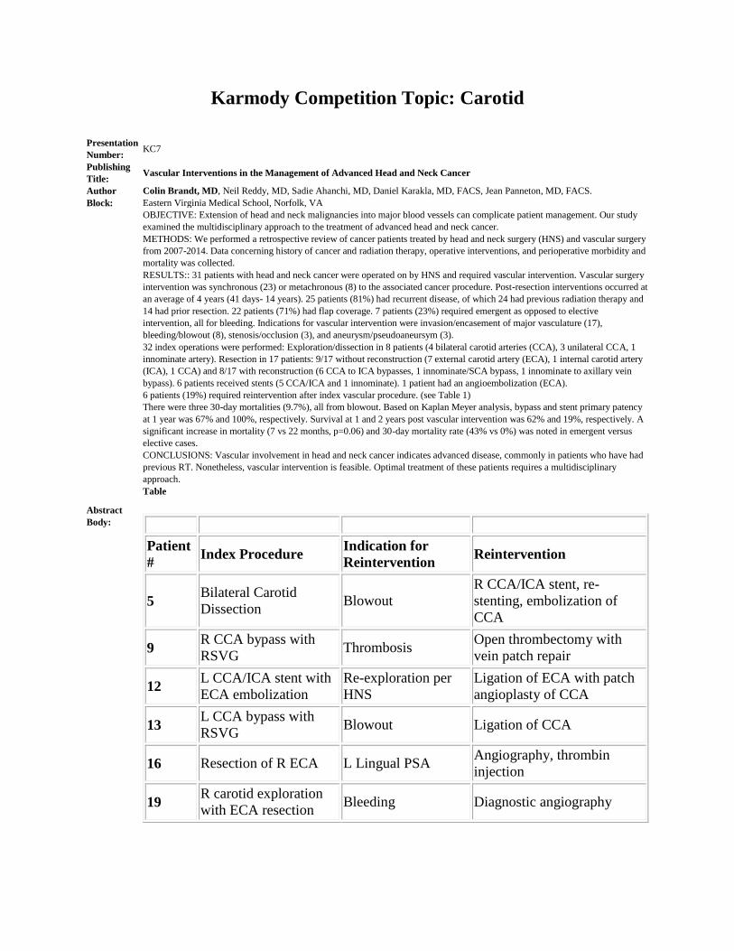
Table
Patient
# Index Procedure
Indication for
Reintervention Reintervention
5 Bilateral Carotid
Dissection Blowout
R CCA/ICA stent, re-
stenting, embolization of
CCA
9 R CCA bypass with
RSVG Thrombosis
Open thrombectomy with
vein patch repair
12 L CCA/ICA stent with
ECA embolization
Re-exploration per
HNS
Ligation of ECA with patch
angioplasty of CCA
13 L CCA bypass with
RSVG Blowout Ligation of CCA
16 Resection of R ECA L Lingual PSA Angiography, thrombin
injection
19 R carotid exploration
with ECA resection Bleeding Diagnostic angiography

Presentation
Number: KC8
Publishing
Title: Carotid Endarterectomy: Gender Disparities and Anesthetic Choice Influences Outcome and Economics
Author
Block:
Elizabeth L. Chou, BS, Michael Sgroi, MD, Nii-Kabu Kabutey, MD, Roy M. Fujitani, MD.
University of California Irvine School of Medicine, Irvine, CA
Abstract
Body:
OBJECTIVE: Carotid endarterectomy (CEA) is the most commonly performed surgical procedure to reduce risk of stroke. The
operation may be performed under local (LA) or general anesthesia (GA). Despite perceived advantages of LA, previous trials have
found no difference in rates of transient ischemic attack, stroke, myocardial infarction and death in CEA under LA compared with
GA. We performed a retrospective review to see if gender may be associated with type of anesthesia and post-operative outcomes.
METHODS: Patients who underwent CEA between 2005-2011 were extracted from the National Surgical Quality Improvement
Program (NSQIP). The cohort was separated by sex and anesthesia type. Primary endpoints included 30-day incidence of stroke
and myocardial infarction. Secondary endpoints included 30-day postoperative local complications, operative time and length of
surgical stay.
RESULTS: Of the 41,442 CEA cases identified, most patients were male (16,874 F, 24,568 M) and most cases were performed
under GA (86% of female cases, 85% female cases). Adjusted multivariate analysis showed no statistical difference between
primary endpoint outcomes based on gender or type of anesthesia used. There was, however, a trend for increased risk of 30-day
post-operative local complications and 30 day incidence of myocardial infarction amongst CEA conducted under GA compared to
LA. Operative time and length of stay was decreased in females, regardless of anesthesia used (mean difference -8.15 [10.09, -
6.21] p <0.0001; 0.34 [0.14, 0.54] p <0.02). Use of general anesthesia was associated with increased operative time, and increased
total length of total surgical stay, regardless of sex, with statistical significance.
CONCLUSIONS: There is no significant difference in post-operative outcomes between women and men regardless of type of
anesthesia used for CEA. GA was found to be associated with increased length of stay and operative time, compared with LA in
women and men. This finding suggests that choice of anesthesia may have significant economic considerations for patients and
institutions. The trend of increased 30 day post-operative local complications and 30 day myocardial infarction amongst GA cases
also support the use of LA for CEA. These factors warrant further evaluation to improve patient outcomes and economic impact of
this commonly performed procedure.
Presentation
Number: KC9
Publishing
Title: Common Carotid Artery Pseudoaneurysm Presenting as Upper Airway Obstruction
Author Block: Gregory T. Clabeaux, D.O.1, Paul M. Anain, M.D.1, Joseph Anain, Sr., MD2. 1Sister's of Charity Hospital, Buffalo, NY 2Sister's of Charity Hospital, Williamsville, NY
Abstract
Body:
OBJECTIVE: In this discussion our intent is to review the etiologies of carotid artery pseudoaneurysm, and to discuss methods
of treatment.
METHODS: Our particular case was managed with careful preoperative evaluation of the imaging and then surgical exploration.
Repair of the vessel after isolation and debridement with GSV patch angioplasty. Upon completion, intraoperative patency was
confirmed with duplex ultrasonography.
RESULTS: The patient's initial complaint on presentation to the ED at the outside facility was that of upper airway compression.
The patient was initially admitted after non contrast ct of the neck with a diagnosis of retropharyngeal abscess. It was only on
subsequent imaging with IV contrast that the pseudoaneurysm was able to be identified definitively.Favorable outcome, no
neurologic deficits or recurrence of pseudoaneurysm in ths particular patient at 1 year follow. up.
CONCLUSIONS: In this particular case, the presentation was atypical in timing, as the only possible identifiable trauma was
three weeks previous, and it did not seem remarkable to the patient. It took extensive questioning and then the patient recalled a
possible mechanism of trauma. Physical exam was also atypical as there was no pulsatility to the mass.
A high index of suspicion must be maintained in the diagnosis of a cervical mass particularly when in the anterior cervical
triangle.

Presentation
Number: KC10
Publishing
Title: Large neck paraganglioma with atypical blood supply from thyrocervical trunk
Author Block: Uwe Fischer, M.D., Ph.D., Mahesh Ramchandani, Orlando Diaz, Alan B. Lumsden.
Houston Methodist DeBakey Heart & Vascular Center, Houston, TX
Abstract
Body:
OBJECTIVES: Paragangliomas of the neck are rare tumors. The most common paragangliomas of the neck are located in the
carotid bifurcation. These tumors are generally supplied by the external carotid artery. We present a case of a large right lower
neck paraganglioma with atypical blood supply from the right thyrocervical trunk.
RESULTS: A 47 year old female presented with a right neck mass. Diagnostic workup included a computer tomography (CT)
and ultrasound of head and neck which revealed a 4.5 cm in diameter hypervascular mass in the right lower neck lateral to the
internal jugular vein. On the left side a smaller hypervascular lesion was discovered in the carotid bifurcation. We first addressed
the large right sided neck mass.
The patient was taken to the angiosuite for embolization of the right sided neck mass prior to excision. Selective angiogram of the
right thyrocervical trunk visualized two main branches feeding the highly vascularized mass. Using a microcatheter system all
branches were selectively cannulated and embolized with ethylene vinyl alcohol (Onyx Liquid Embolic System). A completion
angiogram showed embolization of the entire mass.
The next day, the patient was taken to the operating room for excision of the mass. The mass was located postero-lateral to the
internal jugular vein abutting but not adhered the common carotid artery. The embolized vessels were clearly visible which
facilitated excision and minimized blood loss. The patient tolerated the procedure well and was discharged home on post-
operative day 2. Histologic workup revealed a 4.5 x 3.4 x 2.2 cm paraganglioma.
CONCLUSIONS: Due to the size and vascular structure we decided to embolize the mass prior to resection. We chose the Onyx
Liquid Embolization System which consists of ethylene vinyl alcohol copolymer dissolved in DMSO (dimethyl sulfoxide) and
suspended micronized tantalum powder to provide contrast for visualization under fluoroscopy that precipitates in situ into a
black, spongy embolus which makes it easy to visualize vessels intra operatively. As we could readily identify feeding vessels
resection of the tumor was facilitated with minimal blood loss. We believe that preoperative embolization can be useful in
selected cases, especially verifying the blood supply and assessing the risk of cerebral embolization to minimize the risk of stroke.
Presentation
Number: KC11
Publishing
Title: Extracranial Internal Carotid Artery Aneurysms: A Review Of The Past 20 years
Author Block: Shahin Pourrabbani, MD, Mauricio Szuchmacher, MD, K. V. Krishnasastry, MD, FACS.
North Shore LIJ Medical Center, Manhasset, NY
Abstract
Body:
Extracranial Internal Carotid Artery Aneurysms: A Review Of The Past 20 years
OBJECTIVES: Extracranial Internal Carotid Artery Aneurysm (ICAA) is rare and surgical repair accounts for less than 2% of all
carotid operations. Their significance is linked to the natural history that, if left untreated, ICAA has a high incidence of
significant cerebrovascular morbidity and mortality. In this study, we discuss the pathogenesis, clinical manifestation, diagnosis,
and treatment options of this rare entity.
METHODS: A systematic literature search was conducted using MEDLINE, Cochrane Library and PubMed databases on all
articles pertaining to Extracranial internal carotid artery aneurysm published in the English language between January 1994 and
January 2014. Following keywords were used: carotid artery aneurysms, extracranial carotid artery aneurysms, and extracranial
internal carotid artery aneurysms. A total of 50 review articles were found, and exclusion criteria were intracranial aneurysms,
traumatic and non-traumatic dissections, and non-internal carotid artery aneurysms.
RESULTS: Fifteen articles were included in this study. According to the review of 52 patients, Internal Carotid Artery
Aneurysms tend to develop later in life, at an average age of 37.5 years (range, 3 to 76 years). Among ICAA patients, 28 were
men and 23 were women (M:F ratio 1.2). All patients were symptomatic, and predominant presenting symptoms are TIA or
stroke, Cranial Nerve involvement, and a pulsatile cervical or parapharyngeal mass. Aneurysms were divided into 3 types:
Degenerative Aneurysm (DA) was diagnosed in 21 patients (40.3%), Mycotic (MA) in 6 (11.5%), and Pseudoaneurysm/Trauma
in 8 (15.3%). Surgical intervention was performed in 50 patients (96.1%) and endovascular procedure in 2 (3.8%). Complications
occurred in 8 patients (15.3%), and included residual neurologic deficit, myocardial infarction, and death.
CONCLUSIONS: Although rare, ICA aneurysms should be considered in the differential diagnosis of a mass in the anterior
triangle of the neck. Although recently endovascular therapy became a feasible option, open surgical repair is still the treatment of
choice for most symptomatic patients. Additional randomized controlled trials studying ICAA is therefore necessary to confirm
the conclusions presented here.
Presentation
Number: KC12
Publishing
Title:
“Type & Screen” for CEA is rarely needed: Development of a risk prediction model using the NSQIP database and demonstration
of significant cost-saving potential
Author Block:
Lars Stangenberg, MD PhD, Thomas Curran, MD, Feroze Mahmood, MD, Dominique Buck, MD, Jack McCallum, MD,
Jeremy Darling, Marc L. Schermerhorn, MD.
Beth Israel Deaconess Medical Center, Boston, MA
Abstract
Body:
OBJECTIVES: Preoperative testing for carotid endarterectomy (CEA) often includes blood typing and antibody screen (T&S). In
our institutional experience, however, transfusion for CEA is rare. We assessed transfusion rate and risk factors in a national
clinical database to identify a cohort of patients in whom T&S can be avoided with the potential for substantial cost savings.
METHODS: Using the NSQIP database, transfusion events and timing were established for all elective CEAs in 2012.
Comorbidities and other characteristics were compared for patients receiving intra- or postoperative transfusion and those that did
not using two-tailed t-test or Fisher’s exact test. Using one half of the data set, a point-based risk prediction model that was then
validated on the other half.
RESULTS: Of 7601 patients undergoing CEA in 2012, 139 received at least one transfusion prior to discharge (1.8%). 80% of
transfusions occurred on POD#0 or #1. Hematocrit 1.2 [OR: 3.0; 95%CI: 1.7-5.1], female sex [OR: 1.8; 95%CI: 1.1-3.1] and
preoperative open wound [OR: 5.0; 95%CI: 1.6-16.3] among other risk factors predicted transfusion.
Score was calculated with 1 point for female gender, preoperative dyspnea, preoperative coagulopathy and BMI 1.2 and 8 points
for hematocrit <30. A risk prediction model based on these data produced a C-statistic of 0.84. Application of this model to the
validation set demonstrated a C-statistic of 0.76. 77% of patients in the validation set received a score of 2 or less corresponding
to a transfusion risk of 1.1%. Omitting a T&S in these patients would generate a potential annual cost-saving to NSQIP hospitals
of over $1,000,000 based on our institutional charge.
CONCLUSIONS:While T&S is commonly performed for patients undergoing CEA, transfusion following CEA is rare and well
predicted by a transfusion risk score. Avoidance of T&S in this low-risk population provides a substantial cost-saving opportunity
without compromise of patient care.
Presentation
Number: KC13
Publishing
Title: Carotid Body Tumors
Author Block: M. A. Mansour, M.D., Jennifer J. Watson, MD, Michelle C. Kosovec, MD, Eanas Yassa, MD, Jason D. Slaikeu, MD.
Spectrum Health, Grand Rapids, MI
Abstract
Body:
OBJECTIVES: Carotid Body Tumors (CBT) are rare. Preoperative embolization has been advocated to decrease blood loss and
facilitate resection.
Purpose: To review in detail the clinical management and outcomes of patients presenting with CBT for resection without
preoperative embolization.
METHODS: A retrospective chart review of all patients presenting to our practice for CBT resection. Preoperative and operative
details were reviewed as well as long-term follow-up.
RESULTS: In a 10-year period, ten patients (7 women and 4 men), average age 51 (range 38-60) underwent resection of CBT.
Six patients had a palpable mass and the others had discrete masses detected incidentally on imaging. All patients had either
preoperative CTA or angiography. None had tumor embolization. Two patients required nasotracheal intubation and mandibular
subluxation to facilitate resection. Harmonic scalpel and monopolar cautery were used in all patients. The average tumor size was
3.6 cm (range 0.7-5.8 cm). Classification of the tumors was: Shamblin type I (one patient), type II (5 patients) and type III (5
patients). Blood loss averaged 150 ml. Two patients had residual tumor near the skull base. One patient received postoperative
radiation and the other presented 5 years later with intracranial recurrence. There were no deaths or perioperative strokes. Two
patients had transient swallowing difficulties and one patient with vagus nerve involvement had a permanent deficit.
CONCLUSONS: CBT while rare are quite challenging to resect. Despite a higher incidence of aggressive CBT in our patients,
safe resection was accomplished in most cases without preoperative embolization.

Karmody Competition Topic: Lower Extremity
Presentation
Number: KC14
Publishing
Title: Predictors of Bleeding in Revascularization for Critical Limb Ischemia and Claudication
Author
Block:
Sara L. Zettervall, MD, Peter A. Soden, MD, Dominique B. Buck, MD, John C. McCallum, MD, Jeremy D. Darling, BA, Marc L.
Schermerhorn, MD.
Beth Israel Deaconess Medical Center, Boston, MA
Abstract
Body:
OBJECTIVE: Increased transfusion requirements have been identified as a risk factor for multiple adverse outcomes in patients
undergoing lower extremity revascularization. However, previous studies have not identified clear predictors for bleeding in this
patient population. In this study we aim to identify the predictors for bleeding in patients with critical limb ischemia (CLI) and
claudication.
METHODS: All patients undergoing intervention for CLI or claudication between 2011 and 2012 in the Vascular Targeted NSQIP
database were identified and analyzed separately. Bleeding was defined as transfusion or return to the operating room for bleeding.
Patients undergoing emergent intervention were excluded. Patient demographics and perioperative risk factors including pre-
operative anticoagulation, hematocrit, and preoperative transfusion requirements were identified. Univariate analysis was
completed using Chi-Square, Fisher Exact, and T-test. A stepwise multivariate logistic regression was utilized to compare
independent predictors.
RESULTS: 5163 patients undergoing revascularization were identified including 3067 patients undergoing intervention for CLI
and 2096 undergoing intervention for claudication. A hematocrit less than 35 was an independent risk factor for bleeding regardless
of operative indication (OR 3.0, 95% CI 2.5-3.5). A dependent functional status was an independent predictor of bleeding in
patients undergoing open procedures for critical limb ischemia (OR 1.7, 95% 1.3-2.2). Uncorrected anticoagulation or a bleeding
disorder was an independent predictor for bleeding in claudicants undergoing open procedures (OR 2.1, 95% CI 1.44-2.97). There
are no clear predictors of bleeding for claudicants undergoing endovascular intervention, likely due to very low rates of bleeding.
CONCLUSION: A preoperative hematocrit less than 35 is an independent predictor of bleeding in patients undergoing intervention
for both critical limb ischemia and claudication. Functional status is predictive of bleeding in critical limb ischemia and uncorrected
anticoagulation or bleeding disorders are predictive in patients with claudication. These factors should be accounted for in
preoperative planning to identify patients requiring transfusion in the perioperative period.
Predictors of bleeding by operative intervention
Claudicatio
n and Critical
Limb
Ischemia
Claudication Critical Limb Ischemia
All Cases
Suprainguinal
Endovascul
ar
Supringuina
l Open
Infrainguinal
Endovascul
ar
Infrainguin
al Open
Suprainguinal
Endovascul
ar
Suprainguin
al Open
Infrainguinal
Endovascul
ar
Infrainguin
al Open
Hematocrit <35
3.0 (2.2-4.7)
Nonsignificant
4.1(1.8-9.2) Nonsignificant
3.2 (1.9-5.2)
4.8 (1.9-12.2)
3.2(1.5-6.8) 3.5 (2.1-5.7)
2.2 (1.7-3.9)
Dependent Functional
Status
1.8 (1.4-
2.7)
Nonsignific
ant
Nonsignific
ant
Nonsignific
ant
Nonsignific
ant
Nonsignific
ant
4.8(1.6-
14.3)
Nonsignific
ant
1.8 (1.3-
2.6)
Uncorrected
Anticoagulation or
Bleeding
Disorder
1.6 (1.3-
1.9)
Nonsignific
ant 4.1(1.8-9.3)
Nonsignific
ant
2.5 (1.5-
4.1)
Nonsignific
ant
Nonsignific
ant
Nonsignific
ant
Nonsignific
ant
Preoperative
Sepsis
2.9 (1.7-
5.2)
Nonsignific
ant
Nonsignific
ant
Nonsignific
ant
Nonsignific
ant
Nonsignific
ant
Nonsignific
ant
Nonsignific
ant
Nonsignific
ant
Steroid Use Nonsignificant
Nonsignificant
Nonsignificant
Nonsignificant
Nonsignificant
Nonsignificant
Nonsignificant
Nonsignificant
2.2 (1.4-3.5)
Smoking Nonsignificant
Nonsignificant
Nonsignificant
Nonsignificant
Nonsignificant
Nonsignificant
Nonsignificant
2.2(1.3-3.5) Nonsignificant
Dialysis Nonsignific
ant
Nonsignific
ant
Nonsignific
ant
Nonsignific
ant
Nonsignific
ant
Nonsignific
ant
Nonsignific
ant
Nonsignific
ant
1.8 (1.2-
2.6)
Presentation
Number: KC15
Publishing
Title:
Preoperative Evaluation Prior to Major Lower Extremity
Amputation: Does it Occur?
Author Block: David Hardy, MD1, Xiaoyi Tend, MD2, Sean Lyden, MD1. 1Cleveland Clinic Foundation, Cleveland, OH, USA, 2Cleveland Clinic Foundation, Lyndhurst, OH
Abstract
Body:
OBJECTIVES: Peripheral arterial disease (PAD) affects more than 5 million American adults. Critical limb ischemia (CLI) is a
major consequence of PAD and affects 250,000 Americans per year. For CLI patients who do not undergo revascularization, the
risk of amputation within 1 year is 73% for Rutherford class IV and 95% for patients in class V or VI.
Allie reported that less than half (49%) of amputation patients had any diagnostic vascular evaluation prior to a major lower
extremity amputation. They suggested that every patient with CLI should have a vascular imaging study to evaluate for
revascularization to avoid amputation.
We evaluated all patients who underwent a major amputation and looked at whether or not these patients had a diagnostic
vascular examination or testing prior to their amputation. We propose that all patients have a vascular evaluation exam prior to
major LE amputation and some only need a physical exam.
METHODS: A retrospective analysis of major LE amputations was performed. Patient demographics, comorbidities, type of
amputation, reason for amputation, Rutherford classification, type of preoperative vascular examination, and time since the last
vascular examination were evaluated.
RESULTS: During 2010 to 2013, 281 patients (64.1% male) required major LE amputation. The average age was 65 years
(range, 25-96 years). AKA was performed in 39.1% of patients whereas BKA was performed in 60.9%. Amputation was
performed due to CLI in 92.9% of patients whereas 7.1% of amputations were performed due to diabetes (ulcer, wet
gangrene/sepsis) or other reasons. Preop vascular evaluation was performed in 100% of patients undergoing major amputation.
Pulse and wound physical examination was most common(99.3%) followed by PVR/ABI (78.8%), Angio (54.8%), and CTA
(29.3%), duplex ultrasonography (41.3%), and MRA (0.4%). Amputations most commonly occurred due to Rutherford
classification VI (63.3%) with 97.2% of patients having Rutherford IV-VI classification. Patients with nonsalveagable limbs and
non ambulatory patients did not have additional imaging.
CONCLUSIONS: We demonstrate that 100% of patients undergo preop vascular evaluation prior to major LE amputation at a
tertiary referral hospital. Up to 50% of patients already have non salvageable limbs or are not revascularization candidates and do
not need further diagnostic imaging. Recommending imaging in all individuals with CLI prior to major amputation is a waste of
health care resources and money.

Presentation
Number: KC16
Publishing
Title: Popliteal Artery injuries: A Meta-Analysis and Literature Review of a Complex Injury
Author Block:
Tejas R. Shah, MD, Ans Fakiha, MD, Diego Ayo, MD, Mark A. Adelman, MD, Thomas S. Maldonado, MD, Caron B.
Rockman, MD, Firas Mussa, MD.
NYU Medical Center, New York, NY
Abstract
Body:
OBJECTIVE: Popliteal artery injuries are uncommon but potentially morbid and are associated with a significant risk of
amputation. Previous studies identifying risk factors associated with high amputation rates have been limited to small cohort
studies. Our aim is to conduct a meta-analysis of existing literature to identify predictors of limb loss associated with popliteal
artery injuries.
METHODS: A systematic literature review was conducted on studies reporting amputation rate associated with popliteal artery
injuries using PubMed, Embase and Medline databases. Eligible studies included those that reported on civilian population,
mechanism of injury, and amputation rates. The military population, popliteal pseudoaneurysms, or other associated arterial
injuries were excluded.
RESULTS: 705 articles were identified between 1954-2014. A total of 145 full text articles were reviewed of which 22 articles
met inclusion criteria. 2370 patients with 2394 popliteal artery injuries were identified. 82% were male with a mean age of 32
years. Overall amputation rate was 16.3% (95% CI=[13.8%, 19.2%]). Mortality rate was 3.8% (95% CI=[2.7%, 5.1%]). Injury
distribution included blunt trauma (61%), penetrating injury (38%), and iatrogenic (1%). Patients presenting with a blunt injury
were 2.03x more likely to have amputation than those with penetrating injury (pooled odds ratio = 2.03, 95% CI = [1.78, 2.32],
p<0.0001). Patients that presented with orthopedic fractures were 5.93x more likely to have an amputation (OR = 5.93, 95% CI =
[1.51, 23.39], p=0.011). Among those patients with venous injuries, 19.4 % had amputation (OR = 1.57, 95% CI = [0.77, 3.21],
p=0.213). Mangled extremity severity score (MESS) was stratified in 3 studies. The amputation rate were higher in patient with
high MESS score (50%) than patient with low MESS score (9%) (OR = 9.38, 95% CI = [8.34, 10.56], p<0.001). From 1979 to
2014, the most commonly performed popliteal artery repair technique was saphenous vein grafts (36%) followed by end-end
anastomosis (24%).
CONCLUSIONS:Popliteal artery injuries associated with blunt trauma, concurrent orthopedic injuries, and high MESS score are
at higher risk of lower extremity amputation. Associated venous injury or fasciotomy failed to show any significant association to
limb loss. Popliteal vascular injury remains a challenging clinical entity associated with a significant risk of amputation.
Identifying risk factors associated with higher amputation rates may help guide clinical management of these complex injuries.
Presentation
Number: KC17
Publishing
Title: Anesthesia type for major lower extremity amputation in frail elderly patients does not affect outcomes
Author Block:
Carla C. Moreira, M.D., Denis Rybin, Alik Farber, M.D., Jeffrey A. Kalish, M.D., Mohammad H. Eslami, M.D., Sebastian
Didato, M.D., Jeffrey J. Siracuse, M.D..
Boston University School of Medicine, Boston Medical Center, Boston, MA
Abstract
Body:
OBJECTIVES: The purpose of this study was to determine the impact of anesthesia type; general anesthesia (GA) and
regional/spinal (RA), on outcomes after lower extremity amputation in frail elderly patients.
METHODS: The American College of Surgeons National Surgical Quality Improvement Program (ACS-NSQIP) dataset (2005-
2012) was queried to identify all patients ages greater than 75 years-old with partial or total functional impairment who
underwent major lower extremity amputations with use of GA or RA. To ensure comparability of the groups we used 2:1
propensity matching based on clinically important and significantly different at 0.2 level factors and multivariable analyses
adjusting for the same factors.
RESULTS: There were 3260 patients identified - 702 RA and 2558 GA. The mean age was 82 and 50% were male. Anatomical
distribution was 59% above the knee (AKA) and 41% below the knee (BKA). Patients undergoing GA were more likely to have
impaired sensorium (9% vs. 6%, P=.035), be on anticoagulation or have a bleeding disorder (33% vs. 17%, P<.01), have had a
previous operation within 30 days (16% vs. 10%, P<.01), and were more likely to be operated on by a general surgeon (16% vs.
12%, P=.03). Age and other comorbidities were similar. Propensity matching showed that RA was associated with longer
anesthesia time to surgery (41±31 min vs. 36 ±34 min, P<.01), however there was no difference in operative time (63.2±31 min
vs. 64.8±33 min). There was no difference in complications between GA and RA - specifically 30-day mortality (14.4% vs.
11.7%, P=0.14), postoperative myocardial infarction (MI) (2.9% vs. 3.1%, P=08), pulmonary complications (7.3% vs. 6.7%,
P=0.6), stroke (0.7% vs. 0.9%, P=0.7), UTI (6.7% vs. 6.5%, P=.9), and wound complications (7.6% vs. 7.6%, P=0.75). Median
length of stay for both groups was 5 days. Multivariate analysis of complications and 30-day mortality confirmed that anesthesia
type was not an independent risk factor.
CONCLUSONS: The mode of anesthesia, general vs. regional/spinal, was not found to be associated with perioperative outcomes
following major lower extremity amputation in the frail geriatric population. GA can safely be used in this high risk patient
population.
Presentation
Number: KC18
Publishing
Title: Endovascular Treatment of Blunt Traumatic Bilateral Common Iliac Artery Intimal Injury
Author
Block:
Marissa Toma, MD, Carlos Rosales, MD, Satish Muluk, MD.
Allegheny General Hospital, Pittsburgh, PA
Abstract
Body:
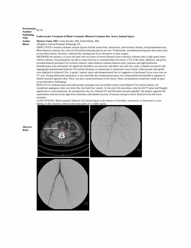
OBJECTIVES-Common traumatic arterial injuries include transections, dissections, arteriovenous fistulas, and pseudoaneurysms.
Blunt bilateral common iliac artery (CIA) intimal shearing injuries are rare. Traditionally, aortobifemoral bypasses have been used
to treat these lesions. However, endovascular stenting may be an alternative to open surgery.
METHODS-We present a 22-year-old male with two hours of severe bilateral lower extremity ischemia after a high-speed motor
vehicle collision. On presentation, he had no motor function or sensation below his knees. CTA of the chest, abdomen, and pelvis
revealed bilateral proximal CIA occlusive injuries. Open bilateral common femoral artery exposure and right iliofemoral
thrombectomy were performed. No significant thrombus was removed, and inflow was still very weak. A sheath was placed, and
angiography demonstrated bilateral CIA intimal shearing; no transections or dissections were evident. Endovascular stent grafts
were deployed in bilateral CIAs. A grade 1 splenic injury and intraperitoneal free air suggestive of perforated viscus were seen on
CT scan. During abdominal exploration, it was noted that the retroperitoneal space was compromised and included a segment of
bluntly denuded sigmoid colon. There was also a small perforation of the ileum. These circumstances would have made an open
revascularization challenging.
RESULTS-A combined open and endovascular technique was successfully used to treat bilateral CIA intimal injuries. On
completion angiogram, there was brisk flow into both iliac vessels. At the end of the procedure, only the left PT pulse had Doppler
signals due to vasoconstriction. By postoperative day two, bilateral PT and DP pulses became palpable. The patient regained full
sensorimotor function in his right lower extremity with limited recovery of sensory and gross motor function in his left lower
extremity.
CONCLUSIONS- Blunt traumatic bilateral CIA intimal injury in the absence of thrombus, transection, or dissection is a rare
finding. In this situation, endovascular stent grafts are a viable option.

Presentation
Number: KC19
Publishing
Title: Early Post-Registry Experience with Drug Eluting Stents in the SFA
Author Block:
Mathew Wooster, Kirsten Dansey, BS, Paul Armstrong, DO, Martin Back, MD, Karl Illig, MD, Brad Johnson, MD, Neil
Moudgill, MD, Peter Nelson, MD, Brian Park, MD, Murray Shames, MD.
University of South Florida, Tampa, FL
Abstract
Body:
OBJECTIVE: Restenosis remains the primary failure mode after stenting of the superficial femoral artery. Drug-eluting stent
technology claims to reduce stent failure and improve durability of endovascular management of SFA occlusive disease. We seek
to present our early experience with the Cook Zilver PTX stent.
METHODS: We have retrospectively reviewed a prospectively collected database of patients undergoing placement of the Cook
Zilver PTX stent for SFA or popliteal disease since its availability to our institution in October 2013. Patients treated with
additional stents to extend coverage of the same lesion were excluded. Patient demographics, comorbidities, concomitant
procedures, TASC classification, procedural details, and follow up were reviewed.
RESULTS: Thirty-one limbs in thirty patients were treated with PTX, five were excluded for concomitant use of non-PTX stents,
leaving 26 limbs in 26 patients for analysis. Indications for intervention were claudication in 17 (65.4%), rest pain in 1 (3.8%)
and tissue loss in 8 (30.8%). A median of 2 PTX stents per limb was used to treat a mean length of 14.2cm +/- 11cm with initial
technical success of 100%. Concomitant inflow (N= 4) or atherectomy (N = 2) interventions were performed in 23%. Sixty-nine
percent of lesions were TASC C (N = 7) or D (N = 11) and 42% were total occlusions. Over a mean 20 week follow up, 2
occlusions were noted (mean 27 weeks), one was treated with surgical bypass, the other with endovascular salvage. Review of
completion angiograms from the index procedures demonstrated severely compromised outflow in the patient later treated with a
fem-distal bypass. A residual stenosis just distal to the stent was noted in the second, which was able to be salvaged with a repeat
angioplasty and additional stent placement. Limb salvage in the series was 92.3% with the two patients requiring major
amputations for infected, non-healing wounds despite patent stents.
CONCLUSION: On mean 20-week follow we have seen 92.3% primary patency and 96.2% secondary patency. A larger number
of patients and longer follow up will be required to determine the true real-world efficacy of this touted drug eluting device, but
early experience is encouraging and warrants continued trial.

Presentation
Number: KC20
Publishing
Title: Mid-term Results of Endovascular Stent Graft Repair of Asymptomatic and Symptomatic Popliteal Artery Aneurysms
Author Block: Andrew Cha, DO, Naiem Nassiri, MD, Randy Shafritz, MD, Saum Rahimi, MD.
Rutgers-Robert Wood Johnson Medical School, New Brunswick, NJ
Abstract
Body:
OBJECTIVES: The treatment of popliteal artery aneurysms (PAA) has evolved from open surgical revascularization to
endovascular stent graft repair with favorable mid-term outcomes. This study reviews a single-center experience of endovascular
popliteal artery aneurysm repair in both asymptomatic and symptomatic patients, including those who presented emergently with
acute limb ischemia.
METHODS: A retrospective review was performed to identify all endovascular PAA repairs between April 2008 and October
2013. Data analysis included age of patients, indication for treatment, number of stent grafts per PAA treated, number of runoff
vessels, perioperative outcome, and limb salvage.
RESULTS: Thirty-five popliteal artery aneurysms (mean size 2.84 ± 1.25 cm) in 27 patients (mean age, 73.5 ± 11.4 years, 96%
men) were treated with endovascular covered stent graft(s) through open (74%) or percutaneous
approach (26%). Indications for treatment included symptomatic presentation in 26% (n = 9), half of these (n = 5) emergent with
acute limb ischemia. Asymptomatic presentation was found in 74% (n = 26). The average number of stent grafts per PAA treated
was 1.9 ± 0.7. Primary patency at 1 year was 91.4%. Limb salvage rate was 97%. There were no perioperative (30-day) deaths.
The mean follow-up was 18.9 months (range 1-55 months). A total of three stent graft failures occurred, all within 90 days (mean
of 50 ± 34 days). Stent graft failure rate was found to be statistically significant in patients younger than 70 years old (P = 0.03),
while the indication for treatment, number of stents, and runoff vessels were not significant contributing factors.
CONCLUSIONS: Endovascular stent graft repair of popliteal artery aneurysms is both safe and technically feasible in patients
with either asymptomatic or symptomatic presentation. Despite favorable mid-term outcomes, early
follow-up and closer observation especially in patients younger than 70 years old may be warranted.

Karmody Competition Topic: Other Presentation
Number: KC21
Publishing
Title: Aortic remodeling after TEVAR for acute complicated type B aortic dissection
Author Block:
Ryan Bishop, BS, Daniel Ocazionez-Trujillo, MD, Kristofer Charlton-Ouw, MD, Rana Afifi, MD, Harleen Sandhu, MD, Samuel
Leake, BS, Naveed Saqib, MD, Maria Codreanu, MD, Sheila Coogan, MD, Charles Miller, PhD, Anthony Estrera, MD, Hazim
Safi, MD, Ali Azizzadeh, MD.
The University of Texas at houston Medical School, Houston, TX
Abstract
Body:
OBJECTIVE: Thoracic endovascular aortic repair (TEVAR) is a treatment option for patients with acute complicated type B
aortic dissection (ACTBAD). The optimal extent of aortic coverage during TEVAR is not well defined. Our current practice
involves coverage of the proximal entry tear with a single device. The purpose of this study was to evaluate aortic remodeling
after TEVAR for ACTBAD.
METHODS: We reviewed TEVAR patients with ACTBAD between 2006-2014. The diameter, total aortic area, true lumen (TL),
and false lumen (FL) were measured at six locations (1. left subclavian, 2. pulmonary artery, 3. left atrium, 4. celiac, 5. lower
renal artery, 6. infrarenal aorta). A specialized radiologist obtained measurements using 3D software (TeraRecon, Fostercity,
CA). Differences in diameter and area were computed and transformed to relative frequency (percent change from baseline).
Percent change was analyzed in its native distribution and as distribution-free rank variables. Data were analyzed by linear
multilevel model, using MIXED procedure in SAS 9.3 (SAS Institute Inc., Cary, NC).
RESULTS: During the study period, 44 patients (median age of 64.5, 73% male) underwent TEVAR for ACTBAD. The 30
mortality, stroke, and paraplegia was (20.5%, 4.55%, 18.2%) respectively. Seventeen patients who had complete imaging datasets
were included in the study. The mean extent of aortic coverage was 19.8 cm. Total aortic diameter was not changed by TEVAR at
any location (p=0.78). TL diameter and area were increased by 100% and 150%, respectively, at locations 2 and 3 (p<0.005). FL
diameter and area were reduced by 50% percent each at locations 1 and 3 (p<0.04). Luminal diameters beyond the stent-graft
were unchanged. The FL was thrombosed over the treated segment in 70% while the FL was patent in the untreated segment of
the aorta in 100%. The median time for follow-up imaging was 36 days (IQR 17-48).
CONCLUSION: Aortic remodeling occurred as expected in the segments covered by the stent-graft, but distal segments were
nchanged. This raises the question of whether exclusion of the proximal entry tear alone is sufficient, or whether extension of
coverage is necessary. Long-term studies are indicated to determine the optimal length of coverage.
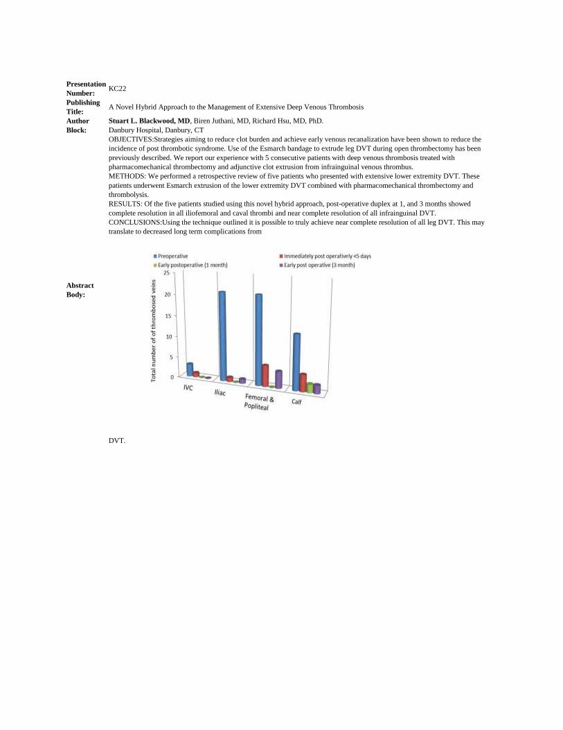
Presentation
Number: KC22
Publishing
Title: A Novel Hybrid Approach to the Management of Extensive Deep Venous Thrombosis
Author
Block:
Stuart L. Blackwood, MD, Biren Juthani, MD, Richard Hsu, MD, PhD.
Danbury Hospital, Danbury, CT
Abstract
Body:
OBJECTIVES:Strategies aiming to reduce clot burden and achieve early venous recanalization have been shown to reduce the
incidence of post thrombotic syndrome. Use of the Esmarch bandage to extrude leg DVT during open thrombectomy has been
previously described. We report our experience with 5 consecutive patients with deep venous thrombosis treated with
pharmacomechanical thrombectomy and adjunctive clot extrusion from infrainguinal venous thrombus.
METHODS: We performed a retrospective review of five patients who presented with extensive lower extremity DVT. These
patients underwent Esmarch extrusion of the lower extremity DVT combined with pharmacomechanical thrombectomy and
thrombolysis.
RESULTS: Of the five patients studied using this novel hybrid approach, post-operative duplex at 1, and 3 months showed
complete resolution in all iliofemoral and caval thrombi and near complete resolution of all infrainguinal DVT.
CONCLUSIONS:Using the technique outlined it is possible to truly achieve near complete resolution of all leg DVT. This may
translate to decreased long term complications from
DVT.

Presentation
Number: KC23
Publishing
Title: Decreased anti-inflammatory (M2) macrophages in a murine model of type II diabetes (T2D) impair wound healing.
Author
Block:
Danielle Horne, MD, Amrita Joshi, PhD, Anna Eliassen, MD, Tina Chen, MD, Dani Campbell, MD, Jordan Knepper, MD, Dawn
Coleman, MD, Peter Henke, MD, Katherine Gallagher, MD.
University of Michigan, Ann Arbor, MI
Abstract
Body:
OBJECTIVES: Diabetic wounds are characterized by a chronic inflammatory state that is maintained by overexpression of pro-
inflammatory cytokines, with a lack of “repair” or anti-inflammatory (M2) macrophages. We hypothesized that altered bone
marrow in type 2 diabetic mice may contribute to the decrease in M2 macrophages seen wounds.
METHODS: Bone marrow derived macrophages (BMDM) were grown via standard conditions from the BM of diet induced obese
(DIO) and wildtype (WT) C57B/6 mice. Macrophages were then treated on day 6 with M2 skewing agents (IL-4/IL-
13)(100ng/mL) and mRNA expression of M2 markers was measured via RT-PCR at 6, 24 and 48 hours post-skewing.
RESULTS: RT-PCR demonstrated decreased transcript for M2 markers (Ym1 and FIZZ1(retn1a)) in the DIO macrophages as
compared to controls. This corresponds with decreased M2 macrophages seen in diabetic wounds. (Figure 1)
CONCLUSIONS: DIO macrophages derived from BM appear to result in decreased Ym1 and FIZZ1, markers of the M2, anti-
inflammatory phenotype, important for wound healing. Manipulation of macrophage phenotypes could allow for development of
new therapies to prevent chronic inflammation and non-healing in diabetic
wounds.

Presentation
Number: KC24
Publishing
Title:
Primary Stent Placement Improves Patency After Endovascular Treatment of Hepatic Artery Stenosis Following Liver
Transplantation
Author
Block:
Linda Le, M.D., William Terral, M.D., Nicolas Zea, M.D., Hernan A. Bazan, M.D., Taylor A. Smith, M.D., George E. Loss, M.D.,
Edward Bluth, M.D., W. Charles Sternbergh, III, M.D..
Ochsner Clinic Foundation, New Orleans, LA
Abstract
Body:
OBJECTIVES: Significant hepatic artery stenosis (HAS) after orthotopic liver transplantation (OLT) can lead to thrombosis with
subsequent liver failure in 30% of patients. While operative intervention or re-transplantation has been the traditional solution,
endovascular therapy has emerged as a less-invasive treatment strategy. Prior smaller studies have been conflicting in the relative
efficacy of angioplasty (PTA) versus primary stent placement for HAS.
METHODS: This was a single center retrospective review of all endovascular interventions for HAS after OLT over a 54 month
period (August 2009-December 2013). Patients with ultrasound imaging with evidence of severe HAS (peak systolic velocity > 400
cm/sec, resistive index of <0.5) underwent endovascular treatment with primary stent placement or PTA. Outcomes calculated were
technical success, primary and primary assisted patency rates, re-interventions, and complications.
RESULTS: Sixty-two interventions for HAS were performed in 42 patients with a mean follow-up of 19.1 ± 15.2 months. The rate
of treated HAS was 6.4% (42/654). Primary technical success was achieved in 95% (59/62) of cases. Initial treatment was with
PTA alone (n=16) or primary stent (n=26). Primary patency rates after initial stent placement were 91%, 81.3%, 77%, and 77% at
1,6,12, and 24 months, respectively, significantly better (P=0.01) when compared to initial PTA (68.8%, 57.1%, 44%) see Figure.
There were 20 re-interventions in 14 patients (8 stents, 6 PTA). The time to initial re-intervention in patients with PTA alone vs.
initial stent was 51 and 105.8 days, respectively. Overall primary patency rates (Kaplan-Meier) were 82%, 70%, 63%, and 50% at
1,6,12, and 24 months, respectively. Overall primary-assisted patency was 96% and 93% at 12 and 24 months. Major complications
were one arterial rupture and two hepatic artery dissections. Long-term risk of HAT in the entire patient cohort was 4.8%.
CCONCLUSIONS: Hepatic artery stenosis after liver transplantation can be treated endovascularly with high technical success
and excellent primary-assisted patency. This series represents the largest reported cohort of endovascular interventions for HAS to
date. Initial use of a stent significantly decreased the need for re-intervention. Avoidance of hepatic artery thrombosis (HAT) is
possible in >95% of patients with endovascular treatment and close follow-up.
Presentation
Number: KC25
Publishing
Title:
Initial Experiences Utilizing Optical Coherence Tomography Imaging in Crossing Femoropopliteal Chronic Total
Occlusions
Author Block:
Christopher M. Paprzycki, MD, Sophia G. Afridi, MD, Nessa E. Miller, MD, Lala R. Hussain, MSc, Matthew H. Recht, MD,
Patrick E. Muck, MD.
TriHealth - Good Samaritan Hospital, Cincinnati, OH
Abstract
Body:
OBJECTIVES: Femoropopliteal chronic total occlusions (CTO) are among the more challenging Trans-Atlantic Inter-Society
Consensus (TASC) II class D lesions to treat from an endovascular approach. Optical coherence tomography (OCT) uses near-
infrared light to optimize intravascular visualization and allows characterization of plaque and vessel wall anatomy. The
Ocelot® catheter (Avinger) is an OCT guided catheter with spiral wedges on the tip rotating in counter directions, and can be used
as an adjunct to fluoroscopy. We reviewed our experience with crossing these complex lesions utilizing OCT guidance.
METHODS: A retrospective review of 28 patients with TASC II class D femoropopliteal lesions was performed where OCT
guidance was used. Procedural success was determined by establishing true lumen guidewire access beyond the CTO. We
assessed for complications including perforation and embolization. RESULTS: Intraluminal crossing was successfully performed
in 22 of 28 cases. The average SFA CTO length was 27.7 cm and the average popliteal CTO length was 15.8 cm. There were two
cases with combined lesions with lengths of 63.1 cm and 27.0 cm. There were no cases of perforation and
embolization. CONCLUSIONS: Optical coherence tomography is a safe and effective technique to assist in intraluminal crossing
of TASC II class D femoropopliteal CTO’s.

Presentation
Number: KC26
Publishing
Title: Celiac Artery Decompression for Median Arcuate Ligament Syndrome: A Single Center Experience
Author Block:
Alec Rezigh, BS1, Rana O. Afifi, MD1, Sapan S. Desai, MD2, Kristofer M. Charlton-Ouw, MD1, Naveed U. Saqib, MD1, Charles
C. Miller, PhD1, Anthony L. Estrera, MD1, Hazim J. Safi, MD1, Ali Azizzadeh, MD1. 1The University of Texas at Houston Medical School, Houston, TX 2Southern Illinois University School of Medicine,
Springfield, IL
Abstract
Body:
OBJECTIVES: Median Arcuate Ligament Syndrome (MALS) results from celiac artery compression by the median arcuate
ligament and is most often associated with chronic abdominal pain. A recent National Inpatient Sample (NIS) study has shown
that only 2.4% of patients with MALS undergo celiac artery decompression (CAD). The objective of this study was to evaluate
the outcomes of MALS after CAD.
METHODS: We collected retrospective data on patients diagnosed with MALS who underwent CAD at our institution. All
patients underwent evaluation by a gastroenterologist to rule out other pathology. All patients diagnosed with MALS underwent
open CAD through a midline incision. Patients completed a postoperative questionnaire used to evaluate specific MALS related
symptoms and their improvement after CAD.
RESULTS: Between 2009 and 2014, we treated 8 patients (5 female, mean age, 58.8 years) with MALS by open CAD.
Abdominal pain was the primary clinical symptom (n=8;100%), followed by nausea and weight loss (n=4;50% for both).
Angiography (inspiration and expiration) was the most utilized imaging modality (n=7, 88%) followed by computed tomography
(CT) (n=6, 75%), magnetic resonance imaging (n=3, 38%) and ultrasound (n=1, 13%). There were no immediate intraoperative or
postoperative complications. All 8 patients reported improvement in abdominal pain and corresponding symptoms based on the
questionnaire. Two patients with residual symptoms underwent post-operative revascularization, one receiving celiac artery
percutaneous transluminal angioplasty (PTA) and the other celiac PTA and stenting (25%). The patients who required
postoperative revascularization had documented preoperative celiac artery stenosis or dissection. The median follow-up time was
7.5 months (range 1 to 63 months).
CONCLUSON: Open CAD appears to be a safe and effective treatment modality for MALS. Patients with intraluminal lesions
(dissection or stenosis) may require additional revascularization.
Presentation
Number: KC27
Publishing
Title:
Computerized Tomography Angiography by Direct Intra-Aortic Low Volume Contrast Injection in Patients Being Evaluated for
EVARs
Author Block: Pedro G. Teixeira, MD, Leonardo Clavijo, Fred A. Weaver, MD.
University of Southern California, Los Angeles, CT
Abstract
Body:
OBJECTIVES: Computerized tomography angiography (CTA) is an important tool for anatomic delineation and pre-procedure
planning in patients who are being considered for endovascular repair of an abdominal aortic aneurysm (EVAR). The iodinated
contrast volume required for conventional CTA in patients with pre-existing renal dysfunction may result in further renal function
deterioration. The aim of this study was to evaluate the utility of CTA using direct intra-aortic injection of a low volume of
iodinated contrast in patients being considered for EVAR.
METHODS: The patients were brought to the angiography suite the day prior to planned EVAR. Through common femoral artery
percutaneous access, the infusion end of a 5F high-flow angiographic catheter was positioned in the mid-thoracic aorta. CTA was
then performed using a multidetector scanner, with image acquisition starting simultaneously with power injection of 10 mL of
iopamidol 76% diluted with normal saline to a total volume of 50 mL (10 mL iodinated contrast + 40 mL saline), at 6 mL/sec
through the angiographic catheter. Aortic enhancement was assessed with a circular region-of-interest cursor within the aorta and
values >=150 HU were considered adequate.
RESULTS: Six patients with AAA and associated chronic renal dysfunction underwent CTA with intra-arterial contrast injection
for endovascular therapy planning. Mean age was 80.2±5.7 years, 67% were male, mean BMI 27.8±2.9 and baseline creatinine
1.7±0.5 mg/dL. Adequate CTA aortic enhancement was obtained in all cases (mean intra-aortic density 259.9±10.5 HU). Using
acquired CTA images EVAR was performed the following day. The total contrast volume used for the CTA and the endovascular
repair combined was 33.3±25.2 mL, with four of the patients receiving less than 30 mL of contrast. No significant change in renal
function occurred perioperatively, as demonstrated by equivalent creatinine clearance pre and post-procedure (36.1±4.6 mL/min
versus 37.8±6.9 mL/min, P = .53). Successful EVAR was achieved in all patients. Patients remained in hospital for an average
3.2±0.4 days. No endoleak or aneurysm growth occurred over a median follow-up of 379 days (range 30-764).
CONCLUSIONS:The use of direct intra-aortic injection of a low volume of iodinated contrast provides an adequate and
reproducible pattern of aortic enhancement on CTA. The CTA so acquired can be used for planning and successful execution of
EVAR while limiting the total volume of iodinated contrast required.

Karmody Competition Topic: Peripheral Presentation
Number: KC28
Publishing
Title: Ultrasound Guided Arterial Access is Associated with Decreased Arterial Complications
Author Block:
Danielle N. Campbell, MD, Adina Robinson, PhD, Andrew Plaska, Hannah Hill, Matthew Sherman, Stefan Garcia, Simanjit
Mand, John E. Rectenwald, MD, Peter K. Henke, MD.
University of Michigan, Ann Arbor, MI
Abstract
Body:
OBJECTIVE: Multiple specialties use percutaneous arterial access to perform a range of interventional vascular procedures.
Vascular Surgeons manage many of the complications of access. We reviewed our institution to assess the rate of access
complications, and factors associated with these complications.
METHODS: All cases involving arterial access completed at our institution from May-December 2010 were identified using CPT
codes (N = 1584 cases). Patient demographics as well as the details surrounding each case (service performing the intervention,
site of access, ultrasound use, French size, micropuncture technique, closure device, peri and post-operative anticoagulation
and/or platelet therapy, and complications) were extracted. Complications were defined as those related to vascular access
specifically. These complications ranged from minor (hematoma, arterial-venous fistula and psuedoaneursym), to serious
(operative intervention, retroperitoneal hematoma, arterial dissection, and thrombosis). Descriptive, univariate, and multivariate
logistical regression statistics were used.
RESULTS: The mean age of our patients was 51 years old, with a mean BMI of 28.3. Most had hypertension (53%) and were
smokers (50%). Only 19% had DM and 16% had PAD. The mean French size used was 5.4, and 51% of cases were completed
using a micropuncture technique. Only 19% of arterial access was completed using ultrasound guidance while 14% of cases
utilized a closure device. Most patients (54%) were anti-coagulated during or prior to their procedure, yet only 18% were
reversed, and 35% remained on post-operative anti-coagulation or anti-platelet therapy. Access related complications occurred in
3% (N = 54) of this cohort. Of the factors assessed, only ultrasound guided access was associated independently with fewer
access complications (OR=0.371; 95% CI: .145-.952, p= .039)
CONCLUSIONS: Arterial access complications were lessened with ultrasound guided access. No other factor was significantly
associated with complications. Further assessment of access techniques may suggest that ultrasound guided access be the standard
of care.

Presentation
Number: KC29
Publishing
Title: Systematic Review of Hospital Mortality: Can we Bend the Curve?
Author Block: M. A. Mansour, M.D., Lindsey M. Korepta, MD, Christopher M. Chambers, MD, PhD, Robert F. Cuff, MD.
Spectrum Health, Grand Rapids, MI
Abstract
Body:
OBJECTIVE: The ostensible goal of participating in quality improvement programs is to improve surgical outcomes. Our
hospital has been submitting data since 2006.
Purpose: To assess whether systematic review of deaths with specific feedback to surgeons can influence behavior and improve
mortality rates.
METHODS: Our hospital participates in NSQIP. Status reports are provided on a quarterly basis. The hospital is ranked based on
calculation of the observed versus expected rate. Surgeons are given feedback on their performance and identification of
opportunities for improvement is done.
RESULTS: We reviewed our results from 4 consecutive years: 2010 to 2013. In 2011, the mortality rate was 0.71% for general
surgery (9 of 1268), 3.8% for colorectal (7 of 180) and 4.5% for vascular (14 of 310). This resulted in decreasing the hospital
ranking to the 9th decile. Detailed analysis revealed that the median age at death was 79.5 years. There were 8 women (26%).
Nearly half the deaths occurred due to futile care or failure to rescue after a life-threatening complication in the postoperative
period. In several cases, the families of these patients elected to withdraw support in the first week postoperatively. The results
were shared with vascular and general surgeons. In 2012, mortality was decreased by half.
CONCLUSION: Surgeons are frequently pressured to operate on moribund patients whose chances of survival are dismal.
Analysis of our hospital mortality rate revealed that many deaths were attributed to futile care or failure to rescue. By sharing this
information with multiple disciplines and early engagement of hospice, a significant decrease in mortality has been achieved.

Presentation
Number: KC30
Publishing
Title: Air pollution association with hospital admissions for peripheral arterial disease and lower extremity amputations
Author Block:
John F. Pearson, M.D., Abimbola B. Pratt, M.D., Michael Shapiro, M.D., John S. Brownstein, M.D., Donald Risucci, Ph.D.,
Marcus D'ayala, M.D..
NY Methodist Hospital, Brooklyn, NY
Abstract Body:
OBJECTIVE: There is increasing evidence that air pollution may have an impact on observed regional variations in disease
incidence and hospital admissions. A recent national study demonstrated that the number of hospital admissions for cardiac and
respiratory diseases was associated with daily fluctuations in airborne fine particulate matter concentrations < 2.5 µm (PM2.5). This
study examines the previously unreported relationship between regional variations in hospital admissions for Peripheral Arterial
Disease (PAD) and regional estimates of PM2.5 concentrations.
METHODS: The Center for Medicare and Medicaid Services Limited Data Set for 2008 was used to identify all U.S. hospital
admissions for PAD among patients insured by Medicare. Annual mean PM2.5 concentration estimates were calculated for each
county in the United States based on 2008 data from the Environmental Protection Agency. Pearson correlations and multiple
regression analyses were used to examine the association between county level PM2.5 estimates and the county level incidence of
PAD admissions and associated surgical procedures (above and below knee amputations).
RESULTS:There were 847668 admissions for PAD, 14407 below knee amputations, and 13466 above knee amputations. The
annual mean PM2.5 level per county was (Mean + SD) 9.88 + 2.04; range: 4.27 - 16.39 across the 3096 counties for which data were
available. Statistically significant direct correlations (p < 0.001) were observed between PM2.5 levels and the percentage of Medicare
admissions for PAD per county (r = 0.21), and those undergoing above knee amputation (r = 0.12). Statistically significant inverse
correlations were observed between PM2.5 levels and age of PAD patients (r = -0.22), and with male gender of PAD patients (r = -
0.11). Multiple regression analyses revealed that the relationship between county annual percentage of Medicare PAD admissions
and PM2.5 was independent of the size of the county populations.
CONCLUSIONS: This study suggests that Medicare patients may have a higher likelihood of hospital admission for PAD if they
live in an area with higher levels of airborne fine particulate matter concentrations. Further, the data suggest that patients admitted
for PAD who reside in more highly polluted regions tend to be younger and are more likely to be female than those living in less
polluted regions. If confirmed in future studies, these results may have significant implications for public health and resource
allocation.

Presentation
Number: KC31
Publishing
Title:
Multivariate Analysis of 319 Iliac Stents Reveals the Strong Impact of Demographic Factors and Lesion Severity on Stent
Patency
Author Block:
Amy Roach, BS, Sebastian Larion, MS, Chad Ammar, MD, Colin Brandt, MD, S. Sadie Ahanchi, MD, David Dexter, MD, Jean
Panneton, MD.
Eastern Virginia Medical School, Norfolk, VA
Abstract
Body:
OBJECTIVES: The aim of our study was to perform a large scale multivariate analysis to identify the demographic, anatomic, or
procedural factors that impact primary iliac stent patency.
METHODS: Iliac stents from 2011- 2013 were retrospectively reviewed. Differences in demographic, anatomical, and procedural
characteristics were analyzed by univariate analysis between groups based on primary patency. Variables that were considered
significant (p<0.05) were brought forward in the cox regression multivariate analysis.
RESULTS: 224 patients underwent primary iliac artery stenting and a total of 319 limbs were analyzed. Average age was 66
years (range 38 to 93), 53% were male, and 57% were Caucasian. Indication for procedure was 64% claudication, 23% rest pain,
and 13% ulcer/gangrene. The cohort included all TASC classifications: 50% TASC A, 25% TASC B, 12% TASC C, and 13%
TASC D. The treated anatomic location was isolated EIA 27%, isolated CIA 55%, and combined CIA/EIA 18%. Intervention
distal to the iliac arteries was performed in 37% of the cohort.
Kaplan Meier (KM) analysis at 1 and 3 years revealed a primary patency of 86%/51%, primary assisted patency 98%/89%, and
secondary patency of 99%/90%. For those patients with critical limb ischemia preprocedurally, limb salvage was 88% at one
year. Based on KM analysis, primary patency (PP) at 1 year for Caucasian patients was 93% versus non Caucasian 79%, P=.001.
PP was 76% in patients <60 years of age, 86% in patients 60-70 years of age, and 97% in patients >70 years of age, with a
significant difference in KM analysis between all groups, P<.001. PP was significantly different for those with and without EIA
occlusion (P<.001), with one year PP by KM analysis 71% and 86%, respectively. PP was also significantly different for those
with and without aortic occlusion, (P=.008), with one year patency of 84% and 87%, respectively. We evaluated five factors on
multivariate analysis and three factors were identified to impact primary patency: Caucasian race (HR, .517; 95% confidence
interval [CI], .313-.852; P=.01) and older age at the time of procedure (HR, .945; 95% CI, .920-.971; P<.001) positively impacted
patency. EIA occlusion (HR, 2.352; 95% CI, 1.294-4.275; P=.005) negatively impacted primary patency.
CONCLUSIONS: In our experience with a large number of iliac interventions, race, age, and EIA occlusion all impacted primary
patency.

Presentation
Number: KC32
Publishing
Title:
Occlusion of infrainguinal bypass is associated with increased postoperative wound infection after major lower extremity
amputations
Author Block: Sajid Shah, Amita Pathak, George Pisimisis, Andrea Garcia, Ronan Allencherril, Neal Barshes, Panos Kougias.
Baylor College of Medicine, Houston, TX
Abstract
Body:
OBJECTIVES: Infections occur frequently after major lower extremity amputations (LEA) performed in patients with the
diagnosis of peripheral arterial disease. We sought to determine whether history of a prior infrainguinal bypass in the amputated
extremity is associated with increased incidence of postoperative wound infection following major LEA.
METHODS: This was a retrospective cohort analysis on consecutive patients who underwent major LEA either below or above
the knee. Indications for LEA included severe rest pain or tissue loss. For each patient included in the analysis a propensity score
was calculated as the probability of having a prior infrainguinal bypass operation in the amputated extremity. Input variables for
this calculation included demographics, comorbid conditions, and urgency and symptomatology of the initial presentation. The
final risk adjustment was performed using the inverse propensity score weighting methodology.
RESULTS: We analyzed 294 major LEA that occurred over a 5 year period and led to 36 (12%) infections. Propensity score
weighting resulting in excellent balance for all the variables that were included in the propensity score calculation. In the final
multivariable model there was no association between native (OR: 1.19, p=0.88) or prosthetic (OR: 0.63, p=0.65) bypass and the
occurrence of postoperative wound infection. However, the presence of an occluded bypass regardless of type of conduit was
associated with infection (OR: 4.7, p=0.03). There was also a trend for increased incidence of wound infection in patients with
lower extremity infection as indication for the amputation (OR: 3.1, p=0.06).
CONCLUSIONS: The presence of an occluded bypass graft regardless of conduit increases the risk of wound infection after
major lower extremity amputation.
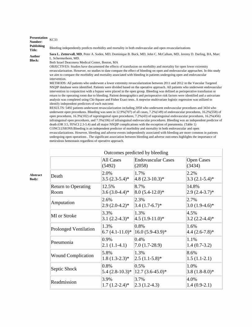
Presentation
Number: KC33
Publishing
Title: Bleeding independently predicts morbidity and mortality in both endovascular and open revascularizations
Author
Block:
Sara L. Zettervall, MD, Peter A. Soden, MD, Dominique B. Buck, MD, John C. McCallum, MD, Jeremy D. Darling, BA, Marc
L. Schermerhorn, MD.
Beth Israel Deaconess Medical Center, Boston, MA
Abstract
Body:
OBJECTIVES: Studies have documented the effects of transfusion on morbidity and mortality for open lower extremity
revascularization. However, no studies to date compare the effect of bleeding on open and endovascular approaches. In this study
we aim to compare the morbidity and mortality associated with bleeding in patients undergoing open and endovascular
intervention.
METHODS: All patients who underwent a lower extremity revascularization between 2011 and 2012 in the Vascular Targeted
NSQIP database were identified. Patients were divided based on the operative approach. All patients who underwent endovascular
intervention in conjunction with a bypass were placed in the open group. Bleeding was defined as perioperative transfusion or
return to the operating room due to bleeding. Patient demographics and perioperative risk factors were identified and a univariate
analysis was completed using Chi-Square and Fisher Exact tests. A stepwise multivariate logistic regression was utilized to
identify independent predictors of each outcome.
RESULTS: 5492 patients underwent revascularization including 2058 who underwent endovascular procedures and 3434 who
underwent open procedures. Bleeding was seen in 12.9%(707) of all cases, 7.2%(149) of endovascular procedures, 16.2%(558) of
open procedures, 16.3%(102) of suprainguinal open procedures, 7.2%(43) of suprainguinal endovascular procedures, 16.2%(456)
infrainguinal open procedures, and 7.3%(106) of infrainguinal endovascular procedures. Bleeding was an independent predictor of
death (OR 3.5, 95%CI 2.3-5.4) and all major NSQIP complications with the exception of pneumonia. (Table 1)
CONCLUSIONS:Bleeding is an independent predictor of morbidity and mortality in both endovascular and open
revascularizations. However, bleeding and adverse events independently associated with bleeding are more common in patients
undergoing open operations . The significant association between bleeding and adverse outcomes highlights the importance of
meticulous hemostasis regardless of operative approach.
Outcomes predicted by bleeding
All Cases
(5492)
Endovascular Cases
(2058)
Open Cases
(3434)
Death 2.0%
3.5 (2.3-5.4)*
1.7%
4.8 (2.3-10.3)*
2.2%
3.3 (2.1-5.4)*
Return to Operating
Room
12.5%
3.6 (3.0-4.4)*
8.7%
8.0 (5.4-12.0)*
14.8%
2.9 (2.4-3.7)*
Amputation 2.6%
2.9 (2.0-4.2)*
2.3%
3.4 (1.7-6.7)*
2.7%
3.0 (1.9-4.6)*
MI or Stroke 3.3%
3.1 (2.2-4.3)*
1.3%
4.5 (1.9-11.0)*
4.5%
3.2 (2.2-4.4)*
Prolonged Ventilation 1.3%
6.7 (4.1-11.0)*
0.8%
16.0 (5.9-43.9)*
1.6%
4.4 (2.6-7.8)*
Pneumonia 0.9%
2.1 (1.1-4.1)
0.4%
7.0 (1.7-28.9)
1.1%
1.4 (0.7-3.2)
Wound Complication 5.8%
1.8 (1.3-2.3)*
1.3%
2.5 (1.1-5.8)*
8.6%
1.5 (1.1-2.1)
Septic Shock 0.8%
5.4 (2.8-10.3)*
0.5%
12.7 (3.6-45.0)*
1.0%
3.8 (1.8-8.0)*
Readmission 3.9%
1.7 (1.2-2.4)*
3.7%
2.3 (1.2-4.3)
4.0%
1.4 (0.9-2.1)
Karmody Competition Topic: Venous/Dialysis Presentation
Number: KC34
Publishing
Title: Duplex Criteria for the Diagnosis of Fat Embolism Syndrome
Author Block: Jason Andre, MD, Stanley Bly, James Pan, MD, Douglas Hood, MD, Kim J. Hodgson, MD, Sapan S. Desai, MD, PhD, MBA.
Southern Illinois University, Springfield, IL
Abstract
Body:
OBJECTIVES: Fat Embolism Syndrome (FES), associated with up to 20% of long bone and pelvic fractures, is a disease process
that can be potentially fatal. FES is often confused with deep venous thrombosis (DVT), and there are no commonly accepted
diagnostic criteria to assist with diagnosis. As the management of these two diseases is markedly different, early and accurate
diagnosis can help guide proper management in these patients.
METHODS: We identified two patients with FES who underwent bilateral lower extremity venous duplex studies after sustaining
long bone fractures. We were able to document sonographic evidence of fat emboli in two patients and use them to generate
diagnostic criteria that distinguish them from DVT.
RESULTS: The first patient was an 83-year-old female with an open distal femur fracture sustained after a fall. Due to concerns
for a possible pulmonary embolism after the onset of pleuritic chest pain, a lower extremity venous duplex study was obtained. A
second patient was an 18-year-old male thrown from a vehicle who sustained bilateral femur fractures. A venous duplex
ultrasound was completed due to concerns for a DVT. In both patients, no DVT was identified, but a discrete, free floating, and
embologenic lesion was identified on ultrasound. Sub-centimeter radio-opaque, spherical lesions were identified passing through
the deep veins. There were no findings consistent with venous augmentation, respiratory variation, compressibility or flow
limitations.
CONCLUSIONS: Fat emboli are distinguished from DVT by their radio-opaque, spherical nature. They are not attached to the
venous wall and are not compressible. As a result, standard signs associated with a DVT are not found with FES. Patients with
ultrasonographic evidence of FES who develop subsequent PE should not be heparinized.

Presentation
Number: KC35
Publishing
Title: Surgeon-Performed Intraoperative Vein Mapping Facilitates Timely Creation of New Arteriovenous Access
Author Block:
Jeffrey B. Edwards, BS, Zachary B. Fang, Susan M. Shafii, MD, Luke P. Brewster, MD, Shipra Arya, MD, Yazan Duwayri,
MD, Ravi K. Veeraswamy, MD, Thomas F. Dodson, MD, Ravi R. Rajani, MD.
Emory University School of Medicine, Atlanta, GA
Abstract
Body:
OBJECTIVES: Preoperative duplex ultrasound is routinely performed prior to upper extremity arteriovenous (AV) access
creation. Socioeconomically disadvantaged patients, however, may not have the resources necessary to obtain such testing.
Focusing on surgeon-performed intraoperative ultrasound, we present a streamlined algorithm to minimize the need for
preoperative testing in resource-poor environments.
METHODS: All patients referred for first-time arteriovenous access creation at an urban county hospital were initially assessed
with a full history and physical exam, including bilateral upper extremity blood pressure measurements. Unless an abnormality
was identified, no other preoperative testing was performed. Patients were consented for creation of arteriovenous access, and all
potential outcomes were explained to them. Intraoperatively, the patient’s non-dominant arm was examined by the operating
surgeon using duplex ultrasound. The cephalic and basilic veins were evaluated for diameter (>2.5 mm), presence of large
branches, and continuity to their draining confluences. Radial and brachial arteries were examined for size, patency, and the
presence of significant calcification. Priority was given to access creation in the following order: radiocephalic, radiobasilic,
brachiocephalic, brachiobasilic, and AV graft placement.
RESULTS: 95 consecutive patients treated with this algorithm were included (age 53 ± 13 years; ESRD 56%). 87 patients (92%)
received a primary AV fistula (37% brachiocephalic, 31% radiocephalic, 24% basilic transposition, and 8% arm AV graft). On
average, intraoperative duplex ultrasound took an additional 6.2 minutes. Unassisted maturation was achieved in 57% of patients.
The six-month assisted primary patency was 65%. There was a 14% complication rate, including an infection rate of 1% and a
clinically significant steal rate of 3%.
CONCLUSION: Permanent AV access creation is able to be performed at success rates similar to previously published results
with no specific preoperative testing other than a thorough physical exam. Intraoperative surgeon-performed venous mapping can
be performed with minimal added operative time, thus obviating the need for any preoperative studies. Such success is able to be
accomplished while maintaining a very high rate of primary fistula creation.

Presentation
Number: KC36
Publishing
Title: Epidemiology and Predictors of Outcome of Postphlebitic Syndrome
Author Block:
Abdallah Naddaf, MD, Nick Nolte, MD, Imran Khan, MD, James Pan, MD, Douglas Hood, MD, Kim J. Hodgson, MD, Sapan
S. Desai, MD, PhD, MBA.
Southern Illinois University, Springfield, IL
Abstract
Body:
OBJECTIVES: Postphlebitic syndrome (PPS) is the most common complication of lower extremity deep vein thrombosis (DVT),
affecting up to one half of patients. PPS is associated with significant patient morbidity and increased healthcare costs. The aim of
this paper is to evaluate the epidemiology and demographics of patients admitted for lower extremity PPS, and to determine risk
factors that impact outcome. A risk score to identify at-risk patients was developed.
METHODS: A retrospective analysis was completed using the Nationwide Inpatient Sample (NIS) from 2002 to 2011. Patients
with lower extremity PPS were identified using ICD-9 codes. Epidemiology, demographics, comorbidities, hospital covariates,
and outcomes were determined. Statistically significant variables and odds ratios were identified by multivariate analysis. A
discriminant function and the area under the receiver operating characteristic curve (AUC) were used to identify factors predictive
of surgical intervention.
RESULTS: The overall incidence of inpatient PPS is 3.1/100,000 and has been decreasing since 2002. The average age of
patients with PPS is 61.2 years, with 48.3% female. Patients diagnosed with PPS with complications (37.8%) such as ulcers are
more likely to undergo an intervention such as venogram (P>0.05), stent (P0.05), venous bypass (P>0.05), vein stripping
(P<0.001), or wound debridement (P<0.001). The median length of stay (LOS) is greater for patients who have PPS with
complications (5 vs. 4, P<0.01), and median cost of care is greater ($7,293 vs. $6,399, P<0.001). Inpatient mortality is greater for
those who have PPS with complications (0.8% vs. 0.4%, P<0.01). PPS with any complication, age under 60, urban hospital
location, and peripheral vascular disease are risk factors that predict the need for intervention.
CONCLUSIONS:PPS is a disabling disease with little known about its true epidemiology and risk factors. PPS with
complications is a major predictor of future interventions and resource burden. The ability to identify at-risk patients may better
guide resource allocation and mitigate the effects of complications from PPS.
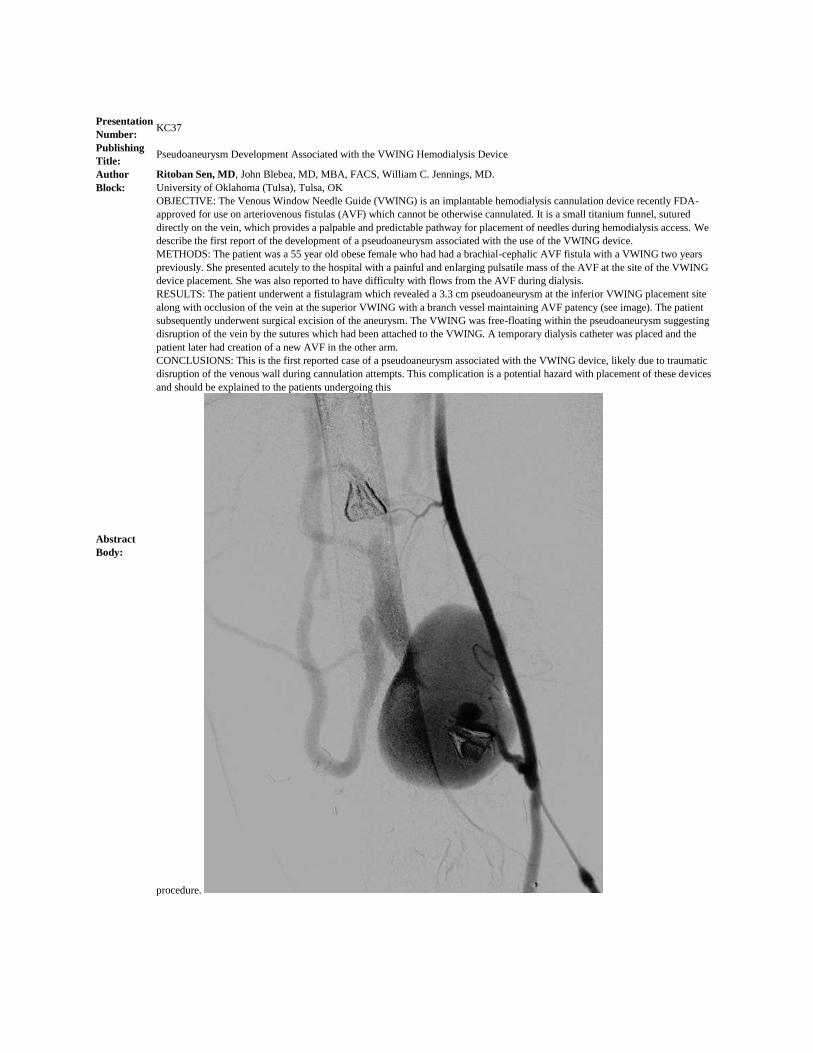
Presentation
Number: KC37
Publishing
Title: Pseudoaneurysm Development Associated with the VWING Hemodialysis Device
Author
Block:
Ritoban Sen, MD, John Blebea, MD, MBA, FACS, William C. Jennings, MD.
University of Oklahoma (Tulsa), Tulsa, OK
Abstract
Body:
OBJECTIVE: The Venous Window Needle Guide (VWING) is an implantable hemodialysis cannulation device recently FDA-
approved for use on arteriovenous fistulas (AVF) which cannot be otherwise cannulated. It is a small titanium funnel, sutured
directly on the vein, which provides a palpable and predictable pathway for placement of needles during hemodialysis access. We
describe the first report of the development of a pseudoaneurysm associated with the use of the VWING device.
METHODS: The patient was a 55 year old obese female who had had a brachial-cephalic AVF fistula with a VWING two years
previously. She presented acutely to the hospital with a painful and enlarging pulsatile mass of the AVF at the site of the VWING
device placement. She was also reported to have difficulty with flows from the AVF during dialysis.
RESULTS: The patient underwent a fistulagram which revealed a 3.3 cm pseudoaneurysm at the inferior VWING placement site
along with occlusion of the vein at the superior VWING with a branch vessel maintaining AVF patency (see image). The patient
subsequently underwent surgical excision of the aneurysm. The VWING was free-floating within the pseudoaneurysm suggesting
disruption of the vein by the sutures which had been attached to the VWING. A temporary dialysis catheter was placed and the
patient later had creation of a new AVF in the other arm.
CONCLUSIONS: This is the first reported case of a pseudoaneurysm associated with the VWING device, likely due to traumatic
disruption of the venous wall during cannulation attempts. This complication is a potential hazard with placement of these devices
and should be explained to the patients undergoing this
procedure.

Presentation
Number: KC38
Publishing
Title: Heparin bonded PTFE does not improve hemodialysis arteriovenous graft function
Author
Block:
Nicolas Zea, MD, Grayson Menard, MD, Linda Le, MD, Hernan A. Bazan, MD, W Charles Sternbergh, MD, Taylor Smith, MD.
Ochsner Clinic Foundation, New Orleans, LA
Abstract
Body:
OBJECTIVES: While heparin-bonded polytetrafluoroethylene (PTFE), when compared to standard PTFE, has been shown to be
beneficial in distal extremity bypass grafts, the data supporting its usage for dialysis access is less clear. We compared the patency
rates, number of interventions, and complications between heparin-bonded (HEP) and non-heparin-bonded (NonHEP) PTFE grafts
placed for dialysis access.
METHODS: A retrospective review of all dialysis access procedures entered into a prospectively maintained vascular surgery
database was performed. Primary end points included functional graft patency, time to graft abandonment, and number of
procedures required to re-establish graft patency following thrombosis. The number of interventions required to maintain graft
patency and graft related complications were also reviewed. Kaplan-Meier curves were used to compare the two groups.
RESULTS: Between January 2013 and March 2014, 301 dialysis access procedures were performed which included 72 AV grafts
(AVG) comprised of 32 HEP (32 6mm straight grafts) and 39 NonHEP (36 4-7mm taper and 3 6mm straight). At a mean follow-up
of 7.35 ± 5.15 months, 22/32 HEP grafts were functional compared to 31/39 NonHEP grafts (67% vs. 79%, p=0.22). Primary,
primary-assisted, and secondary patency at 1, 3, 6 and 12 month follow-up was not significantly different between the HEP and
NonHEP grafts. The incidence of grafts abandoned due to thrombosis (5 HEP, 4 NonHEP, 16% vs. 10%, p=0.51) and time to graft
abandonment were also not different (4.55 ± 3.11 months vs. 2.38 ± 2.72, p=0.13). The number of HEP grafts undergoing an open
or percutaneous thrombectomy was significantly higher than the NonHEP grafts (34% vs. 13%, p=0.03), as was the incidence of
any intervention performed to maintain graft patency (84% vs. 51%, p= 0.002). Kaplan-Meier survival curve failed to show a
benefit in functional patency with HEP vs. NonHEP
PTFE
CONCLUSIONS: We did not demonstrate a benefit to the routine use of heparin-bonded PTFE for AVG creation especially given
the higher cost of these grafts. Functional patency rates were not improved and the rates of re-intervention were higher with heparin
bonded PTFE AV grafts.
Wednesday, April 1st
Scientific Session VII – Venous/Dialysis Presentation
Number: 23
Publishing
Title:
Infraclavicular
First Rib Resection for the Treatment of Acute Venous Thoracic Outlet Syndrome
Author Block:
Jeffrey J. Siracuse, M.D., Paul C. Johnson, M.D., Douglas W. Jones, M.D., Gregory G. Salzler, M.D., Heather L. Gill, M.D.,
Andrew J. Meltzer, M.D., Peter H. Connolly, M.D., Darren B. Schneider, M.D..
New York-Presbyterian Hospital, Weill Cornell Medical College, New York, NY, USA.
Abstract
Body:
OBJECTIVES: Venous thoracic outlet syndrome (VTOS) most commonly is treated by transaxillary, supraclavicular, or
paraclavicular approaches based on surgeon preference. However, we have adopted an infraclavicular approach to VTOS as the
surgical pathology is in the anterior costoclavicular space. We hypothesize that this approach for thoracic outlet decompression
provides excellent access to the costoclavicular space and the axillosubclavian veins for safe and effective treatment of patients
with an acute presentation of VTOS.
METHODS: We retrospectively reviewed all consecutive patients that underwent infraclavicular thoracic outlet decompression
for an acute presentation of VTOS from July 2005 to February 2014 by a single surgeon. Acute presentation was defined as less
than 14 days between the onset of symptoms and catheter directed thrombolysis (CDT). Demographics, primary and secondary
subclavian vein patency, perioperative outcomes, and reinterventions were recorded.
RESULTS: 30 patients underwent an infraclavicular approach for treatment of VTOS. Average age was 33 and 60% were male.
All patients underwent CDT and subsequent infraclavicular first rib resection and intraoperative venography, which was
technically successful in all patients. Intraoperative subclavian vein angioplasty was performed in 70%. Median postoperative
length of stay was 2 days (range 2-6), blood loss was 75 ml (20-200), and operative time was 117 minutes (76-166). Median
follow-up was 78 days (2-483). Ultrasound at follow-up was performed on 24/30 (80%) with all patients having patent subclavian
veins at last follow-up. Reinterventions included 2 cases for rethrombosis and one case of hemothorax. There were no
complications of brachial plexus or phrenic nerve injury. All patients at last follow up were symptom free and subclavian veins
were patent.
CONCLUSIONS: An infraclavicular approach is a safe and effective treatment for acute VTOS. It provides excellent access to
the costoclavicular space for first rib resection and subclavian venolysis while at the same time minimizing the risk of brachial
plexus and phrenic nerve injury.
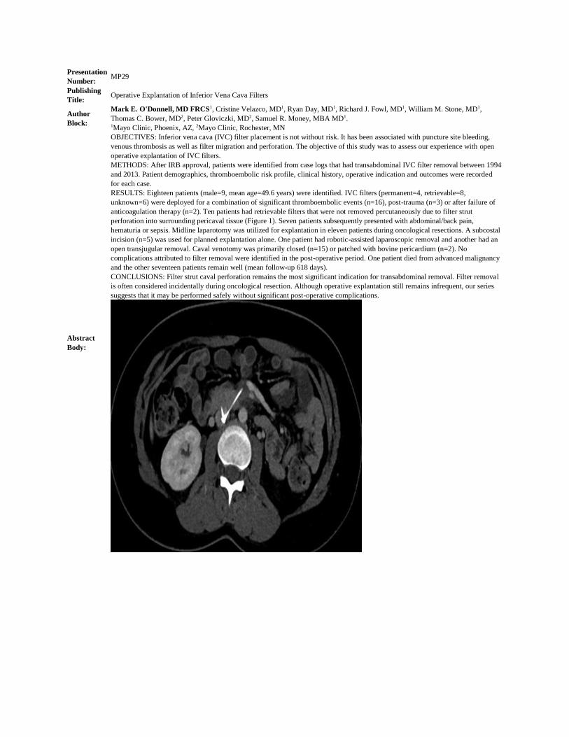
Presentation
Number: MP29
Publishing
Title: Operative Explantation of Inferior Vena Cava Filters
Author
Block:
Mark E. O'Donnell, MD FRCS1, Cristine Velazco, MD1, Ryan Day, MD1, Richard J. Fowl, MD1, William M. Stone, MD1,
Thomas C. Bower, MD2, Peter Gloviczki, MD2, Samuel R. Money, MBA MD1. 1Mayo Clinic, Phoenix, AZ, 2Mayo Clinic, Rochester, MN
Abstract
Body:
OBJECTIVES: Inferior vena cava (IVC) filter placement is not without risk. It has been associated with puncture site bleeding,
venous thrombosis as well as filter migration and perforation. The objective of this study was to assess our experience with open
operative explantation of IVC filters.
METHODS: After IRB approval, patients were identified from case logs that had transabdominal IVC filter removal between 1994
and 2013. Patient demographics, thromboembolic risk profile, clinical history, operative indication and outcomes were recorded
for each case.
RESULTS: Eighteen patients (male=9, mean age=49.6 years) were identified. IVC filters (permanent=4, retrievable=8,
unknown=6) were deployed for a combination of significant thromboembolic events (n=16), post-trauma (n=3) or after failure of
anticoagulation therapy (n=2). Ten patients had retrievable filters that were not removed percutaneously due to filter strut
perforation into surrounding pericaval tissue (Figure 1). Seven patients subsequently presented with abdominal/back pain,
hematuria or sepsis. Midline laparotomy was utilized for explantation in eleven patients during oncological resections. A subcostal
incision (n=5) was used for planned explantation alone. One patient had robotic-assisted laparoscopic removal and another had an
open transjugular removal. Caval venotomy was primarily closed (n=15) or patched with bovine pericardium (n=2). No
complications attributed to filter removal were identified in the post-operative period. One patient died from advanced malignancy
and the other seventeen patients remain well (mean follow-up 618 days).
CONCLUSIONS: Filter strut caval perforation remains the most significant indication for transabdominal removal. Filter removal
is often considered incidentally during oncological resection. Although operative explantation still remains infrequent, our series
suggests that it may be performed safely without significant post-operative complications.
Presentation
Number: MP30
Publishing
Title: Isolated Soleal and Gastrocnemius Vein Thrombosis Revisited: The Proper Treatment
Author Block: Ali Elfandi, MD, Clifford M. Sales, MD, Scott Sundick, MD, Sharen Anghel, MD, Rami Bustami, PhD.
Overlook Medical Center, Summit, NJ
Abstract
Body:
OBJECTIVES: We have advocated for the “non-anticoagulant” approach to the management of isolated soleal or gastrocnemius
vein thrombosis (ISGVT) showing that the addition of therapeutic anticoagulation does not affect the rate of propagation of
thrombi in these vessels. Our original report was based on data through 2009. The current report re-examines our more current
data for the calendar year 2013.
METHODS: All in-patients with a diagnosis of ISGVT made in our ICAVL noninvasive laboratory were included for review.
Only those with a follow-up duplex examination were included in the statistical analysis. Review of the medical record data and
of the duplex ultrasound examinations allowed for the determination of progression of thrombosis
RESULTS: 100 patients were found to have ISGVT in 2013 at our institution_56 (56%) had a follow-up duplex study available
for review and constitute the study group. 32% (n=18) were treated with full-dose anticoagulation (TX group) following the
diagnosis of ISGVT. Compared to our historical control (54% had been so treated in our prior report) this represented a
significant change in practice pattern (p=.006 .) There were no statistical differences between those in the TX group and those in
the NoTX group (those not treated with full-dose anticoagulation) in terms of demographics, risk factors or comorbities. Two of
the 18 patients in the TX group (11%) and 6/38 (16%) in the NoTX group had propagation of their ISGVT (p=1.00)_all of which
propagated to tibial vessels.
CONCLUSIONS: We repeat our strong support for the avoidance of full-dose anticoagulation in the management of ISGVT. This
data shows that propagation is related to factors other than treatment protocols and the addition of therapeutic anticoagulation
only exposes patients to the risks of this treatment without proven benefit. We are impressed to see that the medical staff in our
institution has apparently accepted this information and has begun to alter their practice patterns.
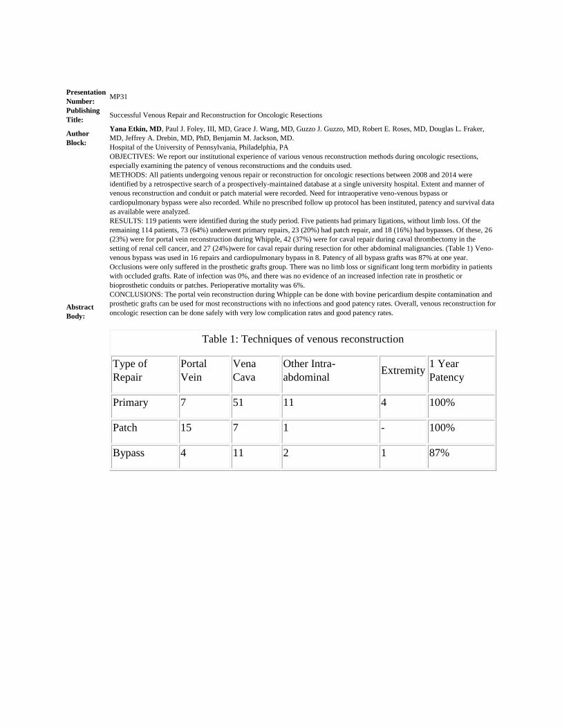
Presentation
Number: MP31
Publishing
Title: Successful Venous Repair and Reconstruction for Oncologic Resections
Author
Block:
Yana Etkin, MD, Paul J. Foley, III, MD, Grace J. Wang, MD, Guzzo J. Guzzo, MD, Robert E. Roses, MD, Douglas L. Fraker,
MD, Jeffrey A. Drebin, MD, PhD, Benjamin M. Jackson, MD.
Hospital of the University of Pennsylvania, Philadelphia, PA
Abstract
Body:
OBJECTIVES: We report our institutional experience of various venous reconstruction methods during oncologic resections,
especially examining the patency of venous reconstructions and the conduits used.
METHODS: All patients undergoing venous repair or reconstruction for oncologic resections between 2008 and 2014 were
identified by a retrospective search of a prospectively-maintained database at a single university hospital. Extent and manner of
venous reconstruction and conduit or patch material were recorded. Need for intraoperative veno-venous bypass or
cardiopulmonary bypass were also recorded. While no prescribed follow up protocol has been instituted, patency and survival data
as available were analyzed.
RESULTS: 119 patients were identified during the study period. Five patients had primary ligations, without limb loss. Of the
remaining 114 patients, 73 (64%) underwent primary repairs, 23 (20%) had patch repair, and 18 (16%) had bypasses. Of these, 26
(23%) were for portal vein reconstruction during Whipple, 42 (37%) were for caval repair during caval thrombectomy in the
setting of renal cell cancer, and 27 (24%)were for caval repair during resection for other abdominal malignancies. (Table 1) Veno-
venous bypass was used in 16 repairs and cardiopulmonary bypass in 8. Patency of all bypass grafts was 87% at one year.
Occlusions were only suffered in the prosthetic grafts group. There was no limb loss or significant long term morbidity in patients
with occluded grafts. Rate of infection was 0%, and there was no evidence of an increased infection rate in prosthetic or
bioprosthetic conduits or patches. Perioperative mortality was 6%.
CONCLUSIONS: The portal vein reconstruction during Whipple can be done with bovine pericardium despite contamination and
prosthetic grafts can be used for most reconstructions with no infections and good patency rates. Overall, venous reconstruction for
oncologic resection can be done safely with very low complication rates and good patency rates.
Table 1: Techniques of venous reconstruction
Type of
Repair
Portal
Vein
Vena
Cava
Other Intra-
abdominal Extremity
1 Year
Patency
Primary 7 51 11 4 100%
Patch 15 7 1 - 100%
Bypass 4 11 2 1 87%
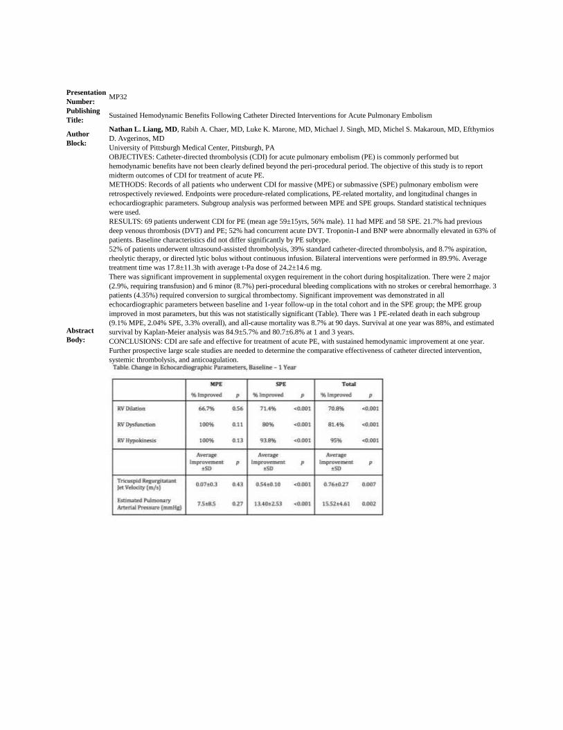
Presentation
Number: MP32
Publishing
Title: Sustained Hemodynamic Benefits Following Catheter Directed Interventions for Acute Pulmonary Embolism
Author
Block:
Nathan L. Liang, MD, Rabih A. Chaer, MD, Luke K. Marone, MD, Michael J. Singh, MD, Michel S. Makaroun, MD, Efthymios
D. Avgerinos, MD
University of Pittsburgh Medical Center, Pittsburgh, PA
Abstract
Body:
OBJECTIVES: Catheter-directed thrombolysis (CDI) for acute pulmonary embolism (PE) is commonly performed but
hemodynamic benefits have not been clearly defined beyond the peri-procedural period. The objective of this study is to report
midterm outcomes of CDI for treatment of acute PE.
METHODS: Records of all patients who underwent CDI for massive (MPE) or submassive (SPE) pulmonary embolism were
retrospectively reviewed. Endpoints were procedure-related complications, PE-related mortality, and longitudinal changes in
echocardiographic parameters. Subgroup analysis was performed between MPE and SPE groups. Standard statistical techniques
were used.
RESULTS: 69 patients underwent CDI for PE (mean age 59±15yrs, 56% male). 11 had MPE and 58 SPE. 21.7% had previous
deep venous thrombosis (DVT) and PE; 52% had concurrent acute DVT. Troponin-I and BNP were abnormally elevated in 63% of
patients. Baseline characteristics did not differ significantly by PE subtype.
52% of patients underwent ultrasound-assisted thrombolysis, 39% standard catheter-directed thrombolysis, and 8.7% aspiration,
rheolytic therapy, or directed lytic bolus without continuous infusion. Bilateral interventions were performed in 89.9%. Average
treatment time was 17.8±11.3h with average t-Pa dose of 24.2±14.6 mg.
There was significant improvement in supplemental oxygen requirement in the cohort during hospitalization. There were 2 major
(2.9%, requiring transfusion) and 6 minor (8.7%) peri-procedural bleeding complications with no strokes or cerebral hemorrhage. 3
patients (4.35%) required conversion to surgical thrombectomy. Significant improvement was demonstrated in all
echocardiographic parameters between baseline and 1-year follow-up in the total cohort and in the SPE group; the MPE group
improved in most parameters, but this was not statistically significant (Table). There was 1 PE-related death in each subgroup
(9.1% MPE, 2.04% SPE, 3.3% overall), and all-cause mortality was 8.7% at 90 days. Survival at one year was 88%, and estimated
survival by Kaplan-Meier analysis was 84.9±5.7% and 80.7±6.8% at 1 and 3 years.
CONCLUSIONS: CDI are safe and effective for treatment of acute PE, with sustained hemodynamic improvement at one year.
Further prospective large scale studies are needed to determine the comparative effectiveness of catheter directed intervention,
systemic thrombolysis, and anticoagulation.

Presentation
Number: MP33
Publishing
Title: Long-Term Patency Of Primary Inferior Vena Cava Reconstructions
Author
Block:
George E. Havelka, MD, Heron E. Rodriguez, MD, Andrew W. Hoel, MD, Mark K. Eskandari, MD.
Northwestern University, Chicgo, IL
Abstract
Body:
OBJECTIVE: Inferior vena cava (IVC) repair after planned and unplanned venotomy may take the form of either interposition
bypass, patch venopasty or lateral venorrhaphy and primary repair. Direct repair of the IVC avoids the use of foreign material, and
allows for an all-autologous repair in an expeditious fashion. Furthermore, primary repair can be accomplished without
compromising venous outflow from the lower extremities and exhibits excellent long-term patency rates.
METHODS: Enrollment consisted of 47 consecutive patients who underwent direct IVC repairs between January 2002 and January
2014. Primary repair followed lateral venorrhaphy for tumor extraction (figure 1) or to repair an iatrogenic or traumatic IVC injury.
Patient demographics, vena cava dimensions and patient outcomes were tabulated.
RESULTS: Most patients underwent en bloc nephrectomy, IVC tumor thrombus extraction and primary lateral venorrhaphy; 92%
had RCC on final pathology. This group consisted of 15 men and 11 women; the average age was 61.0 years (39-83 years); the
mean operative time was 410.3 ± 119.9 minutes; mean estimted blood loss (EBL) was 3395.4 mL and the mean follow-up period
was 38.1 months (1-96 months). Complications included one pulmonary embolism and one case of anuria requiring re-operation.
Five patients required a supra-hepatic IVC clamp associated with a longer operative time compared to those requiring an
infrahepatic clamp (532 minutes vs. 360 minutes, respectively). The group undergoing primary repair for iatrogenic or penetrating
traumatic IVC injury consisted of 15 men and 6 women; the average age was 52.7 years (31-82 years); the mean operative time
was 366.1 ± 137.9 minutes; mean EBL was 1610.1 mL and the mean follow-up period was 20.2 months (3-92 months).
Complications included one pleural effusion requiring chest tube drainage and one hospital acquired pneumonia. There were no
intra-operative deaths nor pulmonary emboli in either group.
Neither group exhibited post-op clinical or radiographic evidence of IVC stenosis or occlusion.
CONCLUSION: Regardless of the indication or the level of IVC clamping, primary IVC repair is a safe, expeditious technique that
provides good long-term patency.
2
Presentation
Number: MP34
Publishing
Title: Proximalization of Arterial Inflow: An Effective Treatment for Ischemic Steal Syndrome After Access Creation
Author Block: Thomas M. Loh, MD, Matthew E. Bennett, MD, Mark G. Davies, MD, PhD, Eric K. Peden, MD
Houston Methodist, Houston, TX
Abstract
Body:
OBJECTIVES: Ischemic steal syndrome (ISS) is a dreaded complication following hemodialysis access creation. Its management
is complex and varied with a majority requiring surgical revision for correction of symptoms. Proximalization of arterial inflow
(PAI) is an alternative to distal revascularization-interval ligation (DRIL) that obviates the need to ligate the axial artery. We
present our experience, of PAI for the treatment of ISS.
METHODS: We retrospectively reviewed consecutive patients who underwent PAI for ISS from April 2008 to June, 2014. Data
collection included demographics, past medical histories, subsequent procedures, volume flows, access usage, limb salvage and
patient survival.
RESULTS: We performed 38 PAI (21 women, 17 men). Indications for surgery were extremity pain in 38 patients, neurologic
dysfunction in 18 patients and tissue loss in 4 patients. 29% had prior surgical intervention for ISS. 74% of patients had a history
of diabetes. 49% had a history of atherosclerotic disease. Time to intervention from creation was 23 months (range, 1-94).
Accesses included 9 radiocephalic, 26 brachiocephalic and 3 brachiobasilic fistulas. Proximalization targets were 1 radial artery, 3
ulnar arteries and 34 proximal brachial arteries. Primary assisted patency at 1 year was 71%. Secondary patency at 1 year was
74%. Symptom resolution was reported as complete in 69% and markedly improved in 22%. 6 patients underwent subsequent
procedures for continued symptoms with 4 fistulas ultimately ligated for ISS. The average flow in the 4 ligated fistulas increased
by 629cc/min (remaining fistulas, average decreased flow of 101cc/min).
CONCLUSIONS: PAI is an effective and durable treatment for ISS without ligating axial flow to the extremity. Early results are
concerning that PAI can potentially increase flow to the access inducing further steal. Patient selection and monitoring of post-op
flow is key for optimizing relief of symptoms and maintaining use of the access. Further investigation into intraop flow
monitoring or combination Proximalization/plication are needed.


