downloaded from 1 on july 21, 2019 by ... · 43 tbf ocr urc msos ctomonlm ay osutbreak c.sases in...
TRANSCRIPT

1
The Borrelia hermsii factor H binding protein, FhbA, is not required for infectivity in mice 1
or for resistance to human complement in vitro 2
3
4
Lindy M. Fine1*, Daniel P. Miller1, Katherine L. Mallory1, Brittney K. Tegels1, Christopher G. 5
Earnhart1, and Richard T. Marconi1,2# 6
7
1Department of Microbiology and Immunology, 2Center for the Study of Biological Complexity, 8
Virginia Commonwealth University Medical Center, Richmond, VA, USA 9
10
11
Running head: FhbA, FH binding and serum resistance of B. hermsii. 12
13
14
IAI Accepts, published online ahead of print on 27 May 2014Infect. Immun. doi:10.1128/IAI.01892-14Copyright © 2014, American Society for Microbiology. All Rights Reserved.
on Decem
ber 25, 2019 by guesthttp://iai.asm
.org/D
ownloaded from

2
#Address correspondence to Richard T. Marconi, [email protected] 15
*Present address: Lindy M. Fine, University of Maryland Center for Environmental Science, 16
Cambridge, MD, USA 17
on Decem
ber 25, 2019 by guesthttp://iai.asm
.org/D
ownloaded from
3
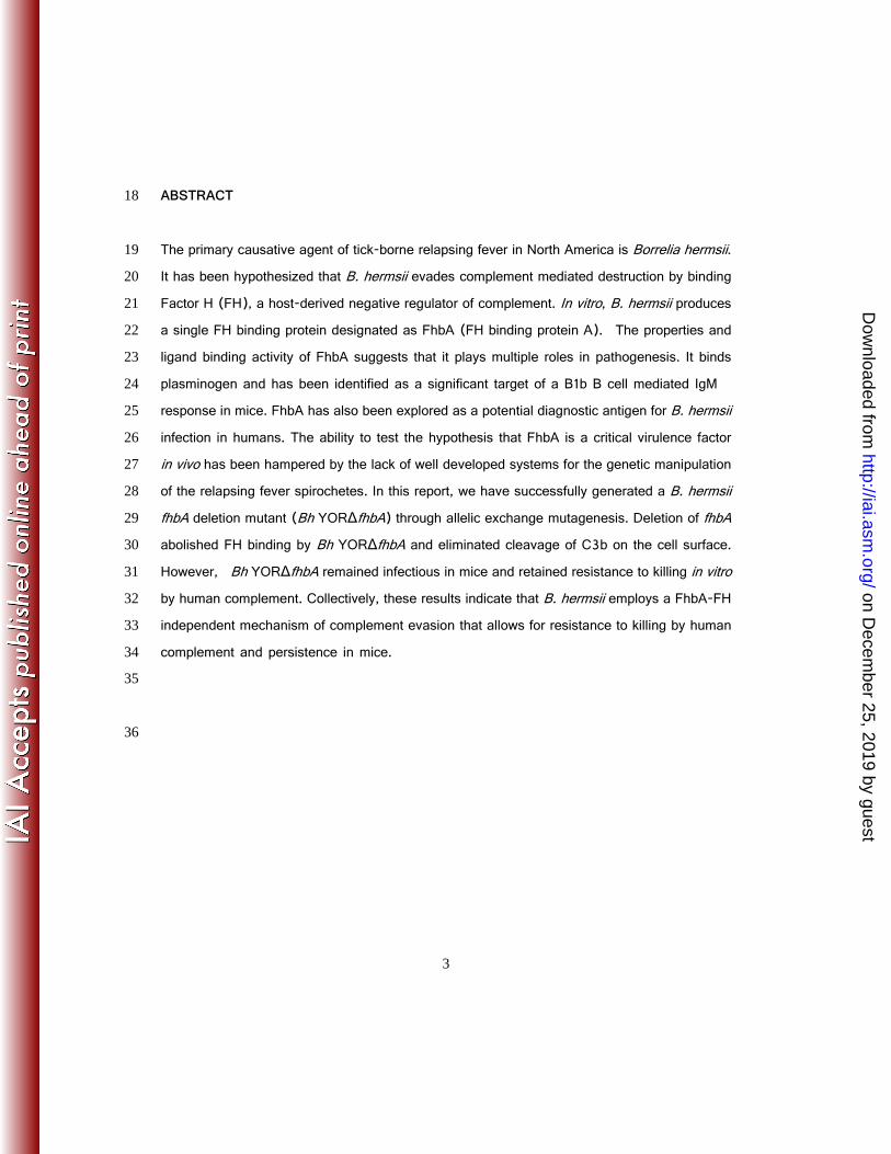
ABSTRACT 18
The primary causative agent of tick-borne relapsing fever in North America is Borrelia hermsii. 19
It has been hypothesized that B. hermsii evades complement mediated destruction by binding 20
Factor H (FH), a host-derived negative regulator of complement. In vitro, B. hermsii produces 21
a single FH binding protein designated as FhbA (FH binding protein A). The properties and 22
ligand binding activity of FhbA suggests that it plays multiple roles in pathogenesis. It binds 23
plasminogen and has been identified as a significant target of a B1b B cell mediated IgM 24
response in mice. FhbA has also been explored as a potential diagnostic antigen for B. hermsii 25
infection in humans. The ability to test the hypothesis that FhbA is a critical virulence factor 26
in vivo has been hampered by the lack of well developed systems for the genetic manipulation 27
of the relapsing fever spirochetes. In this report, we have successfully generated a B. hermsii 28
fhbA deletion mutant (Bh YORΔfhbA) through allelic exchange mutagenesis. Deletion of fhbA 29
abolished FH binding by Bh YORΔfhbA and eliminated cleavage of C3b on the cell surface. 30
However, Bh YORΔfhbA remained infectious in mice and retained resistance to killing in vitro 31
by human complement. Collectively, these results indicate that B. hermsii employs a FhbA-FH 32
independent mechanism of complement evasion that allows for resistance to killing by human 33
complement and persistence in mice. 34
35
36
on Decem
ber 25, 2019 by guesthttp://iai.asm
.org/D
ownloaded from
4
INTRODUCTION 37
Tick-borne relapsing fever (TBRF) is a significant health concern in endemic regions of the 38
world. It is caused by species in the genus Borrelia and is transmitted by Ornithodoros ticks 39
(1). TBRF is among the leading causes of febrile illness in certain regions of Africa, exceeding 40
even that of malaria (2, 3, 4, 5). In parts of Tanzania, estimates suggest that 40% of children 41
under the age of 1 will experience at least one bout of relapsing fever (2). In the Americas, 42
TBRF occurs most commonly as outbreaks. Cases in the United States cluster in high elevation, 43
coniferous forests of the West where the primary etiological agents are B. hermsii and B. 44
turicatae (6-10). TBRF manifests with recurring episodes of high fever that coincide with the 45
emergence of high density (104-8 spirochetes ml-1 blood), antigenically distinct populations of 46
spirochetes (11). Once infected, the TBRF spirochetes rapidly disseminate to distal tissues, 47
organs and the central nervous system (12). 48
49
on Decem
ber 25, 2019 by guesthttp://iai.asm
.org/D
ownloaded from

5
The ability of B. hermsii to establish infection and persist requires that effective strategies be 50
in place for complement evasion. Several recent reviews have summarized the contribution 51
of Factor H (FH) binding in complement evasion by spirochetal pathogens (13,14). FH is a 52
central negative regulator of the alternative complement pathway (15). It serves as a cofactor 53
for factor I (FI)-mediated cleavage of C3b, inhibits formation of the C3 convertase complex 54
and accelerates decay of preformed complex (16,17). Collectively, these processes decrease 55
C3b amplification. B. hermsii, the focus of this study, binds FH via its FhbA protein (18). FhbA 56
has also been demonstrated to bind FH related protein 1 (FHR-1) and plasminogen (19). In 57
contrast to B. burgdorferi, a causative agent of Lyme disease, which produces as many as five 58
different FH binding proteins (20-23), FhbA is the sole FH binding protein of B. hermsii 59
identified to date (18). FhbA is a 24 kD, surface-exposed, lipoprotein (18,19) that is encoded 60
by a gene carried on a genetically stable linear plasmid of 200 kb (lp200). With the exception 61
of high passage derivatives of some strains, FhbA is produced by all strains during in vitro 62
cultivation. Phylogenetic analyses of FhbA have revealed the existence of two distinct FhbA 63
on Decem
ber 25, 2019 by guesthttp://iai.asm
.org/D
ownloaded from
6
variants (FhbA1 and FhbA2) both of which bind FH (24,25). The molecular basis of the 64
FhbA-FH interaction has been investigated using truncation analyses and site-directed 65
mutagenesis (19,24,26). In light of the universal distribution of fhbA among low passage B. 66
hermsii isolates, its expression during infection in humans (23) and its ability to bind serum 67
and complement regulatory proteins we and others have hypothesized that FhbA is an important 68
virulence factor that plays an essential role in B. hermsii=s ability to survive and thrive in blood 69
(18,19,25,27). 70
71
While several spirochete species have been shown to bind FH, a clear correlation between FH 72
binding and serum resistance in vivo has been demonstrated for only a few species (28-32). 73
Primarily due to the lack of well developed genetic manipulation systems for the TBRF 74
spirochetes, the potential role that FhbA plays in serum resistance in vivo has not been 75
assessed. In this report. a B. hermsii YOR fhbA deletion mutant was generated and used to 76
test the hypothesis that FhbA is a critical virulence factor required for survival in the blood of 77
on Decem
ber 25, 2019 by guesthttp://iai.asm
.org/D
ownloaded from
7
infected mammals. The data indicate that FhbA and FH binding are not required for 78
complement evasion in vivo suggesting that B. hermsii employs yet be identified FH 79
independent mechanisms for complement resistance. In addition, this study represents an 80
important technological step forward as it is the first report to describe the inactivation of a B. 81
hermsii virulence factor and subsequent analysis of the resulting mutant in an animal model. 82
83
MATERIALS AND METHODS 84
85
Bacterial strains and cultivation. B. hermsii strain YOR is a well characterized genomic group 86
II isolate (9,18,26). B. hermsii YOR and all derivative strains were cultivated in 87
Barbour-Stoenner-Kelly (BSK)-H complete media supplemented with 12% rabbit serum (37°C; 88
5% CO2) in sealed bottles. Where appropriate, kanamycin (200 μg ml-1) was included in the 89
media. Strain Bh YOR::kanrgfp (33), which was generated in an earlier study and served as 90
a control, carries an identical selectable marker on lp200 as Bh YORΔfhbA but retains fhbA 91
on Decem
ber 25, 2019 by guesthttp://iai.asm
.org/D
ownloaded from

8
(33). Borrelia andersonii MOD-1 (34,35), a species in the B. burgdorferi sensu lato complex, 92
served as control for the C9 deposition assays and was cultivated in BSK-H media with 6% 93
rabbit serum. Escherichia coli strains employed for cloning purposes are described in detail 94
below as appropriate. 95
96
Vector construction and electroporation. Generation of a B. hermsii YOR fhbA deletion mutant 97
was achieved through allelic exchange mutagenesis using a suicide vector. This vector was 98
derived from the pFAEV3 vector which was originally generated to integrate a kanr-gfp cassette 99
into a non-coding region of the 200 kb linear plasmid (lp200) of B. hermsii immediately 100
downstream of fhbA (33). To construct pFAEV3ΔfhbA, fhbA was excised from pFAEV3 using 101
NheI. The construct was then re-ligated to yield pFAEV3ΔfhbA. The plasmid was propagated 102
in E. coli NovaBlue cells (Novagen) and purified with a HiSpeed Plasmid Midi kit according 103
to the manufacturer=s protocol (Qiagen). Prior to transformation, pFAEV3ΔfhbA was 104
linearized with AhdI and PflFI restriction enzymes. Electroporation conditions and limiting 105
on Decem
ber 25, 2019 by guesthttp://iai.asm
.org/D
ownloaded from

9
dilution techniques to obtain clonal populations were as previously described (33). Growth in 106
BSK with kanamycin selected for transformants. Proper integration into lp200 was confirmed 107
by PCR screening. GFP production was confirmed by fluorescence microscopy. 108
109
Assessment of in vitro growth and plasmid composition. B. hermsii growth curves were 110
determined as previously described (33). In brief, equal numbers of actively growing cells 111
were inoculated into fresh media (in triplicate, three independent biological replicates) and 112
maintained at 37°C under 5% CO2 and cell counts were conducted daily (7 days) using dark 113
field microscopy. The average number of cells in ten fields of view (400X magnification) were 114
determined at each time point and averaged. The plasmid content of each strain was assessed 115
using pulsed-field gel electrophoresis (PFGE) as previously described (36) using conditions 116
optimized for separation of linear DNA molecules between 5 and 200 kb in size. DNA was 117
visualized by ethidium bromide staining. 118
119
on Decem
ber 25, 2019 by guesthttp://iai.asm
.org/D
ownloaded from
10
Immunoblot and affinity ligand binding immunoblot (ALBI) assays. B. hermsii cell lysates 120
were fractionated by sodium dodecyl sulfate-polyacrylamide gel electrophoresis (SDS-PAGE) 121
under reducing conditions in 15% precast Criterion gels (Bio-Rad). Proteins were transferred 122
to poly-vinylidene diflouride (PVDF) membranes by electroblotting using standard methods. 123
Immunoblot and FH ALBI assays were performed as previously described (29). In brief, for 124
immunoblot analyses, blots were screened with previously generated mouse anti-FhbA or 125
mouse anti-FlaB antisera (18) at dilutions of 1:1000 and 1:400,000, respectively. 126
Peroxidase-conjugated goat anti-mouse immunoglobulin G (IgG) (1:40,000; Pierce) served 127
as the secondary antibody. For ALBI analyses, blots were incubated with purified human FH 128
(5 ng μl-1) and bound FH detected with goat anti-human FH antiserum (1:1000; Calbiochem) 129
and peroxidase-conjugated rabbit anti-goat IgG (Pierce; 1:40,000). Detection of secondary 130
antibody was achieved using the SuperSignal West chemiluminescent substrate (Pierce). 131
Throughout this study, all assays involving FH were conducted using purified human FH 132
(Complement Tech). Since the FH is purified directly from human serum it possesses all natural 133
on Decem
ber 25, 2019 by guesthttp://iai.asm
.org/D
ownloaded from
11
post-translational modifications. At no point was recombinant FH used in this study. Henceforth, 134
purified human FH is referred to simply as FH. Note that all images of immunoblots and FH 135
ALBI assays were cropped to eliminate blank space from the figures. 136
137
Surface presentation of FhbA. To determine if FhbA is produced and properly presented on 138
the bacterial surface, proteinase K digestion analyses were performed. Cells (3.5 x 10⁸) from 139
log-phase cultures were collected by centrifugation (5,000 x g, 20 min, 4°C), washed with 140
PBS and resuspended in PBS with or without proteinase K (0.2 mg mL-1). Following 141
incubation for 1 hr at room temperature, digestion was inhibited with the addition of 142
phenylmethanesulfonylfluoride (PMSF) (5 μg mL-1). Cells were collected and FhbA 143
expression at the cell surface was detected by immunblot procedures described above. 144
145
Adsorption of FH to viable B. hermsii cells. To determine if FH binds to the spirochetal 146
surface, adsorption assays were performed and FH binding assessed by immunoblotting and 147
on Decem
ber 25, 2019 by guesthttp://iai.asm
.org/D
ownloaded from
12
IFA approaches. Cells (1 x 10⁶) were harvested from mid-log phase cultures by centrifugation, 148
washed with cold phosphate buffered saline (PBS), and suspended in 100 μl PBS 149
(supplemented with 1 mM MgCl2 and 0.15 mM CaCl2). FH (0.5 mg ml-1) was added and the 150
mix was incubated for 1 hr at room temperature. Following three washes with PBS, cells were 151
recovered. A portion of the cells were solubilized, fractionated by SDS-PAGE, transferred to 152
membranes and screened with goat-anti-human FH antiserum as described above. To 153
perform IFA to detect FH on the B. hermsii cell surface, aliquots of the cells were adjusted to 154
a density of 7.0 x 10⁶ spirochetes ml-1, spotted onto slides (10 Φl, Superfrost Plus; Fisher) and 155
air dried. Non-specific antibody binding was prevented by blocking with 3% BSA in PBS-T and 156
then the slides were screened with goat anti-human FH antiserum at a dilution of 1:5000. The 157
slides were washed three times with PBS-T and then incubated with Alexa-Fluor 158
568-conjugated rabbit anti-goat IgG (1:200; Invitrogen). After a final wash, coverslips were 159
mounted using ProLong Gold antifade reagent (Invitrogen) and the slides were assessed using 160
on Decem
ber 25, 2019 by guesthttp://iai.asm
.org/D
ownloaded from
13
fluorescence microscopy. Ten fields of view at 400 X magnification were assessed for each 161
strain. 162
163
C3b cleavage assay. C3b cleavage assays were performed as previously described (29). 164
Briefly, cells (1 x 10⁶) were harvested from mid-log phase cultures by centrifugation, washed 165
with PBS, suspended in PBS (with 10mM MgCl2; 50 μl), and incubated with or without FH (2 166
μg ml-1; 37°C; 1 hr). After three washes, the cells were suspended in PBS containing 10 mM 167
MgCl2, human FI (150 ng; Calbiochem) and human C3b (250 ng; Complement Tech) and 168
incubated for 2 hr at 37°C. Cells were pelleted by centrifugation and the supernatants mixed 169
with SDS-PAGE sample buffer, fractionated by SDS-PAGE, and transferred to PVDF 170
membranes by electroblotting. The blots were screened with goat anti-human C3b antiserum 171
(1:800; Complement Tech). Antibody binding was detected as described above. As a positive 172
control for C3b cleavage purified factor H, FI, and C3b were incubated together in solution with 173
no cells added. 174
on Decem
ber 25, 2019 by guesthttp://iai.asm
.org/D
ownloaded from
14
175
Plasminogen binding assay. Cells were collected, washed with PBS, and immobilized onto 176
ELISA plates in carbonate coating buffer (106 cells ml-1; 100 μl well-1; 4°C; overnight). The wells 177
were washed with PBS-T (PBS with 0.05% Tween) and non-specific binding was inhibited with 178
blocking buffer (PBS with 5% BSA). Plasminogen (10 μg ml-1 in blocking buffer; Sigma) was 179
added and the plates were incubated for 1 hr. Controls included coating one set of wells with 180
Bh YOR-wt cells and incubating with either blocking buffer alone or plasminogen alone. 181
Following three washes, bound plasminogen was detected using goat anti-plasminogen 182
antiserum (1:1000), peroxidase-conjugated rabbit anti-goat IgG (1:40,000), and addition of 183
2,2'-azino-bis(3-ethylbenzthiazoline-6-sulphonic acid) (ABTS) substrate followed by 184
measuring the absorbance at 405 nm. All binding assays were done in triplicate with three 185
independent biological replicates. 186
187
on Decem
ber 25, 2019 by guesthttp://iai.asm
.org/D
ownloaded from
15
Serum sensitivity assay. The serum sensitivity of each strain was assessed using cells 188
harvested from log phase cultures. The cells were incubated in 50% or 100% normal human 189
serum (NHS) (Valley Biomedical) or heat-inactivated (56°C; 30 min) human serum (HIS) at 190
37°C. After 2, 6, and 24 hours, cell viability was assessed using the BacLight LIVE/DEAD 191
kit (Invitrogen) and fluorescence microscopic counts of cells in ten fields under 400X 192
magnification. Methods for staining and microscopy were as described in earlier publications 193
(37). Data are presented as percent survival, calculated as: (average number of intact 194
cells/number intact and disrupted cells) per ten 400X fields of view times 100. B. andersonii, 195
a spirochete belonging to the Borrelia burgdorferi sensu lato complex (35) that is serum 196
sensitive served as a control for complement mediated killing. 197
198
Infectivity analyses. The impact of deleting fhbA on the ability of B. hermsii to infect mice and 199
survive in blood was assessed using C3h/HeJ mice. Mice (n=5 per group; 3 independent 200
experiments) were needle inoculated with each strain (5 x 10⁴ in BSK-H media) and blood 201
on Decem
ber 25, 2019 by guesthttp://iai.asm
.org/D
ownloaded from

16
samples were collected (tail nick) at 0, 3, 4, 7, 10, 14, 18, and 42 days post-infection. To 202
prevent clotting, the blood was diluted 10-fold with 0.15 M trisodium citrate. The presence of 203
spirochetes was assessed by direct examination of blood (diluted 1:20 with PBS) using 204
dark-field microscopy. Data are presented as the average number of spirochetes counted per 205
400X field of view (10 fields) x 20 (dilution factor). Infectivity was also assessed by 206
cultivation. Undiluted blood (6 μl) from each collection time-point was added to BSK-H media 207
(supplemented with 12% rabbit serum, 50 μg mL-1 rifampicin, 20 μg mL-1 fosfomycin, and 2.5 208
ng mL-1 amphotericin B) and incubated at 37°C. Growth was monitored by dark field 209
microscopy and scored as plus or minus. All samples cultivated from the blood of individual 210
mice were analyzed by PCR using fhbA-specific primers (Table 1). All animal experiments 211
performed as part of this study were conducted following the Guide for the Care and Use of 212
Laboratory Animals (eighth edition) and in accordance with protocols peer-reviewed and 213
approved by Virginia Commonwealth University Institutional Animal Care and Use Committees. 214
215
on Decem
ber 25, 2019 by guesthttp://iai.asm
.org/D
ownloaded from
17
Serological analyses. Terminal bleeds were conducted 6 weeks post-inoculation and serum 216
harvested using standard methods. The IgG response to infection was quantified using ELISA 217
assays. ELISA plates were coated with a suspension of wild type B. hermsii YOR cells and 218
screened with serial dilutions of serum collected from each mouse. All methods were as 219
previously described (37). 220
221
To determine if anti-FhbA antibodies developed as a result of infection, immunoblot analyses 222
were performed. Recombinant FhbA was fractionated by SDS-PAGE, transferred to PVDF 223
membrane and screened with pooled sera collected 4 weeks post-infection from mice infected 224
with each B. hermsii strain or from uninfected mice. Methods for detecting anti-FhbA-specific 225
IgG are described above. 226
227
on Decem
ber 25, 2019 by guesthttp://iai.asm
.org/D
ownloaded from
18
Microscopic analyses. All microscopy was performed with an Olympus BX51 microscope 228
fitted with a DP71 camera (Olympus). Dark-field and fluorescence micrographs were recorded 229
with DP Controller 3.1.1.267 software (Olympus). 230
231
RESULTS 232
233
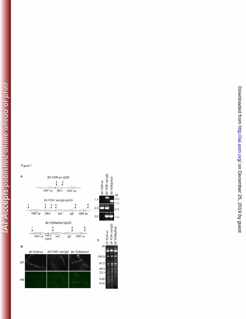
Generation and characterization of a B. hermsii YOR fhbA deletion mutant. The fhbA 234
gene of the B. hermsii YOR isolate was deleted by allelic exchange using the pFAEV3ΔfhbA 235
suicide vector. The replacement of fhbA in the B. hermsii lp200 plasmid with the kanamycin 236
resistance (kanr)-gfp gene cassette was verified by PCR (Figure 1A) and fluorescence 237
microscopy served to confirm expression of the integrated gfp containing cassette in both Bh 238
YORΔfhbA and Bh YOR::kanrgfp strains (Figure 1). Bh YORΔfhbA and Bh YOR::kanrgfp 239
(control strain) were found to have growth rates similar to the wild type parental strain (data 240
not shown). Vectors were also constructed for the purpose of complementing the fhbA deletion, 241
on Decem
ber 25, 2019 by guesthttp://iai.asm
.org/D
ownloaded from
19
however, efforts to obtain complemented strains proved unsuccessful. As detailed below, the 242
inability to generate a complemented strain proved to be irrelevant for this study since deletion 243
of fhbA had no discernable effect on phenotype. 244
245
Plasmid specific PCR has served as the approach of choice for assessing plasmid content of 246
B. burgdorferi after electroporation (38). However, the complete sequence of the plasmid 247
component of B. hermsii is not available and thus as an alternative plasmid content was 248
assessed by pulsed-field gel electrophoresis (Figure 1C). While there are inherent limitations 249
in this approach (potential for co-migrating plasmids and difficulty resolving circular plasmids), 250
no obvious differences in the plasmid content of several clones of wild type B. hermsii YOR 251
(Bh YOR-wt), Bh YORΔfhbA and Bh YOR::kanrgfp were observed. To keep the study size 252
manageable and to minimize animal usage, infectivity analyses were conducted with a 253
single clone of each strain that had plasmid profiles and growth rates consistent with the 254
parental wild type strain. 255
256
on Decem
ber 25, 2019 by guesthttp://iai.asm
.org/D
ownloaded from
20
FhbA production and presentation on the cell surface. Immunoblot analyses using 257
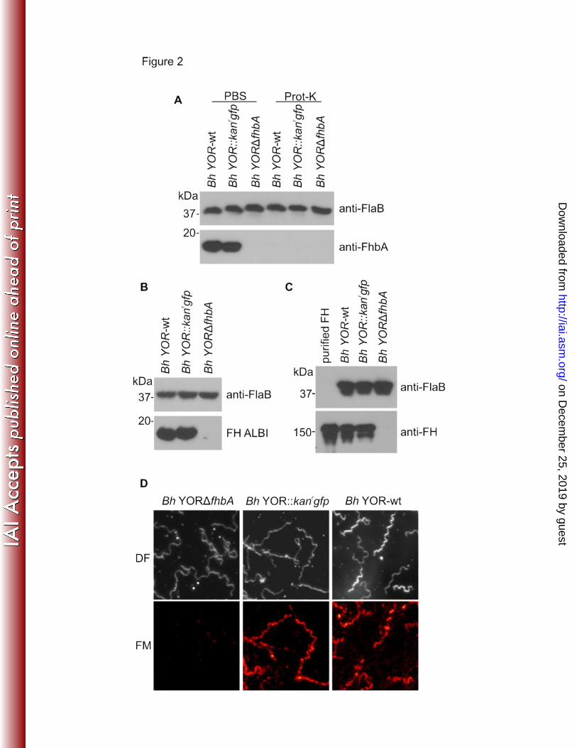
anti-FhbA antisera revealed that FhbA was produced by Bh YOR-wt and Bh YOR::kanrgfp but 258
not Bh YORΔfhbA (Figure 2A). Expression levels were not influenced by temperature as 259
equivalent amounts of FhbA were produced by cultures maintained at 25 and 37 ΕC (data not 260
shown). We also verified that FhbA produced by Bh YOR::kanrgfp is presented on the cell 261
surface in a manner similar to wild type cells using proteinase K digestion assays. Consistent 262
with surface exposure, proteinase K degraded FhbA in the Bh YOR-wt and Bh YOR::kanrgfp 263
strains (Figure 2A) but not FlaB (negative control), an inner membrane anchored protein. 264
265
FH binding assays. To determine if deletion of fhbA eliminates FH binding, cell lysates of each 266
strain were fractionated by SDS-PAGE and tested for FH binding using ALBI assays (18,27). 267
All strains bound FH except Bh YORΔfhbA (Figure 2B, bottom panel). An identical blot was 268
screened with anti-FlaB to verify equivalent loading of the gels (Figure 2B, upper panel). To 269
determine if FH can interact with the B. hermsii cell surface in an FhbA independent manner 270
on Decem
ber 25, 2019 by guesthttp://iai.asm
.org/D
ownloaded from
21
two independent approaches were employed. Pull down assays were performed by incubating 271
mid-log phase cultures of each strain with FH. The cells were pelleted and separated by 272
SDS-PAGE, transferred to membranes and then screened with anti-human FH or anti-FlaB 273
antisera (pull-down control). FH bound to Bh YOR-wt and Bh YOR::kanrgfp but not Bh 274
YORΔfhbA (Figure 2C). FH binding to the cell surface was also assessed using IFA as 275
detailed above. FH was readily detected on the surface of Bh YOR-wt and Bh YOR::kanrgfpr 276
but not on Bh YORΔfhbA (Figure 2D). It can be concluded that FH binding to B. hermsii is 277
strictly dependent on FhbA and that, as suggested in earlier studies (23, 25), FhbA is the sole 278
FH binding protein of B. hermsii. 279
280
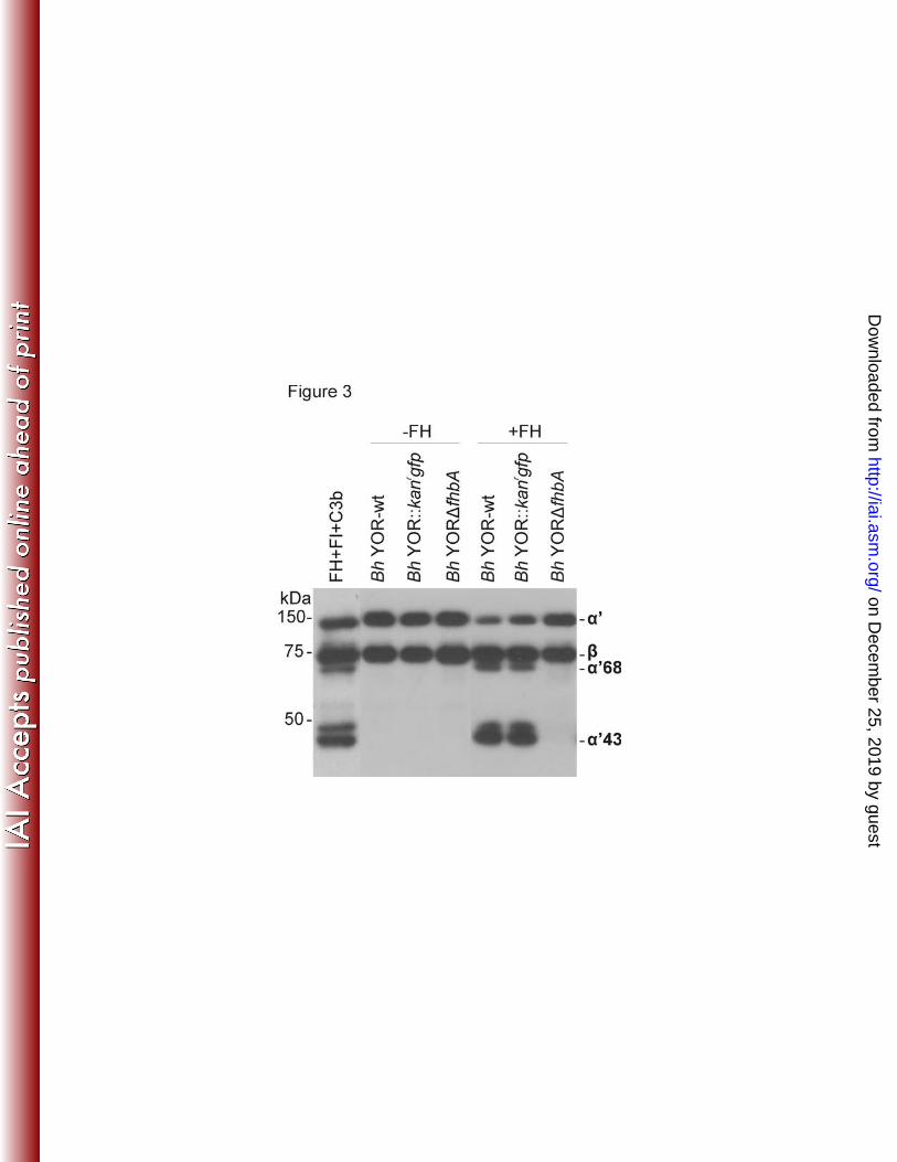
Analysis of C3b inactivation. In previous studies it was demonstrated that FH bound to the 281
surface of B. hermsii YOR efficiently serves as a co-factor for the factor I mediated cleavage 282
of C3b (24). To determine if deletion of fhbA eliminates the ability of B. hermsii to facilitate 283
C3b cleavage, each strain was incubated with human C3b and FI, with and without FH. 284
on Decem
ber 25, 2019 by guesthttp://iai.asm
.org/D
ownloaded from
22
FhbA-expressing strains cleaved C3b while Bh YORΔfhbA did not (Figure 3). It can be 285
concluded that FhbA and FH binding are required for C3b degradation and B. hermsii cannot 286
degrade C3b in a FH-FI independent manner. 287
288
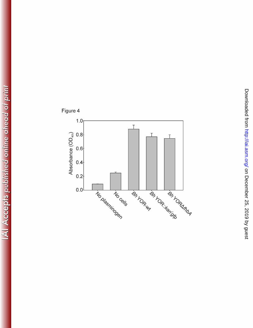
FhbA expression is not required for plasminogen binding. In addition to FH, B. hermsii FhbA 289
binds host-derived, plasminogen (19). Binding of plasminogen and its conversion to plasmin 290
may play an important role in invasion and dissemination (39, 40). Plasminogen binding was 291
assessed using an ELISA format. Statistically significant differences in plasminogen binding 292
were not detected among strains indicating that FhbA is not required for plasminogen binding 293
(Figure 4). 294
295
Analysis of serum sensitivity and complement resistance. To assess the contribution of 296
FhbA and FH binding in complement evasion in vitro, each strain was incubated with 297
complement-active NHS or HIS (complement inactive) and cell killing assessed. B. andersonii 298
on Decem
ber 25, 2019 by guesthttp://iai.asm
.org/D
ownloaded from
23
strain MOD-1 served as a positive control for serum mediated killing. While B. hermsii high 299
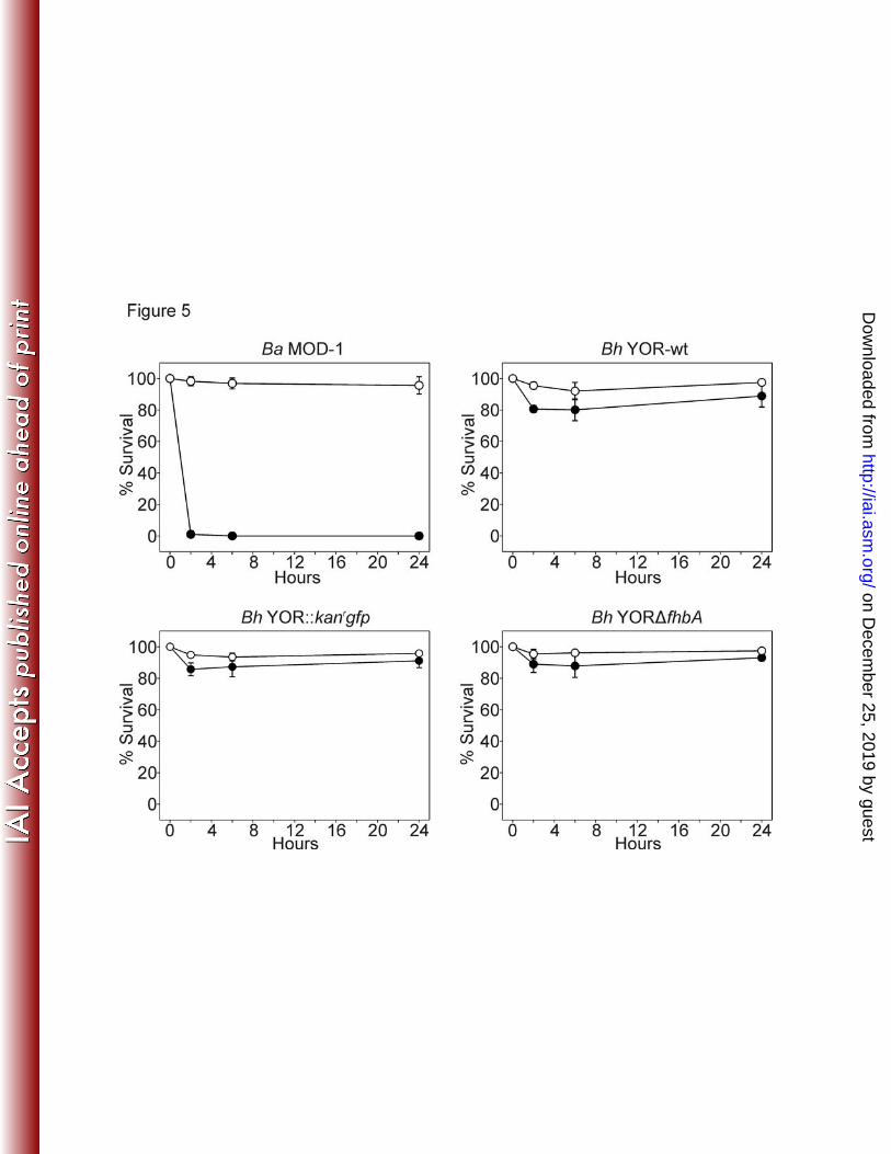
passage laboratory strains have been shown to be of intermediate serum sensitivity and thus 300
could have potentially served as controls, B. andersonii was selected because this species 301
displays a high serum sensitivity phenotype. Bh YOR-wt, Bh YOR::kanrgfp, and Bh YORΔfhbA 302
were serum resistant, even after exposure to 50% human serum for 24 hours (Figure 5). In 303
contrast, B. andersonii was killed within 2 hours. Bh YOR-wt, Bh YOR::kanrgfp, and Bh 304
YORΔfhbA also proved to be serum resistant when incubated in 100% human serum for 24 305
hours (data not shown). Hence, the data presented here do not support the hypothesis 306
previously put forth by our laboratory that the FhbA-FH interaction is required for serum 307
resistance and infectivity (18,25) and suggest that yet to be identified FH-independent 308
mechanisms are involved in complement evasion. 309
310
FhbA and FH binding are not required for B. hermsii to infect mice. To determine if FhbA 311
is required for infectivity and survival in blood, mice were needle inoculated and relative 312
on Decem
ber 25, 2019 by guesthttp://iai.asm
.org/D
ownloaded from
24
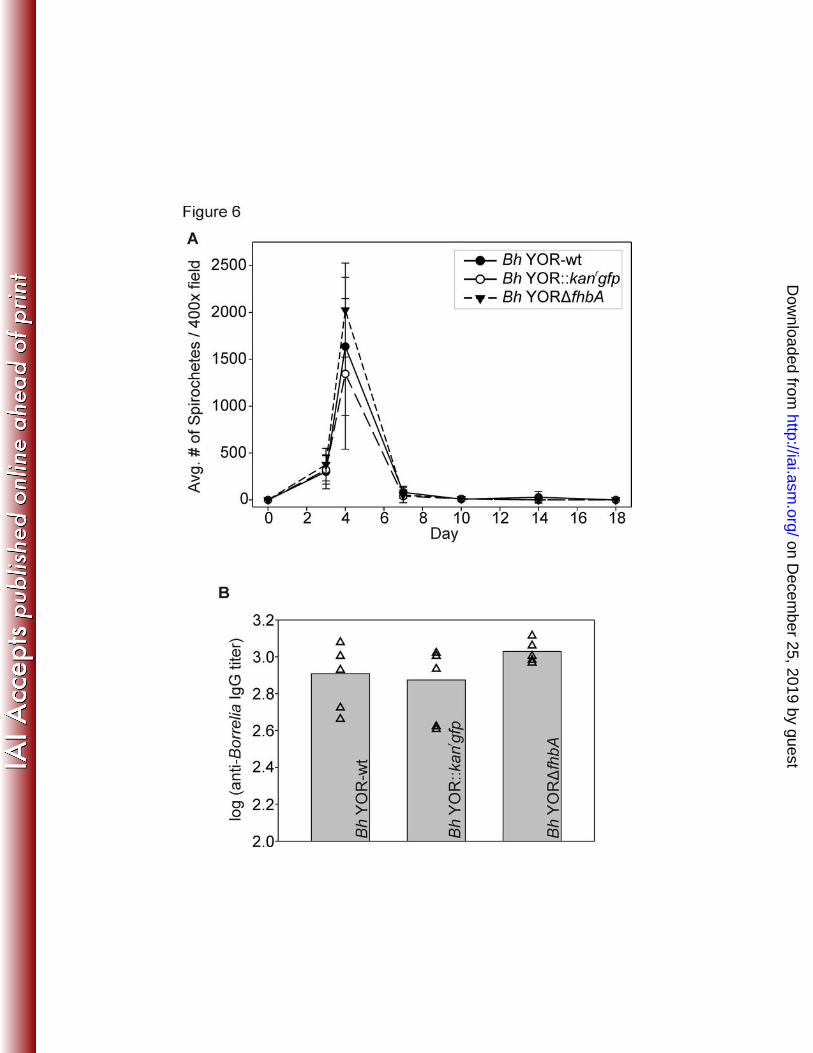
spirochete numbers in blood assessed over time. This was accomplished by visual counting 313
of spirochetes in blood samples using dark field microscopy. All strains were infectious and 314
reached comparable densities in the blood (Figure 6A). A single spirochetemic peak was 315
observed at day 4. Although additional spirochetemic peaks were not observed, cultivation of 316
blood samples revealed that Bh YOR-wt and Bh YOR::kanrgfp and Bh YORΔfhbA persisted 317
in blood up to 14 and18 days, respectively, in individual animals (Table 2). Consistent with 318
this, serological analyses revealed that all mice developed anti-B. hermsii IgG titers (Figure 319
6B). To verify the fhbA genotype of the spirochetes reisolated from blood, PCR analyses were 320
performed using an fhbA primer set. Amplicons were obtained from reisolated Bh YOR-wt and 321
Bh YOR::kanrgfp isolates but not from Bh YORΔfhbA (Figure 7A). To determine if FhbA was 322
produced by each strain during infection, antiserum from each mouse was used to screen 323
immunoblots of r-FhbA. Anti-FhbA antibody was detected in sera collected 4 weeks 324
post-infection from Bh YOR-wt- and Bh YOR::kanrgfp-infected mice but not from mice infected 325
with Bh YORΔfhbA (Figure 7B). 326
on Decem
ber 25, 2019 by guesthttp://iai.asm
.org/D
ownloaded from
25
327
DISCUSSION 328
329
The FhbA protein of B. hermsii binds host derived proteins that are important in complement 330
regulation including FH, FHR-1 and plasminogen (18,19). Based on this and its universal 331
distribution among low passage isolates it has been postulated that FhbA is a virulence factor 332
required for complement evasion (19,25). An earlier study, revealed that the B. hermsii REN 333
strain (which lacks fhbA) displayed greater serum sensitivity than other isolates that produce 334
FhbA (25). However, because the REN used in the cited study had been passaged in vitro 335
>100 times, its increased sensitivity to serum could have resulted from other genotypic or 336
phenotypic changes that can occur during extended cultivation. The ability to directly assess 337
FhbA=s role in complement evasion has been hampered by inherent difficulties of genetically 338
manipulating the TBRF spirochetes. The goal of this study was determine if FhbA and hence 339
on Decem
ber 25, 2019 by guesthttp://iai.asm
.org/D
ownloaded from
26
FH binding is required for B. hermsii to evade complement mediated killing in vitro and to infect 340
mice and persist. 341
342
To address these questions, fhbA was deleted using allelic exchange mutagenesis to yield the 343
Bh YORΔfhbA strain. This deletion mutant displayed similar growth kinetics as the parental wild 344
type strain and PFGE did not reveal the loss of plasmids or genomic rearrangements. Using 345
several different approaches to assess FH binding, it was demonstrated that deletion of fhbA 346
results in the complete loss of FH binding by Bh YORΔfhbA consistent with FhbA being the 347
sole FH binding protein of B. hermsii (18). Surprisingly, deletion of fhbA had no impact on 348
the ability of Bh YORΔfhbA to survive in human serum or to establish infection and persist 349
in mice. All mice developed a single spirochetemic peak during days 3 and 4 post 350
inoculation. While relapses were not evident, spirochetes were readily cultivated from blood 351
collected up to 2 weeks post-inoculation. Hence, the fhbA mutant was able to survive in blood, 352
albeit at low levels, as long as the wildtype strain. It can be concluded that fhbA and FH binding 353
are not required for evasion of complement in human serum and in mice. 354
on Decem
ber 25, 2019 by guesthttp://iai.asm
.org/D
ownloaded from

27
355
The properties of the fhbA deletion mutant suggest that B. hermsii employs FH-independent 356
mechanisms for complement evasion. B. hermsii produces a serine protease (BhpA) that is 357
abundantly expressed in vitro and during infection in mice (41) that could conceivably cleave 358
complement proteins and play a compensatory role in complement evasion in the FhbA deletion 359
mutant. However in this study cleavage of C3b by the fhbA deletion mutant was not observed 360
suggesting that BhpA is not directly involved in cleaving this important opsonin. The potential 361
for BhpA to facilitate complement evasion by potentially cleaving other complement regulators 362
has not yet been directly assessed. Proteolytic inactivation of complement would be a novel 363
immune evasion mechanism for spirochetes and should be considered in future studies. 364
365
Other TBRF spirochetes have been shown to utilize multiple complement evasion mechanisms. 366
In addition to FH, B. recurrentis and B. duttonii also bind C4BP, a central regulator of the 367
antibody-mediated classical complement pathway (42,43). C4BP serves as a cofactor in 368
the FI mediated cleavage of C4b. However, binding analyses suggest that C4BP does not 369
on Decem
ber 25, 2019 by guesthttp://iai.asm
.org/D
ownloaded from
28
bind to B. hermsii (43) (Marconi, RT, unpublished data) and hence C4BP binding does 370
not appear to be involved in B. hermsii serum resistance. B. recurrentis, a louse borne 371
relapsing fever spirochete, binds C1 esterase inhibitor (C1-INH) via the CihC protein 372
(43). C1-INH is a serine protease inhibitor that contributes to the regulation of both the classical 373
and lectin pathways of complement (44). While the binding of C1-INH cannot be excluded 374
for B. hermsii, it would not explain the in vitro resistance to the alternative pathway 375
complement-mediated killing displayed by the FhbA deletion mutant. 376
377
B. hermsii has been demonstrated to recruit and activate plasminogen on its surface (19,45), 378
a process that has been demonstrated to facilitate penetration of endothelial cell monolayers 379
(45). A recent study suggests that plasminogen also has a complement regulatory role 380
(inhibitory) (46). Plasminogen binding analyses revealed that the Bh YORΔfhbA strain binds 381
plasminogen at levels similar to that of Bh YOR-wt. It can be concluded that FhbA is not required 382
for potentially important functions that are mediated by plasminogen binding. 383
384
on Decem
ber 25, 2019 by guesthttp://iai.asm
.org/D
ownloaded from
29
The demonstration that B. hermsii does not require FhbA raises the question as to why FhbA 385
is universal, conserved and constitutively produced? It is possible that FhbA could have 386
functional roles that were not directly tested here such as facilitating penetration of endothelial 387
cells and crossing of the blood brain barrier. Alternatively, FhbA may play an important functional 388
role in the tick. One possibility is that FhbA facilitates complement evasion specifically in ticks 389
during the blood meal. This remains to be tested. A similar hypothesis has been suggested for 390
the FH binding-CspA protein of B. burgdorferi (47). Unfortunately, in contrast to Ixodes ticks, 391
Ornithordoros ticks are not currently commercially available and hence it was not possible in 392
this study to investigate the potential biological role of FhbA in the tick environment. 393
394
In summary, this study is among the first to successfully genetically manipulate a potential 395
virulence factor of a TBRF spirochete and study the mutant strain in vivo. FhbA is known to 396
bind to three human derived ligands: FH, FHR-1 and plasminogen (18,19). However, deletion 397
of fhbA revealed that it is not required for evasion of killing mediated by the alternative 398
on Decem
ber 25, 2019 by guesthttp://iai.asm
.org/D
ownloaded from

30
complement pathway in vitro or for survival in mice when delivered by needle inoculation. It 399
can also be concluded that its function in plasminogen binding is not essential for infectivity. 400
This study suggests that FH/FHR-1 independent mechanisms contribute to B. hermsii=s ability 401
to evade killing by the alternate complement pathway. Future studies utilizing the fhbA 402
deletion mutant will seek to determine if FhbA contributes to other roles during infection and 403
transmission such as invasiveness, adherence or survival in ticks. While the functional role of 404
FhbA remains a question, this study represents an important step forward in the development 405
of tools that will facilitate the study of other B. hermsii virulence factors in vivo. 406
407
on Decem
ber 25, 2019 by guesthttp://iai.asm
.org/D
ownloaded from
31
FOOTNOTES 408
This work is supported in part by grants from NIH to R.T.M (R01-AI037787, R01-DE017401) 409
and to D.P.M. (F31-DE023000). This study was also supported in part by Virginia 410
Commonwealth University=s CTSA (UL1TR000058 from the National Center for Advancing 411
Translational Sciences) and the CCTR Endowment Fund of Virginia Commonwealth University. 412
Its contents are solely the responsibility of the authors and do not necessarily represent official 413
views of the National Center for Advancing Translational Sciences, the National Institutes of 414
Health, or Virginia Commonwealth University. 415
on Decem
ber 25, 2019 by guesthttp://iai.asm
.org/D
ownloaded from
32
REFERENCES 416
New list 417
1 Barbour, A. G., and S. F. Hayes. 1986. Biology of Borrelia species. Microbiological 418
Reviews 50:381-400. 419
2 Cutler, S., and A. Talbert. 2003. Tick-borne relapsing fever in Tanzania -- a forgotten 420
problem? ASM News 69:542-543. 421
3 Dupont, H. T., B. La Scola, R. Williams, and D. Raoult. 1997. A focus of tick-borne 422
relapsing fever in southern Zaire. Clin Infect Dis 25:139-44. 423
4 Jongen, V. H., J. van Roosmalen, J. Tiems, J. Van Holten, and J. C. Wetsteyn. 424
1997. Tick-borne relapsing fever and pregnancy outcome in rural Tanzania. Acta Obstet 425
Gynecol Scand 76:834-8. 426
5 Vial, L., G. Diatta, A. Tall, H. Ba el, H. Bouganali, P. Durand, C. Sokhna, C. Rogier, 427
F. Renaud, and J. F. Trape. 2006. Incidence of tick-borne relapsing fever in west 428
Africa: longitudinal study. Lancet 368:37-43. 429
on Decem
ber 25, 2019 by guesthttp://iai.asm
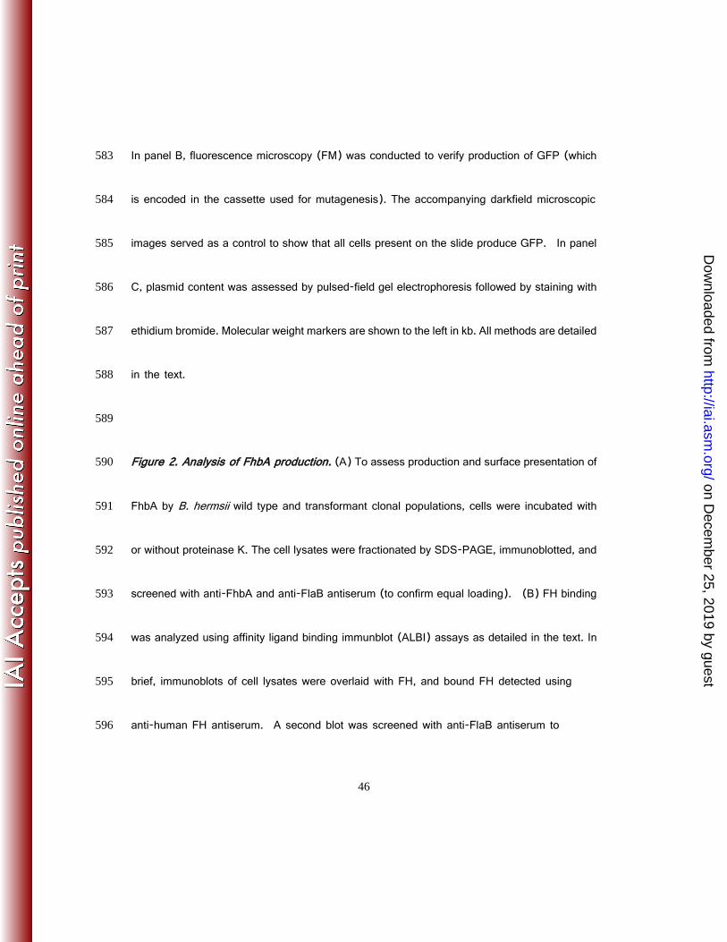
.org/D
ownloaded from
33
6 Dworkin, M., P. Shoemaker, C. Fritz, M. Dowell, and Anderson DE, Jr. 2002. The 430
epidemiology of tick-borne relapsing fever in the United States. Am J Trop Med Hyg 431
66:753-758. 432
7 Dworkin, M. S., J. Anderson, D E, T. G. Schwan, P. C. Shoemaker, S. N. Banerjee, 433
B. O. Kassen, and W. Burgdorfer. 1998. Tick-borne relapsing fever in the 434
Northwestern United States and Southwestern Canada. Clinical Infectious Diseases 435
26:122-131. 436
8. Schwan, T. G., P. F. Policastro, Z. Miller, R. L. Thompson, T. Damrow, and J. E. 437
Keirans. 2003. Tick-borne relapsing fever caused by Borrelia hermsii in Montana. 438
Emerg. Infect. Dis. 9:1151-1154. 439
9 Schwan, T. G., S. J. Raffel, M. E. Schrumpf, and S. F. Porcella. 2007. Diversity and 440
distribution of Borrelia hermsii. Emerg Infect Dis 13:436-42. 441
on Decem
ber 25, 2019 by guesthttp://iai.asm
.org/D
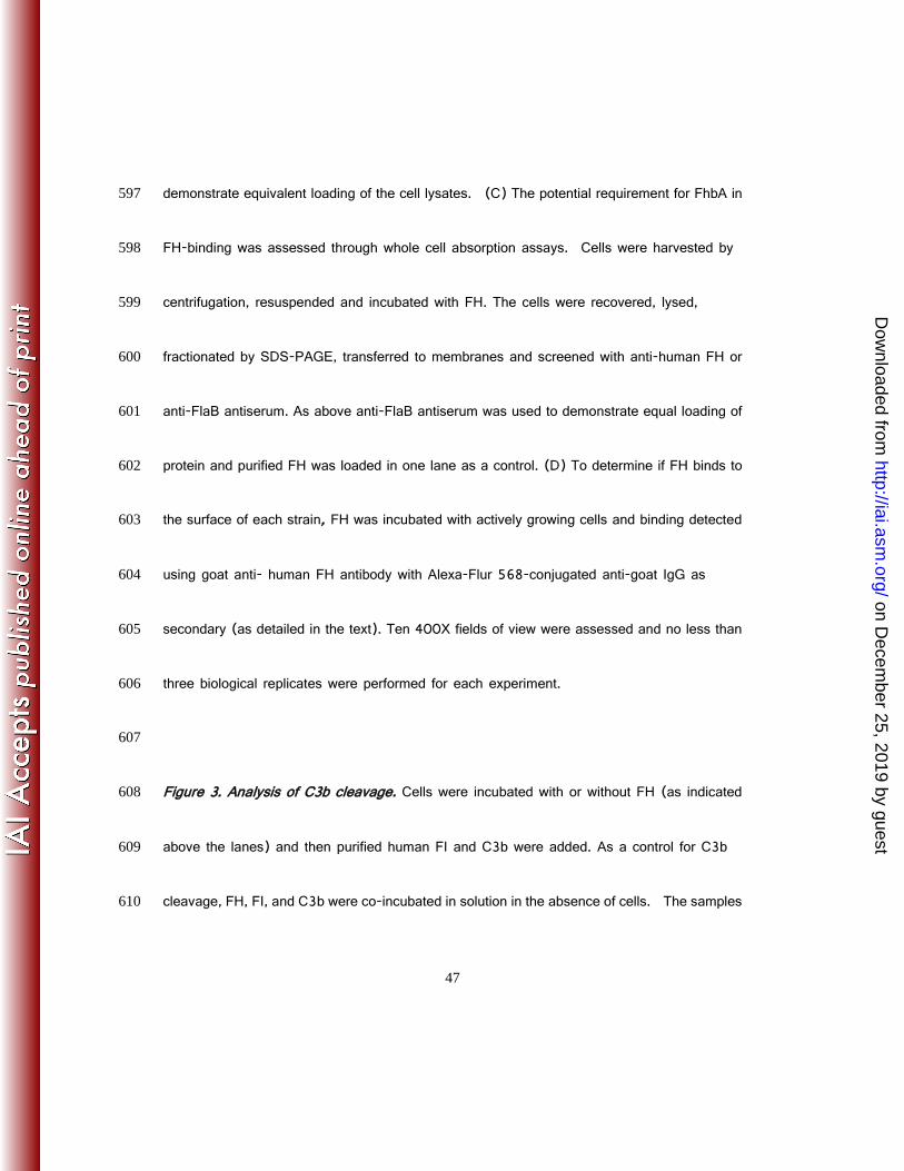
ownloaded from
34
10. Schwan, T. G., S. J. Raffel, M. E. Schrumpf, L. S. Webster, A. R. Marques, R. 442
Spano, M. Rood, J. Burns, and R. Hu. 2009. Tick-borne relapsing fever and Borrelia 443
hermsii, Los Angeles County, California, USA. Emerg Infect Dis 15:1026-31. 444
11. Felsenfeld, O. 1971. Borrelia. Strains, vectors, human and animal borreliosis. Warren 445
H. Green, Inc., St. Louis. 446
12. Cadavid, D., and A. G. Barbour. 1998. Neuroborreliosis during relapsing fever: Review 447
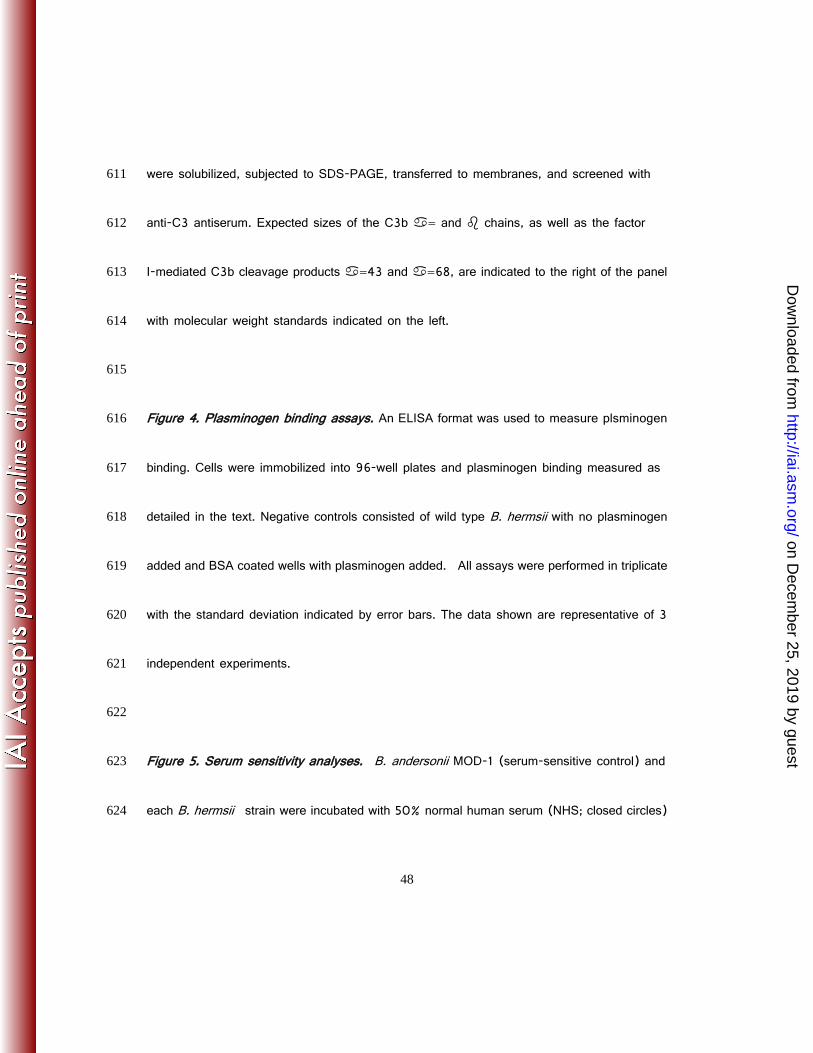
of the clinical manifestations, pathology and treatment of infections in humans and 448
experimental animals. Clinical Infectious Diseases 26:151-164. 449
13. McDowell, J. V., D. P. Miller, K. L. Mallory, and R. T. Marconi. 2012. Treponema 450
denticola: FhbB, Dentilisin, Complement Evasion and the Paradox of Factor H Cleavage, 451
p. 43-62. In M. E. Embers (ed.), The Pathogenic Spirochetes: strategies for evasion 452
of host immunity and persistance. Springer Science+Business Media, New York. 453
on Decem
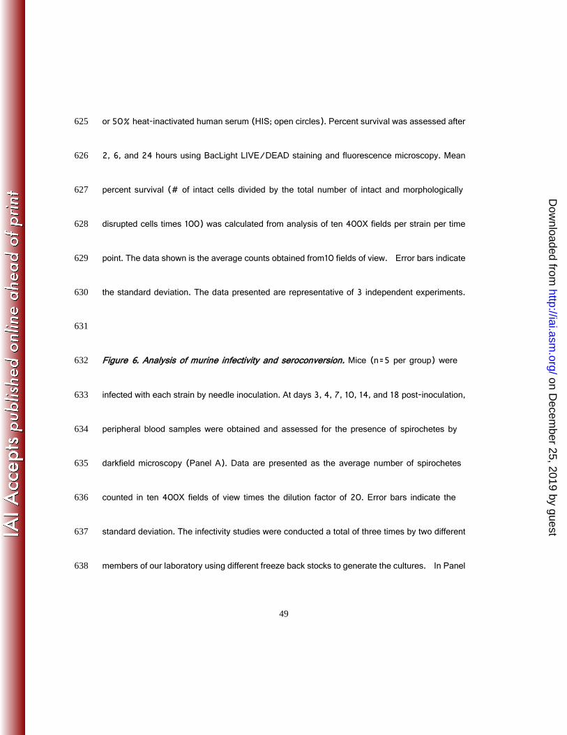
ber 25, 2019 by guesthttp://iai.asm
.org/D
ownloaded from
35
14. Zipfel, P. F., T. Hallstrom, S. Hammerschmidt, and C. Skerka. 2008. The 454
complement fitness factor H: role in human diseases and for immune escape of 455
pathogens, like pneumococci. Vaccine 26 Suppl 8:I67-74. 456
15. Ferreira, V. P., M. K. Pangburn, and C. Cortes. 2010. Complement control protein 457
factor H: the good, the bad, and the inadequate. Mol Immunol 47:2187-97. 458
16. Ruddy, S., and K. F. Austen. 1969. C3 inactivator of man. I. Hemolytic measurement 459
by the inactivation of cell-bound C3. J Immunol 102:533-543. 460
17. Ruddy, S., and K. F. Austen. 1971. C3b inactivator of man. II. Fragments produced by 461
C3b inactivator cleavage of cell-bound or fluid phase C3b. J Immunol 107:742-750. 462
18. Hovis, K. M., J. V. McDowell, L. Griffin, and R. T. Marconi. 2004. Identification and 463
characterization of a linear-plasmid-encoded factor H-binding protein (FhbA) of the 464
relapsing fever spirochete Borrelia hermsii. J Bacteriol 186:2612-8. 465
19. Rossmann, E., P. Kraiczy, P. Herzberger, C. Skerka, M. Kirschfink, M. M. Simon, 466
P. F. Zipfel, and R. Wallich. 2007. Dual binding specificity of a Borrelia 467
on Decem
ber 25, 2019 by guesthttp://iai.asm
.org/D
ownloaded from
36
hermsii-associated complement regulator-acquiring surface protein for factor H and 468
plasminogen discloses a putative virulence factor of relapsing fever spirochetes. J 469
Immunol 178:7292-301. 470
20. Hellwage, J., T. Meri, T. Heikkila, A. Alitalo, J. Panelius, P. Lahdenne, I. J. T. 471
Seppala, and S. Meri. 2001. The complement regulator factor H binds to the surface 472
protein OspE of Borrelia burgdorferi. Journal of Biological Chemistry 276:8427-8435. 473
21. Kraiczy, P., J. Hellwage, C. Skerka, M. Kirschfink, V. Brade, P. F. Zipfel, and R. 474
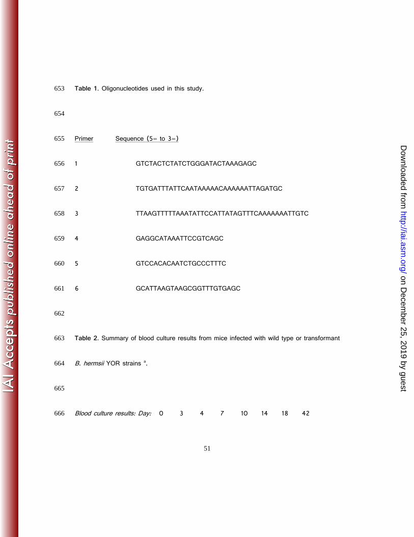
Wallich. 2003. Immune evasion of Borrelia burgdorferi: mapping of a 475
complement-inhibitor factor H-binding site of BbCRASP-3, a novel member of the Erp 476
protein family. Eur J Immunol 33:697-707. 477
22. Kraiczy, P., C. Skerka, and P. F. Zipfel. 2001. Further characterization of complement 478
regulator-acquiring surface proteins of Borrelia burgdorferi. Infect. Immun. 479
69:7800-7809. 480
on Decem
ber 25, 2019 by guesthttp://iai.asm
.org/D
ownloaded from
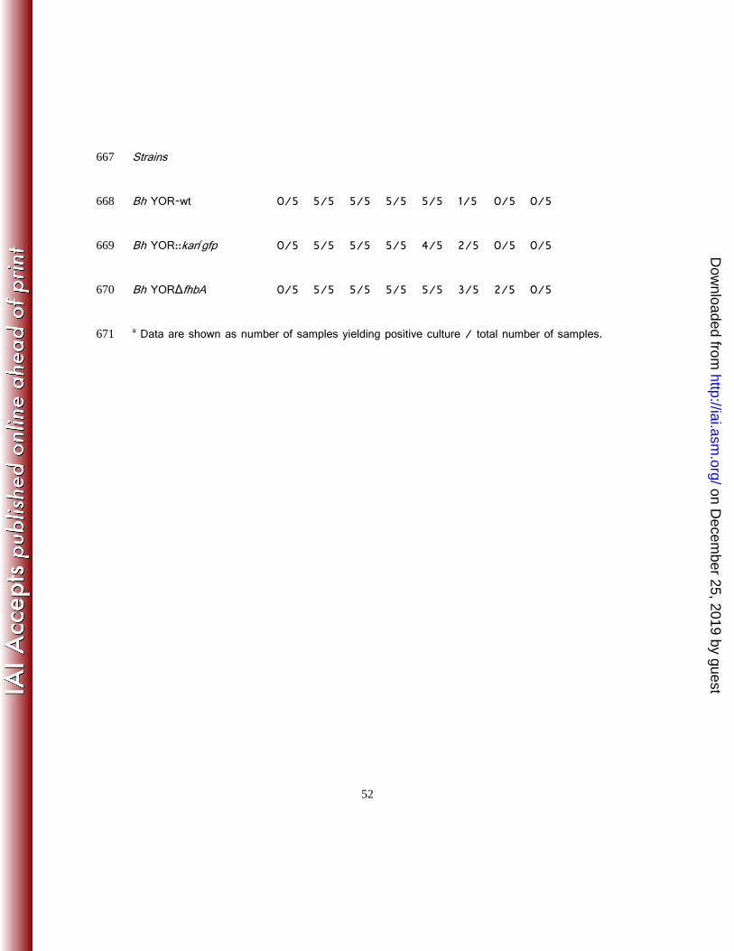
37
23. McDowell, J. V., J. Wolfgang, E. Tran, M. S. Metts, D. Hamilton, and R. T. Marconi. 481
2003. Comprehensive analysis of the factor H binding capabilities of Borrelia species 482
associated with lyme disease: delineation of two distinct classes of factor H binding 483
proteins. Infect Immun 71:3597-602. 484
24. Hovis, K. M., J. P. Jones, T. Sadlon, G. Raval, D. L. Gordon, and R. T. Marconi. 485
2006. Molecular analyses of the interaction of Borrelia hermsii FhbA with the 486
complement regulatory proteins factor H and factor H-like protein 1. Infect Immun 487
74:2007-14. 488
25. Hovis, K. M., M. E. Schriefer, S. Bahlani, and R. T. Marconi. 2006. Immunological 489
and molecular analyses of the Borrelia hermsii factor H and factor H-like protein 1 binding 490
protein, FhbA: Demonstration of its utility as a diagnostic marker and epidemiological 491
tool for tick-borne relapsing fever. Infection and Immunity 74:4519-4529. 492
26. Hovis, K. M., J. C. Freedman, H. Zhang, J. L. Forbes, and R. T. Marconi. 2008. 493
Identification of an antiparallel coiled-coil/loop domain required for ligand binding by the 494
on Decem
ber 25, 2019 by guesthttp://iai.asm
.org/D
ownloaded from

38
Borrelia hermsii FhbA protein: additional evidence for the role of FhbA in the 495
host-pathogen interaction. Infect Immun 76:2113-22. 496
27. McDowell, J. V., E. Tran, D. Hamilton, J. Wolfgang, K. Miller, and R. T. Marconi. 497
2003. Analysis of the ability of spirochete species associated with relapsing fever, avian 498
borreliosis, and epizootic bovine abortion to bind factor H and cleave C3b. J Clin 499
Microbiol 41:3905-10. 500
28. Kraiczy, P., C. Skerka, M. Kirschfink, P. F. Zipfel, and V. Brade. 2001. Mechanism 501
of complement resistance of pathogenic Borrelia burgdorferi isolates. International 502
Immunopharmacology 1:3393-401. 503
29. McDowell, J. V., B. Huang, J. C. Fenno, and R. T. Marconi. 2009. Analysis of a 504
unique interaction between the complement regulatory protein factor H and the 505
periodontal pathogen Treponema denticola. Infect Immun 77:1417-25. 506
30. Meri, T., H. Amdahl, M. J. Lehtinen, S. Hyvarinen, J. V. McDowell, A. 507
Bhattacharjee, S. Meri, R. Marconi, A. Goldman, and T. S. Jokiranta. 2013. 508
on Decem
ber 25, 2019 by guesthttp://iai.asm
.org/D
ownloaded from

39
Microbes Bind Complement Inhibitor Factor H via a Common Site. PLoS Pathog 509
9:e1003308. 48. Zipfel, P. F. 2009. Complement and immune defense: from 510
innate immunity to human diseases. Immunol Lett 126:1-7. 511
31. Zipfel, P. F. 2009. Complement and immune defense: from innate immunity to human 512
diseases. Immunol Lett 126:1-7. 513
32. Zipfel, P. F., C. Skerka, J. Hellwage, S. T. Jokiranta, S. Meri, V. Brade, P. Kraiczy, 514
M. Noris, and G. Remuzzi. 2002. Factor H family proteins: on complement, microbes 515
and human diseases. Biochem Soc Trans 30:971-8. 516
33. Fine, L. M., C. G. Earnhart, and R. T. Marconi. 2011. Genetic transformation of the 517
relapsing fever spirochete Borrelia hermsii: stable integration and expression of green 518
fluorescent protein from linear plasmid 200 (lp200). J Bacteriol 193:3241-3245. 519
34. Lin, T., J. H. Oliver, Jr., L. Gao, T. M. Kollars, Jr., and K. L. Clark. 2001. Genetic 520
heterogeneity of Borrelia burgdorferi sensu lato in the southern United States based on 521
on Decem
ber 25, 2019 by guesthttp://iai.asm
.org/D
ownloaded from
40
restriction fragment length polymorphism and sequence analysis. J Clin Microbiol 522
39:2500-7. 523
35. Marconi, R. T., D. Liveris, and I. Schwartz. 1995. Identification of novel insertion 524
elements, restriction fragment length polymorphism patterns, and discontinuous 23S 525
rRNA in Lyme disease spirochetes: phylogenetic analyses of rRNA genes and their 526
intergenic spacers in Borrelia japonica sp. nov. and genomic group 21038 (Borrelia 527
andersonii sp. nov.) isolates. J Clin Microbiol 33:2427-34. 528
36. Carlyon, J. A., and R. T. Marconi. 1998. Cloning and molecular characterization of a 529
multicopy, linear plasmid-carried, repeat motif-containing gene from Borrelia turicatae, 530
a causative agent of relapsing fever. J Bacteriol 180:4974-81. 531
37. Earnhart, C. G., and R. T. Marconi. 2007. An octavalent lyme disease vaccine 532
induces antibodies that recognize all incorporated OspC type-specific sequences. Hum 533
Vaccin 3:281-9. 534
on Decem
ber 25, 2019 by guesthttp://iai.asm
.org/D
ownloaded from
41
38. Labandeira-Rey, M., and J. T. Skare. 2001. Decreased infectivity in Borrelia burgorferi 535
strain B31 is associated with the loss of either linear plasmid 25 or 28-1. Infection and 536
Immunity 69:446-455. 537
39. Coleman, J. L., J. A. Gebbia, J. Piesman, J. L. Degen, T. H. Bugge, and J. L. 538
Benach. 1997. Plasminogen is required for efficient dissemination of B. burgdorferi in 539
ticks and for enhancement of spirochetemia in mice. Cell 89:1111-1119. 540
40. Nordstrand, A., A. Shamaei-Tousi, A. Ny, and S. Bergstrom. 2001. Delayed invasion 541
of the kidney and brain by Borrelia crocidurae in plasminogen-deficient mice. Infect 542
Immun 69:5832-9. 543
41. Guyard, C., J. M. Battisti, S. J. Raffel, M. E. Schrumpf, A. R. Whitney, J. G. Krum, 544
S. F. Porcella, P. A. Rosa, F. R. DeLeo, and T. G. Schwan. 2006. Relapsing fever 545
spirochaetes produce a serine protease that provides resistance to oxidative stress and 546
killing by neutrophils. Mol Microbiol 60:710-22. 547
on Decem
ber 25, 2019 by guesthttp://iai.asm
.org/D
ownloaded from
42
42. Grosskinsky, S., M. Schott, C. Brenner, S. J. Cutler, M. M. Simon, and R. Wallich. 548
2010. Human complement regulators C4b-binding protein and C1 esterase inhibitor 549
interact with a novel outer surface protein of Borrelia recurrentis. PLoS Negl Trop Dis 550
4:e698. 551
43. Meri, T., S. J. Cutler, A. M. Blom, S. Meri, and T. S. Jokiranta. 2006. Relapsing 552
Fever Spirochetes Borrelia recurrentis and B. duttonii Acquire Complement Regulators 553
C4b-Binding Protein and Factor H. Infect Immun 74:4157-63. 554
44. Davis, A. E., 3rd, P. Mejia, and F. Lu. 2008. Biological activities of C1 inhibitor. Mol 555
Immunol 45:4057-63. 556
45. Coleman, J. L., T. J. Sellati, J. E. Testa, R. R. Kew, M. B. Furie, and J. L. Benach. 557
1995. Borrelia burgdorferi binds plasminogen, resulting in enhanced penetration of 558
endothelial monolayers. Infect Immun 63:2478-84. 559
on Decem
ber 25, 2019 by guesthttp://iai.asm
.org/D
ownloaded from
43
46. Barthel, D., S. Schindler, and P. F. Zipfel. 2012. Plasminogen is a complement 560
inhibitor. J Biol Chem 287:18831-42. 561
47. McDowell, J. V., K. M. Hovis, H. Zhang, E. Tran, J. Lankford, and R. T. Marconi. 562
2006. Evidence that the BBA68 protein (BbCRASP-1) of the Lyme disease spirochetes 563
does not contribute to factor H-mediated immune evasion in humans and other animals. 564
Infect Immun 74:3030-4. 565
566
567
568
on Decem
ber 25, 2019 by guesthttp://iai.asm
.org/D
ownloaded from

45
FIGURE LEGENDS 570
571
Figure 1 . Allelic exchange mutagenesis. Figure 1 . Allelic exchange mutagenesis. The 572
schematics in Panel A (left figure) depict the gene organization near fhbA on lp200 before and 573
after allelic exchange mutagenesis. The upstream and downstream ORFs that are adjacent to 574
fhbA on plasmid lp200 are indicated in the figure as >ORF up= and >ORF dn=, respectively. 575
Primer target sites (black arrowheads), with primer numbers indicated above each arrowhead, 576
are shown. Primer sequences can be found in Table 1. The primers are numbered in the figure 577
as they are in Table 1. To the right of the schematic, the results of PCR analyses conducted 578
to confirm proper insertion of the mutagenesis cassettes are shown (ethidium bromide stained 579
gels). While numerous clones were analyzed, the data presented in panels B and C were 580
obtained with the specific clonal populations that were employed in all subsequent experiments. 581
The criteria used for selecting specific clones for all subsequent work are detailed in the text. 582
on Decem
ber 25, 2019 by guesthttp://iai.asm
.org/D
ownloaded from
46
In panel B, fluorescence microscopy (FM) was conducted to verify production of GFP (which 583
is encoded in the cassette used for mutagenesis). The accompanying darkfield microscopic 584
images served as a control to show that all cells present on the slide produce GFP. In panel 585
C, plasmid content was assessed by pulsed-field gel electrophoresis followed by staining with 586
ethidium bromide. Molecular weight markers are shown to the left in kb. All methods are detailed 587
in the text. 588
589
Figure 2. Analysis of FhbA production. (A) To assess production and surface presentation of 590
FhbA by B. hermsii wild type and transformant clonal populations, cells were incubated with 591
or without proteinase K. The cell lysates were fractionated by SDS-PAGE, immunoblotted, and 592
screened with anti-FhbA and anti-FlaB antiserum (to confirm equal loading). (B) FH binding 593
was analyzed using affinity ligand binding immunblot (ALBI) assays as detailed in the text. In 594
brief, immunoblots of cell lysates were overlaid with FH, and bound FH detected using 595
anti-human FH antiserum. A second blot was screened with anti-FlaB antiserum to 596
on Decem
ber 25, 2019 by guesthttp://iai.asm
.org/D
ownloaded from
47
demonstrate equivalent loading of the cell lysates. (C) The potential requirement for FhbA in 597
FH-binding was assessed through whole cell absorption assays. Cells were harvested by 598
centrifugation, resuspended and incubated with FH. The cells were recovered, lysed, 599
fractionated by SDS-PAGE, transferred to membranes and screened with anti-human FH or 600
anti-FlaB antiserum. As above anti-FlaB antiserum was used to demonstrate equal loading of 601
protein and purified FH was loaded in one lane as a control. (D) To determine if FH binds to 602
the surface of each strain, FH was incubated with actively growing cells and binding detected 603
using goat anti- human FH antibody with Alexa-Flur 568-conjugated anti-goat IgG as 604
secondary (as detailed in the text). Ten 400X fields of view were assessed and no less than 605
three biological replicates were performed for each experiment. 606
607
Figure 3. Analysis of C3b cleavage. Cells were incubated with or without FH (as indicated 608
above the lanes) and then purified human FI and C3b were added. As a control for C3b 609
cleavage, FH, FI, and C3b were co-incubated in solution in the absence of cells. The samples 610
on Decem
ber 25, 2019 by guesthttp://iai.asm
.org/D
ownloaded from
48
were solubilized, subjected to SDS-PAGE, transferred to membranes, and screened with 611
anti-C3 antiserum. Expected sizes of the C3b = and chains, as well as the factor 612
I-mediated C3b cleavage products =43 and =68, are indicated to the right of the panel 613
with molecular weight standards indicated on the left. 614
615
Figure 4. Plasminogen binding assays. An ELISA format was used to measure plsminogen 616
binding. Cells were immobilized into 96-well plates and plasminogen binding measured as 617
detailed in the text. Negative controls consisted of wild type B. hermsii with no plasminogen 618
added and BSA coated wells with plasminogen added. All assays were performed in triplicate 619
with the standard deviation indicated by error bars. The data shown are representative of 3 620
independent experiments. 621
622
Figure 5. Serum sensitivity analyses. B. andersonii MOD-1 (serum-sensitive control) and 623
each B. hermsii strain were incubated with 50% normal human serum (NHS; closed circles) 624
on Decem
ber 25, 2019 by guesthttp://iai.asm
.org/D
ownloaded from
49
or 50% heat-inactivated human serum (HIS; open circles). Percent survival was assessed after 625
2, 6, and 24 hours using BacLight LIVE/DEAD staining and fluorescence microscopy. Mean 626
percent survival (# of intact cells divided by the total number of intact and morphologically 627
disrupted cells times 100) was calculated from analysis of ten 400X fields per strain per time 628
point. The data shown is the average counts obtained from10 fields of view. Error bars indicate 629
the standard deviation. The data presented are representative of 3 independent experiments. 630
631
Figure 6. Analysis of murine infectivity and seroconversion. Mice (n=5 per group) were 632
infected with each strain by needle inoculation. At days 3, 4, 7, 10, 14, and 18 post-inoculation, 633
peripheral blood samples were obtained and assessed for the presence of spirochetes by 634
darkfield microscopy (Panel A). Data are presented as the average number of spirochetes 635
counted in ten 400X fields of view times the dilution factor of 20. Error bars indicate the 636
standard deviation. The infectivity studies were conducted a total of three times by two different 637
members of our laboratory using different freeze back stocks to generate the cultures. In Panel 638
on Decem
ber 25, 2019 by guesthttp://iai.asm
.org/D
ownloaded from

50
B, ELISAs were performed using serum harvested from mice 4 weeks post-inoculation to 639
determine the anti-B. hermsii YOR titers elicited by each infecting strain (as indicated). 640
Geometric means (grey bars) were determined from titers calculated for each mouse (triangles) 641
at 1/3 ODmax. All ELISA assays were conducted in triplicate. The bar graphs represent the 642
average of the titers determined for each group of 5 mice. Individual data points for each mouse 643
are shown 644
645
Figure 7. Analysis of fhbA genotype in recovered isolates and anti-FhbA antibody 646
responses in infected mice. Isolates that were cultivated from mice infected with each strain 647
(indicated in the figure) served as template for PCR amplification of fhbA. Panel A depicts the 648
PCR results. The individual mice within each group were designated as A through E. In panel 649
B, pooled serum harvested from each infected mouse group was screened for the presence 650
of anti-FhbA IgG using immunblot approaches with recombinant FhbA serving as the 651
immobilized antigen. Serum for an uninfected mouse served as a negative control. 652
on Decem
ber 25, 2019 by guesthttp://iai.asm
.org/D
ownloaded from
51
Table 1. Oligonucleotides used in this study. 653
654
Primer Sequence (5= to 3=) 655
1 GTCTACTCTATCTGGGATACTAAAGAGC 656
2 TGTGATTTATTCAATAAAAACAAAAAATTAGATGC 657
3 TTAAGTTTTTAAATATTCCATTATAGTTTCAAAAAAATTGTC 658
4 GAGGCATAAATTCCGTCAGC 659
5 GTCCACACAATCTGCCCTTTC 660
6 GCATTAAGTAAGCGGTTTGTGAGC 661
662
Table 2. Summary of blood culture results from mice infected with wild type or transformant 663
B. hermsii YOR strains a. 664
665
Blood culture results: Day: 0 3 4 7 10 14 18 42 666
on Decem
ber 25, 2019 by guesthttp://iai.asm
.org/D
ownloaded from
52
Strains 667
Bh YOR-wt 0/5 5/5 5/5 5/5 5/5 1/5 0/5 0/5 668
Bh YOR::kanrgfp 0/5 5/5 5/5 5/5 4/5 2/5 0/5 0/5 669
Bh YORΔfhbA 0/5 5/5 5/5 5/5 5/5 3/5 2/5 0/5 670
a Data are shown as number of samples yielding positive culture / total number of samples. 671
on Decem
ber 25, 2019 by guesthttp://iai.asm
.org/D
ownloaded from