downloaded from...
TRANSCRIPT

JPET #259192
1
1. Title Page.
Effect of melatonin and its analogues in the tear secretion.
Francisco Javier NAVARRO GIL1, Fernando HUETE-TORAL2, Almudena CROOKE2, Carmen
Olalla DOMINGUEZ GODINEZ1, Gonzalo CARRACEDO1 and Jesús PINTOR2
1Departamento de Optometría y Visión, Facultad de Óptica y Optometría,
Universidad Complutense de Madrid, Spain
2Departamento de Bioquímica y Biología Molecular, Facultad de Óptica y Optometría,
Universidad Complutense de Madrid, Spain Financial support (a)
This article has not been copyedited and formatted. The final version may differ from this version.JPET Fast Forward. Published on August 1, 2019 as DOI: 10.1124/jpet.119.259192
at ASPE
T Journals on A
pril 26, 2020jpet.aspetjournals.org
Dow
nloaded from

JPET #259192
2
2. Running Title Page
(a) Running tittle: Secretion and melatonin analogues. (b) Corresponding author: Franc isco Javier Navarro Gil, DOO, Msc. Departamento
de Optometría y Visión, Facultad de Optica y Optometría. Universidad Complutense
de Madrid, C/ Arcos del Jalón 118, 28037, Madrid, Spain. e-mail: [email protected]
(c) Number of text page: 20
Tables: 0
Figures: 6
Number of references: 30
Number of words in the abstract: 250
Number of words in the introduction: 382
Number of words in the discussion: 754
(d) Abbreviations:
Ap4A: dinucleotide diadenosine tetraphosphate; IIK7: N-butanoyl-2-(2-methoxy-6H-
isoindolo[2,1-a]indol-11-yl) ethanamine; IOP: intraocular pressure; MTr Melatonin
membrane receptors; MT3: the third melatonin receptor; QR2: quinone reductase 2
(N-ribosyldihydronicotinamide:quinone reductase 2); 5-MCA-NAT: 5-
methoxycarbonylamino-N-cetyltryptamine.
(e )Recommended section assignment: Drug Discovery and Translational
Medicine
This article has not been copyedited and formatted. The final version may differ from this version.JPET Fast Forward. Published on August 1, 2019 as DOI: 10.1124/jpet.119.259192
at ASPE
T Journals on A
pril 26, 2020jpet.aspetjournals.org
Dow
nloaded from
JPET #259192
3
3. Abstract It has been shown that melatonin is an enhancer of tear secretion associated with the
Ap4A. The present study has investigated the isolated action of melatonin and its
analogues, agomelatine, IIK7 and 5MCA-NAT (10 µl at 100 µM) on the tear secretion
applied topically in cornea rabbits, and its relationship with the MT1, MT2 and MT3 /
QR2 receptors. The results show a significant increase of the tear secretion, with
maximal effect at 60 min for the agonist, with values of 138.9 ± 6.5 %, 128.9 ± 6.4 % and
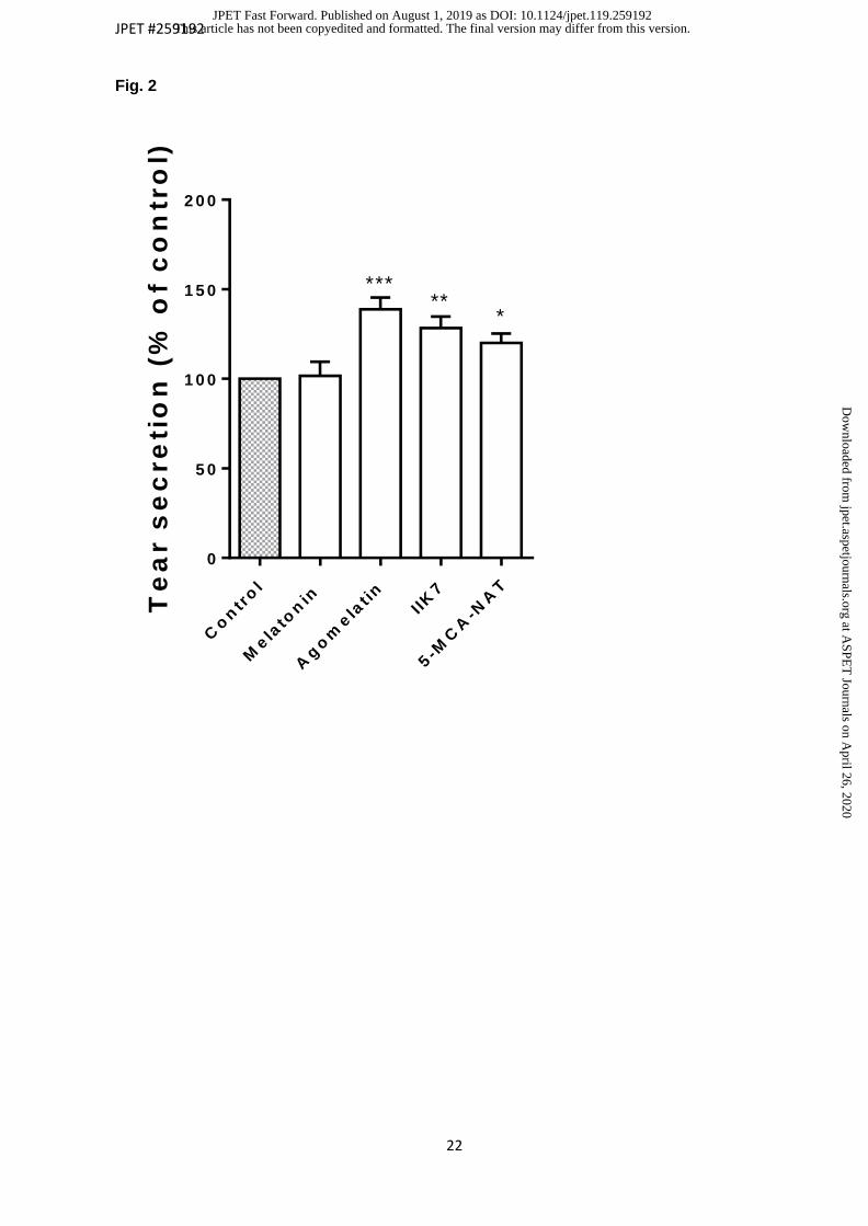
120.0 ± 5.2 % respectively (p < 0.05) (100% control), but not so for melatonin (101.6 ±
7.9%) (p > 0.05). Agonist action was tested combined with the antagonists (10 µl at 100
µM) DH97 (MT2 selective), prazosin (MT3/QR2 inhibitor) and luzindole (non-selective
MTr). DH97 reverses the effect of agomelatine, IIK7 and 5-MCA-NAT up to 30.85 % ±
7.6 %,108 % ± 7.2 % and 87.01 % ± 7.6 % respectively (p < 0.05) (100% control),
Luzindole antagonizes agomelatine and 5MCA-NAT up to 67.35 % ± 7.6 % and 92.12
% ± 8 % (p < 0.05). Prazosin only reverses the 5MCA-NAT action up to 84.2 % ± 7.7 %
(p < 0.05). These results suggest different pathways for the agonists to act through MTr
receptors. Therefore, it can be said that the agomelatine, IIK7 and 5MCA-NAT act as
secretagogues of tear secretion, acting through the receptors MTr, and they could be
considered excellent therapeutic candidates for dry eye treatment.
This article has not been copyedited and formatted. The final version may differ from this version.JPET Fast Forward. Published on August 1, 2019 as DOI: 10.1124/jpet.119.259192
at ASPE
T Journals on A
pril 26, 2020jpet.aspetjournals.org
Dow
nloaded from

JPET #259192
4
4. Significance statement. Currently dry eye with aqueous deficit is treated by adding artif icial tears
palliatively. The study shows that the topical installation in therapeutic doses in rabbit
corneas of three melatonin analogues: agomelatine, IIK7 and 5MCA-NAT, increases
significantly the tear production, but not melatonin, acting through different melatonin
membrane receptors subtypes (MTr). Therefore, this study suggests melatoninergic
compounds could be considered excellent therapeutic candidates for dry eye treatment
and ocular surface diseases occurring with a reduction in tear production.
This article has not been copyedited and formatted. The final version may differ from this version.JPET Fast Forward. Published on August 1, 2019 as DOI: 10.1124/jpet.119.259192
at ASPE
T Journals on A
pril 26, 2020jpet.aspetjournals.org
Dow
nloaded from

JPET #259192
5
5. Introduction Melatonin is a neuro-hormone, released mainly from the pineal gland (Stehle et al., 2011),
that is involved in the control of various physiological actions, many of which are related
with the regulation of circadian rhythms (Pandi-Perumal et al., 2006). The ability to
synthesize and release melatonin is not an exclusive feature of the pineal gland. Melatonin
production has been discovered in the orbital cavity and some eye areas such as the
retina(Cardinali and Rosner, 1971; Alarma-Estrany and Pintor, 2007), the ciliary body
(Martin et al., 1992; Alarma-Estrany and Pintor, 2007), the lens (Alkozi et al., 2018) and
recently melatonin has been found in human tears (Carracedo et al., 2017).
The relationship between the levels of melatonin and some functions in the eye (Samples
et al., 1988) such as the fluctuation of intraocular pressure (IOP) (Rowland et al., 1981;
Liu et al., 2011); are well known. In this sense, the application of melatonin or any of its
analogues has been proposed as therapeutic agents for different ocular conditions (Pintor
et al., 2001; Pintor et al., 2003; Serle et al., 2004; Alarma-Estrany et al., 2008; Ismail and
Mowafi, 2009; Alarma-Estrany et al., 2011; Crooke et al., 2013; Colligris et al., 2014).
On the ocular surface, some studies have demonstrated that the use of melatonin
combined with the dinucleotide diadenosine tetraphosphate Ap4A, instilled in the eye of
New Zealand white rabbits caused a significant increase in tear secretion (Hoyle et al.,
2006).
Melatonin exerts many of its actions by means of membrane receptors, termed melatonin
receptors, divided into MT1, MT2, and MT3 (Alarma-Estrany and Pintor, 2007; Dubocovich
et al., 2010). New studies identify the MT3 with the enzyme QR2 (Boutin and Ferry, 2019).
Beyond these findings, melatonin receptors on the ocular surface have been identified in
structures such as the cornea in different species (Wiechmann and Rada, 2003). In
particular, MT2 receptors have been described in New Zealand white rabbits’ cornea
(Crooke et al., 2015). The existence of melatonin receptors in the lachrymal gland has been
This article has not been copyedited and formatted. The final version may differ from this version.JPET Fast Forward. Published on August 1, 2019 as DOI: 10.1124/jpet.119.259192
at ASPE
T Journals on A
pril 26, 2020jpet.aspetjournals.org
Dow
nloaded from
JPET #259192
6
found, even more the effect of melatonin in combination of Ap4A, increasing tear secretion
had already been reported (Hoyle et al., 2006).
The aim of the present study is to characterize the effect of melatonin and its analogues
(agomelatine, 5MCA-NAT and IIK7) on New Zealand rabbit on tear secretion.
This article has not been copyedited and formatted. The final version may differ from this version.JPET Fast Forward. Published on August 1, 2019 as DOI: 10.1124/jpet.119.259192
at ASPE
T Journals on A
pril 26, 2020jpet.aspetjournals.org
Dow
nloaded from

JPET #259192
7
6. Materials and Methods
6.1 Materials. Melatonin, 5-methoxycarbonylamino-N-acetyltryptamine (5MCA-NAT), N- butanoyl-2-
(2-methoxy-6H-isoindolo[2,1-a]indol-11-yl) ethanamine (IIK7), prazosin , DH97 and
Luzindole were purchased from Tocris (Bristol, UK) and Agomelatine from Santa Cruz
Biotechnology, Inc. (Dallas, USA). All of them were formulated in isotonic saline
containing 1% DMSO from Sigma Aldrich (St. Louis, USA).
6.2 Animals Male New Zealand White rabbits weighing 3-4 kg were placed in individual cages with free
access to food and water and subjected to regular cycles of light/dark (12 h). All the
experiments were performed according to Association for Research in Vision and
Ophthalmology Statement for the Use of Animals in Ophthalmic and Vision Research and
to the European Directive 2010/63/EU.
6.3 Measurement of Tear Secretion Tear secretion was measured with the Schirmer test, according to the protocol described
by Van Bjisterveld (van Bijsterveld, 1969), using Whatman no. 51 paper strips (Schirmer
strips; Whatman, Maidstone, UK) placed in the inferior lid margin of the eye for 5 min. Tear
secretion was measured as the length (millimeters) of the strip wetted by the tears.
6.4 Pharmacological experiments Control experiments were performed by applying 10 µl of saline solution (NaCl 0.9 %)
containing 1% DMSO instilled 15 minutes before any of the other compounds were used.
Melatonin and its agonists were instilled in a volume of 10 µl at 100 µM. Tear secretion
measurements were performed at 5, 15, 60 and 120 minutes after their application (n=24).
Melatonin receptor antagonists, DH97 (a MT2 selective antagonist), prazosin (MT3
/QR2 inhibitor) and luzindole (a non-selective melatonin receptor antagonist), were
This article has not been copyedited and formatted. The final version may differ from this version.JPET Fast Forward. Published on August 1, 2019 as DOI: 10.1124/jpet.119.259192
at ASPE
T Journals on A
pril 26, 2020jpet.aspetjournals.org
Dow
nloaded from

JPET #259192
8
applied at 100µM (10µl) and instilled 15 minutes before the application of the agonists
which were instilled at 100 µM (n=10).
To determine the concentration-response curves, different concentrations of melatonin,
agomelatine, IIK7 and 5-MCA-NAT were tested according to the previous methodology.
Concentrations tested varied from 1µM-1mM (n= 10).
6.5 Statistical analysis Statistical analysis was performed using the software SSPS Statistic 23 (IBM, Chicago;
USA). All data are presented as the mean ± S.E.M. The normal distribution of all variable
for each experiment was assessed with Saphiro-Wilks test. Statistical differences between
baseline and post-instillation for all treatments were calculated using Student t-test for
related samples. In addition, One Way ANOVA test for related samples was used to
analyse the trend in dose-response experiments. Plotting and fitting were carried out with
Prism 6 GraphPad computer program (GraphPad Software, La Jolla, USA). P value < 0.05
was considered statistically significant.
This article has not been copyedited and formatted. The final version may differ from this version.JPET Fast Forward. Published on August 1, 2019 as DOI: 10.1124/jpet.119.259192
at ASPE
T Journals on A
pril 26, 2020jpet.aspetjournals.org
Dow
nloaded from

JPET #259192
9
7. Results
7.1 Effect of melatonin and analogues on tear secretion In order to study the possible effect of melatonin and its analogues on tear secretion, single
applications of these compounds (10 µl at 100 µM) were tested on New Zealand rabbits.
The topical application of melatonin did not modify tear secretion during the experimental
time (101.6 ± 7.9 % at 60 min, figure 1), having a non-statistically significant (p value >
0.05; Student t-test for related samples). On the other hand, a significant increase in tear
secretion was clearly observed, which was maximal above control value (100%
basal product ion) when agomelatine, IIK7 or 5-MCA- NAT was assayed one hour
after their topical application (p value < 0.05; Student t-test for related samples) (figure
1). The values obtained (figure 2) were 138.9 ± 6.5 % for agomelatine, 120.0 ± 5.2 % for
5-MCA-NAT and 128.3 ± 6.4 % for IIK7 (n=24) (p value < 0.05; Student t-test for related
samples).
7.2 Dose-response studies Since agomelatine, 5-MCA-NAT and IIK7 were able to increase tear secretion when
tested at a single dose, the three compounds were tested at several concentrations
ranging from 1µM to 1 mM. As can be seen in figure 3, the melatonin analogues
presented sigmoid curves. Agomelatine presenting a maximal secretion of 129.9 ± 8.2%
above the control value (100% basal production) and presenting a pD2 of 4.28 ± 0.06
(EC50 value of 52.1 µM, n=10). On the other hand, 5-MCA-NAT presented a maximal
effect of 135.7 ± 7.8 % over the control and a pD2 of 4.0 ± 0.02 (EC50 value of 99.5 µM,
n=10). Finally, IIK7 presented a maximal effect of 127.6 ± 2.5 % with a pD2 value of
4.15 ± 0.07 (EC50 value of 69.2 µM, n=10) (p value <0.05; One Way ANOVA test for
related samples for all melatonin analogues tested). Melatonin was also tested, at the
same range of concentrations, in order to see whether it was able to induce tear
secretion. As observed in figure 3, only at very high concentrations did melatonin induce
This article has not been copyedited and formatted. The final version may differ from this version.JPET Fast Forward. Published on August 1, 2019 as DOI: 10.1124/jpet.119.259192
at ASPE
T Journals on A
pril 26, 2020jpet.aspetjournals.org
Dow
nloaded from

JPET #259192
10
a very small increase in tearing, nonetheless, it was not possible to fit a dose-response
curve (p value <0.05; One Way ANOVA test for related samples.
7.3 Antagonist / inhibitor studies As has previously been mentioned, melatonin and analogues display their actions on MT1,
MT2 or MT3 /QR2 melatonin receptors. To investigate the receptor by which melatonin
agonists were acting, some antagonists, luzindole (a non-selective melatonin receptor
antagonist), DH97 (a selective MT2 receptor antagonist) and prazosin (MT3 / QR2
inhibitor) were tested.
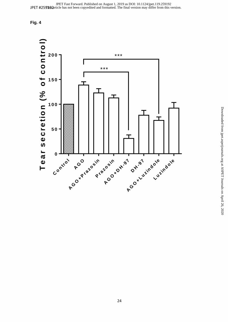
When agomelatine was assayed in the presence of the three mentioned antagonists, it
was possible to observe that only two of them were able to significantly reverse
agomelatine action, the most active being DH97 (figure 4). Luzindole reversed
agomelatine effect below control value (100% basal) up to 67.35 % ± 6.7% while DH97
antagonized up to 30.85% ± 7.6% (n=10) (p value < 0.05; Student t-test for related
samples). Comparing these results of decrease below control with those obtained from
their maximal agonist effect (138.9 ± 6.5 %), the differences are much more significant,
obtaining values of decrease of 71.6 ± 6.7 % and 108.1 ± 7.1 % respectively (p value <
0.05; Student t-test for related samples). Meanwhile, prazosin was not able to
antagonize the agomelatine effect on tear secretion (p value > 0.05; Student t-test for
related samples). When the same experiments were carried out with IIK7 was tested,
and as observed in figure 5, only DH97 was able to significantly revert the
effect of this melatonin agonist up to 108 % ± 7.2 % above control (n=10) (p
value < 0.05; Student t-test for related samples), which means 20.9 % ± 5.5 %
with respect its maximal agonist action (128.9 ± 6.4 %). Finally, when 5-MCA-
NAT was tested with prazosin, DH97 and luzindole, (figure 6) a similar significance
antagonistic and inhibitor effect was noticed, (84.2% ± 7.7 %, 87.01 % ± 7.7 % and
92.12% ± 8 % respectively) (n=10) (p value < 0.05; Student t-test for related samples).
The decrease compared to its agonist action (120.0 ± 5.2 %) is 35.8 ± 6.3 %, 33 % ± 6.3
This article has not been copyedited and formatted. The final version may differ from this version.JPET Fast Forward. Published on August 1, 2019 as DOI: 10.1124/jpet.119.259192
at ASPE
T Journals on A
pril 26, 2020jpet.aspetjournals.org
Dow
nloaded from

JPET #259192
11
% and 27.9 ± 4.5 % for the tree antagonist.
This article has not been copyedited and formatted. The final version may differ from this version.JPET Fast Forward. Published on August 1, 2019 as DOI: 10.1124/jpet.119.259192
at ASPE
T Journals on A
pril 26, 2020jpet.aspetjournals.org
Dow
nloaded from

JPET #259192
12
8. Discussion In the present work, the effect of melatonin receptor agonists, agomelatine, 5-MCA-NAT
and IIK7 of tear secretion is investigated. The results demonstrate that the three
analogues are effective in inducing tear secretion in New Zealand rabbits, but as shown
by the antagonist /inhibitor studies, they did not activate the same melatonin receptors
subtypes. While IIK7 agonist acts in a very specific manner through MT2 receptors,
because its action is only blocked in the presence of DH97 (see fig.5), the results
obtained with 5- MCA-NAT, do not allow us to determine a specific way of action. In
that case, 5-MCA-NAT’s effect is partially inhibited in the presence of the three
antagonists / inhibitor in a similar way (see fig.6). Probably this is due to the action as
partial agonist on MT1/MT2 of 5-MCA-NAT (Vincent et al., 2010). The action of
agomelatine is reversed by two of the three antagonists / inhibitor (see fig.4), showing
an important decrease action with prazosin but without statistical significance and especially
with DH97, suggesting a two-way action through the receptors MT2 and MT3/QR2.
Interestingly in this year the study of Boutin & Ferry (Boutin and Ferry, 2019) identifies MT3
with quinone reductase 2 (QR2). Perhaps the nature of this enzyme is responsible for the
controversial action attributed to the MT3 receptor.
An interesting point to consider is whether the combination of two agonists would be
more effective in the increase of the tear secretion than the individual action. Unpublished
experiments carried out in our lab have demonstrated that the combination of 5-MCA-
NAT and IIK7 only increased tearing 5% above the values obtained by these two
compounds individually. This lack of an additive effect is probably a consequence of both
agonists using melatonin in mainly the same receptors and only 5- MCA-NAT may be
acting through the MT3 /QR2 since its effect has been blocked by prazosin (Huete-
Toral et al., 2015). In this sense, other study has demonstrated that prazosin interplay
with tear by α2-adrenergic receptors in frog cornea model (Chu and Candia, 1988).
This article has not been copyedited and formatted. The final version may differ from this version.JPET Fast Forward. Published on August 1, 2019 as DOI: 10.1124/jpet.119.259192
at ASPE
T Journals on A
pril 26, 2020jpet.aspetjournals.org
Dow
nloaded from

JPET #259192
13
However, this study shows no significant difference between the group treated with
prazosin and the basal control (100%).
The high affinity of melatonin analogues compared to melatonin itself is an interesting
aspect, indicating that, rather than using melatonin to induce tear secretion, it would be
better to use its analogues. This is not the only case, indeed, the compound 5-MCA-NAT
has demonstrated a more robust effect in reducing IOP in rabbits than melatonin does
(Pintor et al., 2003). Also, and on the ocular surface, the ability of melatonin to accelerate
corneal wound healing is enhanced by the MT2 receptor agonist IIK7 (Crooke et al.,
2015). In the present work, agonists of melatonin receptors produced a significant
increase in tear production, the effect of which was dose-dependent. Melatonin did
present an apparent concentration-response behavior, which was only visible at high
concentration, in clear contrast with the effect of its analogues. Such differences may
be due to the best affinity of the analogues acting on melatonin receptors (Alkozi et
al., 2018).
Some comments can be made concerning which of the melatonin analogues would be the
best candidate for a therapy for ocular dryness. Although, agomelatine produce the most
robust effect at 100 µ M (see fig.1 and 2), concentration-response curves showed that
5-MCA-NAT could rise tearing 10% more than agomelatine or IIK7, but increasing its
dose to 300 µM or above. It could have the idea of using any of these compounds to
treat dry eye, as commented, little is known of 5-MCA-NAT and IIK7 regarding toxic
effects, side effects and safety in general, while agomelatine is a compound which is
already on the pharmaceutical market for other purposes (Taylor et al., 2014; Levitan et
al., 2015). Nonetheless, it is important to point out that agomelatine can also acts over
other receptors different from melatonin ones such as 5HT2c (Racagni et al., 2011).
Therefore, taking in account the results found in this study, melatonin agonists
triggering tear secretion without interfering with other receptors, 5-MCA-NAT or IIK7
This article has not been copyedited and formatted. The final version may differ from this version.JPET Fast Forward. Published on August 1, 2019 as DOI: 10.1124/jpet.119.259192
at ASPE
T Journals on A
pril 26, 2020jpet.aspetjournals.org
Dow
nloaded from

JPET #259192
14
should be better choices.
In summary therefore, melatonin analogues can induce a significant increase in tear
secretion in New Zealand rabbits. This is a relevant issue since, to date, there is no
pharmaceutical compound available to treat dry eye, although the existence of artificial
tears trying to relieve the patients of the symptoms are commercially available (Pucker
et al., 2016). Therefore, this study suggests melatoninergic compounds are putative
agents for the treatment of those ocular surface diseases occurring with a reduction
in tear production.
This article has not been copyedited and formatted. The final version may differ from this version.JPET Fast Forward. Published on August 1, 2019 as DOI: 10.1124/jpet.119.259192
at ASPE
T Journals on A
pril 26, 2020jpet.aspetjournals.org
Dow
nloaded from

JPET #259192
15
9. Authorship Contributions Participated in research design: Navarro, Huete-Toral and Pintor
Conducted experiments: Navarro and Huete-Toral
Contributed new reagents or analytic tools: Crooke and Dominguez
Performed data analysis: Navarro, Huete-Toral, Carracedo and Pintor
Wrote or contributed to the writing of the manuscript: Navarro, Huete-Toral, Carracedo and Pintor
This article has not been copyedited and formatted. The final version may differ from this version.JPET Fast Forward. Published on August 1, 2019 as DOI: 10.1124/jpet.119.259192
at ASPE
T Journals on A
pril 26, 2020jpet.aspetjournals.org
Dow
nloaded from

JPET #259192
16
10. References
Alarma-Estrany P, Crooke A, Mediero A, Pelaez T and Pintor J (2008) Sympathetic nervous system modulates the ocular hypotensive action of MT2-melatonin receptors in normotensive rabbits. J Pineal Res 45:468-475.
Alarma-Estrany P, Guzman-Aranguez A, Huete F, Peral A, Plourde R, Jr., Pelaez T, Yerxa B and Pintor J (2011) Design of novel melatonin analogs for the reduction of intraocular pressure in normotensive rabbits. J Pharmacol Exp Ther 337:703-709.
Alarma-Estrany P and Pintor J (2007) Melatonin receptors in the eye: location, second messengers and role in ocular physiology. Pharmacol Ther 113:507-522.
Alkozi HA, Sanchez Montero JM, Doadrio AL and Pintor J (2018) Docking studies for melatonin receptors. Expert Opin Drug Discov 13:241-248.
Boutin JA and Ferry G (2019) Is There Sufficient Evidence that the Melatonin Binding Site MT3 Is Quinone Reductase 2? J Pharmacol Exp Ther 368:59-65.
Cardinali DP and Rosner JM (1971) Retinal localization of the hydroxyindole-O-methyl transferase (HIOMT) in the rat. Endocrinology 89:301-303.
Carracedo G, Carpena C, Concepcion P, Diaz V, Garcia-Garcia M, Jemni N, Lledo VE, Martin M, Pastrana C, Pelissier R, Veselinova A, Wang X and Pintor J (2017) Presence of melatonin in human tears. J Optom 10:3-4.
Chu TC and Candia OA (1988) Role of alpha 1- and alpha 2-adrenergic receptors in Cl- transport across frog corneal epithelium. Am J Physiol 255:C724-730.
Colligris B, Alkozi HA and Pintor J (2014) Recent developments on dry eye disease treatment compounds. Saudi J Ophthalmol 28:19-30.
Crooke A, Guzman-Aranguez A, Mediero A, Alarma-Estrany P, Carracedo G, Pelaez T, Peral A and Pintor J (2015) Effect of melatonin and analogues on corneal wound healing: involvement of Mt2 melatonin receptor. Curr Eye Res 40:56-65.
Crooke A, Huete-Toral F, Martinez-Aguila A, Martin-Gil A and Pintor J (2013) Melatonin and its analog 5-methoxycarbonylamino-N-acetyltryptamine potentiate adrenergic receptor-mediated ocular hypotensive effects in rabbits: significance for combination therapy in glaucoma. J Pharmacol Exp Ther 346:138-145.
Dubocovich ML, Delagrange P, Krause DN, Sugden D, Cardinali DP and Olcese J (2010) International Union of Basic and Clinical Pharmacology. LXXV. Nomenclature, classification, and pharmacology of G protein-coupled melatonin receptors. Pharmacol Rev 62:343-380.
Hoyle CH, Peral A and Pintor J (2006) Melatonin potentiates tear secretion induced by diadenosine tetraphosphate in the rabbit. Eur J Pharmacol 552:159-161.
Huete-Toral F, Crooke A, Martinez-Aguila A and Pintor J (2015) Melatonin receptors trigger cAMP production and inhibit chloride movements in nonpigmented ciliary epithelial cells. J Pharmacol Exp Ther 352:119-128.
Ismail SA and Mowafi HA (2009) Melatonin provides anxiolysis, enhances analgesia, decreases intraocular pressure, and promotes better operating conditions during cataract surgery under topical anesthesia. Anesth Analg 108:1146-1151.
Levitan MN, Papelbaum M and Nardi AE (2015) Profile of agomelatine and its potential in the treatment of generalized anxiety disorder. Neuropsychiatr Dis Treat 11:1149-1155.
Liu H, Fan S, Gulati V, Camras LJ, Zhan G, Ghate D, Camras CB and Toris CB (2011) Aqueous humor dynamics during the day and night in healthy mature volunteers. Arch Ophthalmol 129:269-275.
Martin XD, Malina HZ, Brennan MC, Hendrickson PH and Lichter PR (1992) The ciliary body--the third organ found to synthesize indoleamines in humans. Eur J Ophthalmol 2:67-72.
Pandi-Perumal SR, Srinivasan V, Maestroni GJ, Cardinali DP, Poeggeler B and Hardeland R (2006) Melatonin: Nature's most versatile biological signal? FEBS J 273:2813-2838.
Pintor J, Martin L, Pelaez T, Hoyle CH and Peral A (2001) Involvement of melatonin MT(3) receptors in the regulation of intraocular pressure in rabbits. Eur J Pharmacol 416:251-254.
Pintor J, Pelaez T, Hoyle CH and Peral A (2003) Ocular hypotensive effects of melatonin receptor
This article has not been copyedited and formatted. The final version may differ from this version.JPET Fast Forward. Published on August 1, 2019 as DOI: 10.1124/jpet.119.259192
at ASPE
T Journals on A
pril 26, 2020jpet.aspetjournals.org
Dow
nloaded from
JPET #259192
17
agonists in the rabbit: further evidence for an MT3 receptor. Br J Pharmacol 138:831-836. Pucker AD, Ng SM and Nichols JJ (2016) Over the counter (OTC) artificial tear drops for dry eye
syndrome. Cochrane Database Syst Rev 2:CD009729. Racagni G, Riva MA, Molteni R, Musazzi L, Calabrese F, Popoli M and Tardito D (2011) Mode
of action of agomelatine: synergy between melatonergic and 5-HT2C receptors. World J Biol Psychiatry 12:574-587.
Rowland JM, Potter DE and Reiter RJ (1981) Circadian rhythm in intraocular pressure: a rabbit model. Curr Eye Res 1:169-173.
Samples JR, Krause G and Lewy AJ (1988) Effect of melatonin on intraocular pressure. Curr Eye Res 7:649-653.
Serle JB, Wang RF, Peterson WM, Plourde R and Yerxa BR (2004) Effect of 5-MCA-NAT, a putative melatonin MT3 receptor agonist, on intraocular pressure in glaucomatous monkey eyes. J Glaucoma 13:385-388.
Stehle JH, Saade A, Rawashdeh O, Ackermann K, Jilg A, Sebesteny T and Maronde E (2011) A survey of molecular details in the human pineal gland in the light of phylogeny, structure, function and chronobiological diseases. J Pineal Res 51:17-43.
Taylor D, Sparshatt A, Varma S and Olofinjana O (2014) Antidepressant efficacy of agomelatine: meta-analysis of published and unpublished studies. BMJ 348:g1888.
Vincent L, Cohen W, Delagrange P, Boutin JA and Nosjean O (2010) Molecular and cellular pharmacological properties of 5-methoxycarbonylamino-N-acetyltryptamine (MCA-NAT): a nonspecific MT3 ligand. J Pineal Res 48:222-229.
Wiechmann AF and Rada JA (2003) Melatonin receptor expression in the cornea and sclera. Exp Eye Res 77:219-225.
This article has not been copyedited and formatted. The final version may differ from this version.JPET Fast Forward. Published on August 1, 2019 as DOI: 10.1124/jpet.119.259192
at ASPE
T Journals on A
pril 26, 2020jpet.aspetjournals.org
Dow
nloaded from

JPET #259192
18
11. Footnotes
a) This study is supported (in part) by The Instituto de Salud Carlos III (ISC III), through
the Redes Temáticas de Investigación cooperativa en Salud (RETICS)
(RD16/0008/0001)
This article has not been copyedited and formatted. The final version may differ from this version.JPET Fast Forward. Published on August 1, 2019 as DOI: 10.1124/jpet.119.259192
at ASPE
T Journals on A
pril 26, 2020jpet.aspetjournals.org
Dow
nloaded from
JPET #259192
19
12. Figures Legends
Fig. 1. Time course. Effect of melatonin and analogues on tear secretion tested at 5 min,
15 min 30 min, 60 min and 120 min. Data are presented as the mean ± S.E.M. Topical
single applications of the compounds (10 µl at 100 µM) (n=24). A significant increase of
tear secretion is observed in the case of agomelatine, IIK7 and 5-MCA-NAT. (P value <
0.05; One Way ANOVA for related samples). The topical application of melatonin did not
modify tear secretion during the experimental time. (P value > 0.05; One Way ANOVA for
related samples).
Fig. 2. Maximal effect of melatonin and its analogues on tear secretion at 60 min. Data are
presented as the mean ± S.E.M. P value < 0.05 was considered statistically significant.
Compounds were tested 10 µl at 100 µM (n=24). * P value < 0.05; ** P value < 0.01; *** P
value < 0.001; Student t-test for related samples.
Fig. 3. Dose-response studies. Compounds were tested at several concentrations ranging
from 1µM to 1 mM (n=10). Data are presented as the mean ± S.E.M. The three analogues
presented sigmoid curves. (P value < 0.05; One Way ANOVA for related samples). In the
case of melatonin it was not possible to fit a dose-response curve (P value > 0.05; One
Way ANOVA for related samples).
Fig. 4. Combined action of agomelatine with antagonist: prazosin, DH-97 and luzindole at
60 min. Data are presented as the mean ± S.E.M. P value < 0.05 was considered
statistically significant. Antagonist were applied at 100µM (10µl) and instilled 15 minutes
before the application of the agonist (10µl 100 µM) (n=10). *** P value < 0.001; Student t-
test for related samples.
This article has not been copyedited and formatted. The final version may differ from this version.JPET Fast Forward. Published on August 1, 2019 as DOI: 10.1124/jpet.119.259192
at ASPE
T Journals on A
pril 26, 2020jpet.aspetjournals.org
Dow
nloaded from

JPET #259192
20
Fig. 5. Combined action of IIK7 with antagonist: prazosin, DH-97 and luzindole at 60 min.
Data are presented as the mean ± S.E.M. P value < 0.05 was considered statistically
significant. Antagonist were applied at 100µM (10µl) and instilled 15 minutes before the
application of the agonist (10µl 100 µM) (n=10). ** P value < 0.01; Student t-test for related
samples.
Fig. 6. Combined action of 5-MCA-NAT with antagonist: prazosin, DH-97 and luzindole at
60 min. Data are presented as the mean ± S.E.M. P value < 0.05 was considered
statistically significant. Antagonist were applied at 100µM (10µl) and instilled 15 minutes
before the application of the agonist (10µl 100 µM) (n=10). * P value < 0.05; ** P value <
0.01; *** P value < 0.001; Student t-test for related samples.
This article has not been copyedited and formatted. The final version may differ from this version.JPET Fast Forward. Published on August 1, 2019 as DOI: 10.1124/jpet.119.259192
at ASPE
T Journals on A
pril 26, 2020jpet.aspetjournals.org
Dow
nloaded from

JPET #259192
21
13. Figures.
Fig. 1
This article has not been copyedited and formatted. The final version may differ from this version.JPET Fast Forward. Published on August 1, 2019 as DOI: 10.1124/jpet.119.259192
at ASPE
T Journals on A
pril 26, 2020jpet.aspetjournals.org
Dow
nloaded from
JPET #259192
22
Fig. 2
Te
ar s
ec
re
tio
n (
% o
f c
on
tro
l)
Co
ntr
ol
Mela
ton
in
Ag
om
ela
t in
I IK
7
5-M
CA
-NA
T
0
5 0
1 0 0
1 5 0
2 0 0
***
***
This article has not been copyedited and formatted. The final version may differ from this version.JPET Fast Forward. Published on August 1, 2019 as DOI: 10.1124/jpet.119.259192
at ASPE
T Journals on A
pril 26, 2020jpet.aspetjournals.org
Dow
nloaded from

JPET #259192
23
Fig. 3
This article has not been copyedited and formatted. The final version may differ from this version.JPET Fast Forward. Published on August 1, 2019 as DOI: 10.1124/jpet.119.259192
at ASPE
T Journals on A
pril 26, 2020jpet.aspetjournals.org
Dow
nloaded from
JPET #259192
24
Fig. 4
Te
ar s
ec
re
tio
n (
% o
f c
on
tro
l)
Co
ntr
ol
AG
O
AG
O+P
razo
sin
Pra
zo
sin
AG
O+D
H-9
7
DH
-97
AG
O+L
uzin
do
le
Lu
zin
do
le
0
5 0
1 0 0
1 5 0
2 0 0
***
***
This article has not been copyedited and formatted. The final version may differ from this version.JPET Fast Forward. Published on August 1, 2019 as DOI: 10.1124/jpet.119.259192
at ASPE
T Journals on A
pril 26, 2020jpet.aspetjournals.org
Dow
nloaded from

JPET #259192
25
Fig. 5
Co
ntr
ol
IIK
7
IIK
7+ P
razo
sin
Pra
zo
sin
IIK
7+ D
H97
DH
-97
IIK
7+ L
uzin
do
le
Lu
zin
do
le
0
5 0
1 0 0
1 5 0
2 0 0
Te
ar s
ec
re
tio
n (
% o
f c
on
tro
l)
**
This article has not been copyedited and formatted. The final version may differ from this version.JPET Fast Forward. Published on August 1, 2019 as DOI: 10.1124/jpet.119.259192
at ASPE
T Journals on A
pril 26, 2020jpet.aspetjournals.org
Dow
nloaded from

JPET #259192
26
Fig. 6
Te
ar s
ec
re
tio
n (
% o
f c
on
tro
l)
Co
ntr
ol
5-M
CA
5-M
CA
+P
razo
sin
Pra
zo
sin
5-M
CA
+D
H-9
7
DH
-97
5-M
CA
+L
uzin
do
le
Lu
zin
do
le
0
5 0
1 0 0
1 5 0
2 0 0
***
**
*
This article has not been copyedited and formatted. The final version may differ from this version.JPET Fast Forward. Published on August 1, 2019 as DOI: 10.1124/jpet.119.259192
at ASPE
T Journals on A
pril 26, 2020jpet.aspetjournals.org
Dow
nloaded from