dphil in cancer science university of oxford
TRANSCRIPT

DPhil in Cancer Science University of Oxford
2021 Intake Project Book

1
DPhil in Cancer Science 2021 Intake Project Book
Introduction This booklet provides an overview for prospective students looking to study for a DPhil in Cancer Science at Oxford University, starting in 2021. The Programme provides research based doctoral training for cancer researchers from clinical, biological, engineering, mathematics and statistics backgrounds. Students will receive a world-leading research training experience that integrates an education initiative spanning cancer patient care, tumour biology and research impact; on- and post-programme mentorship; and a specialised, fundamental, subject-specific training tailored to individual research needs. Students participating in the scheme will be offered:
• a choice of interdisciplinary cutting-edge cancer research projects. • the ability to gain a working in-depth knowledge of the fundamentals of cancer biology
and cancer patient care through advanced level seminars. • a world-renowned research environment that encourages the student’s originality
and creativity in their research. • opportunities to develop skills in making and testing hypotheses, in developing new
theories, and in planning and conducting experiments. • an environment in which to develop skills in written work, oral presentation and
publishing the results of their research in high-profile scientific journals, through constructive feedback of written work and oral presentations.
At the end of their DPhil course, students should:
• have a thorough knowledge of the basic principles of cancer research including the relevant literature and a comprehensive understanding of scientific methods and techniques applicable to their research.
• be able to demonstrate originality in the application of knowledge, together with a practical understanding of how research and enquiry are used to create and interpret knowledge in their field.
• have developed the ability to critically evaluate current research and research techniques and methodologies.
• be able to act autonomously in the planning and implementation of research. • have the grounding for an influential cancer researcher of the future.

2
Selection Criteria & Eligibility There are three tracks in the programme as described below, meaning that non-clinicians, undergraduate medical students and post-graduate medical trainees are all eligible to apply for the fully funded (at home rate) studentships. Application Track 1 – Clinical Trainees. Qualified doctors at all stages of training from the foundation training to higher specialist training. Application Track 2 – Medical Undergraduates. Medical students who are currently undertaking a primary medical qualification (MBBS, MBChB or equivalent) Application Track 3A – Non-Clinical/Fundamental Scientist. Science graduates that hold (or be predicted to achieve) the equivalent of a first-class or strong upper second-class undergraduate degree with honours in a biological, medical, or chemical science, as appropriate for the projects offered. Application Track 3B – Non-Clinical/Fundamental Scientist. Science graduates that hold (or be predicted to achieve) the equivalent of a first-class or strong upper second-class undergraduate degree with honours in a engineering, mathematical/data, or physical science, as appropriate for the projects offered. All applicants will be judged on the following:
• commitment and passion to a career in cancer research • evidence of motivation for and understanding of the proposed area of study • commitment to the subject, beyond the requirements of the degree course • preliminary knowledge of relevant research techniques • capacity for sustained and intense work • reasoning ability and academic curiosity.
Funding All offered places are fully funded at the home rate. This includes salary/stipend, University/College fees, and a research consumables budget of £13k p.a.. Salary and stipend provisions are summarised below:
• Application Track 1 – 3 years of salary at Grade E63 or E64 Clinical Researcher rate. • Application Track 2 – 3 years of stipend at the flat rate of £19,000 per annum. • Application Track 3A & 3B – 4 years of stipend at the flat rate of £19,000 per
annum. International applicants are eligible, however funding is limited to the Home level for this programme and therefore international applicants would need to either source further funding or support themselves financially for the remaining fees.

3
How to Apply A detailed summary on how to apply can be found here. In brief, prospective students apply with a prioritised list of three projects selected from this booklet by Friday January 8th 2021. Shortlisted students will be invited to interview in February. If successful, students will be allocated a project on the basis of their ranking during the review process. It is strongly suggested that students contact supervisors of projects they are interested in applying for prior to application.

4
Projects at a Glance Projects are listed below in the following structure “Title – Main Supervisor Eligible Application Tracks”
Targeting DNA repair mechanisms in precision cancer therapies – Prof. Lakin1,2,3A 7
Molecular understanding of patient responses to immunotherapy in gastrointestinal tract cancers – Prof. Lu1,2,3A 9
Mechanisms Of Cure in Acute Myeloid Leukaemia Treated with Intensive Chemotherapy – Prof. Vyas1,2,3A 11
Evaluating the roles of the AHR in melanoma: melanin sensing and regulating the melanoma microenvironment – Dr. Alves1,2,3A 13
Improving immunotherapy by defining the interaction between the bacterial vaccine BCG and the host epigenomic and immune response – Prof. McShane1,2,3A 15
Targeting epigenetic regulators to improve tumour responses to immunotherapy – Prof. Shi1,2,3A 17
Elucidating the Rad51-independent pathway of recombination-dependent replication – Prof. Whitby1,2,3A 19
Analysis of spindle assembly checkpoint function in health and disease – Prof. Gruneberg2,3A,3B 21
Identifying novel regulators of cancer stem cells in pancreatic ductal adenocarcinoma– Dr. Pauklin 1,2,3A 23
Understanding The Therapeutic Efficacy Of Chemotherapy In Pancreatic Cancer at the single cell level – Dr. Sivakumar1,2,3A,3B 25
Heterogeneity of macrophages in colorectal cancer: the role of IRF5 – Prof. Udalova1,2,3A,3B 27
Modelling cancer stem cell dormancy using organoids and advanced 3D culture models – Dr. Boccellato1,2,3A,3B 29
Decoding immune surveillance within the tumour microenvironment: how the extracellular matrix enables escape from immunity – Prof. Midwood1,2,3A 31
UGT8 and microcarrier signalling in the development of breast cancer – Prof. Wilson1,2,3A 33
Studying the role of a chromatin remodelling factor (ATRX) in normal gene expression and in malignancy – Prof. Higgs1,2,3A 35
ARH3/ADPRHL2 as a biomarker for PARP inhibitor sensitivity/resistance – Prof. Ahmed1,2,3A,3B 37
Discovery and mechanistic elucidation of small molecule inducers of myeloblast differentiation for ALL – Prof. Russell3A 39
Single-cell analysis of haematopoietic stem cells in SF3B1 mutant MDS: identification of new therapeutic targets/treatments – Dr. Pellagatti1,2,3A 41

5
Dissecting the biological basis of HLA variation to susceptibility and disease severity in Crohn’s disease & ulcerative colitis as risk factors for the development of colorectal cancer – Prof. Satsangi1 43
The Artemis DNA Repair nucleases – from mechanism to therapeutic inhibition – Prof. Schofield3A 45
Investigating IGFs as immunosuppressive cytokines and cancer risk factors– Prof. Macaulay1,2,3A 47
Genetic and functional characterisation of novel immune escape mutations in mismatch repair deficient endometrial cancer – Dr. Church1,2,3A 49
Mass spectrometric probes for intraoperative brain cancer diagnosis – Prof Vallance1,2,3A,3B 51
Developing an on chip screen for PDAC in high risk groups – Prof Davis1,2,3A 53
Understanding STING regulation in cancer and the crucial role of ubiquitination at the ER – Dr. Christianson1,2,3A 55
Spatial mapping of the bone marrow for improved leukaemia diagnosis using machine learning/artificial intelligence – Dr Royston4 57
Bioengineered gastrointestinal tissues to study neural signalling in cancer development and metastasis – Prof. Bayley1,2,3A,3B 59
Discovery of potent wild-type and surrogate agonist peptides for anti-tumour T Cells – Dr. Fernandes1,2,3A 61
In vitro and in silico models of human induced pluripotent stem cell to investigate the effects of doxorubicin – induced cardiotoxicity – Prof. Zaccolo1,2,3A 63
Genome-wide screening to identify factors impacting on cellular survival upon acute depletion of BRCA2 or PALB2 – Prof. Esashi1,2,3A,3B 65
Galectin-3 promotes glioblastoma emergence from the subventricular zone stem cell niche – Prof. Szele1,2,3A,3B 67
Effects of androgen deprivation on multimodality prostate cancer therapy – Prof. Bryant1,2,3A 69
Characterising the developmental origins in the pathogenesis of mesenchymal tumours of the central nervous system – Prof. Sauka-Spengler1 71
Investigating pathological crosstalk between mature tumour cells and haematopoietic progenitor cells in chronic myelomonocytic leukaemia – Prof. Mead1 73
Mathematics of Lymphoma Immunotherapies: Application of Mechanistic Models to Accelerate and De-Risk Therapeutic Development for Blood Cancers – Prof. Coles1,2,3A,3B 75
Pre-clinical testing of a novel mitochondrial inhibitor (NBS037) in combination with radiotherapy and immunotherapy – Prof. Higgins 1,2,3A 77
Mechanisms of therapeutic response and resistance to BCMA-directed therapy in multiple myeloma- a systems biology approach – Prof. Oppermann1,2,3A,3B 79
Personalised monitoring intervals for cancer surveillance – Dr Oke1,2,3A,3B 81

6
Presentation, Diagnosis and Outcomes of Hodgkin Lymphoma: the role of Primary Care – Prof. Bankhead1,2,3A,3B 83
The impact of microenvironmental components on cellular plasticity in colorectal cancer – Prof. Buczacki1,2,3A 85
The impact of microenvironmental components on cellular plasticity in colorectal cancer – Dr. Jiang1,2,3A 87
The Epitope Abundance-Avidity-Efficacy Axis In Cancer – Prof. Elliot1,2,3A 89
Cell stress and the premalignant niche in ovarian – Prof. Blagden1,2,3A 92
The immune landscape of the pancreas during neoplastic transformation – Prof. O’Neill1,2,3A, 3B 94
7
Targeting DNA repair mechanisms in precision cancer therapies – Prof. Lakin1,2,3A
Primary Supervisor: Nicolas Lakin Additional Supervisors: Peter McHugh Eligibility: Track 1, 2 and 3A students only are eligible to apply for this project. Project Summary Inhibitors of DNA repair have emerged as powerful agents in cancer therapy, either as monotherapies that exploit synthetic lethal interactions between DNA repair pathways, or by increasing the efficacy of chemo- and radiotherapies1. Principal in this strategy is inhibition of Poly(ADP-ribose)-polymerases (PARPs), enzymes that regulate DNA strand break repair, and PARP inhibitors (PARPi) are being used to treat tumours with defects in homologous recombination (HR). However, these strategies are restricted to treating HR-defective ovarian cancers, with limited information on additional synthetic lethal interactions that will broaden the use of PARPi to treat other tumours. By combining our expertise in PARP biology and DNA repair2-6 with cutting edge genome editing, proteomics and cell biology, this research will address this fundamentally important question by characterising novel cancer-related genes that are synthetic lethal with PARP dysfunction. To this end, through a genome-wide CRISPR-Cas9 based screen we identified a novel gene (PASL9) that is synthetic lethal with PARPi and critical to resolve replication-associated DNA damage through a pathway that is often mutated in colorectal cancers. This multidisciplinary hypothesis-driven research will define how PASL9 regulates replication-associated DNA repair and the mechanistic basis of synthetic lethality with PARPi. Our long-term vision is to exploit these findings by developing novel strategies to exploit PARPi to treat a variety of tumours, including digestive cancers. Research Objectives and Proposed Outcomes Background We performed a genome-wide CRISPR-Cas9 screen to identify synthetic lethal interactions with the principal DNA repair PARPs (PARP1/PARP2). A top hit was a gene of unknown function that we named PASL9 (PARP and ATR Synthetic Lethal 9). Independent pasl9Δ cell lines are sensitive to PARP or ATR inhibitors, phenotypes associated with replication-repair defects7, 8. Consistent with PASL9 functioning in replication-associated repair, it fulfils 3 well-established criteria for a role in these pathways: A) PASL9 is recruited to chromatin in response to replication stress induced by hydroxyurea (HU); B) pasl9∆ cells display elevated levels of γH2AX following replication stress; C) pasl9∆ cells are sensitive to HU. Moreover, our data indicate that PASL9 interacts with and functions in the same pathway as HUWE1, a replication-associated repair gene9 that is mutated in colorectal cancers10. Together, these data identify PASL9 as a novel DDR gene that functions with HUWE1 to maintain genome stability. We will define the role of PASL9 in combating replication stress and assess its potential as a target to treat colorectal cancer by pursuing the following objectives: Define the nature of the synthetic lethal interaction between PARP and PASL9 We will define the PARP-dependent pathway that is synthetic lethal with PASL9. Our data indicate that in addition to single strand break repair, PARP1/2 can regulate Rad51-dependent and -independent replication fork recovery pathways6. We will establish which of these pathways is synthetic lethal with PASL9 by assessing cell survival after depleting SSB repair, or replication associated repair (HR; break-induced replication factors in pasl9∆ cells. We will

8
look at the requirements for PASL9 in various aspects of replication dynamics using DNA fibre analysis (replication fork speed, restart and stability) and markers of genome stability associated with defective replication-associated repair (increased chromosome fragmentation, ultrafine anaphase bridges, micronuclei formation etc.). We will assess whether any phenotypes observed are exacerbated by treating pasl9∆ cells with PARPi or ATRi. Define the replication repair mechanisms regulated by PASL9 Whilst PASL9 functions with HUWE1, an E3 ubiquitin ligase that accumulates at stalled replication forks9, the mechanistic basis of this regulation is unknown. PASL9 is synthetic lethal with ATRi and PARPi, suggesting it functions in a repair mechanism that is synthetic lethal with both these agents such as the HR, Fanconi Anaemia, ATM and ATR pathways. We will IP PASL9 protein complexes before or after replication stress and identify PASL9 interactors by mass spectrometry to further inform on which pathways it regulates. We will assess whether HU sensitivity and markers of genome instability of pasl9∆ /huwe1∆ cells (see above) are epistatic with these pathways, in addition to assessing whether biomarkers of pathway activation (ATM/ATR/Chk1 phosphorylation; FANCD2 ubiquitination etc.) and/or assembly of pathway components at DNA damage is defective in pasl9∆/huwe1∆ cells. Mechanistically, we will establish where in the pathway PASL9/HUWE1 function by defining the factors required to assemble PASL9/HUWE1 at stalled/damaged replication forks. Targeting PASL9 defective cancer cells with PARPi We will compare the synthetic lethal interaction of PASL9/HUWE1 with PARPi/ATRi in non-cancer (RPE1; MRC5) and cancer cells (e.g. U2OS; HCT116). Given the increased reliance on certain DNA repair mechanisms in transformed cells11, 12, we will test whether oncogene activation (e.g. cyclin E over-expression) influences the synthetic lethal interaction. Given HUWE1 is synthetic lethal with PARP1/2 and is often mutated in colorectal cancers10, we will assess whether colorectal cancer cells are sensitive to PARPi and if this correlates with HUWE1 mutations. Translational Potential This work will provide fundamental advances in our understanding of how cells tolerate DNA damage. Characterising novel synthetic lethal interactions with genes that are mutated in colorectal cancers will facilitate the use of PARPi to treat tumours beyond those defective in HR, including digestive cancers.
References: 1. M. J. O'Connor. Mol Cell 60: 547-60 (2015). 2. C. A. Couto, et al. J Cell Biol 194: 367-75 (2011). 3. A. R. Gunn, et al. J Cell Sci 129: 3845-3858 (2016). 4. A. L. Kolb, et al. Nucleic Acids Res 45: 10056-10067 (2017). 5. A. Rakhimova, et al. Sci Rep 7: 43750 (2017). 6. G. E. Ronson, et al. Nat Commun 9: 746 (2018). 7. N. Hustedt, et al. BioRxiv (2019). 8. M. Zimmermann, et al. Nature 559: 285-289 (2018). 9. K. N. Choe, et al. EMBO Rep 17: 874-86 (2016). 10. K. B. Myant, et al. EMBO Mol Med 9: 181-197 (2017). 11. L. Costantino, et al. Science 343: 88-91 (2014). 12. S. K. Sotiriou, et al. Mol Cell 64: 1127-1134 (2016).

9
Molecular understanding of patient responses to immunotherapy in gastrointestinal tract cancers – Prof. Lu1,2,3A
Primary Supervisor: Xin Lu Additional Supervisors: Alison Simmons Eligibility: Track 1, 2 and 3A students only are eligible to apply for this project. Project Summary Oesophageal cancer is the 6th most common cancer: in 2012 it caused ~400,000 deaths worldwide and incidence is rising rapidly. Notably, it has been identified by CRUK as an area of unmet need and it is a strategic priority for the CRUK Oxford Centre. Standard care for oesophageal cancer is chemo- or chemo/radiotherapy and surgery. Mortality has remained closely related to oesophageal cancer incidence, with a 5 year survival of <15%, indicating that many oesophageal cancer cells are resistant to existing therapies. Therefore a major clinical challenge is to develop novel and effective therapies for oesophageal cancer.
Immunotherapy is now entering standard clinical care for several cancer types, but only some tumours respond to this mode of treatment. To inform future clinical decision making, we need to identify the molecular differences underlying diverse responses to immunotherapy; this is essential to guide the most effective care for patients. This project presents a unique opportunity to tap into the power of rich, deep-phenotyping datasets from an experimental medicine clinical trial of immune checkpoint therapy in oesophageal cancer, and to follow-up insights with frontier technologies. Blood and tissue samples have been collected at multiple time-points and from several tissue sites from patients throughout the trial. Analysis of these clinical samples is on-going, using state-of-the-art genomic, transcriptomic, epigenomic, proteomic and immunological analysis technologies to enable dissection of the molecular basis for different responses to therapy. Our current work is revealing tantalising initial insights into features in patients who do or do not experience clinical benefit from the treatment. This project will build on our preliminary findings, testing hypotheses using a combination of cutting-edge molecular assays, organoids, co-culture techniques and single-cell sequencing. Analysis of liquid biopsies may also reveal novel biomarkers, such as cell free DNA or autoantibodies. This work will form an essential component of our overall aim to improve future treatment of gastrointestinal cancers and inform for the broader implementation of immunotherapy.

10
Research objectives and proposed outcomes
Background: Reinvigoration of host immune systems to eliminate tumours is one of the most exciting developments in cancer therapy. Therapies are being developed to inhibit pathways that tumours use to evade immune surveillance. Antagonists of the CTLA-4 and PD-1/PD-L1 immune checkpoint pathways (i.e. antibodies to CTLA-4, PD-1 or PD-L1) unleash previously suppressed T-cells to eliminate tumour cells. This strategy - termed immune checkpoint targeting therapy (ICT) - has achieved durable overall survival in patients with highly metastatic tumours. However, only a subset of tumours responds to ICT and understanding why many are resistant to ICT and ICT-related combination therapies is a major scientific challenge. Examining cellular responses before and after interventions is the key to address this problem.
Objectives: The overall aim is to identify molecular differences before and after immune checkpoint therapy and between responders and non-responders. The project will be testing hypotheses arising from analysis of data and samples from patients who have completed treatment in an experimental medicine clinical trial of immune checkpoint therapy in oesophageal cancer, aiming to identify the molecular basis of different responses
Approaches: Key approaches include co-culture techniques and organoid technology to explore interactions between tumour cells and immune cells. Single-cell sequencing may be used to dissect the cellular-level response to altered interactions. Liquid biopsies from patients will be analysed to explore biomarkers, with potential for analysis of the cell free DNA, including the epigenome, antibodies, and other molecular markers.
Mentoring, training and collaboration: The overall strategy for the project and the laboratory research will be supervised by Prof. Xin Lu, Director of the Ludwig Institute for Cancer Research in Oxford, who has extensive experience of mentoring clinical and non-clinical DPhil students. The student will be given training in the necessary cell and molecular biology techniques including single cell genomics and organoids. Prof Simmons also has extensive mentoring experience and will provide expertise in gastroenterology, immunology, and organoid culture. This project will enable the student to benefit from expertise and technologies at both the Ludwig Institute for Cancer Research and Weatherall Institute of Molecular Medicine. The student will have opportunities to integrate with the wider scientific and clinical communities in Oxford through established collaborative networks, and with the national and international communities at conferences. The student will benefit from the training and career development programme at the Ludwig, which includes: regular oral, journal clubs, and skills development in writing, data management and public engagement.
Translational potential of the project: This project is poised to have major implications for guiding future clinical decision making for patients with oesophageal cancer. Specifically, the connections made by the student between molecular characteristics and responses to therapy in the trial of immunotherapy in oesophageal cancer will be vital for developing new clinical stratification models.

11
Mechanisms Of Cure in Acute Myeloid Leukaemia Treated with Intensive Chemotherapy – Prof. Vyas1,2,3A
Primary Supervisor: Paresh Vyas Additional Supervisors: Bilyana Stoilova, Claus Nerlov, Supat Thongjuea, Ryan Beveridge Eligibility: Track 1, 2 and 3A students only are eligible to apply for this project. Project Summary Unmet clinical need: Acute Myeloid Leukaemia (AML) is the most common, aggressive human leukemia. There are ~42 000 new cases of AML in USA and EU/year[1, 2]. Only ~ 15% of patients survive. Most patients die within 12 months of diagnosis. Thus, there is a significant unmet clinical need to improve therapy. Fundamental scientific premise: Within the whole group of AML patients there is a subset of patients (35%), typically younger (less than 65 years of age) who receive intensive conventional combination cytotoxic chemotherapy (anthracyclines and nucleoside analogues), who have a higher cure rate (~65%)[3]. Despite these cytotoxic drugs being in routine clinical use since the 1970’s, the field surprisingly still does not understand why these patients are cured. Conventional wisdom is that these patients are cured, because this intensive cytotoxic therapy kills all AML cells. However, this has never been rigorously proven and alternative hypotheses have not been tested. This proposal aims to understand how these patients are cured. In particular, we will test the hypothesis that patients are cured because there is a “reset” of the patient’s own innate, and acquired, immune system that allows life-long autologous immune-based control of disease after successful reduction of disease burden by intensive cytotoxic chemotherapy. Furthermore, from the science that is proposed here, we aim to understand fundamental general principles that could deliver novel immune therapies for all cancers. Research Objectives Overall Objectives Identify mechanism of cure in AML patients treated with intensive chemotherapy. Cure could result from:
1) increased AML “kill” cells in cured patients versus those that relapse 2) autologous innate 3) acquired immune anti-AML cell response.
Specific Aims 1) Contrast amount of AML cells left after treatment (measurable residual disease, MRD), between cured patients compared to those who relapse. Measure MRD in bone marrow (BM) samples, in patients uniformly treated with standard intensive chemotherapy, after all patients have received exactly the same amount of treatment (after cycle 2 and at end of treatment). 2) If residual disease is detected in any of the samples, characterise the single cell (sc) transcriptome and stage of differentiation (leukemic stem/progenitor cell or more mature cell). (Aims 1 and 2 will specifically measure if there are differences in amount of AML cells left after treatment in cured patients versus those who later relapse). 3) Perform an unbiased sc transcriptomic analysis of innate and acquired immune cells in BM, and peripheral blood (PB), in uniformly treated patients, tested at the same points during treatment (after 2 cycles of chemotherapy and end of treatment), between cured patients compared to those who relapse. 4) If differences are detected in comparable immune cells populations between cured patients versus those that relapse, we will conduct functional experiments. (Aims 3 and 4 will test if there are difference in PB and BM immune cell composition between cured patients versus those that later relapse).

12
Strategy, Methods, Analyses 1) Patient Cohort: 30 uniformly treated, age matched (<70 years old), well-clinically annotated, intermediate and good risk AML patients; 20 cured (>5 years from end of treatment) and 10 relapsed. 3-4 viably frozen sequential BM and PB samples are available from each patient (i) from diagnosis; (ii) after two courses of chemotherapy; (iii) at the end of treatment: (iv) and in those who relapse, at relapse. Collaboration: Haembio (Oxford haematology biobank and UK AML trial group). 2) MRD assessment: two independent orthogonal methods will be employed: next generation sequencing detection of AML-specific mutations using our 108 gene panel (sequencing at 500X)[4] (and Vyas unpublished data) at diagnosis and patient-specific panels[5]. at complete remission and flow cytometry that quantitively detect AML cells at a sensitivity of 1 in 104 to 106 cells)[6, 7] and leukaemic stem cell (LSC)[4, 8-10]. If we detect AML cells we will purify them and assay for combined scRNA-seq and AML mutation a single cell level using the most appropriate method[11, 12]. These methods/computational pipelines are routine the laboratory. This will determine if there are specific transcriptional programs that allow cells to resist therapy. Collaboration: With UK AML AML MRD group and the international European LeukaemiaNet (ELN) MRD group of which Vyas is a founder member. 3) Sc analysis of innate and acquired immune cells. We will perform combined sc 5’ transcriptomics and T-cell receptor and B-cell receptor sequencing and sc CITE-Seq and scATAC-Seq on post treatment sample and end of treatment using the 10X Genomics platform. All these methods and computational pipelines are in routine use in the laboratory. This will be complemented by high-dimensional flow cytometry-based analysis (using 3 30-colour Aurora panels) of the relative frequency and phenotype of effector and regulatory CD4 and CD8 T cell subsets and subpopulations of NK cells, antigen presenting cells and B cells. Further functional experiments will depend on the outcome of the initial analysis. Collaborations: Prof Burrow, Prof Nerlov, Drs Stoilova Thonjuea and Beveridge. Proposed outcomes and translational potential of the project 1) Identify biomarkers of cure that will identify cured patients. For example, it could be that a combination of both MRD and immune markers together, better identify patients who will cured, and those who will relapse. These biomarkers would then have to be validated in a larger independent cohort. 2) Generate proof of concept data, either supporting, or refuting, the importance of autologous immune mechanisms to cure patients. This data would then support further functional studies to identify approaches to manipulate the immune response to create an immune environment seen in cured patients. References 1. Miranda-Filho, A., et al., Epidemiological patterns of leukaemia in 184 countries: a population-based study. Lancet Haematol, 2018. 5(1): p. e14-e24. 2. Society, A.C., Key Statistics for Acute Myeloid Leukemia (AML). https://www.cancer.org/cancer/acute-myeloid-leukemia/about/key-statistics.html. 3.Dohner, H., et al., Diagnosis and management of AML in adults: 2017 ELN recommendations from an international expert panel. Blood, 2017. 129(4): p. 424-447. 4. Quek, L., et al., Clonal heterogeneity of acute myeloid leukemia treated with the IDH2 inhibitor enasidenib. Nat Med, 2018. 24(8): p. 1167-1177. 5. Hourigan, C.S., et al., Impact of Conditioning Intensity of Allogeneic Transplantation for Acute Myeloid Leukemia With Genomic Evidence of Residual Disease. J Clin Oncol, 2020. 38(12): p. 1273-1283. 6. Freeman, S.D., et al., Measurable Residual Disease at Induction Redefines Partial Response in Acute Myeloid Leukemia and Stratifies Outcomes in Patients at Standard Risk Without NPM1 Mutations. J Clin Oncol, 2018. 36(15): p. 1486-1497. 7.Freeman, S.D., et al., Prognostic relevance of treatment response measured by flow cytometric residual disease detection in older patients with acute myeloid leukemia. J Clin Oncol, 2013. 31(32): p. 4123-31. 8. Goardon, N., et al., Coexistence of LMPP-like and GMP-like leukemia stem cells in acute myeloid leukemia. Cancer Cell, 2011. 19(1): p. 138-52. 9. Quek, L., et al., Genetically distinct leukemic stem cells in human CD34- acute myeloid leukemia are arrested at a hemopoietic precursor-like stage. J Exp Med, 2016. 213(8): p. 1513-35. 10. Craddock, C., et al., Azacitidine fails to eradicate leukemic stem/progenitor cell populations in patients with acute myeloid leukemia and myelodysplasia. Leukemia, 2013. 27(5): p. 1028-36. 11.Nam, A.S., et al., Somatic mutations and cell identity linked by Genotyping of Transcriptomes. Nature, 2019. 571(7765): p. 355-360. 12. Rodriguez-Meira, A., et al., Unravelling Intratumoral Heterogeneity through High-Sensitivity Single-Cell Mutational Analysis and Parallel RNA Sequencing. Mol Cell, 2019. 73(6): p. 1292-1305 e8.

13
Evaluating the roles of the AHR in melanoma: melanin sensing and regulating the melanoma microenvironment – Dr. Alves1,2,3A
Primary Supervisor: Pedro Alves Additional Supervisors: Colin Goding Eligibility: Track 1, 2 and 3A students only are eligible to apply for this project.
Project Summary In cancer, it is now recognized that the intra-tumour microenvironment, combined with cell plasticity, leads cells to mount an adaptive survival response that drives metastatic dissemination and contributes to resistance to targeted and immunotherapies. The adaptive response to stress represents a potential key therapeutic vulnerability. Although in melanoma multiple phenotypic states have been identified, how they are established and maintained remains a key issue. One critical microenvironmental factor, is the level and genetically determined types of melanin produced. How melanin influences melanoma progression and therapy resistance, and how interactions between keratinocytes and melanocytes in the skin dictate melanoma susceptibility are poorly understood. We have found that the Aryl hydrocarbon receptor (AHR), a critical hub for sensing environmental signals that plays a crucial role in melanoma initiation, progression, and responses to treatment, binds and responds to melanin. We will therefore investigate how the AHR senses the melanoma microenvironment and shapes anti-tumour responses. In brief, we will assess the AHR as a melanin sensor, and whether ligands in the microenvironment serve as cues for the AHR to sense melanoma status and modulate the immune responses to this cancer. This work will illuminate molecular sensing and signalling pathways underlying melanoma susceptibilities and microenvironmental dynamics, with implications for treatment strategies. This project arises from new insights made from Dr. Moura Alves showing the AHR is able to sense diverse pigments1-3 (Fig.1). This project aims to extend this observation, validating the preliminary data showing the AHR as a sensor of melanin, and also evaluating its role in sensing and shaping the melanoma microenvironment. A role for the AHR in pigmentation has been described4-6, and links have been made to skin cancers and cancer drug therapy7-10, but this project breaks new ground in examining this receptor as a sensor of pigmentation, as a sensor of the melanoma microenvironment and a mediator melanocyte/keratinocyte crosstalk. This project involves establishing zebrafish xenograft and avatar models11 at the LICR. Such models will allow us to: i) further dissect the role of the AHR in sensing melanoma, its microenvironment and the elicited responses (e.g. immune cell recruitment and angiogenesis1-3,12) in an in vivo setting, using both melanoma cell lines and patient derived xenografts; ii) implement an in vivo pipeline for drug screening- initial focus on candidate molecules targeting the AHR, arising from an unbiased drug screen currently being performed at Moura Alves laboratory; and iii) evaluate the impact of AHR targeting therapies in melanoma, tailored to not only the genetically determined pigmentation type (e.g. eumelanin/pheomelanin ratio) or its levels (e.g. total melanin content), but also to the cellular phenotype (e.g. proliferative versus invasive). Research Objectives And Proposed Outcomes: Melanoma is a highly aggressive type of cancer that arises from melanocytes, and is responsible for most skin cancer deaths14. Transformation of melanocytes is triggered by genetic and environmental factors such as UV damage15. As well as the external environment, melanoma initiation and progression are also influenced by interactions in the local cellular environment15. To improve understanding of differing susceptibility to melanoma and responses to targeted and
immune therapies, we need better knowledge of the molecular pathways that mediate crosstalk
between melanocytes, external cues, and other cells in their microenvironment, such as keratinocytes. The AHR is a highly conserved ligand-dependent transcription factor that can bind a vast set of endogenous and exogenous ligands from diverse cellular and tissue microenvironments16. Upon ligand binding, the AHR
Fig. 1- AHR sensing melanin. A) Melanin (cyan) docked to the homology model of human AHR (grey). B, C) Luciferase activity of AHR reporters exposed to B) melanin or C) B16-F10 melanoma cells conditioned medium.

14
regulates different cellular and tissue functions, including cell proliferation and differentiation, apoptosis, expression of inflammatory mediators and recruitment of immune cells16. Recent reports suggest that the AHR is important in tumorigenesis and maintenance of various skin cancers, and is a prognostic factor for melanoma9,17. The carcinogenic effects of UV and several chemical carcinogens are at least partly mediated by the AHR and it has also been implicated in resistance to BRAF inhibitors8,9 and suppression of anti-tumour immune responses9,17. The overarching aim of this work is to investigate the role of the AHR in
sensing the melanoma microenvironment and shaping anti-tumour responses. Dr. Moura Alves recently showed that the AHR senses diverse bacterial molecules, including pigments from Pseudomonas aeruginosa and Mycobacterium tuberculosis1,2 and have unpublished data demonstrating that the AHR is able to sense Aspergillus fumigatus melanin. Consequently, we hypothesized that the AHR
is able to detect human melanins and we have preliminary data suggesting that human and mouse melanin fits into the AHR binding pocket and increases AHR activation in reporter cells (Fig. 1). Importantly, it has been previously shown that the AHR is able to regulate diverse functions and responses in both melanocytes and keratinocytes3,4,7,18,19. For example, FICZ, which is a photooxidation product of tryptophan produced by exposure to UV, is a high affinity ligand of the AHR that is able to modulate the pigmentation status of melanocytes and the expression of inflammatory mediators in keratinocytes4,7,18,19. Therefore, emerging evidence is pointing towards the AHR as a crucial sensor in the melanoma
microenvironment and in shaping anti-tumour responses. Elucidation of how the AHR acts in melanoma will advance knowledge of melanoma biology and provide avenues for developing therapeutic interventions. The main objectives of this PhD project are to:
I) Evaluate the role of the AHR as a melanin sensor; II) Identify AHR-elicited responses in melanocytes and keratinocytes; III) Dissect roles of the AHR in melanocyte-keratinocyte interplay and in the melanoma
microenvironment; IV) Evaluate the impact of AHR modulation on melanoma immune cell recruitment; V) Assess potential crosstalk between the AHR and other signalling pathways in melanoma, with a focus
on MITF20.
Translational Potential: There is extensive current interest in targeting the AHR in cancer 9,10,17. Due to its capacity to bind a set of diverse ligands, the druggability of the AHR is not a limitation. However, it is critical to better evaluate and understand AHR functions in the tumour microenvironment before it can be considered as a rational therapeutic target. The AHR modulates both pro- and anti-tumour responses, by mechanisms that are not fully understood. Thus, to design therapeutic strategies that specifically activate or inhibit the AHR in a particular tumour type/stage, deeper understanding of how the AHR senses the tumour microenvironment is needed. Therefore, our elucidation of how the AHR acts in melanoma will advance knowledge of melanoma biology, shining light on potential underlying mechanisms involved in melanoma susceptibility, such as those linked to melanin type and abundance, and will also provide avenues for prospective development of therapeutic interventions. Dr. Moura Alves was awarded a John Fell Fund to perform an unbiased screen to identify clinically approved drugs with AHR modulatory properties, feeding into this project to test potential drug candidates in the melanoma context. REFERENCES: 1 Moura-Alves, P. et al. Nature, 512, (2014). 2 Moura-Alves, P. et al. Science, 366, (2019). 3 Lozza, L. et al. Sci Rep, 9, (2019). 4 Luecke, S. et al. Pigment Cell Melanoma Res, 23, (2010). 5 Abbas, S. et al. Chem Res Toxicol, 30, (2017). 6 Jux, B. et al. J Invest Dermatol, 131, (2011). 7 Contador-Troca, M. et al. Carcinogenesis, 34, (2013). 8 Corre, S. et al. Nat Commun, 9, (2018). 9 Hidaka, T. et al. Front Med (Lausanne), 6, (2019). 10 Kolluri, S. K. et al. Arch Toxicol, 91, (2017). 11 Costa, B. E., M.F.; Mendes, R.V.; Fior, R. Cells, 9, (2020). 12 Roman, A. C. et al. J Biol Chem, 284, (2009). 13 Puyskens, A. et al. Cell Host Microbe, 27, (2020). 14 Siegel, R. L. et al. CA Cancer J Clin, 67, (2017). 15 Shain, A. H. et al. Nat Rev Cancer, 16, (2016). 16 Stockinger, B. et al. Annu Rev Immunol, 32, (2014). 17 Xue, P. et al. Front Immunol, 9, (2018). 18 Di Meglio, P. et al. Immunity, 40, (2014). 19 Fritsche, E. et al. Proc Natl Acad Sci U S A, 104, (2007). 20 Goding, C. R. et al. Genes Dev, 33, (2019).

15
Improving immunotherapy by defining the interaction between the bacterial vaccine BCG and the host epigenomic and immune response – Prof. McShane1,2,3A
Primary Supervisor: Helen McShane Additional Supervisors: Xin Lu Eligibility: Track 1, 2 and 3A students only are eligible to apply for this project. Abstract The first FDA approved cancer immunotherapy is the only licensed vaccine against tuberculosis, Bacille Calmette Guerin (BCG). In the past 4 decades, BCG has been established as one of the most effective treatment for non-muscle invasive bladder cancer (NMIBC). BCG treatment reduces recurrent rate and prevents progression in around 60% of treated patients. However the non-responsive patients have poor prognosis and high morbidity. Hence there is an unmet clinical need to stratify patients for BCG treatment as well as to understand why the remaining 40% of bladder cancer patients fail to benefit from BCG treatment. Additionally an intriguing question is whether BCG based therapy can be adopted to treat cancer types other than NMIBC. Here we propose to address these important challenges through a multi-disciplinary approach by combining Xin Lu’s molecular and cell biology expertise together with Helen McShane’s biological and clinical understanding of BCG vaccine biology. We propose to study how BCG can alter our immune function in vivo using recently developed state of the art technologies such as ATAC-seq and TAPs-seq to investigate the impact of BCG on the epigenome of collected samples derived from two BCG trials and BCG treated bladder cancer patients. Background and plan of investigation BCG is a live attenuated strain of Mycobacterium bovis and is best known for its ability to protect us against tuberculosis. However the protective efficacy of BCG against tuberculosis is highly variable and there is an urgent need for an improved vaccine. Additionally there has been a resurgence of interest in BCG itself as a protective and therapeutic vaccine. The impact of vaccine delivery routes on the efficacy of BCG is also a subject of intensive study. In particular studies in non-human primates suggest that aerosol or intravenous delivery of BCG is more protective than the licensed intradermal route (2-5). In addition to the specific protective effect of BCG against mycobacterial infection, it has been clear for many decades that BCG may have therapeutic utility beyond tuberculosis. There is an increasing body of evidence suggesting that BCG may confer non-specific protection against all-cause mortality in infants, particularly in low and middle income settings (6). These findings led us to hypothesis that BCG may have a long lasting impact on our immune system. Consistent with this, recent studies suggest that at least some of the non-specific effects are mediated by epigenetic changes and so called ‘training’ of the innate immune system (9). Defining the molecular mechanism(s) and durability of these non-specific protective effects would allow us to stratify patients with bladder cancer who are most likely to respond to BCG, develop more specific interventions and minimise the side effects of BCG. We will utilise a unique collection of samples, taken from healthy volunteers who have received BCG vaccination either by intradermal injection or by aerosol delivery, to interrogate the epigenetic and immunological effects of BCG, both in peripheral blood and in broncho-alveolar lavage fluid (10). Epigenetic regulation is mainly controlled by chromatin remodelling

16
and DNA methylation. Thus we will use ATAC-seq to examine the openness of chromatin and to use a state-of-the-art technologies such as TAPs developed in the Ludwig Institute to interrogate DNA methylation. We will complement these data with flow cytometry and functional assays using PBMCs. The outcome of this study will enable us to comprehensively interrogate whether the observed epigenetic changes are associated with the observed non-specific effects of BCG in the examined immune cells. Having defined potential mechanisms of effect, we will then evaluate blood and bladder biopsy samples taken from patients who have received BCG intravesically as a treatment for their bladder cancer. Samples available: 1. Healthy volunteers, vaccinated with intradermal BCG. PBMC and serum samples taken at baseline (prior to vaccination) and at regular early and late time points after BCG vaccination (Figure 1A). Numbers available vary by time point but 20-40 available. 2. Healthy volunteers, vaccinated with aerosol BCG. PBMC and serum samples taken at baseline (prior to vaccination) and at regular early and late time points after BCG vaccination. Bronchalveolar lavage (BAL) and lung biopsy (in some cases) samples taken at various time points after vaccination (Figure 1B). Numbers available vary by time point but 20-40 for PBMC/Serum and ~3-5 for BAL and lung biopsy 3. Patients with non-invasive bladder cancer, PBMC and serum samples taken at baseline and at regular time points after intravesical BCG installation. Bladder biopsies available at selected time points. Numbers: 5-10.
Figure 1: Schedule for intradermal and aerosol BCG studies A: Intradermal B: Aerosol
Research objectives and proposed outcomes
1. How does BCG impact on the host epigenomic and immune response? 2. How do these epigenomic and immunological effects impact on cancer response
rates? This interdisciplinary project will define the precise mechanisms of therapeutic effect of intravesical BCG, which would allow stratification of patients and improve treatment outcomes. This knowledge will facilitate the development of improved therapies. These results will be of interest for both fields of cancer immunology and vaccinology, and address the increasingly evident link between infectious diseases and cancer. References: 1. Nemes et al, NEJM 2018, 2. Dijkman et al, NM 2019, 3. Sharpe et al, Pharmaceutics 2020, 4. Darrah et al, Nature 2020, 5. Sharpe et al, Tuberculosis, 2016, 6. Higgins et al BMJ 2016, 7. https://www.nice.org.uk/guidance/ng2/resources/bladder-cancer-diagnosis-and-management-of-bladder-cancer-51036766405, 8. Zlotta et al, Can Urol Assoc J, 2009, 9. Kleinnijenhuis et al PNAS 2012, 10. ClinicalTrials.gov Identifiers: NCT02380508; NCT03912207

17
Targeting epigenetic regulators to improve tumour responses to immunotherapy – Prof. Shi1,2,3A
Primary Supervisor: Yang Shi Additional Supervisors: Benoit van den Eynde Eligibility: Track 1, 2 and 3A students only are eligible to apply for this project. Project Summary Cancer immunotherapy, in particular PD-(L)1-directed immune checkpoint blockade (ICB) therapy, has revolutionized cancer treatment. PD-(L)1 inhibitors are FDA-approved to treat a wide range of cancers1, but a majority of cancer patients show only partial, temporary or no response. Thus, there is a clear unmet clinical need to extend the benefits to non-responders and to achieve a long-lasting therapeutic effect. Multiple mechanisms operative in tumors and/or T cells can contribute to the unresponsiveness or “coldness” of tumors, including low antigenic mutations, defects in antigen processing and presentation, lack of T cell infiltration or recognition, and epigenetic factors2. A better understanding of how tumors’ response to T cell immunity and PD-(L)1 blockade therapy is regulated at both genetic and epigenetic levels will reveal new therapeutic strategies for improving ICB. In this project, we will investigate the role of epigenetic regulators in modulating antitumor immunity and tumor responses to immunotherapy. While we and others have identified isolated examples of epigenetic factors that regulate ICB efficacy, the whole network of epigenetic regulators (~1,000 genes) remains to be fully explored. We will carry out in vivo CRISPR/Cas9 screens using an epigenetic factor-focused library, paying particular attention to identifying novel epigenetic targets for turning “cold” tumors “hot”. All hits will be validated in multiple tumor models and mechanistic studies will be conducted to elucidate the underlying epigenetic programs and immunologic basis. These proposed studies will not only uncover new individual epigenetic targets but also possibly reveal a network of epigenetic regulators important for anti-tumor immunity and ICB, which is crucial for future development of combination cancer therapy. Research objectives and proposed outcomes Cancer immunotherapy has shown great success in the treatment of many cancer types, however its potential has been limited by the low or temporary responses of many tumors. One of the great challenges in the field is how to induce a long-lasting response in previous non-responders. Mis-regulation of chromatin modifications has emerged as a main contributor to tumorigenesis3-5, but their roles in the overall response of tumors to the immune system is largely underexplored. Previous work suggests that epigenetic regulators can impact tumor immunogenicity6-16. In addition, we have shown that LSD1, which we discovered as the first histone demethylase in 200417, is a potent inhibitor of antitumor T cell immunity and tumor responsiveness to anti-PD-1 therapy18. LSD1 ablation in “cold” tumors enhances tumor immunogenicity and T cell infiltration, and elicits significant responses of ICB-refractory mouse melanoma to anti-PD-1 therapy. Although a few recent studies have reported genome-wide in vitro CRISPR/Cas9 screens, an unbiased in vivo screen of all ~1000 epigenetic regulators for their ability to modulate tumor response to host immunity and ICB therapy is required. Preliminary data from our lab using a mouse melanoma model and syngeneic mice indicates that our screening set-up is functional; a pilot screen uncovered the enrichment (Ifngr1 and Stat1) and depletion (SWI/SNF chromatin remodeller) controls, whose ablation cause tumor cell resistance and sensitivity to T cell immunity, respectively19,20. We are thus primed to further explore the role of epigenetic regulators in immunity and ICB therapy. In this studentship project, we aim to understand how manipulating epigenetic regulators can impact tumor immunogenicity and overcome resistance of “cold” tumors to ICB by: 1. Performing in vivo CRISPR/Cas9 screens of epigenetic regulators to generate a statistically robust
list of hits that impact tumor response to host immunity and anti-PD-1 therapy. A stable mouse

18
melanoma B16 cell line expressing Cas9 will be transduced with a lentiviral library containing ~4,000 gRNAs targeting ~1,000 genes encoding epigenetic regulators. After 10 days, we transplant these gene-edited B16 cells into three groups of syngeneic mice (TCRα KO, WT and WT plus anti-PD-1). At day 14, whole tumor mass will be harvested to identify significantly depleted gRNAs in WT+anti-PD-1 versus WT mice.
2. Validating the screen hits in multiple tumor models, including an additional mouse melanoma model with defined genetic mutations (BrafV600E/pten-/-) and breast cancer 4T1. For hits that only score in the anti-tumor immunity assay but do not synergize with anti-PD-1, we will determine if they may collaborate with other checkpoint inhibitors, such as anti-CTLA4 and anti-TIM3.
3. Investigating the mechanistic basis of the validated hits. For example, if the hit is an enzyme, we will determine if catalytic activity is required for its role in antitumor immunity. The student will investigate the immunologic and cellular basis of the hit ablation-induced antitumor immunity, including determining CD8+ T cell involvement and functional state by measuring cell proliferation markers, apoptosis indicators, cytokine production levels and cytotoxic molecules. We will characterize the underlying molecular mechanisms using a range of epigenetic tools, including ChIP-seq and ATAC-seq, possibly at the single-cell level.
This will have academic value in furthering our mechanistic understanding of how epigenetic regulators modulate antitumor T cell immunity and ICB and translational potential to identify druggable candidates for drug development and combination therapy in cancer treatment. Collaborations: This award will enable a collaboration for the first time between the Shi and Van den Eynde labs, combining world-leading expertise in cancer epigenetics and tumor immunology respectively. Training: The student will receive training in the necessary cellular, molecular, epigenomic and mouse biology techniques for this project, including CRISPR/Cas9 screening, epigenomic profiling (ChIP-seq, ATAC-seq, single-cell transcriptomic analysis), and FACS. Outside the lab, the Ludwig Institute for Cancer Research runs a bespoke graduate training program, including regular seminars with high-profile external speakers, journal clubs, and training in presentation skills, scientific writing, and data management. Translational potential of the project Increasing tumor responsiveness to immunotherapy would be hugely beneficial to improve the efficacy of cancer treatments. This project will uncover novel epigenetic regulators of antitumor immunity and ICB and their mechanisms of action, and provide insights into how tumor responsiveness or refractoriness to PD-(L)1 blockade is determined by chromatin landscapes. With this knowledge, we hope to identify targets for future drug development to enable combination therapy with ICB to improve patient survival. References: 1 Sharpe AH & Pauken KE Nat Rev Immunol 18, 153-167 (2018). 2 Sharma P et al. Cell 168, 707-723 (2017). 3 Greer EL & Shi Y. Nat Rev Genet 13, 343-357 (2012). 4 Dawson MA & Kouzarides T. Cell 150, 12-27 (2012). 5 Baylin SB & Jones PA. Nat Rev Cancer 11, 726-734 (2011). 6 Chiappinelli KB et al. Cell 162, 974-986 (2015). 7 Roulois D et al. Cell 162, 961-973 (2015). 8 Topper MJ et al. Cell 171, 1284-1300 e1221 (2017). 9 Ghoneim HE et al. Cell 170, 142-157 e119 (2017). 10 Pan W et al. Immunity 47, 284-297 e285 (2017). 11 Fraietta JA et al. Nature 558, 307-312 (2018). 12 Peng D et al. Nature 527, 249-253 (2015). 13 Canadas I et al. Nat Med 24, 1143-1150 (2018). 14 Adeegbe DO et al. Cancer Discov 7, 852-867 (2017). 15 Miao D et al. Science 359, 801-806 (2018). 16 Pan D et al. Science 359, 770-775 (2018). 17 Shi, Y. et al. Cell 119, 941-953 (2004). 18 Sheng W et al. Cell 174, 549-563 e519 (2018). 19 Gao J et al. Cell 167, 397-404 e399 (2016). 20 Manguso RT et al. Nature 547, 413-418 (2017).

19
Elucidating the Rad51-independent pathway of recombination-dependent replication – Prof. Whitby1,2,3A
Primary Supervisor: Matthew Whitby Additional Supervisors: Benoit Kornmann Eligibility: Track 1, 2 and 3A students only are eligible to apply for this project. Project Summary The course to complete genome duplication is littered with obstacles, including DNA lesions and DNA binding proteins, that waylay the replication machinery1. These “replication fork barriers” threaten the successful completion of DNA replication by causing replication fork “collapse”. Fork collapse involves the disassembly and/or remodelling of the replisome, which renders it unable to continue DNA synthesis. The consequent loss of replicative capacity can lead to mitotic catastrophe through the attempted segregation of incompletely replicated chromosomes. To avoid this, cells deploy homologous recombination to repair collapsed forks and restart DNA replication in a process termed recombination-dependent replication (RDR) (also known as break-induced replication [BIR] when initiated from a DNA double strand
break)2,3. Two pathways of RDR have been identified in eukaryotes, which can be distinguished by their differing reliance on the recombinase Rad51 (Figure 1). Rad51-dependent RDR has been a focus of research for over two decades. In contrast, Rad51-independent RDR, which was thought to be a minor and inefficient pathway, has received relatively
little attention and remains poorly understood4,5. However, interest in Rad51-independent RDR was recently re-awakened by discoveries of its importance in humans, where it appears to be especially key for cancer cells to cope with oncogene-induced replication stress and survive without telomerase by driving alternative
lengthening of telomeres6-8. These findings highlight the Rad51-independent RDR pathway as a potential target for novel anticancer therapies. Indeed, a key component of this pathway, Rad52, is currently
being investigated as a target for synthetic lethality-based anticancer therapies9. To fully explore the potential of targeting the Rad51-independent RDR pathway in cancer treatment, we must first determine how the pathway works. The aim of this project is to elucidate the molecular mechanism of Rad51-independent RDR through the identification and characterisation of its component parts. To achieve this goal, the student will take advantage of unique experimental systems and genetic approaches developed by the Whitby and Kornmann labs (see below). Overview of research objectives, methodology and outcomes This project will galvanize a collaboration between the Whitby and Kornmann labs, bringing together the expertise of the Whitby lab in studying DNA repair and recombination, and the
Elucidating the Rad51-independent pathway of recombination-dependent replication
Professors Matthew Whitby and Benoît Kornmann, Department of Biochemistry
i. Abstract (including project importance, aims and relevance to cancer research) The course to complete genome duplication is littered with obstacles, including DNA lesions and DNA binding proteins, that waylay the replication machinery1. These “replication fork barriers” threaten the successful completion of DNA replication by causing replication fork “collapse”. Fork collapse involves the disassembly and/or remodelling of the replisome, which renders it unable to continue DNA synthesis. The consequent loss of replicative capacity can lead to mitotic catastrophe through the attempted segregation of incompletely replicated chromosomes. To avoid this, cells deploy homologous recombination to repair collapsed forks and restart DNA replication in a process termed recombination-dependent replication (RDR) (also known as break-induced replication [BIR] when initiated from a DNA double strand break)2,3.
Two pathways of RDR have been identified in eukaryotes, which can be distinguished by their differing reliance on the recombinase Rad51 (Figure 1). Rad51-dependent RDR has been a focus of research for over two decades. In contrast, Rad51-independent RDR, which was thought to be a minor and inefficient pathway, has received relatively little attention and remains poorly understood4,5. However, interest in Rad51-independent RDR was recently re-awakened by discoveries of its importance in humans, where it appears to be especially key for cancer cells to cope with oncogene-induced replication stress and survive without telomerase by driving alternative lengthening of telomeres6-8. These findings highlight the Rad51-independent RDR pathway as a potential target for novel anticancer therapies. Indeed, a key component of this pathway, Rad52, is currently being investigated as a target for synthetic lethality-based anticancer therapies9.
To fully explore the potential of targeting the Rad51-independent RDR pathway in cancer treatment, we must first determine how the pathway works. The aim of this project is to elucidate the molecular mechanism of Rad51-independent RDR through the identification and characterisation of its component parts. To achieve this goal, the student will take advantage of unique experimental systems and genetic approaches developed by the Whitby and Kornmann labs (see below).
ii. Overview of research objectives, methodology and outcomes This project will galvanize a collaboration between the Whitby and Kornmann labs, bringing together the expertise of the Whitby lab in studying DNA repair and recombination, and the Kornmann lab’s pioneering work in developing the Saturated Transposon Analysis in Yeast (SATAY) technique for rapid genome-wide genetic screening10.
As the key components of Rad51-independent RDR are likely to be well conserved from lower eukaryotes to human, the fission yeast Schizosaccharomyces pombe will be exploited as a tractable system for expediting our understanding of this pathway. The Whitby lab has pioneered approaches to investigate RDR in this organism, including the use of site-specific replication fork barriers to precisely track replication fork collapse and restart at specific genomic sites through a combination of genetic, live cell imaging and in vivo biochemistry approaches11-17. Using these tools, the student will be able to perform a more detailed and accurate in vivo analysis of Rad51-independent RDR than would be possible using other more commonly used approaches in the field, which typically elicit fork collapse using drugs and genotoxins that have pleiotropic and genome-wide effects. Indeed, fission yeast is currently the only organism in which a system for studying Rad51-independent RDR from a site-specific replication fork barrier has been established and validated (Whitby lab unpublished data).
To identify novel components of the Rad51-independent RDR pathway, the student will develop a powerful new genome-wide forward genetic screen based on the SATAY technique pioneered by the Kornmann lab
�1
20
Kornmann lab’s pioneering work in developing the Saturated Transposon Analysis in Yeast
(SATAY) technique for rapid genome-wide genetic screening10. As the key components of Rad51-independent RDR are likely to be well conserved from lower eukaryotes to human, the fission yeast Schizosaccharomyces pombe will be exploited as a tractable system for expediting our understanding of this pathway. The Whitby lab has pioneered approaches to investigate RDR in this organism, including the use of site-specific replication fork barriers to precisely track replication fork collapse and restart at specific genomic sites through a combination of genetic, live cell imaging and in vivo biochemistry
approaches11-17. Using these tools, the student will be able to perform a more detailed and accurate in vivo analysis of Rad51-independent RDR than would be possible using other more commonly used approaches in the field, which typically elicit fork collapse using drugs and genotoxins that have pleiotropic and genome-wide effects. Indeed, fission yeast is currently the only organism in which a system for studying Rad51-independent RDR from a site-specific replication fork barrier has been established and validated (Whitby lab unpublished data). To identify novel components of the Rad51-independent RDR pathway, the student will develop a powerful new genome-wide forward genetic screen based on the SATAY technique
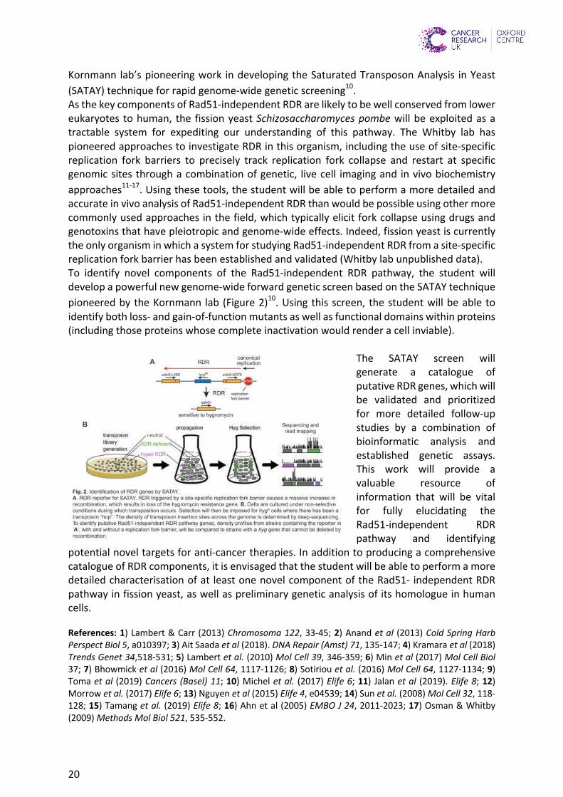
pioneered by the Kornmann lab (Figure 2)10. Using this screen, the student will be able to identify both loss- and gain-of-function mutants as well as functional domains within proteins (including those proteins whose complete inactivation would render a cell inviable).
The SATAY screen will generate a catalogue of putative RDR genes, which will be validated and prioritized for more detailed follow-up studies by a combination of bioinformatic analysis and established genetic assays. This work will provide a valuable resource of information that will be vital for fully elucidating the Rad51-independent RDR pathway and identifying
potential novel targets for anti-cancer therapies. In addition to producing a comprehensive catalogue of RDR components, it is envisaged that the student will be able to perform a more detailed characterisation of at least one novel component of the Rad51- independent RDR pathway in fission yeast, as well as preliminary genetic analysis of its homologue in human cells.
References: 1) Lambert & Carr (2013) Chromosoma 122, 33-45; 2) Anand et al (2013) Cold Spring Harb Perspect Biol 5, a010397; 3) Ait Saada et al (2018). DNA Repair (Amst) 71, 135-147; 4) Kramara et al (2018) Trends Genet 34,518-531; 5) Lambert et al. (2010) Mol Cell 39, 346-359; 6) Min et al (2017) Mol Cell Biol 37; 7) Bhowmick et al (2016) Mol Cell 64, 1117-1126; 8) Sotiriou et al. (2016) Mol Cell 64, 1127-1134; 9) Toma et al (2019) Cancers (Basel) 11; 10) Michel et al. (2017) Elife 6; 11) Jalan et al (2019). Elife 8; 12) Morrow et al. (2017) Elife 6; 13) Nguyen et al (2015) Elife 4, e04539; 14) Sun et al. (2008) Mol Cell 32, 118-128; 15) Tamang et al. (2019) Elife 8; 16) Ahn et al (2005) EMBO J 24, 2011-2023; 17) Osman & Whitby (2009) Methods Mol Biol 521, 535-552.
(Figure 2)10. Using this screen, the student will be able to identify both loss- and gain-of-function mutants as well as functional domains within proteins (including those proteins whose complete inactivation would render a cell inviable).
The SATAY screen will generate a catalogue of putative RDR genes, which will be validated and prioritized for more detailed follow-up studies by a combination of bioinformatic analysis and established genetic assays. This work will provide a valuable resource of information that will be vital for fully elucidating the Rad51-independent RDR pathway and identifying potential novel targets for anti-cancer therapies. In addition to producing a comprehensive catalogue of RDR components, it is envisaged that the student will be able to perform a more detailed characterisation of at least one novel component of the Rad51-independent RDR pathway in fission yeast, as well as preliminary genetic analysis of its homologue in human cells.
iii. Justification for support This project aligns squarely with CRUK and Oxford Centre’s research strategy focusing on genes and epigenetics. It will deliver an important new data resource from which potential targets for novel anti-cancer therapies will be identified. It will also provide a unique and diverse training opportunity for the DPhil student incorporating method development, high throughput screening, big data analysis, and yeast and human genetics. The student will benefit from the support of two well-resourced labs led by PIs with excellent track records both in their research and postgraduate student supervision.
References (hyperlinked to PubMed)
1) Lambert & Carr (2013) Chromosoma 122, 33-45; 2) Anand et al (2013) Cold Spring Harb Perspect Biol 5, a010397; 3) Ait Saada et al (2018). DNA Repair (Amst) 71, 135-147; 4) Kramara et al (2018) Trends Genet 34, 518-531; 5) Lambert et al. (2010) Mol Cell 39, 346-359; 6) Min et al (2017) Mol Cell Biol 37; 7) Bhowmick et al (2016) Mol Cell 64, 1117-1126; 8) Sotiriou et al. (2016) Mol Cell 64, 1127-1134; 9) Toma et al (2019) Cancers (Basel) 11; 10) Michel et al. (2017) Elife 6; 11) Jalan et al (2019). Elife 8; 12) Morrow et al. (2017) Elife 6; 13) Nguyen et al (2015) Elife 4, e04539; 14) Sun et al. (2008) Mol Cell 32, 118-128; 15) Tamang et al. (2019) Elife 8; 16) Ahn et al (2005) EMBO J 24, 2011-2023; 17) Osman & Whitby (2009) Methods Mol Biol 521, 535-552.
�2

21
Analysis of spindle assembly checkpoint function in health and disease – Prof. Gruneberg2,3A,3B
Primary Supervisor: Ulrike Gruneberg Additional Supervisors: Bela Novak Eligibility: Track 2, 3A and 3B students only are eligible to apply for this project. Project Summary Human bodies consist of millions and millions of cells which are produced through the process of cell division. To avoid disease, it is essential that each cell division is carried out correctly. Most importantly, before cells divide, the genetic material, DNA, has to be duplicated and then shared equally between the two daughter cells. If anything goes wrong during this process, DNA may be lost or gained in the daughter cells, resulting in a condition called aneuploidy, strongly associated with cancer [1]. A critical cellular safeguarding mechanism, the spindle assembly checkpoint (SAC), monitors the process of chromosome segregation in healthy cells and prevents aneuploidy from occurring [2, 3]. We propose to use a combination of cutting-edge live cell imaging of normal and tumour cells, and computational analysis and mathematical modelling to analyse the functioning of the spindle assembly checkpoint in tumour and non-tumour cells. This will help us to understand which aspects of spindle assembly checkpoint signaling, and consequently the chromosome segregation process, are altered in cancer cells. During mammalian cell division, it is critical that chromosome segregation and anaphase onset are executed only once all chromosomes have been stably tethered to microtubules via their kinetochores. The success of this process is ensured by the spindle assembly checkpoint. While many molecular details about spindle assembly checkpoint functioning have been elucidated in the past decade [3], key aspects of how this checkpoint functions are still poorly understood. We now know that the spindle assembly checkpoint checks that all chromosomes are attached via their kinetochores to microtubules before segregation of the chromosomes is initiated and that if attachment errors are detected, cell cycle progression is arrested and anaphase entry delayed [3]. However, it is still unclear how the spindle assembly checkpoint sustains a robust cell cycle arrest in the presence of just a few, or indeed just one, unattached kinetochore, yet is promptly silenced and cell cycle progression resumed once the last kinetochore has been attached. We have previously successfully combined experimental work and computational modelling to identify critical concepts and parameters for the functioning of the spindle checkpoint [4, 5]. However, further progress on rate-limiting factors in spindle checkpoint signalling has been hampered by the absence of experimental data on the concentration of proteins involved in spindle checkpoint function and the absence of knowledge of the biological consequences of changing these concentrations. Hence, our aim is now to bring together state-of-the-art genetic manipulation and live cell imaging of human cells with computational modelling to identify which parts of the spindle checkpoint pathway are critical for the robust yet sensitive behaviour of the spindle checkpoint, and how this is altered in cancer cells.
Research objectives and proposed outcomes
Spindle assembly checkpoint gene expression is frequently altered in cancer cells, with both up- and down regulation having been observed [6]. While in yeast cells it is well understood which spindle assembly checkpoint genes are rate-limiting for spindle checkpoint functionality, the distinct physiology of yeast cells precludes a simple translation of yeast data to human cells, and the lack of quantitative data on the human spindle checkpoint limits our understanding of the consequences of such alterations in gene expression for chromosome segregation and cell cycle progression. The aims of this proposal are therefore to close this knowledge gap by analysing the effects of SAC protein downregulation using a combination of cell biology and mathematical modelling.

22
1.) Cell biological analysis of the effects of spindle checkpoint protein downregulation on cell cycle progression in transformed and non-transformed cells We propose to systematically analyse the effect of downregulating known spindle checkpoint components BUBR1, BUB1, BUB3, MAD2, MAD1, CDC20, MPS1, ZW10 and p31comet in the background of transformed or non-transformed cell lines (aneuploid, HPV-transformed HeLa cells versus telomerase immortalised
diploid RPE-1 cells; immortalised non-transformed breast epithelial MCF10A cells in comparison to engineered MCF10 cells expressing constitutive or inducible RasV12 Ras-mutant and matched Ras-WT tumour cell lines) [7, 8]. To enable us to analyse these cell lines in
live cell imaging we will use CRISPR/Cas9-mediated genetical engineering of the cells to express fluorescently tagged cyclin B1 as well as a fluorescently tagged version of the spindle checkpoint protein of interest. Since cyclin B is one of the key proteins that is degraded at anaphase onset (Figure 1), this double tagging will allow us to modulate the levels of the tagged spindle checkpoint protein of interest by RNAi, determine the precise level of residual protein by calibrated fluorescence measurements, and then evaluate quantitatively by live cell measurements of cyclin B-GFP the ability of these cells to arrest cell cycle progression and stop cyclin B degradation in response to spindle poisons. 2.) Computational analysis of spindle checkpoint functionality In cells expressing fluorescently tagged cyclin B1 and SAC protein of interest, we can quantitatively measure the amounts of the proteins-of-interest, as well as cyclin B degradation kinetics (a proxy for cell cycle progression and SAC proficiency) for single cells under conditions when the checkpoint should be on. Together, these measurements allow us to calculate threshold levels of key spindle checkpoint activities [4]. When combined with modelling, these data help us understand which components become rate-limiting for spindle checkpoint maintenance in vivo in situations when levels of checkpoint proteins are reduced. We will carry out this analysis in parallel for transformed and untransformed cells with the aim of understanding which parameters change in cancer cells and how that affects the faithfulness of chromosome segregation in normal and transformed cells. Translational potential
The failure of chromosome segregation can lead to aneuploidy and chromosomal instability, associated with cellular transformation, cancer and resistance to chemotherapy. A detailed understanding of the regulation of the chromosome segregation process is therefore an important goal and may enable us to design targeted therapies to modulate cell division in disease situations such as cancer. Furthermore, given that many cancer cells downregulate the expression of spindle checkpoint proteins [6] and our analysis will enable us to predict the cellular consequences, this information could be pertinent for personalised tumour therapies. References 1. Sansregret, L., and Swanton, C. (2017). The Role of Aneuploidy in Cancer Evolution. Cold Spring Harb Perspect Med 7. 2. Kops, G.J., Weaver, B.A., and Cleveland, D.W. (2005). On the road to cancer: aneuploidy and the mitotic checkpoint. Nat Rev Cancer 5, 773-785. 3. Musacchio, A. (2015). The Molecular Biology of Spindle Assembly Checkpoint Signaling Dynamics. Curr Biol 25, R1002-1018. 4. Hayward, D., Alfonso-Perez, T., Cundell, M.J., Hopkins, M., Holder, J., Bancroft, J., Hutter, L.H., Novak, B., Barr, F.A., and Gruneberg, U. (2019). CDK1-CCNB1 creates a spindle checkpoint-permissive state by enabling MPS1 kinetochore localization. J Cell Biol 218, 1182-1199. 5. He, E., Kapuy, O., Oliveira, R.A., Uhlmann, F., Tyson, J.J., and Novak, B. (2011). System-level feedbacks make the anaphase switch irreversible. Proc Natl Acad Sci U S A 108, 10016-10021. 6. Weaver, B.A., and Cleveland, D.W. (2005). Decoding the links between mitosis, cancer, and chemotherapy: The mitotic checkpoint, adaptation, and cell death. Cancer Cell 8, 7-12. 7. Molina-Arcas, M., Hancock, D.C., Sheridan, C., Kumar, M.S., and Downward, J. (2013). Coordinate direct input of both KRAS and IGF1 receptor to activation of PI3 kinase in KRAS-mutant lung cancer. Cancer Discov 3, 548-563. 8. Matthews, H.K., Ganguli, S., Plak, K., Taubenberger, A.V., Win, Z., Williamson, M., Piel, M., Guck, J., and Baum, B. (2020). Oncogenic Signaling Alters Cell Shape and Mechanics to Facilitate Cell Division under Confinement. Dev Cell 52, 563-573 e563.

23
Identifying novel regulators of cancer stem cells in pancreatic ductal adenocarcinoma– Dr. Pauklin 1,2,3A
Primary Supervisor: Siim Pauklin Additional Supervisors: John Christianson Eligibility: Track 1, 2 and 3A students only are eligible to apply for this project. Project Summary Pancreatic cancer is one of the most lethal malignancies in human due to its highly metastatic characteristics and the poor responsiveness to current therapeutics. Pancreatic tumorigenesis involves a dedifferentiation process of cellular identity and the acquisition of a stem cell-like state of a subpopulation of cells known as cancer stem cells (CSCs). These cells resemble partly to naturally occurring stem cells and are exceptionally important because their developmental plasticity allows them to metastasize and give rise to whole tumours in the organism (1-4). Currently it remains unclear, which transcription factors and epigenetic machineries control the expression of stem cell genes and the stem cell-like identity of pancreatic CSCs. This knowledge would be valuable for developing more efficient pancreatic cancer therapeutics in the future. The research objective of the project is to identify and characterize novel epigenetic machineries and transcriptional regulators which govern gene expression, proliferation and stem cell-like characteristics of pancreatic CSCs. The DPhil project will apply a broad range of cutting-edge research techniques covering human cell culture systems, genome-wide, proteomic, genetic and biochemical methods (4-8). These include human cancer stem cell spheres and pancreatic ductal adenocarcinoma cultures, genome-wide studies (RNA-seq, ChIP-seq, ATAC-seq, ChIA-PET), functional studies (CRISPR/Cas9-mediated gene editing; tumour sphere assays), proteomics (Co-IP / mass-spectrometry), and mechanistic studies (confocal microscopy, flow cytometry, cell sorting, real-time PCR, western blotting, CyTOF). Collectively, this research will provide key insight to the signalling pathways and molecular mechanisms essential for the formation and maintenance of pancreatic CSCs, helping to better understand the tumorigenic process, and to uncover novel ways for diagnosing and treating this lethal cancer.

24
Research objectives and proposed outcomes. The research will uncover key epigenetic mechanisms that control gene expression and thereby the stem cell-like state of pancreatic cancer stem cells. The project has three sub-aims: 1. Performing small molecule screening experiments with compound sets targeting kinases and/or epigenetic mechanisms. The screening library will contain a unique collection of small molecule compounds with biological activity against a range of candidates including a broad range of epigenetic regulators that target distinct enzymes involved in regulating gene expression, as well as proteasome and kinase inhibitors. Our preliminary data has already uncovered interesting candidates for in-depth experiments. 2. Studying the molecular mechanisms of selected candidates from the screen in more detail. This includes functional and mechanistic analysis of candidates by loss-of-function and gain-of-function experiments via CRISPR/Cas9 mediated editing for gene knockdown and overexpression effects on tumour sphere formation and marker expression by flow cytometry, western blotting and confocal microscopy; RNA-sequencing for transcriptomic changes upon candidate knockdown and inhibition by small molecules; ATAC-sequencing for chromatin accessibility analyses; ChIP-sequencing for indentifying candidate binding sites; and ChIA-PET for promoter-enhancer interaction analyses. 3. Using patient samples and animal studies for candidate functional effects on tumorigenesis. We will perform CyTOF and single cell RNA-sequencing data of tumours from primary pancreatic cancer patients upon the treatment of epigenetic inhibitors; mouse tumorigenesis studies by orthotopic transplantation of pancreatic cancer cells with a knockout of candidates. Collaborators from different disciplines. The award will help build a collaboration between researchers with expertise in pancreatic cancer and cancer stem cells (Siim Pauklin); tumorigenesis and cancer therapeutics (John Christianson); epigenetic mechanisms, NGS technologies and computational biology (Udo Oppermann), as well as proteomic specialists at TDI mass-spectrometry facility, and clinical oncologists at NHS Oxford University Hospitals with access to primary pancreatic tumour samples. Translational potential of the project Relevance of the project to cancer. Our research will identify novel therapeutic targets that control epigenetic mechanisms responsible for regulating the stem cell-like characteristics of cancer stem cells in pancreatic ductal adenocarcinoma. Our preliminary experiments from candidate screening have already identified novel compounds that impact pancreatic cancer cell survival and cancer stem cell marker expression. Since we know the target enzymes of these compounds the project will provide valuable information on potential therapeutic candidates that could ultimately be tested in pancreatic ductal adenocarcinoma patients. REFERENCES 1. French, R., Feng, Y., and Pauklin, S. (2020). Targeting TGFβ signalling in cancer: toward context-specific strategies. Trends in Cancer 7, 538-540. 2. Feng, Y., and Pauklin, S. (2020). Two sides of the same coin: the roles of TGF-β in colorectal carcinogenesis. Gastroenterology 20, 30395-4. 3. Stoica, A., Chang C-H, and Pauklin, S. Molecular therapeutics of pancreatic ductal adenocarcinoma: targeted pathways and the role of cancer stem cells. Trends in Pharmacological Sciences (in press). 4. Feng, Y., and Pauklin, S. (2020). Revisiting 3D chromatin architecture in cancer development and progression. Nucleic Acids Res. 2020 Sep 17:gkaa747. doi: 10.1093/nar/gkaa747.

25
Understanding The Therapeutic Efficacy Of Chemotherapy In Pancreatic Cancer at the single cell level – Dr. Sivakumar1,2,3A,3B
Primary Supervisor: Shivan Sivakumar Additional Supervisors: Rachel Bashford-Rogers, Michael Dustin Eligibility: Track 1, 2, 3A and 3B students are eligible to apply for this project. Project Summary Pancreatic cancer has the worst survival of any human cancer1. Breakthroughs in treating this disease have been modest. Our best treatment is chemotherapy with a regime called FOLFIRINOX that extends survival by 5 months in the metastatic setting and cures an extra 10-15% of patients with resectable disease. We have made substantial efforts in the last few years to understand the single cell composition of pancreatic cancer. This project will entail to understand the effects of chemotherapy on the single cell composition of pancreatic cancer and how it effects important cell populations such as the cancer cells, immune cells and fibroblasts. The project will have available a large single cell dataset we have generated of untreated pancreatic cancer including immune cells, cancer cells and fibroblasts to act as a comparator. The fellow’s project will be to elucidate the effect of chemotherapy on the single cell changes in pancreatic cancer. Research Objectives and Proposed Outcomes Objectives:
- To characterise the immune cell subset changes due to chemotherapy of human pancreatic cancer cases
- To characterise the cancer cell transcriptional changes due to chemotherapy of human pancreatic cancer cases
- To characterise the fibroblast population changes due to chemotherapy of human pancreatic cancer cases
- To determine the interactions between cell types that form positive and negative feedback loops resulting tumour microenvironmental changes
The proposed outcome is to generate and analyse single cells from patients with resectable pancreatic cancer who have been treated with chemotherapy before their operation. They will generate data from 20 patients who will be a mix of responders and non-responders. They will process tumour samples into single cells immediately after their operation and sort into immune cells, cancer cells and fibroblasts. These cells will be single cell sequenced and the fellow analysing this and using a previously generated dataset to make their observations. As the tumour tissue will be collected, any interesting cell populations will be validated on these samples. The applicant will be trained in fundamental immunology, in depth analysis of samples from human tissue with techniques such as flow cytometry, single cell sequencing, immunofluorescence as well as transcriptomic and image analysis. They will also appreciate how to frame clinical problems as tractable research questions and translational medicine experimental design. The fellow would also be expected to present the results in national and international meetings. The findings from the study would inform us on how chemotherapy is working in pancreatic cancer and make help us select patients better and help us design better trials. The research itself is highly novel and urgently needed in a cancer of unmet need.

26
This project will show the biological effect of using chemotherapy in pancreatic cancer. The project is a highly collaborative project with the student needing to interface with many departments in the hospital and within the university. Expertise in genetics will be learned from the genomics centre in the Wellcome Trust Centre for human genetics and imaging from the translational pathology laboratory in the university oncology department. Translational potential of the project This work will help identify which subset of pancreatic cancer patients benefit from chemotherapy and help us understand how we could augment different therapeutic agents to gain a better response. This study will give us a real insight into which patients respond and which do not and why this is the case. We envisage that if there is a clear group of responders, we can develop a stratification system so this can feed into national trial platforms such as Precisionpanc to guide how we should treat chemotherapy responsive and unresponsive patients on the national arena. References:
1) Ilic M, Ilic I. Epidemiology of pancreatic cancer. World J Gastroenterol. 2016;22(44):9694-705. doi:10.3748/wjg.v22.i44.9694.
2) De Santiago, I., Yau, C., Heij, L., Middleton, M.R., Markowetz, F., Grabsch, H.I., Dustin, M.L. and Sivakumar, S., 2019. Immunophenotypes of pancreatic ductal adenocarcinoma: Meta-analysis of transcriptional subtypes. International journal of cancer.
3) Sivakumar, S., Abu-Shah, E., Ahern, D., Arbe-Barnes, E.H., Mangal, N., Reddy, S., Rendek, A., Easton, A., Kurz, E., Silva, M. and Heij, L.R., 2020. Immune responses in pancreatic cancer may be restricted by prevalence of activated regulatory T-cells, dysfunctional CD8+ T-cells, and senescent T-cells. bioRxiv.
4) Conroy, T., Desseigne, F., Ychou, M., Bouché, O., Guimbaud, R., Bécouarn, Y., Adenis, A., Raoul, J.L., Gourgou-Bourgade, S., de la Fouchardière, C. and Bennouna, J., 2011. FOLFIRINOX versus gemcitabine for metastatic pancreatic cancer. New England Journal of Medicine, 364(19), pp.1817-1825.
5) Conroy, T., Hammel, P., Hebbar, M., Ben Abdelghani, M., Wei, A.C., Raoul, J.L., Choné, L., Francois, E., Artru, P., Biagi, J.J. and Lecomte, T., 2018. FOLFIRINOX or gemcitabine as adjuvant therapy for pancreatic cancer. New England Journal of Medicine, 379(25), pp.2395-2406.

27
Heterogeneity of macrophages in colorectal cancer: the role of IRF5 – Prof. Udalova1,2,3A,3B
Primary Supervisor: Irina Udalova Additional Supervisors: Fiona Powrie Eligibility: Track 1, 2, 3A and 3B students are eligible to apply for this project. Project Summary The intestinal immune system is a delicately balanced system between tolerance to commensal and food particles and raising an immune response upon infection. Dysregulation of the intestinal immune system can lead to inflammation, which may progress to colorectal cancer (CRC) 1. Macrophages are playing a central role in maintenance of homeostasis, initiation of inflammation, restoration of tissue upon injury and mediation of chemoresistance in tumours. Integrating cues of their immediate tissue microenvironment, macrophages can adapt their functions according to tissue-specific needs and adapt upon change 2. Depending on the polarisation of the tumour-associated macrophages (TAMs), cancer progression and initiation can be hindered or helped 3. Various subsets of macrophages have recently been identified in different tissues using single-cell RNA sequencing (scRNA-seq), highlighting the heterogeneity and plasticity of macrophages 4–7. Furthermore, a role for Interferon Regulatory Factor 5 (IRF5), a master transcription factor involved in regulating the transcription of pro-inflammatory mediators in shaping macrophage polarisation was identified 5,8. IRF5 controls both acute and chronic inflammation and is protective in pathogen clearance 5,9,10. IRF5 has also been identified as a DNA-damage sensor, highlighting a potential beneficial role in CRC 11. Therefore, dissecting the molecular mechanisms involving macrophage polarisation and function is crucial for identification of treatment options for both inflammatory bowel disease (IBD) and CRC. Research objectives and proposed outcomes The aim of this project is to firstly assess the role of IRF5 in macrophages in resolution of inflammation using the Helicobacter hepaticus and anti-IL10R colitis model 12. Previous work in the lab has profiled IRF5-dependent inflammatory CD11c+ macrophages at peak of inflammation using scRNA-seq 5. Based on this work, we aim to profile the heterogeneity of macrophages in resolution of inflammation using targeted mouse models (CX3CR1-IRF5 fl/fl and CCR2-mKate ER2 IRF5 fl/fl). We hypothesize that lack of IRF5 is beneficial for resolution of inflammation as macrophages are being polarised towards a tissue-regenerating phenotype. Furthermore, comparison of macrophage heterogeneity in resolution to CRC will help identify molecular targets in shaping macrophage phenotype and directing towards resolution rather than progression of inflammation into cancer development. It also was suggested that localisation of macrophages within the tumour microenvironment might be a crucial determinant of their function 3. Therefore, assessing the localisation of different macrophage subsets and their interaction cell-cell contacts could also provide further information about their function and potential targeting. The role of TAMs in CRC is yet unclear with various studies suggesting both detrimental and beneficial effects. Thus, in addition to inhibition of IRF5 (as above) we would also consider stimulating IRF5 specifically at tumour sites might improve anti-cancer immunity 6,11. This could be achieved by targeted delivery of adenoviral

28
vector expressing IRF5 (overexpression) or inhibition of IRF5 through phosphorylating kinase inhibition 13. We have an established collaboration with the group of Prof Fiona Powrie, Kennedy Institute, who have developed a number of CRC models and will be able to guide us through. Translational potential of the project T cell immunity, which is beneficial in tumours, is undermined by immunosuppressive myeloid cells, of which a subset of TREM2+ macrophages have been identified as a potential target in tumours 14. Understanding the role of macrophages as pivotal cells in the resolution of inflammation as well as progression of inflammation into CRC will help shaping specific therapies targeting macrophages. IRF5 plays a crucial role in mediating differentiation of infiltrating monocytes into pro-inflammatory macrophages during intestinal inflammation and may therefore be central during resolution and cancer development. References : 1. Mantovani, A., Allavena, P., Sica, A. & Balkwill, F. Cancer-related inflammation. Nature (2008).
doi:10.1038/nature07205 2. Lavin, Y. et al. Tissue-Resident Macrophage Enhancer Landscapes Are Shaped by the Local
Microenvironment. Cell 159, 1312–1326 (2014). 3. Caprara, G., Allavena, P. & Erreni, M. Intestinal Macrophages at the Crossroad between Diet ,
Inflammation , and Cancer. Int. J. Mol. Sci. (2020). 4. Chakarov, S. et al. Two distinct interstitial macrophage populations coexist across tissues in
specific subtissular niches. Science (80-. ). 363, eaau0964 (2019). 5. Corbin, A. L. et al. IRF5 guides monocytes toward an inflammatory CD11c + macrophage
phenotype and promotes intestinal inflammation. Sci. Immunol. 6085, 1–16 (2020). 6. Xiong, Y. et al. Profiles of immune infiltration in colorectal cancer and their clinical significant:
A gene expression-based study. Cancer Med. (2018). doi:10.1002/cam4.1745 7. Norton, S. E., Dunn, E. T., Mccall, J. L., Munro, F. & Kemp, R. A. Gut macrophage phenotype is
dependent on the tumor microenvironment in colorectal cancer. Clin. Transl. Immunol. 5, 76 (2016).
8. Krausgruber, T. et al. IRF5 promotes inflammatory macrophage polarization and TH1-TH17 responses. Nat Immunol 12, 231–238 (2011).
9. Weiss, M. et al. IRF5 controls both acute and chronic inflammation. Proc. Natl. Acad. Sci. U. S. A. 112, 11001–11006 (2015).
10. Pandey, S. P., Yan, J., Turner, J. R. & Abraham, C. Reducing IRF5 expression attenuates colitis in mice, but impairs the clearance of intestinal pathogens. Mucosal Immunol. (2019). doi:10.1038/s41385-019-0165-1
11. Hu, G., Mancl, M. E. & Barnes, B. J. Signaling through IFN regulatory factor-5 sensitizes p53-deficient tumors to DNA damage-induced apoptosis and cell death. Cancer Res. 65, 7403–7412 (2005).
12. Arnold, I. C. et al. CD11c+monocyte/macrophages promote chronic Helicobacter hepaticus-induced intestinal inflammation through the production of IL-23. Mucosal Immunol. 9, 352–363 (2016).
13. Byrne, A. J. et al. A critical role for IRF5 in regulating allergic airway inflammation. Mucosal Immunol. 10, 716–726 (2017).
14. Molgora, M. et al. TREM2 Modulation Remodels the Tumor Myeloid Landscape Enhancing Anti-PD-1 Immunotherapy. Cell 1–15 (2020). doi:10.1016/j.cell.2020.07.013

29
Modelling cancer stem cell dormancy using organoids and advanced 3D culture models – Dr. Boccellato1,2,3A,3B
Primary Supervisor: Francesco Boccellato Additional Supervisors: , Colin Goding, Ahmed Ahmed
Eligibility: Track 1, 2, 3A and 3B students are eligible to apply for this project.
Project Summary Over recent years substantial advances have been made in our understanding of cancer and the development of a range of more effective therapies. Nevertheless, after apparently successful anti-cancer therapy, disease may recur even after many years owing to the presence of therapy-resistant cells. One of the principle causes of relapse is cancer cell dormancy. Some cancer cells stop dividing and enter a dormant state resembling that used by many physiological stem cells that divide to regenerate damaged tissue or replace cells that are naturally turned over. Why and how cells enter or emerge from a state of dormancy is unclear, but understanding how cells become dormant may offer opportunities for therapies designed to reduce relapse. Investigating human dormant cancer stem cells has been difficult due to the lack of appropriate in vitro models, and our current knowledge is extrapolated from experiments in mouse models or steady state analysis of human tumour masses. In this project we aim to use a newly identified biomarker for dormancy to detect and study this cell state ex vivo by using patient-derived biopsies. Further we will use organoids and other advanced cell culture models (the mucosoids) to identify and isolate dormant cells in vitro. The specific aims are to: 1. Characterise the hallmarks of dormant cells; 2. Decipher the signals regulating the generation, maintenance and elimination of dormant cancer cells. This study promises to identify potential therapeutic vulnerabilities in cancer dormancy. To achieve this, we will use single cell RNA-seq, label retention and lineage tracing, 3D culture systems, and live cell fluorescent reporter assays to compare dormant cancer cells with their physiological counterparts.
Research objectives and proposed outcomes
Tumours contain two kinds of non-proliferating cancer cells that are either quiescent or dormant. Most studies on dormancy do not distinguish between these two states. By examining physiological stem cells we have found that a hallmark of dormant, but not quiescent, cells is that they turn off expression of the majority of genes by shutting down transcription, evidenced by absence of an RNA polymerase II (PolII) modification associated with active transcription (Figure 1A). Since low transcription would reduce protein translation and suppress cellular metabolic activity, transcriptional silencing would allow cells to remain inactive for long periods. Importantly, such ‘PolII-negative’ cells are also found in human melanomas, mouse patient derived xenografts (PDX) (Figure 1B) and in 3D models of melanoma (not shown), but are
not observed in 2D culture. The Goding lab has also developed a novel lentivirus-delivered ratiometric fluorescent reporter designed to enable detection of dormant PolII-low cells (Figure 1C). Using this reporter in intestinal organoids revealed that PolII-low cells at the base of the crypt (Figure 1C, open arrows) exhibited both red and green fluorescence, whereas PolII high cells (eg. white closed arrows) were red only. Although still to be tested widely, this reporter potentially enables live dormant cells to be isolated and characterised. The Boccellato lab has developed a homeostatic, long-lived (>1 year) stem cell based human primary cell culture, called mucosoids, for the stomach (Figure 2A,B) and is currently using this technology to cultivate cells from fallopian tubes (Figure 2C,D).
Figure 1. Detecting PolII-low dormant cells. A. Immunofluorescence using PolII-specific antibody (Green) together with lineage markers (red) and DNA (DAPI, blue). B. Detection of PolII-low cells in a melanoma PDX mouse tumour. C. Activity of a ratiometric fluorescent reporter to detect dormant cells. PolII-Low cells (open arrows) marked by absence of blue fluorescence are Green and red, but PolII-High cells are red only.

30
Objectives 1. Characterise the hallmarks of dormant cells. Since PolII-low dormant cells can readily be detected in organoids, 3D culture and in tumours we will use the PolII marker in co-immunofluorescence assays to identify characteristics of dormant cells together with antibodies directed against candidate markers of interest. These include epigenetic marks, cell surface markers associated with minimal residual disease (eg aquaporins, CD36), and markers of stem cell populations identified by lineage tracing experiments in gastric and other organoid systems. In parallel, we will undertake laser-capture microdissection of human and PDX model tumour sections followed by RNA-seq to identify gene expression patterns associated with the PolII-low population, and by using the live cell fluorescent reporter assay in organoids or mucosoids, isolate candidate dormant cells for single cell-RNA-seq. We anticipate that we will identify biomarkers of dormant cells that will be useful for monitoring their generation and response to microenvironmental cues and to therapies, and a gene expression program that will reveal mechanisms underpinning the generation and maintenance of dormant cells. 2. Decipher the signals regulating dormancy. Using the biomarkers identified, including low PolII, we will then vary culture conditions for physiological and cancer organoids and 2D cultures to identify signalling pathways that increase or decrease the proportion of dormant cells. These conditions include hypoxia, nutrient limitation, infection and signalling molecules such as TGF� or TNF� that are important in generating physiological stem cells, as well as targeted and chemotherapies. RNA-seq data will be mined for clues to key signalling pathways that may represent targetable vulnerabilities in dormant cells. The results obtained will generate novel insights into the origins of cancer stem cells, their relationship to physiological stem cells and should identify potential therapeutic vulnerabilities in dormant cells. Collaborations involved:- The project brings together three laboratories (one clinical, two academic) that have not previously collaborated to identify a therapeutic vulnerability in cancer cell dormancy. The preliminary work is based on observations from the Goding lab, which has a long-standing interest in how the microenvironment generates phenotypic heterogeneity in cancer (Garcia Jimenez and Goding, 2019), primarily using melanoma as a model (Rambow et al , 2019). They recently identified PolII-low stem cells in multiple tissues, including in melanomas grown in 3D and PDX models, and have generated of a potential fluorescent reporter for dormancy. Ahmed’s lab has recently identified 4 different cell populations, including stem cells, in the fallopian tube using single cell RNA-seq, and has shown that these populations are also found in ovarian cancer (Hu et al 2020). The Boccellato lab is new to Oxford, but has developed a unique stem cell-dependent mucosoid culture system that accurately recapitulates the multi-lineage, highly polarized gastric epithelium that can be used as an in vitro model for gastric cancer (Boccellato et al 2018). The strength of this proposal comes from the shared interests and complementary expertise of each group in comparing physiological stem cells to those present within cancers, and especially in using the low-PolII status of dormant cells to identify and characterize dormant cells and their relationship to minimal residual disease. Translational potential of the project - The project addresses a critical question in cancer using novel technologies and preliminary observations: what is the nature of cancer cell dormancy? We anticipate that the characterization of dormant cells may enable therapeutic vulnerabilities to be identified, with the potential to prevent and eradicate dormant cancer cells.
References: Boccellato, F. et al., 2018, GUT. García-Jimenéz, C., and Goding, C.R. 2019, Cell Metabolism. Hu et al., 2020 Cancer Cell. Rambow, F., Marine, J.C., and Goding, C.R. 2019. Genes & Dev
Figure 2. A. Schematic of a mucosoid culture. B. Gastric mucosoid culture infected with H.pylori. C. A fallopian tube mucosoid culture stained for the stem cell marker Pax8. D. the same culture stained for CA125 and with visible cilia

31
Decoding immune surveillance within the tumour microenvironment: how the extracellular matrix enables escape from immunity – Prof. Midwood1,2,3A
Primary Supervisor: Kim Midwood Additional Supervisors: Francesca Buffa, Adrian Harris Eligibility: Track 1, 2 and 3A students only are eligible to apply for this project. Project Summary Our immune system detects tumors and activates inflammation to destroy them. However, tumors develop effective strategies to evade elimination, for example switching off cytotoxic T cells that are mobilized to kill them. Re-activating these cells after they have been disarmed by tumors has revolutionized the treatment of people with cancer. However, this approach does not work for all patients, nor all types of tumor, and can be associated with severe autoimmune side effects. The importance of the interplay between tumor cells, stromal cells and immune cells in determining whether a tumor will be eliminated, or will thrive, provided a landmark shift in disease philosophy, opening therapeutic avenues beyond targeting the tumor cell. However, in most solid tumors these cell populations do not exist in isolation, but assemble together within a tumor specific extracellular matrix (T-ECM), which creates the framework of the tumor microenvironment (TME)[1]. Building on our work which revealed that matrix molecules can trigger inflammation, and shape the resultant immune response, we discovered how tumors exploit the immuno-modulatory properties of the matrix to escape immune surveillance. We showed that the T-ECM creates distinct sub-tumoral niches which control immune cell infiltration, localization and phenotype, and we identified new subpopulations of tumor-associated myeloid cells whose behaviour is programmed by microenvironmental cues. We also developed therapeutic antibodies targeting matrix molecules, which prevent tumor growth and spread, by enhancing the infiltration of cytotoxic T cells and macrophages into the tumor, and by driving macrophage polarization towards a tumoricidal phenotype [2, 3]. This project will further investigate how crosstalk between the T-ECM and tumor, stromal and immune cells dictates the balance between an immunogenic and a tolerogenic TME. Using an interdisciplinary approach incorporating state of the art computational genomics, multiplex and high resolution tissue imaging, and therapeutic manipulation of the extracellular matrix, our aims are: 1) to understand the precise immunological roles of distinct components, and compartments, of the T-ECM, and 2) to determine whether targeting molecules within the T-ECM is a viable strategy with which to treat human disease. Research objectives and proposed outcomes Tenascin-C (TNC), an extracellular matrix protein not expressed in healthy adult tissues, but up-regulated in the stroma of many solid cancers, protects tumors from destruction by re-programming inflammation in the TME. In a syngeneic, immunocompetent grafting model of breast cancer, TNC-rich tumors contain high numbers of immune cells that support tumor growth including reparative macrophages and Th17 cells, and low numbers of CD8 cells, compared to tumors lacking TNC (Figure 1A). Analysis of the tumor myeloid compartment at the single cell level revealed previously undiscovered cellular heterogeneity, highlighting subpopulations with discreet functional specialization (B). The balance of myeloid subsets was altered in TNC high vs TNC null tumors; and specific subsets correlated differently with survival in people with breast cancer (C). In mice, tumor-associated macrophages (TAMs) were confined almost exclusively within niches defined by expression of TNC (D), which induced a localized phenotypic switch, downregulating M1-macrophage associated genes and co-stimulatory markers, and upregulating M2-associated genes, yielding TAMs incapable of T cell activation [2]. Treatment with antibodies that block TNC activation of toll-like receptor 4 (TLR4) (anti-FBG or C3) prevented autochthonous tumor growth (E) and spread [2], showing comparable

32
efficacy to checkpoint inhibition alone (anti-PDL1)[2], whilst combination treatment significantly enhanced, and sustained, inhibition of tumor growth (E). Anti-FBG treatment increased TAM infiltration into the tumor compared to isotype treated mice where a significant proportion of TAMs were excluded from the tumor [2], simultaneously upregulating co-stimulatory markers (F).
This project will: 1) further investigate the location, function and ontogeny of novel tumor-associated myeloid subsets using cluster specific markers to image, isolate and deplete subpopulations during murine models of breast cancer, 2) better define the cellular and molecular basis of TNC-mediated tumor growth and spread by examining i) changes in myeloid subpopulation localization and phenotype following treatment with anti-FBG antibodies, and ii)
signalling pathways activated downstream of TNC ligation of TLR4 that contribute to cell re-programming, and 3) identify whether myeloid subpopulations identified in murine breast tumors are conserved in human disease, by i) interrogation of publicly available human breast tumor RNA seq data sets (bulk; single cell (sc) from total cells) and sc data sets generated from sorted CD45+ cells (Harris/Buffa) and ii) imaging biopsy tissue sections (Harris) to map cell-matrix interaction networks in human disease. Together these data will provide a better understanding at a high resolution of how distinct myeloid cell subpopulations drive tumor growth and spread, and how cell-matrix interactions within discreet sub-tumoral niches impact the immune axis in breast cancer. Translational potential of the project and relevance to cancer research or patient care. The move towards treatments that allow the immune system to attack tumor cells has generated much excitement, offering the advantages of a more natural tumor killing mechanism than simply poisoning the tumor, as well as the possibility of generating long lasting immunity against the cancer. The first wave of immune-oncology therapies focused on blocking the tumors ability to de-activate cytotoxic lymphocytes already infiltrating the cancer, allowing existing effector cells to kill tumor cells. The aim of this proposal is to bring forward treatments that allow more efficient generation of effector cells within the tumor, and at metastatic sites, by targeting microenvironmental cues from the T-ECM, to prevent cancer growth and spread. This mode of action is distinct from current and emerging immune-oncology therapies, thereby providing an additional treatment option that may work synergistically with other approaches. Moreover, reducing primary tumor burden as well as control of tumor spread would be a significant step forward in treatment. References: [1] Pickup, M.W., J.K. Mouw, and V.M. Weaver, The extracellular matrix modulates the hallmarks of cancer. EMBO Rep, 2014. 15(12): p. 1243-53). [2] Deligne C, Murdamoothoo D, Gammage AN, Gschwandtner M, Erne W, Loustau T, Marzeda AM, Carapito R, Paul N, Velazquez-Quesada I, Mazzier I, Sun Z, Orend G, Midwood KS. Matrix-Targeting Immunotherapy Controls Tumor Growth and Spread by Switching Macrophage Phenotype. Cancer Immunol Res. 2020 Mar;8(3):368-382. [3] Immobilization of infiltrating cytotoxic T lymphocytes by tenascin-C and CXCL12 enhances lung metastasis in breast cancer. Murdamoothoo, D., Sun, Z., Yilmaz, A., Deligne, C., Velazquez-Quesada, I., Erne, W., Mörgelin, M., Midwood, K.S., Orend, G. Under review EMBO J.
Figure 1. Tenascin-C (TNC) protects tumors from destruction by re-programming inflammation in the TME. (A, C-F, and all other data cited but not shown are published in [2]; B is our unpublished data).

33
UGT8 and microcarrier signalling in the development of breast cancer – Prof. Wilson1,2,3A
Primary Supervisor: Clive Wilson Additional Supervisors: Adrian Harris Eligibility: Track 1, 2 and 3A students only are eligible to apply for this project. Project Summary Intercellular signals and signalling pathways promote the development of all cancers. Traditionally, such signals are considered to be discrete molecular entities like growth factors, but recently, more complex multimolecular complexes, such as extracellular vesicles (EVs), which can simultaneously reprogramme many aspects of cell behaviour, have emerged as key mediators. Using a Drosophila prostate-like genetic model, we have recently identified a novel EV subtype called Rab11a-exosomes, which we showed mediate growth and angiogenic effects in human cancer models (see also recent News and Views article)1. Employing the same model, we have discovered other new signalling complexes called microcarriers (Fig. 1A), which have a neutral lipid core, store signals at their surfacethat also control their fusigenic properties, and can then rapidly release them when the extracellular microenvironment changes2. We hypothesise that these too will provide novel insights into cancer biology and have therapy implications. Genetic analysis of microcarrier biogenesis has revealed that their release is controlled by UDP Glycosyltransferase 8 (UGT8), a ceramide galactosyltransferase, which is the most evolutionarily conserved lipid glycosyltransferase in the animal kingdom3,4. UGT8 knockdown in Drosophila prostate-like cells leads to all microcarriers remaining attached by projections to these cells (Fig. 1B). Even though these flies can transfer sperm to females, they are completely sterile, because microcarriers are not transferred, illustrating the importance of these intercellular signalling structures. UGT8 is highly expressed in several different, more aggressive cancers and particularly in breast cancer5-7. It promotes proliferation, survival and metastasis via mechanisms that are poorly characterised. Interestingly, breast epithelial cells secrete neutral lipid droplets, which carry galactosylceramide at their surface8, using mechanisms that are also not well characterised. Our central hypothesis is that breast cancer cells up-regulate UGT8 to secrete galactosylceramide-coated, neutral lipid-containing, microcarrier-like structures and that these play important intercellular signalling roles.
To test this hypothesis, we will combine the expertise of Prof Wilson’s group, which has discovered microcarriers and the mechanisms by which they are formed using the Drosophila model, with the extensive experience of Prof Harris in basic and clinical breast cancer biology. They have a strong and long-standing track record of collaboration in other areas of tumour cell biology, where Drosophila can inform cancer studies, eg [1,9]. The student will investigate how breast cancer microcarriers and lipid secretion are regulated by UGT8, define microcarrier cargos and determine their functions. This work has the potential to open up a new field in cancer biology, relevant not only to breast cancer, but to other cancers where high level UGT8 expression is a marker for tumour metastasis and therapy resistance7,10.

34
Research objectives and proposed outcomes The project will have the following four research objectives and proposed outcomes, and will fully test our central hypothesis that breast cancer cells secrete neutral lipid in structures equivalent to microcarriers and that UGT8 controls release of these structures and the resulting intercellular signalling: 1. Optimise assays for microcarriers and lipid droplets secreted from breast cancer cell lines: Initially use neutral lipid dyes (eg LipidTox) to stain small volumes of conditioned medium from 2D culture of breast cancer cell lines with high UGT8 expression5,6. These dyes are highly specific and do not stain other secreted multimolecular signalling structures like extracellular vesicles. Also use organoids generated from UGT8-expressing cell lines, eg. PMC42, MCF1011, staining lumen of fixed tissue (and extend to breast carcinoma organoids, if successful). In secreting cells, we will express a GFP protein carrying a glycosylphosphatidylinositol anchor (GFP-GPI), which associates with secreted phospholipid-coated structures and inserts into the outer coat of microcarriers in flies, to test as a vital marker for microcarriers. Confirm microcarrier identity by absence of transmembrane protein cargos, failure to stain with lipid bilayer dyes, immuno-EM and UGT8-dependency (2). Develop assays for measuring size and number of microcarriers. Proposed outcome: Identification of microcarrier-secreting cells and development of microcarrier assays. 2. Characterise the role of UGT8 in microcarrier biogenesis: Block ceramide synthesis, eg with zoledronic acid6, or knockdown UGT8 to test roles in microcarrier biogenesis, measuring microcarrier numbers and size in conditioned medium, and analysing secreting cells and organoids for stalled microcarrier secretion. Proposed outcome: Determine the roles of UGT8 and ceramide in microcarrier biogenesis. 3. Identify cargos carried by microcarriers: Pull-down GFP-GPI-labelled microcarriers or isolate by density gradients on basis of low density, and undertake proteomics analysis of cargos using several cell lines (with Dr Roman Fisher [Target Discovery Institute, with whom we work on exosome analysis). Screen for cargos previously identified in Drosophila, which are already implicated in UGT8 function, eg. Contactin2,12. Start to screen for specific cargos in patient samples, eg. organoids and blood, as the basis for grant applications to CRUK. Proposed outcome: Identification of signalling molecules and other cargos on microcarriers in vitro and in patients. 4. Determine functions of microcarriers: Reduce microcarrier secretion by UGT8 knockdown (or knockdown of cargos from 3) and test effects of resulting conditioned medium and control medium on breast cancer cell growth and migration, blood vessel network formation, etc, with assays guided by cargos found in 3. If time allows, undertake preliminary xenograft experiments to test effects on microcarrier secretion, tumour signalling, growth and metastasis. Proposed outcome: Determine the functions of breast cancer microcarriers. References: 1. Fan S-J et al. (2020) Glutamine deprivation alters the origin and function of cancer cell exosomes. EMBO J. 2020, e1030093 and N&V, van Niel G, Théry C. (2020) Extracellular vesicles: eat glutamine and spit acidic bubbles. EMBO J. 2020, 39:e105119; 2. Wainwright, S.M. et al. (2020) Drosophila Sex Peptide Controls the Assembly of Lipid Microcarriers in Seminal Fluid. BioRxiv, https://doi.org/10.1101/2020.04.24.059238, PNAS, under revision; 3. Ahn SJ, et al. (2011) Comparative analysis of the UDP-glycosyltransferase multigene family in insects. Insect Biochem Mol Biol. 42:133-47; 4. Meech R, et al. (2019) The UDP-Glycosyltransferase (UGT) Superfamily: New Members, New Functions, and Novel Paradigms. Physiol Rev. 99:1153-1222; 5. Owczarek TB, et al. (2013) Galactosylceramide affects tumorigenic and metastatic properties of breast cancer cells as an anti-apoptotic molecule. PLoS One 8:e84191; 6. Cao Q, et al. (2018) Inhibition of UGT8 suppresses basal-like breast cancer progression by attenuating sulfatide-αVβ5 axis. J Exp Med. 215:1679-92; 7. Dzięgiel P, et al. (2010) Ceramide galactosyltransferase (UGT8) is a molecular marker of breast cancer malignancy and lung metastases. Br J Cancer 103:524-31; 8. Bouhours JF, Bouhours D. (1979) Galactosylceramide is the major cerebroside of human milk fat globule membrane. Biochem Biophys Res Commun. 88, 1217-1222; 9. Fan, S-J et al. (2016) PAT4 Levels Control Amino Acid Sensitivity of Rapamycin-Resistant mTORC1 from the Golgi and Affect Clinical Outcome in Colorectal Cancer. Oncogene 35, 3004-15; 10. Al-Mahrouki A, et al. (2017) Microbubble-based enhancement of radiation effect: Role of cell membrane ceramide metabolism. PLoS One 12:e0181951; 11. Marella NV et al. (2009) Cytogenetic and cDNA microarray expression analysis of MCF10 human breast cancer progression cell lines. Cancer Res. 69:5946-53; 12. Traka M, et al. (2002) The neuronal adhesion protein TAG-1 is expressed by Schwann cells and oligodendrocytes and is localized to the juxtaparanodal region of myelinated fibers. J Neurosci. 22:3016-3024.

35
Studying the role of a chromatin remodelling factor (ATRX) in normal gene expression and in malignancy – Prof. Higgs1,2,3A
Primary Supervisor: Douglas Higgs Additional Supervisors: Richard Gibbons Eligibility: Track 1, 2 and 3A students only are eligible to apply for this project. Project Summary Thalassaemia is the most common form of inherited anaemia throughout the world. In all cases, it results from an imbalance in the production of the a-like and b-like globin chains of haemoglobin, leading to a-thalassaemia and b-thalassaemia respectively. The aim of our laboratory is to understand how the globin gene clusters are normally regulated during development and differentiation and how this is perturbed in patients with thalassaemia. By approaching these questions, we are also developing a general understanding of the principles by which mammalian genes are normally switched on and off and how these processes go awry in acquired and inherited human genetic diseases, including cancer. During the course of this work, we have identified about 130 patients who have a rare form of a-thalassaemia which occurs in the context of a pre-malignant condition called the myelodysplastic syndrome (MDS). Many of these patients go on to develop acute myeloid leukaemia. These patients have no pre-existing forms of a-thalassaemia (AT) and so this condition is acquired specifically in the pre-malignant clones of cells in MDS: hence the condition is referred to as the ATMDS syndrome. When we analyse the bone marrow cells of patients with ATMDS we find a distinct constellation of mutations in epigenetic readers, writers and erasers which are also found in other patients with MDS but, importantly, in addition, most patients with ATMDS have mutations in a chromatin remodelling factor called ATRX. This protein was discovered in our laboratory in 1995 as a cause of X-linked a-thalassaemia associated with developmental abnormalities (ATR-X syndrome) and ATRX has more recently been recognised as a tumour suppressor gene in a wide variety of malignant tumours including glioblastoma, melanoma, pancreatic neuroendocrine tumours and a wide range of sarcomas.
By understanding the role of ATRX in vivo, we hope to understand its normal role in gene expression and how this is perturbed in malignancy. Analysis of naturally occurring mutations often provide important clues to mechanism of disease. The key scientific question in this project is how do mutations in ATRX down regulate a-globin gene expression in ATMDS syndrome and why is the effect so much greater in ATMDS compared with ATR-X syndrome. We have been studying both primary cells from patients with ATMDS or ATR-X syndrome and

36
developing much needed erythroid cell models of these diseases. This has been challenging for a variety of reasons but recently we have shown that using single cell analysis we are now able to identify a sub-population of erythroid cells that appear to be more affected by ATRX mutations than others and we are currently investigating why this should be so. Some clues to this will come from analysing the impact of other genes that are mutated in ATMDS syndrome: appropriate cell lines in which such genes have been mutated individually and in combination are now edited and available for further studies. The aims of this project will therefore be to further characterise primary cells and the recently established erythroid cell models of ATMDS syndrome using transcriptional, epigenetic and chromosome conformation studies to analyse how a-globin expression is perturbed in this severe, acquired form of a thalassaemia. All of such experimental approaches are well established in our laboratory. This project will contribute to our understanding of globin gene regulation, the general mechanism(s) by which chromatin remodelling factors normally work and how they may contribute to malignant diseases when mutated. Research objectives and proposed outcomes The role of chromatin remodelling factors is at the forefront of research into gene regulation and cancer. We contribute to many aspects of gene regulation both transcriptional and epigenetic aspects via publication in high impact journals and engagement in international meetings. We have expertise on a wide range of approaches to transcriptional and epigenetic biology in our laboratories. We also provide training in computational biology. We have well established long-standing collaborations with clinical haematologists in the UK (Oxford Haematology and Quek, KCH), EU (Hellstrom, Karolinska) and US (Steensma and Ebert, Harvard) who have been contributing to this programme of work and we have an established network of collaborators in the transcriptional and epigenetic areas of research. Translational potential of the project and relevance to cancer Since we originally identified the ATRX gene we have also shown that the protein is part of a protein complex together with a histone chaperone DAXX and the histone variant H3.3. Mutations in all three components of this complex have now been associated with a wide variety of malignant tumours. Almost all of such tumours maintain their telomeres via the so-called alternative (ALT) pathway of telomere maintenance. This aspect of ATRX is also being studied independently by a member of Oncology (Dr David Clynes) in collaboration with Professor Richard Gibbons (Co-supervisor). The current proposal aims to study the role of ATRX in gene expression. There seems little doubt that understanding the normal biological role of this complex will be of importance in understanding its role in the development of cancer. Relevant Publications 1) Gibbons RJ, Picketts DJ, Villard L & Higgs DR (1995) Mutations in a putative global transcriptional regulator cause X-linked mental retardation with a-thalassemia (ATR-X Syndrome). Cell, 80, 837-845. 2) Gibbons RJ, Pellagatti A, Garrick D, Wood WG, Malik N, Ayyub H, Langford C, Boultwood J, Wainscoat JS & Higgs DR (2003) Identification of acquired somatic mutations in the gene encoding chromatin-remodelling factor ATRX in the a thalassaemia myelodysplasia syndrome (ATMDS). Nat Genet, 34, 1-4. 3) Steensma DP, Gibbons RJ & Higgs DR (2005) Acquired �-Thalassemia in Association with Myelodysplastic Syndrome and Other Hematologic Malignancies. Blood, 105, 443-452. 4) Hanssen LLP, Kassouf MT, Oudelaar AM, Biggs D, Preece C, Downes DJ, Gosden M, Sharpe JA, Sloane-Stanley JA, Hughes JR, Davies B & Higgs DR (2017) Tissue-specific CTCF-cohesin-mediated chromatin architecture delimits enhancer interactions and function in vivo. Nat Cell Biol 19: 952-961. 5) Hughes JR, Roberts N, McGowan S, Hay D, Giannoulatou E, Lynch M, de Gobbi M, Taylor S, Gibbons R & Higgs DR (2014) Analysis of hundreds of cis-regulatory landscapes at high resolution in a single, high-throughput experiment. Nat Genet, 46: 205-212. 6) Hay D, Hughes JR, Babbs C, Davies JOJ, Graham BJ, Hanssen L, Kassouf MT, Oudelaar AM, Sharpe JA, Suciu M, Telenius J, Williams R, Rode C, Li P-S, Pennacchio LA, Sauka-Spengler T, Sloane-Stanley JA, Ayyub H, Butler S, Gibbons RJ, Smith AJH, Wood WG & Higgs DR (2016) Testing the super-enhancer concept by in-vivo dissection. Nat Genet, 48, 895-903.

37
ARH3/ADPRHL2 as a biomarker for PARP inhibitor sensitivity/resistance – Prof. Ahmed1,2,3A,3B
Primary Supervisor: Ahmed Ahmed Additional Supervisors: Ivan Ahel Eligibility: Track 1, 2, 3A and 3B students are eligible to apply for this project. Project Summary To protect the genome from damage organisms have evolved a cellular defence mechanisms termed the DNA damage response (DDR). The DDR includes a diverse set of signal transduction pathways and effector proteins that act to sense DNA lesions and effectively repair the damage, limiting the propagation of genomic instability. Exploiting DDR pathways to specifically target and kill cancer cells has become an attractive therapeutic avenue within cancer research. This is exemplified by the synthetic lethal interaction between PARP inhibition and BRCA1 or BRCA2-deficient tumours1. Ivan Ahel (co-supervisor on this project) laboratory recently identified HPF1 protein as a novel interactor and critical regulator of PARP1 ADP-ribosylation activity upon DNA damage2. Functionally, HPF1 suppresses DNA damage-induced hyper auto-modification of PARP1 and promotes in trans ADP-ribosylation of histones and many other proteins involved in regulation of genome stability. They further demonstrated that HPF1 is a critical specificity factor that allows modification of target proteins by PARP1 on serine residues (Ser-ADPr)3,4. Crucially, the work also identified ARH3 as a hydrolase which specifically removes Ser-ADPr5 and further showed that Ser-ADPr is the major form of ADP-ribosylation following DNA damage6. Taken together, the insights surrounding Ser-ADPr open a large, exciting, and novel area of research into the fundamental understanding of the pathways regulated by this modification. Strikingly, our unpublished data show that ARH3 knockout in model cell lines associates with PARP inhibitor (PARPi) resistance, while ARH3 overexpression is associated with PARPi sensitivity. Based on these results, we hypothesize that ARH3 activity and protein levels affect sensitivity to PARPi, thus representing; i) a predictor for the success of these therapies and, ii) a novel target for further drug development. Currently, PARP inhibitors are used to treat ovarian cancer and several other cancers, and we therefore propose to test the hypothesis that ARH3 expression might be a useful diagnostic tool with which to stratify cancer patients into sub-groups that will be sensitive/resistant to PARPi treatment with a particular focus on ovarian cancer. The mechanism of sensitivity/resistance of cells with deregulated ARH3 expression cells to PARPi is unknown, and elucidating this mechanism will be another goal of this proposed work. Research objectives and proposed outcomes Objective 1. Characterise the effect of ARH3 under- and overexpression in a series of model and primary cancer cell lines on PARP inhibitor sensitivity/resistance. We will collect and test a variety of ovarian cancer cell lines, profiling them for ARH3 protein expression levels and then treating with several different PARPi of varying PARP-trapping capabilities (olaparib, talazoparib, veliparib). To determine the impact of ARH3 protein levels on PARPi vulnerability, we will not only assess drug sensitivity and levels of PARP1, PARG, and ARH3 across a panel of ovarian cancer cell lines, but also assess the impact of systemically varying ARH3 by knockdown, knock out and inducible overexpression in HGSOC lines of defined genotype, including Ovcar8 (BRCA1/2 wt, PARPi resistant), PE01 (BRCA2-mutant, PARPi sensitive), Kuramochi (BRCA2-mutant, PARPi partially sensitive) and COV362 (BRCA1-mutant, PARPi

38
sensitive). Rescue experiments with wild type vs. catalytically inactive ARH3 will assess the suitability of ARH3 as a target for the development of inhibitors. Objective 2. To determine the frequency of ARH3 gene alterations in a larger set of HGSOC samples, we will: i) interrogate data of an ongoing whole exome sequencing study of 504 ovarian cancers searching for ARH3 and PARG copy number alterations and mutations; and ii) perform semi-quantitative detection of ARH3, as well as of PARG, PARP1 and PAR, by immunohistochemistry (IHC) on two independent sets of tissue microarrays (TMAs) containing a total of 1200 ovarian cancers. To augment these analyses, which will be limited by the small number of tumors treated with PARPi, we will also evaluate levels of ARH3, PARG, PARP1 and PAR in patient-derived xenograft (PDX) models that have been assayed for response to single-agent PARPi, including ones that have a high HRD score but did not respond. Objective 3. Elucidating the mechanistic basis for the sensitivity/resistance of cells with deregulated ARH3 expression cells to PARPi (modulation of the PARP-trapping, regulation of DNA repair pathway choice, regulation of the chromatin structure/epigenetic marks). For these studies we will use largely cell biology/biochemical and genomics approaches. This objective will be performed in co-supervisor (Dr Ivan Ahel) laboratory at the Sir William Dunn School of Pathology, University of Oxford.
4. Translational potential of the project Our data suggest that ARH3 protein expression levels in cancer patients might be a marker that confers sensitivity/resistance of the tumour to PARPi, providing a rationale for using PARPi for certain patients. In longer term, understanding the mechanisms of DNA repair and PARPi resistance through studies of ARH3 protein, may reveal new, unexpected avenues for treatments in the future. References 1. Bryant et al (2005) Specific killing of BRCA2-deficient tumours with inhibitors of
poly(ADP-ribose) polymerase. Nature 434, 913-917. 2. Gibbs-Seymour, I., Fontana, P., Rack, J.G., and Ahel, I. (2016) HPF1/C4orf27 Is a PARP-
1-Interacting Protein that Regulates PARP-1 ADP-Ribosylation Activity. Mol Cell 62, 432-442.
3. Bonfiglio, J.J., Fontana, P., Zhang, Q., Colby, T., Gibbs-Seymour, I., Atanassov, I., Bartlett, E.J., Zaja, R., Ahel, I.*, and Matic, I.* (2017) Serine ADP-ribosylation depends on HPF1. Mol Cell 65, 932-940. (*Corresponding authors)
4. Suskiewicz, M.J., Zobel, F., Ogden, T.E., Fontana, P., Ariza, A., Yang, J., Zhu, K., Bracken, L., Hawthorne, W.J., Ahel, D., Neuhaus, D., and Ahel, I. (2020) HPF1 completes the PARP active site for DNA-damage induced ADP-ribosylation. Nature 579, 598-602.
5. Fontana, P., Bonfiglio, J.J., Palazzo, .L, Bartlett, E., Matic, I., and Ahel, I. (2017) Serine ADP-ribosylation reversal by the hydrolase ARH3. Elife Jun 26;6. pii: e28533.
6. Palazzo, L., Leidecker, O., Prokhorova, E., Dauben, H., Matic, I., and Ahel, I. (2018) Serine is the major residue for ADP-ribosylation upon DNA damage. Elife Feb 26;7. pii: e34334.

39
Discovery and mechanistic elucidation of small molecule inducers of myeloblast differentiation for ALL – Prof. Russell3A
Primary Supervisor: Angela Russell Additional Supervisors: Thomas Milne Eligibility: Track 3A students only are eligible to apply for this project. Project Summary The most common childhood cancer is acute lymphoblastic leukaemia (ALL), a disease which leads to the accumulation of immature lymphoid cells in the bone marrow. This is thought to be caused in part by a block in normal lymphocyte differentiation. There has been amazing progress in treating childhood ALL, but unfortunately a subset of childhood ALL continues to be refractory to treatment, especially in those patients that harbour a rearrangement (r) of the Mixed Lineage Leukaemia (MLL) gene. The most common MLLr results in a fusion protein, MLL-AF4, that is responsible for many of the poor prognosis ALL patients. In addition, even for children who are cured, conventional therapies are often toxic and can cause long lasting life-altering effects. Current treatments typically aim to kill abnormal cells via chemotherapy, but our goal is to establish a new paradigm in the treatment of ALL, that is to induce differentiation of ALL blasts. Our inspiration comes in part from the wave of new small molecule therapies for acute myeloid leukaemia (AML) that have been shown to have reduced toxicity compared to conventional therapy and function by causing AML cells to differentiate. Our hope is that by applying the concept of differentiation therapy to ALL we will be able to i) provide novel treatments for refractory ALL such as MLLr leukaemias and ii) develop novel therapies that have fewer toxic side effects than current conventional therapies. Previously, we established an in vitro screen to detect differentiation of AML cells using flow cytometry and used this to identify multiple classes of small molecules which can block proliferation and overcome the differentiation block in AML blasts. Our leading examples are orally bioavailable in mice and are being progressed into in vivo trials to determine efficacy. We have performed some preliminary time-course studies and global RNA-seq analyses to better understand the compounds’ effects at a cellular level. From these data we have shown that our compounds are distinct from other known inducers of differentiation in AML cells. However, we have not yet defined their direct cellular target(s). Preliminary data also suggests that treatment of ALL cells with these compounds impairs their growth in vitro. Our goal in this project is to apply these same compounds, alongside other classes of AML drug candidates which also induce differentiation, to ALL blast cells from a novel MLL-AF4 humanized model (see Figure 1) as well as ALL patient samples to determine i) which novel compounds can disrupt ALL growth; ii) if ALL blasts can be induced to differentiate; iii) how the compound(s) impact the function of the target(s), and (iv) what downstream cellular pathways are impacted by target engagement. Research objectives and proposed outcomes: In this project we aim to use a combination of chemical and biological techniques to address questions (ii)-(iv) for one of these series of molecules. Two parallel approaches will be developed using an integrative approach combining existing cutting-edge expertise in Milne/Russell groups and collaborators Dr Anindita Roy and Prof Irene Roberts: 1. Analyze differentiation of ALL cells using a novel MLL-AF4 ALL model, with a combination of tools as well as novel compounds. Counterscreen for toxicity and/or B-lineage differentiation potential of normal cells (e.g. cord blood) to exclude compounds which exhibit non-specific effects. 2. Identifying compound binding partners in ALL cells through a combination of affinity and photoaffinity proteomics, candidate screening, native intact mass analysis and follow up target validation. 3. Identify key pathways controlled/impacted by compound treatment through a combination of nascent and RNA-seq, CRISPR/CAS9 screening and proteomic analysis.

40
The overall workflow is depicted in Fig. 1. In the environment of the chemistry research laboratories (CRL), training will be provided in chemical synthesis, analytical methods (e.g. NMR, mass spectrometry), medicinal chemistry, drug design, photoaffinity labelling, chemical biology, affinity and photoaffinity protein profiling and proteomics. These techniques are well established in the Russell group, and have been successfully mastered by several DPhil students in recent years (for example Wilkinson et al, 2020). As well as all of these techniques, training will also be provided in how to ask and answer scientific questions in medicinal chemistry and chemical biology. Similarly, a wide range of technical training will be provided in the RDM/WIMM environment including basic cell biology techniques, analytical methods (e.g. FACS), drug screening, RNA-seq and other next generation sequencing techniques, CRISPR/CAS9 screening, basic bioinformatics and dataset analysis. Training will also be provided in answering questions about biological impact and efficacy of compounds. Professor Russell and Professor Milne already collaborate and hold biweekly joint meetings to discuss and monitor research progress. It would be expected that the student would attend these meetings as a primary means to monitor progression in the presence of both academic supervisors so as to ensure coherence and consistency of messaging – a critical factor when students have joint supervision arrangements. It would not be expected that the student would present at every one of these meetings – most likely every other one. These meetings will be critical to set overall objectives and project direction. Together, these distinct environments will provide the unique opportunity for the candidate to bring together two interdisciplinary areas of research to address and important area of unmet medical need in oncology. Translational potential of the project: Accomplishing the goals of this project could potentially impact four specific areas: 1) pharmacodynamic biomarker discovery, 2) yield insights into the basic biology underpinning ALL blast cell differentiation, 3) identifying patient subsets to target in clinical trials, and 4) revealing new molecular targets for future drug discovery endeavours. As the intention with this project is to focus on a mechanistic evaluation of tool compounds this means there would be no commercial restriction or delay to publish the outcomes of the research. The sharing of new data with the scientific community in a timely fashion we anticipate will further the overall goal of identifying and developing drugs that will go into clinical trials to one day impact patient health. References: Godfrey, L.; Milne, T. A.* DOT1L inhibition reveals a distinct subset of enhancers dependent on H3K79 methylation. Nat. Commun. 2019; DOI: 10.1038/s41467-019-10844-3. Wilkinson, I.V.L.; Russell, A.J.* Chemical Proteomics and Phenotypic Profiling Identifies the Aryl Hydrocarbon Receptor as a Molecular Target of the Utrophin Modulator Ezutromid. Angew. Chem. Int. Ed. 2020; DOI: 10.1002/anie.201912392.
Fig. 1: Proposed project workflow.

41
Single-cell analysis of haematopoietic stem cells in SF3B1 mutant MDS: identification of new therapeutic targets/treatments – Dr. Pellagatti1,2,3A
Primary Supervisor: Andrea Pellagatti Additional Supervisors: Adam Mead, Supat Thongjuea Eligibility: Track 1, 2 and 3A students only are eligible to apply for this project. Project Summary The myelodysplastic syndromes (MDS) are common myeloid malignancies.1 There are few effective treatments for MDS and the vast majority of patients will die as a result of their disease. The MDS originate in bone marrow haematopoietic stem cells (HSCs) that are essential for disease initiation and progression, and the eradication of the malignant HSCs is necessary to achieve cure. Splicing factor mutations are the most common mutations found in MDS. The splicing factor gene SF3B1 is the most frequently mutated gene in MDS, and we and others have shown that SF3B1 mutations result in aberrant pre-mRNA splicing.1-3 We recently showed that SF3B1 mutant MDS is a distinct nosologic entity.4 Interestingly we demonstrated that increased R-loops and DNA damage occur in SF3B1 mutant MDS patient bone marrow cells.5 SF3B1 mutations have been identified in other cancers, including chronic lymphocytic leukemia, uveal melanoma, breast cancer and pancreatic cancer, suggesting that somatic mutations in spliceosome genes have an important role in tumorigenesis.1 We will use a single-cell method for high-sensitivity mutation detection with parallel RNA sequencing analysis (TARGET-seq)6 to determine the genetic and transcriptomic heterogeneity of single HSCs from individual MDS patients harbouring SF3B1 mutations. Our approach allows the dection of changes in gene expression levels and also aberrantly spliced transcripts. Dysregulated genes/pathways will be identified using appropriate bioinformatics analyses7 in mutation-defined HSC subpopulations in each MDS patient (Figure 1) and used as input to identify drugs that can target the different HSC subpopulations in an individual patient. The identified drugs will be tested in vitro using long-term culture-initiating cell (LTC-IC) assays to determine the drugs that are cytotoxic to MDS HSCs (with the relevant mutation profile) while sparing the HSCs from healthy controls. Importantly, the drugs identified may also have efficacy in other myeloid malignancies with similar mutation profiles. This approach will lead to the identification of new treatments for SF3B1 mutant MDS and has broad applicability to other haematological malignancies and other cancers.
Figure 1. Sequential mutation acquisition in MDS. An example of clonal hierarchy in an MDS patient harbouring SF3B1, TET2 and CBL mutations is shown. Using TARGET-seq, we will profile the single-cell transcriptome for individual MDS patients and identify different HSC subpopulations on the basis of their mutation status. In a case with SF3B1 as a founder mutation, with TET2 as a subsequent mutation, and CBL as a late mutation, the HSC subpopulations could comprise cells with SF3B1 mutation only, cells with both SF3B1 and TET2
mutations, cells with all three mutations, as well as wildtype cells (non-clonal).
42
Research objectives and proposed outcomes The aim of this project is to identify new therapeutic targets/treatments from the transcriptomic analysis of single HSCs (i.e. the disease-propagating cell population in MDS) from MDS cases with SF3B1 mutation. Our objectives are: 1- To perform single-cell mutational analysis with parallel transcriptomic (RNA-seq) analysis on the HSCs of a group of MDS patients harbouring SF3B1 gene mutations 2- To perform bioinformatics analysis on the single-cell data in order to identify molecular signatures and druggable targets, and drugs known to target them, in each mutation-defined HSC subpopulation in the MDS patients 3- To test the identified drugs in vitro to determine the drugs which are cytotoxic to MDS HSCs (with the relevant mutation profile) while sparing the HSCs from healthy controls We will identify dysregulated genes (both at the expression and splicing levels) and pathways in the HSCs of SF3B1 mutant MDS patients, with the potential to discover novel pathways involved in MDS pathogenesis. This will broaden our knowledge of MDS pathophysiology.
We will identify drugs and drug combinations that can specifically target all HSC subsets within each MDS patient, enabling treatment tailored both to the individual MDS patient (precision medicine), as well as to whole groups of patients with SF3B1 mutations and mutation combinations. Toxicology and safety data may be already available for some of the drugs identified and this will accelerate their introduction into the clinic to treat MDS as a new disease indication for these drugs. This project will benefit from collaboration with Professor Amit Verma (New York), an expert in MDS HSC biology and drug target identification from gene expression data in myeloid malignancy. Translational potential We will identify new drugs and drug combinations for MDS patients with SF3B1 mutations. The drugs identified may also have efficacy in other myeloid malignancies (e.g. acute myeloid leukaemia) and other cancers with SF3B1 mutations. This approach will lead to the identification of new treatments for SF3B1 mutant MDS and has broad applicability to other haematological malignancies and other cancers.
References 1. Pellagatti A, Boultwood J. Splicing factor gene mutations in the myelodysplastic syndromes: impact on disease phenotype and therapeutic applications. Adv Biol Regul 2017; 63:59-70. 2. Dolatshad H, Pellagatti A, et al, Smith CW, Boultwood J. Cryptic splicing events in the iron transporter ABCB7 and other key target genes in SF3B1-mutant myelodysplastic syndromes. Leukemia 2016; 30:2322-2331. 3. Pellagatti A, Armstrong RN, et al, Smith S, Boultwood J. Impact of spliceosome mutations on RNA splicing in myelodysplasia: dysregulated genes/pathways and clinical associations. Blood 2018; 132:1225-1240. 4. Malcovati L, Stevenson K, et al, Pellagatti A, et al, Cazzola M. SF3B1-mutant MDS as a distinct disease subtype: a proposal from the International Working Group for the Prognosis of MDS. Blood 2020; 136:157-170. 5. Singh S, Ahmed D, et al, Pellagatti A, Boultwood J. SF3B1 mutations induce R-loop accumulation and DNA damage in MDS and leukemia cells with therapeutic implications. Leukemia 2020; 34:2525-2530. 6. Rodriguez-Meira A, Buck G, et al, Thongjuea S, Mead AJ. Unravelling Intratumoral Heterogeneity through High-Sensitivity Single-Cell Mutational Analysis and Parallel RNA Sequencing. Mol Cell 2019; 73:1292-1305. 7. Giustacchini A, Thongjuea S, et al, Jacobsen SEW, Mead AJ. Single-cell transcriptomics uncovers distinct molecular signatures of stem cells in chronic myeloid leukemia. Nat Med 2017; 23:692-702.

43
Dissecting the biological basis of HLA variation to susceptibility and disease severity in Crohn’s disease & ulcerative colitis as risk factors for the development of colorectal cancer – Prof. Satsangi1
Primary Supervisor: Jack Satsangi Additional Supervisors: Nicola Ternette Simon Leedham, Rebecca Powell Doherty Eligibility: Track 1 students only are eligible to apply for this project. Project Summary IBD is a chronic, inflammatory disorder of the gastrointestinal tract, driven by dysregulated immune responses to environmental triggers in a genetically susceptible individual. It is well recognised that patients with IBD are at an increased risk of developing colorectal cancer (CRC), the excess CRC risk is estimated at 2.4 [SIR] in UC alone [1]. Moreover, the risk of colitis-associated CRC (CA-CRC) is closely linked with disease extent, duration and severity of inflammation[2, 3]. Understanding the complex genetic-environmental interactions is key to elucidating the pathogenesis of IBD and its complications. HLA class II polymorphisms have now been implicated as potentially causal or contributory to disease progression and severity[4]. Specifically, high-impact genome wide association studies provide unequivocal evidence that the class II variant HLA-DRA/DRB1*01:03 has a primary role in both CD and UC[5]. Early work also linked HLA-DRA/DRB1*01:03 to extensive UC and increased risk of colectomy [6]. Given the role of HLA molecules in antigen presentation for subsequent T-cell stimulation, the strength of this association suggests investigation into potential mechanisms underlying it may provide critical insight into the identity and source of antigens that may play a role in initiating pathogenesis and disease severity, and that lay the basis for the clinical onset of CRC. We hypothesise that HLA-DRA/DRB1*01:03 plays a causal role in colonic IBD via the presentation of specific peptides to aberrant T-cell populations. Using single allele transfected cell lines and mass spectrometry to identify HLA-associated peptide ligands, we will characterise the sequence requirements for binding to the indicated HLA allele and those closely related but unassociated with disease. Having deciphered the antigenic sequence nuances required for presentation, we will proceed to identify peptide antigen candidates implicated in disease progression. We will directly interrogate inflamed and non-inflamed patient tissue and identify the DRA/DRB1*01:03-associated peptide antigens with our immunopeptidomics workflow. We will validate antigens by testing the response to the candidate sequences utilising T-cells from patient PBMCs and colonic biopsies in cultured ELISpots, tetramer binding assays, and flow cytometry for T-cell population characterisation. Using our novel immunopeptidome driven approach and focusing on biopsies from individual patients and healthy volunteers, we will be able to explore these mechanisms in a patient specific manner, identifying antigens unique to a given individual, and potentially allowing the development of personalized immunotherapies. Such potential for immunotherapeutic disease amelioration may serve to also reduce the occurrence of CA-CRC in this population, which remains a significant cause of morbidity and mortality. Objectives and Outcomes
Objective 1: Characterisation of the preferred peptide binding motif for HLA-DRA/DRB1*01:03 with comparative
analysis between APCs and other cell types
We will use CRISPR modified THP-1 cell lines and DLD-1 cells to generate single allele transfected lines, including HLA-DRA/DRB1*01:03 and two closely related controls unassociated with disease (HLA-DRA/DRB1*01:01 and *01:07). We will use HLA-DR specific immunoprecipitation to collect HLA molecule:peptide complexes and subsequently elute bound peptides. We will then analyse the resulting peptide arrays via mass spectrometry to characterise the preferred peptide motif for each allele and to determine if there is any difference in HLA variant peptide motifs based on the cells in which the HLA molecules are expressed. This comparison has particular implications for the link between CD/UC and significantly increased risk of colorectal carcinoma. Outcome: Full Characterisation of Preferred Peptide Binding Motif for CD/UC associated HLA-DRA/DRB1*01:03
in multiple biologically relevant cell types in comparison to closely related alleles.
Objective 2: Identification of patient specific host candidate antigen targets causal to IBD and early CRC
In order to understand the HLA-DRA/DRB1*01:03-associated antigen landscapes directly in affected patient tissues, we will proceed to analyse biopsy samples from 5 patients expressing the relevant haplotypes and experiencing active colonic inflammatory disease, and 5 patients with confirmed, early-stage colitis-associated CRC. We will purify HLA-DR complexes from patient tissue using a DR-specific antibody, and proceed to identify the presented peptide antigens using our LC-MS workflow. In parallel, we will analyse non-inflamed gut tissue, and cancer-free gut tissue from the same individuals as matched controls. We will use the results of Objective 1

44
to shortlist peptide antigens likely indicated in disease. We will further refine the list of candidate antigens by confirming their absence in non-inflamed/cancer-free tissues. Finally, we will interrogate the list of resulting antigens and the proteins from which these antigens derive for their relationship through specific pathways, and prioritise those antigens that have commonalities in their relationship networks. The final list of HLA-
DRA/DRB1*01:03-associated antigens that are likely to be directly associated to the progression of the disease will be further evaluated in T-cell validation studies. Outcome: Identification of patient specific candidate antigens associated with IBD.
Objective 3: Validation of candidate antigens by elucidating their T cell immunogenicity in patient PBMC
We will generate synthetic peptide candidates on the basis of our findings from Objective 2 and determine the capacity each candidate peptide has for stimulating downstream T-cell activity. To do this, we will collect PBMC T-cell isolates from the same patients from which matched biopsy were collected. We will first use these patient PBMC in cultured ELISPOT assays to determine whether specific recognition can be established by IFN-γ, TNF, IL-17 and IL-4 secretion, in comparison to suitable controls. For those antigens that can be confirmed to be immunogenic in the patient PBMC, we will then synthesise tetramers for further evaluation. We will enrich CD4+ and CD154+ T-cells using magnetic beads, and characterise the antigen specific T-cells regarding their expression of integrin-β7 and CCR9, both of which are markers for gut tissue homing. These assays will then be expanded to larger patient cohorts, in order to validate whether the identified antigens are recognised in a wider group of IBD patients. Outcome: Complete workflow that produces full characterisation of individual patient T-cell populations in the
context of patient specific IBD antigen candidates, and therefore identification of patient specific targets for
tolerogenic immunotherapeutics.
Collaborations: The project is a joint venture between Professors Jack Satsangi, Nicola Ternette, Paul Klenerman and Simon Leedham. Professors Satsangi (applicant), Klenerman and Leedham are integrated into the Translational Gastroenterology Unit in the NDM Experimental Medicine department, focussing on gastrointestinal disease and inflammation, immunology, and colorectal carcinoma, respectively. Assoc. Professor Ternette is the Head of the Antigen Discovery Group at the Centre for Cellular and Molecular Physiology, one of the few labs across the world highly specialised in HLA molecular analysis via mass spectrometry. This unique collaboration is thus extremely well-placed to guide the project from multiple perspectives. Academic value: The methods and techniques we propose would be of benefit to the wider scientific community. The Ternette group is specialised in the analysis of HLA molecules, utilising state of the art mass spectrometry technologies. Continued development of well validated, characterised, robust workflows will allow us to contribute to the growing body of HLA class II data required to advance the immunopeptidomics field. Defining an immunopeptidomic approach to characterise the pathogenic relevance of this variant of interest may be extended to other HLA class II gene associations in IBD or furthermore, in other immune-mediated conditions. Translational Potential: By characterising individual patient T-cell populations in the context of patient specific IBD antigen candidates, we will create avenues for the development of innovative, personalised, tolerogenic immunotherapeutic treatments for IBD. Modifying disease severity and progression with ultimately reduce the risk of IBD-associated complications including malignancy. References 1. Annese, V., et al., European Evidence-based Consensus: Inflammatory Bowel Disease and Malignancies. J Crohns Colitis, 2015. 9(11): p. 945-65. 2. Eaden, J.A., K.R. Abrams, and J.F. Mayberry, The risk of colorectal cancer in ulcerative colitis: a meta-analysis. Gut, 2001. 48(4): p. 526-35. 3. Rutter, M., et al., Severity of inflammation is a risk factor for colorectal neoplasia in ulcerative colitis. Gastroenterology, 2004. 126(2): p. 451-9. 4. Ahmad, T., S.E. Marshall, and D. Jewell, Genetics of inflammatory bowel disease: the role of the HLA complex. World J Gastroenterol, 2006. 12(23): p. 3628-35. 5. Goyette, P., et al., High-density mapping of the MHC identifies a shared role for HLA-DRB1*01:03 in inflammatory bowel diseases and heterozygous advantage in ulcerative colitis. Nat Genet, 2015. 47(2): p. 172-9. 6. Satsangi, J., et al., Contribution of genes of the major histocompatibility complex to susceptibility and disease phenotype in inflammatory bowel disease. Lancet, 1996. 347(9010): p. 1212-7.

45
The Artemis DNA Repair nucleases – from mechanism to therapeutic inhibition – Prof. Schofield3A
Primary Supervisor: Christopher Schofield Additional Supervisors: Opher Gileadi, Peter McHugh Eligibility: Track 3A students only are eligible to apply for this project. Project Summary Many cancer treatments rely on inducing DNA damage including interstrand cross-links (ICLs) and double-strand breaks (DSBs). A conserved family of human DNA repair factors, the metallo ß-lactamase MBL-fold enzymes SNM1A, SNM1B/Apollo and SNM1C/Artemis, counteract several crucial forms of therapeutic damage. This project will focus on a DNA repair nuclease known as Artemis. Artemis is an excellent target for sensitising tumours to radiation and increasing the efficacy of the very common form of cancer treatment. Moreover, Artemis also is required for cell survival in tumours that lack the BRCA genes, which control and important DNA repair pathway, meaning that Artemis Inhibitors could act as an effective stand-alone treatment, particularly in some breast and ovarian cancers. Justification for support, Research Objectives and Outcomes. The Schofield, Gileadi, and McHugh labs are already collaborating to determine the mechanistic basis for DNA-PKcs stimulation of the DNA repair endonuclease Artemis. Artemis is required for VDJ recombination during immune system development, but also plays a key role in processing the ‘dirty’ ends (containing damaged bases) present at the termini of DNA double-strand breaks generated by ionising radiation. Therefore, inhibition of Artemis is a compelling strategy to sensitise human tumour cells to the site-directed DNA damage introduced during radiotherapy. Moreover, Artemis is required for cell survival in tumours lacking the BRCA genes, which control and important DNA repair pathway, meaning that Artemis Inhibitors could act as an effective stand-alone treatment, particularly in some breast and ovarian cancers. The work will be underpinned by our extensive preliminary development work on assays and structural studies of metallo-�-lactamase (MBL) fold enzymes and (metallo)-enzyme inhibition (see references). Inhibiting this type of target (intracellular metallo DNA repair enzymes) is unprecedented, but we are confident in our chemical starting points. We expect that this project will involve substantial basic research effort to develop assays and define mechanisms of Artemis, leading to the generation of small molecules with novel modes of inhibition. The work will address basic questions surrounding the mechanisms of genome maintenance and their relevance to cancer cell survival. In the first phase of work, the student would become familiar with a collection of expertise required to produce and characterise nucleases. These involve protein expression and purification, mass-spectrometry-based DNA analysis assays, structural biology and medicinal chemistry platforms and biophysical analysis platforms (X-ray crystallography, MS, ITC, SPR, fluorescence polarisation) that are employed across our groups to characterise DNA modifying enzymes and their ligand interactions. We envisage the project will be tailored to the specific interests and expertise of the student; we outline one project plan below.

46
Using the purified Artemis protein, the student will devise and validate a fluorescence-based screening assay. The assay takes advantage of the experience of our groups in screening for nuclease inhibitors on fluorescence-based platforms. The student would initially use this assay to probe an important, but very poorly understood feature of Artemis biochemistry. Specifically, Artemis contains an auto-inhibitory domain in its C-terminal. This inhibition of enzymatic activity is relieved by phosphorylation targeting this C-terminal domain. Using a combination of mutant forms of the protein generated by the student and reconstituted protein phosphorylation reactions, we will probe the consequences of Artemis phosphorylation on its activity and aim to provide a detailed explanation for the molecular basis of this. This will require the collection of techniques listed above to determine the effect of phosphorylation on the interaction of Artemis with its DNA substrate, how phosphorylation affects key catalytic steps in DNA hydrolysis and also whether phosphorylation induces changes in Artemis structural conformation. Together, this would arm the student with broad expertise in nucleic acid chemistry, enzymology and an introduction to the exciting potential of chemical biology in cancer research. Armed with a strong knowledge of Artemis mechanism and a robust screening assay the student would then search for Artemis inhibitors. We will explore this by undertaking high throughput screens as well as using a panel of several hundred candidate nuclease inhibitors and their relatives from the same chemotypic families. These studies are likely to generate new leads (Kd <100 nM with a 5-fold selectivity for Artemis/SNM1C over related nucleases that can de be further developed in through subsequent medicinal chemistry efforts. The inhibitors will be characterized by the candidate using a broad range of functional biochemical and cell biology assays. For example, we will employ DSB repair assays using Artemis disrupted cells and those harbouring engineered catalytically inactivating mutations to perform functional reporter-based V(D)J recombination assays to validate the cellular activity of candidate Artemis inhibitors, especially in the context of BRCA loss. Translational Impact Cancer cells experience greatly increased stress and damage to their genomes, relative to non-malignant cells. Consequently, tumours are reliant on DNA repair pathways for their propagation. Most tumours, therefore, are sensitive to inhibition of one or more of the pathways that maintain genome stability. We propose to develop small molecule inhibitors to the Artemis DNA repair factor. This will provide new options for cancer treatment since cells that are BRCA defective will be sensitive to these inhibitors. Moreover, Artemis inhibition is an excellent candidate for radiosensitising tumours. Our approach will provide new avenues for treating and sensitising tumours to radiation, but is also likely to reveal previously unanticipated, novel therapeutic anti-cancer strategies. References: 1. Allerston CK et al. The Structures of the SNM1A and SNM1B/Apollo nuclease domains reveal a potential basis for their distinct DNA processing activities. Nucl. Acids Res., 2015, 15;43(22):11047-60 2. Wang AT et al. Human SNM1A and XPF-ERCC1 collaborate to initiate DNA interstrand cross-link repair. Genes and Dev. 2011; 25 (17):1859-70. 3. Sengerová B et al. Characterization of the human SNM1A and SNM1B/Apollo DNA repair exonucleases. J Biol Chem. 2012; 287:26254-67

47
Investigating IGFs as immunosuppressive cytokines and cancer risk factors– Prof. Macaulay1,2,3A
Primary Supervisor: Valentine Macaulay Additional Supervisors: Len Seymour, Clare Verrill, Sarah Blagden Eligibility: Track 1, 2 and 3A students only are eligible to apply for this project. Project Summary This project for a Clinical Research Fellow (CRF) will provide research training that complements our investigation of insulin-like growth factors (IGFs) as cancer risk factors. The project plan is informed by 3 recent findings. First, serum IGF-1 associates with altered expression of IGF receptor (IGF-1R) in malignant prostatic epithelium. Second, IGFs have unanticipated ability to regulate the function of ribonucleotide reductase (RNR) and dNTP supply, effects that are potentially mutagenic. Thirdly, we find that IGFs deregulate proteins and pathways that influence anti-tumour immune responses in prostate cancer cells, complementing reports that IGFs enhance Treg function and suppress autoimmunity in preclinical models. These data lead us to speculate that increased IGF supply drives a pro-mutagenic tumour profile while suppressing the ability to mount an anti-tumour immune response. The CRF will investigate the latter aspect of this hypothesis, using in vitro models and clinical tissues including samples from subjects recruited to an academic trial of IGF blockade. Techniques may include in vitro co-culture, patient-derived explants, IHC and transcriptional profiling, to investigate the relationship between IGF supply and immune cell function. Training in research methods will be provided by Macaulay, Seymour, Verrill and Buffa and Early Phase Trials practice by Blagden. This is an exciting opportunity for training in basic, translational and clinical research. The results will shed light on the contribution of the tumour immune microenvironment to cancer risk and progression, with the goal of developing novel approaches to risk reduction. Research objectives and proposed outcomes Background: Subjects with low serum IGF-1 are strongly protected from cancer (1, 2), while those with high IGF-1 are at increased risk of breast, prostate and colorectal (CRC) cancer (3-5). IGFs are implicated in the association of height with incidence of many solid and haematological cancers, and with aggressive, lethal prostate cancer (6, 7). There is compelling preclinical and clinical evidence that IGF-1 is not just associated with risk, but is causative (2, 8, 9). Our long-term goal is to identify mediators of high IGF-1 in order to develop novel approaches to risk reduction. This aim is supported by Cancer Research UK Early Detection project grant, PCUK Research Innovation Award and Human Immune Discovery Initiative (HIDI) grants to the Macaulay group. Preliminary data: Of relevance to the current project, we recently identified 3 novel effects of varying IGF supply in prostate, breast and CRC models and clinical cases (Fig 1). First, we identified an association between serum IGF-1 and IGF-1R content of malignant epithelium in men with early prostate cancer (Fig 1A-B). This is the first clinical tissue-level change linked to serum IGF-1. IGF-induced IGF-1R upregulation is also reported in infiltrating immune cells in murine CRC (10), representing an initial step to understand how IGF-1 affects cancer risk. Secondly, we find that IGF-1R depletion or IGF neutralizing antibody xentuzumab cause in vitro and in vivo downregulation of the RRM2 subunit of ribonucleotide reductase (RNR), the rate-limiting step for dNTP production. This reduces dNTP supply and induces hallmarks of replication stress including delayed replication fork progression (Fig 1B-D) (11). Conversely, increased IGF supply upregulates RRM2 leading to dNTP pool imbalance (Fig 1E-F). Comparable dNTP imbalance is reported to impair proof-reading by the replicative polymerases, resulting in increased mutation load in other models (12).
Finally, we find that IGF-1 upregulates expression of immunosuppressive cytokines and cell surface checkpoints (Fig 1G-H). We are currently investigating whether these latter changes are direct consequences of cell signaling or of cytoplasmic DNA release secondary to perturbation of DNA replication. IGFs are also reported to suppress Class I expression, enhance function of T regulatory (Treg) cells and secretion of immunosuppressive cytokines, with clinical evidence of low immune cell infiltration in high IGF-1R prostate cancer bone metastases (13-17). Furthermore, recent data implicate IGFs as mediators of resistance to anti-PD-1 antibody therapy (18). These data underpin our hypothesis that IGF-1 drives a pro-mutagenic tumour profile while suppressing the ability to mount an anti-tumour immune response. Research objective: to investigate the hypothesis that immune sequelae of high IGF-1 supply contribute to the risk of cancer development and progression.

48
Figure 1. IGF axis regulates regulates dNTP supply, DNA replication and expression of immune markers A. IGF-1R IHC (a, b) in upper: benign, lower: cancer areas of prostatectomy; IGF1R in situ hybridization (RNAScope; c, d) in cancers with low (c) and high (d) IGF-1R IHC signal, confirming altered regulation at mRNA level. Table: serum IGF-1 associates with IGF-1R in malignant but not benign prostate. B, C. IGF-1R depletion downregulates RRM2 in MCF7 breast cancer cells (B, also prostate cancer, CRC, sarcoma cells, not shown) and delays replication fork progression (DNA fiber assay, C). D. RRM2 downregulation in SKCO-1 CRC xenografts by 3 weeks’ xentuzumab treatment. E, F. IGF-1 upregulates RRM2 and dNTP content. G, H. IGF-treatment of prostate cancer cells upregulates: G, PD-L1 at level of mRNA (left) and cell surface (flow cytometry, right); H, mRNA expression of immunosuppressive cytokines. Experimental plan: The CRF will be supervised day-to-day by PCUK-funded Research Assistant and CRUK-funded Post-doctoral scientist and Bioinformatician, the latter co-supervised with Prof Buffa. 1) Testing effects of manipulating IGF supply on phenotype and function of immune cells. Approaches will include immunoprofiling by flow cytometry and xCELLigence killing assays, quantifying killing as loss of adherence of breast, prostate or CRC cells when killed by non-adherent effector CD8+ T cells as (19). Initial assays will use cancer cell lines and fresh human T cells or cell line TALL-104 co-cultured ± IGF-1 in vitro. These assays will be extended to fresh clinical tumour explants and immune cells sorted from blood and murine Myc-CaP prostate cancers established in wild-type vs high IGF-1 mice (20) being used in our CRUK project. The CRF will also benefit from methods in use in our HIDI project, in which we are collaborating with Prof Seymour to characterise tumour-infiltrating immune cells in prostate cancer. 2) Contribution to an academic clinical trial of IGF blockade. This trial, planned to open in early 2021, will test 4 weeks’ treatment with IGF neutralizing antibody xentuzumab pre-prostatectomy (PI Macaulay, supported by PCUK and Boehringer Ingelheim). Data in Fig 1D and (10) suggest 3-4 weeks treatment is sufficient to influence RRM2 expression and the profile of tumour-infiltrating immune cells. The CRF will use methods developed by Verrill including IHC (21) and 3’-RNAseq on macro-dissected tumour from formalin-fixed paraffin-embedded (FFPE) prostate biopsies. Applying this method to trial subjects’ biopsies and prostatectomies will enable the CRF to assess transcriptional responses to IGF blockade, inferring effects on immune populations as in (22). In preparation for analysis of trial tissues, the CRF will use this method on paired FFPE tissues of 10 non-trial subjects, allowing assessment of transcriptional profiles in the absence of intervention. The results will inform interpretation of trial samples, which will be batch analysed on completion of trial recruitment, predicted Q4 2022. Academic value of the research IGF effects on malignant epithelium are relatively well-characterised. In contrast very little is known about pro-tumorigenic effects of IGFs in the TME. This project has potential to identify proteins and pathways that are novel mediators of high IGF-1 on cancer risk and progression. Translational potential. The overarching aim of this research is to identify and block the key mediators of high circulating IGF-1, which may be novel stromal biomarkers of risk, and targets for risk reduction. References: 1.Guevara-Aguirre et al. Sci Transl Med, 2011. 3: 70ra13. 2.Laron. Endocr Pract, 2015. 21: 1395-402. 3.Group et al. Lancet Oncol, 2010. 11: 530-42. 4.Travis et al. Cancer Res, 2016. 76: 2288-300. 5.Ma et al. J Natl Cancer Inst, 1999. 91: 620-5. 6.Nunney. Proc Biol Sci, 2018. 285. 7.Perez-Cornago et al. BMC Med, 2017. 15: 115. 8.Wu et al. Cancer Res, 2003. 63: 4384-8. 9.Murphy et al. Ann Oncol, 2020. 31: 641-49. 10.Rayes et al. Oncotarget, 2018. 9: 15691-704. 11.Rieunier et al. https://biorxiv.org/cgi/content/short/2020.08.11.245886v1, 2020. 12.Kumar et al. Nucleic Acids Res, 2011. 39: 1360-71. 13.Trojan et al. Science, 1993. 259: 94-7. 14.Pan et al. PLoS One, 2013. 8: e58428. 15.Kooijman and Coppens. J Leukoc Biol, 2004. 76: 862-7. 16.Bilbao et al. EMBO Mol Med, 2014. 6: 1423-35. 17.Nordstrand et al. Clin Exp Metastasis, 2017. 34: 261-71. 18.Ajona et al. Nature Cancer, 2020. 1: 75-85. 19.Cerignoli et al. PLoS One, 2018. 13: e0193498. 20.Cannata et al. Endocrinology, 2010. 151: 5751-61. 21.Rao et al. J Mol Diagn, 2020. 22: 652-69. 22.Steen et al. Methods Mol Biol, 2020. 2117: 135-57.

49
Genetic and functional characterisation of novel immune escape mutations in mismatch repair deficient endometrial cancer – Dr. Church1,2,3A
Primary Supervisor: David Church Additional Supervisors: Nicola Ternette, Marco de Bruyn, Tjalling Bosse, Viktor Koelzer Eligibility: Track 1, 2 and 3A students only are eligible to apply for this project. Project Summary Endometrial cancer (EC) is the most common gynaecological malignancy in developed nations and affects 100,000 women each year in Europe alone. ~25% of ECs carry substantially increased tumour mutation burden (TMB) as a consequence of DNA mismatch repair deficiency (MMRd); a defect which leads to hypermutation and instability at DNA microsatellites (MSI). Interestingly, in contrast to early-stage colorectal cancer (CRC) where MMRd confers a favourable prognosis (presumed due to a T cell response against mutated (non-self) peptides), in EC MMRd is associated with poor outcome. The reason for this discordance has remained unexplained; however, we have recently shown that MMRd elicits a weaker T cell response in EC than it does in CRC. Furthermore, our unpublished analysis of whole genome sequencing (WGS) from >700 ECs in the Genomics England (GEL) 100KGP has identified novel and common loss of function driver mutations in genes required for MHC class I presentation in MMRd cases, providing a possible explanation for this observation. Given that immune checkpoint blockade (ICB) is established therapy in metastatic MMRd EC (and being trialled in early-stage disease), and given that defects in antigen presentation predict resistance to ICB, there is a compelling rationale to define the mechanistic and clinical consequences of these, and other candidate predictors of the immune response in EC. This proposal seeks to do this by combining genetic and immunological analysis of human ECs, with functional analysis of antigen presentation and the immunopeptidome in cell and animal models. The student will gain training and expertise in state-of-the-art experimental methods and bioinformatic analysis, and benefit from a highly collaborative project environment with scope for a period of research abroad. The project sits at the interface of basic and clinical research under the themes of big data, genes and immunity. The work is novel (the candidate immune escape variants we have identified are previously unreported). Our clinical cohorts are world-class (largest collection of ECs with WGS worldwide, practice-changing EC clinical trial cohorts), and the experimental methods we will use cutting edge (immunopeptidomics, multispectral co-immunofluorescence etc). The supervisors and collaborators bring expertise in immunopeptidomics, immunology, pathology, translational science and clinical translation. The proposal has potential for near-term clinical impact through the TransPORTEC group (leading EC trial collaborative). This project represents an outstanding opportunity for a basic scientist, medical student or clinician to gain expertise in experimental and analytical methods and clinically-applied science. WP1. Novel immune escape mutations as determinants of immune response and clinical outcome in MMRd EC Preliminary data: MMRd predicts favourable prognosis in CRC but not EC1,2; unpublished analysis of these tumour types has revealed attenuated T cell response in ECs (Fig. 1A). Correlation with JAK1 mutation status (known immune escape variant) has shown this may be partly explained by loss of IFN-γ signalling (Fig. 1B). Unpublished analysis of 710 ECs from Genomics England EC domain (lead Church) by IntOgen has identified two novel driver genes mutations in which , like JAK1, are strongly enriched in the MMRd subgroup (190 tumours). Both function in MHC class I presentation and are thus plausible immune escape variants in this hypermutated, immunogenic tumour subgroup. Proposed work: The relationship between mutation of novel immune escape genes and other genomic factors (e.g. TMB, neoantigen burden, clonality etc) and transcriptome will be defined in the Genomics England (lead Church) and TCGA

50
(access approved) cohorts. The type, density, and localisation of Intratumoral immune infiltrate will be determined by multispectral co-IF (GE Cell Dive) on FFPE tumour slides in a minimum of 100 MMRd ECs from the Genomics England cohort. Digital pathological analysis of images will be performed in collaboration with Viktor Koelzer (Univ Zurich) in an extension of an existing collaboration3. Correlation of immune infiltrate with novel immune escape mutations and other candidate genomic predictors (eg, TMB, neoantigen burden, JAK1
mutation etc) will be performed by the student (after training) using unsupervised (e.g. random forests) and supervised methods with penalization given high-dimensionality of data. The student may also have the opportunity to travel to Leiden, Groningen or Zurich to contribute to this work or the corresponding analysis of the PORTEC3 trial (450 cases with tumour material). Correlations with clinicopathological variables and clinical outcome (eg Cox PH models) will be performed by the student with all required training provided. Outputs: Genomic, immunological and clinical correlates of novel immune escape mutations in MMRd EC. WP2. Impact of novel immune escape mutations on the immunopeptidome in cell lines and human cancers Preliminary data: The Ternette group have established reliable experimental workflows for the purification of MHC class I and II molecules from cells and the elution and characterisation of the immunopeptidome by mass spectrometry4. In unpublished work, they have extended this to characterise the immunopeptidome in renal cell carcinoma. Exome sequencing of 25 EC cell lines in the Church laboratory reveals several carry the novel immune escape mutations we have identified. Proposed work: To define the impact of novel immune escape mutations on MHC class I presentation and the immunopeptidome we will perform both: (i) re-introduction of candidate genes by stable re-expression (e.g. transduction) in EC cell lines with LOF mutations; (ii) CRISPR-Cas9 knockout of each gene in cells with normal expression. MHC class I pathway components will be interrogated by in-situ methods including live cell imaging where informative. Definition of the impact of genetic re-introduction/loss will be performed by the student under the supervision of a postdoc from the Ternette lab. If successful, experiments will be extended to human cancers (~100 frozen ECs available at present; opportunity for prospective collection). Outputs: Demonstration of the impact of novel immune escape mutations on the MHC class I pathway and antigen presentation. WP3. Impact of novel immune escape mutations on intratumoral immune infiltrate, immunopeptidome and ICB sensitivity in MMRd EC models Preliminary data: Previous work within the Church group has established a relevant preclinical model of EC by deletion of Pten in the murine endometrium. In related work, we have shown that inducing MMRd by Mlh1 loss strongly potentiates tumorigenesis in the context of Apc loss in the mouse intestine (immunophenotyping in progress). We are currently breeding mice to define the impact of MMRd on EC development and immune infiltrate in the context of Pten loss, and the potential modification of this by concomitant Jak1 loss (all alleles already in house). Proposed analyses: We will either import or generate (using the WHG transgenics facility – lead Ben Davies), mice with conditional (‘floxed’) alleles of novel immune escape genes. These will be crossed with Ltf-Cre (endometrial-specific recombinase), and conditional Pten and Mlh1 mice to generate animals with endometrial Pten and Mlh1 loss with or without concomitant loss of novel immune escape genes. In addition to standard tumour analyses, we will perform detailed immunophenotyping by flow cytometry and multispectral co-IF, and define the immunopeptidome using similar methods to those in WP2. The impact of loss of these novel genes on response of MMRd EC to ICB will be examined by therapeutic administration of anti-PD1, with either sacrifice at defined timepoints, or longitudinal imaging. The student will be assisted in mouse husbandry and genotyping by a dedicated Research Assistant in the Church lab, and in experimental analyses by postdoctoral support from the Church and Ternette labs. Outputs: Generation of clinically-relevant models of MMRd EC and functional validation of the impact of novel immune escape mutations on MMRd EC growth, immune response and ICB response. Academic value and collaborations: The novel mouse models and the data they generate will be of substantial value to the academic community, and will help underpin patient stratification for ICB in MMRd EC. Comparison of the murine and human data will extend the existing collaboration between the Church group and the Ternette group as well as with the Bosse/Nijman groups. References: 1. Domingo, E. et al. Lancet. Gastro & Hepatol 1, 207-216 (2016). 2. Stelloo, E. et al. Clin Cancer Res 22, 4215-4224, (2016). 3. Horeweg N et al. Can Immunol Res (accepted manuscript);4. Paes, W. et al. Proc Natl Acad Sci U S A 116, 24748-24759 (2019)

51
Mass spectrometric probes for intraoperative brain cancer diagnosis – Prof Vallance1,2,3A,3B
Primary Supervisor: Claire Vallance Additional Supervisors: Olaf Ansorge Eligibility: Track 1, 2, 3A and 3B students are eligible to apply for this project. Project Summary ‘Oncometabolomics’ links metabolic cancer signatures to tumour genetic subtype, e.g. glioma subtypes may be associated with raised levels of 2-hydroxyglutarate, cystathionine, fumarate, or succinate. Together with detection of specific peptide signatures, highly precise intraoperative real-time diagnostics becomes a reality, and has the potential to replace subjective intraoperative histology. This project aims to develop the technology for this approach and validate it across primary and metastatic brain cancers, including matched primaries such as renal cell carcinoma. This project is multidisciplinary and will provide training in practical and theoretical aspects of both physical chemistry and neuropathology. Two types of mass spectrometry will be employed, one chosen for ease of use and speed of measurement, and one chosen for the high level of detail it can provide on the spatially-resolved chemical composition of the sample. Data from both types of measurement will be analysed with the aid of a variety of machine learning techniques in order to classify samples according to tumour type and to identify important biomarkers associated with tumour type. We aim to develop a robust approach to tumour classification for use by neurosurgeons and neuropathologists, as well as to investigate biomarkers associated with different tumour regions. Our approach can in principle be applied to any cancer, and is therefore of high translational value. To our knowledge, no other UK research group examines complementary rapid-diagnostics and spatially resolved tools for this purpose.
Background The diagnosis of a malignant primary or metastatic brain tumour is devastating for patients and relatives, with median survival of around 14 months even with full medical and surgical treatment. Brain tumours have been identified by Cancer Research UK as a cancer of unmet need, and there are a number of ways in which treatments can potentially be improved. Genome-wide sequencing efforts have revealed that many cancers are associated with mutations affecting metabolic pathways that determine specific nutrient dependency of the cancer cells, thus providing an “Achilles heel” for novel targeted treatments1,2. We postulate that these genomic insights are translatable to rapid and low-cost measurements that can be used as a surrogate for genetic sequencing. Mass spectrometric approaches have the potential to fill this gap.
From a surgical point of view, one of the major challenges lies in the identification of tumour margins during resection. The surgeon aims to achieve maximal tumour resection, which is associated with prolonged survival3, with minimum patient morbidity and no permanent post-operative neurological deficit. However, tumour cells tend to penetrate extensively into the normal brain tissue surrounding the main tumour bulk, and even with fluorescence-guided surgery4,5 it can be difficult to achieve optimal resection. Furthermore, fluorescence-guided surgery cannot currently be used for low-grade tumours, for which optimal resections can in some cases effectively be curative. Mass spectrometry has the potential to provide a sensitive, label-free, almost real-time measurement of tumour (and ‘normal brain’) biomarkers near the tumour margins, offering a powerful and perhaps more sensitive alternative to fluorescence-guided approaches.
We will investigate two types of mass spectrometry for the above purposes. The first uses an atmospheric solids analysis probe (ASAP) coupled to a quadrupole mass spectrometer for rapid single-point measurements on tissue samples, with no need for any special sample preparation. The second employs imaging mass spectrometry to map the chemical composition across the surface of the sample. The two approaches represent opposite ends of the spectrum from the point of view of cost, ease of use, and speed of data acquisition. The former offers ease of use and rapid analysis at

52
relatively low cost, while the latter provides extremely detailed information with high spatial and mass resolution across a large sample area, though requires extremely expensive instrumentation (to which we have access), long acquisition times, and generates very large data sets which need significant processing and analysis to make them tractable. The data from both approaches will be analysed using supervised and unsupervised machine learning techniques in order to determine the extent to which classification of the mass spectra (and therefore the tissue samples) according to tumour type and tumour region (e.g. tumour core, tumour margin, metastasis vs. primary, or normal tissue) can be automated. This process will also provide information on which mass peaks are important for the purposes of classification. We will attempt to assign as many of these individual mass peaks as possible in order to identify biomarkers of interest. For the ASAP measurements we expect to detect primarily lipids and metabolites, while the wider accessible mass range in the imaging mass spectrometry measurements will also allow us to probe peptides and proteins. Research objectives and proposed outcomes The key objectives are:
1. Employ ASAP-quadrupole mass spectrometry to record mass spectra of primary and metastatic brain tumours, including premetastatic samples.
2. Employ matrix-assisted laser-desorption ionisation time-of-flight (MALDI-ToF) imaging mass spectrometry to record spatially resolved mass spectra for a suitably chosen subset of the samples.
3. Investigate a variety of unsupervised and supervised classification and machine learning algorithms (e.g. k-means/hierarchical clustering, linear discriminant analysis, support vector machines, k nearest neighbours, and others) for automated classification of the mass spectra according to tumour type and location within the tumour.
4. Identify mass peaks that are important for distinguishing between the various classifications, and assign the peaks to the relevant chemical constituents of the sample.
5. Correlate the results with data from whole genome sequencing of tumours. Outcomes:
1. A robust method that combines mass spectrometry with machine learning to classify tumours according to their genetic subtype.
2. An understanding of the important biomarkers involved in this classification. 3. Technology that could be deployed for rapid intraoperative diagnostics.
Translational potential of the project The project will develop a mass spectrometric approach to the genetic and metabolomic classification of primary and metastatic brain cancers, providing a novel, rapid diagnostic tool that will provide the blueprint for the development of intraoperative probes and deliver unique insights into fundamental human cancer biology concerning the spatial relationship of genotype and metabolic and peptidomic phenotype. Our technology will eventually be applicable to all cancer types. References: 1.C. Yong, G. D. Stewart, and C. Frezza, Oncometabolites in renal cancer, Nat. Rev. Nephrol. (2019). 2.J. Bi, S. Chowdhry, S. Wu, W. Zhang, K. Masui, and P. S. Mischel. Altered cellular metabolism in gliomas - an emerging landscape of actionable co-dependency targets, Nat. Rev. Cancer, 20(1), 57-70 (2020). 3.N. Sanai, M. Y. Polley, M. W. McDermott, A. T. Parsa, and M. S. Berger, An extent of resection threshold for newly diagnosed glioblastomas. J Neurosurg 115, 3-8 (2011). 4.W. Stummer et al., Fluorescence-guided surgery with 5-aminolevulinic acid for resection of malignant glioma: a randomised controlled multicentre phase III trial, Lancet Oncol., 7, 392-401 (2006). 5.W. Stummer et al., Extent of resection and survival in glioblastoma multiforme: identification of and adjustment for bias, Neurosurgery, 62, 564-576 (2008).

53
Developing an on chip screen for PDAC in high risk groups – Prof Davis1,2,3A
Primary Supervisor: Jason Davis Additional Supervisors: Prof Michael A Silva Eligibility: Track 1, 2 and 3A students only are eligible to apply for this project. Project Summary There is a long held view that there are likely to be detectable changes in the composition of bodily fluids, perhaps several years before the symptomatic onset of disease. This brings with it the possibility of detection, definitive diagnosis and intervention much earlier than is often currently possible. Many of the important markers of disease onset are circulating proteins (the “liquid biopsy”).Their reliable and selective detection in bodily fluids remains, however, challenging, particularly when one seeks to detect multiple molecules simultaneously (a requirement if diagnosis is to be definitive). Currently available methods of protein molecule detection are typically expensive laborious, multi-step, slow, report on only one marker at a time, and are subject to significant user error, nonspecific interference (potentially false positives) and lab-to-lab variation (meaning conflicting clinical results). Pancreatic cancer remains one of the most distressing diseases largely due to late diagnosis and difficulties in effectively treating disease that has spread to distant organs. Its diagnosis occurs, more often than not, when the disease is at a very late stage. In preliminary work this team has identified a panel of serum marker proteins which, in combination, present an exciting opportunity to detect PDAC early in patients at risk (individuals over 50 years of age with new-onset diabetes) and thus enable intervention at a stage where it can truly enable survival. This project specifically seeks to integrate this marker panel into a new reagentless electrochemical detection platform. This is based on following the response of antibody and antibody film capacitors to specific target binding and is designed to support the extremely sensitive, cheap, high dynamic range and multiplexed detection of protein markers in a manner which, ultimately, will require minimal user intervention (a test much akin to the glucose skin prick tests). This proposal, then, tightly integrates state of the art molecular detection protocols, molecular films, polymer interfaces, biophysics etc with the “coal face” of clinical need. It seeks to build on substantial preliminary work in developing a disruptive capability in our ability screen for PDAC. Research Objectives and Proposed Outcomes: The Need - Pancreatic Cancer; The incidence of PDAC is on the rise. In 2013, there were 9,408 new cases in the UK: 4,716 (50%) in males, an incidence rate of 15 per 100,000. For all stages of PDAC combined, the one-year survival rate is 20%, with only 7% of patients surviving five years. Earlier detection greatly improves outcome; the five-year survival for those patients who undergo potentially curative surgery and chemotherapy is now close to 30%.1 Currently the diagnosis of PDAC is mainly established by EUS, CT or MRI scans on presentation with symptoms. Biomarkers capable of identifying PDAC prior to onset of overt symptoms and/or increasing the proportion of patients eligible for surgery could significantly enhance overall survival. An ability to conveniently and effectively screen groups at high risk and enable stratification for risk is badly needed. The Vision: High Risk groups; Approximately 10% of PDAC cases occur in individuals with a family history. An estimated 40%-80% of PDAC patients have new-onset diabetes mellitus (DM) or glucose intolerance at the time of diagnosis of cancer,2 3 making new-onset DM the highest risk group for sporadic PDAC.3 Research suggests that these individuals had early-stage PDAC at the time they were diagnosed with DM. In effect, diabetes was an early warning sign of the presence of cancer. The average time between the diagnosis of DM and the subsequent diagnosis of PDAC is 13 months.4 This provides a significant window for earlier detection of PDAC.5 The incidence of DM in the general population is rising and current clinical modalities for the detection of PDAC (EUS, MRI, CT) are insufficiently accurate (incurring too many false positives) for PDAC screening. To this end, the applicants have developed a panel of biomarkers (below) to facilitate the detection of PDAC in individuals with new-onset DM. This will be integrated into a developed highly-sensitive, low background, reagentless, on-chip electroanalytical assay in producing a convenient platform that screens for up to 4 markers simultaneously. Of assaying methods which are scaleable across a general population, non-lab based and enable the highly sensitive quantification of multiple markers with little user input, electrochemical methods stand out. One of the most sensitive and powerful means of doing this is by electrochemical impedance (EIS).6 We have successfully shown that translation of this approach to clinical samples is viable.7-11 Here we propose to develop new derived reagentless detection tools, receptive and responsive molecular films and an on-chip detection to facilite early, pre-symptomatic, PDAC diagnosis. During the past five years, the PI’s team have been engaged

54
with collaborating clinical teams in establishing electroanalytical platforms capable of the sensitive detection of antibodies in Parkinson’s patient cerebrospinal fluid (CSF) and plasma and protein markers in serum analysis.7, 9,
11 We have demonstrated the dual electronic detection of markers in serum,7 the viability of a triple marker detection chip and sample-normalised exosomal marker detection.12 We have also demonstrated a reliable electroanalytical assay of CA19-9 that reports within minutes. Prior work has identified a number of serum proteins differentially regulated in PDAC.13, 14 15-17 including adiponectin, thrombospondin-1 (TSP-1)18 and interleukin-1 receptor agonist (IL-1Ra). CA19-9 is the only biomarker used in the routine management of PDAC patients. It is known that combining CA19-9 with TSP-1 (AUC 0.86) significantly enhances the performance of CA19-9 (AUC 0.77) in distinguishing PDAC cases from controls up to 2 years prior to diagnosis.18 Moreover, combining adiponectin, IL-1Ra and CA19-9 (AUC 0.96) significantly enhanced the performance of CA19-9 (AUC 0.87) in distinguishing individuals with PDAC from those with long-standing T2DM (p=0.037). Few studies have explored changes occuring in serum in the months prior to PDAC diagnosis, making our data particularly powerful. Developing these markers for the detection of PDAC in patients newly diagnosed with DM could enable a step change in our ability to detect PDAC at a time when intervention would significantly improve outcome for patients. Specific aims:
1. To develop new on-chip antibody-supporting high-surface area polymer films supporting highly selective marker recruitment.
2. To develop and utilise new label free molecular detection protocols based on antibody capacitor films. 3. To increase detection selectivity using slow off rate selectivity protocols. 4. To increase marker detection dynamic range using arrays. 5. To further validate the specificity of CA19-9, Adiponectin, IL1-Ra and TSP-1 in samples from new-onset
DM. 6. To establish an ultrasensitive biomarker chip to simultaneously assay CA19-9, Adiponectin, IL1-Ra and
TSP-1 in human serum. 7. To correlate EIS-derived sensitivities/specificities with those obtained through conventional methods.
The project truly spans cutting edge clinical and physical-analytical science. The investigators have strong track records in the effective execution of multi-disciplinary projects and it will be under this umbrella that the graduate will work. They will be trained in polymer chemistry, biological assays, microfluidics, electroanalysis, sensor development, statistical analysis etc, equipping them for careers in academic or industrial life sciences research. A positive project outcome would be associated with numerous high profile publications and plans to extend the study to new problems/collaborating teams and additional patient cohorts. Translational routes, aided significantly by both a long standing relationship with both industrial (Roche, Somologic and BioRad) and clinical partners, will be through the technology transfer arm of Oxford University. The project lead has relevant IP and is the founder of Osler Diagnostics; based in the centre of Oxford and employing close to 100 people, Osler are now one of the leading diagnostic companies in Europe and currently translating sensor platforms directly to market with the vision of directly empowering patients. References: 1.The Lancet 2017, 389 (10073), 1011-1024. 2.The American Journal of Surgery 1993, 165 (1), 61-67. 3.Gastroenterology 2008, 134 (4), 981-987. 4.Nature Reviews Gastroenterology and Hepatology 2013, 10 (7), 423-433. 5.Diabetes 2017, 66 (5), 1103-1110. 6.Chemical Society Reviews 2013, 42 (13), 5944-5962. 7.Analytical Chemistry 2014, 86 (11), 5553-5558. 8.Biosensors and Bioelectronics 2013, 39 (1), 94-98. 9.Chemical Science 2012, 3 (12), 3468-3473. 10.Bioanalysis 2015, 7 (6), 725-742. 11.RSC Advances 2014, 4 (102), 58773-58777. 12.Analytical Chemistry, 2017, 89 (5), 3184-3190. 13.Journal of Proteomics 2009, 73 (2), 352-356. 14.Methods in molecular biology (Clifton, N.J.) 2010, 658, 281-291. 15.Molecular Cancer 2014, 13 (1), 1-13. 16.Clinical Cancer Research 2016, 22 (7), 1734-1743. 17.Journal of Proteomics 2015, 113, 400-402. 18.Clinical Cancer Research 2016, 22 (7), 1734-43.

55
Understanding STING regulation in cancer and the crucial role of ubiquitination at the ER – Dr. Christianson1,2,3A
Primary Supervisor: John Christianson Additional Supervisors: Eileen Parkes Eligibility: Track 1, 2 and 3A students only are eligible to apply for this project. Project Summary Cancers interact with their surrounding environment (the tumour microenvironment) by remodelling it to contain cells promoting tumour invasion and spread, and resistance to anti-cancer therapies. Innate immune pathways, typically used to defend cells from infection by viral and bacterial pathogens, are hijacked in cancer. The mechanisms by which cancer cells modify innate immunity are currently not well understood. A key pathway is the cGAS-STING pathway – the cytoplasmic sensor cGAS recognises non-self or mislocalised DNA and activates STING (the STimulator of Interferon Genes). STING is embedded in the endoplasmic reticulum (ER) – activation of the STING-mediated interferon response requires oligomerisation and efflux from the ER (1). Fine tuning of this response is paramount, and ubiquitination of STING has emerged as an important post-translational modification capable of modulating these signalling events. Importantly, evidence is emerging of important interferon-independent effects of cGAS-STING signalling which may drive tumour progression. Establishing how ubiquitination and its conjugating machinery impact the cGAS-STING pathway is key to understanding how cancers subvert this pathway to their own ends. This project will biochemically and functionally characterise ER-resident ubiquitination machinery that modulates STING signalling in order to its regulation of the interferon response. While immune checkpoint blockade has transformed outcomes in the treatment of metastatic cancers, the ability of the tumour to prevent an interferon response is an important and unaddressed mechanism of resistance to this therapy. The cGAS-STING pathway is a crucial component of the interferon response, and as such cancers have developed many mechanisms for preventing activation of this pathway. This includes prevention of mobilisation of STING from the ER, the regulation of which is the focus of this project. This project is oriented towards translation and can be partnered with clinical samples from ongoing and imminent trials of STING agonists and other immune therapies in advanced cancer. The supervisors are experts in the fields of protein homeostasis, ubiquitination, cGAS-STING regulation and immunotherapy. Professor Christianson has recently discovered a novel mechanism regulating STING, and Dr Parkes is the principal investigation for STING-targeting trials in the early phase unit. As such, this project aligns closely to the Oncology theme of immunology. Centre funds used to support this project will therefore support the generation of robust pre-clinical data, with a focus on translation to the clinical setting. Funds for this project will include training in novel techniques, access to required equipment, consumables required and ability to present findings at national and international conferences. Research Objectives and Outcomes: Recently, our lab identified a multi-subunit complex organised around ER-resident ubiquitin ligase (E3) RNF26, whose constituents modulate signalling through STING to scale the magnitude of the interferon response (2). We are now investigating how each component of this RNF26 complex impacts STING to contribute to the response. Understanding STING regulation will identify mechanisms of resistance to immune targeting agents (immune checkpoint blockade and STING agonists) in advanced cancers. Objective 1: Molecular dissection of ubiquitin conjugating machinery competing to modify STING in the ER. Genomic editing, gene silencing and dominant negatives will establish relative contributions of ER-resident E3s (RNF26, RNF5, gp78) to STING properties including its; stability/degradation, ubiquitination profile, oligomerisation, trafficking, and activation of the downstream interferon

56
response, in model cell lines. Both mass spectrometry and linkage-specific deubiquitinase sensitivity will aim to reveal the diversity and dynamic nature of ubiquitin chain linkages modifying STING. Outcome: Establishment of key ubiquitination events governing STING in the ER and consequently the magnitude of the interferon response Objective 2: Define how ER-resident cofactors differentially contribute to E3 recognition and ubiquitination of STING. Beginning with RNF26 cofactors (extended to other E3s), IP and proximity-labelling strategies will define E3 complexes’ spatiotemporal organisation and their interaction/s with STING. Bioinformatics will identify key regulatory domains within cofactors while genomic editing, site-directed mutagenesis, and truncations/deletions will and interrogate their impact on STING activity. Interaction/recognition of STING by ER-E3s and their cofactors will be pharmacologically probed for changes using STING agonists and antagonists (currently being developed for clinical applications). Outcome: Characterisation of ER-resident modulators of STING activation to uncover novel factors impacting the interferon response Objective 3: Preclinical validation of STING modulating factors. Identified STING regulating factors will be modified using gene editing and CRISPR-cas9 approaches in syngeneic tumour models. These will be studied in vivo to determine the effect of modulating STING regulating factors on the tumour microenvironment. Using clinically relevant immunotherapeutic agents (for example, anti-PD-1) the role of STING-modulating E3s and co-factors in response to immune checkpoint blockade will be investigated. Response to STING agonists and other novel immunotherapies will be determined. Immune analysis of the tumour microenvironment using flow cytometry and immunohistochemistry will determine the effect of STING modulating on specific immune cell populations. Outcome: Characterisation of targetable mechanisms of STING suppression determining response to cancer immunotherapy. Collectively this research will develop insights into the fundamental cellular controls of immune signalling using both in vitro to in vivo models. Along with ongoing work in the lab, it will form part of our broad effort to define ubiquitination events and mechanisms at the ER responsible for essential cellular homeostatic functions and their relationship to cancer. Translational relevance of the project: This project will address important fundamental and clinical questions relevant to personalising immunotherapy treatment in cancer. Tailoring immune targeting approaches and understanding resistance mechanisms (such as STING repression) has potential to improving clinical responses. In this study novel STING regulating mechanisms will be characterised as potential biomarkers and/or targets for further clinical study. References: (1) Hopfner K and Hornung V (2020) Molecular mechanisms and cellular functions of cGAS–STING signalling. Nature Reviews Molecular Cell Biology. 197: 1-21 (2) Fenech EJ, Lari F, Charles PD, Fischer R, Thezenas ML, Bagola K, Paton AW, Paton JC, Gyrd-Hansen M, Kessler BM, Christianson JC (2020) Interaction mapping of endoplasmic reticulum ubiquitin ligases identifies modulators of innate immune signaling. eLife 2020;9:e57306 DOI: 10.7554/eLife.573 (3) Parkes EE, Walker SM, Taggart LE, et al. Activation of STING-Dependent Innate Immune Signaling By S-Phase-Specific DNA Damage in Breast Cancer. J Natl Cancer Inst . 2017;109(1). doi:10.1093/jnci/djw199.

57
Spatial mapping of the bone marrow for improved leukaemia diagnosis using machine learning/artificial intelligence – Dr Royston4
Primary Supervisor: Daniel Royston Additional Supervisors: Jens Rittscher, Bethan Psaila Eligibility: Track 4 students only are eligible to apply for this project.
Project Summary Leukaemias are blood cancers arising from haematopoietic stem / progenitor cells in the bone marrow. Their diagnosis and classification requires integration of clinical, morphological and genetic findings. Despite major advances in our understanding of the molecular and genetic basis of leukaemias, the complex interactions between neoplastic cells, bone marrow stroma and immune cell infiltrates remain incompletely understood. Improved quantitative analysis of the marrow microenvironment using routinely prepared and widely available biopsy samples has huge potential to improve the diagnosis and monitoring of leukaemia patients and will advance the translation or novel laboratory-based therapeutic strategies into effective treatments. This will require detailed, objective analysis of biopsy samples that is beyond the scope of conventional morphological descriptions provided by pathologists. To this end, we have developed the first machine learning approaches for the automated identification, quantitative analysis, and abstract representation of megakaryocytes that are important features of several leukaemia subtypes. This strategy has established the potential of automated tissue analysis to improve the morphological assessment of bone marrow biopsies in leukaemia patients and offers new opportunities to build sophisticated morphomolecular tools that are ripe for translation into the clinical investigation and management of patients. Recent work from our group has highlighted the potential of computational analysis to augment the diagnosis and classification of certain leukaemias using tissue sections prepared as part of the routine assessment of patients (Figure 1)1. Our quantitative comparisons of sequential tissue samples are beyond conventional pathological assessment and compliments current genomic and cytogenetic approaches for disease diagnosis and monitoring. The aim of this proposal is to expand the computational description of the bone marrow microenvironment to incorporate multiple neoplastic, stromal and immune cell populations along with key signalling pathways implicated in normal and neoplastic haematopoiesis. Automated analysis of these constituent marrow elements in the context of annotated sample cohorts that have already been collected in Oxford will greatly accelerate the translation of the advanced cellular and molecular techniques that are well established for leukaemias by our close collaborators in Oxford.
Figure 1. Overview of the computational pipeline for the disease cohort analysis of MPNs. To effectively build an annotated library of megakaryocytes, assisted annotation tools for identification (A) and delineation (B) have been developed. (C) A library of annotated megakaryocytes from reactive and MPN samples was generated and validated by a hematopathologist. (D) Clustering analysis performed on the library of megakaryocytes identified candidate phenotypes. (E) The phenotypic and topographical profile of megakaryocytes was extracted and used to create abstract representations of each trephine sample. (F) Based on these abstract representations, the analyzed samples can be represented in 2-dimensional space with new samples indexed to annotated disease cohorts.

58
Recent work from our group has highlighted the potential of computational analysis to augment the diagnosis and classification of certain leukaemias using tissue sections prepared as part of the routine assessment of patients (Figure 1). Our quantitative comparisons of sequential tissue samples are beyond conventional pathological assessment and compliments current genomic and cytogenetic approaches for disease diagnosis and monitoring. The aim of this proposal is to expand the computational description of the bone marrow microenvironment to incorporate multiple neoplastic, stromal and immune cell populations along with key signalling pathways implicated in normal and neoplastic haematopoiesis. Automated analysis of these constituent marrow elements in the context of annotated sample cohorts that have already been collected in Oxford will greatly accelerate the translation of the advanced cellular and molecular techniques Research objectives Integrating morphological and topographical features from multiple stromal and cellular components of the bone marrow into unifying disease classifications We have already demonstrated the potential of applying machine learning to biopsy samples by analysing megakaryocytes in the context of myeloproliferative neoplasms, a subset of leukaemias. We now wish to expand and modify this strategy to other stromal and cellular constituents of the marrow including other haematopoietic lineages (erythoid, myeloid etc.) and immune cell subsets to allow a more comprehensive description of the morphological features in diverse leukaemic conditions. This has the potential to rapidly impact on the accurate diagnosis, classification and prognostication of these diseases with the potential to establish accurate disease monitoring using sequential trephine samples taken before and after treatment. The infrastructure and expertise for the development of such analytical pipelines is already established within our group. Analysis and validation of novel therapeutic targets identified from state-of-the-art sequencing and imaging modalities using clinical samples The need to integrate diverse molecular, genomic and immunophenotypic data with tissue morphology using routine diagnostic material is critical to increasing our understanding of leukaemia and evaluating future therapeutics. As part of existing collaborations with groups employing state-of-the art equipment in single cell genomics and multiplex imaging (e.g. iCyTOF, CODEX) this project will comprise an important ‘translational bridge’ by establishing robust, automated pipelines for the analysis and validation of newly identified cellular and molecular targets using clinical bone marrow samples. We will utilise samples obtained and stored locally in addition to material obtained through established collaborations with major trial cohorts (e.g. MAJIC, PHAZAR and PT1) and international cancer centres including Mayo clinic and MSKCC in the United States. Translational potential of the project: The project outlined in this proposal represents a structured approach towards the integration of established in-situ single cell profiling, genomics and transcriptomics facilities and the morphological analysis of clinical tissue samples. The tools developed and validated as part of this project and the subsequent publications will be directly applied to the diagnostic reporting of samples from leukaemia patients. The existing collaborations with leading UK and international haemato-oncology departments, along with prominent clinical trial co-ordinators, have been established with the specific purpose of bringing such computational approaches direct to the clinic. Within the time period of the studentship we plan to have taken the first steps towards the implementation of these platforms and algorithms in clinical care within the local NHS Trust (OUHFT). References: AI-Based Morphological Fingerprinting of Megakaryocytes: a New Tool for Assessing Disease in MPN Patients. Korsuk Sirinukunwattana, Alan Aberdeen, Helen Theissen, Nikolaos Sousos , Bethan Psaila, Adam J. Mead, Gareth D.H. Turner, Gabrielle Rees, Jens Rittscher and Daniel Royston. Blood Adv. 2020 Jul 28;4(14):3284-3294. Doi: 10.1182/bloodadvances.202000230. 2. Single-Cell Analyses Reveal Megakaryocyte-Biased Hematopoiesis in Myelofibrosis and Identify Mutant Clone-Specific Targets.Psaila B, Wang G, Rodriguez-Meira A, Li R, Heuston EF, Murphy L, Yee D, Hitchcock IS, Sousos N, O'Sullivan J, Anderson S, Senis YA, Weinberg OK, Calicchio ML; NIH Intramural Sequencing Center, Iskander D, Royston D, Milojkovic D, Roberts I, Bodine DM, Thongjuea S, Mead AJ. Mol Cell. 2020 May 7;78(3):477-492.e8. doi: 10.1016/j.molcel.2020.04.008.

59
Bioengineered gastrointestinal tissues to study neural signalling in cancer development and metastasis – Prof. Bayley1,2,3A,3B
Primary Supervisor: Hagan Bayley Additional Supervisors: Xin Lu Eligibility: Track 1, 2, 3A and 3B students are eligible to apply for this project. Project Summary Metastasis is responsible for the majority of deaths from cancer. During the past few years, the importance of nervous system in cancer development and metastasis has been recognised. Nerve fibers have been found in some tumor tissues with possible synaptic contact between neural and cancer cells. Further, the nervous system produces neural-related factors to modulate cancer metastatic processes, including cancer cell invasion, migration and colonisation. Understanding complex nervous system-cancer cell interactions could lead to the development of new strategies for the treatment of cancer metastasis. Previously, the Bayley Lab established a droplet-based 3D printing technique for the construction of functional 3D neural tissues. Joint efforts between the Bayley and Lu labs have led to a versatile technique for bioengineering tubular gastrointestinal (GI) tissues. Here we propose a bioengineering approach to fabricate tubular GI tissues for the investigation of neural signalling in cancer progression. The engineered GI tract will have defined layers: an epithelial layer derived from epithelial GI organoids and a sub-epithelial layer containing neural cells with or without fibroblasts and immune cells. These structurally defined tissues will recapitulate the cellular architecture of natural GI tracts. Importantly cancer cells, with specific genetic mutations, and neural cells will be patterned at specific locations in the fabricated GI tissues. An understanding of the role of neural cells in cancer development will aid the development of new strategies for the treatment of cancer metastasis.
Figure 1. Bioengineering techniques developed in Bayley and Lu labs.1,2 A, Printed droplet network with membrane protein as synthetic tissues. B, Gallery of printed 3D droplet network with different patterns. C, Bioengineered GI tract with complex shape (left) and Images from sectioned samples

60
demonstrate the layered structure of the GI tract: H&E staining (right) and immunostaining (bottom). Our approach: We propose to use bioengineered gastrointestinal (GI) tissues to study neural signalling in cancer development and metastasis. We will use our tissue engineering technique2, including 3D droplet-printing1,12 , to generate layered tubular GI tissues (Figure 1). These GI tubes will contain an epithelial layer derived from GI organoids and a subepithelial layer containing neurons with or without fibroblasts and immune cells. Both healthy and cancerous epithelial cells will be used to investigate how cancer develops under the influence of neural cells. Importantly cancer cells with specific genetic mutations will be patterned at different densities at specific locations of the fabricated GI tissues. This will allow us to monitor the subsequent cancer cell proliferation and migration. Further, drug treatments and environment factors, such as acid reflux and infection by pathogens, will be evaluated. Research objectives, proposed outcomes and translational potential of the project: 1) Optimise our tissue engineering techniques to incorporate neural cells in the sub-epithelium layer of the fabricated GI tissues. 2) Incorporate cells, from dissociated normal and diseased duodenum and gastric organoids, to the epithelial layer of the GI tissues. 3) Investigate the effect of neural cells on the proliferation and migration of cancer cells in the tissues. Use single cell sequencing to identify gene expression changes in cancer cells after interaction with neural cells. 4) Develop potential therapeutic strategies, e.g. by drug screening, to block the neural cell-cancer cell communication and stop cancer progress. References: 1. Villar, G., Graham, A. D. & Bayley, H. A tissue-like printed material. Science 340, 48-52, doi:10.1126/science.1229495 (2013). 2. Linna Zhou, C. R. P., Brittany-Amber Jacobs, Xiaoyue Han, Richard Lisle, Hagan Bayley, Xin Lu. Bioengineered tubular gastrointestinal tissues from arrayed droplet networks. Submitted (2020). 3. Reiche, E. M., Nunes, S. O. & Morimoto, H. K. Stress, depression, the immune system, and cancer. Lancet Oncol 5, 617-625, doi:10.1016/S1470-2045(04)01597-9 (2004). 4. Lu, S. H., Zhou, Y., Que, H. P. & Liu, S. J. Peptidergic innervation of human esophageal and cardiac carcinoma. World J Gastroentero 9, 399-403 (2003). 5. Seifert, P. & Spitznas, M. Tumours may be innervated. Virchows Arch 438, 228-231, doi:DOI 10.1007/s004280000306 (2001). 6. Liebig, C., Ayala, G., Wilks, J. A., Berger, D. H. & Albo, D. Perineural Invasion in Cancer A Review of the Literature. Cancer-Am Cancer Soc 115, 3379-3391, doi:10.1002/cncr.24396 (2009). 7. Li, S., Sun, Y. L. & Gao, D. W. Role of the nervous system in cancer metastasis (Review). Oncol Lett 5, 1101-1111, doi:10.3892/ol.2013.1168 (2013). 8. van Zijl, F., Krupitza, G. & Mikulits, W. Initial steps of metastasis: Cell invasion and endothelial transmigration. Mutat Res-Rev Mutat 728, 23-34, doi:10.1016/j.mrrev.2011.05.002 (2011). 9. Yang, E. V. et al. Norepinephrine up-regulates the expression of vascular endothelial growth factor, matrix metalloproteinase (MMP)-2, and MMP-9 in nasopharyngeal carcinoma tumor cells. Cancer Res 66, 10357-10364, doi:10.1158/0008-5472.Can-06-2496 (2006). 10. Okugawa, Y. et al. Brain-derived neurotrophic factor/tropomyosin-related kinase B pathway in gastric cancer. Brit J Cancer 108, 121-130, doi:10.1038/bjc.2012.499 (2013). 11. Drell, T. L. et al. Effects of neurotransmitters on the chemokinesis and chemotaxis of MDA-MB-468 human breast carcinoma cells. Breast Cancer Res Tr 80, 63-70, doi:Doi 10.1023/A:1024491219366 (2003). 12. Zhou, L. et al. Lipid-Bilayer-Supported 3D Printing of Human Cerebral Cortex Cells Reveals Developmental Interactions. Adv Mater, e2002183, doi:10.1002/adma.202002183 (2020).

61
Discovery of potent wild-type and surrogate agonist peptides for anti-tumour T Cells – Dr. Fernandes1,2,3A
Primary Supervisor: Ricardo Fernandes Additional Supervisors: Tao Dong, Benedikt Kessler Eligibility: Track 1, 2 and 3A students only are eligible to apply for this project. Project Summary T cells probe the surrounding environment using the T-cell receptor (TCR) to scan peptides presented by the major histocompatibility complex. The nature and potency of the T cell response towards pathogens or tumor cells is determined by the signaling output from two distinct classes of immune receptors: the TCR and co-receptors, which includes activating and inhibitory checkpoint receptors such as CD28 or PD-1 and CTLA-4, respectively. The latest advances in single-cell sequencing have facilitated the identification of TCRs from clonally expanded, tumor-infiltrating T cells. However, the identification of strong agonist peptides is still notoriously challenging. The aim of this project is to establish a framework to identify potent agonist peptides recognized by effector T cells , with a strong focus in identifying peptides recognized by TCRs from expanded tumor infiltrating lymphocytes. The identification of antigens recognized by the TCR has been challenging given the extreme diversity of the three individual components involved: peptide antigens, TCR and MHC. Our aim is to identify peptides, neoantigens and mimotopes, recognized by the TCR of clonally expanded CD8+ effector and CD4+ T cells in tumor settings. To this end, we will engineer large (> 109) peptide-MHC libraries to be displayed at the surface of yeast cells after which we will use an affinity-based screen to identify peptides recognized by TCRs of interest. This affinity-based approach will be complemented by a functional screen using an engineered system in mammalian cells, whereby the peptide-MHC library is fused to a CAR-like signaling module displayed by T cells. This functional-based selection hijacks the unique sensitivity and specificity of the CD28/CD3 signaling modules to report on a productive TCR/pMHC interaction. Sorting of cells based on the upregulation of activation markers such as CD69 and CD25 will be used to isolate agonist peptides of different potency. The combination of affinity- and activity-based selections will guide the identification of potent agonist mimotopes and the discovery of self-peptides or neoantigens using custom built algorithms to identify closely related wild-type peptides. The identification of peptides recognized by T cells of interest will further enable the production of tumor-specific peptide-MHC tetramers to be used in T cell isolation for detailed phenotypic characterization using a wide-range of techniques such as single-cell transcriptomics and proteomics. Agonist peptide identification combined with single-cell sequencing and quantitative proteomic analysis of relevant T cells will expand our current understanding of the role of diverse T cell subsets during an anti-tumor immune response. The outcome of this research is expected to contribute towards a better understanding of T cell function and to the development of relevant immunotherapies in cancer settings. Furthermore, the discovery of disease-related agonist peptides opens the possibility to modulate T cell activity by peptide immunization, an important first step towards achieving in vivo expansion and activation of tumor-reactive T cells.

62
Research objectives and proposed outcomes The objective of this research plan is to develop a framework for the identification of peptides recognized by T cells of interest with a particular focus in tumor-reactive T cells. We anticipate two main outcomes. First, the engineered peptide-MHC library displayed by yeast and mammalian cells will be made readily available to the scientific community. Second, using the approach described we expect to
identify potent peptide agonists which will facilitate the identification, isolation and expansion/activation of tumor-reactive T cells. These reagents will also be made available to the scientific community working in this field. Translational potential of the project. The deorphanization of TCRs is notoriously challenging and has limited the possibility of expanding (in vivo) tumor-reactive T cells. Checkpoint inhibition blockade using antibodies against PD-1 and CTLA-4 to enhance T cell activity has shown great promise in the clinic, but it is also clear that most patients fail to respond. We anticipate that the next stage of immunotherapy development will involve a combination of checkpoint blockade therapy – which broadly enhances T cell responses and is thus largely unspecific - with peptide antigens specific to tumor-reactive T cells. The research plan we propose will establish a rapid and facile method to discover peptide antigens for tumor-reactive T cells. This project has therefore a significant translational potential by aiming to fill a current gap in the development of effective anti-tumor immunotherapies.
References: Gee MH, Han A, Lofgren SM, Beausang JF, Mendoza JL, Birnbaum ME, Bethune MT, Fisher S, Yang X, Bingham DB, Sibener LV, Fernandes RA, Velasco A, Baltimore, D, Schumacher TN, Khatri P, Quake SR, Davis MM, Garcia KC. Antigen identification for orphan T cell receptors expressed on tumor-infiltrating lymphocytes. (2018) Cell. Jan 25;172(3):549-563.e16. Sibener LV, Fernandes RA, Kolawole EM, Carbone CB, Liu F, McAffee D, Yang D, Su DF, Yu D, Dong S, Gee MG, Jude KM, Birnbaum ME, Goddard WA, Davis MM, Groves JT, Heath JR, Evavold BD, Vale RD, Garcia KC. Isolation of a structural trigger required for TCR signaling from analysis of non-stimulatory peptide-MHC ligands. (2018) Cell. Jul; 174 (3), 672-687. e27. Saligrama N, Zhao F, Sikora MJ, Serratelli W, Fernandes RA, Louis DM, Yao W, Chien YH, Garcia KC, Davis MM. Opposing T Cell Responses in Experimental Autoimmune Encephalomyelitis. (2019) Nature. Aug; 572(7770):481-487. Fernandes RA*, Li C*, Wang G, Yang X, Savvides CS, Glassman CR, Dong S, Luxemberg E, Sibener LV, Birnbaum ME, Benoist C, Mathis D, Garcia KC. Discovery of surrogate agonists for visceral fat Treg cells that modulate metabolic indices in vivo. (2020) eLife. Aug; 9:e58463

63
In vitro and in silico models of human induced pluripotent stem cell to investigate the effects of doxorubicin – induced cardiotoxicity – Prof. Zaccolo1,2,3A
Primary Supervisor: Manuela Zaccolo Additional Supervisors: Christopher Toepfer, Blanca Rodriguez Eligibility: Track 1, 2 and 3A students only are eligible to apply for this project. Project Summary Despite continuous progress in the field of cardio-oncology, cardiotoxicity is still one of the major complications of anti-cancer therapy. Recent advances suggest that adrenergic signalling disfunction may contribute to the cardiotoxicity associated with anthracyclines treatment. This multidisciplinary project combines cutting-edge approaches in real-time imaging, human inducible pluripotent stem cell derived cardiac myocytes and computational models of human cardiac myocytes to investigate the involvement of adrenergic signalling in the cardiotoxic effects of doxorubicin. Anthracyclines, including doxorubicin (DXR), are widely used as antineoplastic agents but their use is compromised by the occurrence of severe cardiomyopathy1. The cardiotoxic effects of anthracyclines are becoming a major health issue due to the increased amount of cancer patient survivors. Notably, the underlying molecular mechanisms of anthracyclines-induced cardiotoxicity remain obscure2, 3. Most of the implicated potential targets, including reactive oxygen species (ROS), and topoisomerases3, appear as double-edge swords, as they also exhibit a vital role in doxorubicin anticancer activity4. Therefore, the design of cardioprotective interventions counteracting doxorubicin cardiomyopathy that would not hinder the anti-tumor activity of the drug represents a great challenge5. Published literature so far has focused mostly on mitochondria dysfunction with consequent oxidative stress, Ca2+ overload, and cardiomyocyte death, leading to heart dysfunction. However, more recent evidence suggest that the mechanisms of cardiotoxicity may be more complex than just mitochondria dysfunction and involve alterations of specific signaling pathways that may be directly targeted by the anticancer drugs itself. Although the underlying mechanisms remain to be elucidated, significant evidence points to maladaptive b-adrenergic stimulation occurring during the acute phase of DXR-induced cardiotoxicity, primarily through the cAMP-PKA pathway, as one of the most critical contributing pathways in the ongoing cardiac damage3, 8-10. cAMP-PKA signaling is now known to be organized at the subcellular level in distinct nanocompartments with tight cross-compartment coordination to maintain control over the complex and often divergent functions of the pathway11. No information is currently available on the effects of DXR on local cAMP/PKA signaling. While most studies addressing DXR-induced cardiotoxicity use animal models, human inducible pluripotent stem cell-derived cardiac myocytes (hiPSC-CM) are emerging as potent models for assessing cardiotoxicity and strategies for cardioprotection12 with obvious advantages for translational applications; similarly, human computational models are powerful tools both for hypothesis generation in mechanistic studies and for in silico drug testing13. This interdisciplinary project combines cellular/molecular approaches and computational modelling to investigate the effects of DXR on local adrenergic signalling, using available hiPSC-CM models that recapitulate both healthy and diseased human cardiac phenotypes14, 15. Research Objectives Cutting edge technologies for local detection of cAMP in living cells using FRET-targeted reporters16 will be used to study how cAMP/PKA signals are affected, at the subcellular level, by DXR treatment and to identify potential targets involved in cardiotoxicity with high spatial resolution. For this, healthy or diseased (genetic models of cardiomyopathy) iPSC-CMs in 3D cultures will be studied alongside simultaneous assessment of calcium transients and cellular contractility using SarcTrack and CalTrack algorithms (see below). Experimental work will
be carried out in combination with in silico modelling and simulation for hypothesis testing and mechanistic investigations.
Experimental characterisation will be integrated into a novel mathematical model of human cardiomyocyte

64
electromechanical activity that will incorporate the local subcellular handling of cAMP/PKA signalling. In detail the funds will be used to pursue the following 3 specific aims: Aim 1: Profiling cAMP in subcellular compartments in iPSC-CMs (collaboration with Professor Mummery, Leiden University). A central component of this proposal is to establish a 3D culture system with highly differentiated CM and amenable to single cell analysis. These protocols will be developed with the assistance of our collaborator Prof C. Mummery who has recently developed a protocol to generate 3D microtissues (MTs) by co-culturing isogenic iPSC-derived CM, endothelial cells and fibroblasts17. In these MTs, CM achieve a superior degree of maturation and have the additional advantages of being easy to set up and are low cost, making them amenable to high-throughput production. Critically, 3D MTs can be easily dissociated to yield high quality single cells. Once the 3D MTs are established, we will use viral transduction of genetically encoded fluorescent reporters of cAMP developed in the Zaccolo laboratory that are targeted to distinct subcellular compartments (cytosol, myofilament, plasma membrane sarcoplasmic reticulum, mitochondria)16 (Fig 1) to study how DXR affects local cAMP signalling in iPSC-CM. The iPSC are tagged with GFP15 as a sarcomeric reporter of contractility. 3D MTs expressing cAMP sensors will be established to test the effects of adrenergic agonists, inhibitors of cAMP
hydrolysis (phospodiesterases inhibitors) and anticancer drugs in different subcellular compartments. Our preliminary experiments show that the cAMP reporters are expressed in human iPS-CM18 and that these cells respond to sympathetic stimulation and PDE inhibition (Fig 1). This aim will provide information of how DXR affects cAMP handling in key subcellular compartments and how these effects can be attenuated by manipulation of adrenergic
signalling. Aim 2: Impact of DXR on CM contractility, calcium transients and cAMP. To link cAMP changes to CM function, here we will develop protocols for simultaneous measurements of cellular contractility, calcium transients and cAMP signals in iPSC-CMs isolated from 3D MTs. cAMP and Ca2+ signals will be assessed using compartment specific fluorescent reporters and fluorescent dyes, respectively. This will be combined with sarcomere length measurements using hiPSC-CM with fluorescent tags on sarcomere proteins (TTN-GFP) and the SarcTrack algorithm developed by Toepfer19. Using this approach, the activity of hundreds of sarcomeres per cell can be quantified, with high statistical power and high fidelity, providing information on sarcomere content, and contractile parameters (Fig 2). This will allow the correlation of cellular calcium transients and contractility to be assayed with cAMP activity. These studies will elucidate how DXR-dependent alterations in local cAMP signals and PKA-dependent phosphorylation impact CM cellular function. Aim 3: Incorporate b-adrenergic signalling changes into human computational models. Incorporation of b-adrenergic signalling changes in CM to human computational models of cardiovascular function and disease will be used for refining hypothesis, testing and validation of experimental findings. This will be performed under the guidance of Blanca Rodriguez, Computer Science, building on modelling and simulation techniques already available (University of Oxford). The Rodriguez group with collaborators have already shown the power of combining iPSC-CM data with computational models to reproduce experimental recordings in silico (13). This work will be extended to include calcium recordings, contractile behaviours and subcellular cAMP signalling. References: 1. Schimmel KJ et al. Cancer Treat Rev, 30:181-91 (2004). 2. Li DLet al. Circulation, 133:1668–1687 (2006). 3. Zhang Set al. Nat Med, 18:1639–1642 (2012). 4. Mordente Aet al, Curr Med Chem, 24:1607–1626 (2017). 5. Octavia Yet al. J Mol Cell Cardiol, 52:1213–1225 (2012). 6. Sala V et al Antiod Red Sig., 32, (2020) 7. Xu L. et al, Cardiovascular Toxicology20:11–19 (2020). 8. Shah P. Am J Cardiol, 124:789−794 (2019). 9. Efentakis P et al Cardiovascular Research 116, 576–591, (2020). 9. Zhang Y et al Circulation. 138:1988–2002 (2018). 10. Roca-Alonso L, et al Cell Death Dis 6: e1754, (2015). 11. D. M. Bers, Y. K. Xiang, M. Zaccolo, Physiology (Bethesda) 34, 240-249 (2019). 12. Schwach V, et al., Front. Cardiovasc. Med. 7:50 (2020). 13. Rodriguez B Alternatives to Laboratory Animals, Vol. 47(5-6) 221–227 (2019). 14. C. N. Toepfer et al., Sarctrack. Circ Res, (2019). 15. C. N. Toepfer et al., Circulation 141, 828-842 (2020). 16 N. C. Surdo et al., Nat Commun 8, 15031 (2017). 17. Giacomelli e. et al, Cell Stem Cells, 26, 862-879 (2020). 18. Y. Dai et al., Sci Rep 10, 209 (2020). 19.D. Barefield et al., J Mol Cell Cardiol 79, 234-243 (2015). 19. M. Paci et.al., Heart Rhythm 14, 1704-1712 (2017).
A
B
C
D E
Figure 2 – A) Representation ofwavelet pair used by SarcTrack to fitsarcomeres in the iPSC-CM GFP-TTNline. B) Simulation of wavelet pairswith degrees of freedom for fittingwhich are used to fit sarcomeres ineach frame of an image stack. C)Representative TTN-GFP iPSC-CMthat has been fit by the wavelet pairsto identify sarcomeres. Colour codingdefines distance spread of sarcomerepairs across the image. D) Meansarcomere shortening of each iPSC-CM cell measured by SarcTrack.MyBPCt/+ is a heterozygous myosinbinding protein C truncating variant.E) Mean relaxation time of each iPSC-CM cell line (WT or MyBPCt/+measured by SarcTrack.

65
Genome-wide screening to identify factors impacting on cellular survival upon acute depletion of BRCA2 or PALB2 – Prof. Esashi1,2,3A,3B
Primary Supervisor: Fumiko Esashi Additional Supervisors: Shibani Nicum, Sarah Blagden Eligibility: Track 1, 2, 3A and 3B students are eligible to apply for this project. Project Summary DNA repair enzymes are crucial for maintaining genomic stability, and defects in the DNA damage response (DDR) is intimately linked to the development of cancer. Defective homology-directed repair (HDR), arising from, e.g., loss of BRCA1/2 or PALB2, is associated with several types of cancer, including breast and ovarian cancer. Cancer cells displaying defective HDR are typically highly sensitive to a number of drugs, including crosslinking agents and poly (ADP-ribose) polymerase (PARP) inhibitors, but the development of resistance to therapy is common and poses a critical clinical problem. Here, we propose a state-of-the-art approach to screen for synthetically lethal interactions and genes conferring resistance to treatment in HDR deficient cells, utilizing genome-wide modulation of transcription in cell lines in which endogenous BRCA2 or PALB2 can be rapidly depleted. In doing so, we hope to identify novel diagnostic markers and therapeutic targets. Individuals with inherited mutations within genes encoding the breast cancer susceptibility 2 (BRCA2) and the partner and localizer of BRCA2 (PALB2) exhibit increased risk to develop a wide range of cancers, including breast cancer, ovarian cancer and acute myeloid leukaemia (AML). It has been widely demonstrated that BRCA2- or PALB-defective cells display increased sensitivity to genotoxic drugs, such as crosslinking agents (e.g., MMC) and poly (ADP-ribose) polymerase (PARP) inhibitors (e.g., olaparib), and these treatments have been used to treat affected patients (1). However, it is becoming increasingly clear that cancers often develop resistance to treatment, leading to a poor prognosis. There is an urgent need to identify the mechanisms by which these cells develop resistance to treatment, and to identify new therapeutic strategies. This study will tackle this important question by a genome-wide screening for factors that modify the survival of BRCA2 or PALB2 defective cells upon MMC or olaparib treatment. The study is expected to reveal, in an unbiased manner, factors that increase or reduce the sensitivity of BRCA2- or PALB2-defective cells against existing cancer drugs, MMC or olaparib. By doing so, we will learn how BRCA2 or PALB2 deficient cells develop drug resistance and how to best tailor the treatment of these types of cancers.
Research objectives and proposed outcomes The genetic concept of synthetic lethality, in which the combination or synthesis of mutations in multiple genes results in cell death, has attracted increasing attention with the advent of clustered regularly interspaced short palindromic repeats (CRISPR)/CRISPR-associated protein 9 (Cas9)-mediated gene editing, and the prospect of finding novel therapeutic targets in hard-to-treat cancers (2). Typically, studies to identify synthetic lethal interactions (SLIs) have been done by genome-wide loss-of-function screens, using, for example, RNA interference (RNAi) or CRISPR knockout (CRISPRko), in knockout cell lines (where both alleles of the gene of interest have been rendered defunct). There are, however, several pitfalls associated with this approach, including the inability to study the direct effects of protein depletion before the phenotype becomes

66
obscured by a range of secondary effects and mutations. To overcome these issues, a new approach to screen for SLIs is in urgent need. In this project, we propose to combine auxin-inducible degron (AID) technology (3, 4) and CRISPR-mediated modulation of transcription, namely CRISPR interference/activation (CRISPRi/a) (5, 6, 7). This allows for highly-specific and non-toxic systematic interrogation of genomes, including studies on the direct effects of protein depletion. Using this approach, we will screen for SLIs and mechanisms of drug resistance in cells, where endogenous BRCA2 or PALB2 can be rapidly depleted. In doing so, we also avoid the stochastic nature of CRISPRko. Our lab has already generated HCT116 cell lines expressing OsTIR1 and endogenously AID-tagged BRCA2/PALB2, and confirmed that, upon auxin-induced depletion of BRCA2 or PALB2, cells exhibit increased sensitivity to a crosslinking agent (MMC) and a PARP inhibitor (olaparib) (Figure 1, and not shown). The next step is to introduce the CRISPRi/a machinery (dCAS9-KRAB/VPR) in these cells, followed by transduction with sgRNA libraries. HCT116 cell lines harboring OsTIR1, mAID-BRCA2/PALB2, dCas9-KRAB/VPR, and sgRNA will then be used to screen for SLIs. Factors identified through this screening will reveal the molecular mechanism by which cell maintain their survival in the absence of BRCA2 or PALB2. Additionally, acquired results will help identify new diagnostic markers for cancers and therapeutic strategies to treat cancers. We will initially collaborate with Dr. Joey Riepsaame, head of genome engineering at the Sir William Dunn School of Pathology, University of Oxford. The award facilitates additional new collaborations with clinicians, Dr. Shibani Nicum and Prof Sarah Blagden, so that the outcome can be directly transferred to a clinical setting. Translational potential of the project: By finding and targeting synthetically lethal interactions, it is possible to take advantage of weaknesses specific to HDR deficient cancer cells, allowing for more effective treatments while simultaneously reducing the rate and severity of the adverse effects. Prior studies have implicated a number of genes as synthetically lethal with BRCA2, PALB2, and PARP1. However, we believe that by using this non-canonical approach, where direct effects of protein depletion can be studied, that we will be able to find novel synthetic lethal interactions while also reducing the false discovery rate. This could, in turn, enable the development of new therapeutic strategies for hard-to-treat HDR deficient cancers (e.g., breast-, ovarian-, and pancreatic cancer), including therapies to sensitize cells to PARP inhibition. This non-canonical pipeline could also prove to be a powerful tool for future synthetic lethal screens. References: 1. Pommier, Y. et al. (2016). Laying a trap to kill cancer cells: PARP inhibitors and their mechanisms of action. Science Translational Medicine, 8, 362ps17. 2. Lord, C. J., & Ashworth, A. (2017). PARP inhibitors: Synthetic lethality in the clinic. Science, 355, 1152–1158. 3. Natsume, T. et al. (2016). Rapid Protein Depletion in Human Cells by Auxin-Inducible Degron Tagging with Short Homology Donors. Cell Reports, 15, 210–218. 4. Nishimura, K. et al. (2009). An auxin-based degron system for the rapid depletion of proteins in nonplant cells. Nature Methods, 6, 917–922. 5. Qi, L. S. et al. (2013). Repurposing CRISPR as an RNA-guided platform for sequence-specific control of gene expression. Cell, 152, 1173–1183. 6. Gilbert, L. A. et al. (2013). CRISPR-mediated modular RNA-guided regulation of transcription in eukaryotes. Cell, 154, 442–451. 7. Gilbert, L. A. et al. (2014). Genome-Scale CRISPR-Mediated Control of Gene Repression and Activation. Cell, 159, 647–661.

67
Galectin-3 promotes glioblastoma emergence from the subventricular zone stem cell niche – Prof. Szele1,2,3A,3B
Primary Supervisor: Francis Szele Additional Supervisors: Eric O’Neill Eligibility: Track 1, 2, 3A and 3B students are eligible to apply for this project. Project Summary
Galectin-3 (Gal-3) has profound roles in promoting inflammation, is uniquely expressed in the adult subventricular zone (SVZ) stem cell niche and is commonly expressed by and regulates cancer. Together with Eric O’Neill, we have found that the pro-inflammatory protein Gal-3 regulates several signaling molecules including Wnt1,2. Whereas Gal-3 increases Wnt signalling in cancer cells, it decreases it in the healthy SVZ. We also demonstrated in pilot work that Gal-3 regulates Hippo/Yap/Taz signaling in human ES cells and apical basal polarity (ABP) of human ESC derived rosette formation. Here, we will test to role of Gal-3 on coordinating Wnt function in ABP. ABP is a key hallmark of SVZ stem cells (apical primary cilia and basal blood vessel contact) and loss of ABP promotes gliomagenesis. Previously, we also showed that the IDHR132H human cancer-relevant mutation promotes gliomagenesis when specifically expressed in the murine SVZ3. The work in this studentship will define how these signalling pathways disrupt ABPin this relevant SVZ gliomagenesis model. The student will use established in vitro and in vivo approaches and also novel state-of-the art live imaging to visualise the generation and evolution of SVZ glioblastomas. This work will position us for future funding to use a powerful new drug screening approach which we developed with the Target Discovery Institute to find small molecule inhibitors of glioma infiltration4. We will test the novel hypothesis that Gal-3 signaling in the SVZ induces
tumorigenesis via loss of ABP5.
Background. Gliomas contacting the SVZ are aggressive, are likely to spread/reappear and thus have a worse prognosis than gliomas not contacting the SVZ6,7. Inflammation predisposes cancer development via cytokines which can activate developmental pathways shifting the tumor toward a more undifferentiated state and increasing the number of cancer stem cells, which are characterised by loss of ABP8. In the brain, the SVZ is a uniquely inflammatory region that can predispose the niche to cancer development by the action of the pro-inflammatory regulator Gal-3 on stem cells5. Gal-3 has been linked with cancer aggressiveness, not only due to its pro-inflammatory role, but because it upregulates several pro-tumorigenic pathways and Gal-3 is correlated with brain tumor grade and prognosis9,10
Pilot data. In unpublished work, the O’Neill and Szele labs have shown that Gal-3 expression is increased in the SVZ during IDH1R132H tumourigenesis. Additionally, macrophages were activated in and around the tumour, which is interesting because they secrete Gal-3 and are chemotactic to it, causing a feed-forward cycle of inflammation11. Using human ES cells we have also shown that anti-Gal-3 antibodies disrupt rosette formation and that Gal-3 and YAP are differentially expressed in the apical and basal poles of stem cells in rosettes (Fig. 1). We further showed that Gal-3 binds to b-catenin1 and hypothesise that this regulates ABP via YAP signaling.
In other work we developed and are using intravital 3-photon (3PM) microscopy with Adam Packer (DPAG), Martin Booth (Engineering) and Chris Xu (Cornell). With 3PM, we can visualise the entire SVZ in live animals at cellular resolution (in revision) and this approach will be used in this project.
Collaboration, mentoring and environment. Szele and O’Neill have collaborated for over 5 years and have published together2,5. We have most recently co-supervised a CRUK/Clarendon-funded DPhil student, Luana Campos Soares. The combination of the IDH mouse we generated, our expertise in Gal-3 biology and its role in tumorigenesis and downstream signaling, 3-photon microscopy and the new small animal MRI make the environment ideal and timely to answer the questions set forth.
Fig. 1

68
Research objectives and proposed outcomes
Workpackage 1. In vitro studies of ABP. Wnt pathways have been implicated in malignancy and “stem-ness” of gliomas, and are a possible therapeutic target12. Since Gal-3 modulates Wnt signalling in opposite directions in cancer compared to the healthy postnatal SVZ, malignant transformation of SVZ cells could involve altered Gal-3 function. The student will modulate Gal-3, Wnt signaling molecules and Yap/Taz in vitro to uncover their role in ABP of healthy and IDH1R132H mutant SVZ stem cells. They will learn and employ the murine neurosphere stem cell assay. This in vitro stem cell assay is well established in the Szele lab and will be used to assess ABP as well as the key features of SVZ lineage progression: self-renewal, proliferation, fate choices and migration. The student will complement this with parallel functional studies in human ES cells (Fig. 1). ABP in rosettes will be examined to dissect its molecular regulation.
Workpackage 2. In vivo studies of ABP. They will next confirm our in vitro findings with in vivo approaches well-established in the Szele lab including knockdown, conditional knockout and rescue experiments of Gal-3 ad Wnt signaling partners in the SVZ. We will determine ABP of SVZ stem cells and loss of ABP in IDH mutant mice. We will also ascertain whether altering Gal-3, Wnt and Yap/Taz signaling in these stem cells affects self-renewing symmetric divisions, increases their proliferation or emigration into surrounding tissues.
Workpackage 3. In vivo studies of inflammation. We will assess the level of in vivo inflammation in the mice generated in WP 2 and this will be correlated with the extent of changes seen in ABP and SVZ stem cell behaviour mapped in WP 2. In other work we will pharmacologically manipulate inflammation in mice to directly determine its effect on ABP of SVZ stem cells. We will determine macrophage infiltration into the gliomagenic lesion by carrying out adoptive transfer experiments with CD68-GFP mice generated in the Greaves Lab, Oxford, whilst monitoring the expression of inflammatory cytokines/chemokines and microglial and macroglial activation.
Workpackage 4. Imaging studies. We will next turn to imaging ABP loss and inflammation in live animals. Based on the results of the above experiments we will judiciously chose which functional manipulations to image. The student will learn 3P microscopy in live mice and 2P time-lapse in slices13, (which is easier and will provide finer temporal and spatial resolution than 3P microscopy). We will use a bespoke 3PM time-lapse in DPAG to determine how the IDH mutation alters the ABP, inflammation, cell proliferation and migration. We will also image tumour evolution in the same mice with the MRI (installed at BSB in South Parks Road).
Translational potential of the project In parallel work, we will carry out pharmaclogical targeting of signalling pathways. We have recently published a unique medium-throughput 3D approach – the “spheroid migration assay” to measure SVZ cell migration/invasion4. In conjunction with the TDI, we plan to use molecular libraries targeted towards the most relevant pathways revealed in Aims 1 and 2. Thus the translational potential of this project is very good as it will screen for molecular targets that limit glioma infiltration. Gal-3 conferred resistance to traditional treatment with chemotherapy and radiotherapy in glioblastoma10, but there are several inhibitors of Gal-3 described, some in clinical trial for cancer14,15. In addition to screening for molecules that block downstream Gal-3 signaling partners we will also determine the role of drect Gal-3 inhibitors in SVZ cel migration with the spheroid migration assay. 1.Al-Dalahmah, Stem Cells, 38:1149, 2020. 2.Al-Dalahmah, Glia, 68:435, 2020. 3.Bardella, Cancer Cell, 30:578, 2016. 4.Ducker, Stem cell reports, 2020. 5.Bardella, Prog Neurobiol, 170:37, 2018. 6.Sanai, New England Journal of Medicine, 353:811, 2005. 7.Mistry, J Neurooncol, 131:125, 2017. 8.Arnold, Cancer Growth Metastasis, 8:1, 2015. 9.Bresalier, Cancer, 80:776, 1997. 10.Wang, Cancer Epidemiology Biomarkers &amp; Prevention, 28:760, 2019. 11.Li, Cell, 167:973, 2016. 12.He, Journal of Cellular Physiology, 234:2217, 2019. 13.Nam, J Comp Neurol, 505:190, 2007. 14.Wdowiak, International journal of molecular sciences, 19:210, 2018. 15.Stegmayr, Sci Rep, 9:2186, 2019.

69
Effects of androgen deprivation on multimodality prostate cancer therapy – Prof. Bryant1,2,3A
Primary Supervisor: Richard Bryant Additional Supervisors: Ian Mills, Jens Rittsher, Freddie Hamdy Eligibility: Track 1, 2 and 3A students only are eligible to apply for this project. Project Summary Radiotherapy (RT) with concomitant androgen deprivation therapy (ADT) is a widely used standard of care for patients with high risk localised or locally advanced prostate cancer. Whilst this treatment is often successful, it can have significant long-term side effects, and a third of patients develop recurrence with limited treatment options. Vascular-targeted photodynamic therapy (VTP) is a novel minimally invasive surgical technique which can focally ablate PCa with high precision. VTP can treat low-risk low-volume prostate cancer with minimal side effects, but it has not been used in combination with other treatments such as RT or ADT. The Bryant laboratory is currently investigating the potential for additive/synergistic effects by combining RT and VTP in pre-clinical immunocompetent murine models of prostate cancer. Initial results have established that sub-lethal doses of RT can have pro- and anti-tumorigenic immune responses within the tumour microenvironment (1), and can alter the function and structure of the tumour microvasculature. These initial experiments have characterised the necessary schedules and doses of RT and VTP for tumour control, and identified the sub-lethal treatment conditions for these immunological and vascular changes. Experiments combining RT and VTP in these pre-clinical models are on-going, testing the hypothesis that these two treatments may lead to benefit when administered sequentially, such that the dose of RT and VTP needed for complete tumour control/cure might be reduced. Such an observation would lead to early phase clinical trials in patients, with the potential to improve clinical practice by reducing RT-related side effects and increasing the cure rate of prostate cancer. However, to date these pre-clinical experiments have not incorporated ADT. Given that ADT plus RT is a standard-of-care for high-risk localised and locally advanced prostate cancer, it is necessary to investigate whether administration of ADT plus RT modulates any additive/synergistic effects of VTP. ADT modulates the tumour immune microenvironment (2-4), and in addition may initially reduce prostate cancer tumour microvascularity and increase hypoxia (5-9) prior to a later revascularisation and reoxygenation phase. This initial prostate cancer tumour vessel degeneration post-ADT may be mediated by dysfunction of androgen-sensitive endothelial cells. Other evidence suggests that ADT results in an improved and highly functional vascularization of prostate cancer tumours one month post initiation of ADT (10-14), which may be driven by initial hypoxia, stabilisation of HIF-1α, expression of HIF-1α target genes including VEGF, and acquisition of alternative mechanisms for androgen-sensitive endothelial cells to reproduce the vascular network. Therefore, it is possible that concomitant ADT influences any beneficial effects of combining RT and VTP, i.e. it may be that combining RT and VT may be best done around one month after commencing ADT, rather than immediately after ADT. It may also be that ADT prior to VTP may obviate the need for RT to induce neovascularisation. This hypothesis requires testing in our established pre-clinical models, as it would have important implications for the design of early phase clinical trials. In conclusion, this research proposal aims to test the effects of ADT on combined RT and VTP treatment of pre-clinical prostate cancer models

70
Figure 1. A) In vivo tumour growth delay of flank prostate cancer allografts post-RT; B) DCE-MRI imaging of flank prostate cancer allografts; C) Immunofluorescence analysis of tumour vasculature. Research objectives and proposed outcomes: The proposed Clinical DPhil project is particularly suitable for an academic surgery or oncology trainee, wishing to gain broad experience in pre-clinical models, multi-modality treatment, with early translational potential. The project is also suitable for a medical undergraduate or science graduate. It aims to understand the complex interplay between ADT, RT and VTP in prostate cancer treatment, such that these effects may be harnessed in improved anti-cancer treatment. The project also aims to cement a recently forged collaboration between the Bryant, Mills and Rittscher laboratories in the study of this theme. Translational potential of the project: Given that ADT plus RT is a standard of care treatment for localised and locally advanced prostate cancer, and given that VTP has already been shown to be a safe and efficacious treatment of low-risk low-volume prostate cancer, combining these treatments with the correct timing and sequence is eminently feasible, although this needs to be informed by pre-clinical results. An early phase clinical trial of these treatment combinations could be in progress within a short timescale (~3 years, though outside of the remit of this DPhil proposal). Such a trial, if successful, could lead to a phase 3 randomised clinical trial investigating this combined treatment approach against standard of care ADT and RT. Such a trial has the potential to change practice by reducing toxicity and improving outcomes for patients with this common malignancy. It also has translational potential for other cancer types, including lung, breast, pancreas and colorectal cancer. References: 1) Philippou. Brit J Cancer 2020. Online ahead of print. 2) Kalina. Cancers 2017;9:13. 3) Wu. Cancers 2019;11:20. 4) Aragon-Ching. Front Biosci 2007;12:4957. 5) Byrne. BJC 2016;114:659. 6) Lekas. Urol Res 1997;25:309. 7) Shabsigh. Prostate 1998;36:201. 8) de la Taille. Prostate 1999;40:89. 9) Hayek. J Urol 1999;162:1527. 10) Røe. Radiation Oncology 2012;7:75. 11) Godoy. Am J Physiol Endocrinol Metab 2011;300:E263. 12) Semenza. Nat Rev Cancer 2003;3:721. 13) Marignol. Cancer Treat Rev 2008;34:313. 14) Stewart GD. BJUI 2009;105:8.

71
Characterising the developmental origins in the pathogenesis of mesenchymal tumours of the central nervous system – Prof. Sauka-Spengler1
Primary Supervisor: Tatjana Sauka-Spengler Additional Supervisors: Olaf Ansorge, Sanjeeva Jeyaretna Eligibility: Track 1 students only are eligible to apply for this project.
Project Summary Primary mesenchymal tumours of the skull base include aggressive meningioma, chordoma and chondrosarcoma. Though hypothesised to arise from different cell types, classification subtypes share histological similarities. All are characterised by slow growth but are locally aggressive and likely to recur with significant morbidity and mortality. Surgery and radiotherapy are currently the only available treatments. There is emerging evidence that meningiomas with particular molecular profiles have a spatial phenotype which strongly correlates with the developmental origin of the meninges. The development of the cranial bones and meninges includes contributions from the neural crest and cranial mesoderm with evidence of signalling interdependence for normal development. Common mutations have been identified across aggressive meningioma, chordoma and chondrosarcoma including mutations affecting hedgehog and PI3K signalling, and SMARCB1. This project seeks to undertake spatial and temporal transcriptomic characterisation of the human embryonic skull base and skull base mesenchymal tumours to unlock new mechanistic insights into the origin of these tumours that may identify new targeted treatments. Research objective and Proposed Outcomes The developmental origin of cancer is increasingly recognised; however, study of relevant cellular and molecular pathways in human tissues is rarely done. This proposal aims to address this, focussing on the interface of neural crest and cranial mesoderm at the skull base, a site of locally aggressive cancers with poor outcomes. Our project is novel; no study has directly addressed the comparative biology of these tumours in relation to human development. Meningiomas are the most common primary brain tumour, and their incidence and prevalence are increasing. Patients with atypical (WHO grade II) and malignant (WHO grade III) meningioma suffer from a high morbidity and mortality, with reported 10-year survival of 63% and 15% respectively. For chordoma, 5-year and 10-year survival is 67.6% and 39.9% respectively, while chondrosarcoma has a recorded 5-year mortality of 11.5% with a median survival of 24 months. Current treatment of surgery and radiotherapy is associated with a high risk of morbidity due to effects on peritumoural critical neurovascular structures. The epigenomic landscape is of increasing importance, particularly given its predictive utility in meningioma, chordoma and chondrosarcoma classification including response to therapeutics1,2. It is thus of mechanistic significance and hypothesised that further understanding of gene regulatory networks underpinning skull base bone and meningeal development could unlock new insights. The meninges are hypothesised to develop from a combination of neural crest and mesoderm, the pattern of this development correlates strongly with the mutation profiles of meningiomas. Chromosomal instability and mutations affecting chromosome 22 (22q) and Hedgehog signalling result in meningiomas in neural-crest cell derived meninges, while somatic mutations affecting PI3K signalling, TRAF7, KLF4 and POLR2A result in meningiomas in the mesodermal-derived meninges (Figure 1). The presence of neural crest cell subpopulations in meningiomas indicates that tumourigenesis may capitalise on existing gene regulatory networks in development. Chordoma is a malignant tumour thought to arise from the embryonic remnant of the notochord, while chondrosarcoma develops from chondrocytes within rests of endochondral cartilage. These are anatomically intimately related to the mesodermally-derived meninges of the skull base, and the driver landscape of these tumours have identified common use of the PI3K and Hedgehog signalling pathways3. Furthermore, chordoma and WHO grade II chordoid meningioma have a striking histological similarity to chordoma and chondrosarcoma. While hyperostosis of bone overlying

72
meningioma and even primary intraosseous meningioma can occur, conversely chondrosarcoma can develop in the meninges mimicking an atypical meningioma4. The meninges and underlying cranial base bone both have contributions from the neural crest and mesoderm, with mouse studies identifying a signalling interdependency for normal development particularly in ossification. This project thus seeks to characterise the gene regulatory networks underpinning normal development of the skull base in humans, while conducting analysis on mesenchymal skull base tumours to identify mechanistic insights.
Figure 1 – Anatomical depiction of meninges with brain and spinal cord removed displaying skull base including tentorium cerebelli on the right side. A) Distribution of meningioma by mutation gene pathway, B) Meningeal development by tissue of origin, and C) Overlying bone development by tissue of origin. This project would be supervised by Professor Sauka-Spengler, an expert on gene regulatory networks in the neural crest, which we will apply to foetal cranial base bone and meningeal tissue and tumour5-8. Oxford is well placed with existing national multi-centre collaborations for meningioma, sarcoma, and chondrosarcoma tissue. These include the Oxford skull base unit (collaboration with Sanjeeva Jeyaretna, lead consultant for skull base surgery and committee member of the British-Irish Meningioma Society) and the Oxford Sarcoma Service (collaboration with Jeremy Reynolds, lead consultant for Oxford’s Bone and Soft Tissue Tumour Service).
Translational Potential There are currently no targeted treatments for aggressive mesenchymal tumours including aggressive (recurrent and WHO grade II-III) meningioma, chordoma and chondrosarcoma. Characterisation of the gene regulatory landscape of these tumours, in the context of discovery work in developmental networks of embryonic tissues, will yield improved understanding of the mechanisms resulting in cellular vulnerability to these tumours and potential targets for future treatments. References: 1. Sahm, F. et al. DNA methylation-based classification and grading system for meningioma: a multicentre, retrospective analysis. Lancet Oncol 18, 682-694, doi:10.1016/S1470-2045(17)30155-9 (2017). 2. Nicolle, R. et al. Integrated molecular characterization of chondrosarcoma reveals critical determinants of disease progression. Nat Commun 10, 4622, doi:10.1038/s41467-019-12525-7 (2019). 3. Presneau, N. et al. Potential therapeutic targets for chordoma: PI3K/AKT/TSC1/TSC2/mTOR pathway. Br J Cancer 100, 1406-1414, doi:10.1038/sj.bjc.6605019 (2009). 4. Korten, A. G., ter Berg, H. J., Spincemaille, G. H., van der Laan, R. T. & Van de Wel, A. M. Intracranial chondrosarcoma: review of the literature and report of 15 cases. J Neurol Neurosurg Psychiatry 65, 88-92, doi:10.1136/jnnp.65.1.88 (1998). 5. Sauka-Spengler, T. & Bronner-Fraser, M. A gene regulatory network orchestrates neural crest formation. Nature Reviews Molecular Cell Biology 9, 557-568, doi:10.1038/nrm2428 (2008). 6. Hockman, D. et al. A genome-wide assessment of the ancestral neural crest gene regulatory network. Nat Commun 10, 4689, doi:10.1038/s41467-019-12687-4 (2019). 7. Williams, R. M. et al. Reconstruction of the Global Neural Crest Gene Regulatory Network In Vivo. Dev Cell 51, 255-276 e257, doi:10.1016/j.devcel.2019.10.003 (2019). 8. Ling, I. T. C. & Sauka-Spengler, T. Early chromatin shaping predetermines multipotent vagal neural crest into neural, neuronal and mesenchymal lineages. Nat Cell Biol 21, 1504-1517, doi:10.1038/s41556-019-0428-9 (2019).

73
Investigating pathological crosstalk between mature tumour cells and haematopoietic progenitor cells in chronic myelomonocytic leukaemia – Prof. Mead1
Primary Supervisor: Adam Mead Additional Supervisors: Lynn Quek Eligibility: Track 1 students only are eligible to apply for this project. Project Summary Chronic myelomonocytic leukaemia (CMML) is characterised by proliferation of myelomonocytic cells, bone marrow (BM) failure (anaemia, thrombocytopenia), and high risk of transformation to acute myeloid leukaemia (AML). It is incurable in most patients (median survival ~30 months), and there are no disease specific treatments available. The mechanism by which the CMML clone exerts a fitness advantage over normal haematopoietic cells is poorly understood. It is increasingly recognised that CMML is associated with chronic inflammation, likely caused by the expansion of pro-inflammatory monocytes due to mutations such as in TET2, found in ~60% of cases. We propose that aberrant cell-cell crosstalk between expanded pro-inflammatory myelomonocytic cells in CMML and both wild-type and CMML haematopoietic stem and progenitor cells (HSPC) is a key driver of clonal expansion, aberrant myelomonocytic differentiation and suppression of normal HSPC in CMML. To explore this, we will apply high-throughput single cell RNA sequencing to characterise the cellular landscape in CMML and normal controls. We will use a novel computational package developed in our laboratory, SCINDY, to identify aberrant cell-cell and ligand/receptor interactions that might be responsible for driving abnormal HSPC function in CMML. We will validate our findings using mass spectrometry and iCyTOF to demonstrate changes in protein levels of these putative interactions, to confirm direct cell-cell interactions and explore alterations in downstream signalling. We will also validate these interactions functionally using co-culture and differentiation assays alongside addition of soluble ligands/small molecule inhibitors/CRISPR knockdown of targets to restore normal HSPC function with the ultimate aim to identify novel druggable targets in CMML. Background and Translational Importance: CMML is associated with an adverse outcome and lack of effective treatments. The genetic basis of CMML has been extensively studied of in the last few years leading to the discovery of a number of recurrent driver mutations within epigenetic modifiers, spliceosome apparatus and cell signalling pathways (Patel, B. J. et al. Leukemia 31, 2815–2823, 2017). However, few patients have a mutation which is directly targetable with small molecule inhibitors. Furthermore, the genetics of CMML does not explain many aspects of the disease phenotype, with many CMML patients carrying the same mutations but with markedly heterogeneous clinical presentation and disease course. This supports that additional epigenetic or cell extrinsic factors are important in shaping CMML pathogenesis. CMML causes a dysregulation of the immune system leading to a chronic state of inflammation that can be detected through the presence of increased levels of proinflammatory cytokines and chemokines in patient serum (Niyongere, S. et al. Leukemia 33, 205–216, 2019). It is not known exactly which cells are responsible for the production of these inflammatory mediators, but monocytes in CMML patients develop a proinflammatory transcriptome with enrichment for cytokine and chemokine signalling, making them a likely candidate (Franzini, A. et al. Blood Adv 3, 2949–2961, 2019). Furthermore, the most frequent driver mutations in CMML are inactivating mutations of TET2, a known regulator of inflammation (Zhang, Q. et al. Nature 525, 389–393, 2015). Single cell sequencing and single cell culture have identified early haematopoietic stem and progenitor cells (Lin-/CD34+/CD38- HSPCs) as being the disease initiating cells and the source of myelomonocytic differentiation bias (Itzykson et al. Blood 121, 2186–2198, 2013 and Wiseman et al. EBioMedicine 58, 102904, 2020). These cells however do not exist in isolation but are surrounded by a complex architecture of multiple different haematopoietic and stromal cells as well as extracellular matrix molecules. HSPC function is profoundly influenced by ligand-receptor interactions with this complex BM microenvironment in both health and disease, for example, in regulating the ‘stress haematopoiesis’ response to inflammation (Batsivari et al. Nat Cell Biol 22, 7–17, 2020). Stress haematopoiesis causes progenitor cells to preferentially differentiate to myelomonocytic cells and suppress red cell production, paralleling key aspects of the CMML phenotype. Cell-cell interactions have also recently been identified as an important cancer specific

74
mechanism promoting leukaemic stem cell quiescence and treatment resistance in chronic myeloid leukaemia (Rothe et al. Cell Stem Cell 27, 110–124.e9, 2020). We therefore hypothesise that crosstalk between mature myelomonocytic cells and HSPCs is an important driver of CMML pathogenesis. The molecular and cellular components of this crosstalk would make excellent targets for treatment of CMML. Indeed, therapies targeting the microenvironment in MDS such as luspatercept are now entering routine clinical practice. Project outline: Work package 1 will focus on identifying aberrant interactions between mature myelomonocytic cells and HSPCs in TET2 mutant CMML by using the high throughput microfluidic-based Chromium platform (10x Genomics) to perform single cell sequencing of all BM mononuclear cells. We have access to 14 well-annotated TET2 mutant CMML patient BM samples via the Oxford based HaemBio biobank. We will analyse a total of 42000 cells from 9 CMML samples with TET2 mutations and 3 age-matched controls. We will use hashtag antibodies to process 6 samples simultaneously and improve cost efficiency. This will produce a comprehensive dataset for further analysis. Using SingCellaR, a computational pipeline for single cell analysis developed by our collaborator Supat Thongjuea, we will comprehensively characterise the cellular landscape in CMML alongside age-matched healthy donor controls, thereby cataloguing all distinct haematopoietic clusters in normal BM and CMML. We will then apply MONOCLE to define differences in the myeloid and erythroid differentiation trajectories and key branch points in normal versus CMML. We will then apply SCINDY, a novel single cell interactome analysis package developed by the Mead lab, to identify putative ligand-receptor interactions that might promote aberrant myeloid differentiation in CMML at the expense of normal haematopoietic development. SCINDY also ranks interactions that are most likely to be biologically relevant by searching publicly available databases. The most promising 30 will be investigated by literature review to choose 10 candidates for further analysis. The Mead Laboratory is experienced in the application of single cell techniques to study malignant haematopoiesis (e.g. Giustacchini et al, Nature Medicine 23, 692-702, 2017 and Rodriguez-Meira et al, Molecular Cell 73,1292-305, 2019). In Work package 2 we will validate these candidate interactions at the protein level through the use of mass cytometry (CyTOF) and imaging CyTOF, CyTOF analysis is established as a technique in the Mead laboratory (Psaila et al, Molecular Cell 78, 477-92, 2020). We will use this to confirm that putative ligands and their receptors are present at the protein level in greater quantity within expanded cell populations in CMML samples and explore whether downstream signalling cascades are triggered by targeting phosphate specific epitopes on downstream molecules. We will also use imaging CyTOF, to visualise cell membrane-based ligand/receptor interactions. This will be performed on BM trephine samples that will be obtained through recruitment of CMML patients from Oxford’s large haematology patient population. By using histological BM samples we will gain architectural information about which cells reside in close proximity to HSPCs in CMML adding a crucial additional layer of validation to prioritise key cell-cell and receptor-ligand interactions. In Work package 3 we will conduct additional functional validation of the candidate interactions and explore direct link to TET2 dysfunction. We have previously developed a primary cell culture assay for differentiating HSPCs from CMML patient samples in semi-solid methylcellulose (colony forming assay) and liquid media to give detailed readouts of HSPC differentiation. We and others have found that early CD34+/CD38- HSPCs are primed to myelomonocytic differentiation in CMML but this is not found in more mature C34+/CD38+ HSPCs. We will adapt this to include co-culture of HSPCs with mature myelomonocytic cells from CMML patients or control samples to investigate whether this alters HSPC function. We will also use this as a model to test our candidate interactions by adding additional ligand to the media or using targeted inhibition/ligand-trap (if available) to recreate changes seen on co-culture. We will then perform CRISPR knockdown of candidate ligand/receptor interactions in CMML patient samples to restore differentiation to normal. Finally, to validate direct role of TET2, we will perform enhanced reduced representation (eRR) methylation sequencing (MethylSeq), using TAPS techniques to generate libraries that discriminate between unmethylated cytosine, 5-methylcytosine and 5-hydroxymethylcytosine. We will look for differential methylation between TET2m progenitors and normal controls, to see if this may be the mechanism of altered gene expression. To ascertain the role of TET2 in modulating of gene expression via chromatin accessibility, we will perform ATAC-seq (as a proxy measure of active or repressive histone marks). These techniques are established in the Mead/Quek laboratories.

75
Mathematics of Lymphoma Immunotherapies: Application of Mechanistic Models to Accelerate and De-Risk Therapeutic Development for Blood Cancers – Prof. Coles1,2,3A,3B
Primary Supervisor: Mark Coles Additional Supervisors: Eamonn Gaffney Eligibility: Track 1, 2, 3A and 3B students are eligible to apply for this project. Project Summary Lymphoma is mostly a disease of older age diagnosed in over 15,000 people in the UK each year and although many people respond to first line chemo and radiotherapy a significant proportion of patients fail to respond effectively in the longer term. Although antibody based therapeutics (e.g. anti-CD20) have had a profound effect on patient treatment, longer term outcomes can be poor. More recently activating immunotherapies, including CART (Chimeric Antigen Receptor T) cells and bi-specific antibodies (dual specificity antibody therapies) have been developed for non-Hodgkin’s lymphoma. However, understanding dosing, efficacy and the management of immune related adverse events, as well as the potential for therapeutic synergy, is clinically challenging and limiting development and application of these therapies in the clinic. Additionally, how to effectively translate from pre-clinical animal models to human disease is unclear. We have previously developed physiological scaled computational models of human, primate and rodent immune cell dynamics to understand human CART cell dosing and we are currently expanding this work to understand CART cell and tumour cell dynamics in lymphoma. In this proposal, we will extend these models to capture the dynamics of endogenous CTLs in both mouse lymphoma models and human patients to determine optimal strategies for bi-specific and combination therapies to optimise the therapeutic window for next generation therapeutics in non-Hodgkin’s lymphomas. Preliminary Data leading to the project: There is an explosion of antibody (including bi-specifics), small molecule and cell based therapeutics targeting haematological cancers and solid tumours, leading to a combinatorial explosion in possible treatments and schedules, to the extent that there are insufficient patients to adopt classical clinical trial approaches to distinguishing the most appropriate treatment for the most appropriate patient. Consequently, there is an extensive interest in, and demand for, novel technologies aimed at improving the exploitation of current and future clinical data to facilitate evidence-based and rationalised decision-making in immuno-oncology. In haematological disorders the mechanism of action of next generation activating immune-therapeutics requires enabling cytotoxic lymphocytes or CART cells to kill target cells. This is dependent on dynamics and interactions with the target cells and thus the pharmacology of the drug (biologic or CART) is dependent on the trafficking of immune cells. This process is key for immunosurveillance of malignant haematopoietic cells: human splenectomy shows a significant increased risk of haematological disorders including non-Hodgkin’s lymphoma, indicating the key role of lymphocyte circulation and the spleen in protection against human haematological disorders (Fig1). To model immune cell dynamics a set of ordinary differential equations (ODEs) were developed for T cell trafficking in the circulatory and lymphatic systems (Fig2) (Brown et al., bioaRxiv, doi: https://doi.org/10.1101/759167 2020). Although cellular therapies including CART cells have shown clinical efficacy in leukaemia and lymphomas, these therapies are delivered in lymphocyte depleted patients and marked by a 100-1000-fold initial cellular expansion. In contrast, activating immunotherapies (e.g. CD20-CD3) are delivered in replete patients and thus trafficking rates of endogenous CTLs to the tumour site are a potential rate-limiting event. Through parameterising the simulation across different species, utilising physiologically relevant datasets, has permitted identification of how T cell trafficking is non-allometric across species due to physiological differences in blood flow. We have validated model predictions using PET/radiological imaging data. To capture CTL function, we have built models incorporating spatial aspects of killer T cell–target cell interactions,

76
thus permitting exploration of tumour microenvironmental changes. Together these technologies provide underpinning technology development to build models of next generation therapeutic approaches for DLBCL.
Research Objectives: In this project we will develop a quantitative computational model to explore next generation treatments for DLBCL focusing on bi-specifics and combination immunotherapies. Specifically, in the PhD project the student will: Objective 1: Develop a computational model of CTL trafficking of human and mouse parameterised using a combination of next generation PET data (human) and Kaede florescent protein (KFP) photo-labelling and tracking of CTLs (mouse), permitting quantitative understanding of CTL dynamics in health and lymphoma. KFP labelled cells will be tracked using confocal microscopy and flow cytometry. Objective 2: Incorporate therapeutic pharmacology and mechanistic biology for bispecific and check-point inhibitors targeting T cells into the simulation to model temporal DLBCL cancer cell dynamics during treatment resulting from T cell mediate killing and inhibit CTL anergy. Objective 3: Develop virtual patient cohorts to understand variability in response and adapt the computational models to reflect anatomic differences in (NOD/SCID/GAMMA) mouse models to better understand translation from commonly use murine lymphoma models and human disease. Expected outcomes: Outcomes from this PhD will be a mechanistic QSP model of DLBCL that permits exploration of animal model data in the context of human disease and can be used to simulate mono and combination immunotherapies for DLBCL. These simulations will be used to identify improved treatments using neural network based genetic evolutionary algorithms (Alden, K et al. EEE/ACM Trans Comput Biol Bioinform, doi: 10.1109/TCBB.2018.2843339 2020). Translational potential: Through working with pharmaceutical industry we aim to have direct impact on accelerating therapeutic approaches, providing an evidence-based approach for combination therapeutics, helping to optimise clinical trial design and identify right patients for different treatment combinations. Role of collaborations: This interdisciplinary project will involve close collaborations with scientists in Oxford, Cardiff and Birmingham to develop a model of non-Hodgkin’s lymphoma treatment, bringing together key expertise in oncology, immunology and mathematics. Relevance to Patient Care: This project will work with primary human clinical trial datasets with the potential to directly impact on patient care. The ultimate long term goal of this approach will be to enable personalised delivery of immunotherapies for lymphoma and to accelerate translation from lab to clinic.

77
Pre-clinical testing of a novel mitochondrial inhibitor (NBS037) in combination with radiotherapy and immunotherapy – Prof. Higgins 1,2,3A
Primary Supervisor: Geoff Higgins Additional Supervisors: Karl Morten Eligibility: Track 1, 2 and 3A students only are eligible to apply for this project. Project Summary The use of radiation in the treatment of cancer has been in existence for nearly a century. Important advances in radiotherapy (RT) technology have led to more precise physical targeting of the tumour to increase efficacy. The identification and exploitation of biological determinants that specifically enhance tumour radiosensitivity is an important clinical objective. Mitochondria are thought to play a critical but not fully understood role in the response to RT. One important variable in the repair of DNA is the ‘oxygen enhancing effect’. Long lasting damage is enhanced by the intra-tumoral level of oxygen which reacts with broken DNA strands creating stable and difficult to repair peroxides structures. Although it is well known that solid tumours typically have a poorly developed vasculature and a reduced presence of oxygen (hypoxia), overall tumour oxygenation is a balance between delivery (vasculature) and consumption (oxygen consumption rate, OCR) through mitochondrial respiration. While much research is ongoing looking to target and normalise the tumour vasculature, targeting of cancer cell mitochondria is an area that has only recently received greater attention. Thus, one of the more critical aspects of hypoxia is it renders cancer cells less responsive to cancer therapies resulting in markedly worse clinical outcomes. Furthermore, hypoxia induced changes in the tumour microenvironment (TME) create a well described immunosuppressive state. Thus, identification of agents that reduce tumour hypoxia may enhance radiosensitivity and T cell anticancer function as well as current immune checkpoint blockade strategies. We recently reported on the ability of a select group of compounds which target both mitochondrial respiration and tumour hypoxia (1). This work demonstrated that pharmacological inhibition of cellular oxygen consumption reversed hypoxia and caused a tumour growth delay when combined with RT. The objective of these proposed investigations is to further explore and understand the mechanistic basis of the cooperation between mitochondrial functional inhibition and radiotherapy sensitivity in the first part and then to examine and determine if this same functional inhibition can sensitise to immune checkpoint inhibition.
Project Proposal: We have been working with an experimental compound in preclinical development called NBS037 (Mitox Therapeutics) whose activity is based on a modified antibiotic which binds and inhibits the mammalian mitochondrial ribosome. The compound possesses tumour-specific mitochondrial accumulation through (i) incorporation of a delocalized cation targeting group (2) and (ii) the reported higher mitochondrial membrane potentials in cancer cells (3, 4). Disruption of mitochondrial ribosomal translation interferes with electron transport chain (ETC) protein expression, activity, and oxygen consumption. We have preliminary data (Figure 1) showing hypoxia levels from NBS037 treated colon (fig 1a, 1c-top graph, 1d) and lung (fig 1b, 1c-bottom) tumour spheroids are reduced in a dose dependent manner (fig 1a, b). Importantly, spheroid size is mostly unaffected (fig 1c), consistent with hypoxia reversal due to reduced OCR.
Figure 2. EF5 probe hypoxia levels (a) control vs ETC inhibitor treated tumours. (b, c) Tumour survival curves. Treatments: ETC inhibitor (ETCi), Checkpoint blocking therapy (aPD-L1), T cell depletion by anti-CD8 antibody (aCD8).
Figure 1. EF5 stain, a marker of hypoxia is shown using HCT116 (a) and H1299 (b) spheroids treated with NBS037 for 48hr. Spheroid diameters (c) are similar compared to controls. (d) EF5 (green) stained spheroids untreated (top) and treated (bottom). Nuclei (blue) stained with Hoechst stain.

78
Objectives and Proposed Outcomes: Objective 1: We have been supplied with NBS037 through a collaboration with Mitox Therapeutics. NBS037 is presently under pre-clinical development but has already shown to effectively target ETC protein expression both in vitro and in vivo as well as OCR in vitro. The studentship will support investigative studies using NBS037 to demonstrate radiosensitisation. Three dimensional (3D) in vitro and in vivo models will be employed. In vitro tumour spheroids, a 3D model of nutrient-restricted tumour-like conditions will be used in these studies and cultured across a range of nutrient restricted tumour-like conditions to examine responses to the inhibitor. Oxygen consumption rates in 2D cultures, hypoxia induction and reversal will be examined. In vivo studies will assess tumour growth and growth delay. Tumour tissue will be examined and analysed using a hypoxia specific probe for its development and reversal under treatment. Additional experiments: examine DNA damage and repair to assess for treatment-induced increases, Colony formation assays in 2D cultures to determine whether NBS037 potentiates IR independently of hypoxia, compound toxicity/tolerability studies in vivo. Objective 2: Cancer immunotherapy using checkpoint blocking strategies is an exciting breakthrough in the treatment arsenal of clinicians but is limited in effectiveness due to an immunosuppressive TME. One of many studies that highlights these effects, Najjar et al (5) showed that tumour hypoxia inhibited T cell activity. Furthermore, deregulated tumour cell oxidative metabolism, not glycolytic metabolism was associated with hypoxia-induced T cell exhaustion and decreased immune activity. Importantly, their analysis of patient samples identified oxidative phosphorylation (OXPHOS) metabolism as being associated with disease progression during PD-1 blockade. It has been demonstrated using either oxygen supplementation (6) or a hypoxia activated pro-drug strategy (7) that responses to anti-PD-1 or anti-CTLA-4 can be enhanced in pre-clinical models. However, these strategies are poorly translatable to the clinic and a more direct strategy using small molecule inhibitors is needed. In preliminary studies from our laboratory using a small molecule ETC inhibitor (referred to as ETCi) we identified reduced hypoxia signal in tumour tissue (figure 2a). Furthermore, this agent was effectively combined with anti-PD-L1 blocking therapy as a significant growth delay was observed compared to anti-PD-L1 or ETCi treatments alone (figure 2b). The growth delay was most likely a specific effect of T cells as it was lost with anti-CD8 induced T cell depletion. Furthermore, combination treated mice with complete tumour regression showed no tumour growth when re-challenged (figure 2c). An effect not observed in treatment naïve mice or inoculation with a different murine cell line (4T1) strongly suggesting the development of a memory, long-lasting and specific anti-tumour immune response. The studies proposed herein will extend these investigations by focusing on the interaction between anti-PD-L1 treatment and ETC inhibition by a mechanistically different inhibitory agent, NBS037. In our preliminary studies with the ETCi, we found no direct effect on T cell activation or effector function, however rotenone and antimycin A, both potent ETC inhibitors, were shown to suppress T cell activation (8). We will expose stimulated splenocytes to NBS037 to determine if T cell activation is inhibited or potentiated by the compound. Growth inhibition and OCR levels will be examined in vitro using the well-established syngeneic murine cancer models CT26 and MC38 models to determine sensitivity to NBS037. Then using the CT26 and MC38 models in vivo, these investigations will seek to determine if NBS037 synergizes with anti-PD-L1 therapy to produce a growth delay or regression. Anti-CD8 antibody will be used as a co-treatment in these studies to deplete cytotoxic T lymphocyte and verify CTL specificity through rescue of a growth delay if it exists. Tumour tissue will be analysed using the EF5 probe to determine if NBS037 causes a reversal of hypoxia. As treatment with a mitochondrial inhibitor could potentially alter the TME and affect other immune cell types, we will profile tumour immune infiltrates using RNAseq, whose analysis will help predict the quantities of specific immune cells. Flow cytometry analysis will be used to verify the RNAseq analysis of immune infiltrates and to assess the levels of tumour-reactive T cells using CT26 and MC38 antigen-specific tetramers. Translational Potential: The successful development of a tumour-specific radiosensitizer would lead to substantial benefits in cancer therapy due to enhanced therapeutic indices. Demonstrating the efficacy of a new therapeutic strategy in combination with radiotherapy and immunotherapy will be important first steps prior to planned clinical studies with NBS037. References: (1) Ashton, T. M., et al. (2016). Nat Commun 7: 12308. (2) Ross, M. F., et al. (2008). Biochem J 411(3): 633-645. (3) Houston, M. A., et al. (2011). Int J Cell Biol 2011: 978583. (4) Bonnet, S., et al. (2007). Cancer Cell 11(1): 37-51. (5) Najjar, Y. G., et al. (2019). JCI Insight 4(5). (6) Hatfield, S. M. et al. (2014) J Mol Med (Berl) 92. (7) Jayaprakash, P. et al. (2018) J Clin Invest 128, 5137-5149. (8) Chang, C. H. et al. (2013) Cell 153, 1239-1251.

79
Mechanisms of therapeutic response and resistance to BCMA-directed therapy in multiple myeloma- a systems biology approach – Prof. Oppermann1,2,3A,3B
Primary Supervisor: Udo Oppermann Additional Supervisors: Karthik Ramasamy, Adam Cribbs , Sarah Gooding , Martin Philpott Eligibility: Track 1, 2, 3A and 3B students are eligible to apply for this project. Project Summary Multiple myeloma (MM) is a cancer of aberrant immunoglobulin-secreting plasma cells residing in the bone marrow. Myeloma recurrently relapses during its clinical course, with inevitable evolution to a fatal multidrug-resistant phenotype. Although MM survival rates have improved due to the availability of new drug classes including immunomodulatory drugs and proteasome inhibitors, patients eventually become resistant to these therapies. Although several novel drug classes are currently investigated, it is only incompletely understood why effective drug molecules only work in subsets of patients. The next wave of therapeutics is targeted against BCMA (B cell Maturation antigen). CART cells, bispecific antibodies and antibody drug conjugates targeting BCMA are in clinical trials.
One such novel therapeutic agent is Belantamab Mafodotin, an antibody-drug conjugate that targets B-cell maturation antigen (BCMA), an essential myeloma surface protein, approved by the FDA recently. Clinical data is encouraging however, a high proportion of patients do not respond, and drug resistance develops in a fraction of patients. The proposed DPhil project will investigate in depth the mechanisms underlying Belantamab Mafodotin drug resistance using a systems-based approach to correlate genomic, epigenomic and proteomic data in ex vivo systems and patient derived myeloma samples (including banked sequential samples from Belantamab treated patients). This will inform on patient populations that are likely to benefit from a targeted therapy.
Research Objectives and Outcomes: Multiple myeloma (MM) is the second most common haematological malignancy and is a clonally heterogeneous cancer of antibody-producing plasma cells residing in the bone marrow. Therapeutic outcomes depend upon underlying genomic and mutational alterations besides evolution in the clonal landscape following exposure to sequential therapy combinations. MM is currently incurable, and it is suggested that the combination of therapy-driven clonal evolution together with the ability of malignant plasma cells to adopt a reversible, microenvironment-dependent dormancy are fundamental reasons why even deep remissions inevitably relapse. It is known that lower minimal residual disease (MRD) levels following induction and consolidation therapy correlate with longer progression free and overall survival. In order to advance towards deeper remissions and potentially towards a cure, suitable approaches to identify optimal patient populations for a given treatment and strategies for targeting persistent and therapy resistant tumour cells are required.
B-cell maturation antigen (BCMA, CD269, TNFRSF17) is a member of the TNF-receptor superfamily and is preferentially expressed in mature B-lymphocytes and malignant plasma cells. The receptor binds to its ligands BAFF and APRIL, leading to NF-kappaB and MAPK8/JNK activation and thus may transduce signals for cell survival and proliferation. Belantamab Mafodotin (GSK2857916) is a novel antibody-drug conjugate (ADC) that targets BCMA, and upon receptor binding and internalisation displays strong cancer cell responses mediated in

80
part by its chemotherapeutic drug conjugate auristatin, a microtubule-targeting agent. The responses to single agent Belantamab Mafodotin therapy in relapsed myeloma are very exciting, showing an exceptional overall response rate of 60% in a Phase 1 study in an extensively pre-treated myeloma patient population1. This monotherapy is a step change in available therapeutics for MM. However, responses are durable only for a proportion of patients: some patients are primary refractory to this agent whilst some patients respond but eventually relapse. Accordingly, the mechanisms driving both response and relapse require systematic scientific evaluation.
Objectives and proposed work: in order to investigate the mechanisms underlying therapeutic response and resistance to BCMA-ADC therapy, a two-tiered approach will be taken. Our laboratory has established a defined set of human myeloma cell lines which represent distinct genomic aberrations and reflect the main determinants of risk status found in myeloma patients. Isogenic wild-type and drug-resistant cell lines are available, including various proteasome inhibitor (PI; bortezomib, carfilzomib) and immunomodulatory drugs (IMiD; lenalidomide, pomalidomide), currently the main myeloma therapeutics. These cell lines have been extensively characterised using a systems approach including transcriptomic (RNAseq), proteomic (including phosphoproteomic and ubiquitomic, as well as mass cytometry) and epigenomic (ATACseq, Hi-C, CHIPseq) techniques. Single-cell data are currently generated to understand the clonal trajectories underlying PI and IMiD resistance. In this DPhil project, the student will first use these well-characterised tools to investigate the responses to Belantamab Mafodotin using the suite of techniques described above. Selected cell lines will then be used to generate BCMA-ADC resistance and will also include experiments with the chemotherapeutic payload auristatin as single agent. Collectively this will deliver essential ex vivo information on candidate pathways that define therapeutic responses to BCMA-directed drugs. By investigating bone marrow samples from patients treated with Belantamab Mafodotin using the systems approach detailed above, the student will be in a position to correlate ex vivo data with patient data. Moreover, single-cell readouts (single cell seq and mass cytometry) used for the patient bone marrow assays will provide important datasets and information on the interactions between myeloma cells and the bone marrow immune environment.
Outcome: Data generated in this project will inform on mechanisms of response and resistance and mode of action of a novel therapeutic in myeloma. The project will provide the student with state-of-the art systems approaches to investigate drug resistance. It is anticipated that the successful student will be trained to advance a future career as clinician scientist or as biomedical researcher in academia or industry.
Translational Potential: This systems approach will deliver new information on how primary and acquired drug resistance against a novel therapeutic develops by defining the dynamic changes in the integrated myeloma cancer and bone marrow microenvironment. The generated large data sets will be instrumental to identify mechanisms underlying the pathways that determine both primary resistance and which are altered during the acquisition of drug resistance. This information is essential to (i) select the appropriate patient population that will best respond to therapy and furthermore (ii) will provide target driven hypotheses to possibly overcome acquired drug resistance to BCMA targeted therapeutics.
References: 1. Trudel et al Lancet Oncol 2018, 19(12): 1641–1653. doi: 10.1016/S1470-2045(18)30576-X

81
Personalised monitoring intervals for cancer surveillance – Dr Oke1,2,3A,3B
Primary Supervisor: Jason Oke Additional Supervisors: Rafael Perera, Brian Nicholson , Richard Hobbs Eligibility: Track 1, 2, 3A and 3B students are eligible to apply for this project. Project Summary:An increasing proportion of precancerous conditions and less advanced cancers are being managed conservatively with surveillance rather than immediate treatment and an increasing number of cancer survivors are being monitored for recurrence. Current surveillance protocols suggest periodic testing to monitor for disease progression. Whilst the evidence for which tests to use is of higher quality, the evidence for the timing of surveillance intervals is often weak with recommendations involving convenient periods of time e.g. every 6 months or annually. Guidelines rarely incorporate patient level risk factors or offer clear guidance on when surveillance could cease. For example, active surveillance for localised prostate cancer involves monitoring changes in PSA or PSA velocity every 3 to 4 months for the first year and every 6 months thereafter (1). We propose the development of a dynamic method to derive personalised monitoring intervals based on the patient’s current risk. Research objectives and proposed outcomes The aim of this project is to develop a statistical monitoring method that could be applied to a wide range of clinical areas including cancer recurrence. We illustrate how the evidence for the frequency of monitoring is often underdeveloped with two examples; the management of Monoclonal Gammopathy of Undetermined Significance (MGUS) and Barrett's Oesophagus. According to the British Society of Gastroenterology guidelines (2) on the diagnosis and management of Barrett’s oesophagus monitoring should take into account the presence of intestinal metaplasia (IM) and length of the Barrett’s segment with patients with Barrett’s oesophagus shorter than 3 cm, with IM, should receive endoscopic surveillance every 3–5 years and patients with segments of 3 cm or longer should receive surveillance every 2–3 years but the quality for the evidence for monitoring is considered low. Similarly, the British Journal of Haematology guidelines for the investigation of newly detected M-proteins and the management of MGUS (3) states that there is “no published evidence on which to base recommendations for the frequency of follow-up and guidance is, of necessity, pragmatic but should seek to take into account information which is known about risk factors for progression and patterns of progression”. After a review of the current evidence and the selection of target applications, the candidate will focus on developing an algorithm that is capable of effectively stratifying a patient by their risk of progression or recurrence and then using this information to inform when to re-measure or cease monitoring. Fundamentally, the more likely the immediacy of risk the more closely we should check the patient’s condition and conversely, if the risk of an event is low then intervals can be safely be extended and eventually stopped. Validation would follow model development and as we currently don’t know the best way to do this, this part of the project would present an opportunity for the development of novel methodology.

82
Academic Value This DPhil studentship has broad relevance to monitoring of precancer and the follow-up of cancer survivors. It addresses two of the strategic scientific themes of the Oxford Centre: Big Data and blood (e.g. MGUS monitoring for progression to myeloma) and digestive cancers (e.g. Barrett's oesophagus monitoring for oesophageal cancer, CEA monitoring for colorectal cancer recurrence). As this is a methodological project we have two supervisors with statistical expertise (Prof Perera and Dr Oke) and two supervisors who are academic GP with expertise in risk prediction modelling (Dr Nicholson and Prof Hobbs). Prof Hobbs and Prof Perera has supervised many DPhil students to completion. Dr Oke is currently supervising 3 DPhil students and taken one student to completion. Collaborations This project will be based in the Nuffield Department of Primary Care Health Sciences but we would expect the candidate to further already established collaborations with experts from oncology and laboratory medicine at Oxford University Hospitals Trust once appropriate clinical areas have been agreed. Translational potential of the project This project has great potential to impact patients and the NHS. More effective monitoring would reduce opportunities for unnecessary harm from overtesting and to focus health system resources to scenarios where monitoring is most likely to produce measurable clinical benefit. References: 1.National Institute for Health and Care Excellence. NG131: Prostate cancer: diagnosis and management. BJU Int. 2019/06/18 ed2019. p. 9-26. 2.Fitzgerald R, di Pietro M, Ragunath K. New British Society of Gastroenterology (BSG) guidelines for the diagnosis and management of Barrett's oesophagus. Gut. 2006;55(4):442. 3.Bird J, Behrens J, Westin J, Turesson I, Drayson M, Beetham R, et al. UK Myeloma Forum (UKMF) and Nordic Myeloma Study Group (NMSG): guidelines for the investigation of newly detected M-proteins and the mana

83
Presentation, Diagnosis and Outcomes of Hodgkin Lymphoma: the role of Primary Care – Prof. Bankhead1,2,3A,3B
Primary Supervisor: Clare Bankhead Additional Supervisors: Graham Collins, David Cutter, Toby Eyre, Richard Hobbs, Brian Nicholson, Rafael Perera Eligibility: Track 1, 2, 3A and 3B students are eligible to apply for this project. Project Summary Lymphoma is the 5th commonest cancer in the UK and is broken down into 2 distinct types: Hodgkin (HL) and Non-Hodgkin lymphoma (NHL). Significant challenges exist within primary care, to recognise and refer a patient with lymphoma. These include:
• The demographic of patients. Classical Hodgkin Lymphoma is the commonest cancer in the teenage and young adult (TYA) population with a peak incidence in the 15 to 35-year-old age group. Diagnosis in younger patients can take longer, with more GP consultations before referral to a specialist (Furness et al, 2017).
• Both NHL and HL are far less common that the ‘big 4’ cancer types of lung, breast, bowel and prostate limiting the experience of primary care physicians in recognising these conditions
• Lymphoma frequently has a non-specific symptom profile with the lack of a specific blood test (or other diagnostic test) to facilitate a diagnosis in general practise (Howell et al, 2019).
The Nuffield Department of Primary Care Health Sciences (NDPCHS) has direct access to linked primary and secondary care electronic health records data resources, including the Clinical Practice Research Datalink (CPRD), QResearch, OpenSafely and the Oxford Research Oxford and Royal College of General Practitioners Clinical Informatics Digital Hub (ORCHID). These longitudinal data sources include millions of patients. The supervisory team also have access to linked routinely collected datasets from Public Health England for all lymphoma patients diagnosed nationally from 1997 to 2017 including cancer registration, hospital episode statistics, cancer waiting times, radiotherapy (RTDS), systemic anti-cancer therapy (SACT), diagnostic imaging, cause of death and linked cardiovascular databases via the Virtual Cardio-Oncology Research Institute (VICORI). These data sources may be harnessed to address specific research questions to improve the diagnosis of lymphoma and the management of lymphoma survivors in primary care. This DPhil project aims to generate evidence to address all or some of the following research questions:
1. What is the potential role of simple currently available blood tests available in primary care and the potential role of more specialist blood tests (e.g. serum CCL17 or TARC) in lymphoma diagnosis?
2. What are the early signs and symptoms of lymphoma, and how can they be identified and acted upon earlier?
3. How are symptoms of lymphoma, such as non-resolving lymphadenopathy, managed in primary care?
4. How are routes to diagnosis, and treatments received, associated with clinical outcomes?

84
5. Does the early diagnosis of lymphoma improve outcomes? 6. What are the long-term health and psychological impacts of lymphoma treatment,
and how can they be managed? This is likely to involve a combination of the following methods developed with the successful DPhil candidate:
• Cohort and case-control studies utilising large linked routinely collected health care datasets
• Evidence synthesis to identify potential biomarkers of use in the diagnosis of lymphoma, and their effectiveness
• Analysis of early laboratory research and clinical records of emerging diagnostic or prognostic tests
Research objectives and proposed outcomes There is scope to develop the research objectives and outcomes within the broad framework of the proposed project. Essentially the objectives are likely to include: utilising routinely collected data to explore the utility of blood and other tests, or combinations of symptom profiles to identify lymphoma with the aim to shorten the diagnostic interval. The team have extensive contacts and collaborations within the cancer research and clinical arena, including both primary and secondary care and have excellent access to large routinely collected healthcare data (CPRD, OpenSafely, ORCHID, QResearch) and registry data (Deaths, Cancer Registrations and specialist Registers). NDPCHS is a member of the NIHR School for Primary Care Research which is a national collaboration of the leading Departments of Primary Care, including close links with Exeter University who also have a focus on cancer research. Translational potential of the project. Harnessing large routine and research datasets to identify signature patterns prior to diagnosis would be translatable to tangible use in clinical care, in a relatively short time period. Earlier diagnosis has been identified as of importance to patients and may be associated with less intensive chemotherapy and radiotherapy with fewer short and life-long detrimental consequences such as psychological consequences, reduced fertility, increased risk of second malignancies and chemotherapy induced cardiomyopathy. In addition, identification of the workload placed on primary care services from patients treated with lymphoma can lead to proper planning and resourcing of care. References: Furness, C.L., Smith, L., Morris, E., Brocklehurst, C., Daly, S. & Hough, R.E. (2017) Cancer Patient Experience in the Teenage Young Adult Population— Key Issues and Trends Over Time: An Analysis of the United Kingdom National Cancer Patient Experience Surveys 2010–2014. Journal of Adolescent and Young Adult Oncology, 6, 450–458. Howell, D.A., Hart, R.I., Smith, A.G., Macleod, U., Patmore, R. & Roman, E. (2019) Disease-related factors affecting timely lymphoma diagnosis: a qualitative study exploring patient experiences. British Journal of General Practice, 69, e134–e145

85
The impact of microenvironmental components on cellular plasticity in colorectal cancer – Prof. Buczacki1,2,3A
Primary Supervisor: Simon Buczacki Eligibility: Track 1, 2 and 3A students only are eligible to apply for this project. Project Summary Colorectal cancer (CRC) is the third commonest solid organ cancer in the world and contributes to significant morbidity, mortality and related healthcare costs. Considerable advances have been made in the understanding of CRC within the last decades. Modern management of CRC uses a combination of surgical resection, chemotherapy or radiotherapy. Despite, progress in these domains, recurrence of tumour (either locally or at distant locations) is common and contributes to poor outcome. It is postulated that cancer stem cells (CSCs) underlie this process of recurrence by fuelling regrowth, mediating resistance to anti-cancer therapies and seeding metastases [1, 2]. These CSCs therefore are important potential therapeutic targets. In spite of the well-accepted Fearon-Vogelstein theory of colorectal carcinogenesis [3], emerging data shows colorectal cancer to be heterogenous, both in terms of mutational background and cellular identity (stem cell versus differentiated) [4]. While targeting CSCs could in theory reduce disease recurrence and progression, recent evidence suggests that certain subpopulations of differentiated cancer cells retain a context dependent ability to revert back to a stem cell like identity that is independent of mutational background [5, 6, 7]. We hypothesise that different clonal subpopulations may interact co-operatively or competitively to achieve these properties [8]. Further, other components of the tumour microenvironment may also be involved in crosstalk to achieve cellular plasticity. Recently published data strongly implicates the stroma and immune system as involved although these reports fail to elucidate how different mutational background changes this paracrine phenomenon [9, 10, 11]. Objectives This project aims to experimentally understand how tumour cell mutational background and proximity to stromal or immune populations changes cancer cell behaviour and fate. Methodology Tools used in the project will include fluorescence-activated cell sorting (FACS), single cell RNA sequencing, organoid technology, flow cytometry and CRISPR targeting. The Buczacki lab already has expertise in applying CRISPR gene editing techniques to primary colon epithelial cell cultures grown as organoids. Combining these contemporary tools enables genetically clean CRISPR-targeted clonal organoid cultures. By sequentially mutating common driver mutations in normal colonic organoids we can generate a robust in vitro tool that can be combined with co-culture to explore cellular interactions and dissect the involvement of specific mutations. Co-culture of organoids with immune and stromal components has previously been shown technically possible and the Buczacki lab already has performed pilot experiments to demonstrate feasibility.

86
Fig 1: Human colon organoids CRISPR targetted to mutate TP53 This project will initially continue with the progress in the lab in generating a CRISPR targeted colon organoid bank containing clonal samples containing combinations of the common driver mutations of CRC. Next co-culture of these organoids with both stromal cells and components of the immune system will be performed. Co-cultures will be screened for phenotypes using live cell imaging, RT-PCR and flow
cytometry. Co-cultures that show positive phenotypes will then be taken forward for multiplexed single cell RNAseq. Single cell data will be analysed using well validated bioinformatic pipelines that enable fate readouts (pseudotime or entropy). Results will be validated using xenograft models in NSG mice (stroma) or humanised mouse models (immune) in collaboration with the Saeb-Parsy Lab (University of Cambridge). The results of the experiments described above should lead to:
1. A characterisation of molecular interactions between tumour and supporting cell populations during early tumorigenesis
2. Robust identification of markers for cell subpopulations that possess plasticity (the ability to revert to CSC)
3. Potential biomarkers of plasticity that may form targets for therapeutic interventions Translational potential of the project. As a consequence of cellular plasticity, simply targeting CSCs in a bid to reduce disease progression or recurrence is inadequate, and instead, reserve cells need to be targeted as well. The results of this project should lead to consistent and accurate identification of these reserve cells thereby paving the way for novel targeted therapies. Technologies generated during the project synergise with other ongoing projects within the Buczacki laboratory and could equally be applied to other tumour types to explore similar interactions. References: 1. O’Brien, C., Pollett, A., Gallinger, S. et al. A human colon cancer cell capable of initiating tumour growth in immunodeficient mice. Nature 445, 106–110 (2007). 2. Batlle, E., Clevers, H. Cancer stem cells revisited. Nat Med 23, 1124–1134 (2017). 3. Fearon ER, Vogelstein B. A genetic model for colorectal tumorigenesis. Cell 61, (5):759-767. (1990) 4. Buczacki SJ, Davies RJ. The confounding effects of tumour heterogeneity and cellular plasticity on personalized surgical management of colorectal cancer. Colorectal Dis 16, (5):329-31 (2014) 5. Kreso A, O'Brien CA, van Galen P, Gan OI, Notta F, Brown AM, Ng K, Ma J, Wienholds E, Dunant C, Pollett A, Gallinger S, McPherson J, Mullighan CG, Shibata D, Dick JE. Variable clonal repopulation dynamics influence chemotherapy response in colorectal cancer. Science 339, (6119):543-8. (2013) 6. van Es JH, Sato T, van de Wetering M, Lyubimova A, Nee AN, Gregorieff A, Sasaki N, Zeinstra L, van den Born M, Korving J, Martens AC, Barker N, van Oudenaarden A, Clevers H. Dll1+ secretory progenitor cells revert to stem cells upon crypt damage. Nat Cell Biol 14, (10):1099-104. (2012) 7. Buczacki SJA, Popova S, Biggs E, et al. Itraconazole targets cell cycle heterogeneity in colorectal cancer. J Exp Med 215, (7):1891-1912. (2018) 8. Cleary AS, Leonard TL, Gestl SA, Gunther EJ. Tumour cell heterogeneity maintained by cooperating subclones in Wnt-driven mammary cancers. Nature 508, (7494):113-7. (2014) 9. van der Heijden M, Miedema DM, Waclaw B, et al. Spatiotemporal regulation of clonogenicity in colorectal cancer xenografts. Proc Natl Acad Sci U S A, 116(13):6140-6145. (2019) 10. Lee HO, Hong Y, Etlioglu HE, et al. Lineage-dependent gene expression programs influence the immune landscape of colorectal cancer. Nat Genet 52, (6):594-603. (2020) 11. Wei C, Yang C, Wang S, et al. Crosstalk between cancer cells and tumor associated macrophages is required for mesenchymal circulating tumor cell-mediated colorectal cancer metastasis. Mol Cancer 18, (1):64. (2019)

87
Investigating the role of inflammation in pancreatic cancer – Dr. Jiang1,2,3A
Primary Supervisor: Shisong Jiang Additional Supervisors: Eric O’Neill Eligibility: Track 1, 2 3A and 3B students are eligible to apply for this project. Project Summary Pancreatic cancer is one of the most lethal type of cancers, with a five-year survival rate of less than 5%. It is usually diagnosed at an advanced stage with limited therapeutic options. There are no efficacious treatments available for patients with advanced pancreatic cancer. This may be due to the highly immunosuppressive tumour microenvironment conferring resistance to conventional therapy
in pancreatic cancer. There is an unmet need to understand cellular and molecular mechanisms underlying the immune microenvironment of pancreatic cancer so that we can design immunotherapy that eliminates pancreatic cancer (Ref 1 and 2).
The aetiology of PC is not clear but inflammation plays an important part. The transcription factor NF-kB, a driving factor for cytokine release in inflammation, is critical in pancreatic cancer (Figure 1). Moreover, many other factors play roles in the process of pancreatic tumorigenesis. For example, 1) proinflammatory cytokines such as TNF, IL-6 and IL-1 have been shown to facilitate PC. Blocking the master proinflammatory cytokine TNF binding to its receptors in PC has certain in vitro advantages but clinical trials of TNF inhibitors in pancreatic cancer have been disappointing (Ref 3 and 4). 2) Genetic factors such as KRAS mutation may lead to inflammatory
pancreatic pathogenesis. 3) Epigenetic dysregulation such as DNA methylation or histone acetylation is related to autoimmunity/inflammation (Ref 5) as well as pancreatic cancer (Ref 6). In general, chronic inflammation (pancreatitis) is one of the major factors that may lead to PC.
From published and unpublished data of lab, we have discovered that among 10-30% of healthy population (Ref 5), there exists autoimmunity against TNF (e.g. autoantibodies and/or autoimmune T cells). In normal situation, the autoimmunity causes no pathogenesis but in TNF-related inflammation, binding of autoantibodies to TNF forms immunocomplex which can stimulate monocytes/T cells to produce more cytokines (e.g. more TNF). This positive feedback loop of TNF production prevents resolution of inflammation. We therefore call the autoimmunity factors Inflammation Enhancer (IE). IE has been found by us in acute inflammatory disease sepsis and chronic inflammatory disease rheumatoid arthritis. In both situations, IE deteriorated the diseases (Figure 2, unpublished data and Ref 7). Since inflammation plays a major role in PC development, we are keen to determine the contribution of the core of NF-kB – TNF pathway- related inflammation to emergence of initiating PC and/or PDAC cells.
Figure 1 TNF induced NF-kB activation and inflammation
Figure 2. positive feedback loop of agonist anti-TNF antibody. TNF stimulates body to produce agonist antibodies, these antibodies will enhance TNF activities including activation of NF-kB leading to more cytokines including TNF secretion. Moreover, epigenetic changes of NF-kB may facilitate inflammation and PC/PDAC.
TNF
NF-kB
Inflammation
PC/PDAC(?)
Agonist anti-TNF Ab
Positive feedback
Posit
ive fe
edba
ck
Inflammation enhancer Epigenetic changes +++
88
Research Objectives and Outcomes Goal 1 – Establish the correlation between inflammation and PC/PDAC Goal 2- Identify factors that affect inflammation The student candidate enrolled in this study will not only gain in-depth understanding of inflammation and its role in tumourigenesis of PC/PDAC but also will master the following technologies:
1. Measurement of cytokines in the patient’s samples – multiplex or ELISA assays 2. Measurement of autoantibodies against cytokines – ELISA assay 3. Measurement of apoptosis, necrosis and necroptosis – these are highly related to
inflammation so we could investigate these cell death related inflammation in pancreatic cells. Shisong Lab has developed a good methods to measure apoptosis/necrosis/necroptosis.
4. The candidate will be trained in the detection and bioinformatic processing of epigenetic data (e.g. 5'methylcytosine and 5'hydoxymethylcytosine) as well as correlation with PC subtypes and inflammatory processes in 1. 2. and 3. This will involve processing FFPE tissue, DNA methylation analysis, illumina EPIC chip processing and bioinformatics using various R packages including iCluster.
References 1. https://www.cancerresearchuk.org/health-professional/cancer-statistics/statistics-by-cancer-
type/pancreatic-cancer/survival#ref-0 2. https://cancerres.aacrjournals.org/content/canres/early/2014/03/25/0008-5472.CAN-14-
0155.full.pdf 3. A multicenter, phase II study of infliximab plus gemcitabine in pancreatic cancer
cachexia. Wiedenmann, P. Malfertheiner, H. Friess, P. Ritch, J. Arseneau, G. Mantovani et al. J. Support Oncol., 6 (2008), pp. 18-25.
4. Disrupting cytokine signaling in pancreatic cancer: a phase I/II study of etanercept in combination with gemcitabine in patients with advanced disease. C. Wu, S.A. Fernandez, T. Criswell, T.A. Chidiac, D. Guttridge, M. Villalona-Calero et al. Pancreas, 42 (2013), pp. 813
5. Surace Anna Elisa Andrea, Hedrich Christian M. The Role of Epigenetics in Autoimmune/Inflammatory Disease. Frontiers in Immunology. 10 (2019), pp.1525
6. Neureiter, D., Jäger, T., Ocker, M., & Kiesslich, T. (2014). Epigenetics and pancreatic cancer: pathophysiology and novel treatment aspects. World journal of gastroenterology, 20(24), 7830–7848.
7. Lu W, Chen Q, Ying S, Xia X, Yu Z, Lui Y, Tranter G, Jin B, Song C, Seymour LW, and Jiang S. Evolutionarily conserved primary TNF sequences relate to its primitive functions in cell death induction. Journal of Cell Science 2016; 129(1): 108 – 120.
8. Herreros-Villanueva, M., Hijona, E., Cosme, A., & Bujanda, L. (2012). Mouse models of pancreatic cancer. World journal of gastroenterology, 18(12), 1286–1294.
9. Torres MP, Rachagani S, Souchek JJ, Mallya K, Johansson SL, Batra SK (2013) Novel Pancreatic Cancer Cell Lines Derived from Genetically Engineered Mouse Models of Spontaneous Pancreatic Adenocarcinoma: Applications in Diagnosis and Therapy. PLoS ONE 8(11): e80580.
10. Jesús Espada, Manel Esteller, Mouse models in epigenetics: insights in development and disease, Briefings in Functional Genomics, Volume 12, Issue 3, May 2013, Pages 279–287.
89
The Epitope Abundance-Avidity-Efficacy Axis In Cancer – Prof. Elliot1,2,3A
Primary Supervisor: Tim Elliot Additional Supervisors: Mark Middleton, Xin Lu, Tao Dong Eligibility: Track 1, 2 and 3A students are eligible to apply for this project. Project Summary The infiltration of tumours with CD8+ T cells (particularly CD103+ Resident memory CD8+ T cells) correlates with better prognosis (1) and a positive outcome in checkpoint blockade immunotherapy (2); and correlates with a tumour gene signature in which the antigen processing and presentation module is upregulated (3). Furthermore, loss of expression of APM genes frequently correlates with poor outcome (4-6); and loss of MHC I heterozygosity during tumour evolution is a marker of poor overall survival (7). Consequently, epitopes targeted by CTL in tumours is currently a subject of fierce interest. Neoantigens, ie epitopes encoded by tumour-specific mutations (or tumour specific post-translational peptide modification) are emerging as crucial targets and although there is some correlation between the mutational burden of a tumour (and therefore the theoretical number of neoepitopes), this is insufficient to explain differences in tumour infiltration with CD8+ CTL and other markers of effective CD8+ T cell mediated immune control []. New paradigms are emerging aimed at understanding (and predicting) the likelihood of specific neoepitopes prompting effective antitumour CTL responses in immunotherapeutic settings such as checkpoint blockade therapy CBT and therapeutic vaccination. These include peptide affinity, homology to microbial peptides and probability scores for TcR recognition. Factors relating to the antigen-processing pathway are also important and include the source and abundance of translated products that enter the processing pathway, processing enzymes including highly polymorphic ERAP1, competition between peptides during the selection process, and the action of tapasin, which varies depending on HLA type of the patient. This project will investigate how these factors control the relative abundance of different peptide:MHC complexes at the cell surface, and how this determines the hierarchy of CTL responses in vivo. The ultimate goal is to identify events in the antigen processing pathway that underpin the generation of CTL that are more likely to improve outcomes following CBT and therapeutic vaccination. Background We have shown that the antigen presentation machinery regulates antigen abundance at steady state on the surface of cells and that this correlates with immunodominance in a DNA vaccine, viral infection and tumour setting. In the latter case, we increased tumour immunogenicity by increasing the abundance of an epitope that is regulated by ERAP1. There are many examples in the literature that indicate an inverse relationship between antigen dose and T cell avidity (including peptide-vaccine studies in cancer patients. We have shown that peptide-specific T cells primed to recombinant virus in mice that lack tapasin – where epitope abundance is significantly reduced, have a functional avidity two orders of magnitude higher than T cells primed in tapasin-competent mice where epitope abundance is much higher. Though the superiority of high avidity T cells in infections and cancer is often asserted, other studies point to the relevance of low-avidity T cells in controlling chronic virus infections and established tumours. Indeed, low-avidity T cells might (a) better distinguish between tumours overexpressing self-antigens and healthy self-tissue, (b) be less sensitive to checkpoint regulation, activation-induced cell death, senescence and exhaustion, leading to protracted survival of functionally-competent T cells, and (c) be less likely to induce tumour escape. In the CT26 tumour model where the immunodominant epitope GSW11 is highly abundant, we observe the priming of a diverse population of GSW11-specific CD8+ T cells which are suppressed or exhausted in the tumour microenvironment. These T cells have a range of avidities, yet the low avidity clones are more readily suppressed by Treg and their expansion (in response to either Treg depletion or immune checkpoint blockade with anti-PD1) correlates with protection in immunotherapy experiments. Moreover, we have found that the immunophenotype of this population, in contrast to high avidity CD8+ T cells recognising the same peptide, resembles that of precursor exhausted T cells seen in chronic viral infection and cancer.

90
Importantly, this population has been shown to be responsive to reinvigoration by anti-PD1 CBT, unlike its terminally differentiated counterpart. Taken together with recent models of epitope fitness – based on their homology to microbial peptides, we have investigated the quality of CTL responses generated in a syngeneic mouse tumour model (CT26) where some target epitopes are derived from an endogenous retroviral glycoprotein that is not expressed in neonatal thymus and therefore superficially resembles a neoepitope with a high quality score. One of these epitopes (GSW11) is particularly interesting because although it has a very low affinity, it is abundant at the cell surface and is especially sensitive to editing in the antigen processing pathway. Previously, we have shown that epitope abundance controls immunodominance for several non-cancer epitopes in two vaccination settings, and we have also shown that the (tumour) cell surface abundance of GSW11 controls its immunogenicity in the CT26 model. GSW11 is sensitive to tapasin editing, and its abundance at the cell surface increases when ERAP1 is inhibited leading to its enhanced immunogenicity. Low affinity CTL recognising this peptide are preferentially suppressed by Treg and can be re-activated upon Treg depletion where they become therapeutically highly effective (Sugyarto et al 2020, Immunotherapy Advances (in press). De-suppression of the same CTL also correlates with therapeutic efficacy in anti-PD1 immunotherapy. We have shown that these CTL have a partially exhausted phenotype similar to those described in chronic viral infection and PD1-responsive human melanoma (Sugyarto et al, in preparation). Taken together, these data suggest that an inverse correlation may exist between the avidity of antitumour CTL and their quality, and furthermore that the generation of low avidity CTL may correlate with a high abundance of epitope presented at the tumour cell surface – a parameter that is ultimately under control of the antigen processing pathway. Proposed Research 3.1 Proof of immunological concepts in mouse models. The relationship between antigen dose, T cell avidity and function in the CT26 model: We have shown that it is possible to enhance the immunogenicity of CT26 by increasing the abundance of the GSW11:Dd complex as a SCT transgene. To test the relationship between p:MHC abundance, immunogenicity, CTL avidity and response to CBT, we will generate a panel of CT26gp90-/- transfectants expressing graded levels of the GSW11:Dd SCT. These will be correlated to tumour growth, TIL specificity, avidity of anti-GSW11 T cells expanded in response to CBT. We have also shown that it is possible to increase the abundance of GSW11:Dd by manipulating ERAP1 expression, and this leads to better immunogenicity. We will isolate TIL from regressing CT26ERAP1-/- tumours and enumerate high and low avidity (GSW11-specific) TIL, using both tetramer run-off experiments and a new technical platform: the Lumicks Movi-Z cell interaction platform which is capable of measuring the force (in pico Newtons) required to separate T cells from their targets using an ultrasound forcefield. The relationship between antigen dose and CD8+ T cell avidity in an anticancer cDNA vaccine setting: Our observation may be relevant to anti-cancer vaccination, where a significant barrier is the tumour-induced exhaustion (or inactivation) of vaccine-induced T cells. We have shown that it is possible to manipulate immunodominance to competing SV40T epitopes delivered as DNA fusion vaccines by altering p:MHC abundance via peptide affinity. We will evaluate the response (relative abundance of the 4 specificities in TIL, and their avidity) of tumour-bearing mice to vaccination with constructs delivering different abundancies of dominant and subdominant epitope. Experiments will be performed initially in B6 mice transplanted with the prostate cancer SV40T-expressing TRAMP-C1 line, then the TRAMP GMM, which uniformly and spontaneously develop prostate tumours driven by SV40T. We will evaluate responses (specificity and avidity) to anti-PD1 CBT in the 40-50% of vaccinated mice in which we observe progressing tumours by isolating low and high avidity CD8+ anti- SV40T TIL at the point where response is evident, using tetramers, quantifying T cells, and characterising their immunophenotype. 3.2 Low avidity CTL in human cancer In HNSCC, we have observed the presence of low avidity oligoclones recognising the well-characterised HPV-1 E6/7 derived HLA-A*0201 restricted LV9 epitope (low tetramer staining example in Figure below). The Lumicks z-movi platform offers the possibility of measuring average T-cell avidities for oligoclonal TIL of unknown specificity, and avidity-sorting them. We are currently validating the platform using the tetramer-sorted cell populations described above and autologous tumour target cells. As part of this

91
project, we will isolate high and low-avidity T cells recovered from TIL as they become available from patient biopsies and/or resected tumours. Where possible, we will integrate this analysis with an ongoing investigation into the correlates of response to combined anti-PD-L1 in combination with chemo-radiotherapy for gastro-oesophogeal cancer (the LUD2015 005 trial in collaboration with Profs Mark Middleton (Oncology) and Xin Lu (Ludwig Institute)). Avidity-sorted CD8+ T cells will be processed for bulk RNAseq to look for different gene signatures, particularly those correlating with precursor vs terminal exhaustion phenotypes, which have been observed by others in TIL. We will relate these data to longitudinal clinical data to determine whether there is a correlation between the expansion of low avidity T cells and tumour regression.
Publications relevant to this project 1. Protective low avidity anti-tumour CD8+ T cells are selectively attenuated by regulatory T cells. Sugiyarto G., Prosser D., Dadas O., Elliott T, James E. bioRxiv 481515; doi: https://doi.org/10.1101/481515 (submitted) 2. HPV Epitope Processing Differences Correlate with ERAP1 Allotype and Extent of CD8+ T-Cell Tumor Infiltration in OPSCC Reeves E, Wood O, Ottensmeier CH, King EV, Thomas GJ, Elliott T, James E. (2019) Cancer Immunol Res DOI: 10.1158/2326-6066.CIR-18-0498 3. Induction of protective anti-tumor immunity through attenuation of ERAAP function. James E., Bailey I., Sugiyarto G. and Elliott T.J. (2013) J Immunol. Jun 1;190(11):5839-46. doi: 10.4049/jimmunol.1300220 4. CD8+ T-cell cross-competition is governed by peptide-MHC class I stability. Galea I., Stasakova J., Dunscombe M.S., Ottensmeier C.H., Elliott T.J., Thirdborough S.M. (2012) Eur J Immunol 42(1):256. 5. Differential suppression of tumour specific CD8+ T cells by Regulatory T cells. James E., Yeh A., King C., Korangy F., Bailey I., Murray N., Van den Eynde B. and Elliott T.J. (2010) J.Immunol. Nov 1;185(9):5048-55.
Figure: Proof of concept that high and low avidity anti E6/E7 CD8+ T cells can be identified among TIL of an HNSCC patient

92
Cell stress and the premalignant niche in ovarian – Prof. Blagden1,2,3A
Primary Supervisor: Sarah Blagden Additional Supervisors: Ahmed Ahmed Eligibility: Track 1, 2 and 3A students are eligible to apply for this project. Project Summary When normal cells are exposed to intrinsic or extrinsic stress they immediately attenuate cap-mediated protein translation and, if the stress is severe or sustained, undergo programmed cell death. Perversely, when cancer cells are exposed to stress they proliferate instead of dying and the stress signal is subverted to drive their malignant behaviour such as encouraging new vessel formation (angiogenesis), chemotherapy resistance, invasion and metastasis (Cubillos-Ruiz et al 2017). It is now known that a class of proteins called RNA binding proteins (RBPs) are crucial for this response. While some RBPs (such as eIF4E) are canonical and required for protein synthesis in all cells, others are present at low levels in normal adult cells but are upregulated in diseases associated with cell stress such as cancer and diabetes whereupon they bind to messenger RNAs (mRNA) encoding stress response proteins and alter their stability and half-life. In this way, the RBPs post-transcriptionally regulate gene expression and ensure that, even when the majority of protein translation is halted, the survival mRNAs are preserved (Chen & Cubillos-Ruiz, 2020) (Backlund et al, 2020) and the cell survives. Interestingly, levels of RBP are high in cancers, for example the RNA binding protein LARP1 is highly expressed in ovarian cancer. We have shown that ovarian cancer cells are “addicted” to LARP1 as it enables then to live in the hypoxic or nutrient-deprived cancer environment. When LARP1 is depleted, the cancer cells die. We have identified that LARP1 is packaged in exosomes and enters the circulation of cancer patients and suspect that it signals to the immune system. Ovarian cancer originates from dysplastic lesions in the fallopian tube called STICs (serous tubal intraepithelial carcinomas) that are present for an average of 6.5 years before becoming ovarian cancer. Although STICs are highly curable with surgical resection, they are “invisible” to blood tests or radiological imaging and the opportunity to intervene is lost. We have found high levels of LARP1 within STICs but also changes in the immune cells around the STIC lesions. In this project, we will investigate how LARP1 regulates the cellular response to ER stress and drive malignant transformation and how it communicates with the immune cells. We will use murine and human tumour organoids and a LARP1-/- mouse model to characterise the role played by LARP1 in tumorigenesis and, specifically, in the formation of ovarian cancer from its origins in the fallopian tube. The overarching aim of this proposal is to explore the contribution of stress and the immune response in driving the formation of ovarian cancer from preinvasive STIC lesions. This has the potential to help identify STIC lesions and reduce the high mortality associated with ovarian cancer.
Techniques Mouse work, crossing GEMMs, extracting fallopian tubes, IHC, multiplex imaging, DNA and RNA extraction and sequencing, proteomic analysis, cell line work, RT-PCR, generating and maintaining organoids.

93
References 1. Hopkins TG et al. The RNA-binding protein LARP1 is a post-transcriptional regulator of
survival and tumorigenesis in ovarian cancer. Nucleic Acids Res. 2016 2. Kim J et al. Cell Origins of High-Grade Serous Ovarian Cancer. Cancers (Basel). 2018 Nov
12;10(11):433 3. Stavraka C, Blagden S. The La-Related Proteins, a Family with Connections to Cancer.
Biomolecules. 2015 Oct 16;5(4):2701-22.

94
The immune landscape of the pancreas during neoplastic transformation – Prof. O’Neill1,2,3A, 3B
Primary Supervisor: Eric O’Neill Additional Supervisors: Tim Elliot Eligibility: Track 1, 2 and 3A/B students are eligible to apply for this project. Project Summary Pancreatic ductal adenocarcinoma (PDAC) is one of the leading causes of cancer death with a 5-year survival below 5%. Poor survival statistics arise from the lack of early symptoms resulting in advanced stage at diagnosis together with mostly ineffective chemotherapeutic regimes. A major difficulty to identifying individuals at-risk of PDAC earlier is that symptoms are vague, non-specific and indicate a very low progression rate even in conditions known to pre-dispose to the disease. Therefore, identifying key biological events associated with progression form pre-neoplastic or benign lesions is key to providing biomarkers that can properly diagnose early. Pancreatic intraepithelial neoplasia (PanIN) lesions are the most common precursor and proposed to originate from both smaller pancreatic ducts and via dedifferentiation of the acinar cells. Inflammation of the pancreas is well described to promote PanIN formation and progression to PDAC, but as not all PanINs progress to invasive carcinoma in situ, diversity of the immune landscape in early lesions is likely to be important in the establishment of disease. Upon progression to PDAC the microenvironment shows extensive fibrotic stroma, an abundance of T-regulatory (Tregs) and M2-polarised tumour associated macrophages (TAMs), blocking immune effector functions. Although there is low immunogenicity, there is variability across in patients with low CD4/CD8 T-cell infiltrates associated with poorer survival. The aim of this project is understand the fine balance between the immune system and the emerging cancer. This project will characterise the establishing tumour immune-microenvironment (TIME) in pre neoplastic tissue to identify key events that result in tumour initiation, but also highlight how evasion mechanisms get embedded and therefore potential intervention strategies for PDAC. The KPC model (LSL-KrasG12D/+;LSL-Trp53R172H/+;Pdx-1-Cre) is a well-defined mouse model of human pancreatic cancer and this rapidly develops spontaneous tumours. The KC model (KrasG12D/+;Pdx-1-Cre) allow identification of early PanIN lesions, with a subset going onto form PDAC with a long latency that more closely mirrors the human condition. We aim to utilise syngeneic orthotopic injection of KPC tumour cells or KC pancreatic ductal organoids together and KC mice to interrogate the immune landscape of benign vs pre-cancerous lesions. We also aim to validate findings in ex-vivo tissue slices of resected PDAC vs comparative healthy tissue, where live tissue responses to interventions can be monitored in real-time. Research objectives and proposed outcomes Our general goals are: 1. To establish the Tumour immunological microenvironment of developing neoplasms in the pancreas

95
2. Identify how the immune suppressive environment becomes established and where intervention strategies are likely to provide benefit. The specific aim for Goal 1 - Establish the immune landscape of developing lesions in the pancreas: We have developed an Aurora Cytek panel covering a broad panel of immune cell markers to give an overview of immune cell populations in blood and tissue. Here we aim to use to determine the tissue resident and emerging immune populations present as a tumour initiates and evolves in the pancreas of KC mice. A secondary aim is to determine whether immune profiling give biomarker information for early detection, monitoring and intervention strategies in per-neoplasm to early disease. The specific aims for Goal 2 - To establish the chronology of how a developing tumour escapes immunoediting and an immune suppressive environment becomes established. A secondary aim is to ascertain key immune signalling events crucial to maintenance of the repressive state and where intervention may induce greater tumour control. We also employ an othrotopic model of pancreatic cancer by injection of KPC tumours cells directly into the pancreas, allowing greater tractability around the onset and monitoring immune landscape. It is anticipated that the information obtained here will lead to a better understand of early lesion biology and will advance in detection as well as potential treatments with immune targeting agents. Translational potential of the project. Describe the relevance of the project to cancer The research has very high translational potential: Through this project, we hope to establish in-depth understanding of the relationship immunity and pancreatic cancer. This project will initiate collaborations between Prof. Tim Elliott (Cancer Immunology, NDM), Prof. Eric O’Neill (Pancreatic Cancer, Oncology as well as research medical oncologists (Mark Middleton and Shivan Shivakumar) and surgeons at the NHS Churchill). As a centre for early diagnosis, pancreatic cancer and immune-oncology any findings here can be rapidly brought forward through our clinical partners to interventional trials.

96
Controlling somatic evolution by manipulating the physico-chemical environment of cancer cells: using single cell physiology to understand selection: Prof Swietach1,2,3A
Primary Supervisor: Pawel Swietach Secondary Supervisor: Daniele Dini, Imperial College (engineering lead) Eligibility: Track 1, 2 and 3A students only are eligible to apply for this project. Project Summary Background and motivation: Cancers cannot grow or metastasise without adequate metabolism to power the relevant biological processes. However, the metabolic profile varies greatly among cancers and correlates with their prognosis, which is exploited clinically in PET scanning for fluorodeoxyglucose (18FDG) uptake. A number of oncogenes have been implicated in increasing metabolic flows, but additional factors also participate setting an upper limit to metabolism. These factors include inhibitory feedback from the ensuing extracellular acidification as well as diffusion-limited oxygen availability. Importantly, the experimental manoeuvres that control these factors could, in principle, be translated to therapies, offering a tangible way of curtailing cancer growth. Critically, however, most in vitro experiments are performed under conditions that inadequately control pH or oxygen partial pressure (pO2), despite acidosis and hypoxia being a signature of the tumour microenvironment. Moreover, poor pH/pO2 monitoring and control contribute to the ‘noise’ of data and hence poor reproducibility. In addition, cancer cell lines vary greatly in terms of their metabolism because of the various combinations of mutations they carry, thus a large panel of cell lines must be screened to identify interventions that most potently affect outcomes in vitro and in vivo for any given cancer cell. To account for heterogeneity in a population of cancer cells, single-cell measurements must be undertaken to investigate the response of the landscape, rather than an ill-defined measure of population-mean. Since metabolism is an ensemble phenotype rather than a unitary genotype, and because somatic evolution selects for a more fit phenotype, rather than genotype, it is critical that single-cell physiology methods are used to define cancer cell behaviours, their dependence on pH and pO2, as well as their responses to interventions targeting the physico-chemical microenvironment. Hypothesis and objectives: We hypothesise that aggressive cancer cell behaviours require an adequate metabolic rate, which is manifested in a subpopulation of cells that are eventually selected for in the oncogenic process. We propose that the survival advantage of these cells relates to their resilience under otherwise adverse environmental conditions. For most cells, low pO2 and pH restrict metabolism, but we propose that certain subpopulations are able to overcome these. Most culture condition, however, do not allow such selection to take place because they do not recapitulate the in vivo conditions. Ultimately, low pH and pO2 are the outcome of cancer cells (rather than an external factor), thus, our experimental designs are based on allowing cultured cells to drive pH/pO2 changes, thus allowing selection to happen at its own pace. We will manipulate the pH/metabolism and pO2/metabolism interplay by changing buffering regime and tortuosity using next-generation hydrogels. Specifically, the gel technology allows us to control diffusivity, porosity and sheer stress in a manner that has hitherto not been possible. By performing high-throughput screens, we will

97
define how specific cell lines respond to finely controlled conditions. We shall screen lines before and after appropriate selection for single-cell readouts of metabolism, such as H+-monocarboxylate transport (a rate limiting step for glycolysis). This will describe how the physico-chemical interventions have affected the phenotypic landscape. Finally, the behaviours of selected versus non-selected cells will be tested using in vitro and in vivo readouts, including xenografting. These findings will define the factors that can curtail selection pressures for a particular cell line (hence mutations), and offer tailored interventions to disrupt cancer growth in patients. Methods, technology, and environment: We have access to a panel of ~100 colorectal and pancreatic ductal adenocarcinoma cells that have been sequenced, characterised for mutations, and studied for metabolic profile. We have developed single-cell physiology methods to screen functional readouts in large cell populations using real-time live-cell imaging. Our industrial partner SBI has provided our lab with the latest technology to monitor pH and pO2 in long-term culture. We also have access to a range of hydrogels developed at Imperial, to control the specific physical properties of the cell environment. Our group is supported by an ERC grant and is part of an EU consortium pHionic.