dr. azila abd aziz vot 71956 - eprints.utm.myeprints.utm.my/id/eprint/2739/1/71956.pdf · gerakan...
TRANSCRIPT
VOT 71956
THE OPTIMIZATION OF THE ENZYME IMMOBILIZATION METHODS FOR
AMPEROMETRIC GLUCOSE BIOSENSORS
(PENGOPTIMUMAN KAEDAH PENYEKAT-GERAKAN ENZIM UNTUK
PENDERIA GLUKOSA AMPEROMETRIK)
AZILA ABD. AZIZ
ANI IDRIS
BADARULHISAM ABDUL RAHMAN
RESEARCH VOTE NO:
71956
Bioprocess Engineering Department
Faculty of Chemical and Natural Resources Engineering
Universiti Teknologi Malaysia
2006

ii
ACKNOWLEDGEMENT
I would like to express my appreciation to all those who have assisted me,
whether directly or indirectly, in conducting this research. There are too many names
to list down. However, special thanks goes to the research assistants, Wong Fui Ling,
Lau Ming Woei, Mohd. Azizul Ashari and Norhasliza Hassan for their dedications
and tireless efforts in making this research work a success.
iii
ABSTRACT
Enzyme immobilization method affects the performance of a glucose
biosensor considerably. In this project, several methods of glucose oxidase
immobilization for peroxide based amperometric glucose biosensor had been
investigated. Parameters such as temperature, matrix concentration and cross-linker
concentration were considered. For the first enzyme immobilization method, the
effect of casting temperature on apparent enzyme activity was investigated. Glucose
oxidase was immobilized in cross-linked poly(vinyl alcohol) at two different
temperatures, 25oC and 4oC. The membranes immobilized at 25°C showed higher
enzyme activity after 25 days compared to membranes immobilized at 4°C. The
membranes immobilized at 25°C were also found to be able to retain enzyme better.
For the second enzyme immobilization method, the effect of matrix concentration on
apparent enzyme activity was investigated. Glucose oxidase was immobilized in
cross-linked poly(vinyl alcohol) at two different PVA concentrations, 10% PVA and
15% PVA. With higher PVA concentration (15%), the enzyme retaining ability was
better. However, the 10% PVA-GOD membranes performed better in terms of
available enzyme activity. Enzyme activity of the 10% PVA-GOD membranes was
approximately 33% higher than the 15% PVA-GOD membranes. For the third
enzyme immobilization method, the effect of matrix concentration on apparent
enzyme activity was once again investigated. However, this time physical enzyme
immobilization method was considered. Glucose oxidase was immobilized in freeze-
thawed PVA at three different PVA concentrations, 5% PVA, 10% PVA and 15%
PVA. This work suggests that the higher the PVA concentration used for
immobilization, the better the retention of the enzyme. Nevertheless, higher PVA
iv
concentration didn’t necessarily correlate well with enzyme activity. 10% freeze-
thawed PVA-GOD membranes have the highest activities. The performances of 15%
freeze-thawed PVA-GOD membranes and 5% freeze-thawed PVA-GOD was
comparable. In terms of kinetic properties, PVA-GOD freeze-thawed membranes
prepared with 10% PVA exhibited the highest Km app compared to the others. This
means that they are more suitable to be used as bio-recognition elements for glucose
biosensors than the other two. For the fourth enzyme immobilization method, the
effect of cross-linker concentration on apparent enzyme activity was investigated.
Glucose oxidase was immobilized within silica sol/PVA and cross-linked with (3-
glycidoxypropyldimethylethoxy)silane. For the membranes prepared with 1:1
(TMOS: 3GPDES), the percentage of enzyme activity which remained at day 40 was
about 51%. Meanwhile, for the membrane prepared with 1:2 (TMOS: 3GPDES) and
1:3 (TMOS: 3GPDES), the percentage of enzyme activity which remained at day 40
was 69% and 58%, respectively. Vmaxapp and Km
app values for membranes prepared
with 1:2 (TMOS: 3GPDES) were the highest indicating that it is most suitable to be
used as the bio-recognition element for a glucose biosensor due to its stability and
kinetic properties.
v
ABSTRAK
Kaedah penyekat-gerakan enzim boleh memberikan kesan yang agak besar
terhadap prestasi biosensor glukosa. Dalam projek ini, beberapa kaedah penyekat-
gerakan enzim glukosa oksides bagi biosensor glukosa amperometrik yang
berdasarkan peroksida telah dikaji. Beberapa parameter seperti suhu, kepekatan
matriks dan kepekatan penyambung-silang telah diambil kira. Bagi kaedah
penyekat-gerakan enzim yang pertama, kesan suhu tuangan terhadap aktiviti nyata
enzim telah dikaji. Glukosa oksides telah disekat-gerak dalam poli(vinil alkohol)
disambung-silang pada dua suhu yang berbeza iaitu 25oC dan 4oC. Membran yang
disekat-gerak pada 25oC menunjukkan aktiviti enzim yang lebih tinggi selepas 25
hari berbanding membran yang disekat-gerak pada 4oC. Membran yang disekat-
gerak pada 25oC juga mampu menyekat-gerak enzim dengan lebih baik. Bagi kaedah
penyekat-gerakan enzim yang kedua, kesan kepekatan matriks terhadap aktiviti nyata
enzim telah dikaji. Glukosa oksides telah disekat-gerak dalam poli(vinil alkohol)
disambung-silang pada dua kepekatan PVA yang berbeza iaitu 10% PVA dan 15%
PVA. Kepekatan PVA yang lebih tinggi (15%) mampu menyekat-gerak enzim
dengan lebih baik. Namun begitu, membran PVA-GOD 10% mempunyai prestasi
yang lebih baik dari segi aktiviti enzim yang sedia ada. Aktiviti enzim membran
PVA-GOD 10% adalah lebih kurang 33% lebih tinggi dari membran PVA-GOD
15%. Bagi kaedah penyekat-gerakan enzim yang ketiga, kesan kepekatan matriks
terhadap aktiviti nyata enzim telah dikaji sekali lagi. Namun begitu, kali ini kaedah
penyekat-gerakan enzim yang digunakan ialah kaedah fizikal. Glukosa oksides telah
disekat-gerak dalam PVA dibeku-cair pada tiga kepekatan PVA yang berbeza iaitu
5% PVA, 10% PVA dan 15% PVA. Kerja ini mengesyorkan bahawa kepekatan
PVA yang lebih tinggi yang digunakan untuk proses penyekat-gerakan akan mampu
menyekat-gerak enzim dengan lebih baik. Namun, kepekatan PVA yang lebih tinggi
vi
tidak semestinya mempunyai korelasi yang baik dengan aktiviti enzim. Membran
PVA-GOD dibeku-cair 10% mempunyai aktiviti yang paling tinggi. Prestasi
membran PVA-GOD dibeku-cair 15% dan membran PVA-GOD dibeku-cair 5%
adalah hampir sama. Dari segi sifat kinetik, membran PVA-GOD dibeku-cair yang
dihasilkan menggunakan 10% PVA menunjukkan Kmapp yang paling tinggi
berbanding yang lain. Ini bermakna membran ini lebih sesuai digunakan sebagai
elemen pengenalpasti unsur biologi untuk biosensor glukosa berbanding dua kaedah
sebelum ini. Bagi kaedah penyekat-gerakan enzim yang ke empat, kesan kepekatan
penyambung-silang terhadap aktiviti nyata enzim telah dikaji. Glukosa oksides
disekat-gerak dalam silika sol/PVA dan disambung-silang dengan (3-
glaisidoksipropildimetiletoksi)silan. Bagi membran yang disediakan menggunakan
1:1 (TMOS: 3GPDES), peratusan enzim aktiviti yang tinggal selepas hari ke 40 ialah
lebih kurang 51%. Bagi membran yang disediakan menggunakan 1:2 (TMOS:
3GPDES) dan 1:3 (TMOS: 3GPDES), peratusan enzim aktiviti yang tinggal selepas
hari ke 40 ialah 69% dan 58%. Vmaxapp dan Km
app bagi membran yang disediakan
menggunakan 1:2 (TMOS: 3GPDES) adalah yang paling tinggi menunjukkan
bahawa membran ini paling sesuai digunakan sebagai elemen pengenalpasti unsur
biologi untuk biosensor glukosa kerana kestabilannya dan sifat kinetiknya.
vii
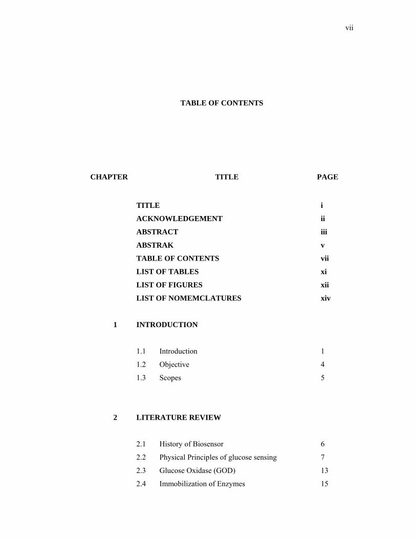
TABLE OF CONTENTS
CHAPTER TITLE PAGE
TITLE i
ACKNOWLEDGEMENT ii
ABSTRACT iii
ABSTRAK v
TABLE OF CONTENTS vii
LIST OF TABLES xi
LIST OF FIGURES xii
LIST OF NOMEMCLATURES xiv
1 INTRODUCTION
1.1 Introduction 1
1.2 Objective 4
1.3 Scopes 5
2 LITERATURE REVIEW
2.1 History of Biosensor 6
2.2 Physical Principles of glucose sensing 7
2.3 Glucose Oxidase (GOD) 13
2.4 Immobilization of Enzymes 15
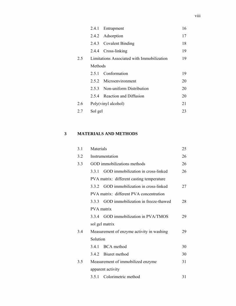
viii
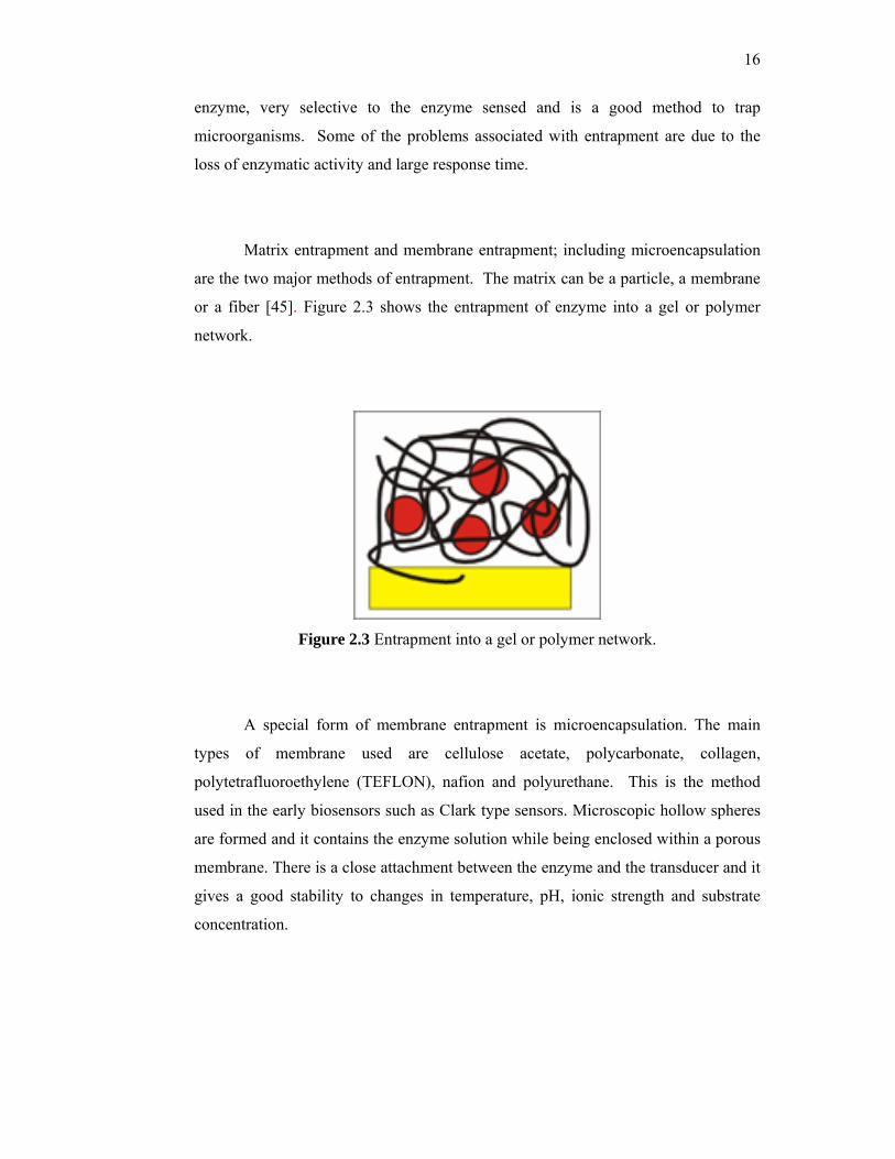
2.4.1 Entrapment 16
2.4.2 Adsorption 17
2.4.3 Covalent Binding 18
2.4.4 Cross-linking 19
2.5 Limitations Associated with Immobilization 19
Methods
2.5.1 Conformation 19
2.5.2 Microenvironment 20
2.5.3 Non-uniform Distribution 20
2.5.4 Reaction and Diffusion 20
2.6 Poly(vinyl alcohol) 21
2.7 Sol gel 23
3 MATERIALS AND METHODS
3.1 Materials 25
3.2 Instrumentation 26
3.3 GOD immobilizations methods 26
3.3.1 GOD immobilization in cross-linked 26
PVA matrix: different casting temperature
3.3.2 GOD immobilization in cross-linked 27
PVA matrix: different PVA concentration
3.3.3 GOD immobilization in freeze-thawed 28
PVA matrix
3.3.4 GOD immobilization in PVA/TMOS 29
sol gel matrix
3.4 Measurement of enzyme activity in washing 29
Solution
3.4.1 BCA method 30
3.4.2 Biuret method 30
3.5 Measurement of immobilized enzyme 31
apparent activity
3.5.1 Colorimetric method 31
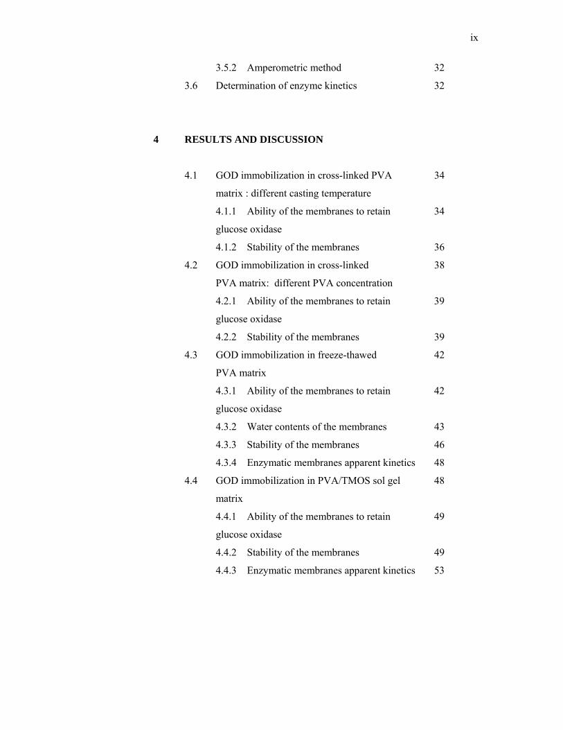
ix
3.5.2 Amperometric method 32
3.6 Determination of enzyme kinetics 32
4 RESULTS AND DISCUSSION
4.1 GOD immobilization in cross-linked PVA 34
matrix : different casting temperature
4.1.1 Ability of the membranes to retain 34
glucose oxidase
4.1.2 Stability of the membranes 36
4.2 GOD immobilization in cross-linked 38
PVA matrix: different PVA concentration
4.2.1 Ability of the membranes to retain 39
glucose oxidase
4.2.2 Stability of the membranes 39
4.3 GOD immobilization in freeze-thawed 42
PVA matrix
4.3.1 Ability of the membranes to retain 42
glucose oxidase
4.3.2 Water contents of the membranes 43
4.3.3 Stability of the membranes 46
4.3.4 Enzymatic membranes apparent kinetics 48
4.4 GOD immobilization in PVA/TMOS sol gel 48
matrix
4.4.1 Ability of the membranes to retain 49
glucose oxidase
4.4.2 Stability of the membranes 49
4.4.3 Enzymatic membranes apparent kinetics 53
x
5 CONCLUSIONS AND RECOMMENDATIONS
5.1 Conclusions 54
5.1.1 GOD immobilization in cross-linked 54
PVA matrix: different casting temperature
5.1.2 GOD immobilization in cross-linked 55
PVA matrix: different PVA concentration
5.1.3 GOD immobilization in freeze-thawed 55
PVA matrix
5.1.4 GOD immobilization in PVA/TMOS 56
sol gel matrix
5.2 Recommendations 57
REFERENCES 59
xi
LIST OF TABLE
TABLE TITLE PAGE
2.1 General characteristics of glucose oxidase 14
xii
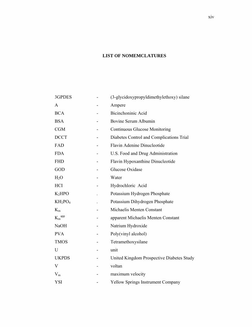
LIST OF FIGURES
FIGURE NO. TITLE PAGE
2.1 Overall topology of glucose oxidase. 13
2.2 Subunit structure of GOD showing FAD (red spacefill). 14
2.3 Entrapment into a gel or polymer network . 16
2.4 Adsorption to the surface. 17
2.5 Covalent linkage to the electrode or a self-assembled 18
monolayer.
4.1 Leaking profiles for PVA-GOD membranes 35
immobilized at 25°C and 4°C.
4.2 Comparison of apparent enzyme activity for PVA-GOD 37
membranes immobilized at 25°C and 4°C.
4.3 Leaking profiles for PVA-GOD membranes immobilized 40
with 10% and 15% of PVA.
4.4 Stability of the PVA-GOD membranes immobilized 41
with 10% and 15% of PVA.
4.5 Leaking profiles for PVA-GOD freeze-thawed membranes 44
prepared using 5% PVA, 10% PVA and 15% PVA.
4.6 The relationship between the concentration of the PVA matrix 45
of the freeze-thawed PVA-GOD membranes and their water
contents.
xiii
4.7 Stability of freeze-thawed PVA-GOD membranes 47
prepared using 5% PVA, 10% PVA and 15% PVA.
4.8 Leaking profile for PVA/TMOS-GOD membranes. 51
4.9 Stability of the PVA/TMOS-GOD membranes. 52
xiv
LIST OF NOMEMCLATURES
3GPDES - (3-glycidoxypropyldimethylethoxy) silane
A - Ampere
BCA - Bicinchoninic Acid
BSA - Bovine Serum Albumin
CGM - Continuous Glucose Monitoring
DCCT - Diabetes Control and Complications Trial
FAD - Flavin Adenine Dinucleotide
FDA - U.S. Food and Drug Administration
FHD - Flavin Hypoxanthine Dinucleotide
GOD - Glucose Oxidase
H2O - Water
HCI - Hydrochloric Acid
K2HPO - Potassium Hydrogen Phosphate
KH2PO4 - Potassium Dihydrogen Phosphate
Km - Michaelis Menten Constant
Kmapp - apparent Michaelis Menten Constant
NaOH - Natrium Hydroxide
PVA - Poly(vinyl alcohol)
TMOS - Tetramethoxysilane
U - unit
UKPDS - United Kingdom Prospective Diabetes Study
V - voltan
Vm - maximum velocity
YSI - Yellow Springs Instrument Company
CHAPTER 1
INTRODUCTION
1.0 Introduction
Diabetes is a disease in which the body loses the ability to regulate the level
of glucose in the blood. In a person without diabetes, the body is able to regulate the
amount of glucose in the blood between 3.5 to 6.5 mM with the help of insulin. In
diabetes, the auto-regulation of glucose fails and the blood glucose level of a diabetic
sufferer may vary between 1 to 30 mM . This can lead to hyperglycemia (too much
glucose) or hypoglycemia (too little glucose). Hyperglycemia can result in long term
damage to organs and hypoglycemia can result in coma or death due to too little
glucose reaching the brain.
Diabetics have to monitor their blood glucose closely in order to remain
healthy and to decrease the risks of serious complications of the disease. The
Diabetes Control and Complications Trial (DCCT), which began in 1983 and
stretched over a period of 10 years, examined the effect of tight regulation of glucose
levels on the complications suffered by type 1 diabetics [1]. The study followed two
groups of over 1441 diabetes sufferers. One group used a standard regime of glucose
2
measurements and insulin shots and the other group maintained tight control over
their blood glucose level by checking their blood glucose levels and injecting
themselves with insulin more frequently. Over time, the group that monitored their
blood glucose levels more closely suffered less complications related to the disease
compared to the other group. A computer simulation that was performed based on
the DCCT results predicted a longer life span and a better quality of life for diabetics
who controlled their blood glucose level tightly. The computer program estimated
that patients who regulate their blood glucose level tightly can expect an additional 5
years of life, 6 years free of amputations, 8 years of sight, and 6 years free from
kidney disease [2]. Another study that was carried out on non-obese type 2 Japanese
diabetic patients showed that intensive insulin therapy prevented the advancement of
microvascular complications, which supported the results of the DCCT [3]. Recently
the results of a new study, the United Kingdom Prospective Diabetes Study
(UKPDS) were released. As in the two previous studies, this study demonstrated
similar effects of an intensive insulin treatment policy on type 2 diabetic patients [4].
The studies on intensive insulin treatment demonstrated that the health of
diabetics depends on their ability to tightly control their blood glucose levels. Fewer
health complications will decrease the cost of diabetes-related healthcare. Home
testing by diabetics will undoubtedly decrease the cost of diabetes even further as
fewer numbers of doctors’ visits will be necessary. Thus, the method of choice for
blood glucose control would be self-determination of one’s own blood glucose level
using a glucose biosensor.
Therefore, it is of no surprise that currently, the development of a successful
glucose biosensor is one of the most financially attractive areas in medical
diagnostics. The various studies have proven that frequent monitoring can reduce the
cost of diabetes-related healthcare. The increase in healthcare costs around the world
necessitates the introduction of cost-effective, diagnostic glucose-testing kits. In
addition, human lifespan in many developed countries has increased, resulting in a
substantial aging population, which leads to a rise in the incidence of type 2 diabetes.
3
These are among the factors behind the need for continuing research in the area of
blood glucose monitoring.
Over the years, a wide range of methods for measuring blood glucose
concentration has been studied. The first ones to materialize were glucose assays
based on glucose oxidase and reagents impregnated in paper strips, which give
diabetics a qualitative indication of their blood glucose level. Then, the paper strips
were combined with a reflectometer [5].
Another method employs the concept of glucose concentration determination
using amperometric techniques. Glucose concentration can be measured by
electrochemically following the decrease in oxygen concentration as the reaction
proceeds [6-7], the production of hydrogen peroxide [8-10] or the change in pH with
the production of D-gluconic acid. The oxygen and hydrogen peroxide based glucose
sensors are the so-called first generation amperometric glucose sensors. Second
generation glucose sensors make use of mediators to shuttle electrons from the
enzyme to the electrode [11-13]. Third generation amperometric glucose sensors are
based on the use of conducting organic salts or polymers. The films are grown
electrochemically and glucose oxidase is entrapped in the membranes. Polypyrrole
[14] and polyaniline [15] have been investigated as potential membrane materials for
the entrapment of the enzyme. To date, however, amperometric glucose biosensors
based on hydrogen peroxide continue to dominate the field of glucose sensor
research due to its simplicity.
An important consideration in the practical application of glucose biosensors
is the operational life of the sensing element. Considerable research effort has been
focused on the method of immobilizing glucose oxidase since the technique
employed may influence the available activity of the enzyme and thus affect the
performance of the sensor. However, narrow measuring range and low current
response, which are a direct consequence of the effectiveness of the immobilized
4
glucose oxidase, are still considered a problem. Most of these problems stem from
conformational changes that occur when the enzyme is being immobilized.
Some of the conventional methods of immobilization that have been
investigated include covalent attachment to a reactive insoluble support, physical
adsorption to a solid surface, physical entrapment in polymeric gels, and cross-
linking with a bifunctional agent such as glutaraldehyde, often in combination with
adsorption or physical entrapment [16]. Newer approaches include the entrapment of
glucose oxidase in electrochemically grown polymeric films such as polyaniline and
polypyrrole [17]. GOD has been immobilized on methacrylate copolymers [18],
polyaniline [19,20], poly(phenol) films [21], poly(vinyl ferrocene) films [22],
ferrocenyl-acrylamide-acrylic acid copolymer films [23], polypyrrole films [24] and
cellulose acetate [25].
In this project, various methods of glucose oxidase immobilization for
peroxide based amperometric glucose biosensor were investigated. Numerous
parameters (such as temperature, concentration, etc.) that can affect the activity of
the enzyme were considered in order to determine the optimized method for GOD
immobilization. The “old school” method of changing one control variable at a time
was employed for the optimization.
1.2 Objective
The objective of this project was to examine various ways to improve the
performance of immobilized glucose oxidase membranes for peroxide based
amperometric glucose biosensors.
5
1.3 Scopes
The scopes for this project were as follows:
a) Immobilization of glucose oxidase using different methods. Various
immobilization parameters were investigated.
b) Determination of the performance of the immobilized glucose oxidase
membrane. Various characteristics of the membrane were investigated.
CHAPTER 2
LITERATURE REVIEW
2.1 History of Biosensor
Biosensor history started in 1962 when American Scientist Leland C. Clark
studied the electrochemistry of oxygen reduction at a platinum metal electrode,
pioneering the use of an oxygen sensor [26]. The electrode was eventually coined a
“Clark Electrode”. Clark then placed entrapped glucose oxidase next to the platinum
electrode and followed the activity of the enzyme by following the changes in
oxygen concentration, thereby turning the chemosensor into a glucose biosensor.
The first step towards commercial exploitation was taken by the Yellow
Springs Instrument Company (YSI) in the 70s. YSI collaborated with Clark to
develop a series of laboratory-scale glucose sensors. A lot of work went into finding
suitable membrane that rendered the GOD-platinum electrode technique reproducible
and accurate.
Today, mass marketing of home-monitoring glucose systems has become a
reality. At present, most of the home-monitoring glucose systems on the market are
enzyme-photometric or amperometric methods and are of the in vitro type. However,
7
the commercial market is changing rapidly. Cygnus GlucoWatch Biographer [27], a
minimally invasive glucose sensing system based on iontophoresis, and the Minimed
Continuous Glucose Monitoring (CGM) system [28], a minimally invasive
transcutaneous glucose sensor, have received U.S. Food and Drug Administration
(FDA) approval.
2.2 Physical Principles of glucose sensing
Over the years, a wide range of methods for measuring blood glucose
concentration has been studied. The first ones to materialize were glucose assays
based on glucose oxidase and reagents impregnated in paper strips. When blood is
placed on the paper strip, hydrogen peroxide that is produced from the enzymatic
reaction oxidizes an oxygen acceptor in the presence of peroxidase to form a color
change. The intensity in the color of the paper strips will give the diabetics a
qualitative indication of their blood glucose level. However, the qualitative nature of
the tests prompted researchers to turn to other approaches that can provide more
quantitative results. One approach is to combine the use of the paper strips with a
reflectometer. When light is shined on the test pad, it will be reflected differently
according to the intensity of the color of the test strip. The reaction reflectance will
be measured electronically and a blood glucose concentration value is displayed.
Systems using absorbance photometry utilize two wavelengths to measure the
reaction reflectance instead of the single wavelength used by most reflectance
photometry systems [5]. In recent versions of the reflectance-based method, apart
from the glucose oxidase-peroxidase-dye optical method, glucose oxidase has also
been coupled with other reagents such as the glucose oxidase-prussian blue method,
glucose oxidase-organic mediator optical method and others. Some systems use
hexokinase instead of glucose oxidase for glucose detection [29].
8
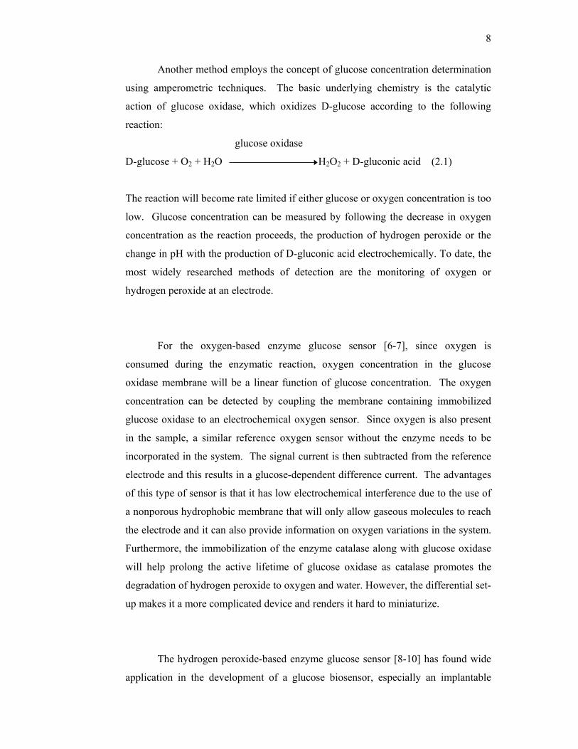
Another method employs the concept of glucose concentration determination
using amperometric techniques. The basic underlying chemistry is the catalytic
action of glucose oxidase, which oxidizes D-glucose according to the following
reaction:
glucose oxidase
D-glucose + O2 + H2O H2O2 + D-gluconic acid (2.1)
The reaction will become rate limited if either glucose or oxygen concentration is too
low. Glucose concentration can be measured by following the decrease in oxygen
concentration as the reaction proceeds, the production of hydrogen peroxide or the
change in pH with the production of D-gluconic acid electrochemically. To date, the
most widely researched methods of detection are the monitoring of oxygen or
hydrogen peroxide at an electrode.
For the oxygen-based enzyme glucose sensor [6-7], since oxygen is
consumed during the enzymatic reaction, oxygen concentration in the glucose
oxidase membrane will be a linear function of glucose concentration. The oxygen
concentration can be detected by coupling the membrane containing immobilized
glucose oxidase to an electrochemical oxygen sensor. Since oxygen is also present
in the sample, a similar reference oxygen sensor without the enzyme needs to be
incorporated in the system. The signal current is then subtracted from the reference
electrode and this results in a glucose-dependent difference current. The advantages
of this type of sensor is that it has low electrochemical interference due to the use of
a nonporous hydrophobic membrane that will only allow gaseous molecules to reach
the electrode and it can also provide information on oxygen variations in the system.
Furthermore, the immobilization of the enzyme catalase along with glucose oxidase
will help prolong the active lifetime of glucose oxidase as catalase promotes the
degradation of hydrogen peroxide to oxygen and water. However, the differential set-
up makes it a more complicated device and renders it hard to miniaturize.
The hydrogen peroxide-based enzyme glucose sensor [8-10] has found wide
application in the development of a glucose biosensor, especially an implantable
9
version, due to its simple sensor configuration that facilitates ease of miniaturization.
Unlike oxygen, hydrogen peroxide is not present in the sample to be analyzed, so no
differential set-up is needed. However, it suffers from an intrinsic problem, the
interference from small endogenous analytes such as urate and ascorbate and some
common drugs such as acetaminophen. These species are electro-active at the
detection potential of hydrogen peroxide.
The interfering effects of most of the endogenous species have been shown to
be effectively eliminated by the use of a number of internal membranes such as
cellulose acetate[30] or Nafion® [31]. Since these membranes impart negative
charges by the presence of residual carboxyl groups or sulphonate groups, the
transport of anionic species such as ascorbate and urate can be easily retarded.
However, since acetaminophen is an uncharged molecule, it can still cause a
considerable bias in the sensor output even with these membranes. A number of
studies have reported that the interference caused by acetaminophen can be reduced
by using composite membranes such as cellulose acetate and Nafion®[32]; and γ-
aminopropyltriethoxysilane , cellulose acetate and Nafion® [33].
The oxygen and hydrogen peroxide based glucose sensors are the so-called
first generation amperometric glucose sensors. Second generation glucose sensors
make use of mediators to shuttle electrons from the enzyme to the electrode [11-13].
This type of system is supposed to eliminate the dependency of the enzymatic
reaction to oxygen. If the system is oxygen deficient, the glucose sensor will become
insensitive to glucose and will only respond to changes in oxygen concentration.
However, as oxygen remains in the system, the mediator must be able to compete
effectively for the electrons. Ferrocene and its derivatives are the best-known
artificial electron carriers.
10
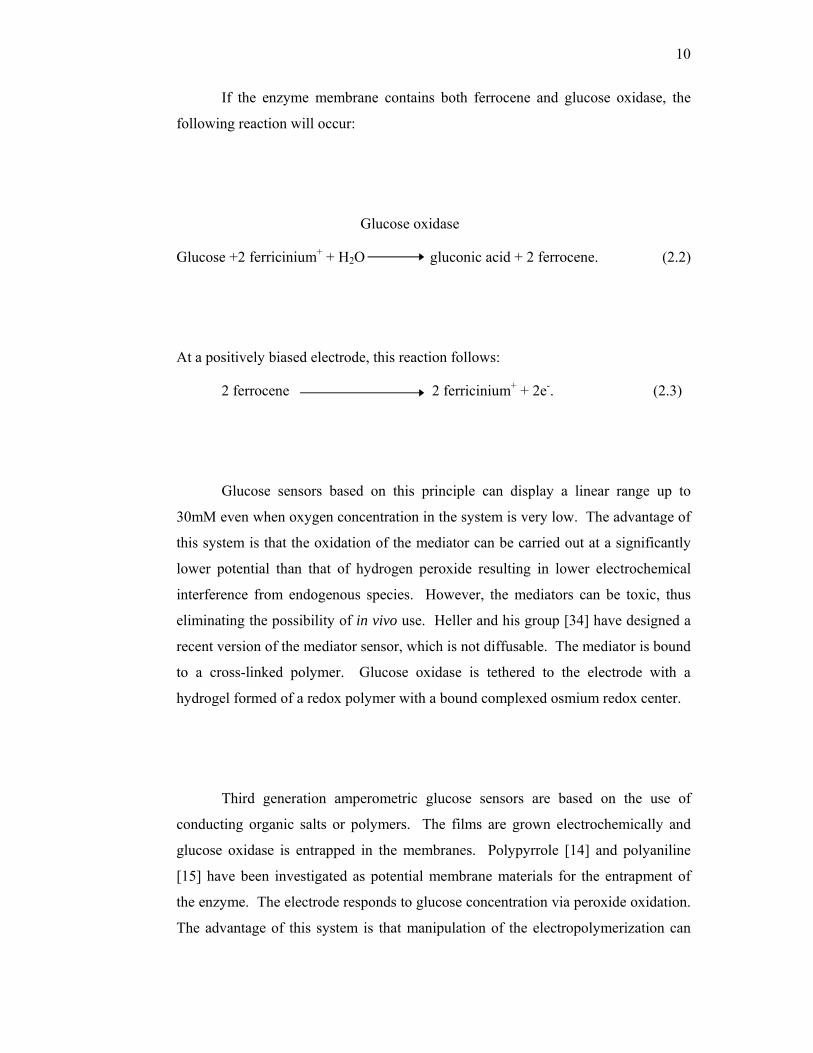
If the enzyme membrane contains both ferrocene and glucose oxidase, the
following reaction will occur:
Glucose oxidase
Glucose +2 ferricinium+ + H2O gluconic acid + 2 ferrocene. (2.2)
At a positively biased electrode, this reaction follows:
2 ferrocene 2 ferricinium+ + 2e-. (2.3)
Glucose sensors based on this principle can display a linear range up to
30mM even when oxygen concentration in the system is very low. The advantage of
this system is that the oxidation of the mediator can be carried out at a significantly
lower potential than that of hydrogen peroxide resulting in lower electrochemical
interference from endogenous species. However, the mediators can be toxic, thus
eliminating the possibility of in vivo use. Heller and his group [34] have designed a
recent version of the mediator sensor, which is not diffusable. The mediator is bound
to a cross-linked polymer. Glucose oxidase is tethered to the electrode with a
hydrogel formed of a redox polymer with a bound complexed osmium redox center.
Third generation amperometric glucose sensors are based on the use of
conducting organic salts or polymers. The films are grown electrochemically and
glucose oxidase is entrapped in the membranes. Polypyrrole [14] and polyaniline
[15] have been investigated as potential membrane materials for the entrapment of
the enzyme. The electrode responds to glucose concentration via peroxide oxidation.
The advantage of this system is that manipulation of the electropolymerization can
11
give a film that extends the linear range for glucose detection and reduces oxygen
dependence.
Another concept that is rapidly gaining interest is the fluorescence-based
optical glucose affinity sensor. Glucose detection is via competitive displacement of
a fluorescently-labeled competing sugar ligand from an immobilized protein with
binding sites for certain carbohydrates. With increasing glucose concentration, the
labeled competing sugar ligand will be displaced from the immobilized protein
causing it to diffuse freely into the volume illuminated by a light source. This will
lead to a glucose concentration-related increase in fluorescence. The concept was
first developed using concanavalin A as the protein and high molecular weight
fluorescein labeled dextran as the competing sugar ligand [35]. As a substitute to the
single fluorophore technique, both the protein and the competing ligand can be
labeled. Meadows, et al. [36] labeled high molecular weight dextran and
concanavalin A with fluorescein isothiocyanate and tetramethylrhodamine
isothocyanate, respectively. Two optical fibers were used with one employed to
detect fluorescein isothiocyanate emission to determine glucose concentration and
the other one employed to detect tetramethylrhodamine isothocyanate emission to
account for drift and changes in configuration.
In recent years, the concept of non-invasive optical glucose sensors has also
garnered attention. Light in the near infrared or other region of the spectrum is
beamed on to a relatively transparent part of the body such as an ear lobe or finger
web [37]. Glucose absorbs near infrared light in the 1000 to 2500 nm region where
skin and tissue are non-absorbing. The light signal is processed by mathematical
filtering techniques to maximize the signal that can correlate with blood glucose
concentration. This concept has also been attempted with the technique of attenuated
total reflectance infrared spectroscopy; however, the penetration depth is much
smaller than that of the near infrared light [38]. Although the concept of an optical
non-invasive sensor is appealing, the lack of adequate selectivity is a major problem.
Other biological species in the area along with tissue structures can interfere with
12
glucose measurement. The systems also suffer from complexity in obtaining
accurate calibration as the correlation between signal and blood glucose can change
with time, thus rendering it useless for real time monitoring.
Several approaches to non-invasive transdermal extraction of tissue fluids
have also been considered as substitutes to blood sampling. Even though this
method does not make use of any new working principle of glucose sensing, the fact
that it can provide an attractive alternative to frequent blood collection makes it
worth mentioning. This method is also termed the minimally-invasive technique of
blood glucose measurement by some. Tissue fluid can be extracted by means of
suction or reverse iontophoresis [39, 40]. In the reverse iontophoresis method, an
externally applied potential is used to promote molecular transport through the skin.
The transdermally-extracted samples can be analyzed using any type of glucose
sensing technique ex vivo. The samples seem to be less aggressive to the sensor than
blood, thus minimizing biofouling. However, there are some points that still need
clarifying such as long term effects of current on skin; lag in response time as fluid
collection might require at least 15 minutes; the possibility of inconsistency in
glucose recovery at different sites and the frequency of sensor recalibration.
The description above is not intended to be a comprehensive summary of all
the physical principles used for glucose determination. Among those that are not
mentioned here are potentiometric glucose electrodes [41], sensors based on
microcalorimetry [42] and others [43, 44].
13
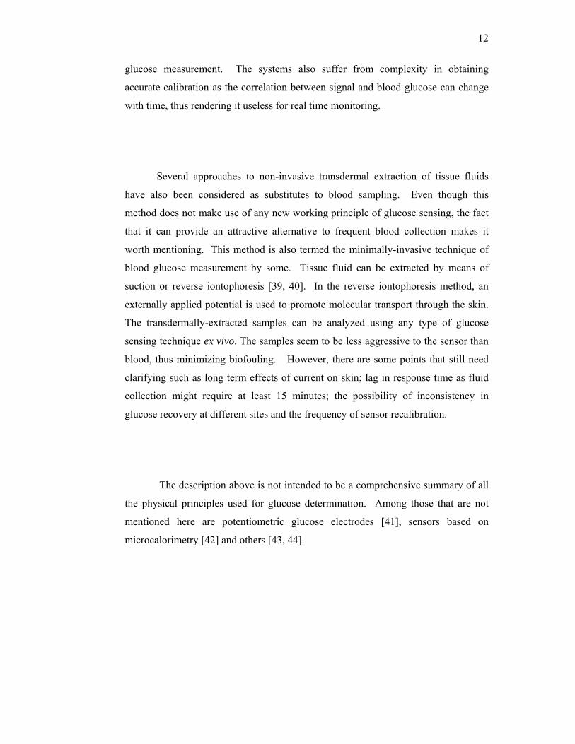
2.3 Glucose Oxidase (GOD)
Various methods of glucose sensing have been discussed in detail in part 2.2.
One popular method involves the use of glucose oxidase (GOD). GOD is a highly
specific enzyme for D-glucose, from the fungi Aspergillus Niger and Penicillium,
which catalyses the oxidation of β-glucose to glucono-1,5-lactone, which
spontaneously hydrolyses in the absence of enzyme to gluconic acid using molecular
oxygen or artificial electron acceptor.
GOD is a dimeric protein as shown in figure 2.1 with a molecular weight of
160, 000 Dalton. It contains one tightly bound flavin adenine dinucleotide (FAD)
per monomer as cofactor which means that each enzyme will have two FAD-sites.
FAD can be released from the protein following partial unfolding of the protein since
it is not covalently bound. FAD in GOD is shown in figure 2.2.
Figure 2.1 Overall topology of Glucose oxidase.
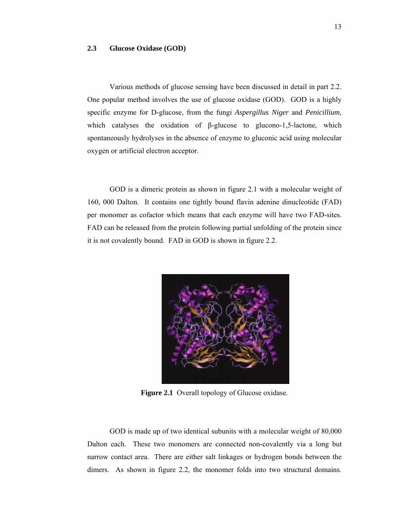
GOD is made up of two identical subunits with a molecular weight of 80,000
Dalton each. These two monomers are connected non-covalently via a long but
narrow contact area. There are either salt linkages or hydrogen bonds between the
dimers. As shown in figure 2.2, the monomer folds into two structural domains.
14
One of the domains involved with substrate binding and the other binds FAD. The
corresponding dimensions of the dimer are 70 Å x 55 Å x 80 Å. Properties of GOD
are shown in table 2.1.
Figure 2.2 Subunit structure of GOD showing FAD (red spacefill)
Table 2.1: General characteristics of glucose oxidase
Characteristics Description
Molecular weight 160, 000 Dalton
Composition
The enzyme consists of two identical polypeptide chain
subunits (80, 000 Dalton) covalently linked by disulfide
bonds. Each subunit contains one mole of Fe and one
mole of FAD. The molecule is to be approximately
74% protein, 16% neutral sugar and 2% amino sugars.
It is indicated that the FAD is replaceable with FHD
(Flavin-Hypoxanthine Dinucleotide) without loss of
activity.
Optimum pH 5 with broad range 4-7
Specificity
The enzyme is highly specific for β-D-glucose. The α
anomer is not acted upon. 2-deoxy-D-glucose, D-
mannose and D-galactose exhibit low activities as
substrate.
15
Inhibitors Ag+, Hg2+, Cu2+. FAD binding is inhibited by several
nucleotides.
Stability
Dry preparations are stable for years when stored cold.
Solutions are reasonably stable under a variety of
conditions.
2.4 Immobilization of Enzymes
Enzyme immobilization is defined as the restriction of enzyme mobility in a
fixed space. In order to make a viable biosensor, the biological component (enzyme)
has to be properly attached to the transducer. Design, preparation procedure and
immobilization procedures is the key to construct a successful biosensor so that
enzymes should be stabilized and easy to be contacted by substrates. Choice of
immobilization technique to immobilize enzyme is extremely important in terms of
biosensor operational stability and long-term use. Methods selected must be
compatible with the enzyme and substrates. Poor technique will result in significant
loss of enzyme activity and thus low sensor response. A critical step in fabrication of
these devices is effective enzyme immobilization while maintaining free diffusion of
the substrates to the enzyme layer.
2.4.1 Entrapment
Entrapment is a physical method to immobilize or physical enclosure of
enzymes in a small space. Enzyme remains free in the solution, but restricted in
movement by the lattice structure of a gel. Pore size of the gel lattice is controlled so
that the structure is tight enough to prevent enzyme leakage while allowing free
movement of substrates and products. This method is applicable to many enzymes,
may provide no or little perturbation of the native structure and function of the
16
enzyme, very selective to the enzyme sensed and is a good method to trap
microorganisms. Some of the problems associated with entrapment are due to the
loss of enzymatic activity and large response time.
Matrix entrapment and membrane entrapment; including microencapsulation
are the two major methods of entrapment. The matrix can be a particle, a membrane
or a fiber [45]. Figure 2.3 shows the entrapment of enzyme into a gel or polymer
network.
Figure 2.3 Entrapment into a gel or polymer network.
A special form of membrane entrapment is microencapsulation. The main
types of membrane used are cellulose acetate, polycarbonate, collagen,
polytetrafluoroethylene (TEFLON), nafion and polyurethane. This is the method
used in the early biosensors such as Clark type sensors. Microscopic hollow spheres
are formed and it contains the enzyme solution while being enclosed within a porous
membrane. There is a close attachment between the enzyme and the transducer and it
gives a good stability to changes in temperature, pH, ionic strength and substrate
concentration.
17
2.4.2 Adsorption
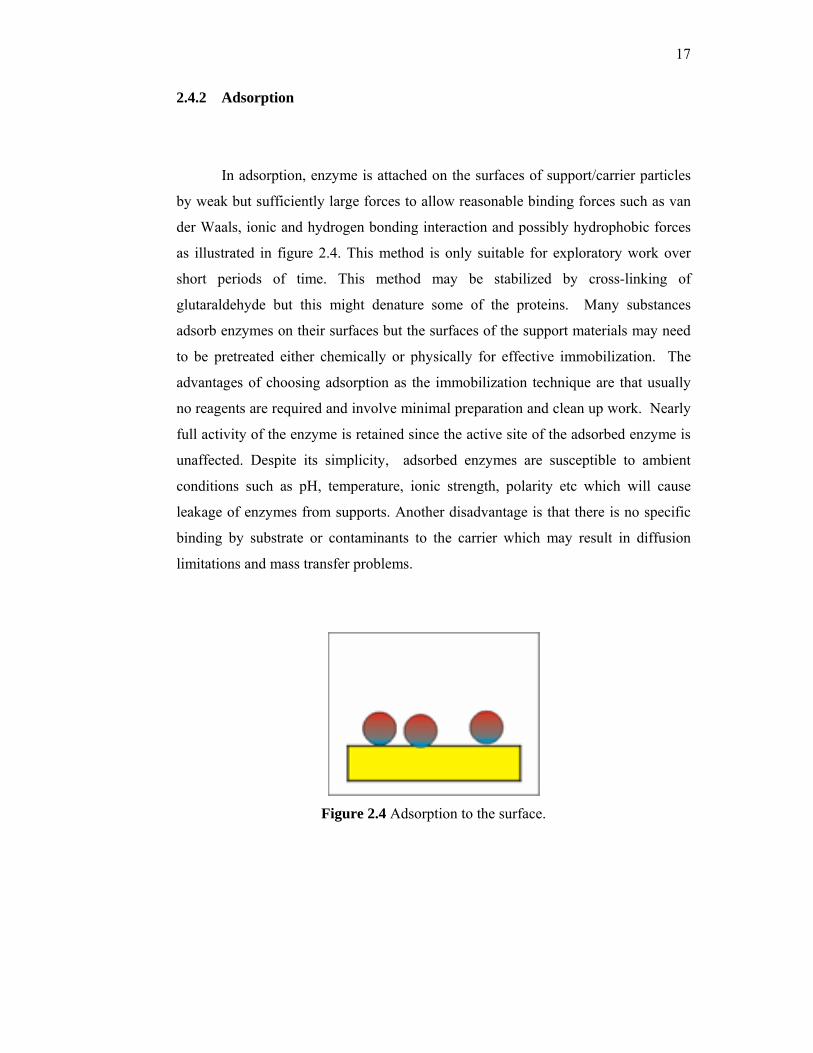
In adsorption, enzyme is attached on the surfaces of support/carrier particles
by weak but sufficiently large forces to allow reasonable binding forces such as van
der Waals, ionic and hydrogen bonding interaction and possibly hydrophobic forces
as illustrated in figure 2.4. This method is only suitable for exploratory work over
short periods of time. This method may be stabilized by cross-linking of
glutaraldehyde but this might denature some of the proteins. Many substances
adsorb enzymes on their surfaces but the surfaces of the support materials may need
to be pretreated either chemically or physically for effective immobilization. The
advantages of choosing adsorption as the immobilization technique are that usually
no reagents are required and involve minimal preparation and clean up work. Nearly
full activity of the enzyme is retained since the active site of the adsorbed enzyme is
unaffected. Despite its simplicity, adsorbed enzymes are susceptible to ambient
conditions such as pH, temperature, ionic strength, polarity etc which will cause
leakage of enzymes from supports. Another disadvantage is that there is no specific
binding by substrate or contaminants to the carrier which may result in diffusion
limitations and mass transfer problems.
Figure 2.4 Adsorption to the surface.
18
2.4.3 Covalent Binding
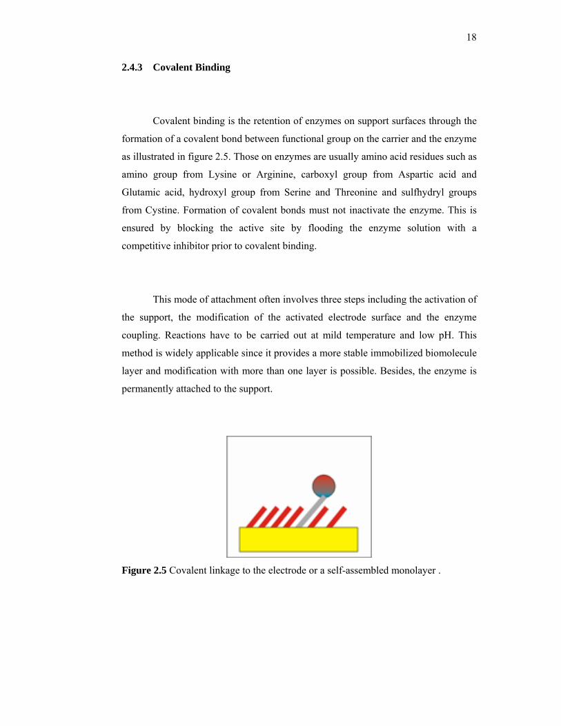
Covalent binding is the retention of enzymes on support surfaces through the
formation of a covalent bond between functional group on the carrier and the enzyme
as illustrated in figure 2.5. Those on enzymes are usually amino acid residues such as
amino group from Lysine or Arginine, carboxyl group from Aspartic acid and
Glutamic acid, hydroxyl group from Serine and Threonine and sulfhydryl groups
from Cystine. Formation of covalent bonds must not inactivate the enzyme. This is
ensured by blocking the active site by flooding the enzyme solution with a
competitive inhibitor prior to covalent binding.
This mode of attachment often involves three steps including the activation of
the support, the modification of the activated electrode surface and the enzyme
coupling. Reactions have to be carried out at mild temperature and low pH. This
method is widely applicable since it provides a more stable immobilized biomolecule
layer and modification with more than one layer is possible. Besides, the enzyme is
permanently attached to the support.
Figure 2.5 Covalent linkage to the electrode or a self-assembled monolayer .
19
2.4.4 Cross-linking
This method joins the enzyme to each other or to another supporting material
to form a large 3D structure. This can be done by using bi-functional agents such as
gluteraldehyde, bis-diazobenzidine and 2,2-disulfonic acid. In cross-linking,
enzymes can be cross-linked with glutaraldehyde to form an insoluble aggregate,
adsorbed enzymes may be cross-linked or cross-linking may take place following the
impregnation of porous support material with enzyme solution. Cross-linking is a
useful method to stabilize adsorbed enzyme. It may cause significant changes in the
active site of enzymes and thus, tests must be done to ensure that the active site
remains free and available for catalytic activity. It may result in severe diffusion
limitations and poor mechanical strength or rigidity.
2.5 Limitations Associated with Immobilization Methods
With immobilized enzymes, reaction rates depend not only on substrate
concentration and kinetic constants but also on immobilization effects [46]. These
effects are due to the alteration of the enzyme by the immobilization process.
2.5.1 Conformation
Conformation changes of the enzyme caused by immobilization usually
decrease the affinity to the subtrate (increase Michaeles constant, Km). Furthermore,
a partial inactivation of all, or the complete inactivation of a part of the enzyme
molecules may occur (decrease of maximum velocity, Vm).
20
2.5.2 Microenvironment
Ionic, hydrophobic or other interaction between the enzyme and the matrix
(microenvironmental effects) may also result in changed Km and Vm values. These
essentially reversible effects are caused by variations in the disassociation equilibria
of charged groups of the active center.
2.5.3 Non-uniform Distribution
A non-uniform distribution of substrate and/or product between the enzyme
matrix and the surrounding solution affects the measured (apparent) kinetic constants.
2.5.4 Reaction and Diffusion
In biosensors, the enzyme reaction proceeds in a layer separated from the
measuring solution. The substrate reaches the membrane system of the biosensor by
convective diffusion from the solution. The rate of this external transport process
depends essentially on the degree of mixing. In the multilayer system in front of the
sensor, the substrates and products transferred by diffusion. Slow mass transfer to
and within the enzyme matrix leads to different concentrations of the reaction
partners in the measuring solution and in the matrix. Diffusion and the enzyme
reaction do not proceed independently of one another. They are coupled in a
complex manner.
21
2.6 Poly(vinyl alcohol)
Poly(vinyl alcohol) (PVA) is manufactured by the hydrolysis of polyvinyl
acetate. Even after a prolonged hydrolysis, PVA generally retains around 1 to 2
mole percent of acetate groups. The amount of residual acetate groups affects the
physical and chemical properties of PVA, as they are hydrophobic relative to the
hydroxyl groups. The residual acetate groups can interfere with inter-molecular and
intra-molecular forces such as hydrogen bonding. Highly hydrolyzed PVA has
strong hydrogen bonds within and between molecules [47].
PVA, unlike many polymers, is soluble in water. However, owing to strong
internal hydrogen bond, it only goes into solution at higher temperatures, around
900C. Aqueous solutions of PVA exhibit non-Newtonian behavior at room
temperature [48]. PVA can be cross-linked chemically or physically to form a
hydrogel. Chemically, the polymer can be cross-linked by any bifunctional agent
that can react with organic hydroxyl groups. Some of the various chemicals that can
cross-link PVA are glutaraldehyde, formaldehyde, maleic acid and boric acid. Cross-
linking can also be achieved physically using ultraviolet light in the presence of
photo-sensitizers, by electron beam or by γ-radiation. Another physical method of
cross-linking PVA is through freeze thaw cycles, where physical bonds are formed
[49].
Cross-linking causes PVA to be insoluble in water. Furthermore, by
controlling the cross-link density of this material, a variety of transport properties can
be obtained. In addition, poly(vinyl alcohol) is also considered biocompatible.
Protein adsorption onto cross-linked PVA has been shown to be negligible, thus the
potential of minimizing fibrotic capsule development around implantable PVA exists
[50]. Hence, due to its promised biocompatibility, ease of manipulation and
hydrophilicity, PVA has been used extensively in biomedical applications. In the
biomedical area, PVA has found use in hernia treatment [51], in artificial heart valve
22
replacement [52] and as a drug carrier in controlled drug release system [53], among
others.
In dense hydrogels, diffusion of solutes is determined by the cross-linking
density of the network, or mesh size, and the degree of swelling. The “pores” are
the open spaces between the cross-link points and are not fixed. Different theories
have been proposed to describe solute diffusion in hydrogels. Polymer chains are
hypothesized to hinder solute diffusion through one or a combination of these
methods: by physically obstructing the passage of solute, by increasing the
hydrodynamic drag on the solute molecule or by reducing the available free volume
for the solute [54].
Transport properties of cross-linked of PVA has been studied extensively.
Korsmeyer and Peppas [55] examined the correlation between the degree of cross-
linking and hydration, on the diffusion of drugs in PVA hydrogels ; Reinhart and
Peppas [56] studied the influence of the degree of cross-linking of PVA hydrogels on
the diffusion of bovine serum albumin (BSA) ; Li and Barbari [57, 58] investigated
protein transport through surface modified poly(vinyl alcohol) hydrogels; Dai and
Barbari [59, 60, 61] studied the transport properties of poly(vinyl alcohol) with mesh
size asymmetry based on gradient cross-linking with glutaraldehyde and examined
the possibility of using the modified PVA for cell encapsulation or bioseparations;
and Hideto et al. [62] analyzed the diffusion of solutes with molecular weights
ranging from 180-66,000 in cross-linked PVA with free volume theory. The various
properties of PVA have made them suitable for enzyme immobilization.
23
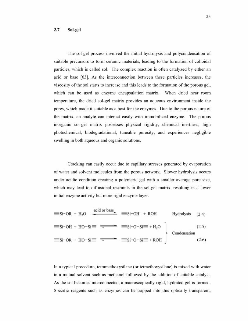
2.7 Sol-gel
The sol-gel process involved the initial hydrolysis and polycondensation of
suitable precursors to form ceramic materials, leading to the formation of colloidal
particles, which is called sol. The complex reaction is often catalyzed by either an
acid or base [63]. As the interconnection between these particles increases, the
viscosity of the sol starts to increase and this leads to the formation of the porous gel,
which can be used as enzyme encapsulation matrix. When dried near room
temperature, the dried sol-gel matrix provides an aqueous environment inside the
pores, which made it suitable as a host for the enzymes. Due to the porous nature of
the matrix, an analyte can interact easily with immobilized enzyme. The porous
inorganic sol-gel matrix possesses physical rigidity, chemical inertness, high
photochemical, biodegradational, tuneable porosity, and experiences negligible
swelling in both aqueous and organic solutions.
Cracking can easily occur due to capillary stresses generated by evaporation
of water and solvent molecules from the porous network. Slower hydrolysis occurs
under acidic condition creating a polymeric gel with a smaller average pore size,
which may lead to diffusional restraints in the sol-gel matrix, resulting in a lower
initial enzyme activity but more rigid enzyme layer.
In a typical procedure, tetramethoxysilane (or tetraethoxysilane) is mixed with water
in a mutual solvent such as methanol followed by the addition of suitable catalyst.
As the sol becomes interconnected, a macroscopically rigid, hydrated gel is formed.
Specific reagents such as enzymes can be trapped into this optically transparent,
(2.4)
(2.6)
(2.5)
24
stable host matrix by simply adding them to the sol prior to its gelation. Such
molecules become entrapped in the growing covalent gel network rather than being
chemically bound to the water-rich inorganic matrix, so that enzyme activity can be
maintained. These materials have been used in numerous applications including
solid-state electrochemical devices, chemical sensors, catalysts, and nonlinear and
optic applications.
Ratio of TMOS and water of the stock sol-gel solution is one of the most
significant process parameters for controlling the pore size of the matrix. An R value,
which is the water/alkoxide ratio, of 1:3.7 was seen to be optimal [64]. Higher R
value causes increase in the rate of hydrolysis resulting in a more particulate gel.
Different alkoxides may give different properties to the resulted sol-gel
matrix. Two types of alkoxides are usually applied, the silica alkoxide,
tetramethoxysilane (TMOS) (C4H12O4Si), and metal alkoxides, alumina (aluminium
isopropoxide)(Al[OCH(CH3)2]3). .
CHAPTER 3
MATERIALS AND METHODS
3.1 Materials
Glucose oxidase (EC 1.1.3.4, type X-S, 157,500 units/g solid) from
Aspergillus niger and horseradish peroxidase (EC 1.11.1.7, type VI from horseradish,
330 purpurogallin units/mg solid) were purchased from Sigma.(3-
glycidoxypropyldimethylethoxy)silane was purchased from Fluka. Poly (vinyl
alcohol) with average molecular weight of 70,000 – 100,000, O-dianisidine tablets
(10 mg substrate/tablet) and glutaraldehyde (1.2% w/v aqueous solution) were
obtained from Sigma Chemical Co. Tetramethoxysilane (TMOS), sodium potassium
tartrate tetrahydrate, D-glucose monohydrate, copper sulfate pentahydrate, sodium
hydroxide, potassium dihydrogen phosphate (KH2PO4) and potassium hydrogen
phosphate (K2HPO4) were obtained from MERCK. All chemicals were used as
received.
26
3.2 Instrumentation
Electrochemical measurements were carried out using a Metrohm µ Autolab
Type 111 potentiostat. A conventional three-electrode electrochemical cell was
employed. A platinum electrode was used as the working electrode (WE), a
platinum auxiliary electrode was used as the counter electrode (CE) and a
Ag/AgCI/KCI was employed as the reference electrode (RE). Colorimetric
measurements were done using a Hitachi V-100 UV spectrophotometer.
3.3 GOD immobilizations methods
3.3.1 GOD immobilization in cross-linked PVA matrix: different casting
temperature
25µL of 10 wt% aqueous PVA solution was mixed with a cross-linking
solution, which consisted of 15µL 10% v/v acetic acid (buffer), 10µL 50% v/v
methanol (quencher), 5µL 10% v/v sulfuric acid (catalyst) and 28µL of 1.2 % w/v
glutaraldehyde, so that it ended up with a cross-linking ratio (CR) of 0.06. Cross-
linking ratio is defined as the ratio of the moles of glutaraldehyde per mole of PVA
repeat unit.
36µL of the solution was added to 6µL GOD solution (200 mg/mL). An
aliquot of the mixture was pipetted quickly onto a glass slide, air-dried for 20
minutes and covered with another glass slide. The two glass slides were clamp
together, to prevent the membrane from contracting, and left for either 2 days at 4°C
27
or 24 hours at 25°C. Membrane thickness was controlled with aluminium spacer
tape where 3 layers of aluminium spacer tape were stuck on the first glass slide.
Following the polymerization, the slides were soaked in 35mL of phosphate
buffer, pH 6.0 for a prescribed amount of time at room temperature in a petri dish.
The slides were unclamped carefully and the layer was gently stripped from the slide.
The washing solutions were collected for enzyme assay purposes and the membranes
were swollen in 5mL of phosphate buffer at 4°C. The buffer was changed every 6
hours for the first day, every 12 hours for the second day, and 24 hours thereafter,
and analyzed for any sign of enzyme activity.
3.3.2 GOD immobilization in cross-linked PVA matrix: different PVA
concentration
25µL of either 10 wt% or 15 wt% aqueous PVA solution was mixed with a
cross-linking solution, which consisted of 15µL 10% v/v acetic acid (buffer), 10µL
50% v/v methanol (quencher), 5µL 10% v/v sulfuric acid (catalyst) and 28µL of
1.2 % w/v glutaraldehyde, so that it ended up with a cross-linking ratio (CR) of 0.06.
Cross-linking ratio is defined as the ratio of the moles of glutaraldehyde per mole of
PVA repeat unit.
36µL of the solution was added to 6µL GOD solution (200 mg/mL). An
aliquot of the mixture was pipetted quickly onto a glass slide, air-dried for 20
minutes and covered with another glass slide. The two glass slides were clamp
together, to prevent the membrane from contracting, and left for 24 hours at 25°C.
Membrane thickness was controlled with aluminium spacer tape where 3 layers of
aluminium spacer tape were stuck on the first glass slide.
28
Following the polymerization, the slides were soaked in 35mL of phosphate
buffer, pH 6.0 for a prescribed amount of time at room temperature in a petri dish.
The slides were unclamped carefully and the layer was gently stripped from the slide.
The washing solutions were collected for enzyme assay purposes and the membranes
were swollen in 5mL of phosphate buffer at 4°C. The buffer was changed every 6
hours for the first day, every 12 hours for the second day, and 24 hours thereafter,
and analyzed for any sign of enzyme activity.
3.3.3 GOD immobilization in freeze-thawed PVA matrix
60µL of PVA solution (5%, 10% or 15% w/v) was added to 10µL of 280
mg/ml GOD solution. An aliquot of the mixture was pipetted quickly onto a polymer
block and covered with another polycarbonate block. The two blocks were clamped
together in order to prevent the membrane from contracting. The membrane was
then left to freeze at -200C for 12 hours. After freezing the membrane was thawed at
40C for 12 hours. The process was repeated 5 times. The membrane thickness was
controlled with aluminum spacer tapes.
After the freezing and thawing process, the blocks were soaked in 40 ml of
phosphate buffer, pH 6.0 at room temperature. The blocks were slowly unclamped
and the membrane was gently stripped from the block. 1mL of the washing solution
was collected for enzyme assay purposes. The buffer was changed every 6 hours for
the first day, every 12 hours for the second day and 24 hours thereafter and analyzed
for any sign of enzyme activity.
29
3.3.4 GOD immobilization in PVA/TMOS sol gel matrix
TMOS, (3-glycidoxypropyldimethylethoxy)silane, H2O, methanol and
57.5µL of HCI were mixed together, in a volume ratio of 1 : x : 3.7 : 3.7 : 0.0013,
where x was 1, 2 and 3 respectively. The solution was stirred for 30 minutes. 115 µL
of HCI was later added and the sol gel solution was stirred for 1 hour. 25µL of PVA
solution was mixed with 100µL of the sol gel solution. 90µL of the solution was then
added to 15µL of 140 mg/ml GOD solution. An aliquot of the mixture was pipetted
quickly onto a glass slide. The membrane was air-dried for 20 minutes and then
covered with another glass slide. The two glass slides were clamped together to
prevent the membrane from contacting and left for 24 hours at 25ºC. Membrane
thickness was controlled with aluminium spacer tapes.
After 24 hours, the slides were soaked in 25mL phosphate buffer solution in a
petri dish at 25ºC. The slides were carefully unclamped and the membrane layer was
gently stripped from the slide. The membrane was swollen in 5mL phosphate buffer
solution and kept at 4ºC. 1mL of the washing solution was collected for enzyme
assay purposes. The buffer was changed every 6 hours for the first day, every 12
hours for the second day and 24 hours thereafter and analyzed for any sign of
enzyme activity.
3.4 Measurement of enzyme activity in washing solution
Enzyme activity in the washing solution was measured using either BCA
method or Biuret method.
30
3.4.1 BCA method
A Standard working solution (SWR) was prepared by mixing reagent A (0.04
g/mL CuSO4.5H2O) and reagent B (0.01 g/mL BCA, 0.02 g/mL Na2CO3.H2O,
0.0016 g/mL Na2C4H4O6.H2O, 0.004 g/mL NaOH and 0.0095 g/mL NaHCO3) in a
ratio of 50:1.
Bovine Serum Albumin (BSA) was used as the protein standards. The
concentrations used were from 0 mg/mL – 1.0 mg/mL. 2 mL of SWR was added into
each test tubes that contained 0.1 mL of either the BSA solution or the washing
solution. The tubes were incubated at 370C for 10 minutes. The absorbance for the
standards and unknowns were recorded at 450 nm.
3.4.2 Biuret method
6.0g sodium potassium tartrate tetrahydrate (NaKC4H4O6.4H20) was
dissolved in 500mL distilled water. 1.5g of copper sulfate pentahydrate
(CuSO4.5H2O) was then added and dissolved into the solution. After that, 300mL of
10%w/v sodium hydroxide (NaOH) was added slowly with stirring. Finally, the
solution was diluted to 1L with distilled water. The solution was stirred until
homogenous. The Biuret reagent was stored in a Schott® bottle covered with
aluminium foil and kept in shaded place for subsequent use due to its sensitivity to
light.
Bovine Serum Albumin (BSA) was used as the protein standards. The
concentrations used were from 0 mg/mL – 12.5mg/mL. For the protein assay, 3 mL
of biuret reagent was added into each test tube that contained 2 mL of either BSA
31
solution or the washing solution. Then the tubes were incubated at 37 ºC in a water
bath for 15 minutes. The absorbance of each solution was measured against a blank
(biuret solution without BSA) at a wavelength of 550nm.
3.5 Measurement of immobilized enzyme apparent activity
Immobilized enzyme apparent activity was measured using either
colorimetric method or amperometric method.
3.5.1 Colorimetric method
The chromogen solution was prepared by diluting 0.1mL of 1% w/v O-
dianisidine in 12mL of phosphate buffer at pH 6.0. 450µL of 25%w/v of aqueous
glucose solution (allowed to mutarotate overnight) and 150µL of 200µg/mL
peroxidase were added to 3.75mL of the chromogen solution. The mixture was
incubated at room temperature for temperature equilibration. For enzyme activity
calibration curve, 150µL of GOD with the activity ranging from 0 to100 mU/mL was
mixed with the mixture. For apparent immobilized enzyme activity determination,
the membrane was dipped in a 5mL universal bottle and the solution was stirred. For
both sets of experiments, reaction was allowed to proceed at room temperature for 10
minutes. 300µL of 4M HCl was added to stop the reaction. However, for the
apparent enzyme activity assay, the membrane was removed prior to the addition of
the acid. The amount of colour formed was measured by reading the absorbance
value at 450nm. Apparent enzyme activity was determined using a standard
32
calibration curve. One unit of activity causes the oxidation of one micromole of O-
dianisidine per minute at 25°C and pH 6.0 under the conditions specified.
3.5.2 Amperometric method
For amperometric experiments, a conventional three-electrode
electrochemical cell was employed. A platinum electrode was used as the working
electrode (WE), a platinum auxiliary electrode was used as the counter electrode
(CE) and a Ag/AgCI was employed as the reference electrode (RE).
The enzymatic membrane was attached to the surface of the working
electrode. Before used, the electrode was rinsed with deionized water and immersed
in 15mL of phosphate buffer with pH 7.0. Voltage at 0.7 V vs Ag/AgCl was applied
to the system. The electrochemical response was let to stabilize. Freshly prepared
enzymatic membranes could attain a stable electrochemical response after 5 to 10
minutes of rinsing. After the current had stabilized, a prescribed amount of stock
glucose solution was injected into the cell to make up a 5 mM glucose solution. The
change in current is proportional to the apparent enzyme activity immobilized in the
membrane. The amperometric studies were performed at 25oC.
3.6 Determination of enzyme kinetics
The enzymatic membrane was attached to the surface of the working
electrode. Before used, the electrode was rinsed with deionized water and immersed
33
in 15mL of phosphate buffer with pH 7.0. Voltage at 0.7 V vs Ag/AgCl was applied
to the system. The electrochemical response was let to stabilize. Prescribed amount
of stock glucose solutions were injected sequentially to the electrochemical cell to
make up a variety of glucose solution concentrations. Upon each addition of glucose
solution, the current was let to stabilize before the next glucose injection. All
experiments were performed at a temperature of 25ºC. Lineweaver-Burke Plots were
plotted and the Km and Vmax of the immobilized enzyme were obtained from the
slope and intercept of the graph.
CHAPTER 4
RESULTS AND DISCUSSIONS
4.1 GOD immobilization in cross-linked PVA matrix : different casting
temperature
For this part of the research, the immobilization method investigated was
immobilization of glucose oxidase in cross-linked PVA matrix. Two different
casting temperatures were studied, which were 4oC and 25oC to investigate the
effects of casting temperature on the effectiveness of the enzyme immobilization
procedure.
4.1.1 Ability of the membranes to retain glucose oxidase
In order to investigate the effectiveness of the immobilization method, which
in this case was entrapment by cross-linked PVA at 0.06 cross-linking ratio and two
different casting temperatures namely 4oC and 25oC, protein contents of the washing
solutions were determined. The washing solution (phosphate buffer) was changed

35
every 6 hours for the first 24 hours and every 12 hours thereafter. Biuret method
was used to determine the total protein. The protein concentration determined from
the experiment was used as an indicator for enzyme leakage, which led to the
indication of the ability of the PVA-GOD membrane in retaining the glucose
oxidase. Figure 4.1 shows that the total protein concentrations of the washing
solutions for the PVA-GOD membranes demonstrate a declining profile for the
whole period of investigation both for membranes immobilized at temperature 25°C
and 4°C. Within 36 hours, the protein concentrations have reached zero. No sign of
protein, and hence glucose oxidase, was in the washing solution. This indicates that
the immobilization method was effective enough in immobilizing glucose oxidase in
cross-linked PVA membranes.
0.0000
0.0100
0.0200
0.0300
0.0400
0.0500
0.0600
0.0700
0.0800
0.0900
0.1000
0 5 10 15 20 25 30 35 40
Time(h)
Prot
ein
Con
cent
ratio
n(m
g/m
L)
25 degree Celcius
4 degree Celsius
Figure 4.1 Leaking profiles for PVA-GOD membranes immobilized at 25°C and
4°C.
36
As a rough comparison between the PVA-GOD membranes immobilized at
25°C and 4°C, the enzyme leakage of the membranes immobilized at 25°C was
higher during the initial investigation period compared to membranes immobilized at
4°C. However, the membranes immobilized at 25°C stopped leaking earlier than the
membrane immobilized at 4°C.
4.1.2 Stability of the membranes
The apparent activity of the immobilized glucose oxidase immobilized in
cross-linked PVA was determined using colourimetrical enzyme assay based on the
oxidation of o-dianisidine through a peroxidase-coupled system. The dye produced
from the reaction resulted in the colour intensity of the assay solution, which was
determined photometrically at 450nm [65]. Furthermore, the stability of the
repeated-use PVA-GOD membranes was examined as well as the decay of apparent
enzyme activity over time and limited lifetime of the enzyme layer of a biosensor
have been reported [66]. The enzyme activity of the membranes was tested within
25 storage days. The membranes were prepared and stored in phosphate buffer at 4
°C. The first enzyme assay was carried out on membranes that had been stored for 3
days.
Figure 4.2 shows the apparent enzyme activity of the membrane immobilized
at 25°C and 4oC. From the graph, it can be observed that for both types of
membranes, the activity of the enzyme increased from day 3 to about day 10 before
becoming rather stable from day 10 to day 25. This increase might be due to
diffusion problems as the membranes, which are hydrogels, might not still be at their
equilibrium states in terms of water content. The diffusion property of a membrane
can be examined by determining the water content of the PVA-GOD membrane.
From the water content, the mesh size or the cross-linking density can be estimated.
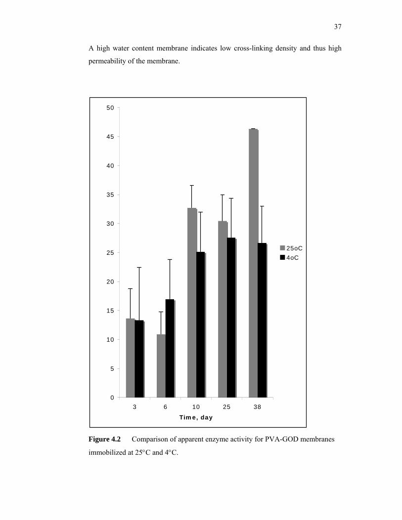
37
A high water content membrane indicates low cross-linking density and thus high
permeability of the membrane.
0
5
10
15
20
25
30
35
40
45
50
3 6 10 25 38
Time, day
25oC
4oC
Figure 4.2 Comparison of apparent enzyme activity for PVA-GOD membranes
immobilized at 25°C and 4°C.
38
The reason the membranes were immobilized at lower temperature was to
reduce the exposure of the enzyme to high temperature in order to preserve the
enzyme activity. Longer exposure of enzyme to high temperature will denature the
enzyme and reduce the enzyme lifetime. From day 10 to day 25, it was observed
that the enzyme activity of the membrane at temperature 25°C was higher than the
membrane immobilized at 4°C, although the opposite was expected. This might due
to insufficient time for the polymerization process at the lower temperature even
though the membranes formed at lower temperature were allowed to polymerize for
an additional 12 hours compared to the membranes immobilized at 25°C. The
different polymerization procedures might have resulted in membranes with
different water contents thus the membranes might have different cross-linking
ratios and thus different properties.
4.2 GOD immobilization in cross-linked PVA matrix: different PVA
concentration
For this part of the research, the immobilization method investigated was
immobilization of glucose oxidase in cross-linked PVA matrix. Two different
concentrations of PVA we were studied, which were 10% PVA and 15% PVA to
investigate the effects of matrix concentrations on the effectiveness of the enzyme
immobilization procedure.
39
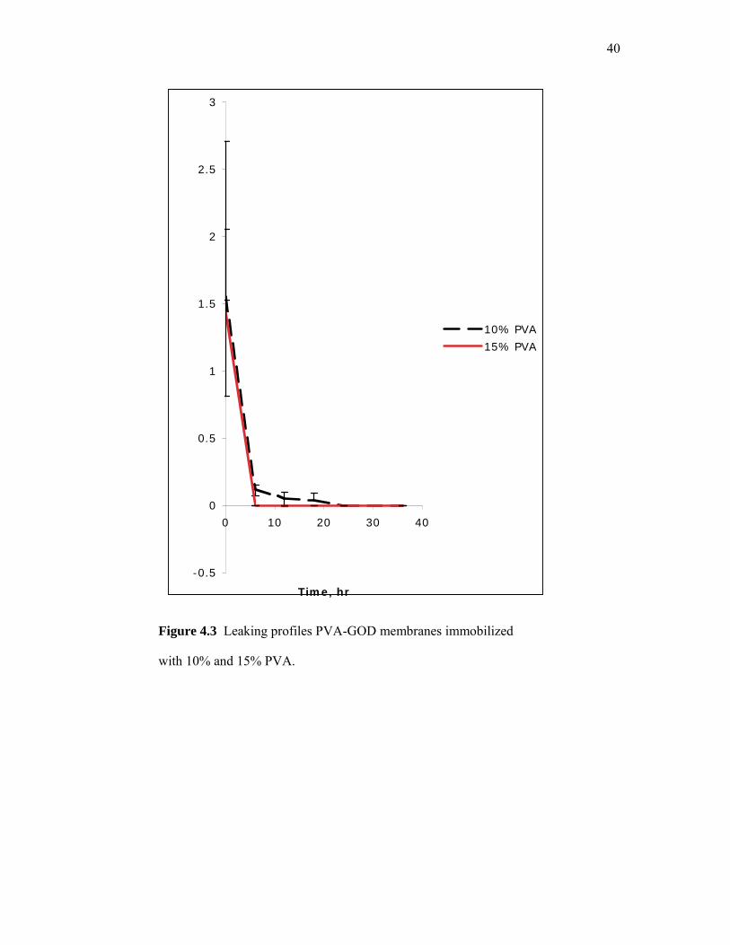
4.2.1 Ability of the membranes to retain glucose oxidase
For both types of membrane, 10% and 15% PVA matrix, the cross-linking
ratios were 0.06. As indicated earlier, after an enzyme immobilization process, the
enzymes that were not properly immobilized will leak out from the membrane.
Usually, immobilization process that employs chemical cross-linking agents will
result in membranes with comparatively short leaking period. Figure 4.3 shows that
leaking profile for GOD immobilized in 10% and 15% PVA at the cross-linking ratio
of 0.06. The period of leaking was shorter if higher concentration of PVA (15%)
was used. This suggests that with higher concentration of PVA, more matrices were
available to entrap the enzyme, thus suppressing the leaking more effectively.
The total amount of the equivalent protein leaked from enzymatic membranes
prepared using 10% PVA was 20% higher than the amount leaked from enzymatic
membranes prepared using 15% PVA. Its period of leaking was also 18 hours longer
respectively.
4.2.2 Stability of the membranes
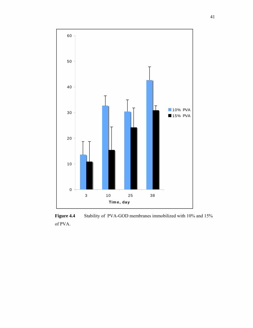
Figure 4.4 shows that the apparent activities of the immobilized enzyme
became stable at around day 10. As explained earlier in 4.2 the increase in activity
from day 3 to day 10 might be due to hydration equilibrium problems. Enzymatic
membranes prepared using 10% PVA showed approximately 33% higher activity
than the ones prepared using 15% PVA. This might be because even though the
15% PVA matrix was more effective in retaining the enzyme, the tighter matrix may
impose greater diffusional barrier towards glucose and hydrogen peroxide diffusion
and thus limiting the enzymatic membrane response.
40
-0.5
0
0.5
1
1.5
2
2.5
3
0 10 20 30 40
Time, hr
10% PVA
15% PVA
Figure 4.3 Leaking profiles PVA-GOD membranes immobilized
with 10% and 15% PVA.
41
0
10
20
30
40
50
60
3 10 25 38
Time, day
10% PVA
15% PVA
Figure 4.4 Stability of PVA-GOD membranes immobilized with 10% and 15%
of PVA.
42
4.3 GOD immobilization in freeze-thawed PVA matrix
For this part of the research, the immobilization method investigated was
immobilization of glucose oxidase in freeze-thawed PVA matrix. Compared to the
two previous methods, this procedure is a physical method. Three different matrix
concentrations were studied, which were 5% PVA, 10% PVA and 15% PVA to
investigate the effects of matrix concentrations on the effectiveness of the enzyme
immobilization procedure.
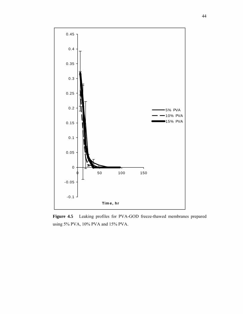
4.3.1 Ability of the membranes to retain glucose oxidase
BCA method, which is more sensitive than Lowry method, was used to
determine the total protein contents in the washing solution. The total protein
contents represent the amount of enzyme that leaked out of the membrane and thus
become an indicator of the ability of the membranes to retain glucose oxidase.
Figure 4.5 shows the leaking profile for freeze-thawed PVA-GOD membranes
prepared with 5% PVA, 10% PVA and 15% PVA. PVA-GOD freeze-thawed
membranes prepared with 5% PVA, 10% PVA and 15% PVA stopped leaking after
72 hours, 48 hours and 36 hours respectively. This indicates that the more
concentrated the PVA used for the matrix, the higher the cross-links that were
formed and the higher the amount of enzyme that was retained in the matrix.
43
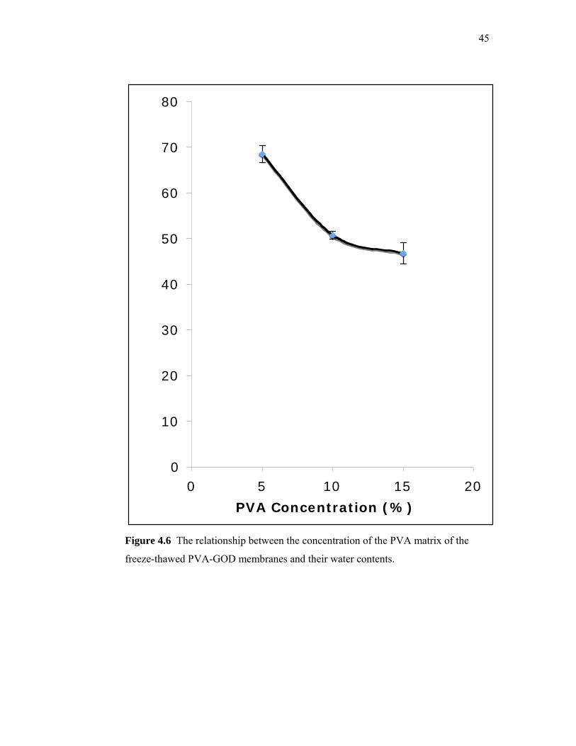
4.3.2 Water contents of the membranes
One method that can be used to estimate the cross-link density of a cross-linked
membrane is by measuring its water content. Figure 4.6 shows the relationship
between water content and PVA concentrations of the PVA-GOD freeze-thawed
membranes. As expected membranes prepared using 5% PVA have higher water
contents than membranes prepared using 10% and 15% PVA. This suggest that
membranes made with 15% PVA had the highest cross-link density followed by
membranes prepared with 10% PVA and 5% PVA respectively. Water content is
also a good indicator of the permeability of the membranes. The higher the water
content, the lower the permeability of the membranes.
44
-0.1
-0.05
0
0.05
0.1
0.15
0.2
0.25
0.3
0.35
0.4
0.45
0 50 100 150
Time, hr
5% PVA
10% PVA
15% PVA
Figure 4.5 Leaking profiles for PVA-GOD freeze-thawed membranes prepared
using 5% PVA, 10% PVA and 15% PVA.
45
0
10
20
30
40
50
60
70
80
0 5 10 15 20
PVA Concentration (%)
Figure 4.6 The relationship between the concentration of the PVA matrix of the
freeze-thawed PVA-GOD membranes and their water contents.
46
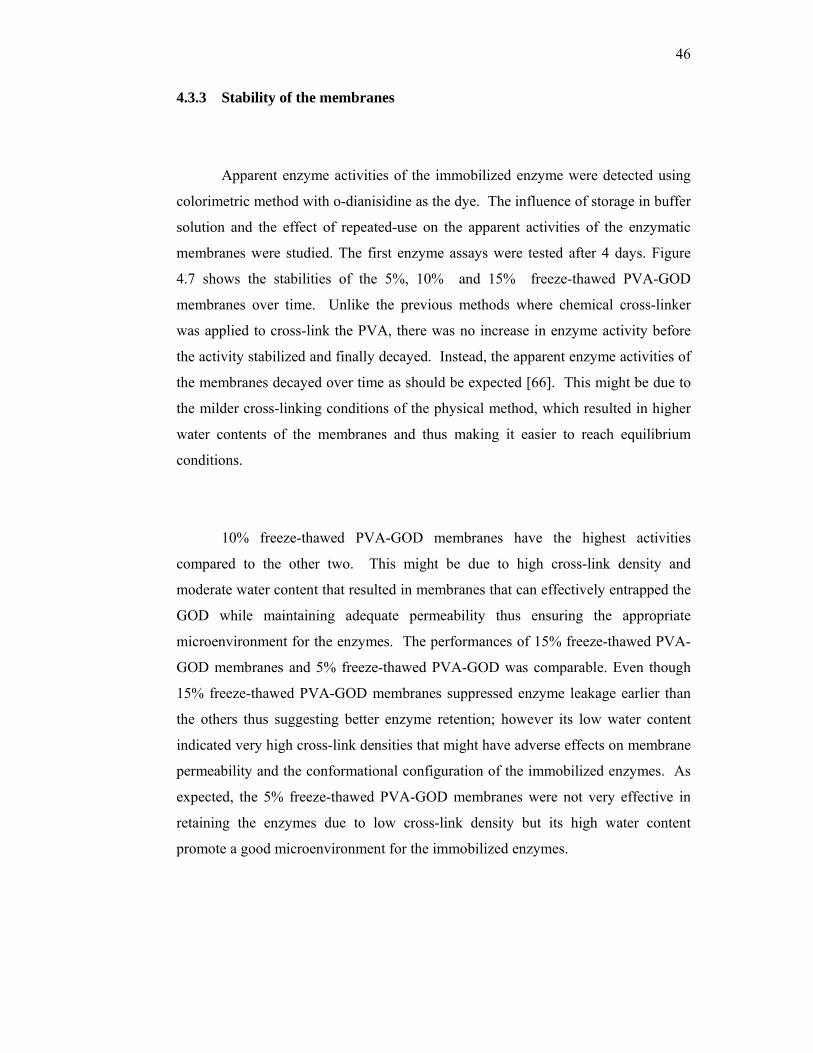
4.3.3 Stability of the membranes
Apparent enzyme activities of the immobilized enzyme were detected using
colorimetric method with o-dianisidine as the dye. The influence of storage in buffer
solution and the effect of repeated-use on the apparent activities of the enzymatic
membranes were studied. The first enzyme assays were tested after 4 days. Figure
4.7 shows the stabilities of the 5%, 10% and 15% freeze-thawed PVA-GOD
membranes over time. Unlike the previous methods where chemical cross-linker
was applied to cross-link the PVA, there was no increase in enzyme activity before
the activity stabilized and finally decayed. Instead, the apparent enzyme activities of
the membranes decayed over time as should be expected [66]. This might be due to
the milder cross-linking conditions of the physical method, which resulted in higher
water contents of the membranes and thus making it easier to reach equilibrium
conditions.
10% freeze-thawed PVA-GOD membranes have the highest activities
compared to the other two. This might be due to high cross-link density and
moderate water content that resulted in membranes that can effectively entrapped the
GOD while maintaining adequate permeability thus ensuring the appropriate
microenvironment for the enzymes. The performances of 15% freeze-thawed PVA-
GOD membranes and 5% freeze-thawed PVA-GOD was comparable. Even though
15% freeze-thawed PVA-GOD membranes suppressed enzyme leakage earlier than
the others thus suggesting better enzyme retention; however its low water content
indicated very high cross-link densities that might have adverse effects on membrane
permeability and the conformational configuration of the immobilized enzymes. As
expected, the 5% freeze-thawed PVA-GOD membranes were not very effective in
retaining the enzymes due to low cross-link density but its high water content
promote a good microenvironment for the immobilized enzymes.

47
Figure 4.7 Stability of freeze-thawed PVA-GOD membranes prepared using 5%
PVA, 10% PVA and 15% PVA.
0
2
4
6
8
10
12
14
16
18
5 9 16 30 40 55
Time (day)
5% PVA
10% PVA
15% PVA
48
4.3.4 Enzymatic membranes apparent kinetics
The apparent kinetic properties of the enzymatic membranes were obtained
using Lineweaver-Burke plot. Apparent Vmax for PVA-GOD membranes with 5%,
10% and 15% PVA was 13.3 mM/min, 26.12 mM/min and 17.53 mM/min
respectively. The apparent Michaelis constant (Kmapp) for PVA-GOD membranes
with 5%, 10% and 15% PVA was 4.8 mM, 5.95 mM and 5.15 mM, respectively. The
low values indicated that the enzymes had a high affinity for substrate, as was
usually the case for enzyme in solutions. This implied that the freeze-thaw physical
immobilization method was a mild method that resulted in a good microenvironment
for the enzymes. However, low Kmapp limits the detection limit of the biosensor that
uses this type of enzymatic membrane as its biological recognition element. Other
methods such as the use of diffusional outer membrane have to be employed to
extend the linear range of substrate detection.
4.4 GOD immobilization in PVA/TMOS sol gel matrix
For this part of the research, the immobilization method investigated was
immobilization of glucose oxidase in PVA/TMOS sol gel matrix. To improve the
retention of enzyme in the matrix, (3-glycidoxypropyldimethylethoxy) silane
(3GPDES), a cross-linker was used to cross-link the TMOS based sol-gel matrix.
The work focused on the effect of cross-linker concentration on the performance of
the enzymatic membranes.
49
4.4.1 Ability of the membranes to retain glucose oxidase
The washing solutions were analyzed for the presence of protein to
investigate the effectiveness of immobilizing GOD in cross-linked silica sol and poly
vinyl alcohol membrane incorporated with (3-glycidoxypropyldimethylethoxy)
silane. Phosphate buffer solution was changed every 6 hours for the first day, every
12 hours for the second day and 24 hours thereafter and was analyzed for any sign of
protein leakage. Using the Biuret method for determination of total protein, the
leaking of glucose oxidase (GOD) in the washing solution was determined.
Figure 4.8 shows the total protein concentrations of the washing solution for
the PVA/TMOS-GOD membranes for membranes with 1:1, 1:2 and 1:3 TMOS to
3GPDES volumetric ratios, respectively. Within 72 hours, the protein concentrations
in the washing solutions for all three types of membranes have reached zero. This
means that the immobilization method was effective enough to retain GOD in the
cross-linked silica sol and PVA membrane incorporated with (3-
glycidoxypropyldimethylethoxy) silane. The enzymatic membranes with the higher
concentration of the cross linking agent [(3-glycidoxypropyldimethylethoxy) silane]
was expected to show better enzyme retention. However, figure 4.4.1 shows that the
leakage profiles for the three membranes were not much different indicating that the
method was effective for all membranes.
4.4.2 Stability of the membranes
The immobilized membranes were placed in the phosphate buffer solution
(pH 7) when not in used. The stability of the membranes was investigated using
amperometric measurement where the glucose stock solution was injected into the
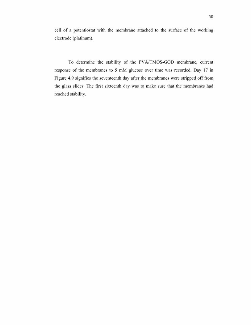
50
cell of a potentiostat with the membrane attached to the surface of the working
electrode (platinum).
To determine the stability of the PVA/TMOS-GOD membrane, current
response of the membranes to 5 mM glucose over time was recorded. Day 17 in
Figure 4.9 signifies the seventeenth day after the membranes were stripped off from
the glass slides. The first sixteenth day was to make sure that the membranes had
reached stability.
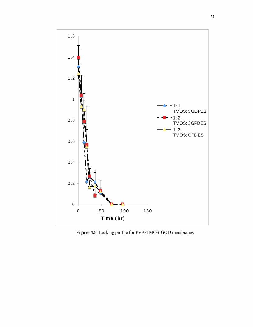
51
0
0.2
0.4
0.6
0.8
1
1.2
1.4
1.6
0 50 100 150
Time (hr)
1:1TMOS:3GDPES1:2TMOS:3GPDES1:3TMOS:GPDES
Figure 4.8 Leaking profile for PVA/TMOS-GOD membranes
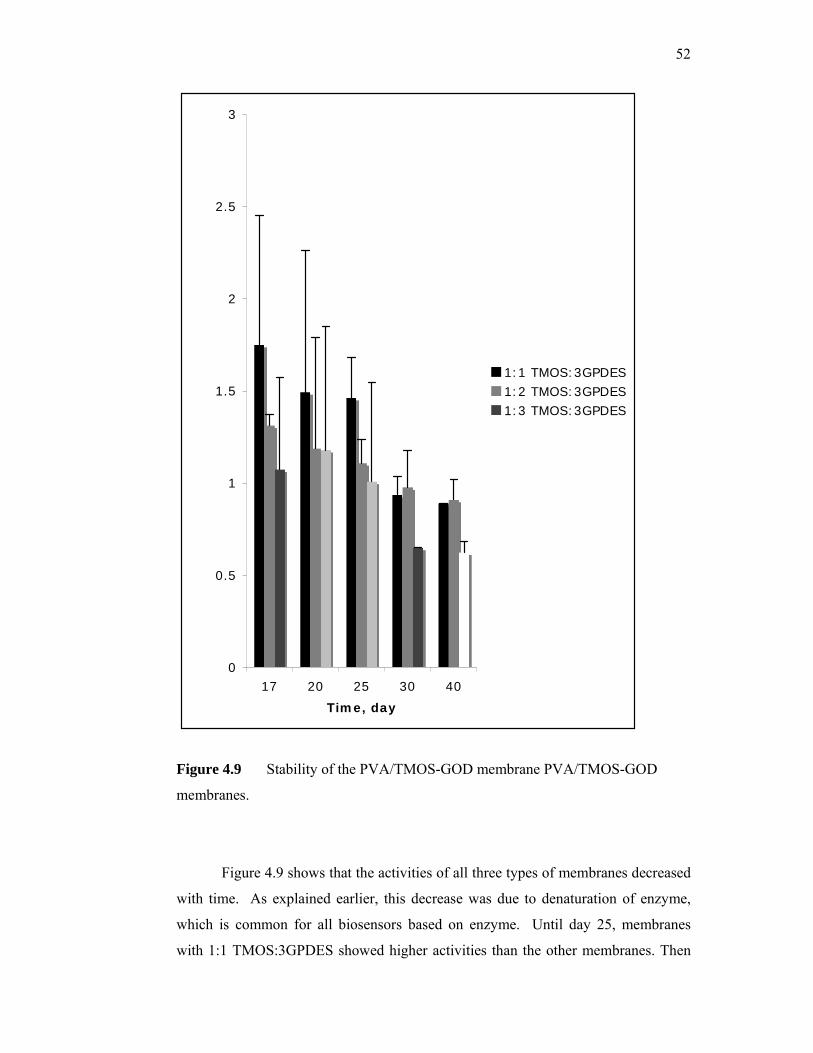
52
Figure 4.9 Stability of the PVA/TMOS-GOD membrane PVA/TMOS-GOD
membranes.
Figure 4.9 shows that the activities of all three types of membranes decreased
with time. As explained earlier, this decrease was due to denaturation of enzyme,
which is common for all biosensors based on enzyme. Until day 25, membranes
with 1:1 TMOS:3GPDES showed higher activities than the other membranes. Then
0
0.5
1
1.5
2
2.5
3
17 20 25 30 40
Time, day
1:1 TMOS:3GPDES1:2 TMOS:3GPDES1:3 TMOS:3GPDES
53
the activities decreased rather rapidly. At day 40 only 51% of the initial activity
remained. Membranes with 1:2 TMOS:3GPDES were quite stable with 69% of the
initial activity remained at day 40. For membranes with 1:3 TMOS:3GPDES, 58% of
the initial activity remained at day 40.
4.4.3 Enzymatic membranes apparent kinetics
The apparent kinetics of the immobilized membranes was investigated using
amperometric measurements. The kinetic parameters for the catalytic reaction, Km
and Vmax were estimated from modified Lineweaver-Burke plot. The Km and Vmax of
the enzyme immobilized in the membrane were obtained from the slope and intercept
of the graph.
max
1InterceptV
= max
SlopeVKm =
Both parameters are substrate specificity factors used to indicate catalytic efficiency
and biosensor performance. High values indicate better biosensor performance.
At high glucose concentrations a platform response is observed, showing a
characteristic of the Michelis-Menten kinetic mechanism. For 1:1 TMOS:3GPDES
enzymatic membrane, the Vmax value was 5.6 µA and the Km value was
approximately 1.5 mM. For 1:2 TMOS:3GPDES enzymatic membrane, Vmax was
6.0 µA and Km was approximately 1.8 mM. For 1:3 TMOS:3GPDES enzymatic
membrane 3, the Vmax and Km values were 5.2 µA and 1.5 mM, respectively.
CHAPTER 5
CONCLUSIONS AND RECOMMENDATIONS
5.1 Conclusions
5.1.1 GOD immobilization in cross-linked PVA matrix: different casting
temperature
In this work, the basic working principle of a glucose biosensor especially the
enzyme layer was studied. GOD was immobilized in cross-linked PVA matrix. The
effect of casting temperature on enzymatic membrane performance was investigated.
The immobilization of glucose oxidase in cross-linked poly(vinyl alcohol)
was shown to able to produce membranes with sufficient enzyme activity. The
enzyme activity range detected by membrane immobilized at 4°C was 13.33mU/mL
- 26.67mU/mL. The enzyme activity range for membranes immobilized at 25°C was
10.91mU/mL – 30.45mU/mL. The membranes immobilized at 25°C show higher
enzyme activity in 25 days compared to membranes immobilized at 4°C. The
membranes immobilized at 25°C were found to be able to retain enzyme better.
55
5.1.2 GOD immobilization in cross-linked PVA matrix: different PVA
concentration
In this work, the effects of PVA concentration on the performance of cross-
linked PVA-GOD membranes in terms of enzyme leakage and available activity
were investigated. The cross-linker used was glutaraldehyde and the cross-linking
ratio was 0.06. The experiments were conducted at room temperature.
With higher PVA concentration (15%), the enzyme retaining ability was
better. For membranes prepared with 15% PVA, total enzyme leakage was 20%
lower than the ones prepared with 10% PVA. The 10% PVA-GOD membranes
leaked for 16 hours longer than the 15% PVA-GOD membranes. However, the 10%
PVA-GOD membranes performed better in terms of available enzyme activity.
Enzyme activity of the 10% PVA-GOD membranes was approximately 33% higher
than the 15% PVA-GOD membranes. This can be attributed to better permeability
and micro-environment of the 10% PVA-GOD membranes.
5.1.3 GOD immobilization in freeze-thawed PVA matrix
In this work, GOD was physically immobilized in PVA through the freeze-
thaw method. The immobilization method was milder than using chemical cross-
linker. The effect of PVA concentration on enzymatic membranes performance was
investigated.
This work suggests that the higher the PVA concentration used for immobilization,
the better the retention of the enzyme. Nevertheless, higher PVA concentration of
immobilization didn’t necessarily correlate well with enzyme activity. 10% freeze-
thawed PVA-GOD membranes have the highest activities. This might be due to high
cross-link density and moderate water content that resulted in membranes that can
56
effectively entrapped the GOD while maintaining adequate permeability thus
ensuring the appropriate microenvironment for the enzymes.
The performances of 15% freeze-thawed PVA-GOD membranes and 5%
freeze-thawed PVA-GOD was comparable. Even though 15% freeze-thawed PVA-
GOD membranes suppressed enzyme leakage earlier than the others thus suggesting
better enzyme retention; however its low water content indicated very high cross-link
densities that might have adverse effects on membrane permeability and the
conformational configuration of the immobilized enzymes. As expected, the 5%
freeze-thawed PVA-GOD membranes were not very effective in retaining the
enzymes due to low cross-link density but its high water content promote a good
microenvironment for the immobilized enzymes.
In terms of kinetic properties, PVA-GOD freeze –thawed membranes
prepared with 10% PVA exhibited the highest Km app compared to the others. This
means that they are more suitable to be used as bio-recognition elements for glucose
biosensors than the other two.
5.1.4 GOD immobilization in PVA/TMOS sol gel matrix
In this work, GOD was immobilized in PVA/TMOS sol gel matrix. A
chemical cross-linker was used to improve retention of enzyme. The effect of cross-
linker concentration on enzymatic membranes performance was investigated.
The immobilization of glucose oxidase in silica sol/poly vinyl alcohol
membrane incorporated with (3-glycidoxypropyldimethylethoxy) silane was shown
to be able to produce membranes which acceptable stability. For the membranes

57
prepared with 1:1 TMOS:3GPDES, the percentage of enzyme activity which
remained at day 40 was about 51%. Meanwhile, for the membrane prepared with 1:2
TMOS:3GPDES and 1:3 TMOS:3GPDES, the percentage of enzyme activity which
remained at day 40 was 69% and 58%, respectively.
From the kinetics studies, it was calculated that Vmaxapp and Km
app values for
membranes prepared with 1:1 TMOS:3GPDES were 5.7 µA and 1.5 mM,
respectively. Meanwhile, Vmaxapp and Km
app values for membranes prepared with 1:2
TMOS:3GPDES were 6.0 µA and 1.8 mM, respectively. For membranes prepared
with 1:3 TMOS:3GPDES, the Vmaxapp and Km app values were approximately 5.2 µA
and 1.5 mM, respectively. Thus membranes prepared with 1:2 TMOS:3GPDES is
most suitable to be used as the bio-recognition element for a glucose biosensor due to
its stability and kinetic properties.
5.2 Recommendations
Throughout the experiment, controlling the thickness and size of the
membranes are rather challenging. However, producing membranes with uniform
size and thickness are essential, as it will affect the available enzyme activity and
permeability of the membrane.
In this work, the enzyme activity, kinetic and stability studies were
investigated. But, in order to get a more comprehensive study of the immobilized
glucose oxidase, it should include several other tests such as interference test,
reproducibility test and optimization of the biosensor.
58
For the interference test, it should be done to detect possible effects of several
common chemicals such as ascorbic acid and uric acid to the biosensor. This test is
particularly important for enzymatic membranes meant for amperometric glucose
biosensor.
Reproducibility test should be conducted to determine if the enzymatic
membranes are able to respond to the varying glucose concentration in an acceptable
time and with reproducible output response.
59
REFERENCES
1. The Diabetes Control and Complications Trial Research Group: The Effect of
Intensive Treatment of Diabetes on the Development and Progression of
Long Term Complications in Insulin-dependent Diabetes Mellitus (1993).
New Eng J Med. 329, 977-986.
2. The Diabetes Control and Complications Trial Research Group: Lifetime
Benefits and Costs of Intensive Therapy as Practiced in The Diabetes Control
and Complications Trial (1996). JAMA. 276, 1409-1415.
3. Ohkubo, Y., Kisikawa H., Araki, E., Miyata, T., Isami, S., Motoyoshi, S.,
Kojima, Y., Furoyoshi, N. and Shichiri, M. (1995). Intensive Insulin Therapy
Prevents The Progression of Diabetic Microvascular Complications in
Japanese Patients with Non-insulin-dependent Diabetes Mellitus: A
Randomized Prospective Study. Diabetes Res Clin Pract, 28, 103-117.
4. UK Prospective Diabetes Study Group: Intensive Blood Glucose Control with
Sulphonylureas or Insulin Compared with Conventional Treatment and Risk
of Complications in Patients with Type 2 Diabetes (1998). Lancet. 325, 837-
853.
5. Cooppan, R. (1994). General Approaches to The Treatment of Diabetics. In:
Kahn, C. R. and Weir, G.C. eds. Joslin’s Diabetes Mellitus. Philadelphia: Lea
& Febriger.
60
6. Updike, S. J. and Hicks, G. P. (1967). The Enzyme Electrode. Nature. 214,
968-988.
7. Gough, D. A., Leypoldt, J.K. and Armour, J. C. (1982). Progress Towards A
Potentially Implantable Enzyme-based Glucose Sensor. Diabetes Care. 5,
190-198.
8. Schichiri, M., Askawa, N., Yamasaki, Y., Kawamari R. and Abe, H. (1986).
Telemetry Glucose Monitoring Device with Needle-type Glucose Sensor: A
Useful Tool for Blood Glucose Monitoring in Diabetic Individuals. Diabetics
Care. 9, 298-301.
9. Turner, R. F. B., Harrison, D. J., Rajotte, R. V. and Baltes, H. P. (1990). A
Biocompatible Enzyme Electrode for Continuous In-vivo Monitoring in
Whole Blood. Sensors and Actuators B. 1, 561-564.
10. Moatti-Sirat, D., Capron, F., Poitout, V., Reach, G., Bindra, O. S., Zhang, Y.,
Wilson, G. S. and Thevenot, D. R. (1992). Towards Continuous Glucose
Monitoring: In-vivo Evaluation of A Miniaturized Glucose Sensor Implanted
for Several Days in Rat Subcutaneous Tissue. Diabetologia. 35, 225-230.
11. Turner, A. P. F. (1998). The Advantages and Disadvantages of Mediated
Amperometric Biosensor for In-vivo Monitoring. EEC Workshop: The
Interface between Biology and Sensors. Cranfield Biotechnology Center, UK:
Milton Keynes.
12. Janata, J. (1992). Chemical Sensors. Anal. Chem. 64, 196R-219R.
13. Janata, J. and Bezegh, A. (1988). Chemical Sensors. Anal. Chem. 67, 62R-
74R.
61
14. Bartlett, P. N. and Whitaker, R. G. (1987). Electrochemical Immobilization
of Enzymes. Part 1. Theory. Part 2. Glucose Oxidase Immobilized in Poly-n-
methylpyrrole. J. Electroanal. Chem. 224, 37-48.
15. Shinohara, H., Chiba, T. and Aizawa, N. (1988). Enzyme Microsensor for
Glucose with an Electrochemically Synthesized Enzyme–polyaniline Film.
Sensors and Actuators. 13, 79-86.
16. Barker, S. A. (1987). In: Turner, A.P.F., Karube, I. and Wilson, G.S. eds.
Biosensors: Fundamentals and Applications. Oxford: Oxford University
Press.
17. Hall, E. A. H. (1990). Biosensors. UK: Open University Press, Milton
Keynes.
18. Hall, C. E. and Hall, E. A. H. (1993). Covalent Immobilization of Glucose
Oxidase on Methacrylate Copolymer for Use in an Amperometric Glucose
Sensor. Anal. Chim. Acta. 281, 645-653.
19. Parente, A. H., Marques, E. T. A., Azeuedo, W. M., Diniz, F. B. Melo, E. H.
M. and Filho, J. L. (1992). Glucose Biosensor using Glucose Oxidase
Immobilized in Polyaniline. Appl. Biochem. Biotech. 37, 267-273.
20. Contractor, A. Q., Sureshkumar, T. N., Narayanan, R., Sukeerthi, S., Rakesh,
L. and Srinivasa, R. S. (1994). Conducting Polymer-based Biosensors.
Electrochim. Acta. 39, 1321-1324.
62
21. Bartlett, P. N and Caruana, D. J. (1992). Electrochemical Immobilizations of
Enzymes. Part V: Microelectrodes for the Detection of Glucose Based on
Glucose Oxidase Immobilized in Poly(phenol) Film. Analyst (Lond). 117,
1287-1292.
22. Chen, C. J., Liu, C. C. and Savinell, R. F. (1993). Polymeric Redoz Mediator
Enzyme Electrodes for Anaerobic Glucose Monitoring. . J. Electroanal.
Chem. 348, 317-338.
23. Calvo, E. J., Danilowicz, C. and Diaz, L. (1993). Enzyme Catalyst at
Hydrogel-mediated Electrodes with Redox Polymer Mediator. J. Chem. Soc.,
Faraday Trans. 89, 377-384.
24. Shin, M. C. and Kim, H. S. (1995). Effects of Enzyme Concentration and
Film Thickness on The Analytical Performance of a Polypyrrole / Glucose
Oxidase Biosensor. Anal. Letter. 28, 1017-1031.
25. Bindra, D. S., Zhang, Y., Wilson, G. S., Sternberg, R. Thevenot, D. R.,
Moatti, D. and Reach, G. (1991). Design and In-vitro Studies of a Needle
Type Glucose Sensors for Subcutaneous Monitoring. Anal. Chem. 63,1692-
1696.
26. Karubem I, Seki A. and Sode, K (1990) . Microbiosensor and Immunosensors
In: Microbial Enzymes and Biotechnology. Second edition. England: Elsevier
425-450.

63
27. Tierney, M. J., Jayalakshmi, Y., Parris, N. A., Reidy, M. P., Uhegbu, C. and
Vijayakumar, P. (1999). Design of a Biosensor for Continual Transdermal
Glucose Monitoring. Clin. Chem. 45, 1681-1683.
28. Rebrin, K.Steil G. M., von Antwerp, W. P. and Mastrototaro, J. J. (1999).
Subcutaneous Glucose Predicts Plasma Glucose Independent of Insulin:
Implications for Continuous Monitoring. Am. J. Ped. 277, E561-E571.
29. Henning, T. P. and Cunningham, D. D. (1998). Biosensor for Personal
Diabetes Management. In: Ramsay, G. ed. Commercial Biosensors:
Application to Clinical, Bioprocess and Environment Sample. Canada: John
Wiley & Sons. 5.
30. Yamasaki., Y., Ueda, N., Nao, K., Sekiya, M., Kawamari, R., Schichiri, M.
ana Kamada, T. (1989). Direct Measurement of Whole Blood by a Needle-
type Sensor. Clin. Chim. Acta. 93, 93-98.
31. Harrison, D. J., Turner, R. F. B. and Baltes, H. P. (1988). Characterization of
Perfluorosulfonic Acid Polymer Coated Enzyme Electrodes and a
Miniaturized Integrated Potentiostat for Glucose Analysis in Whole Blood.
Anal. Chem. 60, 2002-2007.
32. Zhang, Y., Hu, Y., Wilson, G. S., Moatti-Sirat, D., Poitout, V., Reach, G.
(1994). Elomination of The Acetominophen Interference in an Implantable
Glucose Sensor. Anal. Chem. 66, 1183-1188.
33. Matsumoto, T., Furusawa, M., Fujiwara, H., Matsumoto, Y. and Ito, N.
(1998). A Microplanar Amperometric Glucose Sensor Unsusceptible to
Interference Species. Sensors and Actuators B Chemicals. 49, 68-72.
64
34. Helller, A., Csöregi, E. and Schmidtke, D. W. (1995). Design and
Optimization of a Selective Subcutaneously Implantable Glucose Electrode
Based On Wired Glucose Oxidase. Anal. Chem. 67, 1240-44.
35. Schultz, J. S., Mansouri, S. and Goldstein, I. J. (1982). Affinity Sensor: A
New Technique for Developing Implantable Sensors for Glucose and Other
Metabolites. Diabetes Care. 245-253.
36. Meadows, D. L. and Schultz, J. S. (1993). Design, Manufacture and
Characterization of an Optical Fiber Glucose Affinity Sensor Based on
Homogenous Fluorescence Energy Transfer Assay System. Anal. Chim. Acta
280, 21-30.
37. Cui, W., Ostrander, L. E. and Lee, B. Y. (1990). In-vivo Reflectance of Blood
and Tissue as a Function of Light Wavelength. IEEE Trans. Biomed. Engin.
37, 632-639.
38. Marbach, R. and Heise, H. M. (1995). Optical Diffuse Reflectance Accessory
for Measurements of Skin Tissue by Near Infrared Spectroscopy. Applied
Optics. 34, 610-621.
39. Kayashima, S., Arsi,T., Kikuchi., M., Nagata, N., Ito., N., Kuriyama, T. and
Kimura, J. (1992). Suction Effusion Fluid from Skin and Constituent
Analysis: New Candidate for Interstitial Fluid. Am. J. Physio. 263, H1623-
H1627.
40. Tamada, J. A., Bohannon, N. J. Y. and Potts, R. O. (1995). Measurements of
Glucose in Diabetic Patients using Non-invasive Transdermal Extraction.
Nature Med. 1, 1198-2001.
65
41. Proctor, A., Castner, J. F., Wigard, L. B and Hercules, D. M. (1985). Electron
Spectroscopic (ECSA) Studies of Platinum Surfaces used for Enzyme
Electrodes. Anal. Chem. 57, 1644-1649.
42. Xie, B., Hedberg, U., Mecklenburg, M. and Danielsson, B. (1993). Fast
Determination of Whole Blood Glucose with a Calorimetric Micro-biosensor.
Sensors and Actuators B Chemicals. 14, 15-16.
43. Malmros, M. K., Gulbinski, J. and Gibbs, W. B. (1987/8). A Semiconductive
Polymer Film Sensor for Glucose. Biosensor. 3, 71-87.
44. Muehlbauer, M. J., Guilbeau, E. J. and Towe, B. C. (1989). Model for a
Thermoelectric Enzyme Electrode Glucose Sensor. Anal. Chem. 61, 77-83.
45. Shuler, M. L. and Kargi, F. (2002). Bioprocess Engineering Basic Concepts.
2nd ed. USA: Prentice Hall.
46. Imai, K.,Shiomi T., Uchida., K. and Miya, M.(1985). Immobilization of
Enzyme into polyvinylalcohol membrane. Biotechnology and Bioengineering.
28.
47. Winfried Hartmeier (1986). Immobilized Biocatalyst: An introduction. New
York: Springer-Verlag Berlin Heidelberg.
48. Sakurada, I. and Sore, Y. (1997). Rheological Studies of a Concentrated
Solution of PVA. I: Dependence of Apparent Viscosity on Shear and the Age
of Solution. Kobunshi Kagaku. 10, 1953.
49. Finch, C. A. (1992). Poly(vinyl alcohol): Developments. New York: John
Wiley and Sons.
66
50. Miller, D. R. and Peppas, N. A. (1988). Diffusional Effects During Albumin
Adsorption on Highly Swollen Poly(vinyl alcohol) Hydrogel. Eur. Polym. J.
24, 611.
51. Ratner, B. D. and Hoffman, A. S. (1976). Synthetic Hydrogels for
Biomedical Applications. In: Hydrogel for Medical and Related Applications.
Andrade, J. D. ed. ACS Symp. Ser. 31, 1-29.
52. Szycher, M. and Bernhard, W. F. (1983). Hemocompatible Interfaces in
Artificial Heart Devices in Biocompatible Polymers, Metals and Composites.
Szycher, M. ed. Lancaster, PA: Techronic Publishers.
53. Korsmeyer, R. W., Gury, R., Doelker, E., Buri, P. and Peppas, N. A. (1983).
Mechanisms of Solute Release from Hydrophilic Polymers. Int. J. Pharm. 15,
25.
54. Amsden, B. (1998). Solute Diffusion in Hydrogels: An Examination of the
Retardation Effecct. Polym. Gels. And Networks. 6, 13.
55. Korsmeyer, R. W. and Peppas, N. A. (1981). Effect of Morphology of
Hydrophilic Polymeric Materials on the Diffusion and Release of Water
Soluble Drugs. J. Membrane Science. 9, 211-227.
56. Reinhart, C. T. and Peppas, N. A. (1984). Solute Diffusion in Swollen
Membranes. Part II: Influence of Cross-linking on Diffusive Properties. J.
Membrane Science. 18, 227-229.
57. Li, R. H. and Barbari T. A. (1994). Protein Transport Through Membranes
based on Toluene Diisocyanate Surface-modified Poly(vinyl alcohol) Gels. J.
Membrane Science. 88, 115-125.
67
58. Li, R. H. and Barbari T. A. (1996). Characterization and Mechanical Support
of Asymmetric Hydrogel Membranes based on the Interfacial Cross-linking
of Poly(vinyl alcohol) with Toluene Diisocyanate. J. Membrane Science.111,
115-122.
59. Dai, W. S. and Barbari T. A. (1999). Hydrogel Membranes with Mesh Size
Assymmetry based on the Gradient Cross-linking of Poly(vinyl alcohol). J.
Membrane Science. 156, 67-69.
60. Dai, W. S. and Barbari T. A. (2000). Gel-impregnated Pore Membranes with
Mesh-size Asymmetry for Biohybrid Artificial Organs. Biomaterials. 21,
1363-1371.
61. Dai, W. S. and Barbari T. A. (2000). Hollow Fiber-supported Hydrogels with
Mesh-size Asymmetry. J. Membrane Science. 171, 79-86.
62. Matsuyama, H., Teramoto, M. and Urano, H. (1997). Analysis of Solute
Diffusion in Poly(vinyl alcohol) Hydrogel Membrane. J. Membrane Science.
126, 151-160.
63. Liu, Z., Liu, B., Zhang, M., Kong, J. and Deng, J. (1999). Al2O3 Sol-Gel
Derived Amperometric Biosensor for Glucose. Elsevier: 135-141.
64. Lillis, B., Grogan, C., Berney, H. and Lane, W. A. (2000). Investigation into
Immobilisation of Lactate Oxidase to Improve Stability. Sensors and
Actuators B Chemicals. 68, 109-114.
65. Doretti, L., Ferrara, D., Gattolin, P., and Lora, S. (1997). Amperometric
Biosensor with Physically Immobilized Glucose Oxidase on a PVA Cryogel
Membrane. Talanta. 44: 859-866.

68
66. Doretti, L., Ferrara, D., Gattolin, P., and Lora, S., Schiavon, F. and Veronese,
F. M. (1998). PEG-Modified Glucose Oxidase Immobilized on a PVA
Cryogel Membrane for Amperometric Biosensor Applications. Talanta. 45:
891-898