dr. bhupendra m. shah dr. kamlesh b. sainiimagsb.com/uploads/journal/july-2018.pdfand...
TRANSCRIPT
I.M.A.G.S.B. NEWS BULLETIN / JULY - 2018 Vol. 13 No. 7
I.M.A.G.S.B. NEWS BULLETIN / JULY - 2018 Vol. 13 No. 712
Dear Memberes
Seasons greetings.
Every body enjoying monsoon particularly south Gujarat and Saurashtra regions friends. Our
journey of this month started on 1st July celebrating as a doctors day. We are feeling proud that our
noble profession is celebrated as a Doctors day all over India.
On 1st July, Ahmedabad Medical Association arranged AMACON at AMA house with wonderful
scientific programs. We had inaugurated this program. On this occasion, Women Doctors Wing
(WDW) installation ceremony arranged. Dr Bhupendra Shah had installed WDW under leadership of
Dr Monaben Desai as a chairperson and Dr Gargi Maheshbhai as a secretary. We congratulate whole
team and best wishes for their new journey . We assured whole hearted support .
On Doctors day, many local branches had organized blood donation camp as a part of Doctors
day celebration from 1st July to 7th July. Himatnagar branch also organized blood donation camp and
its branch member Dr Dilip Pujara had donated 75th time on this day. Many local branches had
arranged press conference and public awareness program against violence and zero tolerance as per
guidelines of IMA headquarters.
Our PPS Managing Director, Dr Bipinbhai And Dr Anilbhai Nayak had chaired and inaugurated
PPS seminar at Surat.
Under leadership of IMA GSB, the representative of various association like Orthopaedics, Neuro
Surgeons and General Surgeons had meeting with Smt. Jayanti Ravi, Commissioner of Health;
Dr Dholakia, Director of Medical services and other official of Govt. regarding Vahan Akasmat Yojana
and discussed our concerns against this scheme and they assured that our suggestions to put in front
of Honourable Deputy CM Shree Nitinbhai Patel, positively.
The Government is giving us real good reasons to sit together and work to save the health
providers to eventually save the health of the nation, on the other hand the Government expects us
to fulfill their deficiencies and provide health care to the people of our country, whereas on the other
hand, they look out for all means to punish us for even minor clerical errors by bringing harsh
regulation against the medical profession.
Our Honorable Prime Minister declares 'End TB by 2025' and then the Government asks IMA to
take up this project to eradicate TB by sensitizing all medical professionals in Private Sector and
increase notification of TB cases. The irony is that although they know 70% of the health care in the
country is provided by private doctors and small nursing homes & hospitals, however they only
regard public sector hospitals as their means for dissemination of populist health projects and
programs.

I.M.A.G.S.B. NEWS BULLETIN / JULY - 2018 Vol. 13 No. 713
DR. BHUPENDRA M. SHAH DR. KAMLESH B. SAINI
(President, G.S.B.I.M.A.) (Hon. State Secy. G.S.B.I.M.A.)
The Government's Ayushman Bharat Mission may be a good populist move on paper but if the
Government listens to & accept our suggestions and involves us in framing rules and projects
costing leading to acceptable packages, only then can it serve its purpose to change the health
scenario of the country.
All our IMA leaders of every State will find out and suggest the minimum acceptable packages
for common procedures and medical packages as this can only get us some good reasonable return
of our hard work which is our habit when providing care to our patients.
The proposed amended National Medical Commission Bill 2018, pending consideration before
the Loksabha in all its likelihood could be taken up for consideration at the ensuing Monsoon session
in the Loksabha scheduled from 18th July, 2018.
The net result which is vividly seen is that the cosmetic and illusory changes which the
Government of India, has proposed are more of a facade as they do not serve any meaning purpose
in any way.
This Bill still remains anti-poor, anti-federal and non representative in character.
As such, it is imperative that the unity and solidarity invoked by the Indian Medical Association
to resist this draconian Bill is the only way and therefore by vigilance and invocation of timely and
planned initiatives and moves counteracting the intent and obstinacy of the Government of India, is
key to strategy by the IMA through the collective unity and wisdom alike.
Against all odds a dent has been made which has to be taken to its logical finality with courage,
conviction and unity as the sole weapon as depiction of our resistance with a commitment that 'No
National Medical Commission'.
The IMA has proposed alternatives in all its details in the form of 'Amended Indian Medical
Council Act, 1956 and National Medical Grants Commission Bill 2017 in the larger interest of medical
education, profession and country as a whole.
We sincerely hope that the ensuing Monsoon Sessions of the Parliament will not give the
medical profession any jitters and some maturity will prevail upon the Government for the
betterment of the health of the nation.
Again On 27th - 28th October, Gimacon 2018, organised by IMA Himatnagar. All members are
requested to register. In our tenure, this is the second edition of Journal of IMA GSB.
We congratulate Dr. K R Sangavi and his whole team for their hard work and wonderful job. Thanks
for your kind cooperation and support.
Long Live IMA
19
Dear friends,
While putting this issue of Gujarat Medical Journal (GMJ) in your hands, we are happy that now very regularly
we are publishing GMJ.
You all know that GMJ is indexed in Index Copernicus International” (ICI), and all the issues of GMJ since 2015
can be viewed on
Obviously the Gujarat Medical Journal should look for impact factor which is the next big thing for any
scientific journal. To achieve this we need to put in a lot of efforts. We would like to request all the research mined
doctors in Gujarat who are into research and publications to seriously consider GMJ for their manuscripts. We are
also making all efforts to make our journal website at par with any leading medical journal. We hope to bring in
many more value added features to our journal within a reasonable period of time.
Our country and particularly, Gujarat has entered in the field of medical tourism. People from developed and
under developed countries come here for treatment and we provide world best treatment to them at a cheaper
rates then that is available in developed countries. Apart from big cities of Gujarat like Ahmedabad, Surat. Vadodra
and Rajkot-Bhavnagar, even small centers like Anand and Nadiad provide world class treatment in the field of
cardiology and nephrology. Our hospitals and expertise are world class and that pushes the medical tourism in
Gujarat far ahead. From our own domestic population also we get large number of patients. This provides
opportunities for research to our doctors. Now we have better infrastructure facilities for data collection and
access to world data, for comparison. It has provided a big boost to research wok in our state.
Without making any compromise with our laid down policy, we have made all the efforts to make GMJ more
informative and more interesting so that large number of our colleagues read it and utilize the knowledge and
information provided in it. For this, we welcome your suggestions and comments also.
We would like to mention here, the determined and committed efforts made by our enthusiastic and
dynamic past president Dr. Yogendra Modi and hon. Secretary of IMA GSB Dr. Kamlesh Saini along with our ex-
editor Dr. Amitbhai P. Shah and Dr . Urvesh Shah (GCS medical college Ahmedabad) in maintaining our indexation
status with Index Copernicus International. Dr. Urvesh Shah's painstaking efforts in up loading all the issues of GMJ
on Index Copernicus website made them available there. We extend our sincere thanks to Dr. Cinmay Shah
(Bhavnagar medical college) for helping us time to time.
Our sincere thanks to GSB president Dr. Bhupendra Shah and hon. secretary Dr. Kamlesh Saini for
encouragement and suggestions and giving us free hand in publication of this journal. We are also grateful to GSB
past presidents Dr Kirtibhai Patel, Dr. Jitubhai Patel and Dr. Mahendrabhai Desai for their guidance and help.
With regards,
https://journals.indexcopernicus.com/search/details?id=43553
I.M.A.G.S.B. NEWS BULLETIN / JULY - 2018 Vol. 13 No. 7

20
2017-2018
DR. TUSHAR B. PATEL
DR. KETAN PATEL
DR. MUKUND B. PATEL
DR. PARESH GOLWALA
DR. GIRISH MODI
DR. S. S. VAISHYA
DR. BHARAT KAKADIA
AHMEDABAD
WEST ZONE
CENTRAL ZONE
VADODARA
SURAT
SOUTH ZONE
RAJKOT ZONE
AHMEDABAD
WEST ZONE
CENTRAL ZONE
VADODARA
SURAT
SOUTH ZONE
RAJKOT ZONE
DR. JASWANT DARBAR
DR. KASHYAP C. DAVE
DR. JITESH DESAI
DR. PARESH MAJMUDAR
DR. DHIREN PATEL
DR. VANRAJSINH MAHIDA
DR. CHETAN LALSETA
FAMILY WELFARE SCHEME
HOSPITAL BOARD OF INDIA
CHAIRMANDR. PARTH N. PATEL
DR. BIPIN M. PATEL
DR. BHUPENDRA M. SHAH HIMATNAGAR
MOB. 94260 04474
DR. YOGENDRA S. MODI AHMEDABAD
MOB. 98240 21444
DR. KAMLESH B. SAINI AHMEDABAD
MOB. 96019 49252
DR. DEVENDRA R. PATEL
DR. ANIL D. PATEL MEHSANA
I.M.A.G.S.B. NEWS BULLETIN / JULY - 2018 Vol. 13 No. 7
ORIGINAL ARTICLE
GMJ 21 I.M.A.G.S.B. NEWS BULLETIN / JULY - 2017 Vol. 12 No.7
I.M.A. G.S.B. NEWS BULLETIN (Gujarat Medical Journal)
Vol. : 13 JULY-2018 Issue : 07
CONTENTS
*
*
State President and Hon. Secretary's Message ............................................................................................ 16
From the Desk of Editors ...................................................................................................................................19
*
*
*
*
*
*
*
*
*
*
*
Public Awareness Survey about Anesthesia and Anesthesiologist (A study of 210 cases)
23
Clinico-demographic profile of patients admitted in PICU at tertiary care centre in Ahmedabad.
29
“Hearing results of ossicular chain reconstruction with cartilage in type III tympanoplasty in
tubotympanic disease”
34
A Clinical Study : Role of Flexible Fiberoptic Bronchoscopic Biopsy in Diagnosis of Lung Cancer
39
Serum Homocysteine In Acute Ischaemic Cerebrovascular Stroke
45
Study and Clinical Evaluation of 25 cases of Primary Amenorrhea
48
A Clinico-Pathological Study of Anemias in Geriatric Age Group
59
Pre-Operative concerns of a patient in a Rural setup :RURAL SETUP: An Indian Perspective
64
A Comparative Study of Dosage of MGSO4in Indian Women with Eclampsia.
69
Intralesional Bleomycin in the treatment of Orbital Lymphangioma- Original Article
73
Role of Blood Components Transfusion in Obstetrics
76
Dr. Jigar Dodiya*, Dr. Vandana Parmar**,Dr. Vrinda Oza***, Dr. Mehul Vaghela*, Dr. Upendrakumar Parmar*....
Dr. Purvi R. Patel* Dr. Halak J. Vasavada** Dr. Snehal V. Patel *** Dr. Rohan Mehta**** Dr. Nirav Rathod...............
Dr. Sujan R.Patel*, Dr. Rupa P. Parikh**...............................................................................................................
Dr. Nalin T. Shah*, Dr. Kandarp Nimavat**, Dr. Smit Patel**, Dr. Chintan Shah**, Dr.Akash Dodiya**.....................
R S Roy*, H Patel**, M Marwadi*, R K Chavda***.....................................................................................
Dr Tushar M Shah*, Dr Foram Lalcheta**,Dr Rini Buddhdev*** .......................................................................
Amit H.Agravat*, Trushali Dobariya**, Gauravi A Dhruva***................................................................................
Heta Rajyaguru*, Dipak Rajyaguru**, Pravin Baldaniya**, Gayatri Raval***........................................................
Dr. Bhavesh B. Airao*, Dr. Nishant H. Patel** Dr. Bipin R. Shah***...........................................................
Dr. Nitin Trivedi*, Dr. Stuti N. Trivedi**, Dr. Manasi Modi**, Dr. Rutuvi Parikh**..............................................
Dr. Kruti J. Deliwala*, Dr.Akila H. Parmar**, Dr. Rina V. Patel***, Dr. Parul T. Shah**** , Dr. Devangi S. Munshi*.....

CASE REPORT
GMJ 22 I.M.A.G.S.B. NEWS BULLETIN / JULY - 2017 Vol. 12 No.7
*
*
*
*
*
*
Cervical Ectopic Pregnancy
79
A Case Report of Adrenomyeloneuropathy- A variant of X linked Adrenoleukodystrophy
81
Supratentorial Recurrence of Medulloblastoma, A Rare Case Report
83
A case presentation of thyroid storm in a case of complete hydatidiform mole
86
Mandatory Submission Form.... 89
Instruction of Authors 90
Dr.Hina .V.Oza*,Dr.Hafsa Vohra**, Dr.Nikunj Navadiya***,Dr.Narbat Chaudhary****....................................
Dr. Mitesh V Chandarana*, Dr. Heli S Shah*, Dr. Swati PTrivedi*, Dr. MayankAPatel**, Dr. Navneet Shah***.....
Dr. Tushar V. Soni*, Dr. Sandeep Kala**, Dr. Kushal N. Shah***, Dr. Yaksh Sompura****..................................
Dr Saloni B Naik*, Dr Bhavesh B Airao**, Dr Bipin R Shah***.........................................................................
....................................................................................................................
...................................................................................................................................
I.M.A. G.S.B. NEWS BULLETIN (Gujarat Medical Journal)
Vol. : 13 JULY-2018 Issue : 07
DISCLAIMER
Opinions in the various articles are those of the authors and do not reflect the views of I
ndian MedicalAssociation, Gujarat State Branch. The appearance of advertisement is not a guarantee
or endorsement of the product or the claims made for the product by the manufacturer.
KEY WORDS : Anaesthesia, Anaesthesiologist, Patient knowledge
ABSTRACT
INTRODUCTION :
METHOD :
RESULTS :
CONCLUSION :
Anaesthesiologist has always been a “behind the screen” specialist. Despite its phenomenal
growth in the recent past, there is inadequate public knowledge regarding the speciality as well as the exact role of
anaesthesiologist.
The present study was a survey conducted using predesigned questionnaire containingquestions
related to the awareness and knowledge of anaesthesia among patients, under graduate medical students, post
graduate medical students, nursing students and general public.
Only 46% knows that anaesthesiologists are fully trained medical doctors. Only 30% responder knew
that their role is also in I.C.U, 25% knew that their role in pain management. 81.4% afraid of post-operative pain,
45.2% afraid their recovery from anaesthesia. Only 149 responderswereaware about information given in
consent.76.6% responders did not realise that monitoring, pain management and care of haemodynamic could be
under the control of anaesthetists.
In conclusion, this study identified deficiencies in patients' knowledge of anaesthesia and the role
of anaesthetists. On-going work is still required in order to better explain the wide scope of this profession to the
public.
Public Awareness Survey about Anesthesia and Anesthesiologist (A study of 210 cases)
ORIGINAL ARTICLE
Dr. Jigar Dodiya*, Dr. Vandana Parmar**,Dr. Vrinda Oza***, Dr. Mehul Vaghela*, Dr. Upendrakumar Parmar*
*Resident, **Professor, ***Assistant professor
Department of Anesthesia, P.D.U. Medical College, Rajkot.
Correspondence Address : Dr. Jigar D.Doiya
“Saria”, Sunpram Park-2, Jamnagar Road, Rajkot.
E-mail : [email protected]
INTRODUCTION
Anaesthesiologist has always been a “behind the screen”
specialist. Despite its phenomenal growth in the recent
past, there is inadequate public knowledge regarding
the speciality as well as the exact role of
anaesthesiologist. Many, if not all, practicing an
anaesthesiologist have struggled at some point with issue
relating to status and image of the speciality. It has been
felt that thought the role of anaesthesiologist is crucial, he
doesn't get the due deserve .There is a general feeling
that the speciality of anaesthesia carries low profile when
compared to other medical specialities .Despite great
evolution in anaesthesia, the network media doesn't
emphasize the role of anaesthesia team in successful
outcome of surgery. Rather it focuses on legal aspects
surrounding the patients' perioperative complications and
demises .
One role that has been much talked about recently is that
of the
With all this back ground, study was conducted to know
about:-
1
2
3
4
PERI-OPERATIVE PHYSICIAN .5
1] Patients' and general public perception about
anaesthesiologist and anaesthesia.
2] To impart information to the patients about
anaesthesia at the time of pre-operative visit.
The study of public awareness survey about anaesthesia
and anaesthesiologist was carried out in P.D.U. Medical
College after approval of Institutional Ethics
Committee.We included 210 subjects from these seven
groups.
Group 1: Patients enrolled for surgeries
Group 2: Nursing students
Group 3: Under graduate M.B.B.S. students
Group 4: Post graduate students in medicine student
Group 5: common public with literacy level <10 standard
Group 6: common public with literacy level graduates
Group 7: common public with literacy level post graduates
Questions inquiring about role of anaesthesiologist,
presence of anaesthesiologist inside the theatre,
MATERIAL AND METHOD
I.M.A.G.S.B. NEWS BULLETIN / JULY - 2018 Vol. 13 No. 723

Attitude of patients towards anaesthesia -
Attitude of responders aboutAnesthesia procedure -
OBSERVATION AND RESULT
1) I would like to meet the anaesthetist before my
operation every time.
1 2 3 4 5 6 7
2) A good operation result requires teamwork from both
the surgeon and anaesthetist.
1 2 3 4 5 6 7
3) I am more nervous about the anaesthetic than the
surgery itself.
1 2 3 4 5 6 7
4) Anaesthetists should tell you what all the possible
complications of an anaesthetic are, no matter how
serious.
1 2 3 4 5 6 7
Where,
1. strongly agree 2.Agree
3. Somewhat agree 4.Neither agree nor disagree
5. Somewhat disagree 6.Disagree
7. Strongly disagree
1. Is he-charge of operation theatre ?
2. Is he responsible for perioperative care of the
patients ?
3. Does he also work outside the operation theatre ?
4. Do you want to know your anaesthetist ?
5. Do you want to choose your anaesthetist ?
6. Are you afraid of operation ?
7. Are you afraid of anaesthesia ?
8. Are you afraid that you will not come out of
anaesthesia ?
9. Are you afraid of post-operative pain ?
10. Do you like general anaesthesia ?
11. Do you like spinal anaesthesia ?
12. Have you been anaesthetized before ?
13 You thinkAnaesthesia is necessary for surgery ?
14 Are you aware of the information given in the consent
form which you or your relatives have signed for
approval of surgery?
15. 16 In case of any future surgery would you like to
know about your anaesthetist ?
The study was carried out on 210 persons, 30 in each
group which consisted of patients, nursing staff, under
graduate medical students, post graduate medical
students and general public aging above 16 years and of
either sex.
qualification; also about various types, concerns,
complications of anaesthesia were asked. The exclusion
criteria were who cannot understand importance of the
study, language of communication mentally challenged
patients, unwilling be involved in the study
We had distributed questionnaires at different time to
patients aged 16 and above undergoing elective
surgeries in our hospital, Under graduate medical
student, Post graduate medical student, nursing student
and general public and they were asked to fill. The
questions were framed to assess patients' knowledge
about anaesthesiologist and their work and patients'
attitudes towards anaesthesiologists. The data was
expressed as percentage.
A1. Fully trained medical doctors who are specialists in
their field
A2. Non specialist medical doctors who give
anaesthetics
A3. University graduates who can give anaesthesia
A4. Nurses who are trained to give anaesthesia
A5. Hospital technicians trained to give anaesthesia
B1. Giving anaesthesia for operation
B2. Looking after patients in the intensive care unit
B3. Helping to treat patient with a lot of pain
B4. Performing major surgery
B5. Emergency in the hospital generally
B6. Helping to treat patient in pain after surgery
B7. Emergency transfers of patients from hospital and
accident sites
B8. Difficulty or specialised intravenous access and
monitoring
C1. Putting the patient to sleep
C2. Monitoring the heart , blood pressure and breathing
C3. Helping the surgeon with the actual surgery
C4. Treating any medical problems that may occur
C5. Making sure the patient wakes up in good shape
C6. Making sure the patient in pain free
C7. Supervising the patients in the recovery room
QUESTIONNAIRES
Perception of anaesthetists' qualification -
Perception of anaesthetists' roles -
Perception of anaesthetists' responsibilities -
I.M.A.G.S.B. NEWS BULLETIN / JULY - 2018 Vol. 13 No. 724

As per table 2 shows that among nursing only 56%, patients group only 10%, public educated < 10 std. only 26%,
public educated up to graduate only 5(16%) and public educated up to postgraduate only 26% thought that
anesthetists were medically qualified.
Table 2 : Perception of Anesthetists' qualification
Table : 1 Demographic Profile
Variable Total
Age(years)
18-30 155
30-50 30
>50 25
Sex
Male 104
Female 106
Table 1 shows demographic profile (age and sex) of all groups
Nursing Patient MBBS PG Medical < 10 std Post Graduate Total
student
(n=30)
(n=30) (n=30) (n=30) (n=30) Graduate (n=30) (n=210)
(n=30)
A1 17 3 30 30 2 5 8 95
A2 10 15 0 0 5 9 15 54
A3 3 2 0 0 8 5 1 19
A4 0 7 0 0 9 7 5 28
A5 0 3 0 0 6 4 1 14
Nursing Patient MBBS PG Medical < 10 std Post Graduate Total
student
(n=30)
(n=30) (n=30) (n=30) (n=30) Graduate (n=30) (n=210)
(n=30)
B1 30 30 30 30 30 30 30 210
B2 3 0 26 30 0 1 3 63
B3 4 1 20 26 0 1 2 54
B4 2 1 0 0 0 0 0 3
B5 3 0 25 30 0 0 0 58
B6 2 0 17 25 0 0 0 44
B7 3 0 22 26 0 0 0 51
B8 5 0 20 25 0 0 0 50
Table 3: Perception of Anesthetists' roles
As per Table 3 subjects among all groups realized that anesthetist involved in giving anesthesia for operations. Only
63(30%) have knowledge about role of anesthetist in I.C.U., poorest knowledge in patients and maximum in medical
students. Only 54(25%) have knowledge about role of anesthetist in Pain Management. Only 58(28%) have
knowledge about role of anesthetist in Emergency management.Only 44(25%) have knowledge about role of
anesthetist in post-operative Pain Management. Only 51(24%) have knowledge about role of anesthetist in
Emergency transfer of patients from hospitals and accident sites. Only 50(25%) have knowledge about role of
anesthetist in difficult I.V. access.
I.M.A.G.S.B. NEWS BULLETIN / JULY - 2018 Vol. 13 No. 725

Table 4 : Perception of Anesthetists' responsibilities
Nursing Patient MBBS PG Medical < 10 std Post Graduate Total
student
(n=30)
(n=30) (n=30) (n=30) (n=30) Graduate (n=30) (n=210)
(n=30)
C1 30 30 30 30 30 30 30 210
C2 4 0 20 24 0 0 1 49
C3 1 0 0 0 1 0 0 2
C4 3 0 14 20 0 1 1 39
C5 12 6 25 28 6 10 12 99
C6 22 20 30 30 20 22 25 169
C7 2 0 22 25 0 0 0 49
As per Table 5 and graph all subject considered anesthetist to be responsible for putting the patients to sleep. 76.6%
did not realize that all vitals (pulse, BP and breathing) were under control of anesthetist. There was only 39(18%)
appreciation that treating medical problem during surgery, giving necessary drugs and monitor vital signs are the
responsibilities of the anesthetist.
Table 5:Attitude of patients towards anaesthesia
Group Meet anes bf
ot (median)
Require team
work (median)
More nervous
about anaesthetic
(median)
Anae. Should tell
complication
(median)
1 1 1 1.5 1
2 4 4 5 2.5
3 1 1 4 1
4 2 1 6 1
5 4 4 4.5 2
6 4 4 5 2
7 3 3 5 2
As per table 5 majority person (64.3%) in all groups would like to meet the anesthetist before operation. (65%)
feel that a good operation requires teamwork from both the surgeon and anesthetist. Medical students (100%),
nursing students (93%) and post graduate people (64%) were giving more positive response.Responder more
nervous about anesthesia. Nervousness about anesthesia more among patients group and illiterate. Most
responder preferred to know all the complication of an anesthesia, no matter how serious they are.
As per table 2 of our study, when we asked what is the
qualification of the anaesthetist? Out of 210 only 95(46%)
knows that fully trained medical doctors who are
specialists in their field. 30% of them didn't know that they
are doctors. This study supported by Birva N kharaet al
study on knowledge about anaesthesia and perception
about anaesthesiologist among patients at a rural tertiary
care Hospital . There were total 300 participants enrolled
in the study. Only 233 participants believe that
anaesthetist is a doctor.
All responder knew that anaesthesiologist put patient to
sleep, only 30% responder knew that their role is also in
6
DISSCUSSION
Anaesthetist provides medical care to patients in variety
of situation including preoperative evaluation,
consultation with surgical team, creation of a plan for
anaesthesia tailored to each patient, airway
management, intra operative life support, I.C.U.,
provision of pain control, intra operative diagnostic
stabilization, proper post-operative management of
patients, chronic pain control, emergency transport of
patient. The speciality of anaesthesia has revolutionized
by development of new anaesthetic agents, techniques
and development in monitoring system.
I.M.A.G.S.B. NEWS BULLETIN / JULY - 2018 Vol. 13 No. 726

Table 6 : Attitude of responders about Anesthesia procedure
Nursing Patient MBBS PG Medical < 10 std Post Graduate Total
student
(n=30)
(n=30) (n=30) (n=30) (n=30) Graduate (n=30) (n=210)
(n=30)
1 10 2 10 20 1 2 2 47
2 10 1 8 20 0 1 2 42
3 6 0 25 29 0 2 2 64
4 15 8 26 30 8 10 15 112
5 20 8 27 29 8 10 12 114
6 26 25 25 28 26 27 25 182
7 12 15 22 18 15 21 21 124
8 19 18 9 4 18 15 12 95
9 29 25 22 20 26 24 25 171
10 20 22 20 25 19 22 23 151
11 10 8 10 5 11 8 7 59
12 2 5 2 3 3 2 1 18
13 30 25 30 30 22 25 27 189
14 22 18 28 30 10 18 23 149
15 30 16 30 30 12 17 24 159
As per table 19 all over only 22.4% have knowledge that anesthetist is in charge of operation theatre. Only 20%
have knowledge that anesthetist is responsible for peri operative care of patient. Only 54.3% were wanted to choose
their anesthetist. 81.4% afraid of post-operative pain. 72% like general anesthesia. Only 28% like spinal anesthesia. All
over 90% thoughtAnesthesia is necessary for surgery. 70.9% aware of the information given in the consent.
I.C.U. Only 25% knew that their role in pain management.
There is very little knowledge about the role of
anaesthetists in emergency transfer of patient from
hospital to tertiary centre and accident site to hospital,
also role of anaesthetists in difficult intravenous access,
airway management and life support skills. Similar study
done by UditaNaithaniet al found that Patients aware of
Anaesthesiologist's role in ICU, painless labour and relief
of chronic pain were only 7.33%, 12.67%and 4.67%
respectively. The role of an anaesthesiologist outside the
OT - ICU, painless labour & pain clinic was not known to
amajority of patients .
Most of respondents considered anaesthetist to be
responsible for actually putting the patient to sleep. 76.6%
responders did not realise that monitoring, pain
management and care of haemodynamic could be under
the control of anaesthetists. There was also little
appreciation that treating medical problems during
surgery (18.6), giving necessary drug, and care and
stability of vitals (23.33%) are responsibilities of the
anaesthetist. There was little knowledge (23.33%) about
role of anaesthetist in post-operative period in recovery
2
room. Our study also consistent with Bhattarai B, et al
found that 72.5% of respondents who knew
anaesthesiologists have definitive role in the theatre, still
they thought the role was limited to administering drugs
and monitoring patients intraoperative, whereas 27.5%
had added information about the role of anaesthesiologist
in the post-operative period .
Majority of respondents would like to see the anaesthetist
preoperatively and many would like to see the
anaesthetist after the operation to find out how the
anaesthesia proceed and want to know presence of
anaesthetists throughout their procedure. Inour study
65% responders believe that good operation result
requires team work from both the surgeon and
anaesthetists.Respondents were more nervous about
anaesthesia than surgery. Most of respondents (92.3%)
preferred to know all the possible complications of related
to drugs and procedure of anaesthesia, in any serious
conditions.
In our study, 81.4% afraid of post-operative pain, 45.2%
afraid their recovery from anaesthesia and regain the
same condition after anaesthesia.
4
I.M.A.G.S.B. NEWS BULLETIN / JULY - 2018 Vol. 13 No. 727

SeetharamanHariharan done study suggested that the
most common fear of patients regarding
anaesthesiareported in many studies is 'not waking up'
after anaesthesia. Postoperative pain is the next
common fear which was expressed by patients across
the world .
In our study shows that only 149 out of 210 responder
aware about information given in consent. Illiteracy could
also be the reason; busy hospital schedule not giving
doctors enough time to explainthe risk factors could be
canother reason.Similar study was done by Wafaa E.
Ismaeil et al. Only27.64% of the patients were aware of
the informationgiven in the consent form which they or
their relatives had signed .
In conclusion, this study identified deficiencies inpatients'
knowledge of anaesthesia and the role of anaesthetists.
Majority of patients regardthe anaesthetist as a medical
specialist, the actual roles of anaesthetists in I.C.U.
management, pain management, emergency transfer of
patients, difficult i.v. access, responsibility of
anaesthetists during operation and attitude towards
anaesthesia remain poorly understood.On-goingwork is
still required in order to better explain the wide scope of
this profession to the public. Thiswould help to alleviate
fear among surgical patients, as well as increase the
attractiveness of this specialty among medical students
and interns.
1
7
CONCLUSION
REFERENCES
1. SeetharamanHariharan Knowledge and attitudes of patients
towards anaesthesia and anaesthesiologist. A Rewiew
Anaesthesia en Mexico 2009;21(3):174-178
2. UditaNaithani, DharamPurohit, Pramila Bajaj public
awareness about anaesthesia and anaesthesiologist: A
Survey Indian Journal of anaesthesia 2007;51(5):420-426
3. JumanaBaaj, Mohamad Said MaaniTakrouri, Badeah M.
HussanAyyaf Saudi patients' knowledgr and attitude toward
anaesthesia and anaesthesiologist M.E.J. ANESTH 18(4),
2006
4. Bhattarai B, Kandel S,Adhikari N Perception about the role of
anaesthesia and anaesthesiologist among the paramedical
staffs: perspective from medical college in Nepal Nepal.
KthmanduUni Med J 2012;38(2):51-54
5. Webster NR. The anaesthetist asperioperative physician.
Anaesthesia 2000; 55:839-840.
6. Birva N Khara, Krutika B Rupera1, Kinjal R Gondalia, Hemlata
V Kamatknowledge about anaesthesia and perceptionabout
anaesthesiologists among patients at arural tertiary care
hospital: a cross sectionalsurveynational journal of medical
research Volume 3 I Issue 4 I Oct – Dec 2013
7. Wafaa E. Ismaeil Awareness of the public about anesthesia
and anesthesiologists inEgypt Ain Shams Journal of
AnesthesiologyVol 4-2; July 2011
I.M.A.G.S.B. NEWS BULLETIN / JULY - 2018 Vol. 13 No. 728

ABSTRACT
Introduction :
Aims & objectives:
Materials & Method:
Result:
Conclusion:
Advances in paediatrics especially in the field of intensive care have dramatically improved the
prognosis for the critically ill children. Numerous conditions that were previously fatal are now treatable, and many
children who previously would have sustained a permanent disability now recover completely.
is to study the demographic, clinical profile, morbidity, mortality & outcome of the patients
admitted in P.I.C.U.
It is a prosspective observational study conducted at a Paediatric I.C.U of tertiary care centre
(Shardaben general hospital) affiliated with medical college during September 2017 to June 2018.
Out of 242 Children admitted to PICU 48.13% of the patients were infants, 53.30% were male & 46.70%
female. 74.80 % of the patients were directly admitted in PICU while rest were shifted from ward when the patient
deteriorated. Out of total patients, 53.31% patients had respiratory system involvement, 19.84% CNS, 18.5% multi
system, 5.79% GIT, 0.83% renal, 0.83% CVS and 0.83% endocrine system involvement. .As per outcome status,
80.16% were discharged & 19.84% were expired. Among the expiries, Respiratory involvement (37.50%) was
most common as primary system involvement on presentation.
In children who required admission in PICU maximum were male infants. The occurrence of disease
critical enough to warrant PICU admission, were maximum involving respiratory system.
Clinico-demographic profile of patients admitted in PICU at tertiary care centre in Ahmedabad.
ORIGINAL ARTICLE
KEY WORDS : P.I.C.U., Clinical profile, Demography, Respiratory system, Outcome.
Dr. Purvi R. Patel* Dr. Halak J. Vasavada** Dr. Snehal V. Patel *** Dr. Rohan Mehta**** Dr. Nirav Rathod****
*Assistant Professor **Associate Professor ***Professor & Head of Unit ****Resident Doctor Department of Pediatrics,
N.H.L. Medical college Shardaben Municipal General Hospital, Ahmedabad
Correspondence Address : Dr. Purvi R. Patel
95, Madhavbag Tenaments, Nirnaynagar, Ahmedabad-382481
E mail: [email protected]
I.M.A.G.S.B. NEWS BULLETIN / JULY - 2018 Vol. 13 No. 729
INTRODUCTION
Advances in pediatrics especially in the field of intensive
care have dramatically improved the prognosis for the
critically ill children. Numerous conditions that were
previously fatal are now treatable, and many children
who previously would have sustained a permanent
disability now recover completely. The current evidence
suggests that the sickest subgroup of critically ill children
are less likely to die if treated in pediatric intensive care
unit (PICU) in a tertiary care hospital.
Advances in the care of the critically ill child have not
come cheaply, and there are problems in terms of costs,
resources, technical problems, and stress on staff, family
and child. To some, intensive care appears over-
invasive, unreasonably expensive, frequently ineffective,
and a drain on the limited health service resources. So,
for the optimal management of the critically ill children,
the pediatrician dealing with the children should be
familiar with critical complications of all childhood
diseases, and be aware how invasive intensive care can
significantly improve the prognosis.
MATERIALS & METHOD
STUDY TYPE
STUDY SITE
STUDY PERIOD
INCLUSION CRITERIA
EXCLUSION CRITERIA
• It is a prospective observational study.
Paediatric I.C.U of Shardaben Municipal
General Hospital , affiliated to N.H.L. Medical
College,Ahmedabad.
September 2017 to June 2018
All the patients from the age of 1 month to 18
years who were admitted in the paediatric I.C.U.
fulfilling admission criteria as per department
protocol.
All children 1 month to 18 years of age admitted
to paediatric ward and shifted to PICU.
· All DAMApatients.
•
•
•
•

I.M.A.G.S.B. NEWS BULLETIN / JULY - 2018 Vol. 13 No. 730
•
•
Patients expired within 2 hours.
All surgical cases.
Out of 60 bedded ward, the PICU in the institute is a 3
bedded with 3 ventilators. It was a Well-equipped PICU
with adequate Nursing staff and round-the clock
pediatrician.
Mechanical ventilation
Impending respiratory failure
Stridor
Oxygen requirement to maintain spo2 >94
Severe respiratory distress
Respiratory rate >70 per min
Chest indrawing- use of accessory muscles of respiration
Prolonged episodes of apnoea
Comatose patients – GCS <11, altered sensorium
Prolonged or Multiple convulsions
All type of poisonings
Shock / circulatory failure
Systemic bleeding
Recovery from critical illness requiring monitoring
All ward patients with potential unstable conditions
requiring close monitoring.
is an acute process characterized by the body's
inability to deliver adequate oxygen to meet the
metabolic demands of vital organs and tissues.
CRITERIAFOR P.I.C.U.ADMISSIONS
Shock(1)
Table I : Age wise distribution
Age of child Number of Percentage Number Percentage
patients of total of patients of total
<1 year 117 48.34% 128 44.8%
1-5 year 76 31.41% 111 38.8%
5-12 year 47 19.42% 35 12.2%
12-18 year 02 0.83% 12 4.2%
Total 242 286
Table II. : Source of admission
Number of patients Percentage of total
From ward 61 25.20%
Direct PICU 181 74.80%
Total 242
M.O.D.S. :
OBSERVATION AND DISCUSSION
(2)Multiple organ dysfunction syndrome
was defined as progressive dysfunction of two or
more major organ systems in a critically ill patient that
makes it impossible to maintain homeostasis without
medical intervention.
In present study, out of total 242 patients, maximum
patients are infants (48.3%). This is comparable to a
study conducted in southern India by Sujay kumar
Earan et al(4) which shows 53.4% of the total to be
infants. This was similar to that reported by Rady(5),
Einloft et al(6)(40%), and Lanetzki et al(7).In the age
group of 1-5 year, there were 31.41% patients, which
was 27.7% in the study by Sujay Kumar Earan et
al(4).
The higher percentage of PICU admissions in infants
can be attributed to a number of factors.
• Low immunity
Malnutrition more in infants
Higher rates of infectious diseases in infants as
compared to other group.
Over crowding
The lower percentage of PICU admissions in age
group >12 years attributed to following factors.
Lower incidence of infectious diseases in
adolescence.
More adolescence attending medical OPD than
paediatrics OPD in most set up.
•
•
•
•
•

I.M.A.G.S.B. NEWS BULLETIN / JULY - 2018 Vol. 13 No. 731
Table IV: Primary system involved on presentation
Respiratory System 129 53.31% 40.2% 25.5% 43%
C.V.S 2 0.83% 3.78% 20.9%
C.N.S 48 19.84% 16% 16.16% 12.2%
G.I.T 14 5.79% 9.9% 6.06% 1%
Renal 2 0.83%
Multi system 45 18.5%
Endocrine 2 0.83%
Total 242
Number of Percentage Sujay kumar Rukamani Ali Mohammed
patients of total Earan et al(4) et al(10) et al(3)
Table III. : Number of patients transferred from other hospital Which were admitted directly into P.I.C.U.
Duration of stay at
previous hospital patients
Number of
< 24hrs 16
1-3 days 45
3-4 days 12
>= 5 days 09
In the present study, out of 242 patients, almost 75% were
directly admitted in PICU and the rest were shifted from
pediatric ward when they deteriorated and fulfilled criteria
for PICU admission. This is comparable to study by Asim
Khurshid et al(8) showed that, total admissions from
emergency were 70%, from the ward were 20% and from
operation theatre 10%.
Monitoring is an essential component of pediatric care
and it is important to react at first sign of worsening and
shift such patients to PICU so that overall survival can be
improved. This shows the lack of awareness and literacy
regarding early consultation to general practitioner, at
PICU or referral doctor.
In the present study out of 181 patientsadmitted in
P.I.C.U. , 82 were referred from other hospital . Out of 82
patients ,16 patients (19.5%) came within24 hours of
outside admission. 45 patients (54.8%) came within 1-3
days of outside admission. 12 patients (14.6%) came
within 3-4th day of outside admission. Whereas 09
patients (10.9%) came on or after 5th day of outside
admission. Rest of the 99patients out of 181 admitted
directly in P.I.C.U. came directly from home. Out of these
99 patients which were directly admitted to P.I.C.U. , 67
patients were seeking consultation on O.P.D. basis at
different clinics
Out of 242 patients admitted in P.I.C.U 129 (53.3%) were
primarily due to respiratory cause of which 85 (65.89%)
were due to pneumonia, 44 (34.1%) patients presented
with bronchiolitis were as 2 (0.01%) patientswere found
to have congenital lung anomalies.
Study by BandyaSahoo et al(11)revealed that infectious
diseases (20.7%), respiratory diseases (19.1%) and
central nervous system diseases (14.3%) were the Major
Primary Causes of admission into PICU and Study by
Batista et al.(9) showed that most hospitalizations were
primarily due to clinical conditions involving respiratory
illnesses such as pneumonia, bronchiolitis, and asthma.
This means that overall incidence of respiratory system
infections like pneumonia, bronchiolitis andALTB, HRAD
is higher and these patients are more vulnerable to get
PICU admissions as compared to GI system disorders.
In the study done by Suresh Goyal et al(12) 20% were of
respiratory system. In the same study CNS, GI,
Infectious disease, CVS, hematological were
respectively 19.2%, 18.9%, 18.9%, 8.9%, 3.9%. This
may be due to different spectrum of illness prevalent in
that locality and differing criteria for PICU admission and
the level of PICU setting..

I.M.A.G.S.B. NEWS BULLETIN / JULY - 2018 Vol. 13 No. 732
Table V : Morbidity Profile:
Number of patients percentage of total
Shock 78 32.23%
Mechanical ventilation 60 24.79%
M.O.D.S 33 13.64%
Local complication of
Intravenous access 11 16.41%
Bed sores 0 0%
Table VI : Outcome
Number of Percentage Number of Percentage
patients of total patients of total
Discharged 194 80.16% 249 87.1%
Expiry 48 19.84% 37 12.9%
Total 242 286
Present study Ali mohammed et al(3)
In present study, 78(32.23%) were in shock, 60(24.79%)
needed mechanical ventilation, 33(13.64%) had
M.O.D.S, 11(16.41%) had local complication of
Intravenous access.
In a study done by Paridon B et al(13) showed that, out of
79 children admitted to PICU 44 (56%) children were
with septic shock.
This is comparable to study by Bandya Sahoo et
al(11)which showed 25% and by Villeneuve A(14)
showed 21.4% had MODS during PICU stay.
This is comparable to the study done by Rukmani et
al(10) showed 20.68% patients required mechanical
ventilation.
In present study, out of 242 patients admitted in PICU,
194 (80.16%) were discharged and 48(19.84%) were
expired. This is comparable to study done by Asim
Khurshid et al(8) which shows 19.07% mortality,
Rukmani et al(10) showed out of 396 PICU admissions,
27(7.07%) died, mortality of 6.7% and 16.7% recorded
in India by Khilnani et al(15) and Bellad et al(16)
respectively. Study by Batista et al(9) showed 514
(84.4%) patients were discharged from the unit and 95
(15.6%) died.
The occurrence of disease critical enough to warrant
PICU admission, was maximum in infancy and involving
respiratory system.Most of patients who need PICU care
CONCLUSION
were directly admitted in PICU.Most of patients who
expired were of respiratory system involvement.
Important factors that may have contributed to survival in
these patients include adequate manpower and
equipment and provision of continuous medical education
on pediatric critical care from time to time for staff by the
institution.
The reasons behind the association between admission
source and fatal outcome are still unclear. In the present
study, we found that mortality was higher among patients
with comorbidities, regardless of source of admission.
Prevalence of comorbidities was similar among patients
originating from wards when compared with those
transferred from OR; however, mortality was significantly
higher among the former. The underlying hypothesis is
that, unlike OR patients, certain patients transferred from
wards would also have severe comorbidities refractory to
treatment and/or acute diseases with poor response to
routine treatment. Furthermore, these patients are also
more likely to have had prior prolonged stays, favouring
colonization and infection by resistant microorganisms,
which, in patients weakened by comorbidities and
prolonged hospital admission, could result in
unfavourable progression.
In addition, indication for PICU admission depends on the
clinical judgment of the hospital team. It is possible that
the team responsible for the patients in the wards may
eventually delay shifting them to the PICU while they are

I.M.A.G.S.B. NEWS BULLETIN / JULY - 2018 Vol. 13 No. 733
clinically deteriorating. This potential delay in transferring
a patient to intensive care in more severe scenarios may
be related to worse prognosis. On the other hand, we
cannot exclude the hypothesis that patients admitted to
the wards with complex diseases and poor prognosis
may have been admitted to the PICU, where they
subsequently died. Compared with all other patients,
those admitted from wards had more oncologic/
hematologic, neurologic, multiple, gastroenterological/
hepatic and genetic comorbidities. Another factor that
may be associated with PICU admission of patients with
poor prognosis is the difficulty medical teams may have
in adopting only palliative, rather than curative, therapy.
PICU Paediatric intensive care unit
MODS Multiple Organ Dysfunction Syndrome
IAP Indian Academy of Paediatrics
ALTB Acute Laryngotracheobronchitis
CVS Cardio Vascular System
CNS Central Nervous System
GIT Gastro Intestinal System
HRAD Hyper Reactive Airway Disease
OR Operation Room
ABBREVIATION
REFERENCES
1. Definition of "Shock". http://medical-dictionary.thefreed-
ictionary.com/shock
2. Al-Khafaji A, Sharma S, Eschun G, Pinsky MR. Multiple Organ
Dys func t i on Synd rome in Seps i s : Medscape .
.http://emedicine.medscape.com/article/169640-overview
3. Ali mohammed ali Zaakouk, Mohamed abdel malik Hasaan,
Mohammed Fathy Hassan et al, Demographic criteria, clinical
profile and outcome in PICU of El-Hussein university hospital,
Cairo, Egypt. Al- Azahar journal of ped.vol.16 no.2 june 2013,
1-17
4. Kumar S, Varadarajan P, Sangareddi S. Study of Vasoactive
Infusions through Peripheral Line. pediatric Oncall online
journal.April-June 2015;12.
5. Rady HI. Profile of patients admitted to pediatric intensive care
unit, Cairo University Hospital: 1-year study. Ain-Shams J
Anaesthesiol 2014;7:500.
6. Einloft PR, Garcia PC, Piva JP, Bruno F, Kipper DJ, Fiori RM.
Epidemiological profile of sixteen years in a pediatric intensive
care unit. Rev Public Health 2002;36:728-33.
7. Lanetzki CS, de Oliveira CA, Bass LM, Abramovici S, Troster
EJ. The epidemiological profile of pediatric intensive care
center at hospital IsraelitaAlbert Einstein. Einstein (Sao Paulo)
2012;10:16-21.
8. Asim Khurshid, Ghazi Khosa, Sara Rubab, Admission Source
and Mortality in a Pediatric Intensive Care Unit,p.j.m.h.s vol. 9,
no. 1, jan – mar 2015
9. Batista NO, de Rezende Coelho MC, Trugilho SM, Pinasco
GC, de Sousa Santos EF, Ramos-Silva V. Clinical-
epidemiological profile of hospitalised patients in paediatric
intensive care unit. J Hum Growth Dev 2015;25:187-93.
10. Dr. J. Rukmani, Dr. N. Kumar, Clinical profile and outcome of
PICU in a tertiary care hospital in south India, RAJAR Volume 3
Issue 5 May 2017, 902-907
11. Sahoo B, Patnaik S, Mishra R, Jain MK. Morbidity pattern and
outcome of children admitted to a paediatric intensive care unit
of Eastern India. Int J Contemp Pediatr 2017;4:486-9.
12. Goyal S, Dhyani A. Epidemiological Profile, Outcome Analysis
and PRISM III Score of Patients Admitted in PICU at Bal
Chikitsalaya, M.B Hospital, R.N.T. Medical College, Udaipur.
International Journal of medical pediatrics and oncology,
October-December 2015; 1(1); 1-5.
13. Paridon B, Sheppard C, Guerra G, Joff A. Timing of antibiotics,
volume, and vasoactive infusions in children with sepsis
admitted to intensive care.Critical care 2015;19(1):293.
14. Villeneuve A, Joyal J, Proulx F, Ducruet T, Poitras N, Lacroix J.
Multiple organ dysfunction syndrome in critically ill children:
clinical value of two lists of diagnostic criteria. Annals of
Intensive Care 2016;6:40.
15. Khilnani P, Sarma D, Singh. Demographic profile and outcome
analysis of tertiary level pediatric intensive care unit. Indian J
Paediatr. 2004;71:587-91.
16. Bellad R, Rao S, Patil VD, Mahantshetti NS. Outcome of
intensive care unit patients using pediatric risk of mortality
(PRISM) score. Indian Pediatr. 2009;46:1091-2.

ABSTRACT
Abbreviations:
This article is to evaluate the functional and hearing outcomes using different cartilages for ossicular chain
reconstruction in cases of tubotympanic disease where type III tympanoplasty (minor columella) with canal wall up
mastoidectomy is done.
The retrospective study is performed at tertiary referral institute which included 30 patients with tympanic
membrane perforations requiring tympanic membrane and ossicular chain (type III) reconstruction. Patients with
disease extending beyond the aditus requiring canal wall down mastoidectomy were excluded. Ossicular chain
reconstruction was performed using different cartilages like tragal cartilage, conchal cartilage or homologus septal
spur cartilage.
Patients were assessed by comparing pre operative air bone gap( ABG ) with post operative air bone gap(ABG)
and thus hearing improvement was assessed at the end of 12 weeks.
CSOM: Chronic suppurative otitis media - OCR: Ossicular chain reconstruction
ETF: Eustachain tube function ABG:Air bone gap
“Hearing results of ossicular chain reconstruction with cartilage in type III
tympanoplasty in tubotympanic disease”
ORIGINAL ARTICLE
KEY WORDS : Tympanoplasty, minor columella, cartilages
Dr. Sujan R.Patel*, Dr. Rupa P. Parikh**
*3rd year resident, **Professor & Head of Department, ENT Department,
SMIMER Medical College & Hospital, Umarwada, Surat.
Correspondence Address : Dr.Sujan R.Patel
303/ Riddhi, Happy home Near china gate- 2, new city light road,
Surat-395007 E mail: [email protected]
I.M.A.G.S.B. NEWS BULLETIN / JULY - 2018 Vol. 13 No. 734
Chronic suppurative otitis media is worldly prevalent
disease with otorrhoea, hearing loss, otalgia. Chronic
suppurative otitis media (CSOM) is of two types,
tubotympanic (mucosal) and atticoantral( squamous).
CSOM tubotympanic type is the main indication for
Tympanoplasty. It is a disease of the middle ear with a
wide range of pathology affecting ossicular chain,
mastoid air cell system, facial nerve and eustachain tube.
Tympanoplasty is defined as “an operation performed to
eradicate disease in the middle ear and to reconstruct the
hearing mechanism, without mastoid surgery, with or
without tympanic membrane grafting.” Tympanoplasty
techniques for chronic otitis media were first developed in
Germany and the temporalis fascia was first used by
Heermann . The goals of a successful tympanoplasty
procedure are creation of an intact and mobile tympanic
membrane, mucosalized and aerated middle ear, and a
mobile ossicular conductive apparatus. An intact
ossicular chain is one of the most desirable attributes of a
tympanoplasty procedure and represents the most
favorable hearing outcome .
1
2
INTRODUCTIONSince the fundamental principles of Tympanoplasty were
introduced by Wullstein and Zollner, surgery of the ear has
been directed towards the restoration of functional ear by
ossicular chain reconstruction. Zollner and Wullstein
provided a classification of Tympanoplasty that focused on
the type of ossicular chain reconstruction (OCR) needed.
The five types of Tympanoplasty they described refer to the
most lateral intact structure on which the conductive
mechanism will be constructed.
Type I : perforation in tympanic membrane to be repaired
with graft. All three ossicles to be present and mobile. Thus,
OCR is not needed.
Type II : defective or absent handle of malleus but intact
incus and stapes. Graft kept to an intact incus and stapes.
Type II a: necrosis of handle of malleus,, graft kept over
remnant malleus and long process of incus
Type II b: malleus and stapes assembley( incus
transposition), joining handle of malleus with head of
stapes
Type II c: reconstruction between head of stapes and
tympanic membrane
3
I.M.A.G.S.B. NEWS BULLETIN / JULY - 2018 Vol. 13 No. 735
Type III: malleus and incus are absent. Intact mobile
stapes superstructure is present and the tympanic
membrane or graft remains directly on the stapes
superstructure.(Columella effect)
Type IV: describes an absent or eroded superstructure of
stapes with mobile stapes footplate,exteriorized with
reconstruction of tympanic membrane as a round window
baffle.
( Baffle effect)
Type V: stapes footplate is fixed but round window is
functioning. Another window is created on horizontal
semicircular canal and covered with a graft. (Fenestration
operation)
Modified Wullstein classification has been introduced
Type I: repair of tympanic membrane without altering
ossicular chain
Type II: repair of ossicular chain with restoration of lever
mechanism
Type III: has 3 other subtypes
Minor columella – reconstructive material kept between
stapes head and graft
Major columella – reconstructive material kept between
stapes footplate and graft
Stapes columella – graft directly on stapes head
Type IV: mobile stapes footplate directly exposed to
incoming sound from ear canal, graft placed to shield the
round window
Type Va: fenestarion of horizontal semicircular canal
Type Vb: fixed stapes footplate removed, oval window
sealed by tissue graft
Certain conditions, however, mandate a type III ossicular
reconstruction due to ossicular erosion (resorptive
osteitis, cholesteatoma induced, granulations around
ossicular chain) or ossicular disarticulation and removal
for epitympanic disease clearance . This can be
associated with poorer hearing outcomes due to the
primary pathology itself or the less than optimal
prosthesis designs, materials and implant extrusions .
The use of cartilage in the middle ear has been suggested
for use on a limited basis to manage retraction pockets for
many years . It has been shown that cartilage is well
tolerated by the middle ear, and long-term survival is the
norm. Jansen found autologus tragal cartilage and
autologus or preserved allogenic nasal septal cartilage
suitable for tympanic membrane to stapes head
interposition
This prospective and observational study is focused on
tubotympanic type of CSOM in which modified type III
4
5
6,7
8
9
Tympanoplasty with minor columella and reconstruction
of the tympanic membrane done and analyses the
audiometric pattern of hearing loss in CSOM patients
undergoing Tympanoplasty pre- operatively, and assess
the outcome of the surgery in terms of improvement in
hearing after 3 months later postoperatively. For the
reconstruction of the functioning ossicluar chian we used
different types of cartilages. We commonly used tragal
cartilage, conchal cartilage or homologus nasal septal
spur cartilage. Homologus septal spur cartlige had been
harvested from nasal surgery and preserved in 70% ethyl
alcohol from patients with all serological test for
transferble diseases were negative.
To evaluate the functional and hearing outcomes using
different cartilages for ossicular chain reconstruction in
tubotympanic disease type III tympanoplasty (minor
columella) with canal wall up mastoidectomy
This is a prospective study to assess the result of surgical
treatment of tubotympanic type CSOM with type III
tympanoplasty with minor columella with canal wall up
mastoidectomy. The study is also done to assess the
result of hearing improvement after ossicular chain
reconstruction with cartilage over stapes head. Adult,
pediatric and revision cases were included and patients
with epithelium/cholesteatoma requiring canal wall down
mastoidectomy were excluded from the study.The
procedure to be performed was explained to the patients
and their relatives and written informed consent was
taken.
All the patients with discharging ear were treated
conservatively first using antibiotics, antihistaminics and
topical ear drops. Nasal endoscopy had been done to rule
out any nasal or nasopharyngeal pathology.
Pre operative assessment of status of ear before surgery,
ETF, type of hearing loss were done and recorded.
Detailed history, cl inical and otomicroscopic
examinations were carried out after taking informed
written consent of patients. X ray of both mastoids in
Schuller's view in all patients were done.
Pre operative pure tone audiometry was carried out in 1
week or less before surgery on OPD basis. The hearing
was tested at frequencies 500Hz, 1000 Hz,
2000Hz,4000Hz. The air conduction threshold and bone
conduction threshold averages were calculated. The air
bone gap (ABG) was calculated taking differences
between air conduction and bone conduction threshold.
Once a dry ear was achieved the patient underwent
tympanoplasy with canal wall up mastoidectomy. All the
AIMS AND OBJECTIVES
MATERIAL AND METHODS
:
:
I.M.A.G.S.B. NEWS BULLETIN / JULY - 2018 Vol. 13 No. 736
surgeries were done in general anaesthesia. After
clearing the disease
from the middle ear and mastoid, the status of ossicular
chain was assessed. Type of tympanoplasty procedure
was planned according to the status of the middle ear and
ossicular chain.
In all cases we found that there was no continuity between
incus and stapes or just a fibrous band between eroded
incus and stapes with absent of round window reflex. In all
cases tympanoplasty with canal wall up mastoidectomy
with ossicular chain reconstruction was performed in one
stage by post aural incision. Temporalis fascia was used
as a graft material for tympanic membrane
reconstruction.
We had used tragal cartilage, conchal cartilage or
homologus nasal septal spur cartilage for ossicular chain
reconstruction. We kept one of this cartilage kept over
stapes head and round window reflex was check for
ossicular chain continuity. Temporalis fascia graft was
kept over this assembly supported with antibiotic soaked
gelfoam.
The allograft septal spur cartilage is a firm cartilage and
does not undergo softening post-operatively over long
term. It is shaped using a 15 number blade such that the
length is enough to just protrude through the neotympanic
membrane. The superior contact surface of the cartilage
should be sloping, have enough contact area (at least 5
sq.mm) and the margins should be smooth so that there
are no sharp edges against the tympanic membrane. The
inferior portion of cartilage is drilled using 0.6mm diamond
burr to form a socket for stapes head stapedius tendon
and a slit is designed using 11 number blade for stapedius
tendon .10
From conchal cartilage, strip is cut and at one end of strip,
small socket is made to accommodate stapes head.
Other end of cartilage strip is kept in groove made near
tympanic sulcus lateral to annulus. TFG kept over this
assembly.
Sometime for tragal cartilage, palisades technique is
used. After putting the single piece of tragal cartilage as
describe above for chonchal cartilage, other similar
pieces were kept above and below it to support it.
Patients were followed up post operatively on 7th day for
stitch removal and after 3 weeks, 2 months and 3 months
for assessment of graft uptake. Post operative pure tone
audiometric thresholds for air conduction and for bone
conduction were recorded on 12th week. Post operative
ABG was calculated. Hearing improvement was
calculated by pre operative and post operative ABG
evaluation.
In present study, 30 patients were included. There were
total 23 females and 7 males. In which 9 patients had
bilateral perforation while 21 had unilateral perforation, in
which 12 patients were operated on right side and 18
patients were operated on left side. All 30 patients had
complain of ear discharge from affected ear, 22 patients
had complain of decrease hearing, 19 patients had
complain of earache and 2 patients had complain of
ringing in ear. No patient had complain of giddiness and
facial deviation.
OBSERVATION
Homologus nasal cartilage with facet for stapedius
tendon over stapes head
Table I. Distribution of patients according to age
group( in years)
Table II. Pre operative hearing threshold.
In 30 patients average mean preoperative AC threshold
was 54.375(±11.31) dB and mean post-operative AC
threshold was 40.66(±13.09) dB. Mean pre-operative BC
was 9.875(±7.03) dB and mean post-operative BC was
9.95(±7.32) dB. The mean pre-operative Air Bone Gap
was 44.91(±8.49) dB and postoperative air bone gap was
Age group(yrs) No of patients
0-10 0
11-20 10(33.33%)
21-30 12(40%)
31-40 6(20%)
41-50 2(6.67%)
>50 0
Pre operative ABG (db) No of patients
25-30 2
30-35 2
36-40 3
41-45 9
46-50 7
>50 7

I.M.A.G.S.B. NEWS BULLETIN / JULY - 2018 Vol. 13 No. 737
30.70(±9.90) dB. The mean air bone gap closure was
14.20(±9.58). This hearing gain was statistically highly
significant (p<0.001).
Out of 30 patients, we used tragal cartilage in 11 patients,
conchal cartilage in 4 patients, while homologus nasal
septal spur cartilages in 15 patients. In tragal cartilage use
we had meanABG improvement of 14.09±0.74 db while in
conchal cartilage mean ABG improvement is
17.18±14.00db while in septal cartilage mean ABG
improvement is 13.5±8.85db. All this hearing
improvement is statistically highly significant.
Out of 30 patients, pre operatively 1 patient had mild
hearing loss while 1 patient had severe hearing loss and
rest 28 patients had moderate hearing loss. After
tympanoplasty type III with ossicular chain reconstruction
with cartilage, 11 patients had hearing within normal limit
while 19 patients had mild hearing loss.
The primary goal of surgery of COM is the achievement of
a safe, dry ear and hearing improvement. Ossicular chain
reconstruction has always been a challenge in disease
involving incus and stapes. The ideal prosthesis for
ossiculoplasty should be bio-compatible with surrounding
tissue, stable, cost effective, capable of optimal sound
transmission, easy to harvest and easy to handle during
surgery.
Comparative study on outcome of ossiculoplasty using
different materials in different ossicular status of middle
ear had already been done by various authors previously.
DISCUSSION
Table III : Post operativeABG gain.
Post operative ABG
gain (db)(Hearing gain) No of patients
0-10 12
11-15 7
16-20 5
21-25 1
26-30 3
31-35 1
>35 1
Table IV. Overall hearing improvement
Hearing improvement No of patients
0-25 (normal range) 11
26-40 ( mild deafness) 19
Goode and Nishihara14 reported that the “ideal”
ossiculoplasty should have the following characteristics:
prosthesis mass < 40mg; proper tension of the
prosthesis; angle between tympanic membrane and
stapes< 45°, prosthesis with head angulated at about 30°
to increase the surface area connected to the tympanic
membrane.
Many materials have been proposed to restore sound
transmission in case of lysis or absence of incus with
intact stapes. Current techniques tend to use
biocompatible materials: mastoid cortical bone, incus
transposition, hydroxyapatite partial ossicular
replacement prosthesis(PORP) , titanium PORP and also
autologus or homologus cartilage graft. The
biocompatibility of ossicular prosthesis now appears to
have been improved but their cost still remains a problem.
So we decided to use autologus or homologus cartilages.
Similar study has been done by Solamaz11 and group for
perichondium attached cartilage island graft in
tympanoplasty with tragal cartilage. In their study, pre
operative mean ABG was 56.58 ± 10.27 dB HL and post
operative mean ABG was 44.84 ± 12.45 dB HL in type III
tympanoplasty with tragal cartilage while in our study we
had pre operative mean ABG 44.91(±8.49) dB and
postoperative air bone gap was 30.70(±9.90) dB, which
was significant.
Guneri12 reported a mean gain of 20 dB (64%) in a
retrospective clinical study presenting their experience
with cartilage grafts in ear surgery while we had 55%
patient with mean gain of 15db or more; postoperative
hearing levels were improved in all type III
tympanoplasties with ossicular reconstruction with canal
wall up mastoidectomies.
Similar study is also done by Masahiro Okada13 in which
post operative mean ABG in 56 patients was 17.1± 6.9 dB
HLby putting auricular cartilage in type III tympanoplasty.
Similar study is also done by Querat and group15 with
mean pre operative ABG 23.6±11.7db HL operated with
tragal or conchal cartilage over stapes head. Mean post
operativeABG is 18.8±14.1db HL.
We analazed the hearing results after 3 months of
surgery. It was generally accepted that short term results
would be better than long term results in ossiculoplasties
and absorption and / or extrusion of ossiculoplasty
materials were among the reason cited.
Tympanoplasty is a beneficial procedure for hearing
improvement and the eradication of the disease but
ossicular reconstruction still presents challenges. In our
study we had a remarkabable improvement of hearing
CONCLUSION

I.M.A.G.S.B. NEWS BULLETIN / JULY - 2018 Vol. 13 No. 738
after various cartilages used in type III tympanoplasty with
canal wall up mastoidectomy. Ossiculoplasty with
different cartilages over stapes head has good outcome in
hearing. Postoperative PTA-ABG results demonstrated a
significant improvement in our study for cartilage use in
ossicular chain reconstruction.
Obvious limitations of the present study include its
relatively small sample size. More research with a larger
sample size and longer follow-up would certainly shed
more light on the subject.
REFENECES
1. Storrs LA. Myringoplasty with the use of fascia grafts. Arch
Otolaryngol 1961; 74: 65.
2. Aslan Felek S, Islam A, Celik H, Demirci M, Samim E, Kose KS
(2009) The functional and anatomic results of canal wall down
tympanoplasty in extensive cholesteatoma. Acta
Otolaryngol129:1388–1394
3. Wullstein H. Theory and practice of tympanoplasty.
Laryngoscope 1956;66:1076-1093
4. Merchant SN, Rosowski JJ, Shelton C. Reconstruction of
middle ear. Ballenger's otorhinolaryngology 17. 17th ed.
Hamilton, Ont.: B C Decker; 2009: 240-45.
5. Duckert LG, Makielski KH, Helms J (2002) Management of
anterior epitympanic cholesteatoma: expectations after
epitympanic approach and canal wall reconstruction. Otol
Neurotol 23:8–13
6. Bahmad F Jr, Merchant SN (2007) Histopathology of ossicular
implants in chronic otitis media. Ann Otol Rhinol Laryngol
116:181–191
7. Huttenbrink KB, Zahnert T, Wustenberg EG, Hofmann G (2004)
Titanium clip prosthesis. Otol Neurotol 25:436–442
8. Sheehy JL. Surgery of chronic otitis media. English
otolaryngology. Philadelphia: Harper and Row;1985: 1-86.
9. Jansen, C.(1963). Cartilage tympanoplasty. Laryngoscope, 73:
1288-1302.
10. Mahadevaiah A, Parikh B. Surgical techniques in chronic otitis
media and otosclerosis. Chapter 9, ossiculoplasty. Shaping of
cartilage graft: 98
11. Solamaz F. The audiological and take results of perichondrium
attached cartilage island graft in tympanoplasty:PACIT. ACTA
otorhinolaryngologica ita lica 2016;36:275-281
12. Guneri EA, Ikiz AO, Erdag TK, et al. Cartilage tympanoplasty:
Indications, techniques and results. J Otol-Head Neck Surg
2009;38:362-8.
13. M. Okada.(2014).Auris Nasus Larynx 41:153–159
14. Goode R, Nishihara S. Experimental model of ossiculoplasty.
Otolaryngol Clin NAm. 1994;27;663-75
15. Querat C, Martin C. Canal wall up mastoidectomy with intact
stapes. Comparison of hearing results between cartilage and
PORP in stapes and impact of malleus removal and total
reinforcement of the tympanic membrane by cartilage.
European Annals of Otorhinolaryngology, Head and Neck
disease, 131(2014):2011-216

ABSTRACT
Aim & Objectives :
Materials &
Method:
Observation & Conclusion:
Fiberoptic bronchoscopy is very safe and highly sensitive investigation for patients suspected of having lung
cancer. It allows one to visualize the tracheobronchial tree and perform a variety of diagnostic and therapeutic
procedures. To study the clinical profile of patients suspected of having lung cancer, the
etiological factors, the complications and safety of the procedure & diagnostic yield of the procedure.
This is a prospective study conducted in the Pulmonary Medicine Department of B. J. Medical College
and Civil Hospital, Ahmedabad from November 2015 to December 2016. We performed flexible bronchoscopy
and took biopsy for histopathological examination along with BAL and Endobronchial Brushings in 44 patients
suspected of lung cancer after through clinical examination and investigations. Out
of 44 patients 40 were diagnosed lung cancer with histopathological confirmation by bronchoscopy while rest 4
patients were diagnosed with CT guided biopsy. Yield of bronchoscopic biopsy is 90.91%, of BAL is 20.45% and of
brushings is 70%. Adenocarcinoma is the most common type (50%). Only 4.54% patients developed
complications. Mortality is 0%. Thus flexible fiberoptic bronchoscopy is very safe procedure.
A Clinical Study : Role of Flexible Fiberoptic Bronchoscopic Biopsy in Diagnosis of Lung Cancer
ORIGINAL ARTICLE
KEY WORDS : Flexible Fiberoptic Bronchoscopy, Lung Cancer
Dr. Nalin T. Shah*, Dr. Kandarp Nimavat**, Dr. Smit Patel**, Dr. Chintan Shah**, Dr. Akash Dodiya**
Professor and HOU*, Resident Doctor** Pulmonary Medicine Department (T.B. & Chest Diseases)
B. J. Medical College & Civil Hospital, Asarwa, Ahmedabad.
Correspondence Address : Dr. Nalin T. Shah
Pulmonary Medicine Department (T.B. & Chest Diseases)
B. J. Medical College & Civil Hospital, Asarwa, Ahmedabad
I.M.A.G.S.B. NEWS BULLETIN / JULY - 2018 Vol. 13 No. 739
INTRODUCTION
Fiberoptic bronchoscopy is very safe and highly sensitive
investigation for patients suspected of having lung
cancer. It is performed in the bronchoscopy room or suit,
under local anaesthesia and conscious sedation, with
routine cardiorespiratory monitoring but without
endotracheal intubation, mechanical ventilation or
general anaesthesia. It allows one to visualize the
tracheobronchial tree and perform a variety of diagnostic
and therapeutic procedures i.e broncho alveolar lavage,
transbronchial needle aspiration, endobronchial lung
biopsy, endobronchial brushings and transbronchial lung
biopsy. In the present study, we describe our experience
with the fiberoptic bronchoscopy and biopsy for
diagnostic purposes.
To study the clinical profile of patients suspected of having
lung cancer, the etiological factors, the radiological
manifestations, the complications and safety of the
procedure & diagnostic yield of the procedure.
This is a prospective study conducted in the Pulmonary
Medicine Department of B. J. Medical College and Civil
Hospital, Ahmedabad from November 2015 to December
2016. After through clinical examination and
investigations we performed flexible bronchoscopy and
took biopsy for histopathological examination along with
BAL and Endobronchial Brushings in 44 patients having
AIMS AND OBJECTIVES
MATERIALS AND METHODS
symptoms higly suggestive of lung cancer i.e.
hemoptysis, clubbing and chest x-ray or CT evidence of
obstructive airway lesion or mass lesion.
The patient was kept nil by mouth for 3 hours. In cases of
COPD and asthma they were given nebulisation with
levosalbutamol 1.25mg (2-4ml) and ipratropium bromide
500ug (2-4ml), 15-20 min prior to bronchoscopy.
Intramuscular atropine was given as anticholinergic to
reduce airway secretions, prevent vasovagal reactions &
reduce reflex bronchoconstriction whenever appropriate.
As anesthetic agent nasal jelly of 2% lignocaine and 10%
lidocaine spray (10 mg per actuation, usually two to five
actuations) as oropharyngeal topical anaesthesia were
used. Intravenous Midazolam was given to the patient to
maintain conscious sedation (maximum up to 10 mg).
The patient was in supine position with continuous pulse
oximetry and heart-rate monitoring. 'Spray-as-you-go'
delivery was used to administer lidocaine to the larynx
and lower airways. Full examination of tracheobronchial
t ree was done andbronchoalveolar lavage,
endobronchial brushings and endobronchial or
transbronchial biopsies were taken. For biopsy open cup
forceps with/without spike were used and at least 5
samples were taken. Biopsy samples were immediately
fixed with 10% neutral buffered formalin. Endobronchial
brush slide were fixed with 95% ethanol or 100%
methanol. The final diagnosis was made in light of
histopathologic examinations with all the previous
investigations.

I.M.A.G.S.B. NEWS BULLETIN / JULY - 2018 Vol. 13 No. 740
OBSERVATIONS
Age & Gender:
Age (Yrs) 21-30 31-40 41-50 51-60 61-70 71-80 81-90 Total
Patients 1 3 9 13 13 4 1 44
Most common age of presentation is 51 to 70 years. Patients less than 40 years of age are few. The
average age is 57.88 years. 37 (84.09%) patients were male and 7 (15.91%) were female.
Cough is the most common symptom
(88.64%). Chest pain is second most
common (68.18%). Signs and symptoms
related to SVC obstruction i.e. facial
swelling & prominent neck veins are
rarest (4.55%). Most (27) (61.36%)
presented within 3 months of symptoms.
As seen endobronchial lesion is most common
finding(62%). Mucosal abnormalities (20%) and
constricted airway (18%) are other common
findings.
In biopsy Adenocarcinoma is the most
common cancer (43.18%) followed by
Squamous cell carcinoma (36.36%).
BAL fluid analysis yielded useful
information in only 20.45% patients.
Brushings were taken in only 10
patients out of which 7 provided useful
information.*BAL & brush showing
atypical cells but not diagnostic of any
particular subtype.
Symptoms Patients
Cough 39
Hemoptysis 7
Dyspnoea 22
Chest pain 30
Fever 10
Clubbing 16
Anorexia 17
Weight loss 10
SVC obstruction 2
Findings Patients Percent
Endobronchial lesion 31 70.45
Mucosal abnormalities 10 22.73
Constricted airway 9 20.45
Mucoid/mucopus secretions 3 6.82
Normal 1 2.27
Adenocarcinoma 19(43.18%) 4(9.09%) 2(20%)
Squamous cell ca. 16(36.36%) 2(4.55%) 1(10%)
Small cell ca. 2(4.55%) 0 0
Large cell ca. 1(2.27%) 0 0
Carcinoid tumor 1(2.27%) 0 0
Metastatic ca. 1(2.27%) 0 0
Atypical Cells 0 2 (4.55%)* 4(40%)*
Undiagnosed 4(9.09%) 36(81.82%) 3(30%)
Total 44(100%) 44(100%) 10(100%)
Duration (Months) Patients
1 10
2 11
3 6
4 3
5 4
6 - 12 10
Total 44
Smoking:
Bronchoscopic Findings:
Histopathological Diagnosis:
Pack Years 0-5 6-10 11-15 16-20 21-25 26-30 >30 Total
Patients 2 4 8 8 3 4 1 30
30 (68.18%) patients were smokers, all were male.
Symptomatology:
Diagnosis Patients
Biopsy BAL Brushings

I.M.A.G.S.B. NEWS BULLETIN / JULY - 2018 Vol. 13 No. 741
Comparison of yield:
Procedure Biopsy Brushings BAL
Yield 90.91% 70% 18.18%
Thus yield of bronchoscopic biopsy is highest (90.91%) in all
investigations.
All the four undiagnosed case were confirmed malignant by CT guided biopsy. 3 were peripherally located
adenocarcinoma while 1 was centrally located squamous cell carcinoma.
As described Squamous cell carcinoma is the most common (57.69%) centrally located cancer while
Adenocarcinoma is most common (88.89%) in periphery.
As seen both in male and female patients
adenocarcinoma is the most common cancer accounting
for 50% of cases.
Type Male Female Total
Adenocarcinoma 18 4 22
Squamous Cell Ca. 15 1 16
Small Cell Ca. 2 1 3
Other 2 1 3
Total 37 7 44
CT guided biopsy:
Cancer Types according to Gender:
Squamous cell carcinoma is more common in smokers
(50%) while adenocarcinoma is more common is non-
smokers (71.43%). 85.71% of the female patients has
exposure to bio-mass fumes. Only non-exposed female
had adenocarcinoma.
Type Smoker Non-smoker Total
Adenoca. 12 10 22
Squamous Cell 15 1 16
Small Cell 2 1 3
Other 1 2 3
Total 30 14 44
Cancer Type according to Smoking:
Cancer Types based on Location:
Types Adenoca. Squamous Cell Ca. Small Cell Ca. Other Total
Central 6 15 3 2 26
Peripheral 16 1 0 1 18
Total 22 16 3 3 44
a poor sensitivity for peripheral lesions <2cm in diameter.
The sensitivities of CT guided biopsy is excellent
especially for peripheral lesions but there is a trend
toward lower sensitivity for smaller lesions (<2cm in
diameter). Studies using CT guidance had a significantly
higher sensitivity than those using fluoroscopy guidance.
The data on safety of the flexible bronchoscopy available
in some studies suggests that it is a very safe procedure.
Although false positive diagnoses are rare, non-
diagnostic results are not uncommon.
Complications:
DISCUSSION
Only two patients had complications. Hemoptysis was
mild and was controlled easily. Only one patient had
dyspnea but was relieved within short time. There was
zero mortality.
The sensitivity for endobronchial lesion is high,
especially for biopsies and brushings. The sensitivity is
lower for peripheral lesions, in which cytobrushing shows
the highest sensitivity, followed by transbronchial
biopsies and BAL washings. Flexible bronchoscopy has

I.M.A.G.S.B. NEWS BULLETIN / JULY - 2018 Vol. 13 No. 742
Age Profile:
Author AverageAge
Cohen & Saha 53 years
Guleria et al 57.2 years
Jindal &Behera 54.3 years
CM Shetty 60.5 years
Present study 57.88 years
Previously lung cancer occurred in middle aged male but now it is also seen
in females as well. Average age is 57.88 years. Lung cancer below age of 40
years is rare. In my study 9% were below the age of 40 years. The reason for
this age of presentation is mainly increased prevalence of tobacco smoking.
Smokers have a high risk as compare to non-smokers
and direct correlation has been found between
cumulative dose of exposure and risk as well as reversal
of risk following cessation of smoking. Even after 30
years, risk remains higher for ex-smokers compared to
non-smokers or never smokers. In present study 30 out of
44 cancer patients were smokers.
Author Total Cases Smokers
CM Shetty 81 (100%) 75 (93.0%)
Gupta et al 279 (100%) 227 (81.6%)
Prasad 400 (100%) 284 (71.0%)
Solanki et al 50 (100%) 42 (84.0%)
Present Study 44 (100%) 30 (68.18%)
Gender Profile:
Lung cancer is predominant in
males but my study also shows
increased rate in females. The
higher incidence in males is due to
greater habit of smoking and
greater exposure to industrial
c a r c i n o g e n i c a g e n t s . T h e
increasing rates in females are
probably due to increased habit of
smoking in female population.
Author Patients Male Female Ratio
Cohen et al 417 383 (91.8%) 34 (8.1%) 11:1
Guleria et al 120 106 (88.3%) 14 (11.6%) 7.6:1
Chhajed PN 73 59 (81%) 14 (19%) 4.2:1
Prasad 400 324 (81%) 75 (19%) 4.3:1
Solanki et al 50 41 (82%) 9 (18%) 4.6:1
Present Study 44 37 (84.09%) 7 (15.91%) 5.1:1
Smoking Profile:
Symptomatology:
The average duration before
consultation is 4 months. Cough,
chest pain and dyspnoea are
common. Dyspnea is usually due to
associated COPD, collapse, tumor,
consolidation and pleural effusion.
Cough is productive as most have
COPD with secondary infection. An
ill-defined, intermittent, aching
chest pain is also seen.
Cancer involves both lungs equally
though there is predominant upper
lobe involvement.
Symptoms Cohen et al Guleria et al Shetty Present Study
Chest Pain 76.0% 42.0% 35.0% 68.18%
Cough 78.0% 46.0% 55.5% 88.64%
Dyspnoea 22.0% 48.0% 43.2% 50.0%
Haemoptysis 37.0% 48.0% 35.8% 15.91%
Anorexia &
Weight Loss 44.0% 50.0% 82.0% 38.64%
Radiological Manifestations:
Study Right Lung
Upper
Lobe
Middle
Lobe
Lower
Lobe
Upper
Lobe
Lower
Lobe
Left Lung
ASYTaha et al 37.14% 11.43% 5.71% 42.86% 2.86%
Present Study 29.55% 13.64% 6.82% 34.09% 11.36%

I.M.A.G.S.B. NEWS BULLETIN / JULY - 2018 Vol. 13 No. 743
Histological Pattern:
Bronchoscopy yield and comparison of location:
Bronchoscopy Yield:
Complications:
In old studies Squamous cell was the most
common cancer. But in recent years
adenocarcinoma has replaced it. This is seen in
study done by Noronha et al and in the present
study.
Fiberoptic bronchoscopy is very
sensitive for central tumors but
not for peripheral.
In our study biopsy showed highest yield (90.91%).
According to latest British thoracic society guidelines
for fiberoptic bronchoscopy every bronchoscopy unit
should try to bring yield above 80%.
Mortality rate is 0%. Thus flexible bronchoscopy is a very
safe procedure.
Author
Study
Histology
Bronchoscopy Positive
Squamous
Squamous cell ca.
Central CentralPeripheral Peripheral
Adenoca. Small cell
Adenocarcinoma
Shetty CM 44.4% 18.5% 17.2%
Gupta RC 42.0% 20.0% 18.0%
Prasad 46.5% 18.5% 18.2%
Noronha et al 26.2% 43.8% 8.0%
Present Study 36.36% 50% 6.8%
Solanki et al 23 (100.00%) 0 2 (66.67%) 1 (33.33%)
Mac et al 125 (87%) 14 (9.74%) 24 (44%) 30 (56%)
Present Study 15 (93.75%) 1 (6.25%) 6 (31.57%) 13 (68.42%)
Study Complications Mortality
Wlademir et al 1.7% 0.1%
De Blic et al 6.9% 0.0%
Present Study 4.0% 0.0%
Study Biopsy BAL Brushings
Liam et al 70.98% 31.1% 37.96%
K. Roth et al 60.70% 6.90% 23.00%
AB Fuladi et al 76.92% 70.76% 61.53%
V H Mak et al 62.77% 45.74% 44.15%
Present study 90.91% 18.18% 70.00%
lesion. Endobronchial lesion is the most common
(70.45%). In HPE of biopsy 90.91% patients were
diagnosed cancer. Yield of bronchoscopic biopsy is
90.91%, of BAL is 18.18% and of brushings is 70%.
9.09% Undiagnosed cases which went under CT
guided biopsy - 75% were peripherally located
adenocarcinoma and 25% were centrally located small
cell carcinoma. Adenocarcinoma is the most common
type (50%). Squamous cell carcinoma is second most
common type (36%).Adenocarcinoma is most common
SUMMARY
Most common age group is 51 to 60 years. The average
age is 57.88 years. 8.09% are male and 15.91% are
female. 68.18% patients had duration of symptoms
below 4 months. Cough is most common symptom,
present in 88.64%. 68.18% patients are smoker. 85.71%
of the female cancer patients has exposure to bio-mass
fuel fumes. Both lungs are similarly involved by cancer.
Upper lobes are most commonly involved (63.64%).
59.09% have central lesion and 49.91% have peripheral

I.M.A.G.S.B. NEWS BULLETIN / JULY - 2018 Vol. 13 No. 744
13. The chest x-ray a survival guide : Gerald de Lacey, Simon
Morley, Laurence Berman - Saunders Elsevier
14. British Thoracic Society guideline for diagnostic flexible
bronchoscopy in adults - 2013
15. Diagnostic yield of flexible bronchoscopic procedures in lung
cancer patients according to tumour location - Liam CK1,
Pang YK, Poosparajah S.
16. Predictors of diagnostic yield in bronchoscopy: a retrospective
cohort study comparing different combinations of sampling
techniques - Kjetil Roth, Jon A Hardie, Alf H Andreassen,
FriedemannLeh and Tomas MLEagan
17. Value of Washings, Brushings, and Biopsy at Fibreoptic
Bronchoscopy in the Diagnosis of Lung Cancer AB Fuladi, RP
Munje, BO Tayade
18. A Prospective Cooperative Study of Complications following
Flexible Fiberoptic Bronchoscopy - Wlademir Pereira Jr.,
M.D.a, Daniel M. Kovnat, M.D., F.C.C.P.b, Gordon L. Snider,
M.D., F.C.C.P
19. J. de Blic, V. Marchac, P. Scheinmann - European Respiratory
Journal 2002 20: 1271-1276
20. Value of washings and brushings at fibreoptic bronchoscopy in
the diagnosis of lung cancer - V H Mak, I D Johnston, M R
Hetzel, and C Grubb
peripherally located cancer - in 88.89% and squamous
cell carcinoma is most common centrally located cancer
- in 57.69%. Only 4.54% patients developed
complications. Mortality is 0%. Thus flexible fiberoptic
bronchoscopy is very safe procedure.
In patients having complains of cough, dyspnea, chest
pain, hoarseness of voice, anorexia, weight loss and
having symptoms of SVC obstruction with history of
smoking and having parenchymal lesions on chest x-ray,
lung cancer must be suspected. Flexible fiberoptic
bronchoscopy with biopsy is very safe procedure for
confirming the diagnosis with very high yield.
CONCLUSION
REFERENCES
1. Travis WD, Brambilla E, Noguchi M, et al. International
association for the study of lung cancer/american thoracic
society/ european respiratory society international
multidisciplinary classification of lung adenocarcinoma. J
ThoracOncol. 2011;6: 244–285.
2. Khuder SA. Effect of cigarette smoking on major histological
types of lung cancer: a meta-analysis. Lung Cancer.
2001;31:139–148.
3. Tomashefski JF Jr, Connors AF Jr, Rosenthal ES, Hsiue IL.
Peripheral vs central squamous cell carcinoma of the lung. A
comparison of clinical features, histopathology, and survival.
Arch Pathol Lab Med. 1990;114:468–474.
4. Minna J, Schiller J. Harrison's Principles of Internal Medicine.
17 ed. New York, NY: McGraw-Hill; 2008.
5. Mark VHF, Johnston. Value of washing and brushing at FOB in
diagnosis of lung cancer .Thorax1990;373-376
6. S.K Jindal, Behera K. Clinical spectrum of Primary lung cancer
:Review of Chandigarh experience of 10 yrs :lung india
1990;8:94-98
7. Prasad R, James P, Kesarwani V, Gupta R, Pant MC,
Chutervadi A, Shrivastav AN : Clinopathological study of
bronchogenic carcinoma ; respiratory 2004 ,9:557-560
8. CM Shetty, BN Klakhkar, VSS Gangadhar, NR Ramchandran :
Changing patterns of bronchogenic carcinoma : A statistical
variation of a reality
9. Chhajed PN, Athavale AU, Shah AC, Clinical and pathological
profile of 73 patients With lung carcinoma : is the picture
Changing - associations Physicians of India 1990;47
10. Gupta RC, Purohit SD, Sharma MP, Bhardwaj : Primary
bronchogenic carcinoma : clinical profile of 279 cases from
mid-west rajasthan. Indian chest diseases allied sciences
1998;40;109-16
11. Jain NK, Madam A, Sharma TN, et al. A study of 109 cases of
bronchogenic carcinoma JAPl19889;7:379-82
12. Mak VH, Johnston lD, Hetzel MR, et al. Value of washings and
brushings at fiberoptic bronchoscopy in the diagnosis of lung
cancer. Thorax 1990; 45:373-376
ABSTRACT
Background:
aterial
Results:
Conclusion:
There is evidence that high serum homocysteine is a poor prognostic factor in patients with acute
ischaemic cerebrovascular stroke. Our study showed a close correlation of serum homocysteine and National
Institutes Of Health Stroke Scale Score (NIHSS score) in patients of acute ischaemic cerebrovascular stroke.
M and Methods: 50 indoor cases of acute ischemic cerebrovascular accidents were studied. All patients
underwent serum homocysteine determination and the NIHSS score of all the patients at admission were
calculated.
Most patients with a better prognosis as determined by their lower NIHSS score had a lower serum
homocysteine level, and most with a worse prognosis as per their higher NIHSS score and a higher serum
homocysteine level.
Higher level of serum homocysteine is associated with a poorer short term prognosis. Thus, it may
act as an indicator of short term prognosis in acute ischaemic cerebrovascular stroke.
Serum Homocysteine In Acute Ischaemic Cerebrovascular Stroke
ORIGINAL ARTICLE
KEY WORDS : Homocysteine, Acute ischaemic cerebrovascular stroke, NIHSS Score
R S Roy*, H Patel**, M Marwadi*, R K Chavda***,
*Assistant Professor in Medicine, Parul Inst. Of Med. Sciences And Research, Baroda-391760.
**Assistant Professor in Medicine, GMERS,Baroda.
***Professor in Medicine, Parul Inst. of Med.Sciences And Research, Baroda-391760
Correspondence Address : Dr. Hetal Patel,
B/5, Maruti Society, Karodiya Road, Gorwa,Vadodara-390016.
Email : [email protected]
I.M.A.G.S.B. NEWS BULLETIN / JULY - 2018 Vol. 13 No. 745
INTRODUCTION
Homocysteine is a sulfhydryl-containing amino acid
derived from the essential amino acid Methionine, which
is abundant in animal sources of protein. The metabolic
pathway that converts methionine to homocysteine is
essential for the proper functioning of many
biomolecules, including DNA, proteins, phospholipids,
and neurotransmitter.
However, homocysteine also plays a major role in the
causation of acute ischaemic cerebrovascular stroke.
1. Induces DNA hypomethylation and expression of
genes known to mediate cell growth and
differentiation.
2. Induces oxidative stress.
3. Induces vascular inflammation by altering
expression of tumor Necrosis factor – a and
inducible Nitric Oxide (NO) synthase.
4. Induces endothelial dysfunction as a result of
increased oxidative stress, decreased bioavailability
EFFECTS OF HOMOCYSTEINE THAT MAY BE
R E L E VA N T T O AT H E R O G E N E S I S A N D
THROMBOGENESIS LEADING TO STROKE: 1-5
ATHEROGENESIS :
of Nitric Oxide (due to increased oxidative stress) and
increased inflammation.
5. Alters hepatic and macrophage lipoprotein
metabolism, in part by enhancing uptake of modified
low density lipoprotein.
6. Induces hypertrophy and altered mechanics in the
micro-circulation and increased intima media
thickness.
1. Induced tissue factor expression in monocytes.
2. Modulates leukocyte endothelium interactions.
3. Increases platelets aggregation.
4. Enhance binding of lipoprotein (a) to fibrin.
5. Interferes with several clotting factors.
Thus, there seems to be a correlation between serum
homocysteine level with the prognosis of acute ischemic
stroke, as higher level is associated with a poorer prognosis
in animal studies.
We did this study to note if there is any relationship between
serum homocysteine level and the NIHSS score in patients
of acute ischaemic cerebrovascular stroke.
THROMBOGENESIS:

I.M.A.G.S.B. NEWS BULLETIN / JULY - 2018 Vol. 13 No. 746
youngest patient was 35 years old, and the oldest patient
was 85 years old. The mean age of the patients was 59.4
years.In the present study 70% of the patients were
males and 30% were females. Males are more frequently
affected than females. In the present series, the ratio is
2.3 : 1 in favour of males, which confirms to that of other
workers.(Table , and Chart )Ι Ι
2 patients with NIHSS score more than 10 had their
serum homocysteine less than or equal to 15.0 mg/dl,
while 37 patients had it at serum homocysteine levels
more than 15.0 mg/dl.
Likewise, 6 patients with NIHSS score less than 10 had
their serum homocysteine less than 15.0 mg/dl, while
only 5 patients had it at serum homocysteine levels more
than 15.0 mg/dl.(Table II, and ChartII).
MATERIAL AND METHODS
RESULTS AND DATA ANALYSIS
In the present study, 50 indoor cases of ages more than
35 were studied who had acute ischemic
cerebrovascular stroke within previous 72 hours as
diagnosed by clinical examination and confirmed by
either a CT Scan or by an MRI Scan.
Patients presenting with hemorrhagic stroke/
subarachnoid hemorrhage/ cerebral venous sinus
thrombosis or presenting with ischemic stroke after 72
hours of onset were excluded.Also, Patients with other
predisposing illnesses, other than classical risk factors
for stroke (e.g. HT, DM, hyperlipidemia, IHD, previous
TIA/ stroke) which may alter results of the study were
excluded.
Detailed history of the patient included in the study was
taken. They were asked in details about their symptoms,
the onset, duration and progress of the same, as also
associated other symptoms. They were specifically
asked for symptoms of headache/ vomiting/ vertigo/ gait
imbalance/ speech disturbances/ sensory symptoms/
visual complains among the other symptoms.
Past history regarding any illnesses was elicited.
Especially history for risk factors for stroke, i.e.
hypertension, diabetes, ischemic heart disease and
previous TIA/stroke was asked as also about treatment
of the same.
Complete nervous system examination was done
systematically along with examination of the other
systems like respiratory, cardiovascular, gastrointestinal
and musculoskeletal systems. After that, clinical
diagnosis was made.
The NIHSS score of all the patients at admission were
calculated.
All patients underwent serum Homocysteine
determination. In our study, a serum homocysteine level
above 15μmol/Lwas considered as significant.
Patients also underwent the following investigations : Hb,
TC, DC, Urine examination, RBS, FBS, PP2BS, S.
Cholesterol, blood urea, serum creatinine, serum
electrolytes, Liver function test, E.C.G., Fundus
examination, C.T./ M.R.I. Scan examination, Lipid profile,
X-ray chest PA view, 2D- ECHO, USG- KUB in certain
cases.
In the current study, most of the patients 36% were
belonging to the age group 55-64 years, followed by 28%
in 65-74 years, 18% in 45-54 years, 10% in 35-44 years,
6% in 75%84 years and 2% aged 85 years.The
6-12
≥
Table : Age and Sex distribution in the study.Ι
AGE GROUPMALE
No. of
Patients
FEMALE
No. of
Patients
TOTAL
35-44 4 1 5
45-54 9 0 9
55-64 7 11 18
65-74 12 2 14
75-84 2 1
85 1 0 1
TOTAL 35 15 50
>
Chart : Age and Sex chart in the studyΙ
Table-II : Correlation of serum homocysteine and NIHSS score
Serum Total
Homocysteine
NIHSS
10≤
NIHSS
> 10
TOTAL
≤15 mg/dl 6 2 8
> 15 mg/dl 5 37 42
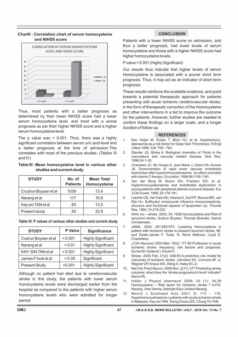
CONCLUSION
REFERENCES
Patients with a lower NIHSS score on admission, and
thus a better prognosis, had lower levels of serum
homocysteine and those with a higher NIHSS score had
higher homocysteine levels.
P value < 0.001 (Highly Significant)
Our results thus indicate that higher levels of serum
Homocysteine is associated with a poorer short term
prognosis. Thus, it may act as an indicator of short term
prognosis.
These results reinforce the available evidence, and point
towards a potential therapeutic approach for patients
presenting with acute ischemic cerebrovascular stroke,
in the form of therapeutic correction of the Homocysteine
and other interventions in a bid to improve the outcome
for the patients. However, further studies are needed to
confirm these findings on a larger scale, and a longer
duration of follow-up.
1. Den Heijer M, Koster T, Blom HJ, et al. Hypertomocy-steinaemia as a risk factor for Deep Vein Thrombosis. N EnglJ Med.1996; 334: 759 – 762.
2. Stamler JS, Slivka A. Biological chemistry of Thiols in thevasculature and vascular related disease. Nutr Rev.1996;54:1-30.
3.
4. Van der Berg M, Boers GH, Franken DG, et al.Hyperhomocysteinaemia and endothelial dysfunction inyoung patients with peripheral arterial occlusive disease. EurJ Clin Invest. 1995; 25:176-181.
5. Leerink CB, Van Ham AD, Heeres A, Duif PF, Bouma BN, vanRijn HJ. Sulfhydryl compounds influence immunoreactivity,structure and functional aspects of lipoprotein (a). ThrombRes. 1994; 74:219-232.
6. AHA; Inc – stroke. 2003; 34; 1258 Homocysteine and Risk ofrecurrent stroke. Gudrun Boysen, Thomas Brander, HanneChristiensen.
7. JAMA. 2004; 291:565-575. Lowering Homocysteine inpatient with Ischemic stroke to present recurrent stroke, MI,and Death,James F. Toole, N. Rene Malinow, Lloyd E.Chambless.
8. J.Clin Neurosci.2003 Mar; 10(2): 177-80 Profession in acuteischemic stroke: frequency, risk factors and prognosis.Sumer M, Ozdemir I, Erturk O.
9. Stroke. 2000 Feb; 31(2): 448-55 A predictive risk model foroutcomes of ischemic stroke. Johnston KC, Connors AF Jr,Wagner DP, Knaus WA, Wang X, Haley EC Jr.
10. Nat Clin Pract Neurol. 2006 Nov; 2(11): 577 Predicting strokeoutcome: what does the 'stroke prognostocrit level' indicate?Sacco RL.
11. Indian J Physiol pharmacol 2009; 53 (1): 34-38Homocysteine – Risk factor for Ischemic stroke ? A.P.S.Narang , Indu Verma, Satinder Kaur,Anisha Narang.
12. Neurol J Southeast Asia 2001; 6: 113 – 119.Hyperhomocystinaemia n patients with acute ischemic strokein Malaysia. Kay-sin TAN, Toong Chow LEE, Chong Tin TAN.
Chambers JC, Mc Gregor A, Jean-Marie J, Obeid OA, Kooner
JS, Demonstration of rapid onset vascular endothelialdysfunction after hyperhomocysteinaemia : an effect reversiblewith vitamin C therapy. Circulation. 1999;99:1156-1160.
STUDY No. of
Patients
Mean Total
Homocysteine
Crudrun Boysen et al 1039 13.4
Narang et al 117 16.8
Kay sin TAN et al 83 13.5
Present study 50 32.9
I.M.A.G.S.B. NEWS BULLETIN / JULY - 2018 Vol. 13 No. 747
ChartII : Correlation chart of serum homocysteine
and NIHSS score
Thus, most patients with a better prognosis as
determined by their lower NIHSS score had a lower
serum homocysteine level, and most with a worse
prognosis as per their higher NIHSS score and a higher
serum homocysteine level.
The p value was < 0.001. Thus, there was a highly
significant correlation between serum uric acid level and
a better prognosis at the time of admission.This
correlates with most of the previous studies. (Tables III
and IV)
Table III : Mean homocysteine level in various other
studies and current study.
STUDY P Valve Significance
Cudrun Boysen et al < 0.001 Highly Significant
Narang et al < 0.01 Highly Significant
KAY- SIN TAN et al < 0.001 Highly Significant
James F took et al = 0.05 Significant
Present Study <0.001 Highly Significant
Table IV: P values of various other studies and current study.
Although no patient had died due to cerebrovascular
stroke in this study, the patients with lower serum
homocysteine levels were discharged earlier from the
hospital as compared to the patients with higher serum
homocysteine levels who were admitted for longer
period.

ABSTRACT
AIMS
SUBJECTS AND METHODS
RESULTS AND CONCLUSION
Menstruation is the endpoint of a series of events which begin in the cerebral cortex and terminates at the uterine
tissues in the HPO axis. Primary amenorrhea can be due to disorder of the outflow tract or uterine target organs
: The objective of this study is to note the various causes, complete clinical picture and the management in
25 such cases of Primary Amenorrhea. : This is A Prospective study done in 25
cases .They were investigated ,managed and patients were called up for follow up for their response to treatment.
: Out of 25 cases studied, maximum cases presented at 14-16 years of age , with
chief complaint of primary amenorrhea, out of which 8% were married, 13 cases were of MRKH syndrome which
was the most common cause of PrimaryAmenorrhea.
Study and Clinical Evaluation of 25 cases of Primary Amenorrhea
ORIGINAL ARTICLE
KEY WORDS : Primary Amennorhea, MRKH syndrome, imperforate hymen
Dr Tushar M Shah*, Dr Foram Lalcheta**,Dr Rini Buddhdev***
*AssociateProfessor ,** 2nd year OBGY resident,***1st year OBGY resident in Obstretics & Gynecology Department
B.J Medical College, Civil Hospital, Ahmedabad.
Correspondence Address : Dr Tushar M Shah
A-902, Status Tower, Opp. Doordarshan, Nr. SAL Hospital, Drive-in-Road, Thaltaj,
Ahmedabad-380054
INTRODUCTION
Primary Amenorrhea is absence of menstruation and
secondary sexual characters by age of 14 years or
absence of menstruation regardless of secondary sexual
characters by age of 16 years.Menstruation, also known
as a 'period' or 'monthly', is a regular discharge of blood
and tissue from the inner linning of the uterus through
vagina . For a normal menstruation to occur the genetic
make up, neuroendocrinological, embryological
development is must .Any abnormality in above leads to
amenorrhea. Mentruation marks the transition from a girl
to a woman. It is the physical manifestation noticed of all
pubertal changes and last to occur . Incidence of
Primary Amenorrhea is <1%.Development of female
genital organs takes place from MULLERIAN DUCT
(Paramesonephric Duct). Sexual development starts
after 8 weeks.In absence of Y chromosome, functional
testes and lack ofAMH, Mullerian Duct develops and form
uterus, fallopian tubes and upper vagina and at the same
time in absence of testosterone, Formed from coelomic
Epithelium.Hymen is formed by invagination of posterial
wall of urogenital sinus and usually ruptures-
spontaneously in perinatal period. .Menarche is the end of
cascade of events occurring in puberty.
Causes of primary amenorrhea can be classified
according to WHO as:
1. HYPOGONADOTROPIC
HYPOGONADISM (GroupI)
(1)
(2)
(4)
[decreased estrogen, normal or low FSH,and
no lesion in hypothalamic-pituitary region]
Physiologic Delay.
Kallman syndrome
CNS tumors
2. HYPERGONADOTROPIC
HYPOGONADISM (Group II)
[decreased estrogen, but raised FSH]
3. DEVELOPMENT DEFECT OF
GENITALTRACT (GroupIII)
The objective of this study is to evaluate the age of
presentation, presenting symptom, clinical findings of
general and local examination, causes, outcome and
management of 25 cases of primary Amenorrhea
presented in Tertiary care hospital for a duration of 1 year
from November 2016 to November 2017.
This is a prospective study. A methodical, systemic
approach was developed to achieve best diagnosis.
Evaluation of primary Amen or rheaisdesigned to
separate there productive system into its distinct
structural components such as Genital outflow track,
Uterus, Ovaries, Pituitary, Hypothalamus and to test
functional integrity of each beginning at lowest level and
OBJECTIVES
MATERIALS AND METHODS
I.M.A.G.S.B. NEWS BULLETIN / JULY - 2018 Vol. 13 No. 748

Table 1: Age distribution of Patients
Out of 25 cases, 3 of the patients were married and
22 were unmarried.
Causes of PrimaryAmenorrhea:
MRKH syndrome is the most common cause of
Primary Amennorhea in our Study i.e 52% . These
patients had normal secondary sexual characters
that was symmetric with age. They had normal
Development of breasts which indicates normal
ovarian function(6). All had normal karyotyping
(46XX).MRI is gold standard to diagnose any
congenital anomalies . These patients were
explained about their pathology and counseled
regarding their menstrual and reproductive function.
Primary goal of management in MRKH syndrome is
creation of functional vagina. They were explained
about vaignaplasty and were called for follow-up
before 6 months of their marriage. MRKH syndrome
is associated with renal, cardiac and skeletal
malformations . Thus all the cases of MRKH
syndrome underwent USG KUB, 2D echo and X-ray
spine. 16% of them were associated with abnormal
USG KUB.
(3)
(7)
There were 7 cases of imperforate hymen,
5 presented with complaint of cyclical lower
abdominal pain and 2 case with urinary retention. All
of them had normal symmetrical secondary sexual
characters and normal breast development
indicating normal ovarian function.All the cases were
manged surgically and 1 case with recurrent
Hematomet ra and Hematoco lpos , to ta l
hysterectomy was done, Ovaries were preserved
Of 3 patients with transverse vaginal septum, both
had resumedmenses regularly post resection of
septum and vaginoplasty.
Age Percentage
11- 13 Years 6 (24%)
14- 16 Years 9 (36%)
17- 19 Years 4 (16%)
19- 21 Years 3 (12%)
> than 21 Years 3 (12%)
Table 2 : Presenting Symptoms
Symptom Percent age
PrimaryAmenorrhea 13 (52%)
Cyclical abdominal pain 5 (20%)
Ambiguous Genitalia 3 (12%)
Urinary Retention 4 (16%)
USG KUB Percentage
Normal 7 (53%)
Horse shoe kidney 1 (0.7 %)
Fused Crossed Ectopic
Kidney 1 (0.7%)
Absent Kidney 2 (0.15%)
Table 3 : Causes
Causes Percentage
MRKH Syndrome 13(52%)
Imperforate Hymen 10(40%)
Turner syndrome 1 (4%)
Uterus didelpysis blind
vaginablind vagina 1(4%)
Presenting Symptoms:
progressing systemicallyto higher levels until the cause is
determined. 25 Case of primary Amenorrhea were
studied in detail for following parameters-
Age ofpresentation
Presenting complaint
MaritalStatus
Past history of any medicalillness
FamilyHistory
General Built (Height, weight, BMI and armspan)
Presence of secondary sexual characters (according
to tanner'sstaging)
Localexamination(P/A,P/vulval,P/S,P/V)
Hormonal profile(FSH,LH, Estrogen,AMH,TSH)
Radiological investigations ( USG,MRI)
Special investigations(CT –IVP)
Karyotyping
Presenting age was in range from 11 to 25 years.
RESULTS
I.M.A.G.S.B. NEWS BULLETIN / JULY - 2018 Vol. 13 No. 749

Patient with turner syndrome presented with c/o
primary amenorrhea with short stature,shield
chest(widely spaced nipples) with underdeveloped
breast and absent pubic and axillary hairs (Tanner
stage I). MRI pelvis suggested of 6X4 mm of
hypoplastic uterus with absent ovaries. Karyotyping
s/o mosaicism 45,x,(92)/46,x,i(x)(910)(8) , 92% of
cell with XO and 8% of cells with XX. She was given
withdrawal with Oral contraceptive pills(MALA-N)
and since then patient is menstruating normally.2D
ECHO and USG KUB was normal .
One patient had uterus didelphis with 2 separate
cervix with two localized
collection in vaginal fornix, right ovary 6×3 cm
complex cyst, and blind vagina In this patient
laprotomy was done to remove bothuterus and
vaginoplasty was done
3. Troiano RN. Magnetic resonance imaging of mullerian duct
anomalies of the uterus. Top MagnReson Imaging 2003;
14:269–79.
4. Griffin JE, Edwards C, Madden JD, Harrod MJ, Wilson JD:
Congenital absence of the vagina. The Mayer-Rokitansky-
Kuster-Hauser syndrome. Ann Intern Med 1976, 85:224-236.
++PubMedAbstract
5. Folch M, Pigem I, Konje JC: Mullerian agenesis: etiology,
diagnosis, and management. ObstetGynecolSurv 2000,
55:644-649. PubMedAbstract | Publisher Full Text
6. Varner RE, Younger JB, Blackwell RE: Mulleriandysgenesis. J
Reprod Med 1985, 30:443-450. PubMedAbstract
7. Strubbe EH, Willemsen WN, Lemmens JA, Thijn CJ, Rolland
R: Mayer-Rokitansky-Kuster -Hauser syndrome: distinction
between two forms based on excretory urographic,
sonographic, and laparoscopic findings. Am J Roentgenol
1993, 160:331-334.
8. VARNER, R.E., J.B. YOUNGER & R.E. BLACKWELL.1985.
9. Mulleriandysgenesis. J. Reprod. Med. 30: 443
10. Marc Fritz,LeonSperoff clinical gynecologic endocrinology
and infertility 8thedition pg 124-127.
11. MarcFritz,LeonSperoff clinical gynecologic endocrinology
and infertility 8thedition pg 435-450.
CONCLUSION
REFERENCES
Amenorrhea has got multifactorial etiology. For patients
with Amenorrhea physical examination should focus on
pubertal development and possible genital outflow
obstruction. Mullerianagenesis(MRKH syndrome) was
the most common cause of primary amennorhea. Early
surgery offered to patients may reduce patients suffering ,
help restore patients outflow tract and may preserve
fertility in some cases .Research on mullerian anomalies
is limited and further studies are needed.Out of 25 cases
of Primary Amenorrhea , 52% had MRKH syndrome
which was the most common cause of primary
amenorrhea 28% imperforate Hymen, 12% had
transverse vaginal septum.
1. John A. Rock, Howard W. Jones. TeLinde' s Operative
Gynaecology . 10th ed. New Delhi :Lipincott Williams and
Wilkins; 2009 . p. 550-551
2. John A. Rock, Howard W. Jones. TeLinde' s Operative
Gynaecology . 10th ed. New Delhi :Lipincott Williams and
Wilkins; 2009 . p. 509-511
I.M.A.G.S.B. NEWS BULLETIN / JULY - 2018 Vol. 13 No. 750

INTRODUCTION
Anemia in the elderly is an extremely common problem
that is associated with increased morbidity and poor
health related quality of life. It is easy to overlook anemia
in the elderly since symptoms like fatigue, weakness or
shortness of breath may be attributed to aging process
itself and should never be accepted as an inevitable
consequence of aging. A progressive statistical increase
in the number of elderly persons has been observed as a
universal phenomenon. Thus, anemia in the elderly
patients is an emerging global health problem for the 21st
Century which negatively impacts the quality of life.
Aging by itself is unlikely to cause anemia. Hemoglobin
levels in the healthy older individuals do not change
significantly from 60 to 98 years of age. Changes that
occur commonly during aging, increase the risk of
anemia, thus explaining the association of anemia with
old age. These include reduced ability to absorb essential
nutrients, decreased hematopoietic reserve and reduced
sensitivity to erythropoietin.
[1]
[2]
[2]
[3]
OBJECTIVES
MATERIALS AND METHOD
To study the clinico-hematological patterns of
anemia in the elderly patients 60 years and above.
To detect the morphological types of anemia
prevalent amongst them.
To know common etiology for anemia.
To know various associated disorders.
The present study is a descriptional cross sectional study
which was conducted in the Department of
Pathology,P.D.U.Government Medical College,Rajkot
over period of one year i.e. 1st August 2016 to 31st
September 2017. All the indoor patients who were60
years and above and clinically diagnosed as anaemic
were included.Routine haematological investigations.
Peripheral Blood smear examination using Field stain and
Le ishman s ta in ,Comple tehemogram.Spec ia l
investigations like Iron studies, Reticulocyte count, Perl's
ABSTRACT
INTRODUCTION :
AIMS AND OBJECTIVES:
MATERIALS AND METHODOLOGY :
RESULTS :
CONCLUSION :
Anemia in the elderly is a major health problem in India and many parts of the world, since it
signifies an underlying disease and is associated with poor clinical outcome like increased morbidity and poor
health related quality of life. Since symptoms like fatigue or shortness of breath associated with anemia may be
attributed to aging process, anemia can be easily overlooked in the elderly.
Clinico-hematological patterns and morphological types of anemia in elderly are
manifold, hence, this study was undertaken to determine them and to know associated disorders
The present study was conducted on a sample size of 200 patients (A one
year study) who were60 years and above and clinically diagnosed as anemic. Routine haematological
investigations including Peripheral Blood smear examination and Completehemogram were done. Special
investigations like Bone-marrow examination, Iron studies etc. were done whenever required.
Males were more affected than females and patients in the age group of 60-69 years were
affected the most. The most common presenting symptom was generalized weakness. Normocytic Norm chromic
anemia was the most common morphological type and chronic diseases were the commonest etiological factors.
Despite modern diagnostic advances, geriatric anemias still remain under-reported and
inadequately investigated, necessitating evaluation of even mild anemias. Prompt diagnosis and definite
categorization helps in appropriate management of anemias.
A Clinico-Pathological Study of Anemias in Geriatric Age Group
ORIGINAL ARTICLE
KEY WORDS : Normocytic Normochromic Anemia; Anemia of chronic disease, Elderly
Amit H.Agravat*, Trushali Dobariya**, Gauravi A Dhruva***
*Associate Professor, **Resident Doctor, ***Professor & Head, Department of Pathology,
P.D.U. Government Medical College, Rajkot.
Correspondence Address : Dr. Trushali Dobariya
Resident Doctor, Department of Pathology, P.D.U. Government medical college, Rajkot
E-mail : [email protected]
I.M.A.G.S.B. NEWS BULLETIN / JULY - 2018 Vol. 13 No. 759

I.M.A.G.S.B. NEWS BULLETIN / JULY - 2018 Vol. 13 No. 760
Table 2 : Relation with Symptoms and Sign
The above table indicates that, maximum number of
subjects 154 were in the age group of 60-70years, 33
subjects in the age group of 71-70 years and 13 were
subjects in the age group of 80 years and above.
Figure 1 : Pie diagram representing Gender wise
distribution of Cases
In this study, non-specific symptoms were most
commonly associated with anemia, followed by
symptoms and signs of Respiratory disease, gastro-
intestinal diseases, carcinoma, nutritional disorders, liver
and renal diseases.
Table 3 : Peripheral Blood Smear Patterns
In the present study, we observed that, the most common
morphological type of anemia was Normocytic
normochromic (45%) followed by Hypochromic
microcytic (30%), Dimorphic (16%), Normocytic
hypochromic anemia (5%) and Macrocytic (4%) which
was the least common.In present study 28 % patient were smoker, 23% were
with hypertension, 23.5 % were having Diabetes mellitus,
20 % with blood loss and 11 % were using NSAIDS
The above figure show that, 53% subjects were males
and 47 % werefemales in the present study.
Figure 2: Correlation withAssociated Factors
Age group Total Number Percentage
(years) (n=200) (100%)
60-65 120 60 %
66-70 34 17 %
71-75 20 10 %
76-80 13 6.5 %
81-85 9 4.5 %
85-90 4 2 %
Total 200 100 %
Associated
factor
Total number Percentage
n=200 (100%)
Respiratory 36 18 %
Gestrointestinal 30 15 %
Carcinomas 27 13.5 %
Nutritional disorders 26 13 %
Liver 14 7 %
Renal 13 6.5 %
Non specific 54 27 %
Total 200 100 %
Peripheral Blood Total Percentage
Smear Findings Numbern=200 100%
Normo-Normocytic
chroimc Anemia 90 45 %
Hypochroic Microcytic
Anemia 60 30 %
Dimorphic Anemia 32 16 %
Normocytic
Hypochromimc Anemia 10 5 %
Macrocytic Anemia 8 4 %
Total 200 100 %
47%
53%
Male Female
Stain and bone-marrow examination etc. were done
whenever required.
OBSERVATIONS AND ANALYSIS
Table 1 : Age wise distribution of Cases

Figure 3: Pie diagram representing causes of Anemia
I.M.A.G.S.B. NEWS BULLETIN / JULY - 2018 Vol. 13 No. 761
We observed in the present study that, anemia due to
chronic disease (52.%) was the most common type
followed by Iron deficiency anemia (17%), anemia due to
other nutritional deficiencies (13%), anemia due to blood
loss and the least common was anemia due to
hemolysis.(1%)
In the present study, more males were found to be anemic as compared to females. A similar gender wise distribution
was noted in the study by Wasimkhatib et al , Ramya et al . Study conducted by Bird et al in which 51 % were male
41 % females. Our result is differs from study done by S.Amarneel et al , Joosten et al , Mathew RongJie Tay et al in
which Females were more anemic.
[5] [6]
[4] [7] [8]
In the present study, patients in the age group of 60-70 years were maximally affected which is in concurrence with the
study by S.Amarneel et al.
Nisha TR et al , and Kiranaithal et al whereas, in a study done by Tabea Geisel et al , patients in the age group of
80-90 years were maximally affect.
[9] [10] [11]
Figure 4: Age group wise Grading of Anemias
Table 4 : Comparative Study of Gender wise distribution of Geriatric Anemias
Table 5: Comparative Study of Maximally Affected Age Group
Gender
Present
study
(Rajkot)
[2017]
n=200
S.amarneel
et al
(Bhavnagar
Gujarat)
[ 2015]
n=42
Wasim
Khatib
et al ( karad,
Maharashtra
[2016]
n=256
Ramya
et al,
(Puduchhery)
[2016]
n=675
Joosten
et al
(Belgium)
[1992]
n=178
Mathew Rongjie
Tay et al.
(Singapore)
[2011]
n=424
Male 53.9 % 51 %
71.4 % 61.2 % 51.4 %
53 % 28.6 % 38.8 % 48.6 %
Female 47 % 46 % 49 %
Age
group
(Years
Present
study
(Rajkot,
Gujarat)
2017
n=200
S.Amarneelet
al.
(Bhavnagar,
Gujarat)
2015
n=42
Nisha TR
et al
(Kozhikode,
Kerala)
[2017]
n=826
KiranAithal
et al
(Dharwad
Karnataka)
[2017]
n=100
Tabea Geisel
et al.
(Germany)
[2017]
n=388
50-59 - - 18.3 % - -
60-70 77 % 61.25 % 44 % 70 % 13.9 %
71-80 17 % 27.5 % 26.8 % 23 % 40.2 %
81-90 6 % 11.25 % 10.8 % 7 % 46 %
Above graph is showing that out of 200 patients 91 were
having Grade 2
( moderateAnemia).
DISCUSSION

I.M.A.G.S.B. NEWS BULLETIN / JULY - 2018 Vol. 13 No. 762
In the present study, most common underlying cause of anemia is anemia of chronic disease, This finding is in concurrence with
the study by Guyatt et al , Nisha TR et al and Joosten et al in which, chronic disease was maximally responsible for anemia
followed by iron deficiency anemia.. In hematological malignancy Chronic myeloid leukemia was present in 4( 0.8 %) subjects in
present study, correlates with KiranAithal et al in which 1% was noted. Chronic leukemia and lympho-proliferative disorder was
noted in 0.4 % which is differs from the study conducted by Nish TR et al having 9.7 % and Vijay Tailak et al 70having 2.2 % of
Chronic leukemia and lympho-proliferative disorder. In present study Myelodysplastic syndrome is present in 2 ( 0.4%) subjects
concurrent with study done by Vijay Tailak et al having 1.4% of subjects with Myelodysplastic syndrome.
[12]
[13]
In the present study, highest number of the subjects are with moderate degree ( Grade II) of anemia. This finding is in
concurrence with the study by Suma J.K. et al andJoosten et al.
Our result differs from the study conducted by Nish TR et al, Ramya et al, in which majority of elderly were having mild
anemia( Grade I ).
Table 6 : Comparative Study of contributory causes resulting in Anemia
Table 7 : Comparative Study of Grading of Anemia
Cause of Anemia
Grade of Anemia
Present
study
(Rajkot,
Gujarat)
[2017] n=200
Present
study
(Rajkot,
Gujarat)
[2017] n=200
Nisha TR
et al
(Kozhikode
Kerala)
[2017] n=500
Nisha TR
et al
(Kozhikode,
Kerala)
[2017] n=826
Guyatt
et al.
(Ontario,
Canada)
[1990] n=259
Suma J.K.
et al
(Mysore)
[2013]
n=114
Joostenet
al
(Belgium)
[1992]
n=178
Ramya
et al,
(Puducherry)
[2016]
n=675
Mathew
RongjieTay
et al.
( Singapore)
[2011] n=424
Joosten
et al
(Belgium)
[1992]
n=178
Iron deficiency Anemia 17 % 12.2 % 36.3 % 15 % 13 %
Anemia of Chronic disease 52 % 48.9% 43.6 % 41.5 % 29.3 %
Nutritional anemia 13 % 6.9 % 8.10 % 5.5 % 13 %
Blood loss 17 % 8.5 % - 7.0 % -
Hematological malignancy 1 % 18.5 % 2.70 % 11 % 0. 7 %
Others 1 % 5 % 9.3 % 20 % 44 %
Mild (10-12gm/dl) 28 % 68.8 % 19.29 % 80.9 % 29.2 %
Moderate(7-10 gm/dl) 46 % 26.3 % 16.7 % 16.7 % 57.9 %
Severe (<7 gm/dl) 26 % 4.9 % 2.4 % 2.4 % 12.9 %
Renal 10 % 12 % 50 % - 56.6 %
Respiratory 28 % 36.4 % 17.5 % 34. 8 % -
Carcinoma 20 % 15.2 % - 34.8 % 12.04
Arthritis 8 % 12.1 % 12.5 - 15.7
Table 8: Comparative Study of Associated Co-morbidities
Associated
Comorbidities
Present
study
(Rajkot
Gujarat)
[2017] n=130
Suma J.K.
et al
(Maysore)
[2013]
n=33
KiranAithal
et al
(Dharwad
Karnataka)
[2017] n =100
Mathew
RongjieTay
et al.
(Singapore)
[2011] n=23
Tabea Geisel
et al
(Germany)
[2017]
n=83
GI Disorder 23 % 18.2 % - 8.69 % 15.66
Liver 11 % 6.0 % 20 % 21.73 % -

I.M.A.G.S.B. NEWS BULLETIN / JULY - 2018 Vol. 13 No. 763
In present study Respiratory Disease is associated in most of the subjects (28.13 %)which correlates with study
conducted by Suma J.K. et al(36.4%) and Mathew RongjieTay (34.8 %),next common condition associated was
Gestro intestinal disorder .In hematological malignancy Chronic myeloid leukemia was present in 4( 0.8 %)
subjects in present study, correlates with KiranAithal et al 68 in which 1% was noted. Chronic leukemia and
lympho-proliferative disorder was noted in0.4 % which is differs from the study conducted by Nish TR et al
having9.7 % and Vijay Tailak et al 70 having 2.2 % of Chronic leukemia and lympho-proliferative disorder.
studyMyelodysplastic syndrome is present in 2(0.4%) subjects concurrent with study done by Vijay Tailak et al et
al having 1.4% of subjects with Myelodysplastic syndrome
CONCLUSION
REFERENCES
Despite the modern diagnostic advances, geriatric
anemia still remain under-reported and inadequately
investigated, especially when mild, thereby necessitating
evaluation of even mild anemias in this vulnerable
population. Non specific symptoms like fatigue and
weakness should not be ignored attributing it to normal
aging process as it can be important signal to presence of
anemia. Improved definitions of anemia and more
detailed investigations like bone marrow aspiration and
biopsy also help to define the subtypes of anemia,
thereby facilitating prompt and accurate diagnosis to
ensure appropriate patient management.
1. Gary J Vanasse, Nancy Berliner. Anemia in elderly patients: An
emerging problem for the 21stcentury. Harvard Medical
School, Hematology 2010; 271-4.
2. Molivares, Eltertrompf, Capurro MT, Wegner D. Prevalence of
anemia in elderly subjects living at home: role of micronutrient
deficiency and inflammation. European Journal of Clinical
Nutrition 2000;54:834-9.
3. Guralnik Jack M, Eisenstaedt RS, Luigi Ferrucci. Prevalence of
anemia in persons 65 years and older adults. Blood. 2004
Oct;104:2263-9.
4. S Amarneel, N. Sheth, Pattern of anemia in elderly age group;
International journal of scientific research and reviews
2015,49(2),51-56.
5. Wasim M.Khatib,TasneemV.Bisht,Anup N Gosavi;
Clinopathological profile of anemia in elderly-Aone year study;
IOSR-JDMS Volume 15,issue 9 (September 2016)PP 85-87.
6. Ramya Gandhi, NityaSelvaraj, Vimal Mourouguessine;Study
of prevalence and morphological pattern of anaemia in adult
and geriatric population: A hospital based study, J Evolution
Med. Dent. Sci 2016; 5(69):5041-5044.
7. Joosten E, Pelemans W, Hiele M, Noyyen J. Prevalence and
causes of anemia in a geriatric hospitalized population.
Gerantol1992;38:111-7.
8. Mathew RongjieJie Tay, Yong Youong. Prevalence and risk
factors of anaemia in older hospitalized patients. Singapore
Health Care 2011;20(2):71-9.
9. ThundiparambilRaghavanNisha,Gopalan Nair Rajan, Prasad
PH; Evaluation ofanemia in patients aged 55 years and above
in a tertiary care setting in north kerala,J Evolution
Med.Dent.Sci.2017;69(48):3694-3697.
10. KiranAithal, KirankumarMeti, Sathvik Jain; A study of anemia in
elderly patients admitted at tertiary center.Sch.J.App.Med. Sci,
2017; 5(4D): 1483-86.
11. Tabea Geisel, Julia Martin, Bettina Schulze:An Etiologic Profile
of anemia in 405 Geriatric patients.Volume2014.
12. Gordon H Guyatt, ChristoherPallerson, Monmoud Ali, Joel
Singer, Mark Levine, IraneTurpie, et al. Diagnosis of iron-
deficiency anemia in elderly. American Journal of Medicine.
1990;88:205-9.
13.Tilak V, Tilak R.; Geriatric anemia- A public helath crisis in
hematology. Indian J PrevSoc Med 2012;43(2).

ABSTRACT
Objective :
Method :
Results :
Conclusion :
To identify and appraise the concerns different patients have prior to undergoing surgery in a rural set
up. In this prospective survey, 207 patients completed a questionnaire regarding their concerns prior to
undergoing surgery. Patients responded by answering the questions and sharing their concerns and fears. The
study was conducted over a period of one year (2016 – 2017). Patient population consisted of 207
consenting adults undergoing elective major and minor surgery in a rural setup. Patients included 149 men, 57
women and 1 newborn. The male:female ratio being 2.6:1.0. The age of the patients ranged from 10 years to 75
years. 150 patients were scheduled to undergo urological procedures, and remaining 57 were to undergo gastro
intestinal surgeries. Out of all the concerns listed, 35.27% of the total patients who were also found to be unskilled
laborers had foremost concern regarding the hospital and medical expenses. The female patients had soaring
apprehensions about their family, children and other household works (24.64%). 18.36% patients confirmed
having issues concerning postoperative backaches owing to spinal anesthesia. 7.73% patients were found to
have mixed concerns. And only about 14% of patients said that they had no fear regarding their surgeries.
In a rural setup, preoperative counseling of a patient by the doctor definitely helps in post operative
recovery. In case of female patients, counseling of family members is also equally important. This data provides
information that will be helpful in preoperative patient counseling and in generating awareness for patients
undergoing surgeries and their relatives.
Pre-Operative concerns of a patient in a Rural setup :RURAL SETUP: An Indian Perspective
ORIGINAL ARTICLE
KEY WORDS : Pre-operative concerns, Rural Setup, Counseling
Heta Rajyaguru*, Dipak Rajyaguru**, Pravin Baldaniya**, Gayatri Raval***
*Intern, ** Urologist, ***Gynecologist
Urocare Super Speciality Hospital, Mehsana, Sadbhavna Trust Hospital Mahuva, Bhavnagar.
Correspondence Address : Dr. Heta Rajyaguru
Urocare Super Speciality Hospital, Narayan Shopping Centre, O/s. Gopinala,
Mehsana-384002. E mail: [email protected]
I.M.A.G.S.B. NEWS BULLETIN / JULY - 2018 Vol. 13 No. 764
Precutaneous Nephrolithotomy (PCNL), Ureteroscopy
(URS), Transurethral Resection (TUR), Laparoscopic
Appendectomy and Laparoscopic Cholecystectomy are
some of the most commonly performed operations in
Urology and Gastroenterology. Although information
concerning the clinical and surgical outcome is abundant,
little attention has been focused on the concerns of the
patient prior to undergoing these minimal invasive or
other major invasive surgeries. Going under the knife and
being hospitalized is a stressful experience that affects
the physical and psychological well being of a patient,
which can adversely have an effect on post operative
recovery and to some extent mental health of the patient
and their relatives as well. Studies have reported that lack
of preoperative preparation and counseling of patients,
not addressing postoperative concerns and anxiety, and
being insensitive to the negative thoughts and belief
system of patients and their relatives are remarkably
related with psychological and psychiatric disorders. In
addition, review of literature have recommended that
health-care professionals, especially surgeons, need to
INTRODUCTION counsel the patient and their family members before and
after surgery, but doctors are usually unenthusiastic and
least interested to provide essential information and
counseling related to surgery and do not empathize
toward the patients day-to-day concerns. In a rural set up
patients with lower educational qualification and families
belonging to lower socioeconomic group were also found
to be least concerned and minimally informed.
Unexpected sickness, non preparation for hospital stay,
fears allied to surgical procedures, post operative follow
ups, hospital and medical expenses, long hours of wait
and caregivers responsibi l i t ies adds to the
inconveniences and increases the stress and anxiety
among patients and their families. Counseling is the
guidance offered by trained therapist or doctors to help
the patient and their family members resolve social,
personal or medical problem. Offering counseling
sessions is too time-consuming for today's busy surgeon
owing to the increased outflow of patients and also due to
the lack of training in communication. The purpose of this
study is to document and examine the concerns patients
have prior to undergoing above mentioned as well as

I.M.A.G.S.B. NEWS BULLETIN / JULY - 2018 Vol. 13 No. 765
other Urological and Gastro Intestinal procedures in both
a large Trust Hospital and a moderately sized Urologic
Private Practice.
Two hundred and seven (207) patients undergoing
various urological and gastro-intestinal surgeries
completed a questionnaire covering 12 items, regarding
their personal data, concerns, issues and anxiety prior to
surgery. Patients who could not understand English and
Gujarati, who had psychiatric illness or mental
retardation, or who refused to give consent and a few
pediatric cases were excluded. The patients were given
the survey after having met their surgeon before the
surgery and also after the surgery was over. All the
patients were interviewed when they were most
comfortable resting in their cots in the presence of their
relatives. The patients were surveyed at both a large trust
hospital in Mahuva – Sadbhavna Trust Hospital, Kalsar,
Mahuva, District Bhavnagar, Gujarat, India. And a
moderate sized private practice hospital – Urocare
Superspeciality Hospital, Mehsana, Gujarat, India. The
information covered during the survey and the methods of
delivery were standardized for all patients. General
patient demographics, medical histories, current issues
about anxiety regarding surgery, thoughts regarding
same issues post surgery and how the anxiety, concerns
and issues were taken care of were all documented.
Patients responded verbally and concerns were noted
down and listed. Responses were examined for
association with patients gender; i.e male or female, the
type of surgery whether urological or gastrointestinal,
private set up or trust hospital, the socioeconomic status
of the patient (according to Prasad's classification),
occupation of the patient, if the patient had had previous
surgery, if the patient had a previous surgical complication
and if the patient had discussed risks, concerns, and
anxieties with the concerned surgeon.
A total of 207 patients were interviewed in this perspective
survey over a period of one year between 2016 to 2017, in
a trust hospital and a private care hospital. The mean age
of the respondent was 42.5 years (range 10 years to 75
years), 135 respondents were younger then 55 yrs and 66
respondents were aged 55 yrs or older (Table 1). 57 of the
total 207 respondents were females and the remaining
150 were males respondents (Table 2). 72.46% of the
patients were to undergo urological surgeries and the
remaining 27.54% were to undergo gastrointestinal
surgeries (Table 3 and Pie chart 1). There were 125
patients surveyed at the trust hospital and 82 surveyed at
the private care hospital (Pie Chart 2). Almost all of the
PATIENTS AND METHODS
RESULTS
female patients (24.64%), 51 women had concerns
regarding their household work and their families as to
who will take care of the household chores/children in
their absence.Around 20.77% (43 patients) had concerns
regarding pain relief, symptoms relief, recurrence of
disease, medication, and recurrent hospitalization. 18.36
% (38 patients) had had previous surgeries and all of them
had experienced discomfort regarding backache due to
spinal anesthesia, which made them anxious and they
showed fear of anesthesia. 14.50% (30 patients) were
either unskilled workers like laborers', watchmen,
carpenters, car mechanics, tailors, farmers, goldsmiths
etc. (Pie chart 3), all of these patients were mainly
concerned about the expenses regarding the surgery and
the medication and the number of days they will miss at
work. 7.73% (16 patients) shared mixed concerns and
issues like fear of surgery and hospitalization, caretakers
role and responsibilities, anxiety etc. And the remaining
14 % (29 patients) had no fears regarding their surgeries
(Table 4).
Table 1 : Age Distribution
Table 2 : Sex Distribution
Pie Chart 1 : Type of Surgery
Sex No. of patients Percentage
Male 150 72.46%
Female 57 27.54%
Total 207 100%
Age range No. of patients Percentage
= < 20 years 20 9.66%
21 – 40 years 44 21.25%
41 – 60 years 100 48.32%
61 – 80 years 43 20.77%
Total 207 100%

I.M.A.G.S.B. NEWS BULLETIN / JULY - 2018 Vol. 13 No. 766
Table 4 : Preoperative concerns of the patients
Table 3 : Type of Surgery and Procedures carried out
Type of surgery Procedure No. of patients Percentage
Urology PCNL 60 40%
TUR 36 24%
URS 3 22%
Hernioplasty 09 6%
Nephrectomy 03 2%
Stricture Urethra 03 2%
VesicoVaginal Fistula 03 2%
Litholepexy 03 2%
Total 150 100%
Gastrointestinal Lap. Appendectomy 15 26.31%
Lap. Cholecystectomy 12 21.05%
Hemorrhoids 12 21.05%
Open Appendectomy 08 14.03%
Open Cholecystectomy 03 5.26%
ERCP 02 3.51%
Epigastric Hernia 02 3.51%
Liver abscess 01 1.76%
Superficial Parotidectomy 01 1.76%
Ileostomy closure 01 1.76%
Total 57 100%
Concerns and Issues No. of patients Percentage
Who will take care of the household chores/children
(specially females patients). 51 24.64%
Pain relief, symptoms relief, recurrance of disease,
medication, recurrent hospitalization. 43 20.77%
Fear of anesthesia (post operative backaches). 38 18.36%
Missing no. of days of work
(patients working on daily wages). 30 14.50%
Mixed concerns and issues like fear of surgery and
hospitalization, caretakers role and responsibilities, anxiety etc. 16 7.73%
No Fears, Anxieties and Concerns 29 14.00%
Total 207 100%

I.M.A.G.S.B. NEWS BULLETIN / JULY - 2018 Vol. 13 No. 767
Pie Chart 2 : Type of Rural setup
Pie chart 3 : Occupation of the patients
DISCUSSION
Precutaneous Nephrolithotomy (PCNL), Ureteroscopy
(URS), Transurethral Resection (TUR), Laparoscopic
Appendectomy and Laparoscopic Cholecystectomy are
some of the most commonly performed minimal invasive
procedures in urology and gastroenterology that can be
extremely distressing to the patients. Surgeons have
appropriately focused their attention on outcomes after
these surgeries, but patient concerns prior to undergoing
theses procedures have received little attention. Pain
after the surgery, length of recovery, expenses, fear of
anesthesia, care of the family members in absence
(specially for female patient) were the most notable
concerns of the group as a whole prior to any of the
procedures.
Any surgery be it major or minimally invasive is a terrifying
experience for the patient. It was observed that most of
the time, neither the patients nor their family members are
aware of the illness or the surgical procedures to be
carried out. Thus, patients and their caregivers who are
awaiting their turn for the proposed operation face and
experience loads of worries and tensions which includes
stress, anxiety, depression and may turn out to be
uncooperative to the doctor and the treatment. Hence
being sensitive and understanding towards the concerns
of the patients and giving them pre operative counseling
sessions will alleviate the fears, clarify concerns related to
surgery, duration of surgery, role of anesthesia, and to
prepare the patients psychologically for surgical
interventions, it will also give them a reasonable
understanding of the planned surgery and its prognosis,
further it will create awareness amongst them regarding
the procedure and post surgical pain and complications if
any. Studies have reported that giving preoperative
counseling sessions has a positive effect on the patient
and it minimizes the postoperative stress level, the pain
and anxiety of the patients as well as their relatives.
Another significant intervention needed in these
counseling sessions is identifying, appraising and
understanding the psychosocial necessitates, issues and
concerns of both patients and their family members.
Postoperative counseling too is equally necessary and
helpful. This phase begins immediately after surgery and
continuous until patient is discharged from the hospital.
The purpose of the postoperative counseling is to deal
with the postoperative fears, anxiety, concerns and other
emotional issues if any. It also provides supportive care
for patients and their relatives and builds up a rapport with
the concerned doctor. The other day-to-day concerns
such as suture removal, wound cleaning and dressing,
pain education and management, drug compliance, and
explaining the likely healing and recovery would be of
immense assistance for the patients and their family
members. A brief knowledge about illness and providing
necessary guidance and counseling helps the patient to
adjust with postoperative environment.
The family members of the patient too needs to be
thoroughly educated regarding the illness, the causes,
the procedure, the post operative prognosis and side
effects, their role and their responsibilities in the hospital
and at home. Studies have also reported that patients
and their caretakers who are contented and satisfied with
preoperative and post operative counseling sessions
show significant improvement and demonstrate good
psychological adjustments. Patients and their relatives
who have not received any counseling or those that are
dissatisfied with the sessions are more prone to develop
psychological and psychiatric disorders. Consequently,
both pre operative and postoperative counseling of
patients and their relatives is crucial.
In a rural setup be it private or a trust hospital
preoperative and post operative counseling by the
concerned operating surgeon definitely has many
positive effects on patient's physical and mental recovery,
coping efforts, attitude, it reduces anxiety and improves
mood disturbances. Counseling helps in allowing the
patient and family members to prepare for the future. In
CONCLUSION

I.M.A.G.S.B. NEWS BULLETIN / JULY - 2018 Vol. 13 No. 768
12 Preoperativeconcernsof patients undergoing endoscopic sinus
surgery. Yeung JC1, Brandt MG, Franklin JH, Doyle PC,
Rotenberg BW, Kilty SJ. 2014 Aug;4(8):658-62. doi:
10.1002/alr.21330. Epub 2014Apr 9.
13. REVISTA BRASILEIRA DE ANESTESIOLOGIA “Preoperative
patient education: can we improve satisfaction and reduce
anxiety?”: Jaime Ortiz a, Suwei Wang, MacArthur A. Elayda,
DanielA. Tolpinc
14. The Effect of Preoperative Counseling on Patient Fear From
Visual Experience During Vitrectomy Surgery Under Regional
Anesthesia.https://clinicaltrials. gov/ct2/show/NCT01383629
15. Robert T. Trousdale, Brain J. McGrory, Daniel J. Berry, Michael
W. Becker and William S. Harmsen. Patients' Concerns prior to
undergoing Total Hip and Total Knee Arthroplasty. Mayo clin
Proc.1999;74:978-982
case of female patients, counseling of relatives and other
family members is also very important. In a concluding
note, a doctor plays an important role by providing
informational, educational and by addressing the
concerns and the psychosocial issues of patients and
their family members. The concerned surgeon ensures
retaining the patient for follow-up and adhering them to
treatment schedules in all the stages of their illness, be it
preoperative or post operative, a trust hospital or a private
setup. Therefore, every hospitalized patient undergoing
any major or minor surgical procedure and their family
members should be provided with pre operative, post
operative and lastly pre discharge counseling sessions
for effectively addressing the psychosocial needs and
concerns of the patients and caretakers in a rural set up.
REFERENCES
1. Birudu Raju and Krishna Reddy. Are Counseling Services
Necessary for the Surgical Patients and their Family Members
during Hospitalization? J Neurosci Rural Pract. 2017 Jan-Mar;
8(1): 114–117. Available from : https://www.ncbi.nlm.nih.gov
/pmc/articles/PMC5225691/
2. Leino-Kilpi H, Vuorenheimo J. Patient satisfaction as an
indicator of the quality of nursing care. [Last cited on 2016 Jul
12];Nord J Nurs Res. 1992 12:22–8. Available from:
http://njn.sagepub.com/content/12/3-4/22.short
3. White CA, Hunt JC. Psychological factors in postoperative
adjustment to stoma surgery. Ann R Coll Surg Engl.
1997;79:3–7. [PMC free article] [PubMed]
4. Shuldham C. A review of the impact of pre-operative education
on recovery from surgery. Int J Nurs Stud. 1999;36:171–7.
[PubMed]
5. Birudu R, Pandey P, Arivazhagan A, Krishna Reddy N, Santosh
V. Qualitative study on psychosocial needs of persons with
glioblastoma in India. [Last cited on 2016 Jul 13]; Int J Res Sci
I n n o v . 2 0 1 4 1 : 7 2 – 8 0 . A v a i l a b l e f r o m :
http://www.rsisinternational.org/Issue3/72-80.pdf .
6. Reddy K, Durai Pandi A, Ahemed A. Hand Book of Psychiatric
Social Work. 1st ed. Bangalore: National Institute of Mental
Health and Neurosciences; 2007. Psychosoical interventions in
neurosurgical setting; pp. 228–36.
7. Counseling Services You May Need. [Last cited on 2016
July12].Available from: http://www.cancer.org
8. Fearon KC, Ljungqvist O, Von Meyenfeldt M, Revhaug A,
Dejong CH, Lassen K, et al. Enhanced recovery after surgery:A
consensus review of clinical care for patients undergoing
colonic resection. Clin Nutr. 2005;24:466–77. [PubMed]
9. Wong J, Wong S, Nolde T, Yabsley RH. Effects of an
experimental program on post-hospital adjustment of early
discharged patients. Int J Nurs Stud. 1990;27:7–20. [PubMed]
10. Cebeci F, Celik SS. Discharge training and counseling increase
self-care ability and reduce post discharge problems in CABG
patients. J Clin Nurs. 2008;17:412–20. [PubMed]
11. Patient Education and Counseling (Volume 23, Issue 4, 1994,
Pages 41-47) “Preoperative patient education: Evaluating
postoperative patient outcomes”. Bonnie Juve' Meeker
https://doi.org/10.1016/0738-3991(94)90059-0

ABSTRACT
ObjectiveTo assess the safety and efficacy of a low dose magnesium sulfate regimen for the management of
eclampsia in Indian women.Aims & Objectives: 1)To identify the cases of eclampsia who come to dept. Of
obstetrics &gynaecology of C.U.Shah medical college, surendranagar. 2)To treat some cases with Pritchard
regimen & study the efficacy & safety profile. 3)To treat some cases with Low-dose regimen & study the efficacy &
safety profile. 4)To compare the efficacy & safety profile of the two regimens.Methods:A loading dose consisting of
4 gram of magnesium sulfate intravenously plus 6 gram intramuscularly (3 gram in each buttock) was followed by 4
gram intramuscularly every 4 hours, for 24 hours beyond the last seizure. This regimen was evaluated
prospectively with 50 women with eclampsia and the results were compared with Pritchard regimen which was
used in 50 women with eclampsia.Results: Conclusion:The low dose regimen was associated with a lower seizure
recurrence and a slightly lower maternal mortality. This studyconfirm that Low-dose regime is as effective and
safer than Prichard regime.
A Comparative Study of Dosage of MGSO4in Indian Women with Eclampsia.
ORIGINAL ARTICLE
KEY WORDS : : Eclampsia, Low dose magnesium sulphate, Pritchard regimen
Dr. Bhavesh B. Airao*, Dr. Nishant H. Patel** Dr. Bipin R. Shah***
*Associate Proffesor, **Senior Resident, ***Proffesor& HOD, Dept. of Obstetrics & Gynecology,
C. U. Shah Medical college & Hospital, Surendranagar.
INTRODUCTION
Hypertensive disorders remain among the most
significant and intriguing unsolved problems in obstetrics.
Hypertensive disorders complicate 5 to 10 percent of all
pregnancies, and together they are one member of the
deadly triad along with hemorrhage and infection that
contributes greatly to maternal morbidity and
mortality.Pre-eclampsia is identified in 3.9% of all
pregnancies . The World Health Organization (WHO)
systematically reviews maternal mortality worldwide, and
in developed countries, 16% of maternal deaths were
reported to be due to hypertensive disorders2. Together
preeclampsia – eclampsia account for 40,000 maternal
deaths every year with most of the mortalities occurring in
the developing countries. In India, they account for 5% of
all maternal deaths with most of them occurring due to
eclampsia. Eclampsia is derived from the Greek word
meaning flash of lightening, to shine forth. Eclampsia is
defined as the occurrence of generalized tonic-clonic
convulsion in women with pre-eclampsia not caused by
any other neurological or medical disorders. The major
breakthrough in the management of eclampsia came
when Dr. J A Prichard published his standard protocol in
19843. Currently the most commonly used regimens of
magnesium sulfate administration is the standard
regimen of Pritchard3,4,5,6. Pritchard regime sometimes
1
ends up with toxicity, due to its high dosage (34 g)
because the women Pritchard studied were obese and
well-nourished from developed country, the same dosage
may not apply to the lean and malnourished of developing
countries like India. Pritchard himself stated that “If a
woman is known to be or appear to be small, the dose
should probably be limited.” - J. A. Pritchard, American
Journal of Obst. &gyn vary according to the patient's
weight or body mass index. However this has never been
adequately evaluated7. With this background Low-dose
protocol (26 g) has been formulated to suit our Indian
women with eclampsia to reducing magnesium sulfate
toxicity without compromising its efficacy in controlling
seizures. This study is to compare the efficacy and safety
profile of two different regime, Pritchard regime and low
dose regime in Indian women with eclampsia.
A prospective study was conducted at a tertiary care
teaching hospital from December 2014 to October 2016 in
100 patients with eclampsia. A written informed consent
was taken from patient and/or relative for participation in
this study. All cases of Eclampsia (Antepartum /
Intrapartum / Postpartum) were included in the study.
Other cases of convulsions like Epilepsy, meningitis,
encephalitis, cerebrovascular accidents, rupture of
aneurysm were excluded. Eclampsia with severe
MATERIALS AND METHODS
Correspondence Address : Dr. Nishant H. Patel
Dept. of Obstetrics & Gynecology,, C. U. Shah Medical college & Hospital,
Surendranagar-363001. E-mail : [email protected]
I.M.A.G.S.B. NEWS BULLETIN / JULY - 2018 Vol. 13 No. 769

Eclampsia, Following observations were recorded
incidence of eclampsia is more in unbooked cases than
that of booked cases. Majority of patients with eclampsia
were in the age-group of 21-30 years 72%, 20% were in
the age-group of 20years and below, whereas 8%
patients were 31 years and above. Incidence of
eclampsia is more in primi para than that of multi para.
61% patients were primi para and 39% patients were multi
para. In this study, 55% of patients were had 3-5
convulsions, 36% of patients were had 1-2 convulsion
and 9% of patients were had more than 5 convulsions
before starting of treatment. In this study, 68% of patients
were conscious, 27% of patients were semi-conscious
and 5% of patients were unconscious at the time of
admission. Patients with weight of 45-49 kg 23 (46%)
patients received Pritchard regime and 22 (44%) patients
received Low-dose regime. Patients with weight of 50-55
kg 27 (54%) patients received Pritchard regime and 28
(56%) patients received Low-dose regime.In this study,
incidence of eclampsia is more in 36 wks of gestation and
above, 52% of patients were had gestational age of 36
wks and above, 35% of patients were had gestational age
of 30-34 wks and 13% of patients were had gestational
age of 28 wks and below.Ante partum eclampsia is much
more common. 86% of patients were of Ante partum
eclampsia, 10% patients were of Post partum eclampsia
and 4% of patients were of Intra partum eclampsia. 54%
of patients were had systolic blood pressure 150-159 mm
Hg and diastolic blood pressure 100-109 mm Hg and 46%
of patients were had blood pressure 160/110 mm Hg on
admission. Patients who were treated with low-dose
regime from them 8 (16%) patients had 1 episode of
recurrence of convulsion and patients who were treated
with Pritchard regime from them 2 (4%) patients had 1
episode of recurrence of convulsion.patients who were
treated with Pritchard regime had developed much more
toxicity, 8 (16%) had absent DTR and 2 (4%) had
respiratory depression. Patients who were treated with
low-dose regime had developed very less toxicity, 2 (4%)
had absent DTR and no any patient developed respiratory
depression.Patients with body weight of 45-49 kg, 23
patients who received pritchard regime, 6 of them
developed toxicity, and patients with body weight of 50-55
kg, 27 patients who received Pritchard regime, 4 of them
developed toxicity. Patients with 45-49 kg, 22 patients
received Low-dose regime, 2 patients developed toxicity,
and no patient developed toxicity with body weight of 50-
55 kg. Patients who treated for eclampsia, 19% were
deliverd FTND (full term vaginal delivery), 3% were
vacuum delivery, 43% were PTVD (preterm vaginal
delivery) and 35% were delivered by cesarean section.
Patients who treated with Pritchard regime, 34 (68%) had
good perinatal outcome, 10 (20%) poor and 6 (12%) had
≥
complication such as CVA, HELLP Syndrome, Renal
failure, Oliguria, Pulmonary oedema, massive
hemorrhage and shock and suffering from known medical
diseases like Diabetes mellitus, Heart disease, Jaundice,
Blood dyscrasia were also excluded. Out of the 100
patients, 50 patients were randomly selected for Prichard
regimen and given intravenous Loading dose of 4 g
mgso4 (20 ml of 20%) iv f/b intramuscular loading dose of
5 g mgso4 (10 ml of 50%) im in each buttock,
Maintenance dose of 5 g mgso4 (10 ml of 50%) im was
given 6 hrly on alternate buttock. Total dose of
magnesium sulfate was 34 g. Other 50 patients were
given Low-dose regimen with intravenous Loading dose
of 4 g mgso4 (20 ml of 20%) iv f/b intramuscular loading
dose of 3 g mgso4 (6 ml of 50%) im in each buttock,
Maintenance dose of 4 g mgso4 (8 ml of 50%) im was
given 6 hrly on alternate buttock. Total dose of
magnesium sulfate was 26 g, 23.53% less than Pritchard
regime. Detail history was elicited and examination was
carried out. Weight of the patient was obtained from
antenatal card for booked patients and mamta card for
unbooked ones. Abdominal examination was also carried
out, height of fundus, presentation of the fetus and Uterine
contractility was assessed by palpation. FHS were
ausculted using a doppler. Per vaginum examination was
carried out at the time of admission and repeated every 4
hourlyand then according to progress of labor. All patients
were monitored hourly by pulse, BP, respiratory rate,
Spo2, level of consciousness, knee jerk, auscultation of
lungs, urine output. Maintenance dose was given as per
schedule if their urine output > 30ml/hour, deep tendon
reflexes present, respiratory rate > 14/min and pulse
oximetry 96%. Evidence of magnesium toxicity was
detected by absence of deep tendon reflexes, respiratory
rate and pulse oximetry. Continuous catheterization and
I.V fluid was given very cautiously. If convulsions
persisted after start of therapy additional 2gm mgso4
(20% solution) given intravenously. All necessary
investigations like CBC, urinary protein, blood group,
RBS, serum creatinine, SGPT, serum electrolyte, serum
bilirubin, blood coagulation profile, HIV, HBsAg, test were
done. The additional antihypertensives used were oral
nifedipine, oral labetalol and intravenous labetalol as per
the management protocol practiced in our hospital.
Patients under study were either induced, allowed to
deliver spontaneously or if indicated cesarean section
was performed. Progress of labor was monitored.
Mothers were kept under close observation at least for 72
hours post-partum.
From the prospective study was conducted from
December 2014 to October 2016 in 100 patients with
≥
OBSERVATION
I.M.A.G.S.B. NEWS BULLETIN / JULY - 2018 Vol. 13 No. 770
I.M.A.G.S.B. NEWS BULLETIN / JULY - 2018 Vol. 13 No. 771
with the study done by other authors,incidence of
eclampsia is higher among the age group of 21-30 years.
In Prosunbera et al study it is 87.6%, in Cherukuri karuna
el at study it is 82%, Nautiyal el at study it is 88% and in
present study it is 72%
8
9 10
Eclampsia is much more common, in primi para, it is
correlates with the study done by other authors fromIndia
and around the world. Present study showing 61% of
patients with eclampsia were primi para and 39 % were
multi para. Sultana N reported 73 % patients were primi
para, Cherukuri karuna el at reported 78% and
Ruchiranautiyal el at reported 72% patients were primi
para in their study. As is evident from this study ante
partum eclampsia is most common type of eclampsia, it is
correlates with the study done by other authors from India.
Present study showing 86% had ante partum eclampsia,
10% had post partum eclampsia and 4 % had intra partum
eclampsia. R sasikala el at reported 83% ante partum
eclampsia, jayatinath el at reported 74% ante partum
11
9
10
12
13
Efficacy of Low-dose regime in different study
Study Total no. Of patients Control of convulsion
Cherukuri karuna el at9
50 94%
Jayatinath el at13
50 86%
Present study 50 84%
Statistical study for Comparison of safety profile of two regimes
Regimen Total no. of
patients
No. of patients
developed
toxicity
No. of patients
not developed
toxicity
Statistical Study
Pritchard regime 50 10 40 x2 =6.061
df = 2
p < 0.05
(0.048291)Low -dose regime 50 2 48
Statistical study for Comparison of efficacy of two regime
Regimen Total no. of
patients
No. of patients
of convulsion
No. of patients
with rec urrence without
recurrence of
convulsion
Statistical Study
Pritchard regime 50 2 48
x2 = 4
df = 2
p > 0.05
(0.135335)Low -dose regime 50 8 42
IUD (intra-uterine death). With low-dose regime 41 (82%)
had good perinatal outcome, 3 (6%) poor and 6 (12%) had
IUD (intra-uterine death). Patients who were treated with
Pritchard regime, 41 (82%) had systolic blood pressure
120-129 mm Hg and diastolic blood pressure 90-99 mm
Hg, 9 (18%) had blood pressure of 130/90 mm Hg on
discharge. With low-dose regime, 38 (76%) had systolic
blood pressure 120-129 mm Hg and diastolic blood
pressure 90-99 mm Hg, 12 (24%) had blood pressure of
130/90 mm Hg on discharge.
In this study, 100 cases of eclampsia, who came to the
Dept. of Obstetrics and Gynaecology of a tertiary care
teaching hospital, have been studied during the period of
December 2014 to October 2016. Detailed analysis has
been done and the results have been compared with the
statistics available from Indian authors and other authors
around the world. As is evident from this studycorrelates
≥
≥
DISCUSSION

As the Low-dose regimen lowered the drug dose for
each patient, it substantially lowered the overall cost
of treatment.
The results of present comparative study of both
regimes confirm that the Low-dose is adequate for
Indian women with eclampsia who weigh less than
the western counter parts.
The present comparative study of both regime
confirm that Low-dose regime is as effective and
more safer than Prichard regime.
REFERENCES
1. Martin JN Jr, Owens My, Keiser SD, et al: Standardized
Mississippi protocol treatment of 190 patients with HELLP
syndrome: slowing disease progression and preventing new
major maternal morbidity. Hypertens Pregnancy 31(1):79,
2012
2. Khan KS, Wojdyla D, Say L, et al: WHO analysis of causes of
maternal death:Asystematic review. Lancet 367:1066, 2006
3. Pritchard JA, Cunningham FG, Pritchard SA. Parkland
Memorial hospital protocol for treatment of eclampsia;
evaluation of 245 cases.Am J Obstetrics 1984; 148:951-63
4. Pritchard JA. The use of the magnesium ion in the
management of eclamptogenic toxemias. SurgGynecol
Obstet 1955; 100: 131-40
5. Pritchard JA: Management of severe Preeclampsia and
eclampsia. SeminPerinatol 1978; 2: 83
6. Pritchard JA. The use of magnesium sulphate in pre
eclampsia. J Reprod Med 1979; 23: 107-14
7. Witlin Andrea. Prevention and treatment of eclamptic
convulsions. Clinical ObstetGynecol, 1999. 42 (3)
8. ProsumBera, NiharBhoi, Pushpanjali, Kumudini, Chintamani,
Mamata, Tushar, Shweta: Study of efficacy of low dose
magnesium sulphate regimen as compared to standard
regimen (pritchard) in the management of eclampsia. J of
Evolution of Med and Dent Sci/ eISSN- 2278-4802, pISSN-
2278-4748/ Vol. 4/ Issue 61/ July 30, 2015
9. Cherukuri Karuna Kumari, YelamanchiliAnuragamayi, D.
Rajya Lakshmi, Syamala: A comparative study of low dose
magnesium sulphate regime vs standard dose pritchard
regime in the management of eclampsia in indian scenario. J
of Evolution of Med and Dent Sci/ eISSN- 2278-4802, pISSN-
2278-4748, Vol. 3, Issue 39,Aug 28, 2014
10. RuchiraNautiyal, Akansha Srivastava, Nidhi Chauhan,
Hemant Kumar: Feasibility of low dose magnesium sulphate
for eclampsia – a randomized study. Indian Journal of
Obstetrics and Gynecology Research 2016;3(2):121-125
11. Sultana N, Begum K, Begum A, Akhter U: A Lower Dose of
Magnesium Sulphate for Control of Convulsion in Eclamptic
Women of Bangladesh. Bangladesh J ObstetGynaecol, 2010;
Vol. 25(2) : 71-76
12. R.Sasikala, A Devi Lakshmi: Efficacy of Low dose Magnesium
Sulphate in Indian Eclamptic Women. IOSR Journal of Dental
and Medical Sciences, e-ISSN: 2279-0853, p-ISSN: 2279-
0861.Volume 15, Issue 8 Ver. V,August, 2016, PP 28-30
13. R.Sasikala, A Devi Lakshmi: Efficacy of Low dose Magnesium
Sulphate in Indian Eclamptic Women. IOSR Journal of Dental
and Medical Sciences, e-ISSN: 2279-0853, p-ISSN: 2279-
0861.Volume 15, Issue 8 Ver. V,August, 2016, PP 28-30
eclampsia and Cherukuri karuna el at9 reported 76% ante
partum eclampsia in their study.
As is evident from above table Low-dose regime is
efficient to control convulsions in indian women.As shown
in table, Cherukuri karuna el at9 study 94% patients were
control convulsions, another study Jayatinath el at13
shows 86% of patients were control convulsions and
present study shown 84% of patients were control
convulsions.
Statistical study for Comparison of safety profile of two
regimes
Above Table shows whether safety profile of Low-dose
regime is more than Pritchard regime on basis of
development of toxicity. The Chi-square test was applied
and its value came to be 6.061. The p-value for the above
table for degree of freedom (df) = 2 is 0.048291 i.e. p-
value < 0.05. This means that the difference between the
two groups is statistically significant.
This shows that patients with eclampsia if they receive
Low-dose regime, they having less chances of
development of toxicity. This Statistical study shows that
Low-dose regime is more safer than Prichard regime in
indian women with eclampsia.
Above table shows whether efficacy of Low-dose regime
and Pritchard regime are equval on basis of recurrence of
convulsion. The Chi-square test was applied and its value
came to be 4. The p-value for the above table for degree of
freedom (df) = 2 is 0.135335 i.e. p-value > 0.05.
This Statistical study shows that efficacy of Low-dose
regime and Pritchard regime is same.
From the above observations and discussion, following
conclusion has been drawn:
Present study concludes that Low-dose of
Magnesium Sulfate is as effective as Standard
Pritchard regime in the control and prevention of
recurrent convulsions.
Low-dose Magnesium Sulfate is sufficient to prevent
recurrence of convulsion in 84% of cases.
Dose required for control of convulsion with Low-
dose Magnesium Sulfate is 23.53% less than that of
Pritchard regime.
Present study conclude that Pritchard regime end up
with more magnesium sulfate related toxicity on
clinical monitoring, 20% cases developed toxicity.
There was minimum Magnesium Sulfate related
toxicity on clinical monitoring with Low-dose regime
as compared to Pritchard regime.
CONCLUSION
I.M.A.G.S.B. NEWS BULLETIN / JULY - 2018 Vol. 13 No. 772

KEY WORDS : Orbital, Lymphangiomas, Bleomycin
ABSTRACT
A prospective study of 15 patients of orbital lymphangioma, diagnosed clinically and radiologically, is presented.
Age of patient varied from 2 to 45 years. Treatment of patients was combination of intralesional injection Bleomycin
in all patients with surgical excision in some where cyst was anteriorly situated. Dose of drug was 0.5 mg /Kg body
weight, not exceeding 10 units at a time. A 24 g needle was passed into the orbital cyst and fluid was aspirated.
Keeping needle in same position, drug was injected into the cyst. Follow up after 6 months,4 patients had total
regression clinically and on B scan .Seven patients had partial regression and injection was repeated. In 2 patients
as mass was anteriorly situated, surgical excision with injection of dye in posterior lesions. In 2 patients there were
residual asymptomatic cysts in posterior part and so patients were kept under observation. No significant side
effects were observed.
Intralesional Bleomycin in the treatment of Orbital Lymphangioma- Original Article
ORIGINAL ARTICLE
I.M.A.G.S.B. NEWS BULLETIN / JULY - 2018 Vol. 13 No. 7
Correspondence Address : Dr. Nitin V. Trivedi
49, A, Lad Society, Avataran Nursing Home, Saneshpress Road, Bodakdev,
Ahmedabad-380015. E-mail : [email protected]
INTRODUCTION
AIM
MATERIAL
Although benign and representing only 1 to 3 percent of all
orbital masses, orbital and periorbital lymphangiomas are
an obst inate bunch: These hamartomatous,
nonencapsulated tumors interwine with surrounding
normal tissue, increase in size with infections, and create
proteinaceous or blood-filled cysts that can bleed
spontaneously—despite the absence of an identifiable
blood supply.
The study was conducted to find out the clinical effect of
Inj Bleomycin on clinically diagnosed orbital
lymphangiomas.
This is a prospective non randomized clinical study
carried out in Nagri Eye Hospital, Ahmedabad between
January 2015 and April 2016. Patients having complains
indicating lymphangioma were investigated radiologically
with B scan ultrasonography, CT scan and MRI. If
diagnosed as lymphangioma, patients were included in
the study. Clinical features were proptosis, bluish
conjunctival mass, subconjunctival hemorrhage with
proptosis or dilated blood vessels, or rarely
hemolacryma(bloody tears). There was a history of upper
respiratory tract infection(URTI) in 5 patients. Four
patients gave a history of sudden increase in the amount
of proptosis with URTI. One patient gave a past history of
operated by a neurosurgeon for swelling on forehead 5
years back. On systemic examination, a two year old child
showed a brownish vascular tuft on the hard palate in the
mouth.
Age of the patient varied from 2 year to 45 years and all
were reported to have developed within past five years of
life. There were 8 females and 7 males. It was unilateral in
all cases. None had a positive family history.
Two treatment modalities were employed in the study. If
the mass was anteriorly situated and well localized, the
surgical excision was done with injection of Bleomycin in
the microcysts .If the lesion was big and posteriorly
situated, an attempt was made to expose the anterior
extent of the lesion which on examination under
microscope appeared as bluish pearly lobules. A 24
gauge needle on 5 cc empty syringe was inserted in the
mass towards center and posterior pole. With suction,
fluid was aspirated , which was dark brown in all cases
with some times fresh blood was aspirated along with.
This led to collapse of the macrocyst and the proptosis
reduced. A gentle pressure was maintained and with
needle in place syringe was detached. Another syringe
containing bleomycin solution was attached and the
solution was injected into the cyst.Amount of fluid injected
was either equivalent to the amount of fluid aspirated or
METHOD
Dr. Nitin Trivedi*, Dr. Stuti N. Trivedi**, Dr. Manasi Modi**, Dr. Rutuvi Parikh**
*M.S,, Ophthalmology, **3rd Year Resident, Department of Ophthalmology,
M & J Institute, Asarwa, Ahmedabad.
73

high spatial resolution. CT findings correlate well with
surgical and histological findings MRI on T1 signal shows
blood as hyperintense but rest of the lesion will be
hypointense. On T2 signal lesion gives hyperintense
signals. In Flow Void Phenomenon tumor vessels can be
delineated without the use of contrast agents
Treatment options for Lymphangioma can be broadly
categorized into:
Observation
Surgical excision
Non-surgical interventions
Surgery can be carried out for well localized small lesions
or as a part of debulking. Non surgical therapy is in the
form of scleroing agents like ethyl alcohol, Na
tetradecyl sulphate, ethanolamine oleate, OK-432,
Doxycycline and Bleomycin. Cryotherapy, radiotherapy
or CO2 laser are useful in selected cases.
Inj bleomicin intralesionally is a new modality which is
found to be useful in lymphangioma. Its mechanism of
action is:
1. Cytotoxicity mediated by DNA cleavage at the level
of linker DNAbetween nucleosomes
2. Proptosis is induced in rapidly growing immature
cells including those of vascular malformations
3. Specific Sclerosing effect on vascular endothelium
cells
Bleomycin is a cytotoxic antitumor antibiotic. It can be
administered intralesionally by transcutaneous injections
and effective as a modulator of vascular anomalies. It
provides “Non-surgical” and “Scarless” treatment. It is
predictable and significant response rates are observed
in vascular malformations including microcystic lymphatic
malformations.
15 (15 mg) units of bleomycin powder is available in a vial.
A solution is reconstituted with 1.5 ml of 0.9% normal
saline yielding a 1unit/ml concentration A 24 Gauge
needle attached with a 5cc syringe is used to administer
the dose Dose of bleomycin in 1 session 0.5 - 1 unit /Kg
not exceeding 10 mg at a time. Its side effects are
minimum with a big safety margin. A rare complication is
pulmonary fibrosis which is dose dependent .Erythema,
edema, pain at the site of the injection are some of the
minor complications which can be easily treated . Local
skin necrosis and eschar formation at the site of injection
are also rare.
according to pre injection dose calculated. Constant firm
pressure was maintained and pad was applied to prevent
upthrust. Eye was bandaged with moderate pressure.
Injection Bleomycin is available in vial. A fresh drug is
prepared It is diluted in 5 cc saline. Dose of drug was 0.5
mg /Kg body weight, not exceeding 10 units at a time.
Patients were followed up on the first post operative day to
examine the status of proptosis, chemosis, visual acuity
and fundus to see the condition of optic disc . Next follow
ups were done after 15, 30, 45 and 60 days. Late follow up
was at the end of one year. Follow up after 6 months, 4
patients had total regression clinically and on B scan
.Seven patients had partial regression and injection was
repeated. In 2 patients as mass was anteriorly situated,
surgical excision with injection of dye in posterior lesion
was done with total regression. In 2 patients there were
residual asymptomatic microcysts in posterior part and so
patients were kept under observation. No significant side
effects were observed.
Lymphangiomas are rare vascular hemartomata of
lymphatic channels
They are hemodynamically isolated from the vascular
system and are most commonly found in head and neck
region.They consist of enlarged, non-capsulated
channels lined with a single layer of endothelium. The
lesion is a hemartoma- abnormal growth of endothelial
lined channels interspersed with normal tissue
Histopathology study of lymphangioma shows infiltrative
endothelium like channels, with a sparse cellular network
and lymphocytes. Lymphatic follicles are also seen in the
walls of the tumor. Red blood cells are not present unless
secondary hemorrhage has occurred.Orbital imaging is
essential to help make correct diagnosis and also, to
determine the extent of the lesion .Superficial lesions can
be identified clinically but deep lesions can be diagnosed
only with the use of radiology. USG is the first line of
investigation and should be performed in all the patients.
In capillary hemangiomas - USG shows high amplitude,
closely packed echoes from vessel walls adjacent to
blood filled spaces. Lymphangiomas have a similar USG
pattern, however, with very wide separations of echoes
due to larger fluidlakes. CT scan plays a very important
role in diagnosis as well as for depiction of extent of the
disease due to its multiplaner reformation capability and
OBSERVATION
DISCUSSION
I.M.A.G.S.B. NEWS BULLETIN / JULY - 2018 Vol. 13 No. 774
4. Intralesional Bleomycin in Lymphangioma: An Effective and
Safe non Operative Modality of Treatment; V Kumar, P
Kumar, A Pandey, D K Gupta, R C Shukla, S P Sharma, and
ANJ CutanAesthet Surg. 2012AprJun; 5(2): 133–136.
5. Baskin D, Tander B, Bankaglu M. Local Bleomycin injectin in the
treatment of lymphangioma
6. Kertesz Z, Bala G, Bancu S, Gozar H, Virgil G, Horvath E, Pavai
Z. Bleomycin therapy for lymphangioma. Chirugia(Bucur)2011;
106: 103-7(PubMed)
7. Rozman Z, Thambidorai RR, Zaleha AM, Zakaria Z, Zulfiquar
MA. Lymphangioma: Is intralesional Bleomycin sclerotherapy
effective? Biomed Imaging Interv J. 2011;7:e18[PMC free
artcle][PubMed]
Limitation of study:
1. Shorter follow up.
2. No controlled study with other drugs. Small number
of patients. Not all cases diagnosed on
histopathology.
Intralesional bleomicin is one of the effective non
invasive methods for the treatment of orbital
lymphangiomas. It is safe without major complications.
CONCLUSION
REFERENCES
1. AnnieStuart,ContributingWriterNovel Approaches to Orbital
Lymphangioma ,Eye Net MagazineInterviewing Kenneth V.
Cahill, MD, JillA. Foster, MD, and Mary O'Hara, MD,2014.
2. Caroline Godding et al; IntralesionalBleomycin: A Potential
Treatment for Refractory Orbital Lymphangiomas;
OphthalPlastReconstrSurg, Vol. 30, No. 3, 2014
3. Jason L Acevedo; Non-surgical therapies for
lymphangiomas: a systemic review;Otolaryngology–Head
and Neck Surgery (2008) 138, 418-424
I.M.A.G.S.B. NEWS BULLETIN / JULY - 2018 Vol. 13 No. 775

ABSTRACT
Appropriate and rational use of blood/components is essential for ensuring availability for the needy as well as
preventing risks of transfusion-transmitted diseases and saving resources. The safety, adequacy, and
effectiveness can only be achieved if unnecessary transfusions can be prevented. Recombinant factor VII a is a
new adjunct for treatment of massive haemorrhage and should be considered, if available. Among all the causes
Hemorrhage is leading cause of maternal mortality and intensive care unit admission in obstetrics. Thus, leading
cause of maximum transfusion of Blood and Blood component in obstetric Practice.
Role of Blood Components Transfusion in Obstetrics
ORIGINAL ARTICLE
KEY WORDS : Blood transfusion, Obstetric Practice, Rational use.
Dr. Kruti J. Deliwala*, Dr. Akila H. Parmar**, Dr. Rina V. Patel***, Dr. Parul T. Shah**** , Dr. Devangi S. Munshi*
*Assistant Professor, **3rd Year Resident, ***Associate Professor, ****Head of Department
Department of Obst. & Gynec., Smt. NHL Municipal Medical College, Ellisbridge, Ahmedabad
Correspondence Address : Dr. Rina V. Patel
New Highess Society, Opp. New Khajakhana, B/h. Jalaram Nagar,
Diiwaniya Mustafa Masjid, Veraval-362265. E-mail : [email protected]
I.M.A.G.S.B. NEWS BULLETIN / JULY - 2018 Vol. 13 No. 776
INTRODUCTION
Obstetric is branch of which requires maximum
transfusion of blood and blood components. Modern
blood management generally recognize the importance
of blood conservation, both from the perspective of the
patient, who benefits from the avoidance of an
unnecessary transfusion, and society, which benefits
from appropriate consumption of limited resources.
In obstetrics, the pregnant woman on the other hand, by
virtue of her anatomy and physiology, presents unique
challenge to the obstetrician. Discussion on blood and
blood components in obstetrics, by its nature, is about
obstetrics hemorrhage. Hemorrhage is the leading cause
of maternal mortality and intensive care unit admission in
India as well as worldwide in obstetrics[1]. In developing
country like India, where prevalence of under nutrition
and anemia is very high, minimal amount of blood loss
during parturition may lead to hemorrhagic shock. In this
situation gold standard management is transfusion of
blood and blood components, it has become possible to
transfuse the components which are necessary. In certain
obstetrical conditions (e.g. IUFD, ABRUPTIO
DPLACENTA, HELLP Syndrome etc.) where
hematological changes are likely to occur, prophylactic
transfusion of various component can be considered.
The goal of caring for these women is to obtain a healthy
outcome with minimal morbidity for both mother and baby,
while minimizing the use of allogeneic blood products.
Establishment and maintenance of facilities that allow
prompt and appropriate administration of blood and blood
components are absolute requirements for acceptable
obstetric care. With the use of blood and blood
components therapeutically as well as prophylactically
can reduce the mortality and morbidity in obstetrics.
Our Aim is to analyze and list out use of blood and blood
components at different period of pregnancy and delivery
and to study the prophylactic role and response of
transfusion of Blood and Blood components.
In hospital based, prospective study of randomly selected
100 patients, admitted in obstetric department. Which
included women with pregnancy, delivery and postpartum
period in which blood and blood components were given
at our institution from OCTOBER 2016 TO SEPTEMBER
2017 were studied.
History and detailed examination of patients who
presented with obstetrics emergency were recorded.
Patients were transfused whole blood or blood
components as per laboratory and systematically
tabulation and charts were made.
AIMS AND OBJECTIVES
MATERIALS AND METHODS

I.M.A.G.S.B. NEWS BULLETIN / JULY - 2018 Vol. 13 No. 777
There is total 93 patients who has anemia. Among which
90 patients has given PCV. Blood is needed for immediate
transfusion in cases of excessive hemorrhage at birth with
Hb<7g/dl (Kalaivani et al ).one common indication of
transfusion in woman is severe anemia and prevalence of
which south Asian countries is high, highest in India with
half of the global maternal deaths due to anemia
[2]
[3]
This Table shows that there is only 11 patients whom
whole Blood was given there is only 10 patient who not
required PCV transfusion and 86 patients were
transfused about 6 to 10 PCV.
Component given Nil 1 to 5 6 to 10 11 to 15 16 to 20
No of Pt. given Whole Blood 89 11 - - -
No. of Pt. GivenPCV 10 86 4 - -
No. of Pt. given FFP 21 46 25 8 -
No. of Pt. given PRC 21 37 31 8 3
No. of Pt. No. of component Given
Distribution of Pt. According to component given
In obstetrics patients with microvascular bleeding require
platelet transfusion when platelet count is below 50000. In
our cases 79 patients were transfused with FFP, while
there is 43 patients in altered coagulation profile thus FFP
given prophylactically.
Above chart shows that 43 cases had abnormal
coagulation profile according to laboratory parameters,
which included tests like Prothrombin time (PT), activated
partial Thromboplastin time (APTT), fibrin degradation
product (FDP) and D- dimer, bleeding time, clotting time
and clot retraction time.
OBSERVATION AND DISCUSSION
In the present study 100 cases were randomly selected,
presented in Obstetric department in whom blood and
blood components were given at our institution. Out of the
total 100 cases, 23 cases were of ante partum, 56 cases
were of intra partum and 21 cases were of postpartum.
Observation and analysis was carried out.
The results of this study are as under:
As along with Massive transfusion to prevent dilutuional
coagulopathy PRC was given to 79 Cases. 31 patient
had platelet count more than 1,60,000 mm3 of which 16
patient were given PRC on prophylactic Bases.

I.M.A.G.S.B. NEWS BULLETIN / JULY - 2018 Vol. 13 No. 778
Above table and chart shows the mode of delivery in
cases of the study. 51 patients delivered by LSCS, 46 by
vaginal route and 4 cases had instrumental delivery of
which, 3 cases by forceps and 1 by vacuum.
Fetal Outcome and Obstetrics Emergency
Diagnosis of Patients in Whom Transfusion Was Done
PIH and its complication 24
Abruptio Placenta 21
PPH 20
Placenta previa 6
Rupture uterus 5
Uterine inversion 1
Severe anemia 43
Thrombocytopenia 13
Jaundice 9
DIC 4
Malaria 1
Sickle cell anemia 1
Von willebrand Disease 1
Thalessemia minor 1
Aplastic anemia 1
Diagnosis No of patients
Direct causes
Indirect causes
Here 43 patients has severe anemia and other major
cause is hemorrhagic ,thus obstetric hemorrhage is major
cause of perinatal and maternal mortality[4]. Hemorrhage
is leading cause of ICU admission[1].
Mode of Delivery
Out of 100 deliveries, there were 73 live births and 27 still
births. In obstetrics emergency cases there is high fetal
morbidity and mortality.
In the present study out of 100 cases there were 7
maternal deaths. cause of death was DIC in 2 cases,
eclampsia in 2 cases, PPH in 2 cases, Jaundice in 1 case
hepatic encephalopathy.
Distribution of patients according to iron supplementation
in cases of obstetrics emergency.
Above table suggests that majority of (82%) patient had
taken iron supplementation during their pregnancy and
still obstetrics emergency is prevalent in them. According
to WHO, most common cause of anemia is nutritional and
iron deficiency anemia which contributes about 75-95%
which is followed by folic acid and vitamin B12
deficiency[5].
Hemorrhage is the leading cause of maternal mortality
and morbidity in developing countries. Proper intra
partum monitoring and prediction of complications before
they occur. Active management of third stage of labor can
minimize the blood loss. Components can be give
according to requirement. Blood bank facilities should be
available to peripheral health centers. Instead
prophylactic administration of blood components has
definitive Role and may prevent morbidity and mortality in
cases if obstetrics emergency.
SUMMARY
CONCLUSION
REFERENCES
1. RCOG. Blood Transfusion in Obstetrics. Green Top Guideline
Number 47. 2007.
2. Kalaivani K. Prevalence & consequences of anaemia in
pregnancy. Indian J Med Res 2009;130:627-3
3. India's undernourished children: A call for reform and action,
World Bank Report. Available from: http://www. s i t e
resources.wor ldbank.org/Health Nutrition Population/
Resources/2816271095698140167/ India Undernourished
ChildrenFinal.pdf. [Last accessed on 2014Apr]
4. The World Health Report 2005. Make every mother and child
count. Available from: http://www.who.int/whr/2005/en. [Last
accessed on 2014Apr]
5. WHO. Micronutrient deficiency: Battling iron deficiency
anaemia: The challenge, 2004. Available from: http://www.
who.int/nutrition/topics/ida/en/. [Last accessed on 2014Apr]
Mode of delivery No. of patients
Vaginal 45
LSCS 51
InstrumentalDelivery Forceps 3
Vacuum 1
Fetal outcome No. of newborns
Live 73
Still birth 27
Iron supplementation No. of patients
Yes 82
No 18

Cervical Ectopic Pregnancy
CASE REPORT
I.M.A.G.S.B. NEWS BULLETIN / JULY - 2018 Vol. 13 No. 779
Dr.Hina .V.Oza*,Dr.Hafsa Vohra**, Dr.Nikunj Navadiya***,Dr.Narbat Chaudhary****
*Associate professor, **Assistant professor, ***Second year Resident,****Third year Resident
Department of Obstetrics and Gynaecology, B J Medical College, Civil Hospital, Ahmedabad
Correspondence Address : Dr. Hina V. OzaB-310, New PG Hostel, Civil Hosital, Asarwa, Ahmedabad-380016.
E-mail : [email protected]
KEY WORDS : Cervical, Ectopic, Pregnancy
INTRODUCTION
CASE REPORT
Cervical ectopic pregnancies account for less than 1% of
all pregnancies, with an estimated incidence of one in
2500 to one in 18,000 with potential grave consequences
if not diagnosed and treated early enough . Its etiology is
still unclear. We present a case and ultrasound images of
an early cervical ectopic pregnancy in a primigravida who
was successfully treated with suction and evacuation.
A20 year old primigravida, referred from a Private hospital
as a case of cervical ectopic pregnancy, with active
married life of 4 months conceived spontaneously,
presented with 2 months of amenorrhea with complaints
of abdominal pain and bleeding P/V for 2 days. Her last
menstrual period was on 9/11/15 followed by spotting on
17/12/15. Her past menstrual cycles were regular,
moderate and painless. There was no history of taking TT
injection or undergoing MTP, abortion and contraception.
On admission, her pulse rate was 96/min, BP- 120/80 mm
of Hg, SpO2- 99% on air. On examination, her abdomen
was soft. On per speculum examination spotting was
present and Os appeared to be closed. Per vaginal
examination was withheld in view of cervical ectopic
pregnancy. Initial investigations revealed Hb- 9.80 gm/dl,
WBC- 14,200/mm3, platelets- 2,44,000, RBS-85mg/dl,
RFT,LFT and electrolytes were WNL -hCG-7052.33
mIU/l. Her USG pelvis revealed an ill-defined G sac like
structure measuring 12mm corresponding to 6 weeks and
1 day in cervical canal. Fetal pole was seen but fetal
cardiac activity was absent, with empty uterine cavity.
The patient was posted for Suction evacuation. Dilatation
followed by suction and evacuation was done under
short general anaesthesia and the tissue specimen was
sent for histopathological examination. 1 unit PCV was
given intraoperatively. Histopathological examination
showed cervical tissue with chorionic villi and
trophoblastic proliferation suggestive of Ectopic cervical
pregnancy.
[1,2]
, β
DISCUSSION
Cervical pregnancy results due to implantation of a
fertilized ovum in the endocervical canal below the level
of internal os with a reported incidence of less than 0.1%
of all pregnancies . Early and timely diagnosis is critical
for successful treatment and to avoid complications.
Clinical manifestations include a period of amenorrhoea
followed by profuse and often painless vaginal bleeding.
Abdominal pain or cramps may occur in less than one
third of patients. Even with advanced diagnostic
modalities and reduction in current maternal mortality
rates, Cervical pregnancy remains a life-threatening
condition[3]. Although predisposing factors like
endometrial damage after curettage or chronic
endometritis, leiomyoma, intrauterine devices, in vitro
fertilization and primary embryo anomaly are implicated
in the pathogenesis of Cervical pregnancy, the rarity of
the condition has prevented any retrospective studies,
and the association of Cervical pregnancy with all these
factors remains weak .
Criteria for the diagnosis of cervical pregnancy (Rubin
1911)
1. Cervical glands must be adjacent to the placental
attachment.
2. Placental attachment to the cervix must be situated
below the entrance of the uterine vessels or below the
peritoneal reflection of the anterior and posterior
surfaces of the uterus.
3. Fetal elements must be absent from the corpus uteri.
Because strict anatomical and histological criteria
necessitate a hysterectomy for a complete study of the
entire uterus, Palman and McElin proposed 5 more
clinically practical criteria for the diagnosis of this
condition.
1. Uterine bleeding without cramping pain following a
period of amenorrhoea.
[1,2]
[4,5]
[6]
[7]

I.M.A.G.S.B. NEWS BULLETIN / JULY - 2018 Vol. 13 No. 780
2. A soft, enlarged cervix equal to or larger than the
fundus (the hour glass uterus).
3. Products of conception entirely confined within and
firmly attached to the endocervix.
4. Aclosed internal cervical os.
5. Apartially opened external os.
Figure 1 Endocervical location of the gestational sac
demonstrated with echogenic midline vaginal cavity
proximally (asterisk) and the echogenic decidual
reaction in the endometrial cavity distally (arrow).
Figure 2. Peritrophoblastic blood flow in Color Doppler.
Sonographic demonstration of an intrauterine pregnancy
represents extremely valuable evidence against the
possibility of an extrauterine pregnancy. It should be
understood that heterotopic pregnancies do occur and
their incidence is increasing. Patients undergoing
ovulation induction are at a higher risk. Endovaginal
transducers greatly enhance visualization of early
pregnancies. In ectopic pregnancies one may visualize a
pseudogestational sac in which there will be only a single
echogenic layer (instead of the two concentric echogenic
rings of a true gestational sac) surrounding an
intraendometrial fluid collection. Colour Doppler
(especially transvaginal) will demonstrate focal
“peritrophoblastic” flow that demonstrates a low
resistance pattern on pulsed Doppler waveform
analysis[8]. It tends to show a focal area of arterial flow
adjacent to the sac that is more intense than other colour
flashes in the uterus. Because of low diastolic resistance,
this area of colour flow will appear continuous or nearly
continuous during real time examination. As with
intrauterine pregnancies, extrauterine pregnancies will
also show focally recognizable intense areas of colour
flow. But they may demonstrate a high resistance
pattern. Sonography is a pivotal examination in
assessing ectopic pregnancy. In majority of cases, this
diagnosis can be promptly made. A sonographic
impression of cervical pregnancy is correct in 87.5% of
cases. MRI maybe helpful in unusual or complicated
cases.
Treatment modalities can be medical or surgical. Single
or multidose intramuscular Methotrexate is effective in
80-90% of cases of early cervical ectopic pregnancy.
Surgical modalities includeDilatation & Evacuation or
Hysterectomy. The main complication of D&E is a high
incidence of severe haemorrhage which can be reduced
by preoperative measures like transvaginal ligation of
cervical branches of the uterine arteries, cervical
encerclage, angiographic uterine artery embolization,
intracervical vasopressin injection, balloon catheter
tamponade of the implantation site after evacuation. It is
desirable to avoid hysterectomy to enable future child
bearing.Even though cervical ectopic pregnancy is very
rare, there should be increased awareness of the
condition. Timely and prompt diagnosis is essential for
successful treatment and also to avoid interventions
which could lead to severe haemorrhage necessitating
hysterectomy.
REFERENCES
1. Leeman LM, Wendland CL. Cervical ectopic pregnancy.
Diagnosis with endovaginal ultrasound examination and
successful treatment with methotrexate. Arch Fam Med.
2000;9:72–7.
2. Cepni I, Ocal P, Erkan S, Erzik B. Conservative treatment of
cervical ectopic pregnancy with transvaginal ultrasound-
guided aspiration and single-dose methotrexate. Fertil Steril.
2004;81:1130–2.
3. Mashiach S, Admon D, Oelsner G, Paz B, Achiron R, Zalel Y.
Cervical Shirodkar cerclage may be the treatment modality of
choice for cervical pregnancy. Hum Reprod. 2002;17:493–6
4. Mashiach S, Admon D, Oelsner G, Paz B, Achiron R, Zalel Y.
Cervical Shirodkar cerclage may be the treatment modality of
choice for cervical pregnancy. Hum Reprod. 2002;17:493–6
5. Fylstra DL, Coffey MD. Treatment of cervical pregnancy with
cerclage, curettage and balloon tamponade. A report of three
cases. J Reprod Med. 2001;46:71–4
6. Rub in IC. Cerv ica l p regnancy. Surg Gyneco l
Obstet.1911;13:625
7. Paalman RJ McElin TW. Cervical pregnancy; review of the
literature and presentation of cases. Am J Obstet Gynecol.
1959;77:1261-70
8. Emerson DS, Cartier MS, Altieri LA, Felker RE, Smith WC,
Stovall TG, Gray LA. Diagnostic efficiency of endovaginal
color doppler flow imaging in an ectopic pregnancy screening
program. Radiology 1992; 183: 413–20

A Case Report of Adrenomyeloneuropathy- A variant of X linked Adrenoleukodystrophy
CASE REPORT
I.M.A.G.S.B. NEWS BULLETIN / JULY - 2018 Vol. 13 No. 781
Dr. Mitesh V Chandarana*, Dr. Heli S Shah*, Dr. Swati P Trivedi*, Dr. Mayank A Patel**, Dr. Navneet Shah***
*DM Neurology Resident, **Associate professor DM Neurology, ***Consultant Endocrinologist
Department of Neurology,V.S.Hospital,Ahmedabad,India.
Correspondence Address : Dr. Mitesh V. ChandaranaB-18, Doctors Quarters, N.H.L. Municipal Medical Collee, V.S. Hospital, Ellisbridge,
Ahmedabad-380007. E-mail : [email protected]
KEY WORDS : X- linked adrenoleukodystrophy (X-ALD), Very long chain fatty acid (VLCFA), Adrenomyeloneuropathy
INTRODUCTION
X-ALD is the most common peroxisomaldisorder.The
adult neurologic variants ofALD affect approximately 30%
of the males and 15% to 20% of female heterozygotes.
Adrenomye-loneuropathy (AMN) is the most common
form. Patients develop gradually progressive spastic
paraparesis, sensory ataxia, sphincter dysfunction, pain
in the legs and impotence. Neuroimaging of brain is
normal or may show moderately increased signal
intensities of the pyramidal tracts in brainstemand internal
capsules on FLAIR and T2 sequencesanddorsal cord
atrophy.
A 33 years old gentleman presented to our hospital with
insidious onset gradually progressive both lower limb
weakness with tightness (proximal followed by distal)&
urinary urge incontinence since 10 years. He didn't have
upper limb weakness, sensory complains, back or neck
pain. There was no seizure, loss of consciousness,
memory impairment, abnormal behavior, decreased or
double vision, dysphagia, dysarthria or nasal
regurgitation of food. He didn't have blood transfusion or
indescent sexual exposure.At present, he is on crutch
support.In past, he had history of generalized
hyperpigmentation of skin. Patient was put on oral
steroids with which he improved over time. Family history
was unremarkable.
On general examination, he had cushingoid face.He had
normal higher mental and cranial nerve function.On motor
2
3
1
CASE PRESENTATION
ABSTRACT
We, hereby, report a case of 33 years old menwith 10 years history of progressive spastic paraparesis with
bladder involvement. His MRI dorsal spine was suggestive of dorsal cord atrophy & on nerve conduction velocity
he had sensorimotor axonal polyneuropathy affecting both lower limbs. His very long-chain fatty acids (VLCFA)
levels were raised. His cortisol level was reduced. He is currently being treated with oral steroids & physiotherapy.
Adrenomyeloneuropathy is a rare X-linked inherited disorder of peroxisomes. It is caused bymutations in the
ABCD1 gene that encodes the peroxisomal membrane protein ALDP which is involved in the transmembrane
transport of VLCFA ( C22). A defect in ALDP results in elevated levels of VLCFA in plasma and tissues. X linked
Adrenoleukodystrophy (ALD) has three main phenotypes: Addison-only, adrenomyeloneuropathy, and cerebral
ALD. CerebralALD is further divided into childhood and adolescent/adult onset.
≥
examination, both lower limbs were spastic. Motor power
was 3-/5 in both lower limbs proximally & 2/5 distally.
Upper limb strength was normal. On Sensory
examination, there was impaired joint position & vibration
sense upto both metatarsophalangeal joints. All deep
tendon reflexes were 3+ except bilateral ankle
hyporeflexia (1+) with bilateral extensor plantar
response. Cerebellum, skull, back & spine were normal.
Routine lab investigations including hematological,
biochemical parameters were within normal limits. CSF
examination was also normal. HIV, HBsAg, HCV, RPR,
ANA were nonreactive. B12 & Thyroid profile were within
normal limits. MRI spine (figure 1) showed dorsal cord
atrophy & Brain imaging (figure 2,3) was suggestive of T2
& FLAIRhyperintensity in pons, splenium of corpus
callosum, bilateral peritrigonalwhite matter & left
cerebellum.NCV study showed sensorimotor axonal
polyneuropathy affecting lower limbs. Considering
Figure 1 (MRI spine: Dorsal cord atrophy)

I.M.A.G.S.B. NEWS BULLETIN / JULY - 2018 Vol. 13 No. 782
DISCUSSION
In 1976, an X-linked adult onset progressive myelopathy
that was often associated with Addison's disease was
reported.4,5 A year later, the term adrenomyelone-
uropathy (AMN) was proposed. The phenotypes of X-
linked ALD are childhood cerebral ALD, adolescent or
adult cerebral ALD, adrenomyeloneuropathy, Addison's –
only phenotype, atypical ALD and asymptomatic ALD.It is
an X-linked peroxisomal disorder in which VLCFA,
accumulate within cells (especially those in adrenal
cortex, Leydig cells in testes and myelin producing cells)
as a result of defective beta-oxidation within the
peroxisome.6The disease is caused by mutations in the
ABCD1 gene that encodes the peroxisomal membrane
protein ALDP, involved in the transmembrane transport of
VLCFA. This defect results in elevatedVLCFA in plasma
and tissues.The neurologic syndrome is one of slowly
progressive spastic paraparesis, hyperreflexia with a
symmetric distal neuropathy beginning in the 3rd or 4th
decade of life. Sphincter disturbances (+/- sexual
dysfunction), cerebellar ataxia and intellectual
deterioration may occur as later manifestations.Adrenal
function is normal in up to 30% ofAMN patients.
Brain MRI is normal or show T2 & FLAIR
hyperintensitiesof the pyramidal tracts in brainstem and
internal capsules.MRI of the spinal cord shows dorsal
cord atrophy. The definitive diagnostic test forALD is the
serum VLCFA particularly absolute concentration of
C26:0 as well as the C24:0/C22:0 and C26:0/C22:0
ratios.6Other tests include serum ACTH and baseline
cortisol levels. The diagnosis is confirmed by ABCD1
mutation analysis. ForAMN there is no effective disease
modifying therapy available yet. Although Lorenzo's oil
(LO) had great promise, the disease progresses even
when plasma VLCFA are normalized by LO treatment.
Other treatment options for AMN in research include
lovastatin & antioxidants.
High index of clinical suspiction should be kept in any
young male with long standing spastic paraparesis with
bladder involvement with or without history of adrenal
insufficiency. Typical clinical picture along with MRI
findings helps in early diagnosis which is confirmed by
elevated serum VLCFA levels & ABCD1 genetic
analysis. Our case went undiagnosed for almost ten
years until the evolution of neurological symptoms,
which reveals the unawareness of this entity in
developing countries.
1.
kr
1923, 68:217–244.
CONCLUSION
REFERENCES
Marc Engelen, Stephan Kemp; X-linked adrenoleukody-
strophy (X-ALD): clinicalpresentation and guidelines for
diagnosis,follow-up and management; Orphanet Journal of
Rare Diseases 2012, 7:51
2. Ashok J. Kumar, Wolfgang Ko¨hler, Bernd Kruse, Sakkubai
Naidu, Ann Bergin, David Edwin, and Hugo W. Moser; MR
Findings in Adult-Onset Adrenoleukodystrophy; AJNR Am J
Neuroradiol16:1227–1237, June 1995
3. Griffin JW, Goren E, Schaumburg H, Engel WK, Loriaux L.
Adrenomyeloneuropathy:a probable variant of
adrenoleukodystrophy. Neurology 1977;27:1107–1113
4. Siemerling E, Creutzfeldt HG: Bronzekrankheit und
sklerosierendeEnzephalomyelitis. Arch PsychiatrNerven
5. Budka H, Sluga E, Heiss WD: Spastic paraplegia
associated with Addison'sdisease: adult variant of adreno-
leukodystrophy. J Neurol 1976, 213:237–250.
6. Patricia I. Rosebush, Sarah Garside; The Neuropsychiatry
of Adult-Onset Adrenoleukodystrophy; The Journal of
Neuropsychiatry and Clinical Neurosciences 1999;
11:315–327
Figure 3 (MRI Brain; FLAIR hyeprintensity in bilateral
corticospinal tract)
Figure 2 (MRI Brain T2 hyperintensity along internal capsule)
p r e s e n t i n g s y m p t o m s a n d p a s t h i s t o r y o f
hyperpigmentation, further investigations were planned.
Serum cortisol was low (2.32 ug/dl) while ACTH level was
normal. CT abdomen with contrast showed bilateral renal
calculi with normal suprarenal glands. Based on clinical &
typical MRI findings along with laboratory parameters,
diagnostic possibility of adrenomyeloneuropathy was
considered & VLCFA level was sent and it was suggestive
of elevated levels of C26:0 & increased C26/C22 ratio. At
present he is being treated with oral glucocorticoids as well
as mineralocort icoids, ant ispast ic i ty drugs &
physiotherapy.

Supratentorial Recurrence of Medulloblastoma, A Rare Case Report
CASE REPORT
I.M.A.G.S.B. NEWS BULLETIN / JULY - 2018 Vol. 13 No. 783
Dr. Tushar V. Soni*, Dr. Sandeep Kala**, Dr. Kushal N. Shah***, Dr. Yaksh Sompura****
*Professor of Neurosurgery,, **3rd Year Resident Doctor,
Department of Neurosurgery, Sheth V. S. General Hospital, Ellisbridge, Ahmedabad
***2nd Year Resident Doctor, ****4th Year Resident Doctor
Correspondence Address : Dr. Kushal Shah14, Yogiji appt., Opp. Shankar Ashram, Rajnagar, Paldi, Ahmedabad – 380007
E-mail : [email protected]
KEY WORDS : Medulloblastoma, Recurrence
ABSTRACT
Medulloblastoma is the most common brain tumor in children accounting for approximately 20% of all childhood
brain tumors and 40% of all paediatric posterior fossa tumours with 70% of cases below 10 years. The peak age
incidence is 5 years. Initial therapy includes surgical resection and radiation of the entire neuro-axis. Recurrence is
common and typically occurs within 2 years of initial diagnosis. We report a case of supratentorial recurrence 5
years after initial diagnosis. A 17 year old male presenting 5 years after initial diagnosis with isolated right frontal
recurrence. Late recurrence to the supratentorial region is uncommon and long term follow-up is recommended in
these patients.
INTRODUCTION
Medulloblastoma is the most common brain tumor in
children accounting for approximately 20% of all childhood
brain tumors and 40% of all pediatric posterior fossa
tumours with 70% of all cases below 10 years .
Occurrence in the adult population is also well
documented, but only accounts for 1% of adult tumors.
Medolloblastoma is very aggressive tumour and
recurrence is common even after adequate
recommended treatment. Treatment includes surgical
resection followed by radiotherapy of the entire neuro-axis
with chemotherapy. Recurrence of this tumor is well
recognized and may require salvage therapy. Time to
recurrence typically occurs within two years of initial
diagnosis in the pediatric population. The locations of
recurrence most commonly present as posterior fossa,
spinal, supratentorial and bony metastases. Spinal
seedling is common but supratentorial recurrence is a rare
phenomenon. Supratentorial recurrence is reported to be
in the frontal and subfrontal region.
A 17-year-old male with a history of surgery
for posterior fossa space occupying lesion (Figure 1) at 12
years of age, removed surgically with histopathology
suggestive of medulloblastoma and treated with adjuvant
craniospinal irradiation with chemotherapy presented with
history of convulsion, headache and left sided weakness
for 7 days. Patient was conscious and oriented. The right
pupil was semidilated and sluggish reacting and left pupil
was normal size and normally reacting. The left upper
extremity had 4/5 strength. MRI Scan showed a space
occupying lesion in right frontal region (figure 2). Patient
1
1
2,3,4,5
5
4,5
Case report:
was operated with near total excision of tumor followed by
radiotherapy. Patient is kept on regular follow up and is
doing well and post-operative MRI scan suggested total
clearance (figure 3)
.Figure 1
Figure 2.

I.M.A.G.S.B. NEWS BULLETIN / JULY - 2018 Vol. 13 No. 784
Figure 3
DISCUSSION
Medulloblastoma, a malignant tumor typically arising from
the cerebellar vermis in young children and lobes in older
children, represents 4–8% of all intracranial tumors, and
is the most common malignant central nervous system
tumor of childhood, with approximately 80% occurring in
patients under 15 years of age.4,6,7 It accounts for
15–25% of all childhood brain tumors in comparison with
only 1% of adult intracranial neoplasms, with a slight male
predominance in both groups.4,6,7,8 Although primary
treatment of medulloblastoma is successful in a high
percentage of patients, but early recurrence is seen in
adults. Tumor recurrence may be either local or
metastatic.9 Presenting features of medulloblastoma are
genera l ly re la ted to hydrocepha lus ( found
radiographically in 97% of patients) and cerebellar
dysfunction which include: headache, nausea/vomiting,
truncal ataxia, and unsteady gait.5 Approximately 83%
are found in the midline in children, while 49% of adult
tumors are lateral.7
Spinal metastasis are commoner than supratentorial
metastasis.10,11 Supratentorial metastasis occur
commonly in the frontal lobe, subfrontal region near the
orbital roof or cribriform plate.4,5,12 Leptomeningeal
metastasis are more common than supratentorial
recurrences which appear as mass lesions.13,14
Medulloblastoma tends to seed the cerebrospinal fluid
resulting in extensive leptomeningeal involvement, and
treatment therefore requires surgical resection followed
by neuro-axis radiotherapy and chemotherapy.15,16
With the advent of CT scan and MRI, supratentorial
metastasis would be seen more commonly than
presumed earlier.17 Frequent supratentorial recurrences
seen in the frontal and the subfrontal region have been
attributed to the pooling of the tumor cells in the prone
position in the frontal region and also due to under-dose
of radiation to that region.12,18 Hence it is
recommended that medial frontal-basal cisterns to be
included in the radiotherapeutic regime.
Age at diagnosis has been shown to be a significant
predictor of time until relapse.8 The majorities of pediatric
recurrences are within two years of initial diagnosis, and
are in the posterior fossa.4,6,8 Those with tumor-free
period equal to the age at diagnosis plus nine months
may be considered cured (Collin's Law). Treatment of the
recurrence with radiation, chemotherapy, or surgery can
produce useful palliation in some patients.19 In view of
the incidence of local recurrences, Sure (1995)
recommended 3 monthly scan of neuraxis in the first
three postoperative years and six monthly scans
thereafter to catch early recurrences and metastatic
disease.18
Recurrence of medulloblastoma is common and often
occurs in the posterior fossa. Spinal dissemination of
medulloblastoma is more common than supratentorial
recurrences and known as drop metastasis.
Supratentorial recurrence of medulloblastoma in frontal
region is a rare phenomenon. We present a case of a 17
years old male who presented 5 years following his initial
diagnosis of medulloblastoma with symptoms of
supratentorial recurrence. Continued close follow-up is
needed in pediatric patients after resection and radiation
for medulloblastoma given their unexpected recurrence
pattern.
CONCLUSION
REFERENCE
1. Giordana MT, Schiffer P, Lanotte M, Girardi P, Chio A.
Epidemiology of adult medulloblastoma. Int J Cancer.
1999;80:689–92.
2. Brown WD, Tavaré CJ, Sobel EL, Gilles FH. Medulloblastoma
and Collins' law : a critical review of the concept of a period of
risk for tumor recurrence and patient survival. Neurosurgery.
1995;36:691–7.
3. Buglione M, Triggiani L, Grisanti S, Liserre R, Buttolo L,
Gipponi S, et al. Retreatment of recurrent adult
medulloblastoma with radiotherapy : a case report and review
of the literature. J Med Case Rep. 2013;7:64.
4. Roka YB, Bista P, Sharma GR, Adhikari D, Kumar P. Frontal
recurrence of medulloblastoma five years after excision and
craniospinal irradiation. Indian J Pathol Microbiol.
2009;52:383–5.
5. Sun LM, Yeh SA, Wang CJ, Huang EY, Chen HC, Hsu HC, et
al. Postoperative radiation therapy for medulloblastoma--
high recurrence rate in the subfrontal region. J Neurooncol.
2002;58:77–85.
6. Sanders MA, Vitaz T, Rosenblum M, Plaga AR, Parker JC, Jr,
Parker JR. 46-year-old man with a spinal cord mass. Brain
Pathol. 2011;21:109–12.

I.M.A.G.S.B. NEWS BULLETIN / JULY - 2018 Vol. 13 No. 785
7. Sarkar C, Pramanik P, Karak AK, Mukhopadhyay P, Sharma
MC, Singh VP, et al. Are childhood and adult
medulloblastomas different? A comparative study of
clinicopathological features, proliferation index and
apoptotic index. J Neurooncol. 2002;59:49–61.
8. Tabori U, Sung L, Hukin J, Laperriere N, Crooks B, Carret AS,
et al. Distinctive clinical course and pattern of relapse in
adolescents with medulloblastoma. Int J Radiat Oncol Biol
Phys. 2006;64:402–7.
9. Albright AL, Wisoff JH, Zeltzer PN, Boyett JM, Rorke LB,
Stanley P. Effects of medulloblastoma resection on outcome
in children: A report from Children's Cancer Group.
Neurosurgery. 1996;38:265–71.
10. Arseni C, Constantinescu A, Danaila L. Supratentorial
dissemination of medulloblastoma. Neurol Neurochir
Psychiatr. 1976;119:299–306.
11. Garcia EM, Diaz GJ, Soto TO, Simon de las HR, Mateos BF,
Munoz A, et al. Cerebellar medulloblastomas in childhood:
Supratentorial metastasis. Neurologia. 1993;8:131–4.
12. Hardy DJ, Hope Stone HF, McKenzie CG, Scholtz CL.
Recurrence of medulloblastoma after homogenous field
radiotherapy. Report of three cases. J Neurosurg.
1978;49:434– 40.
13. Meyers SP, Wildenhain S, Chess MA, Tarr RW. Post-operative
evaluation for intracranial recurrence of medulloblastoma: MR
findings with gadopentatedimeglumine. ANJR Am J
Neuroradiol. 1994;15:1425–34.
14. Gerlach R, Kieslich M, Van de Nes J, Galow W, Seifert V.
Sup ra ten to r i a l l ep tomen ingea l me tas tas i s o f
medulloblastoma without cerebellar tumor recurrence. Acta
Neurochir (Wien) 2002;144:201–4.
15. David KM, Casey AT, Hayward RD, Harkness WF, Phipps K,
Wade AM. Medulloblastoma : is the 5-year survival rate
improving? A review of 80 cases from a single institution. J
Neurosurg. 1997;86:13–21.
16. Nigri F, Telles C,Acioly MA. Late obstruction of an endoscopic
third ventriculostomy stoma by metastatic seeding of a
recurrent medulloblastoma. J Neurosurg Pediatr.
2010;5:641–644.
17. Clark EE, Hattner RS. Brain scintigraphy in
recurrent medulloblastoma. Radiology. 1976;119:633–6.
18. Sure U, Bertalanffy H, Isenmann S, Brandner S, Berghorn
WJ, Seegar W, et al. Secondary manifestation of
medulloblastoma: Metastasis and local recurrence in 66
patients.Acta Neurochir (Wien) 1995;136:117–26.
19. Mealey JJr, Hall PV. Medulloblastoma in children. Survival
and treatment. J Neurosurg. 1977;46:56–64. Mealey JJr,
Hall PV. Medulloblastoma in children. Survival and treatment.
J Neurosurg. 1977;46:56–64.

A case presentation of thyroid storm in a case of complete hydatidiform mole
CASE REPORT
86
Dr Saloni B Naik*, Dr Bhavesh B Airao**, Dr Bipin R Shah***
*3rd year resident, **Associate Professor, ***Professor & HOD, Dept. of Obstetrics & Gynaecology,
C U Shah Medical College, Surendranagar
INTRODUCTION
CASE REPORT
Hydatidiform Mole (H. Mole) occurs in 1:1000
pregnancies worldwide. Incidence is higher in Asian
countries. Prevalence of hyperthyroidism during
complete molar pregnancy is as high as 7% . This is
attributed to excess of Human Chorionic Gonadotropin
(HCG), which has a weak intrinsic thyroid stimulating
activity. Clinical hyperthyroidism due to trophoblastic
disease is cured by evacuation of molar tissue. Here we
present a case of thyroid storm in a case of complete H.
Mole.
A 35 year old woman, gravida 5, para 4, presented to
Emergency room with history of 2.5 months of
amenorrhoea and complaints of intermittent vaginal
bleeding. On examination, she had fever (101 degreees
Fahrenheit), tachycardia (146 bpm) and a blood pressure
of 122/84 mm Hg and tachypnea with respiratory rate of
30 breaths per minute. There was no exophthalmos and
her extraocular movements were normal. The thyroid
gland was palpable and of normal size. Cardiovascular
examination revealed sinus tachycardia without
murmurs, rub or gallops. Breath sounds were equal
bilaterally, without rhonchi or wheezes. Pallor was
present. There was no peripheral edema. Per abdomen
examination revealed uterine size of 24-26 wks (much
more than her history of amenorrhoea). There was
bleeding p/v, with passage of white grape-like vesicles.
Ultrasonography of her enlarged uterus revealed that the
uterine cavity was significantly distended and filled with
an echogenic soft-tissue mass that had small cystic
components, most compatible with complete molar
pregnancy. Theca lutein cysts were present in both
ovaries.
Her investigations revealed an haemoglobin level of 7.3
gm/dl, total count, differential leucocyte count and platelet
count were normal, RBS was 63 mg/dl, Beta-HCG
[1]
Correspondence Address : Dr Saloni B Naik
Professor & HOD, Dept. of Obstetrics & Gynaecology,
C U Shah Medical College, Surendranagar
>10000 and a positive urine pregnancy test. Her Thyroid
Function Tests revealed a S.TSH of 0.009 (normal value
0.27-4.20 microIU/ml), fT4 5.59 (normal value 0.8-2.0
ng/dl) and T3 465 (normal value 40-180 ng/dl). Chest
X-ray was normal.
As it was a case of thyroid storm, patient was shifted to
ICU and hyperthyroidism was controlled by giving
T. Propranolol (40) 1-0-1 and T. Carbimazole (10) 1-1-1.
Supportive treatment was given in the form of intravenous
fluids, injectable antibiotics, antipyretics and Inj.
Dexamethasone (8 mg) 8 hrly. Anaemia was corrected by
giving Inj. PCV 3 units (1 unit/day). On her 2nd day in ICU,
her S. Potassium was found to be low (2.81) (normal
value 3.8-5.6 mEq/L). Hence potassium correction was
done by giving Inj. KCl 2 amp in 500 ml NS over 6 hrs.
When patient was stabilized on 3rd day, suction-
evacuation was carried out under sedation (Inj. Propofol
150 mg + Inj. Fentanyl 100 microgram) and paracervical
block (Inj. Lignocaine 2% 10 ml). Post-operatively, patient
was given intravenous fluids and injectable antibiotics.
T. Propranolol was stopped on 1st post-op day.
T. Carbimazole was continued. Dexamethasone was
tapered and stopped in the next 4 days. She was
discharged on 7th day with advice to follow-up regularly
with beta-HCG levels and to use barrier contraceptives
until the beta-HCG levels returned to normal.
Abnormal proliferation of trophoblast leads to formation of
H. Mole. Since trophoblast produces HCG, there is
excess of HCG in such patients. Gestational
Trophoblastic Disease (GTD) with thyrotoxicosis is a rare
clinical scenario, but thyroid hyperstimulation by HCG
can have severe clinical consequences. Complete H.
Mole most commonly presents with vaginal bleeding
occurring at 6 to 16 weeks of gestation in 80 to 90% of
cases . Tisne et al reported the first case of clinical
hyperthyroidism in a patient with hydatidiform mole in
DISCUSSION
[2]
I.M.A.G.S.B. NEWS BULLETIN / JULY - 2018 Vol. 13 No. 7
87
1955 . The glycoprotein hormone HCG is a specific
tumour marker for trophoblastic diseases. The analogy in
the structure between HCG and TSH can cause cross-
reactivity with their receptors. It has been shown that the
homology in the HCG and TSH molecules, as well as in
their receptors, is likely to be responsible for the cross-
reactivity of HCG with the TSH receptor . Glinoer has
estimated that for every 10000 mIU/ml increase in serum
HCG, free T4 increases by 0.1 ng/dl and TSH decreases
by 0.1 mIU/ml . Molecular variants of HCG found in
molar pregnancies have increased thyrotropic potency .
Use of both general and spinal anaesthesia has been
reported for evacuation of the mole. General anaesthesia
maybe the preferred technique in hypotensive bleeding
patients scheduled for emergency evacuation. In stable
patients, spinal anaesthesia is preferable due to its non-
tocolytic properties and safety in hyperthyroid patients.
Intravenous fluids and blood must be administered
judiciously as these patints have a propensity to develop
pulmonary edema. Thyroid storm is an extreme
accentuation of thyrotoxicosis. Marked tachycardia,
arrhythmias, pulmonary edema and congestive cardic
failure may occur . If unrecognised, the condition is
invariably fatal. Combined use of propylthiouracil, iodide
and dexamethasone restores serum T3 concentration to
within normal range within 24 to 48 hours. In the absence
of cardiac insufficiency, beta blockers to ameliorate
symptoms should be started preoperatively.
Women with a history of molar pregnancy are at a risk of
further molar pregnancies. Much has been reported in
the literature about the incidence, recurrence and
persistence of H. Mole. Thyroid Function tests should be
mandatory in all women with hydatidiform mole and these
women should be stabilized with beta blockers an anti-
thyroid medication prior to induction of anaesthesia for
their surgical evacuation. Vigilant montoring and
intensive care should be extended into the post-
operative period because there is a likelihood of
occurrence of cardiopulmonary complications, thyroid
storm and disseminated intravascular coagulation.
[3]
[4]
[5]
[6]
[7] [8]
[9]
CONCLUSION
REFERENCES
1. Shapter AP, McLellan R. Gestational trophoblastic disease.
Obstet Gynecol Clin NorthAm 2001;28:805-17
2. Lurain JR. Gestational trophoblastic disease I: epidemiology,
pathology, clinical presentation and diagnosis of trophoblastic
disease and management of hydatidiform mole. Am J Obstet
Gynecol 2010;203:531-9. EpubAugust 21, 2010
3. Tisne L, Barzelatto J, Stevenson C. Study of thyroid function
during pregnancy-puerperal state with radioactive iodine. Bol
soc Chil Obstet Ginecol 1955;20:246-51.[in Spanish]
4. Yoshimura M, herdman JM. Thyrotropic action of human
chorionic gonadotropin. Thyroid 1995;5:425-34
5. Glinoer D. The regulation of thyroid function in pregnancy:
pathways of endocrine adaptation from physiology to
pathology. Endoer Rev 1997;18:404-33
6. Pekary AE, Jackson IM, Goodwin TM et al. Increased in-vitro
thyrotropic activity of partially sialated human chorionic
gonadotropin extracted from hydatidiform moles of patients
with hyperthyroidism. J Clin Endocrinol Metlab 1993;76:70-4
7. Erol DD, Cevryoglu AS, Uslan I. Preoperative preparation and
general anaesthesia administration with sevoflurane in a
patient who develops thyrotoxicosis and cardiogenic
dysfunction due to a hydatidiform mole. The Internet Journal of
Anaesthesiology.2004 Available from: http://www.ispub.com/
journal/the_internet_journal_of_anesthesiology/archive/volu
me_8_number_1_12.html [last accessed on 2010Aug 20]
8. Solak M, Akturk G. Spinal anaesthesia in a patient with
hyperthyroidism due to hydatidiform mole. Anesth Analg
1993;77:851-2
9. Malyer RH, Trivedi, padhiyar NN et al. ARDS in a case of
vesicular mole with secondary hyperthyroidism. J Assoc
Physicians India.2004;52:992-3
I.M.A.G.S.B. NEWS BULLETIN / JULY - 2018 Vol. 13 No. 7

5. No Objection Certificate from the institute / hospital is mandatory, along with the article sent for the publication
6.
I.M.A.G.S.B. NEWS BULLETIN / JULY - 2018 Vol. 13 No. 7
Phone : (079) 2658 73 70 E-mail : [email protected], [email protected]
88

I.M.A.G.S.B. NEWS BULLETIN / JULY - 2018 Vol. 13 No. 7
Phone : (079) 2658 73 70 E-mail : [email protected], [email protected]
89