dr magdi sasi cns examination 1
TRANSCRIPT
Central Nervous System
DR MAGDI AWAD SASI

Central nervous system (CNS)– includes the brain and spinal cord
Peripheral nervous system (PNS)– composed of the muscles ,NMJ , peripheral nerves (( cranial and spinal nerves)), roots and plexuses
Autonomic system – comprises the sympathetic and parasympathetic system (controls smooth muscle action)


Forebrain – cerebrum and diencephalon
Midbrain – mesencephalon
Hindbrain – cerebellum,pons and the medulla oblongata (sometimes called the brain stem)

Signs of UMN Upper Motor Neuron damage: 1- Muscle weakness. 2- Hypertonia ----------spasticity. 3- Hyperreflexia.( deep tendon reflex) +/- clonus. 4- Babinski sign 5- Absent abdominal & cremasteric reflexes 6- In long standing cases there is wasting( disuse). 7- Spasticity is hallmark of the UMN disease.
Spasticity is a state of sustained increase in muscle tension in response to muscle lengthening, in particular, with passive movements.
8-Pseudobulbar palsy is hallmark of the UMN disorder

Signs of LMN lesions
1.Weakness or paralysis of muscles 2- Hypotonia 3- Absent tendon reflexes (Hyporeflexia). 4- Fasciculation 5- Absent or flexor planter response 6- Muscle wasting.

Localization of level of UMNL Sites = Brain , Brain stem , Spinal cord 1- Cortical lesions localized loss of
function e.g. monoplegia, aphasia, apraxia
2- Internal capsule ---- hemiplegia 3- Brain stem lesions ----- crossed
hemiplegia 4- Spinal cord lesions ------ Ipsi-lateral
weakness.

((ANTERIOR CIRCULATION)) Higher Cortical Function
Aphasia, Apraxia , Agnosia Cranial Nerves: normal except 7th&unless
forced eye deviation cerebellar Function: normal Motor: Weakness of face/arm>leg (or vice versa) Sensory:
Sensory abnormality of face/arm>leg Deep Tendon Reflexes:
Hyper-reflexia Babinski’s reflex

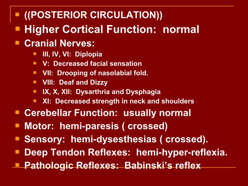
((POSTERIOR CIRCULATION)) Higher Cortical Function: normal Cranial Nerves:
III, IV, VI: Diplopia V: Decreased facial sensation VII: Drooping of nasolabial fold. VIII: Deaf and Dizzy IX, X, XII: Dysarthria and Dysphagia XI: Decreased strength in neck and shoulders
Cerebellar Function: usually normal Motor: hemi-paresis ( crossed) Sensory: hemi-dysesthesias ( crossed). Deep Tendon Reflexes: hemi-hyper-reflexia. Pathologic Reflexes: Babinski’s reflex

CEREBELLUM
Higher Cortical Function: normal Cranial Nerves: usually normal Cerebellar Function:
Nystagmus Flaccid dysarthria
Motor: Normal bulk and strength with ipsilateral hemi-hypotonia Intention tremor Axial instability with dysmetria
Sensory: Normal Deep Tendon Reflexes: Normal Pathologic Reflexes: Normal (plantar flexing to plantar stimulation

((SPINAL CORD LESION)) Higher Cortical Function: normal Cranial Nerves: normal Cerebellar Function: normal Motor:
weakness (extensors worse than flexors) below the lesion Hypertonia below the lesion with spasticity
Sensory: horizontal level usually lower than the lesion, poorly
localizing , may be somewhat asymmetric
Deep Tendon Reflexes: Hyper-reflexia below the level, possibly clonus
Pathologic Reflexes: loss of superficial reflexes (abdominal, cremasteric, anal
wink)
Babinski’s reflex


Cranial Nerves
DR MAGDI AWAD SASI

1. NAME 2. NUCLEUS 3. FUNCTION 4. HOW TO EXAME 5. DEFECT INFECTION ISCHEMIA INFLAMMATION TUMOUR T2RY TB DEGENERATIVE

Olfactory Nerve I
Sense of smell Damage causes impaired sense of smell

Test with alcowipes, coffee .
Unilateral anosmia may be significant
Bilateral anosmia: commonest cause viral
Classical pathology:olfactory groove meningioma
Basal skull fractures another potential cause (unilateral or bilateral)

Optic Nerve II
Provides vision Damage causes blindness in visual field

Visual acuity Visual fields to confrontation Colour vision Light reflex Pupillary reflexes (II and III) Fundoscopy (papilloedema, optic atrophy,
retinitis pigmentosa)

VISUAL ACUITY
CORRECTED (ie brain not lens)
Each eye separately
Snellen charts for distance and near vision reading charts for near vision
If unable, finger counting, hand movements, perception of light

VISUAL FIELDS
Often forgotten but very important
First do a bilateral screening test: will uncover the majority of significant visual field defects immediately
Go on to check each eye separately, ask about scotomata
Mention checking for blind spot enlargement


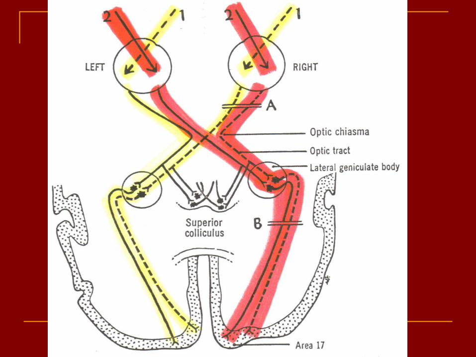
COMMON FIELD DEFECTS
HOMONOMOUS HEMIANOPIA: lesion posterior to the optic chiasm (eg posterior cerebral artery territory infarction)
BITEMPORAL HEMIANOPIA: lesion at the optic chiasm (eg pituitary tumour)
BLINDNESS ONE EYE: lesion in eye, retina or optic nerve

PUPILLARY RESPONSES
Light reflex is the clinically significant one
Afferent limb = II, efferent limb = III
Look at pupillary sizes
Direct and consensual response Look for afferent pupillary defect (optic nerve
lesion)

One large pupil: IIIrd nerve palsy, iris problem (eg traumatic midriasis), unilateral dilator eye drops
Small pupil: Horner’s syndrome, Argyll-Roberston pupil (small, irregular, reacts to accommodation but not to light)
Bilateral small pupils: drugs (opiates), pontine lesion (haemorrhage

HORNER’S SYNDROME
Oculosympathetic paralysis A good lateralising sign but a poor localising
sign Ptosis, miosis and sometimes unilateral
anhydrosis of face Look especially at neck, supraclavicular
fossa and hand (Pancoast’s tumour)

. A patient who has been exhibiting various endocrine abnormalities has an MRI scan of the head. This scan reveals a small tumor of the pituitary gland. As this tumor expands superiorly what visual field defect will this patient exhibit?
A. Left or right monocular blindness
B. Binasal hemianopsia
C. Left or right homonymous hemianopsia
D. Bitemporal hemianopsia


Eye movements (III, IV and VI)
III: OCULOMOTOR NERVE: all extraocular muscles,Except SO & LR ,
Also carries parasympathetic (constrictor) fibres to pupil, and
Fibres to levator palpebrae superioris

Look at eyes in primary position of gaze
IIIrd nerve palsy: ptosis ,eye often ‘down and out’
VI nerve palsy: often eyes convergent
(unopposed medial rectus)
Look at pupils
Look for ptosis

Oculomotor Nerve III
Somatic and Autonomic motor function Eye movement (Superior, inferior, medial rectus muscles and inferior
oblique muscle), opening of eyelid (levator palpebrae superioris), constriction of pupil (circular muscle), focusing (ciliary muscle and accomodation)

Fibers extend from the ventral midbrain, pass through the superior orbital fissure, and go to the extrinsic eye muscles
Functions in raising the eyelid, directing the eyeball, constricting the iris, and controlling lens shape
The latter 2 functions are parasympathetically controlled
Parasympathetic cell bodies are in the ciliary ganglia

Damage causes
1. Drooping eyelid ((ptosis))
2. Dilated pupil
3. Double vision
4. Difficulty focusing and inability to move eye in certain directions
5. Down and out {{eye}}


Trochlear Nerve IV
Eye movement (superior oblique muscle) Damage causes double vision and inability to
rotate eye inferolaterally

Abducens Nerve VI
Provides eye movement (lateral rectus m.) Damage results in inability to rotate eye
laterally and at rest eye rotates medially


Diagnosing complete 6th cranial nerve palsies is easy, but determining their etiology can be more challenging. Excluding increased intracranial pressure and papilledema (by looking for retinal venous pulsations during funduscopy) is important. MRI or CT can help exclude intracranial mass lesions, hydrocephalus, and direct nerve compression by lesions in the orbit, cavernous sinus, and base of the skull. Lumbar puncture determines the CSF opening pressure and can detect leptomeningeal inflammatory, infectious, or neoplastic infiltrates entrapping the 6th nerve. A collagen vascular screen helps exclude a vasculopathic process. In many cases, 6th nerve palsies resolve once the primary disorder is treated.

Trigeminal Nerve V

MOTOR SENSORY REFLEX 3 SENSATIONS + 3 BRANCHES 3 MUSCLES FUNCTIONS MASTICATIONS

Most important function is sensory Ophthalmic, maxillary and mandibular divisions Test with light touch and pinprick in all 3
divisions, comparing each side Corneal reflexes (afferent limb V, efferent limb
VII) Know something about trigeminal neuralgia
(examination is normal in these cases)

Ophthalmic branch – sensations from nasal cavity, skin of forehead, upper eyelid, eyebrow, nose
Maxillary branch – sensations from lower eyelid, upper lips and gums, teeth of the maxilla, cheek, nose, palate, pharynx
Mandibular branch – sensations from teeth of the mandible, lower gums and lips, palate, tongue. Motor function of temporalis and masseter muscles.
Damage produces loss of sensation and impaired chewing
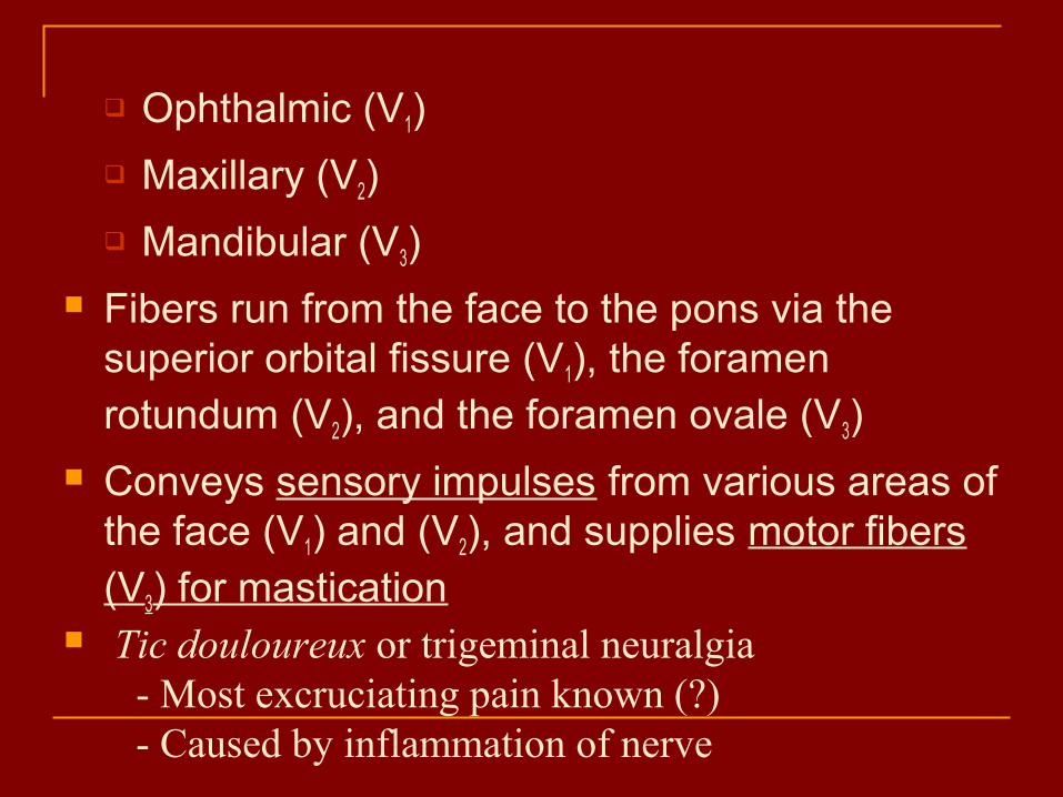
Ophthalmic (V1)
Maxillary (V2)
Mandibular (V3)
Fibers run from the face to the pons via the superior orbital fissure (V1), the foramen rotundum (V2), and the foramen ovale (V3)
Conveys sensory impulses from various areas of the face (V1) and (V2), and supplies motor fibers (V3) for mastication
Tic douloureux or trigeminal neuralgia - Most excruciating pain known (?) - Caused by inflammation of nerve
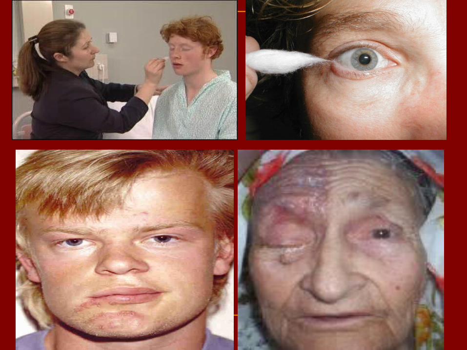

Facial Nerve VII


Motor - facial expressions
Autonomic Motor - salivary and lacrimal glands, mucous membranes of nasal and palatine mucosa
Sensory - taste on anterior 2/3’s of tongue
Damage produces sagging facial muscles and disturbed sense of taste (no sweet and salty)

DIFFERENTIATE AN UPPER MOTOR NEURON FROM A LOWER MOTOR NEURON LESION
Upper motor neuron lesion: milder, spares the forehead, no Bell’s phenomenon

Branches of Facial Nerve
Clinical test: Test anterior 2/3’s of tongue with substances such as sugar, salt,; test response of tear glands to ammonia fumes; test motor functions by asking subject to close eyes, smile, whistle, frown, raise eyebrows, etc.

Upper facial territory is supplied by bilateral motor cortices
Lower facial territory is supplied only by contralateral motor cortex
Therefore, unilateral central lesions spare upper face Lesions distal to geniculate ganglion
Mostly motor abnormalities
Lesions proximal to geniculate ganglion Motor, gustatory & autonomic abnormalities


Bell's Palsy
Unilateral facial paralysis of sudden onset Unknown cause. The mechanism presumably involves
swelling of the nerve due to immune or viral disease, with ischemia and compression of the facial nerve in the narrow confines of its course through the temporal bone.

Symptoms of Bell’s Palsy --Symptoms usually start suddenly, and range from mild to severe. They may
include: Twitching in face Weakness in face Face feels stiff or pulled to one side Droopy eyelid or corner of mouth Drooling due to inability to control facial muscles Facial Paralysis of one side of the face, makes it hard to close one eye Change in facial expression (for example, grimacing) Dry eye or mouth Loss of sense of taste Difficulty with eating and drinking Pain behind or in front of the ear, may occur 1-2 days before muscle
weakness Sensitivity to sound (hyperacusis) on the side of the face affected Headache
--These symptoms of Bell's palsy usually begin suddenly and reach their peak within 48 hours


The affected side becomes flat and expressionless, but patients may complain instead about the seemingly twisted intact side.
In severe cases, the palpebral fissure widens, and the eye does not close.
The patient may complain of a numb or heavy feeling in the face, but no sensory loss is demonstrable. A proximal lesion may affect salivation, taste, and lacrimation and may cause hyperacusis.

Weakness of the entire half of the face distinguishes Bell's palsy from supranuclear lesions (eg, stroke, cerebral tumor), in which the weakness is partial, affecting the frontalis and orbicularis oculi less than the muscles in the lower part of the face. Bell's palsy must be differentiated from unilateral facial weakness due to other disorders of the facial nerve or its nucleus, chiefly geniculate herpes (Ramsay Hunt's syndrome), middle ear or mastoid infections, sarcoidosis, Lyme disease, petrous bone fractures, carcinomatous or leukemic nerve invasion, chronic meningeal infections, and cerebellopontine angle or glomus jugulare tumors

A 24 y. o. woman presents to her physician with an inability to close her right eye. Physical exam reveals weakness of the right orbicularis oculi. Which of the following symptoms would likely also be present?
A. Double vision
B. Inability to feel the face
C. Inability to chew
D. Hyperacusis
E. Inability to shrug the shoulder

61
A 49 year old woman is in a motor vehicle accident and sustains a closed head injury. A CT scan does not show any intracranial hemorrhage but reveals a small tumor at the cerebellopontine angle of the brain. Which of the following nerves is most likely to be affected by this tumor?
A. Facial nerveB. Glossopharyngeal nerveC. Abducens nerveD. Trigeminal nerveE. Vagus nerve

Vestibulocochlear Nerve VIII
Special Sensory Provides hearing (cochlear branch) and sense of
balance (vestibular branch) Damage produces deafness, dizziness, nausea,
loss of balance and nystagmus

For clinical examination purposes, forget the vestibular element
Check hearing approximately in each ear
If reduced, determine whether conductive (BC >AC) or sensorineural (AC>BC) deafness


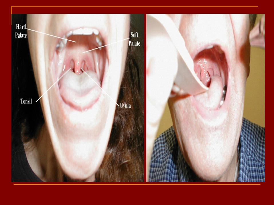
GLOSSOPHARYNGEAL (IX) AND VAGUS (X) Tested together Speech, palate, cough, swallow, (gag reflex) Bulbar palsy: bilateral LMN lesions of IX and
X: poor palatal movement, nasal speech, nasal regurgitation of fluids
Pseudobulbar palsy: bilateral UMN lesions: ‘hot potato’ speech, no nasal regurgitation, additional frontal lobe signs

Glossopharyngeal Nerve IX
Somatic motor – Swallowing and voice production via pharyngeal muscles
Autonomic motor - salivation, gagging, control of BP and respiration Sensations from posterior 1/3 of tongue including taste Sensations from baroreceptors and chemoreceptors Damage results in loss of bitter and sour taste and impaired swallowing,
blood pressure anomalies (with CN X).

68
A physician is performing a cranial nerve exam on a patient. While testing the gag reflex it is noted that when the left side of the pharyngeal mucosa is touched, the patient gags and his uvula deviates to the left. When the right side is touched, the patient does not gag. Which of the following is the most likely location of his lesion?
A. Left glossopharyngeal and vagus nervesB. Right glossopharyngeal and vagus nervesC. Right glossopharyngeal nerve onlyD. Right vagus nerve onlyE. Left glossopharyngeal nerve only

Vagus Nerve X Sensations from skin at back of ear,
external acoustic meatus, part of tympanic membrane, larynx, trachea, espophagus, thoracic and abdominal viscera
Sensations from bararoceptors and chemoreceptors
Special sensory – taste from epiglottis and pharynx
Somatic motor – Swallowing and voice production via pharyngeal muscles
Autonomic motor – smooth muscle of abdominal viscera, visceral glands secretions, relaxation of airways, and normal or decreased heart rate.
Damage causes hoarseness or loss of voice, impaired swallowing, GI dysfunction, blood pressure anomalies (with CN IX), fatal if both are cut

Dyspnea Dysphagia Dysphonia Dysarthria Emotional liability if UMN Total destruction incompatible with life

ACCESSORY NERVE (XI)
Cranial and spinal roots Cranial roots: sternocleidomastoid (note
direction of head turn) Spinal roots: trapezius (shoulder shrug)

Accessory Nerve IX
Swallowing, head, neck and shoulder movement via trapezius and sternocleidomastoid and pharyngeal muscles
Damage causes impaired head, neck, shoulder movement


It is purely motor. It has two roots, CRANIAL& SPINAL. The spinal root arises from the anterior horn cells of the upper 5 cervical segments, &it enter the skull through foramen magnum.
These fibers are joined by the cranial root which arises from the caudal part of the nucleus ambigus & together they leave the skull through the jugular foramen with the vagus.
In the jugular foramen the cranial root fibers join the vagus to be distributed along with fibers of the vagus to the pharynx and larynx.
This part of the nerve cannot be tested separately. The spinal part supplies the sternomastoid and upper part of the trapeziuz.

Hypoglossal Nerve XII
Tongue movements for speech, food manipulation and swallowing
If both are damaged – can’t protrude tongue If one side is damaged – tongue deviates towards
injured side

HYPOGLOSSAL NERVE
Movement of the tongue Look for wasting and fasiculation of the
tongue Deviation of tongue on protrusion Tongue movements including power

Fibers arise from the medulla and exit the skull via the hypoglossal canal
Innervates both extrinsic and intrinsic muscles of the tongue, which contribute to swallowing and speech
If damaged, difficulties in speech and swallowing; inability to protrude tongue

Function of the Cranial Nerves
Olfactory (I) Smell
Optic (II) Vision
Oculomotor (III) Eye movement (Inf Rec/Med Rec/ Sup Rec/Inf Oblique); Focusing (Iris and Lens)
Trochlear (IV) Eye movement (Sup Oblique)

Function of the Cranial Nerves
Trigeminal (V) Chewing, sensation of scalp, face, teeth (Not tongue) Somatosensory information (touch, pain) from the face and head; muscles for chewing.
Abducens (VI) Eye Movement (Lat Rectus)

Function of the Cranial Nerves
Facial (VII) Taste (anterior 2/3 of tongue); Somatosensory information from ear; Controls muscles used in facial expression.
Vestibulocochlear (VIII)
Vestibular branch: Posture, balance, equilibrium Cochlear branch: Hearing
Glossopharyngeal (IX)
Taste (posterior 1/3 of tongue); Somatosensory information from tongue, tonsil, pharynx; Controls some muscles used in swallowing.

Function of the Cranial Nerves
Vagus (X) Sensory, motor and autonomic functions of viscera (glands, digestion, heart rate)
Accessory (XI) Swallowing and head movement (Trapezius and SCM = Sternocleidomastoid muscle)
Hypoglossal (XII) Controls muscles of tongue

THANKS FOR PATIENCE