dr. meg sleeper: the abcs of ecgs back to basics (part i
TRANSCRIPT

1
The ABCs of ECGsBack to Basics Part I
Meg Sleeper VMD, DACVIM (cardiology)University of Florida Veterinary School
Lecture outline
• Electrical properties of the heart• Action potentials• Normal intracardiac conduction• ECG interpretation (cases)
Electrical properties of the heart
• Automaticity• Excitability• Refractoriness• Conductivity
Automaticity
• Only pacemaker cells are normally capable of beating spontaneously
• Gradual diastolic reduction in action potential (becoming less negative) toward the cell�s threshold potential.
Excitability
• All resting myocytes are capable of responding to an effective stimulus by generating an action potential.
Refractoriness
• Period of recovery following excitation when cells cannot respond to stimuli. Excitability is gradually restored.

2
Conductivity
• Intercalated discs in the ends of muscle fibers give the atria and ventricles the property of a syncytium. Therefore, if propagation is blocked along the preferential conducting pathway, depolarization can still spread directly from one cell to the next (a slower process).
• Conduction speed is dependent on cell size (slower in smaller cells), and is normally slowest at the AV node.
Contractility
• Peak tension developed by myocardial cells at a specific resting fiber length.
• ECG gives no information regarding contractility or pump function
Cardiac cell action potential
P T
QRS
ECG
Ventricular cell action potential
4
0
1 2
3
4Resting
Na+ In
Ca++ In
K+ Out
Resting-90 mV
Pacemaker cell action potential
Ik inward currentinactivated
ICa
-50-60 mV
4
Pacemaker Cells
Sinus node
AV node
Purkinje Fiber
Analysis of Cardiac Arrhythmias• Site of impulse origin
– Supraventricular: SA node, atria, AV node– Ventricular
• Rate: atrial and ventricular
• Timing– Premature beats: occur early in the sequence of
normal beats– Escape beats: occur after a pause in the sequence of
beats
3
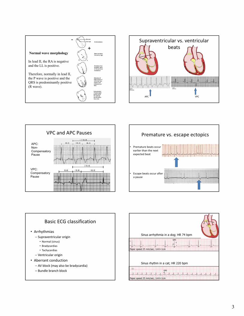
Normal wave morphology
In lead II, the RA is negativeand the LL is positive.
Therefore, normally in lead II,the P wave is positive and the QRS is predominantly positive(R wave).
-+
APC VPC
Supraventricular vs. ventricular beats
VPC and APC Pauses
VPC:CompensatoryPause
APC:Non-CompensatoryPause
Premature vs. escape ectopics
• Premature beats occur earlier than the next expected beat
• Escape beats occur after a pause
Basic ECG classification
• Arrhythmias– Supraventricular origin
• Normal (sinus)• Bradycardias• Tachycardias
– Ventricular origin• Aberrant conduction– AV block (may also be bradycardia)– Bundle branch block
P
QRST
PQRS
Sinus arrhythmia in a dog; HR 74 bpm
Sinus rhythm in a cat; HR 220 bpm
Paper speed 25 mm/sec; 1mV=1cm
Paper speed 25 mm/sec; 1mV=1cm

4
ECG #17 year old mc Jack Russell terrier50 mm/sec; 1mV=1cm
R waves regularly irregular
ECG #17 year old mc Jack Russell terrier50 mm/sec; 1mV=1cmHR@ 90 bpm
Normal QRS morphology and the same in all complexes
ECG #17 year old mc Jack Russell terrier50 mm/sec; 1mV=1cm
HR@ 90 bpm
P waves present for every QRS with consistent PR
P waves have variable amplitude
ECG #17 year old mc Jack Russell terrier50 mm/sec; 1mV=1cmHR@ 90 bpm
Summary
• Normal heart rate• Supraventricular (normal morphology)• Variable P wave with normal and consistent
PR intervals
• Wandering pacemaker/Sinus arrhythmia
Treatment: None necessary
ECG #23 year old mc MIXB; automobile accident25 mm/sec; 1mV=1cm

5
RR regularity: slight irregularity vs. regular
ECG #23 year old mc MIXB; automobile accident25 mm/sec; 1mV=1cm
HR@150 bpm
QRS morphology: normal and wide (two different morphologies)
ECG #23 year old mc MIXB; automobile accident25 mm/sec; 1mV=1cm
HR@150 bpm
Fusion beat
P waves: present for some of the QRSs (narrow QRSs)
ECG #23 year old mc MIXB; automobile accident25 mm/sec; 1mV=1cm
HR@150 bpm
Summary
• Normal heart rate• Episode of ventricular rhythm with a normal
heart rate• Diagnosis:
– Accelerated idioventricular rhythm with underlying sinus arrhythmia
Treatment: Ventricular antiarrhythmic only if rate is fast enough to negatively impact bloodpressure; generally self limiting
ECG #3Paper speed 25 mm/sec; 1mV=1cm3 year old Labrador retriever; presenting for an episode of weakness

6
RR regularity: regular with abrupt transitions
ECG #3Paper speed 25 mm/sec; 1mV=1cm3 year old Labrador retriever; presenting for an episode of weakness
HR@ 300/150 bpm
QRS morphology: narrow/normal
ECG #3Paper speed 25 mm/sec; 1mV=1cm3 year old Labrador retriever; presenting for an episode of weakness
HR@ 300/150 bpm
P waves: not always visible; transition begins with premature P wave
ECG #3Paper speed 25 mm/sec; 1mV=1cm3 year old Labrador retriever; presenting for an episode of weakness
HR@ 300/150 bpm
Summary
• Tachycardia with normal QRS morphology– >>supraventricular
• Paroxysmal transitions– >>not sinus
• Diagnosis:– Supraventricular tachycardia or paroxysmal
atrial tachycardia
Treatment for supraventricular tachycardia
• Diltiazem– 0.5-5 mg/kg q 8 hours (oral)
– 0.1-0.2 mg/kg IV bolus, then 2-6 mcg/kg/min CRI
• Digoxin– 0.003-0.005 mg/kg q 12 hours (oral)
• Beta blocker– Atenolol: 0.25-2.0 mg/kg q 12-24 hours (oral)
– Esmolol: 50-100 mcg/kg IV bolus every 5 min up to 500 mcg/kg maximum; 25-200 mcg/kg/min CRI
ECG #4Paper speed 25 mm/sec; 1mV=1cm5 year old great Dane, no clinical signs
!

7
ECG #4Paper speed 25 mm/sec; 1mV=1cm5 year old great Dane, no clinical signs
HR- 160 bpm
!
RR regularity: irregular
ECG #4Paper speed 25 mm/sec; 1mV=1cm5 year old great Dane, no clinical signs
HR- 160 bpm
!
QRS morphology: normal width to increased with variable morphology ( )
P waves present for most complexes but not all
Summary
• Normal to mildly increased heart rate with irregular rhythm
• Occasional premature, wide morphology complexes
• Rhythm diagnosis:– Ventricular premature complexes; multiform
Treatment ventricular ectopy
• In asymptomatic dogs, there is no evidence that starting anti-arrhythmic therapy will reduce the risk of a fatal arrhythmia
• The arrhythmia may be a sign of structural myocardial disease, particularly in breed predisposed to acquired heart disease.
• Recommend further evaluation:– Echocardiogram– Holter monitor
CASTCardiac Arrhythmia Supression Trial
(N Engl J Med. 1989;321:406-412.}
There was an increased mortality in post-myocardialinfarction patients given Class 1c antiarrhythmic agents.
ECG example #5Paper speed 25 mm/sec; 1mV=1cm5 year old Boxer, an episode of syncope
!

8
ECG example #5Paper speed 25 mm/sec; 1mV=1cm5 year old Boxer, an episode of syncope
HR- 160 bpm; brief period of 300 bpm
!
RR regularity: irregular
!
QRS morphology: normal width and increased width complexes
ECG example #5Paper speed 25 mm/sec; 1mV=1cm5 year old Boxer, an episode of syncope
HR- 160 bpm; brief period of 300 bpm
P waves present for most complexes but not all
Summary
• Normal to mildly increased heart rate with periods of rapid heart rate
• Some premature, wide morphology complexes• Rhythm diagnosis:– Ventricular premature complexes and non-
sustained ventricular tachycardia
Treatment ventricular ectopy• In symptomatic dogs, while there is no evidence that starting
anti-arrhythmic therapy will reduce the risk of a fatal arrhythmia, effective therapy will reduce clinical signs
• Ventricular anti-arrhythmics to consider for non-sustained ventricular tachycardia:– Mexilitine: 5-8 mg/kg three times daily (oral)– Sotalol: 1-2 mg/kg twice daily (oral)
• The arrhythmia may be a sign of structural myocardial disease, particularly in breed predisposed to acquired heart disease.
• Recommend further evaluation:– Echocardiogram– Holter monitor
ECG # 63 year old MIXB; normal25 mm/sec; 1 mV=1cm
RR regularity: irregular
ECG # 63 year old MIXB; normal25 mm/sec; 1 mV=1cm
HR@ 100bpm
QRS morphology: narrow/normal

9
P waves: occasional un-conducted P waves ( )
ECG # 63 year old MIXB; normal25 mm/sec; 1 mV=1cm
HR@ 100bpm
PR interval progressively lengthens prior to blocked P wavePattern mirrors sinus arrhythmia RR variation
ECG # 63 year old MIXB; normal25 mm/sec; 1 mV=1cm
HR@ 100bpm
Summary
• Normal heart rate with irregular rhythm
• Occasional blocked P waves • QRS morphology normal• Rhythm diagnosis:
– Second degree AV block type 1 (Wenchebach)
Treatment: Generally none necessary; usually asymptomatic and athletic dogs
ECG # 79 year old toy poodle with degenerative valve disease; currently on furosemide, enalapril and pimobendan25 mm/sec; 1 mV=1 cm
Rhythm: irregularly irregular
No P waves
QRS morphology: normal/narrow
ECG # 79 year old toy poodle with degenerative valve disease;currently on furosemide, enalapril and pimobendan25 mm/sec; 1 mV=1 cmHR@ 160 bpm
Summary
• Irregularly, irregular rhythm• Lack of P waves• Normal QRS morphology• Rhythm diagnosis:
– Atrial fibrillationTreatment: in a small breed dog with under-lying heart disease, control heart rate with digoxin and/or diltiazem

10
ECG example #8Paper speed 25 mm/sec; 1mV=1cm9 year old cocker spaniel; no clinical signs
! RR regularity: irregular; predominantly long RR cycles (some longer than 2 seconds), occasional short cycles
QRS morphology: narrow/normal
ECG example #8Paper speed 25 mm/sec; 1mV=1cm9 year old cocker spaniel; no clinical signsHR-50 bpm
P waves: intermittent; morphology variable
ECG example #8Paper speed 25 mm/sec; 1mV=1cm9 year old cocker spaniel; no clinical signsHR-50 bpm
Summary
• Bradycardia (sinus bradycardia)• Negative P waves correspond to long RR
cycles>>junctional escape beats• Occasional premature beats
• Diagnosis:
– Sick sinus syndromeDifferentiate from high vagal tone>>
Atropine response test (0.04 mg/kg IV or IM)
Treatment
• If atropine response test is normal, look for underlying disease that is causing elevated vagal tone (respiratory, GI, ocular or neurologic)
• If abnormal atropine response test, gold standard therapy would be pacemaker. If not possible consider medical management with:– Propantheline bromide: 0.25-5 mg/kg q 8-12 hour (oral)– Terbutaline: 1.25-5 mg/dog PO q 8-12 hour (oral)– Theophylline: 10 mg/kg q 12 hour (extended release;
oral)
ECG #912 year old DSH; no clinical signs25 mm/sec; 1 mV= 1 cm

11
RR regularity: regular
QRS morphology: normal width to slightly increased with right axisdeviation
ECG #912 year old DSH; no clinical signs25 mm/sec; 1 mV= 1 cmHR@100 bpm
P waves: regular and asynchronous with QRSs
ECG #912 year old DSH; no clinical signs25 mm/sec; 1 mV= 1 cmHR@100 bpm
Summary
• Bradycardia• Regular RR and PP, but
asynchronous• PR intervals variable• Diagnosis:– Complete heart block or third
degree AV block
Treatment complete heart block
• Depends on symptoms• If asymptomatic:
– No treatment, monitoring for secondary heart disease
• If symptomatic:– Pacemaker vs medical management
• Terbutaline– 0.1 mg/kg q 8 hours (oral)
• Theophylline– 4 mg/kg q 12 hours (oral)– 19 mg/kg q 24 hours (extended release; oral)
ECG #10Paper speed 50 mm/sec; 1mV=1cm7 year old Newfoundland; no clinical signs
I
II
III
aVR
aVL
RR regularity: irregular
ECG #10Paper speed 50 mm/sec; 1mV=1cm7 year old Newfoundland; no clinical signsHR@ 150 bpm

12
Wide QRSs QRS= 4.5 mm or 4.5 X 0.02 seconds= 0.90 sec; Normal < 0.60 sec (canine)
ECG #10Paper speed 50 mm/sec; 1mV=1cm7 year old Newfoundland; no clinical signsHR@ 150 bpm
No P waves
ECG #10Paper speed 50 mm/sec; 1mV=1cm7 year old Newfoundland; no clinical signsHR@ 150 bpm
Differentials for wide complex QRS
• Ventricular in origin • Electrolyte disturbance- most often
hyperkalemia• Aberrant conduction- bundle branch block
Summary
• Irregular RR• No P waves• QRS prolongation most likely due
to right bundle branch block (ventricular rhythm should be regular)
• Rhythm diagnosis: atrial fibrillation with RBBB
Diagnostics and treatment
• Bundle branch blocks– No treatment
• Lone atrial fibrillation vs. rapid atrial fibrillation– Echocardiogram to evaluate heart structure– Rate control if necessary
• Digoxin: 0.003-0.005 mg/kg q 12 hours (oral)• Diltiazem: 0.5-5 mg/kg q 8 hours (oral)
– Cardioversion