dr. skoog is professor of plastic surgery, university of
TRANSCRIPT

The Use of Periosteal Flaps in the Repair
of Clefts of the Primary Palate
TORD SKOOG, M.D.
Uppsala, Sweden
In recent years, the repair of the maxillary defect in cleft lip and palate
deformity has attracted an increasing interest, resulting in the develop-
ment of elaborate and tedious programs for correction. In this paper, a
new principle for primary repair is presented. The principle involves the
use of periosteal flaps for the purpose of creating bony continuity, pre-
venting maxillary collapse, and contributing to the restoration of sym-
metry of the alveolar arch.
The facial bones gre, for the most part, of intramembranous origin and
their early development is well known from histogenetic studies. After a
mass of primary cancellous bone has been laid down, there appears a
peripheral concentration of mesenchyme. This periosteal layer has great
bone-forming potentiality and deposits parallel lamellae about the can-
cellous center of the growing bone. One would expect the osteogenetic
capacity of the periosteum to be most pronounced during the period of
growth. The formation at an early age of new bone arising from the peri-
osteum of the facial skeleton may in fact be deduced from clinical ob-
servations. Some cases treated at this department are particularly instruc-
tive in this respect: two of them will be reported briefly.
Case 1. A 4-month-old girl referred because of a melanotic progonoma in-
volving the maxillary bone required unilateral, subtotal removal of themaxilla. The resection was performed leaving all the periosteum in situ.
Complete bone regeneration took place and x-ray films two years after theoperation revealed, except for the missing teeth and dental anlages, a normallooking maxilla with a maxillary sinus. This case has been more extensively
reviewed by Korlof (1).
Case 2. In a boy with a congenital cleft of the primary palate, maxillary
collapse occurred following soft tissue repair. During orthodontic expansion,at the age of five years, a substantial bone bridge developed spontaneously
between the premaxilla and the lateral maxillary segment. The possible ex-planation, suggested in a previous report of this case (5), was that in theprimary repair the periosteal membranes had united across the cleft.
Based on these observations, suggestive in regard to the osteogenetic
capacity of the periosteum of facial bones in children, a new principle has
been adopted in the repair of the maxillary defect in cleft primary palate.
The details of surgical technique and a preliminary report on results are
included in this paper. .
Dr. Skoog is Professor of Plastic Surgery, University of Uppsala.
332

PERIOSTEAL FLAPS 3393
Operative Technique
Closure of the cleft primary palate within the alveolar region has become
a fairly standardized procedure based on the original description by Veau
(6). The technique we use to accomplish lining on the nasal aspect is
illustrated in Figure 1, upper left and right. On the lateral side, the in-
cision made along the border of the cleft is extended in front between the
upper and lower nasal cartilages: posteriorly it reaches about 2 em behind
the alveolus. The incision is carried through mucous membrane and peri-
osteum down to bone. The lateral border of the piriform aperture and the
maxillary cleft are exposed and a mucoperiosteal flap is raised from the
inner aspect of the bone. By extensive subperiosteal elevation, this flap
can easily be advanced medially. On the medial border of the cleft, an
incision is made along the vomer and carried over the anterolateral aspect
of the premaxilla towards the anterior edge of the septum. The surface of
the premaxilla bordering the cleft is completely denuded of its rather thin
FIGURE 1. Operative procedure designed for the creation of periosteal continuityacross the maxillary cleft. Upper left and right shows lining of the nasal aspect.Lower left and right shows that the periosteal flap (a) is raised on the anterioraspect of the maxilla and rotated 180° into position across the bony cleft. Fordetails, see text.

334 Skoog
and firmly adherent periosteum, with care being taken not to injure the
delicate bone structures. The periosteal elevation is continued on the
vomer and the mucoperiosteal-perichondrial flap thus mobilized is then
approximated to the corresponding lateral flap with 3-0 chromic catgut
sutures, tied on the nasal side. In this closure, broad apposition of the
periosteal surfaces is attempted and for that purpose a second row of
sutures may, if possible, be placed on the oral side. Should there be any
tension on the suture line, relaxing incisions are made on the lateral flap
in an area of overlying bone.
It is a well-known fact that a nasal floor reconstructed in this fashion,
which is left for epithelialization on the oral side or covered with a mucosal
flap taken from the buccal sulcus, is not capable of laying down bone
within the alveolar cleft. In order to make use of the phenomenon of sub-
periosteal bone formation, this repair of the nasal floor has been supple-
mented to provide within the alveolar region a second layer of periosteal
lining brought in on the anterior aspect; that is, periosteal continuity is
created on both aspects of the maxillary cleft.
At the age of between three and six months, when the initial repair of
the cleft primary palate is generally performed, the periosteum investing.
the lateral segment of the maxilla forms a substantial membrane which
is easily stripped off on the facial as well as on the palatal aspect. After
trials with different sizes and shapes of flap, it was found that the perios-
teum covering most of the anterolateral aspect of the maxilla had to be
mobilized to provide a flap that in a complete cleft would give good cover
and bridge the gap to reach the premaxilla without tension (Figure 1,
lower left and right). In the planning, one must remember that the peri-
osteal membrane has an outer layer composed of coarse fibrous connective
tissue which lacks elasticity and therefore is not easily adjusted in flap
transfer. The flap is based medially along the infraorbital margin and on
the lateral aspect of the nasal pyramid. From this location, a periosteal
flap is raised by incising in a lateral direction below the infraorbital fora-
men, and the original incision for exposure in the vestibulum is extended
laterally to meet this upper border. The dissection to expose the flap is
carried out in the tissue plane immediately superficial to the periosteum
and the infraorbital nerve is not injured. The flap is then rotated through
almost 180° to its new position across the cleft and sutured to the peri-
osteal edge on the anterior aspect of the premaxilla. The upper border of
the flap may also be sutured to the periosteal flaps of the previously re-
constructed floor of the nose. It is possible that the degree of rotation and
the proportions of this flap will occasionally impair the blood supply to
the extent that the distal part may only survive as a free graft.
In incomplete clefts, regularly characterized by a wide piriform aperture
and a depressed nasal floor, correction was attempted according to the
same principle. Laterally, the periosteum on both the inner and outer
aspect of the bone was advanced; in addition, a periosteal flap was placed

PERIOSTEAL FLAPS 335
across the anterior, denuded part of the nasal floor at a level considered
normal.
In the present series of cases, repair of the cleft lip was performed in
conjunction with the creation of periosteal continuity within the maxillary
cleft. The single clefts were repaired according to a method described in
1957 and modified in 1963 (8, 4). The bilateral clefts were repaired ac-
cording to a technique reported in 1965 (5).
Results
Altogether cight cases have been operated upon according to the prin-
ciple described. In none of them did complications occur following surgery.
The series includes four patients with complete clefts of the primary
palate, two with bilateral clefts, and two with unilateral clefts. One of the
bilateral cases had a periosteal flap repair on one side only. Since the
complete clefts are of particular interest in the evaluation of postoperative
results, they are reported in more detail in order to illustrate the different
stages of repair and the observations made (Figures 2 to 5).
FIGURE 2. Upper left, a three-month-old girl at the time of repair of the cleftprimary palate. A large periosteal flap was placed across the maxillary cleft asillustrated in Figure 1. Upper right, 2% months postoperatively, the alveolar arch
has become symmetrical and there is no tendency to collapse. Lower left and right,
x-ray films, taken 2% and 3% months, respectively, after the operation, show new
bone extending from both the lateral and the medial borders of the maxillary cleft.
At the later stage (right), the formation of bone is more widespread within the
cleft and bone structures are more dense.

336 Skoog
FIGURE 3. Left, a three-month-old boy, when the cleft of the primary palatewas repaired (June, 1964) using periosteal flaps as described in text. Right, condi-tion 10 months after the operation. Note accurate alignment of maxillary segments.X-rays show that the abnormal position of 1+ was present already before eruptionand prior to surgery.
On examination, there was less depression of the cleft side than is nor-
mally found following simple soft tissue repair (Figure 2). The regular
finding of a marked step in the maxillary framework corresponding to the
alveolar cleft was also less noticeable.
In both cases of complete bilateral clefts, the premaxilla became firmly
consolidated into the alveolar arch, and this applied to the case in which
a periosteal flap was used on only one side. These clinical findings can
most reasonably be interpreted as the result of bone formation within the
clefts.
The effect of the surgical procedure described on the maxillary configura-
tion at the level of the alveolar processes was carefully studied. In three
out of five complete clefts, there was an approximation of the alveolar
segments into end-to-end contact, producing a symmetrical arch form. In
two clefts, good contact was achieved within the arch but there was slight,
insignificant overlapping of the premaxillary segment. In the latter cases,
examination of the preoperative maxillary casts revealed that collapse
was present to the same degree prior to soft tissue repair. Though pre- and
postoperative variations in this respect are great, as has been demonstrated
by Pruzansky and Aduss (2), the dynamic effects of soft tissue repair,
themselves related to methods of closure and surgical technique, are of
fundamental importance for the final arch form. Searring and the crea-
tion of unphysiological mechanical conditions can only increase the in-
cidence and degree of maxillary collapse and deformity. The observations
made are therefore of interest even in a limited number of clefts because
of their uniform character, showing that in no case did maxillary collapse
occur following surgery. The modelling influence of the surgical procedure
used was found to be excellent, both in the unilateral and the bilateral
clefts. In fact, the relationship of the maxillary segments obtained in this
series of cases by soft tissue repair only compared favorably with the best
to be obtained by pre- or postsurgical orthopedics. An evaluation of the
PERIOSTEAL FLAPS 337
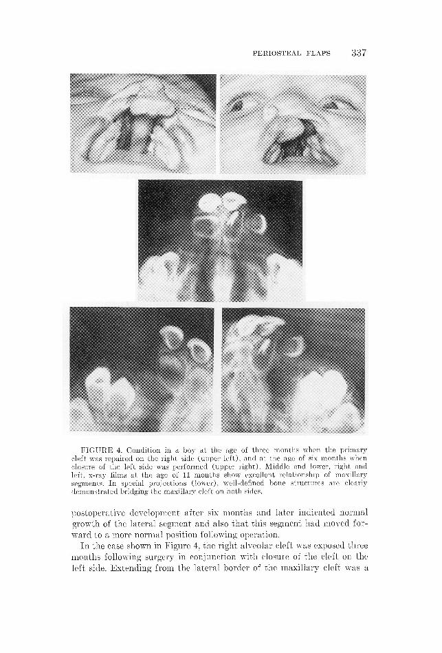
FIGURE 4. Condition in a boy at the age of three months when the primarycleft was repaired on the right side (upper left), and at the age of six months whenclosure of the left side was performed (upper right). Middle and lower, right andleft, x-ray films at the age of 11 months show excellent relationship of maxillarysegments. In special projections (lower), well-defined bone structures are clearlydemonstrated bridging the maxillary cleft on both sides.
postoperative development after six months and later indicated normal
growth of the lateral segment and also that this segment had moved for-
ward to a more normal position following operation.
In the case shown in Figure 4, the right alveolar cleft was exposed three
months following surgery in conjunction with closure of the cleft on the
left side. Extending from the lateral border of the maxillary cleft was a

FIGURE 5. Left, complete bilateral cleft in a girl, aged three months, at thetime of repair on the left side. The right side was repaired three months later. Alateral periosteal flap for cover was used on the latter side only. Right, the resultnine months following the second operation. The premaxilla was then firmly con-solidated into the alveolar arch.
narrow bridge of bone, corresponding in size and shape to the periosteal
flap that had been shifted from the lateral segment at the time of repair.
In this early case of the series, a fairly small triangular flap had been used
which just reached the medial border, as did the newly formed bone. A
distinct periosteal layer enclosed the new bone; it was easily stripped off,
but appeared thinner than periosteum normally found in this region at
that age. At the donor site for the periosteal flap, complete regeneration
had taken place and the new periosteum was almost indistinguishable
from the surrounding periosteal layer except that it was more adherent
to the bone. In closing the wound, the periosteum within the cleft was
partly separated from the new bone by subperiosteal packing with hemo-
statice sponge. This may explain why more bone had formed when x-ray
films were taken three months later.
Roentgenograms taken three and six months postoperatively were not
always easy to interpret because of the fineness of the structures and the
lack of density of the new bone. Well-defined bridges across the cleft and/
or processes extending from both the medial and lateral borders were most

PERIOSTEAL FLAPS 339
easily distinguished (Figure 4). For the rest, the maxillary cleft contained
tissue that radiologically could not be differentiated with certainty from
nonspecific soft tissue apart from minor spots and streaks of greater den-
sity. The case illustrated in Figure 2 shows how such tissue is transformed
into typical bone structure at a later stage. -
In a case of incomplete cleft with a depressed nasal floor, a solid sub-
cutaneous ridge could be felt corresponding to the periosteal flap three
months following operation. X-ray laminography revealed symmetrical
conditions and a not sharply defined bone contour within this area.
Naturally, follow-upstudies of these cases have to be continued over
the entire growth period to provide conclusive evidence. Comments with
regard to the applicability of this new principle in the repair of maxillary
deformity in clefts of the primary palate, its biological nature and an-
ticipated late effects, will not be included in this preliminary report. Nor
will suggestions on the many possible variations in surgical techniques be
presented until further experience is gained.
Summary
This report suggests a new surgical approach to maxillary restoration
in patients with cleft primary plate. An operative technique for primary
repair of complete clefts has been developed utilizing periosteal flaps for
the purpose of creating bony continuity between the premaxilla and the
lateral maxillary segment, preventing maxillary collapse, and contributing
to the restoration of symmetry of the alveolar arch. In incomplete clefts,
the same principle has been applied to reduce the bony deficiency. Eight
cases treated according to the technique described were examined three
and six months postoperatively with regard to the maxillary configuration,
especially approximation of the alveolar segments, and the formation of
new bone. Though it is not yet possible to make an assessment concerning
the long term advantages of the procedure proposed in this paper, the
early results are promising in all respects, and the principle evolved ap-
pears to be of great value in restoring anatomical and functional conti-
nuity to the cleft primary palate.Akademiska sjulkhuset
Uppsala, Sweden
References
1. Kortor, B., and BrErastrOnM, R., Melanotic progonoma of the maxilla. Acta chir.
Scand., 129, 292-299, 1965.2. Pruzansky, S., and Apuss, H., Arch form and the deciduous occlusion in complete
unilateral clefts. Cleft Palate J., 1, 411-418, 1964.
3. Sxrooc, T., A design for the repair of unilateral cleft lips. Amer.:J. Surg., 95, 223-
226, 1958.4. Sxooc, T., Porous tape in wound closure, skin grafting and wound dressing. Acta
chir. Scand., 126, 383-887, 1968. '
5. T., The management of the bilateral cleft of the primary palate. Plastic
reconstr. Surg., 35, 140-147, 1965.
6. Vrav, V., Bec-de-ligvre. Paris: Masson & Cie, 1938.