draft benefit definition: early and locally advanced ... · 2.1 this is a recommendation for the...
TRANSCRIPT

Page 1 of 22
Draft benefit definition: Early and locally
advanced breast cancer

Page 2 of 22
Contents
1. Introduction .......................................................................................................................................... 4
2. Epidemiology ....................................................................................................................................... 4
3. Scope and purpose.............................................................................................................................. 4
4. Diagnostic procedures ......................................................................................................................... 6
5. Staging and risk assessment ............................................................................................................... 8
6. Management of localised disease ...................................................................................................... 10
7. Management of locally advanced disease ......................................................................................... 13
8. Reference .......................................................................................................................................... 19

Page 3 of 22
Disclaimer:
The breast cancer benefit definition has been developed for the majority of standard patients. These benefits
may not be sufficient for outlier patients. Therefore regulation 15h may be applied for patients who are
inadequately managed by the stated benefits. The procedure codes are just an indication of applicable
procedure codes, however some significant procedure codes may not have been included. The benefit definition
does not describe specific in-hospital management such as theatre, anaesthetists, anaesthetist drugs, supportive
medication and nursing care. However, these interventions form part of care and are prescribed minimum
benefits.

Page 4 of 22
1. Introduction
1.1 The legislation governing the provision of the prescribed minimum benefits (PMBs) are contained in
the regulations enacted under the Medical Schemes Act 131 of 1998. In respect of some of the
diagnosis treatment pairs (DTPs), medical scheme beneficiaries find it difficult to know their
entitlements in advance. In addition, medical schemes interpret these benefits differently, resulting
in a lack of uniformity of benefit entitlements.
1.2 The benefit definition project is coordinated by the Council for Medical Schemes (CMS) and aims to
define the PMB package and to guide the interpretation of the PMB provisions by relevant
stakeholders. The guidelines are based on the available evidence of clinical and cost effectiveness
taking into consideration affordability constraints and financial viability of medical schemes in South
Africa.
2. Scope and purpose
2.1 This is a recommendation for the diagnosis, treatment and care of individuals with early and locally
advanced breast cancer in any clinically appropriate setting as outlined in the Medical Schemes
Act.
2.2 The purpose is to improve clarity in respect of funding decisions by medical schemes, taking into
consideration evidence based medicine, affordability and in some instances cost-effectiveness
3. Epidemiology
3.1 Breast cancer is the most common cancer in women both in the developed and less developed
world. In 2012, 1.7 million women were diagnosed with breast cancer while the prevalence stood at
6.3 million women. According to the World Health Organisation (WHO) Breast cancer was also the
most common cause of cancer death among women with 508 000 deaths in 2011 and 522 000
deaths in 2012. Breast cancer was also the most frequently diagnosed cancer among women in
140 of 184 countries worldwide [1]
3.2 Although breast cancer is thought to be a disease of the developed world, almost 50% of breast
cancer cases and 58% of deaths occur in less developed countries. Incidence rates of breast
cancer vary greatly worldwide from 19.3 per 100,000 women in Eastern Africa to 89.7 per 100,000
women in Western Europe. In contrast to Eastern Africa, breast cancer was the most commonly
diagnosed cancer and the leading cause of cancer death among women in Southern Africa (9000
cases, 4500 deaths)[2].

Page 5 of 22
3.3 Breast cancer survival rates vary greatly worldwide, ranging from 80% or over in North America,
Sweden and Japan to around 60% in middle-income countries and below 40% in low-income
countries[3]. The low survival rates in less developed countries can be explained mainly by the lack
of early detection programmes, resulting in a high proportion of women presenting with late-stage
disease, as well as by the lack of adequate diagnosis and treatment facilities.
3.4 Currently in South Africa 10% of patients with breast cancer present with stage 1 diseases and the
remainder presents with 30% each for stages two three and four[4]. According to the South African
National Cancer Registry, Breast cancer was the most prevalent cancer amongst women with a
lifetime risk of 1:35[5].
Table 1: Possible ICD 10 codes to identify breast cancer
ICD 10 WHO description Comments
Z12.3 Special screening examination for neoplasm of breast
C50.0 Malignant neoplasm, nipple and areola
C50.1 Malignant neoplasm, central portion of breast
C50.2 Malignant neoplasm, upper-inner quadrant of breast
C50.3 Malignant neoplasm, lower-inner quadrant of breast
C50.4 Malignant neoplasm, upper-outer quadrant of breast
C50.5 Malignant neoplasm, lower-outer quadrant of breast
C50.6 Malignant neoplasm, axillary tail of breast
C50.8 Malignant neoplasm, overlapping lesion of breast
C50.9 Malignant neoplasm, breast, unspecified
D05.0 Carcinoma in situ, lobular carcinoma in situ
D05.1 Carcinoma in situ, intraductal carcinoma in situ
D05.7 Carcinoma in situ, other carcinoma in situ of breast
D05.9 Carcinoma in situ, of breast, unspecified
4. Screening
4.2 Current evidence does not support the use of mammogram for screening women below 40 years
and women above 69 years[6, 7].
4.1 Screening mammogram is a PMB level of care for women between the ages of 45 to 69 years.

Page 6 of 22
5. Diagnostic procedures
Women with signs and symptoms of breast cancer must undergo triple assessment for diagnosis. Triple
assessment consist of clinical examination, imaging and pathological assessment [8, 9].
5.1 Clinical assessment:
5.1.1 The diagnostic work-up of early breast cancer starts with assessment of general health status
which includes the complete history of the patient, family history relating to cancers, physical
examination and biochemical examination.
5.1.2 Clinical examination includes bimanual palpitation of the breasts and complete examination of
tall systems (bones, liver, brain and lungs) to assess distant metastases.
5.1.3 Blood tests such as liver function test, renal function tests, calcium and phosphates to assess
general health and metastatic disease are PMB level of care [8].
5.2 Imaging
Imaging plays a crucial role for classifying and sampling both palpable and non-palpable breast
abnormalities, as well as for defining the extent of breast tumours, both locally, loco-regionally, and at
distant sites.
5.2.1 Diagnostic mammogram is indicated for most women with positive screening mammogram [8,
10].
5.2.2 Ultrasound is indicated for symptomatic younger women (women less than 40 years,) as they
have dense breast tissue and high risk of false negatives on mammogram. Ultrasound with
mammogram has a better diagnostic value as compared to either test alone in symptomatic
women [11-13].
5.2.3 Magnetic resonance Imaging (MRI) of the breast is not routinely recommended. MRI is PMB
level of care in cases of family history of breast cancer, women from families not tested or
inconclusively tested for BRCA mutation with 20-30% lifetime risk or greater familial breast
cancer associated with BRCA mutations, breast implants, lobular cancers and when the
findings of conventional imaging are inconclusive [8, 14, 15].

Page 7 of 22
5.2.4 Positron Emission Tomography - Computed Tomography (PET-CT) scan, three dimensional
mammographic ultrasound and computed tomography scan are not PMB level of care for
diagnosis[16] [8, 17]
5.3 Pathological assessment
5.3.1 Ultrasound guided core needle biopsy is the method of choice for diagnosing breast cancer.
Core needle biopsy has been shown to reliably distinguish between in-situ and invasive
cancers, allow evaluation of more histological, prognostic and predictive factors in breast
cancer [18, 19].
5.3.2 Fine needle aspiration (FNA) is indicated as the first-line pathologic investigation for palpable
breast lesions. In the case non-palpable lesions, suboptimal sampling and localization remains
the main cause of false negative results[20]. Using ultrasound to guide FNA decreases the
number of false negative results and increases the sensitivity and specificity of FNA[21, 22]
5.3.3 Excision biopsy is considered a reference standard method of evaluating a suspicious breast
lesion. However, the availability of core needle biopsy has limited the role of open surgical
biopsy which places the patient at risk of experiencing morbidities. A less invasive method of
evaluation of breast lesions is preferred[23].
5.3.4 Frozen section biopsy is not a PMB level of care. Frozen section biopsy has been shown to
have a limited role in the diagnosis of carcinoma and is not recommended on small lesions (<
1cm), where the pathologist believes that freezing will distort subsequent tissue
morphology[23]. Current evidence discourages the use of Frozen section for evaluation of
resection margins that are grossly free of tumour and on a breast excision specimen removed
because of mammographic calcifications [22, 23].
5.4 Evaluation of the Axilla
5.4.1 Axillary lymph nodal status remains an important prognostic factor because treatment of breast
cancer is influenced by the presence of and number of axillary lymph nodes involved.
5.4.2 Sentinel lymph biopsy is a PMB level of care for women with operable breast cancer and
multicentric tumours, with ductal carcinoma in situ (DCIS) who will undergo mastectomy, who
previously underwent breast and/or axillary surgery, or who received preoperative/neoadjuvant

Page 8 of 22
systemic therapy are offered SLNB [24, 25]. SLNB should not be performed routinely for all
patients with an initial diagnosis of DCIS [8, 15, 26].
5.5 Histological assessment
5.5.1 Progesterone receptor (PR) and HER2 status is determined on all breast cancers and breast
cancer recurrences [27, 28]. Immunohistochemical staining can be performed on the core
needle or excision biopsy.
6. Staging and risk assessment
The TNM classification is a universally accepted system that is used to stage breast cancer. TNM
staging takes into account the size of the tumour (T), whether the cancer has spread to the lymph
glands (lymph nodes) (N), and whether the tumour has spread anywhere else in the body (M – for
metastases)[8].
6.1 Current guidelines for the management of women with early breast cancer generally recommend
against the routine use of staging imaging to detect asymptomatic distant metastases at the time of
diagnosis [8, 29-32].
6.2 Chest x-ray is considered for patients with clinically positive axillary nodes, large tumours, and
clinical signs and/or laboratory values suggesting metastases to determine the presence of
pulmonary metastases.
6.3 Bone scan is considered for patients with clinically positive axillary nodes, large tumours, clinical
signs and/or laboratory values suggesting metastases, bone pain to determine the presence of
metastases to bone[32]
6.4 Magnetic Resonance Imaging (MRI) is indicated in patients with clinically positive axillary nodes,
large tumours, clinical signs and/or laboratory values suggesting metastases to determine
metastatic regions[17]
6.5 Computed tomography (CT scan) is considered for patients with clinically positive axillary nodes,
large tumours, and clinical signs laboratory values suggesting metastases to determine metastatic
regions[17]
6.6 [18F]-fluorodeoxyglucose Positron emission tomography–computed tomography (FDG-PET/CT) is
indicated only when conventional methods are not conclusive in determining metastases [33, 34].
According to the Radiological Society of South Africa, PET-CT scan is only indicated if:

Page 9 of 22
There is a significant chance of distal disease as determined by axillary dissection or where
conventional imaging is equivocal.
The PET-CT scan can result in up to 57% change of stage and management compared to
other CI (conventional imaging).
The PET-CT scan has a high accuracy (86% vs. 77% for CT alone) for nodal and distal
metastases in patient with infiltrating ductal carcinoma.
This procedure should is not a PMB level of care unless distant metastasis is suspected with
unequivocal results.
6.7 Post-operative pathological assessment is done according to the primary TNM system and
maximum diameter of tumours removed, the total number of lymph nodes removed and number of
positive lymph nodes and the extent of metastases in the lymph nodes. Age, tumour stage, ER
expression and histological grade are used to estimate the probability of recurrence and death from
breast cancer [10]
Table 2: Diagnostic work-up for breast cancer
Procedure Indication
Blood tests Full blood count Standard pre-operative assessment for possible bone marrow metastasis
Liver function tests Total Bilirubin Baseline tests to assess possible liver involvement
Albumin
Alanine transminase
Aspartate transminase
Alkaline Phosphatase
Renal function tests Urea Assessment of possible obstructive renal symptoms
Creatinine
Electrolyte
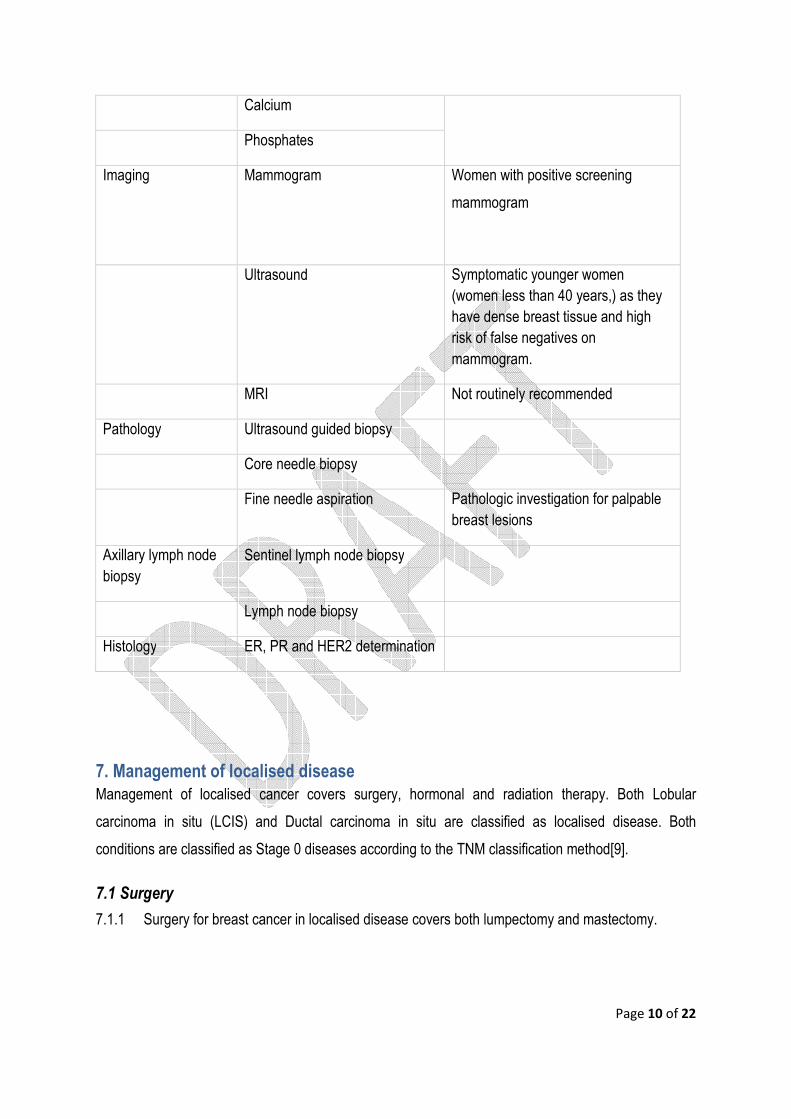
Page 10 of 22
Calcium
Phosphates
Imaging Mammogram Women with positive screening
mammogram
Ultrasound Symptomatic younger women (women less than 40 years,) as they have dense breast tissue and high risk of false negatives on mammogram.
MRI Not routinely recommended
Pathology Ultrasound guided biopsy
Core needle biopsy
Fine needle aspiration Pathologic investigation for palpable breast lesions
Axillary lymph node biopsy
Sentinel lymph node biopsy
Lymph node biopsy
Histology ER, PR and HER2 determination
7. Management of localised disease
Management of localised cancer covers surgery, hormonal and radiation therapy. Both Lobular
carcinoma in situ (LCIS) and Ductal carcinoma in situ are classified as localised disease. Both
conditions are classified as Stage 0 diseases according to the TNM classification method[9].
7.1 Surgery
7.1.1 Surgery for breast cancer in localised disease covers both lumpectomy and mastectomy.

Page 11 of 22
7.1.2 Lumpectomy without lymph node surgery together with or without radiation is indicated in
women with DCIS. However, the option of lumpectomy alone should be considered only in
cases where the patient and the physician view the individual risks as low[15].
7.1.3 Patients with DCIS and evidence of widespread disease (i.e. disease in 2 or more quadrants)
require total mastectomy with or without sentinel node biopsy. Although mastectomy provides
maximum local control, long-term cause-specific survival with mastectomy appears to be
equivalent to that with excision and whole breast irradiation [9, 35].
7.1.4 Both lumpectomy and mastectomy are PMB level of care for women with early breast cancer
7.2 Radiation therapy
7.2.1 Radiation therapy is covered in the treatment of patients with DCIS.
7.2.2 Results of clinical trials have shown that radiotherapy after local excision for DCIS, as
compared with local excision alone, reduces the overall number of both invasive and non-
invasive recurrences in the ipsilateral breast [36-38].
7.2.3 External beam radiation therapy (EBRT) is indicated for localised disease.
7.2.4 There are 3 types of EBRT: conventional radiotherapy, 3D conformal radiation therapy (3D-
CRT) and intensity modulated radiotherapy (IMRT).
7.2.5 The South African Oncology Consortium (SAOC) does not recommend the use of partial breast
irradiation as standard therapy because of concerns regarding the long term efficacy of such
therapy.
7.2.6 Supporting evidence has shown that the outcome of conventional therapy versus that of 3D-
CRT and IMRT do not differ [39, 40].
7.2.7 Conventional radiotherapy is therefore indicated as standard treatment of care.
7.3 Hormone therapy
7.3.1 The use of hormonal therapy in the management of DCIS remains uncertain.
7.3.2 Currently Tamoxifen, Letrozole, Anastrozole and Exemestane are used in the treatment of
locally advanced breast cancer.
7.3.3 Tamoxifen is a well-established drug in the treatment of breast cancer and therefore is covered
as a PMB level of care.
7.3.4 Aromatase inhibitors are currently being investigated for the adjuvant therapy of DCIS and
therefore not covered as a PMB level of care.

Page 12 of 22
Clinical Evidence
Two randomised trials have studied the use of tamoxifen in the management of DCIS. The National
Surgical Adjuvant Breast and Bowel Project (NSABP) B-24 trial was a randomised controlled trial of
BCS and adjuvant RT with tamoxifen or placebo in 889 women. The median follow up was 13.6 years.
The results of the study at 5 years showed that women in the tamoxifen group had fewer breast cancer
events (8·2 vs. 13·4%, p=0·0009), fewer cumulative incidence of all invasive breast-cancer events
(4·1% vs. 7.2%), fewer incidences of ipsilateral breast (2·1% vs. 4.2%) and contralateral breast (1·8%
vs. 2.3%). An increase in the rate of endometrial cancer was reported in the tamoxifen group (1.53 vs.
0.45 per 1000 patients in the placebo group)[41]
The United Kingdom, Australia, and New Zealand DCIS trial was a randomised 2x2 factorial study of
RT, tamoxifen or both for locally excised DCIS. Two hundred and forty two women were randomised to
receive tamoxifen and radiotherapy. Out of these patients, 25 developed a new breast event, ten DCIS
and 14 an invasive cancer. Tamoxifen plus radiotherapy significantly reduced all ipsilateral new breast
events (p<0·0001) but had no effect on contralateral new breast events (p=0·2). There were no
significant differences in new breast events between patients randomly assigned to radiotherapy and
tamoxifen and those randomised to radiotherapy alone. Patients randomised to radiotherapy and
tamoxifen had significantly reduced ipsilateral new breast events compared with those randomised to
tamoxifen alone (p<0·0001) but not contralateral new breast events (p=0·5). [38].
The benefit from endocrine therapy with tamoxifen or an aromatase inhibitor in low-risk breast cancer
(for example small tumours < 2 cm, grade 1, lymph node-negative) is very small and needs to be
weighed with the effects on quality of life [15]. Currently, aromatase inhibitors are being investigated for
the adjuvant therapy of DCIS and therefore should not be used in routine care [42].
7 .4 Surveillance/Follow-up
7.4.3 Follow-up of patients with localised disease includes interval history and physical examinations
every 6 to 12 months for 5 years and then annually as well as yearly diagnostic mammography.
7.4.4 Patients treated with breast-conserving therapy should have follow-up mammography
performed 6 to 12 months after completion of breast-conserving radiation therapy.
Page 13 of 22
7.4.3 Ultrasound or MRI is not offered routinely post-treatment in patients who have been treated for
early invasive breast cancer or DCIS [8, 9, 15].
8. Management of locally advanced disease
8.1 Patients with locally advanced disease include those with operable (Stage I, IIA, IIB, IIIA: T0 -T3
with a N1-2; N2 with any T1–T3) and inoperable disease at presentation (Stage IIIB: T4a, skin; T4b,
chest wall; T4c (a1b) with N1-N2) and those with inflammatory disease (Stage IIIC: N3 with any T,
T4d)[9, 43].
8.2 The treatment of locally advanced breast cancer includes a combination of systemic chemotherapy,
surgery, hormonal therapy and radiotherapy to optimize the chance of cure[44].
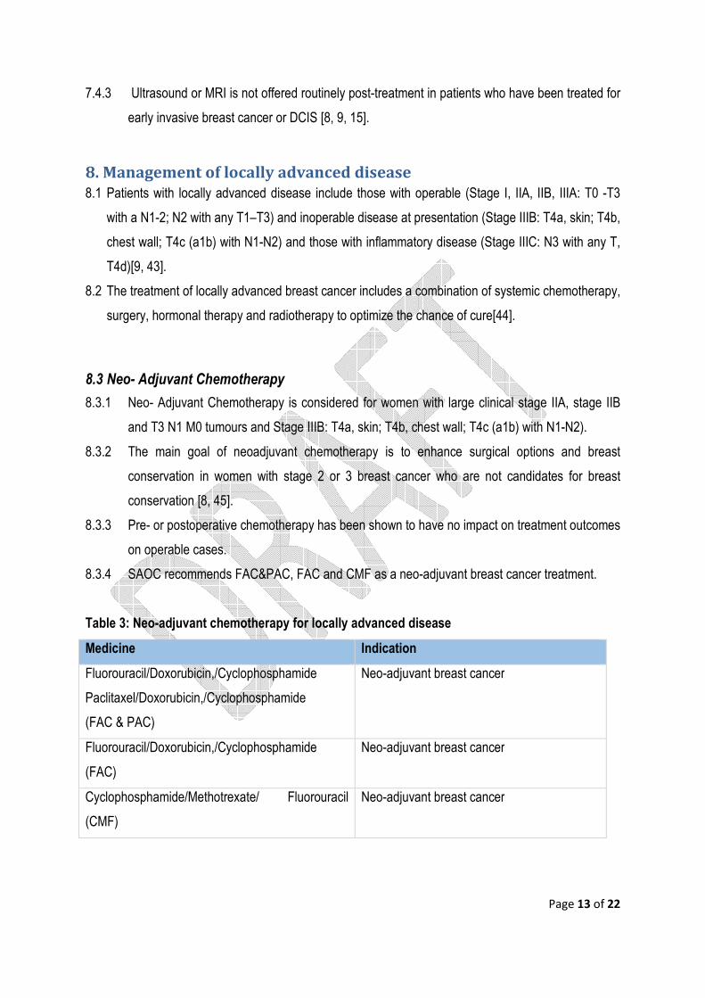
8.3 Neo- Adjuvant Chemotherapy
8.3.1 Neo- Adjuvant Chemotherapy is considered for women with large clinical stage IIA, stage IIB
and T3 N1 M0 tumours and Stage IIIB: T4a, skin; T4b, chest wall; T4c (a1b) with N1-N2).
8.3.2 The main goal of neoadjuvant chemotherapy is to enhance surgical options and breast
conservation in women with stage 2 or 3 breast cancer who are not candidates for breast
conservation [8, 45].
8.3.3 Pre- or postoperative chemotherapy has been shown to have no impact on treatment outcomes
on operable cases.
8.3.4 SAOC recommends FAC&PAC, FAC and CMF as a neo-adjuvant breast cancer treatment.
Table 3: Neo-adjuvant chemotherapy for locally advanced disease
Medicine Indication
Fluorouracil/Doxorubicin,/Cyclophosphamide
Paclitaxel/Doxorubicin,/Cyclophosphamide
(FAC & PAC)
Neo-adjuvant breast cancer
Fluorouracil/Doxorubicin,/Cyclophosphamide
(FAC)
Neo-adjuvant breast cancer
Cyclophosphamide/Methotrexate/ Fluorouracil
(CMF)
Neo-adjuvant breast cancer

Page 14 of 22
8.4 Surgery
8.4.1 Mastectomy with axillary lymph node dissection or breast-conserving therapy with lumpectomy,
axillary dissection and whole breast irradiation are indicated as a primary breast treatment of
women with stage I and stage II breast cancers[9].
8.4.2 Randomised control trail (RCTs) comparing breast conserving surgery with mastectomy found
no significant difference in terms of survival or recurrence of disease.
8.4.3 Bilateral mastectomy of un-diseased breast is excluded as a PMB level of care.
8.5 Surgery to the Axilla
8.5.1 Axillary Lymph Node surgery is indicated if there is an axillary disease. The intention of axillary
clearance is to prevent axillary relapse.
8.5.2 Axillary lymph node sampling or clearance may also be used for staging; however axillary
lymph node clearance may constitute overtreatment in some patients.
8.6 Adjuvant Chemotherapy
7.5.1 Adjuvant chemotherapy is indicated in patients with operable and inoperable disease [8, 9, 15,
46].
Table 4: Adjuvant chemotherapy for locally advanced disease
Medicine Indication Comment
Doxorubicin,/Cyclophosphamide(AC) Low risk adjuvant breast
cancer
Fluorouracil/Doxorubicin,/Cyclophosphamide
(FAC)
Low adjuvant breast cancer
Cyclophosphamide/Methotrexate/
Fluorouracil (CMF)
Low adjuvant breast cancer
Fluorouracil/Epirubicin/Cyclophosphamide
(FEC)
Docetaxel
High risk adjuvant breast
cancer
Node positive fit
patients
Paclitaxel/Doxorubicin,/Cyclophosphamide
(PAC)
High risk adjuvant breast
cancer
Younger patients with
higher risk of relapse

Page 15 of 22
Doxorubicin,/Cyclophosphamide (AC)
Fluorouracil/Doxorubicin,/Cyclophosphamide
(FAC)
High risk adjuvant breast
cancer
Doxorubicin 75 & CMF 21 High risk adjuvant breast
cancer
Cyclophosphamide/ Epirubicin/ Fluorouracil
(CEF) 28
High risk adjuvant breast
cancer
Highly selected high
risk patients, ER-
PgR- Her2+++; node
positive
8.7 Hormonal therapy
8.7.1 Patients with invasive breast cancers that are ER- or PR- positive are considered for adjuvant
endocrine therapy regardless of patient age, lymph node status, or whether adjuvant
chemotherapy is to be administered. [9, 15].
8.7.2 Tamoxifen is indicated for 10 years in patients with non-metastatic hormone receptor positive
breast cancer.
8.7.3 Aromatase inhibitors are indicated after 2-3 years of Tamoxifen in intermediate and high-risk
patients, or after completing 5 years of Tamoxifen
8.7.4 LHRH AGONIST (Goserelin, Zoledronic acid) + Aromatase Inhibitors are indicated for
premenopausal with Tamoxifen contraindication
Table 5: Hormonal therapy for locally advanced disease
Medicine Comment
Tamoxifen Adjuvant Tamoxifen for 10 years in patients with non‐metastatic
hormone receptor positive breast cancer
Anastrozole Postmenopausal women with ER-positive early invasive breast
cancer who are high risk and who have been treated with Tamoxifen
for 2–3 years.
Letrozole Postmenopausal women with ER-positive early invasive breast

Page 16 of 22
cancer who are high risk and who have been treated with Tamoxifen
for 2–3 years.
Exemestane Postmenopausal women with ER-positive early invasive breast
cancer who are high risk and who have been treated with Tamoxifen
for 2–3 years.
LHRH Agonist (Goserelin,
Zoledronic acid) +
Aromatase Inhibitors
Premenopausal with Tamoxifen contraindication
Clinical Evidence
Evidence from the ATAC trial showed anastrozole to be an effective and well-tolerated endocrine option
for the treatment of postmenopausal patients with early breast cancer. The randomized, double-blind
trial, compared tamoxifen (20 mg) with anastrozole (1 mg) alone, and the combination of anastrozole
plus tamoxifen (combination), as adjuvant endocrine treatment for postmenopausal patients with early
breast cancer. A total of 9366 patients with operable invasive breast cancer following completion of
primary therapy were included in the study. Median duration of therapy was 30.7 months and median
follow-up was 33.3 months. The results of the study showed a significant improvement in disease free
survival (DFS) (hazard ratio (HR) =0.81, 95% confidence interval (CI) (0.71-0.96), P=0.013) and time to
relapse in the anastrozole compared with tamoxifen (HR=0.79, CI (0.67-0.94), P=0.008), which
improved even further in the ER+ and/or PR+ subgroup (HR=0.73, CI (0.59-0.90), P=0.003). The
incidences of hot flushes, thromboembolic events, Ischaemic cerebrovascular events, vaginal
bleeding/discharge and endometrial cancer were significantly reduced with anastrozole compared with
tamoxifen (P<0.03 for all). Musculoskeletal disorders and fractures were significantly reduced in
patients receiving tamoxifen compared with those on anastrozole (P<0.03 for both). No increase in hip
fractures was seen for anastrozole versus tamoxifen (11 versus 13, respectively[47].
Evidence from the BIG 1-98 phase III, double-blind trial of 8010 postmenopausal showed that women
with endocrine-responsive early breast cancer had a reduction in breast cancer recurrence and
mortality when using letrozole monotherapy when compared to tamoxifen. The monotherapy

Page 17 of 22
comparison included patients randomized to tamoxifen × 5 years (n=2459) or letrozole × 5 years
(n=2463).The results of the study showed however that sequential treatments involving tamoxifen and
letrozole does not improve outcome when compared with letrozole monotherapy[48].
The results of a double-blind, randomized trial to test whether, after two to three years of tamoxifen
therapy, switching to exemestane was more effective than continuing tamoxifen therapy for the
remainder of the five years of treatment showed that exemestane therapy significantly improved
disease-free survival as compared with the standard five years of tamoxifen treatment. A total of 4742
patients were enrolled in the study. After a median follow-up of 30.6 months, 183 events were recorded
in the exemestane group and 266 in the tamoxifen group. Overall survival was not significantly different
in the two groups, with 93 deaths occurring in the exemestane group and 106 in the tamoxifen group.
Severe toxic effects of exemestane were rare. Contra lateral breast cancer occurred in 20 patients in
the tamoxifen group and 9 in the exemestane group (P=0.04)[49].
8.8 Radiation therapy
8.8.1 After mastectomy and axillary dissection, radiotherapy has been shown to reduce both
recurrence and breast cancer mortality in women with one to three positive lymph nodes in
clinical trials[50, 51].
8.8.2 External beam radiation therapy is indicated for localised disease. There are 3 types of EBRT:
conventional radiotherapy, 3d conformal radiation therapy (3d-CRT) and intensity modulated
radiotherapy.
8.8.3 The South African Oncology Consortium does not recommend the use of partial breast
irradiation as standard therapy because of concerns regarding the long term efficacy of such
therapy.
8.8.4 Supporting evidence has shown that the outcome of conventional therapy versus that of 3d-
CRT and IMRT do not differ.
8.8.5 Conventional radiotherapy is therefore covered as a PMB level of care.
Page 18 of 22
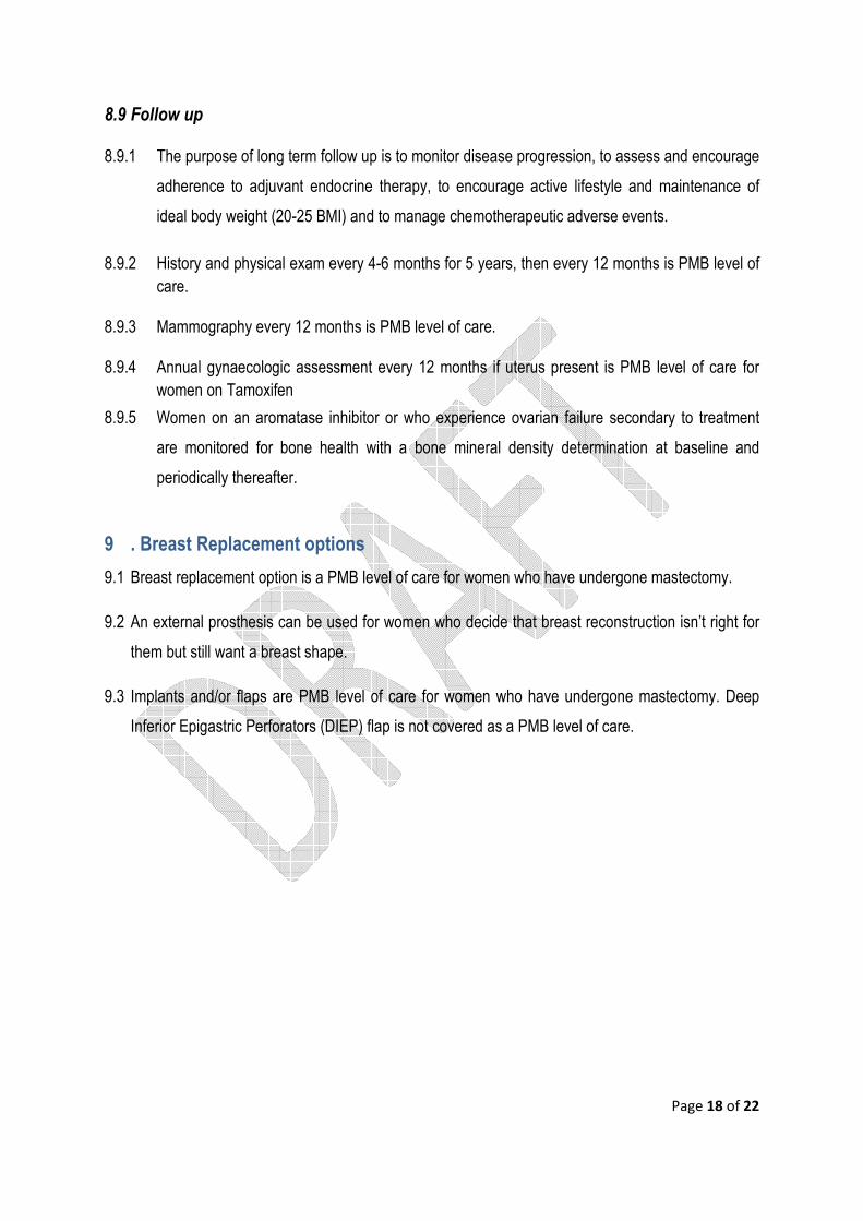
8.9 Follow up
8.9.1 The purpose of long term follow up is to monitor disease progression, to assess and encourage
adherence to adjuvant endocrine therapy, to encourage active lifestyle and maintenance of
ideal body weight (20-25 BMI) and to manage chemotherapeutic adverse events.
8.9.2 History and physical exam every 4-6 months for 5 years, then every 12 months is PMB level of care.
8.9.3 Mammography every 12 months is PMB level of care.
8.9.4 Annual gynaecologic assessment every 12 months if uterus present is PMB level of care for women on Tamoxifen
8.9.5 Women on an aromatase inhibitor or who experience ovarian failure secondary to treatment
are monitored for bone health with a bone mineral density determination at baseline and
periodically thereafter.
9 . Breast Replacement options
9.1 Breast replacement option is a PMB level of care for women who have undergone mastectomy.
9.2 An external prosthesis can be used for women who decide that breast reconstruction isn’t right for
them but still want a breast shape.
9.3 Implants and/or flaps are PMB level of care for women who have undergone mastectomy. Deep
Inferior Epigastric Perforators (DIEP) flap is not covered as a PMB level of care.

Page 19 of 22
10. Reference
1. WHO, Latest world cancer statistics. 2013, World Health Organisation: Geneva, Switzerland. 2. Soceity, A.C. Global Cancer: Facts and Figures. 2011. 3. Coleman, M.P., et al., Cancer survival in five continents: a worldwide population-based study
(CONCORD). The Lancet Oncology, 2008. 9(8): p. 730-756. 4. Karusseit, V. Locally advanced breast cancer in the developing countries. University of
Pretoria. 5. Cansa. Cancer Statistics. 2007 [cited 2014; Available from:
http://www.cansa.org.za/files/2014/06/NCR-2007-Cancer-Statistics.pdf. 6. WHO, Guidelines for the early detection and screening of breast cancer, O.M.N. Khatib and A.
Modjtabai, Editors. 2006, World Health Organisation. 7. USPSTF, Screening for breast cancer: U.S. Preventive Services Task Force recommendation
statement. Ann Intern Med, 2009. 151(10): p. 716-26, W-236. 8. Senkus, E., et al., Primary breast cancer: ESMO Clinical Practice Guidelines for diagnosis,
treatment and follow-up. Ann Oncol, 2013. 24 Suppl 6: p. vi7-23. 9. NCCN, Breast Cancer. 2013. 10. Cardoso, F., et al., 1st International consensus guidelines for advanced breast cancer (ABC 1).
Breast, 2012. 21(3): p. 242-52. 11. Hooley, e.a., Screening US in Patients with Mammographically Dense Breasts: Initial
Experience with Connecticut Public Act 09-411. Radiology, 2012. 265(1). 12. Nothacker, M., et al., Early detection of breast cancer: benefits and risks of supplemental
breast ultrasound in asymptomatic women with mammographically dense breast tissue. A systematic review. BMC Cancer, 2009. 9: p. 335.
13. Cardoso, F., et al., The European Society of Breast Cancer Specialists recommendations for the management of young women with breast cancer. Eur J Cancer, 2012. 48(18): p. 3355-77.
14. Mann, R.M., et al., MRI compared to conventional diagnostic work-up in the detection and evaluation of invasive lobular carcinoma of the breast: a review of existing literature. Breast Cancer Res Treat, 2008. 107(1): p. 1-14.
15. NICE, Early and locally advanced breast cancer:diagnosis and treatment. 2009. 16. Africa, R.S.o.S. PET-CT referral guidelines. 2013. 2. 17. James, J.J., et al., CT staging for breast cancer patients with poor prognostic tumours. Breast,
2012. 21(6): p. 735-8. 18. Wendy Bruening, J.F., Kelley Tipton, Jonathan R. Treadwell, Jason Launders, Karen
Schoelles,, Systematic Review: Comparative Effectiveness of Core-Needle and Open Surgical Biopsy to Diagnose Breast Lesions. Ann Intern Med., 2010. 152: p. 238-246.
19. Tamaki, K., et al., Comparison of core needle biopsy (CNB) and surgical specimens for accurate preoperative evaluation of ER, PgR and HER2 status of breast cancer patients. Cancer Sci, 2010. 101(9): p. 2074-9.
20. Sauer, T., et al., Fine-needle aspiration cytology in nonpalpable mammographic abnormalities in breast cancer screening: results from the breast cancer screening programme in Oslo 1996–2001. The Breast, 2003. 12(5): p. 314-319.
21. Marta WesołaA–D, M.J., The Diagnostic Efficiency of Fine Needle Aspiration Biopsy in Breast Cancers – Review. Adv Clin Exp Med, 2013. 22(6): p. 887–892.
22. WHO, Guidelines for management of breast cancer, W.H. Organisation, Editor. 2006. 23. Association of Breast Surgery at, B., Surgical guidelines for the management of breast cancer.
Eur J Surg Oncol, 2009. 35 Suppl 1: p. 1-22. 24. Salem, A., Sentinel lymph node biopsy in breast cancer: a comprehensive literature review. J
Surg Educ, 2009. 66(5): p. 267-75.

Page 20 of 22
25. Schwartz, G.F., Clinical Practice Guidelines for the Use of Axillary Sentinel Lymph Node Biopsy in Carcinoma of the Breast: Current Update. The Breast Journal, 2004. 10(2): p. 85–88.
26. Son, B.K., et al., Ductal carcinoma in situ and sentinel lymph node biopsy. J Breast Cancer, 2011. 14(4): p. 301-7.
27. Hammond, M.E., et al., American Society of Clinical Oncology/College Of American Pathologists guideline recommendations for immunohistochemical testing of estrogen and progesterone receptors in breast cancer. J Clin Oncol, 2010. 28(16): p. 2784-95.
28. Wolff, A.C., et al., Recommendations for human epidermal growth factor receptor 2 testing in breast cancer: American Society of Clinical Oncology/College of American Pathologists clinical practice guideline update. J Clin Oncol, 2013. 31(31): p. 3997-4013.
29. Robert E. Myers, M.J., Kathy Pritchard, Mark Levine,§ Tom Oliver,† and the Breast Cancer Disease Site Group of the Cancer Care Ontario Practice Guidelines Initiative, Baseline staging tests in primary breast cancer: a practice guideline. CMAJ, 2001. 164(10): p. 1439-44.
30. Puglisi, F., et al., Baseline staging tests after a new diagnosis of breast cancer: further evidence of their limited indications. Ann Oncol, 2005. 16(2): p. 263-6.
31. DANIELA MÜLLER, G.K.a.R.O., Staging Procedures in Primary Breast Cancer. ANTICANCER RESEARCH, 2008. 28: p. 2397-2400.
32. Kasem AR, D.A., Daniell S, Sinha P., Bone scan and liver ultrasound scan in the preoperative staging for primary breast cancer. Breast J, 2006. 12(6).
33. Dose J1, B.C., Bachmann S, Bohuslavizki KH, Berger J, Jenicke L, Habermann CR, Jãnicke F, Comparison of fluorodeoxyglucose positron emission tomography and "conventional diagnostic procedures" for the detection of distant metastases in breast cancer patients. Nucl Med Commun, 2002. 23(9): p. 857-64.
34. Mahner, S., et al., Comparison between positron emission tomography using 2-[fluorine-18]fluoro-2-deoxy-D-glucose, conventional imaging and computed tomography for staging of breast cancer. Ann Oncol, 2008. 19(7): p. 1249-54.
35. Patani, N., et al., Ductal carcinoma in-situ: an update for clinical practice. Surg Oncol, 2011. 20(1): p. e23-31.
36. Goodwin, A., et al., Post-operative radiotherapy for ductal carcinoma in situ of the breast. Cochrane Database Syst Rev, 2013. 11: p. CD000563.
37. Holmberg, L., et al., Absolute risk reductions for local recurrence after postoperative radiotherapy after sector resection for ductal carcinoma in situ of the breast. J Clin Oncol, 2008. 26(8): p. 1247-52.
38. Cuzick, J., et al., Effect of tamoxifen and radiotherapy in women with locally excised ductal carcinoma in situ: long-term results from the UK/ANZ DCIS trial. The Lancet Oncology, 2011. 12(1): p. 21-29.
39. Olivotto, I.A., et al., Interim cosmetic and toxicity results from RAPID: a randomized trial of accelerated partial breast irradiation using three-dimensional conformal external beam radiation therapy. J Clin Oncol, 2013. 31(32): p. 4038-45.
40. Smith, B.D., et al., Accelerated partial breast irradiation consensus statement from the American Society for Radiation Oncology (ASTRO). Int J Radiat Oncol Biol Phys, 2009. 74(4): p. 987-1001.
41. Fisher, B., et al., Tamoxifen in treatment of intraductal breast cancer: National Surgical Adjuvant Breast and Bowel Project B-24 randomised controlled trial. Lancet, 1999. 353: p. 1993–2000.
42. Chlebowski, R.T. and N. Col, Postmenopausal women with DCIS post-mastectomy: a potential role for aromatase inhibitors. Breast J, 2012. 18(4): p. 299-302.
43. Seung, A.H.S.a.A.H., Early and Locally advanced breast cancer. p. 19.

Page 21 of 22
44. Tamara Shenkier, L.W., Mark Levine, Ivo Olivotto, Timothy Whelan, Leonard Reyno, Clinical practice guidelines for the care and treatment of breast cancer: 15. Treatment for women with stage III or locally advanced breast cancer. CMAJ, 2004. 170(6): p. 983-994.
45. Connolly, R.M. and V. Stearns, Current approaches for neoadjuvant chemotherapy in breast cancer. Eur J Pharmacol, 2013. 717(1-3): p. 58-66.
46. GIORDANO, S.H., Update on Locally Advanced Breast Cancer. The Oncologists, 2003: p. 521-530.
47. Buzdar, A. and A.t. group., 'Arimidex' (anastrozole) versus tamoxifen as adjuvant therapy in postmenopausal women with early breast cancer--efficacy overview. J Steroid Biochem Mol Biol, 2003. 86()(3-5): p. 399-403.
48. Regan, M.M., et al., Assessment of letrozole and tamoxifen alone and in sequence for postmenopausal women with steroid hormone receptor-positive breast cancer: the BIG 1-98 randomised clinical trial at 8·1 years median follow-up. The Lancet Oncology, 2011. 12(12): p. 1101-1108.
49. Coombes, R.C., et al., A Randomized Trial of Exemestane after Two to Three Years of Tamoxifen Therapy in Postmenopausal Women with Primary Breast Cancer. n engl j med, 2004. 350(11).
50. G. Yavas, C. Yavas, and S. Akyurek, Postmastectomy radiation therapy in locally advanced breast cancer. Exp Oncol, 2013. 35(4): p. 258–266.
51. EBCTCG, Effect of radiotherapy after mastectomy and axillary surgery on 10-year recurrence and 20-year breast cancer mortality: meta-analysis of individual patient data for 8135 women in 22 randomised trials. The Lancet, 2014. 383(9935): p. 2127-2135.
52. Metcalfe, K.A., et al., Changes in psychosocial functioning 1 year after mastectomy alone, delayed breast reconstruction, or immediate breast reconstruction. Ann Surg Oncol, 2012. 19(1): p. 233-41.

Page 22 of 22