draft report support for chemical nomination and selection process
TRANSCRIPT
DRAFT REPORT
SUPPORT FOR CHEMICAL NOMINATION AND SELECTION
PROCESS OF THE NATIONAL TOXICOLOGY PROGRAM
EXECUTIVE SUMMARY OF DATA
CINNAMALDEHYDE
DECEMBER 14, 1989
Submitted to:
NATIONAL TOXICOLOGY PROGRAM
Submitted by:
Arthur D. Little, Inc.
Disclaimer The information contained herein is based on data from current published literature and is believed to be accurate. However, no warranty is expressed or implied regarding the accuracy ofthese data or the results to be obtained from the use thereof
TABLE OF CONTENTS ~
I. Introduction .........................................................................................................2
II. Chemical and Physical Properties .......................................................................... .3
III. Production!Use........................................................................................................5
A. Production...................................................................................................5
B. Use .........................................................................................................6
IV. Exposure/Regulatory Status ....................................................................................7
A. Consumer Exposure ....................................................................................7
B. Occupational Exposure ...............................................................................7
C. Environmental Exposure .............................................................................8
D. Regulatory Status ........................................................................................8
V. Toxicological Effects ...............................................................................................9
A. Acute .........................................................................................................9
1. Animal Data ....................................................................................9
2. Human Data .................................................................................... 1 0
3. Case Reports ................................................................................... 1 0
B. Subchronic/Chronic ..................................................................................... 12
1. Animal Data .................................................................................... 12
2. Case Reports ...................................................................................13
C. Carcinogenicity............................................................................................ 15
1. Animal Data ....................................................................................15
2. Human Data .................................................................................... 17
D. Mutagenicity/Genetic Toxicology ............................................................... 17
1. Animal Data .................................................................................... 1 7
E. Teratology/Reproductive Toxicology .........................................................21
1. Animal Data ....................................................................................21
F. Immunotoxicity ...........................................................................................22
VI. Chemical Disposition ..............................................................................................23
A. Animal Data ................................................................................................23
B. Human Data ................................................................................................25
VII. Biochemical. Toxicology ..........................................................................................25
1. Animal Data ................................................................................................25
2. Human Data ................................................................................................31
VIII. Structure/Activity Considerations ..........................................................................37
I. INTRODUCTION
Exposure to Cinnamaldehyde results primarily from its widespread use as a flavor and
fragrance ingredient in food, beverages, medical products, cosmetics and perfumes. There
are numerous reports in the literature describing cases of skin sensitization reactions
resulting from both occupational and consumer exposure to Cinnamaldehyde. This
compound has also been found to cause severe skin irritation following acute exposure.
There are conflicting reports concerning the mutagenicity of Cinnamaldehyde.
Cinnamaldehyde has been found to be mutagenic to Bacillus subtilis, Drosophila
melanogaster, Chinese hamster ovary cells, mouse leukocytes, hamster fibroblasts and
Salmonella strain TAlOO. However, Cinnamaldehyde has also been found, by other
authors, to be non-mutagenic to this strain ofSalmonella and is reportedly non-mutagenic
in other test systems. In addition, there are conflicting reports concerning the
teratogenicity of this compound. In one study, Cinnamaldehyde was found to induce limb
malformations in chick embryos. Although there are no data available which associate
Cinnamaldehyde with carcinogenic effects in animals or humans, the transforming
capacity of this compound has been demonstrated in vitro. In addition, two related
compounds, 3, 4, 5-Trimethoxy Cinnamaldehyde and Cinnamyl Anthranilate, have been
found to be animal carcinogens. Because Anthranilic Acid was observed to be non
carcinogenic, it is believed that the Cinnamyl moiety may play a role in the
carcinogenicity of Cinnamyl Anthranilate.
Based on both the concern about the possible carcinogenicity of the Cinnamyl moiety,
and the importance ofCinnamaldehyde as a flavor ingredient in food, the Food and Drug
Administration (FDA) has nominated Cinnamaldehyde as its priority chemical for fiscal
year 1989. Cinnamaldehyde was originally nominated to the National Toxicology Program
(NTP) in 1979, at which time the Chemical Nomination and Selection Committee
(currently the Chemical Evaluation Committee) recommended that this compound be
selected for testing. However, because of scheduled budget cuts and subsequent
reallocation of resources, toxicological studies on Cinnamaldehyde were not performed.
2
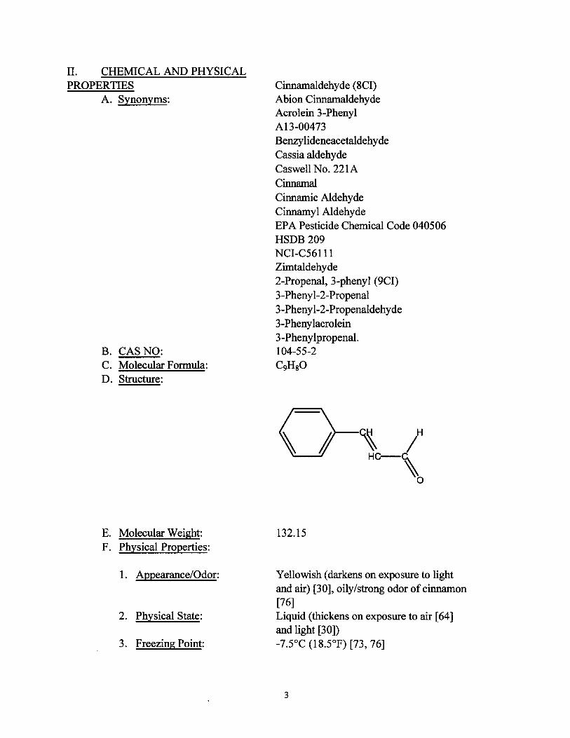
II. CHEMICAL AND PHYSICAL PROPERTIES
A. Synonyms:
B. CASNO: C. Molecular Formula: D. Structure:
E. Molecular Weight: F. Physical Properties:
1. Appearance/Odor:
2. Physical State:
3. Freezing Point:
Cinnamaldehyde (8CI) Abion Cinnamaldehyde Acrolein 3-Phenyl A13-00473 Benzylideneacetaldehyde Cassia aldehyde Caswell No. 221A Cinnamal Cinnamic Aldehyde Cinnamyl Aldehyde EPA Pesticide Chemical Code 040506 HSDB 209 NCI-C56111 Zimtaldehyde 2-Propenal, 3-phenyl (9CI) 3-Phenyl-2-Propenal 3-Pheny 1-2-Propenaldehyde 3-Phenylacrolein 3-Phenylpropenal. 104-55-2 C9HsO
132.15
Yellowish (darkens on exposure to light and air) [30], oily/strong odor ofcinnamon [76] Liquid (thickens on exposure to air [64] and light [30]) -7.5°C (18.5°F) [73, 76]
3
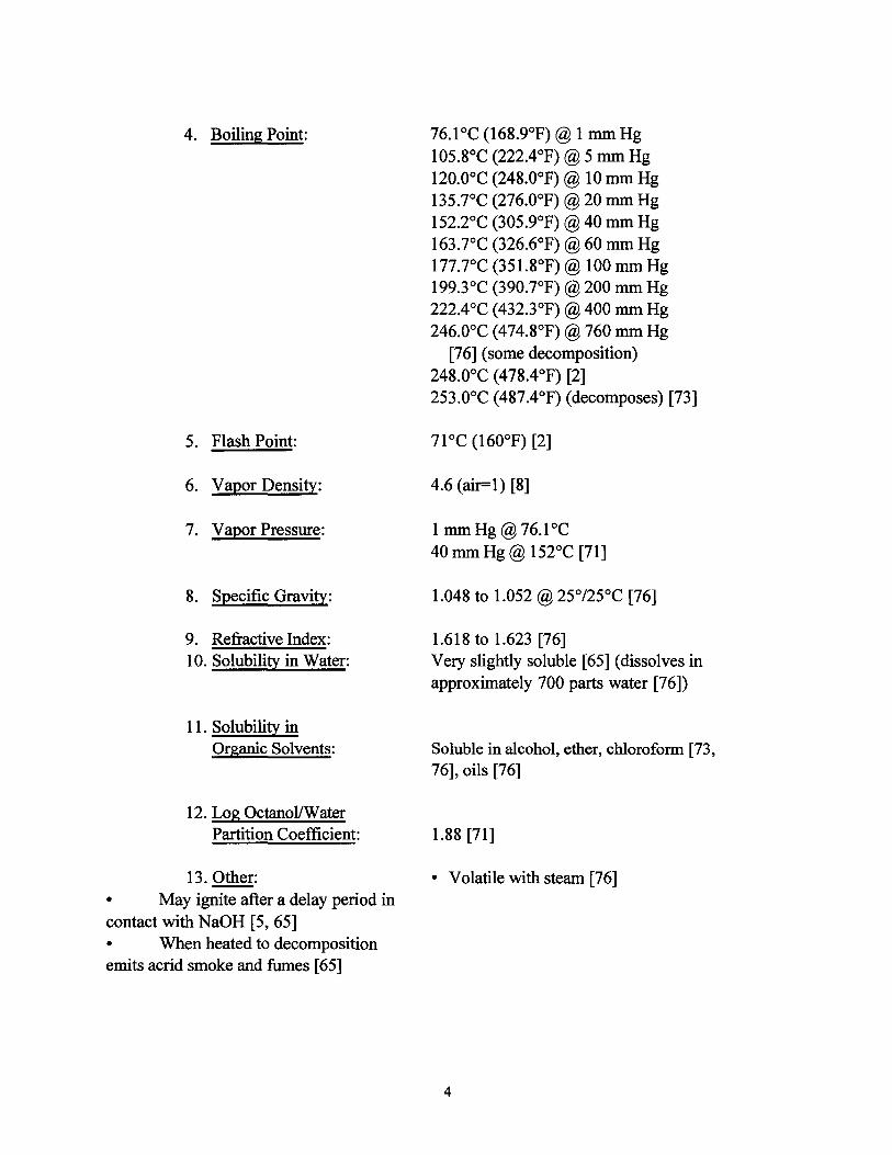
4. Boiling Point: 76.1 oc (168.9°F)@ 1 mm Hg 105.8°C (222.4°F)@ 5 mm Hg 12o.ooc (248.0°F)@ 10 mm Hg 135.7°C (276.0°F)@ 20 mm Hg 152.2°C (305.9°F)@ 40 mm Hg 163.7°C (326.6°F)@ 60 mm Hg 177.7°C (351.8°F)@ 100 mm Hg 199.3°C (390.7°F)@ 200 mm Hg 222.4°C (432.3°F)@ 400 mm Hg 246.0°C (474.8°F)@ 760 mm Hg
[76] (some decomposition) 248.0°C (478.4°F) [2] 253.0°C (487.4°F) (decomposes) [73]
5. Flash Point:
6. Vapor Density: 4.6 (air=l) [8]
7. Vapor Pressure: 1 mm Hg @ 76.1 oc 40 mm Hg@ 152°C [71]
8. Specific Gravity: 1.048 to 1.052@ 25°/25°C [76]
9. Refractive Index: 1.618to 1.623 [76] 10. Solubility in Water: Very slightly soluble [65] (dissolves in
approximately 700 parts water [76])
11. Solubility in Organic Solvents: Soluble in alcohol, ether, chloroform [73,
76], oils [76]
12. Log Octanol/Water Partition Coefficient: 1.88 [71]
13. Other: • May ignite after a delay period in contact with NaOH [5, 65] • When heated to decomposition emits acrid smoke and fumes [65]
• Volatile with steam [76]
4
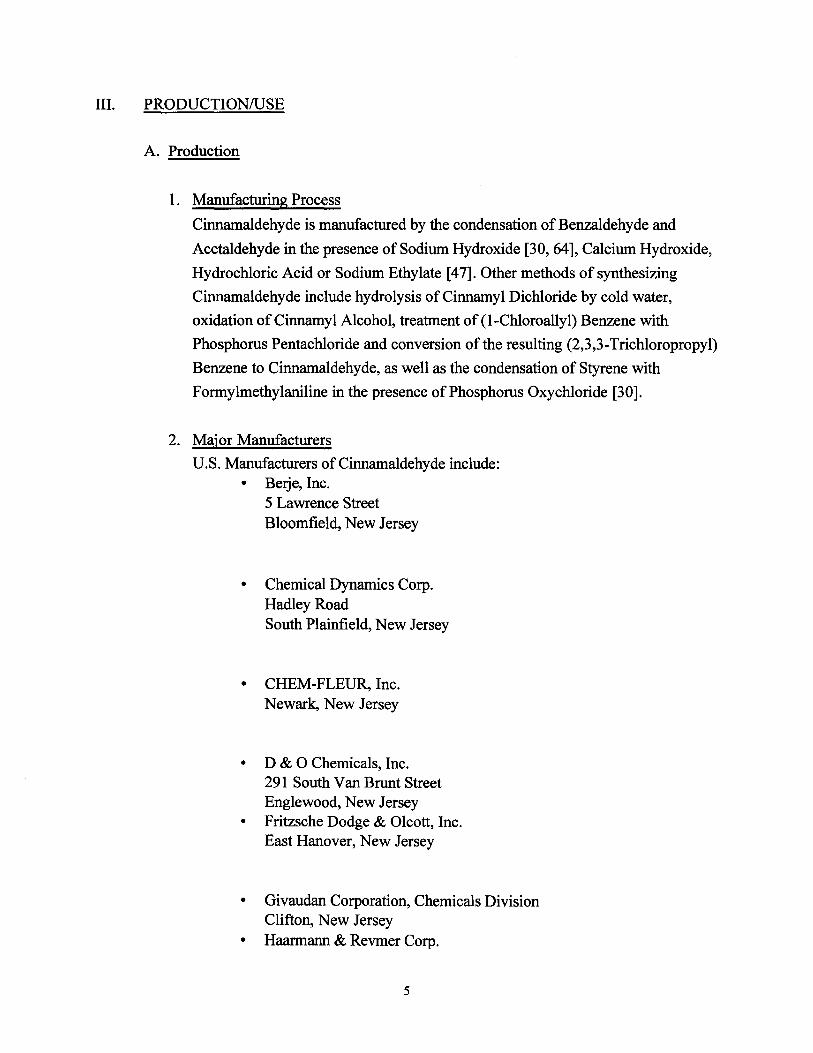
Ill. PRODUCTION/USE
A. Production
1. Manufacturing Process
Cinnamaldehyde is manufactured by the condensation of Benzaldehyde and
Acetaldehyde in the presence of Sodium Hydroxide [30, 64], Calcium Hydroxide,
Hydrochloric Acid or Sodium Ethylate [47]. Other methods of synthesizing
Cinnamaldehyde include hydrolysis of Cinnamyl Dichloride by cold water,
oxidation of Cinnamyl Alcohol, treatment of (1-Chloroallyl) Benzene with
Phosphorus Pentachloride and conversion of the resulting (2,3,3-Trichloropropyl)
Benzene to Cinnamaldehyde, as well as the condensation of Styrene with
Formylmethylaniline in the presence of Phosphorus Oxychloride [30].
2. Major Manufacturers
U.S. Manufacturers ofCinnamaldehyde include: • Berje, Inc.
5 Lawrence Street Bloomfield, New Jersey
• Chemical Dynamics Corp. Hadley Road South Plainfield, New Jersey
• CHEM-FLEUR, Inc. Newark, New Jersey
• D & 0 Chemicals, Inc . 291 South Van Brunt Street Englewood, New Jersey
• Fritzsche Dodge & Olcott, Inc . East Hanover, New Jersey
• Givaudan Corporation, Chemicals Division Clifton, New Jersey
• Haarmann & Revmer Corp .
5

70 Diamond Road Springfield, New Jersey
• NIP A Laboratories, Inc. 3411 Silverside Road Wilmington, Delaware
• Penta Manufacturing Company P.O. Box 1448 Fairfield, New Jersey
• Quest International Fragrances USA Inc. 400 International Drive Mount Olive, New Jersey
• Universal Oil Products Company East Rutheford, New Jersey [7,47]
3. Volume
In 1977, 911,730,000 grams ofCinnamaldehyde were produced in the United
States as reported by the United States International Trade Commission. Between
1978 and 1989, production data on this compound were not published.
There are no export or import data available in the current literature for
Cinnamaldehyde. The following import data have been reported for Cinnamon Oil:
in 1972, 1.85 x 10⁷ grams of Cinnamon Oil were imported to the United States. In
1975, 1.9 x 10⁷ grams were reportedly imported [47].
B. Use
Cinnamaldehyde is used primarily in the flavor and fragrance industries for imparting
a cinnamon flavor and/or fragrance to various types of foods, beverages, medical
products, and perfumes. This chemical is used in the liquor industry for flavoring
liqueurs and cordials. Cinnamaldehyde has also been used as a rubber reinforcing
agent, a filtering agent, an attractant for termites, a corrosion inhibitor for sulfuric acid
baths to clean galvanized iron and zinc, as an emulsion fog inhibitor for photographic
6
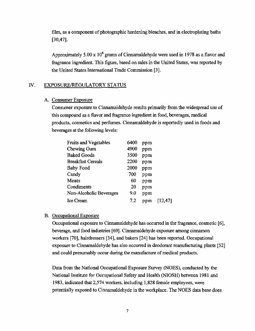
film, as a component ofphotographic hardening bleaches, and in electroplating baths
[30,47].
Approximately 5.00 x 10⁸ grams ofCinnamaldehyde were used in 1978 as a flavor and
fragrance ingredient. This figure, based on sales in the United States, was reported by
the United States International Trade Commission [3].
N. EXPOSUREffiEGULATORYSTATUS
A. Consumer Exposure
Consumer exposure to Cinnamaldehyde results primarily from the widespread use of
this compound as a flavor and fragrance ingredient in food, beverages, medical
products, cosmetics and perfumes. Cinnamaldehyde is reportedly used in foods and
beverages at the following levels:
Fruits and Vegetables 6400 ppm Chewing Gum 4900 ppm Baked Goods 3500 ppm Breakfast Cereals 2200 ppm Baby Food 2000 ppm Candy 700 ppm Meats 60 ppm Condiments 20 ppm Non-Alcoholic Beverages 9.0 ppm
Ice Cream 7.2 ppm [12,47]
B. Occupational Exposure
Occupational exposure to Cinnamaldehyde has occurred in the fragrance, cosmetic [6],
beverage, and food industries [69]. Cinnamaldehyde exposure among cinnamon
workers [70], hairdressers [34], and bakers [24] has been reported. Occupational
exposure to Cinnamaldehyde has also occurred in deodorant manufacturing plants [52]
and could presumably occur during the manufacture of medical products.
Data from the National Occupational Exposure Survey (NOES), conducted by the
National Institute for Occupational Safety and Health (NIOSH) between 1981 and
1983, indicated that 2,574 workers, including 1,828 female employees, were
potentially exposed to Cinnamaldehyde in the workplace. The NOES data base does
7
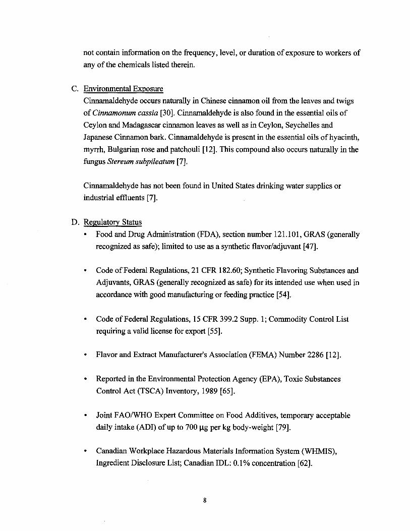
not contain information on the frequency, level, or duration of exposure to workers of
any of the chemicals listed therein.
C. Environmental Exposure
Cinnamaldehyde occurs naturally in Chinese cinnamon oil from the leaves and twigs
of Cinnamonum cassia [30]. Cinnamaldehyde is also found in the essential oils of
Ceylon and Madagascar cinnamon leaves as well as in Ceylon, Seychelles and
Japanese Cinnamon bark. Cinnamaldehyde is present in the essential oils of hyacinth,
myrrh, Bulgarian rose and patchouli [12]. This compound also occurs naturally in the
fungus Stereum subpileatum [7].
Cinnamaldehyde has not been found in United States drinking water supplies or
industrial effluents [7].
D. Regulatory Status
• Food and Drug Administration (FDA), section number 121.101, GRAS (generally
recognized as safe); limited to use as a synthetic flavor/adjuvant [47].
• Code ofFederal Regulations, 21 CFR 182.60; Synthetic Flavoring Substances and
Adjuvants, GRAS (generally recognized as safe) for its intended use when used in
accordance with good manufacturing or feeding practice [54].
• Code ofFederal Regulations, 15 CFR 399.2 Supp. 1; Commodity Control List
requiring a valid license for export [55].
• Flavor and Extract Manufacturer's Association (FEMA) Number 2286 [12].
• Reported in the Environmental Protection Agency (EPA), Toxic Substances
Control Act (TSCA) Inventory, 1989 [65].
• Joint F AOIWHO Expert Committee on Food Additives, temporary acceptable
daily intake (ADI) of up to 700 Jlg per kg body-weight [79].
• Canadian Workplace Hazardous Materials Information System (WHMIS),
Ingredient Disclosure List; Canadian IDL: 0.1% concentration [62].
8
• There is no OSHA permissible exposure limit (PEL) or ACGIH recommended
threshold limit value (TL V).
V. TOXICOLOGICAL EFFECTS
A. Acute
1. Animal Data
Exposure to Cinnamaldehyde had been found to affect the central nervous,
cardiovascular, and digestive systems. This compound has also reportedly caused
contact urticaria, diarrhea, depression and coma in animals following acute
exposure.
Cinnamaldehyde has been found to have both inhibitory and excitatory effects on
the central nervous system of mice. Intraperitoneal administration of this
compound at doses higher than 100 mg/kg was observed to cause a transient
excitation (running fit) followed by a depression in activity [72].
Cinnamaldehyde has been observed to affect the cardiovascular system ofdogs
and guinea pigs. Intravenous administration of 5-10 mg/kg to male and female
Mongrel dogs was found to reduce blood pressure and increase respiratory rate
and femoral blood flow. Heart rate was observed to increase simultaneously with
the fall in blood pressure, and thereafter to return to baseline.
A fall in blood pressure was also observed in male guinea pigs following
intravenous administration of Cinnamaldehyde at a dose of 1 mglkg. Heart rate
was lowered by 15 percent following administration of this compound at a dose of
5 mg/kg, while femoral blood flow was observed to increase. In experiments using
isolated guinea pig hearts, Cinnamaldehyde administered at doses ranging from 50
to 500 Jlg was found to increase heart beat rate and to induce arrhythmias at does
greater than 250 J..Lg.
Cinnamaldehyde has also been observed to affect the digestive systems of rats and
mice. In male, dd mice, Cinnamaldehyde was found to have an inhibitory effect on
intestinal propulsion following intraperitoneal administration at a dose of 250
mg/kg. In addition, Cinnamaldehyde was observed to decrease stress-induced
gastric erosion at an intraperitoneal dose of250 mg/kg. In male, Wistar rats, this
9
compound was found to inhibit spontaneous gastric contraction at an intravenous
dose of 5 mglk:g. Oral administration of Cinnamaldehyde at a dose of 500 mglk:g
reportedly increased biliary excretion. Cinnamaldehyde did not change the pH
value of gastric perfusate at intravenous doses up to 10 mg/k:g [18].
Cinnamaldehyde has been found to induce nonimmunologic contact urticaria in
guinea pigs, rats and mice, with symptoms ranging from slight erythema to
extensive local erythema and edema accompanied by tingling, burning and itching,
following application of a 20% solution to the earlobes. The thickness of the
earlobes was measured before, during and after the application. Maximal ear
swelling was observed 20 to 50 minutes after the application of Cinnamaldehyde
and reportedly decreased during the three-hour observation period [32].
Acute expose to Cinnamaldehyde has been found to cause diarrhea and depression
in rats. High, acute doses of this compound have induced coma in rats [ 48]. Acute
systemic toxicity values for Cinnamaldehyde are presented in Table 1.
2. Human Data
Acute exposure to Cinnamaldehyde may result in skin, eye [58], respiratory [47]
and gastrointestinal irritation. Systemic effects from acute exposure are believed to
be limited [16]. Acute toxicity data available for Cinnamaldehyde is restricted
primarily to this compound's effect on the skin.
Cinnamaldehyde has been found to cause severe skin irritation
followingapplication of40 mg for 48 hours [48]. A 3 percent solution of
Cinnamaldehyde in petrolatum was not found to cause skin irritation after a 48
hour closed-patch test on humans. However, an 8 percent solution was found to
be severely irritating to the skin, and the concentration had to be reduced to 2
percent for the test to be completed [56].
The acute toxicity of Cinnamaldehyde has been assessed in vitro using cultured
human KB cells. A dose response curve was obtained following a 72-hour, KB cell
exposure to various concentrations of Cinnamaldehyde. The 72-hour ID501 was
determined to be 19.50 J.lg/ml. This was compared to a 72-hour ID50 value of70.0
J.lg/l for Saccharo-myces cerevisie tested under identical conditions[43].
10
2. Case Reports
Cinnamaldehyde has been found to cause contact urticaria in children. Children
being treated for contact urticaria were patch tested for skin reaction to a variety
of fragrances and food additives. Children who developed palpable pruritic
erythema 20 minutes after exposure were considered positive for contact urticaria
reactions. Twelve out of 125 children reportedly had a positive patch test result
for Cinnamaldehyde [60].
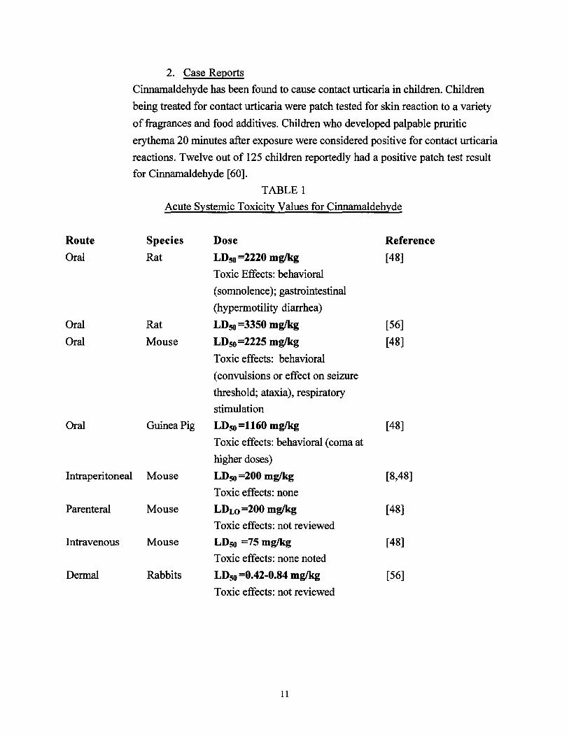
TABLE 1
Acute Systemic Toxicity Values for Cinnamaldehyde
Route Species Dose Reference
Oral Rat LD50 =2220 mglkg [48]
Toxic Effects: behavioral
(somnolence); gastrointestinal
(hypermotility diarrhea)
Oral Rat LD50 =3350 mglkg [56]
Oral Mouse LD50 =2225 mglkg [48]
Toxic effects: behavioral
(convulsions or effect on seizure
threshold; ataxia), respiratory
stimulation
Oral Guinea Pig LD5o=1160 mglkg [48]
Toxic effects: behavioral (coma at
higher doses)
Intraperitoneal Mouse LD50 =200 mglkg [8,48]
Toxic effects: none
Parenteral Mouse LDLo =200 mglkg [48]
Toxic effects: not reviewed
Intravenous Mouse LD50 =75 mglkg [48]
Toxic effects: none noted
Dermal Rabbits LD50 =0.42-0.84 mglkg [56]
Toxic effects: not reviewed
11
B. Subchronic/Chronic
1. Animal Data
The data available in the literature concerning the subchronic and chronic
toxicology of Cinnamaldehyde in animals primarily concerns the sensitizing effect
of this chemical. The contact sensitization potential of Cinnamaldehyde has been
tested in female, Balb/C mice maintained on a diet supplemented with vitamin A
acetate2• The sensitization protocol included an induction period of two weeks
followed by a total of six topical applications of a 30 percent Cinnamaldehyde
solution to the shaved abdomen and thorax. This was followed one week later by a
topical challenge of 15 percent Cinnamaldehyde to both ears. Ear thickness was
measured before the challenge as well as 24 and 48 hours after the challenge. The
percent increase in ear thickness was determined, and the statistical significance of
increased ear thickness was assessed by the Mann Whitney U test. A compound
was classified as a sensitizer if the Mann Whitney test was significant at P# 0.01 ,...::::..or the Mann Whitney test was significant at P not> 0.05, and in addition 2 -
animals had increases in ear thickness twice that of the highest control increase. -One mouse from the group of ten tested was found to have an increase in ear
thickness 24 hours after the challenge that was 100 percent greater than the
highest increase in the control group, while six mice had increases in ear thickness
after the challenge that were determined to be 50 percent greater than the highest
increase in the control group. The Mann Whitney test was found to be significant
at P< 0.01, classifying Cinnamaldehyde as a contact sensitizer [36].
Effects observed following dietary administration of Cinnamaldehyde to male and
female rats over a sixteen week period at a concentration of 10,000 ppm include
slight hyperkeratosis of the squamous portion of the stomach lining and slight
swelling of the hepatic cells. When administered at doses of2 mg on alternate days
to two generations of rats for 223 and 21 0 days respectively, Cinnamaldehyde
was found to cause an increase in liver weight by 20 percent in the first generation
and 22 percent in the second.
The maximum tolerated dose (MTD) of Cinnamaldehyde defmed as the maximum
single dose tolerated by a group of five mice following six intraperitoneal
injections over a two week period was determined to be 0.25 glkg [56].
12
2. Case Reports
Numerous case reports describe the skin sensitization potential of
Cinnamaldehyde in humans. Skin sensitization has been found to occur following
both occupational and consumer exposure to this compound. In some cases, the
skin sensitization caused by Cinnamaldehyde has been found to be permanent
[58]. The following cases of chronic contact dermatitis from occupational
exposure to Cinnamaldehyde are presented in the literature:
A case ofallergic contact dermatitis from exposure to Cinnamaldehyde at an air
freshener manufacturing plant has been reported. A 43 year old man who had no
history of non-occupational exposure to perfumed products developed an itchy
eruption on his fingertips which began one month after he began working at the
plant. The eruption was confined to his hands and consisted of erythematous
scaling patches with indistinct borders on the fingertips and the dorsal surfaces of
both hands. In his job, the employee added various fragrances to a dispensing
machine that subsequently applied the fragrances to pads used to make household
air freshening devices. In addition, the employee served as a maintenance person
and was frequently exposed to full-strength perfume concentrates from
malfunctioning equipment.
Patch tests were performed on the employee using the European Standard Patch
Test Series. The worker only developed an allergic response to Cinnamaldehyde.
The eight fragrance concentrates to which the worker was exposed were
subsequently analyzed for their Cinnamaldehyde content; three of the eight
fragrances were found to have detectable levels of Cinnamaldehyde. It was
concluded that the allergic contact dermatitis most likely resulted from repeated
skin contamination with full-strength perfume concentrates [52].
An increased incidence of fragrance-related occupational dermatitis among a group
ofcoal miners being treated for eczematous skin problems has been documented.
Thirty five miners, 55 male non-miners and 30 female non-miners were patch
tested over a period of eighteen months using the International Contact Dermatitis
Research Group (ICDRG) Standard Series. Forty-five percent of the coal miners
were found to be fragrance sensitive while 20 percent of the male, and 13 percent
of the female non-miners had positive patch test results. Cinnamaldehyde
13
reportedly caused the highest number of positive responses among the male
miners and the male non-miners tested; 14 of the miners and 7 ofthe non-miners
developing positive patch test results after 96 hours. The increased incidence of
allergic contact dermatitis among the coal workers is believed to be related to a
highly perfumed body lotion used at the coal mine [15].
A high incidence ofoccupationally-related allergic skin reactions was also reported
among factory workers in a Danish spice manufacturing plant. Almost all of the
workers exposed to high concentrations of Cinnamaldehyde during the
manufacture of cinnamon spice substitutes developed sensitivity to
Cinnamaldehyde [56].
During an eight-year study, 66 hairdressers who were being treated by
dermatologists for contact dermatitis were patch tested to the North American
Contact Dermatitis Group Standard Screening Trays and to a hairdressers'
screening tray. Cinnamaldehyde was found to produce allergic skin reactions in
1.5% of the hairdressers tested [34].
The following cases of chronic contact dermatitis from consumer exposure
(toothpaste, cosmetics, fragrances) to Cinnamaldehyde are reported in the
literature:
Over a six-month period, a 25 year-old woman reportedly developed perioral
leukoderma caused by a Cinnamaldehyde-containing toothpaste. The leukoderma
around the woman's mouth began at the oral commissures and had spread above
and below the lips. Porcelain-white depigmentation of the skin lateral to the oral
commissures was observed. In addition, leukoderma of the perioral skin adjacent
to the borders of her lips was marginated by a thin border of hyperpigmentation.
Patch testing was performed using the routine screening series of the North
American Contact Dermatitis Group (NACDG) which included a 2 percent
solution of Cinnamaldehyde in petrolatum. A positive (2+) papular reaction to
Cinnamaldehyde was observed 48 and 96 hours after exposure.
It was subsequently determined that two years before the onset of the
leukoderma, the woman had begun using a Cinnamaldehyde-containing toothpaste.
Six months after she switched to a non-Cinnamaldehyde-containing toothpaste,
the perioral leukoderma almost completely disappeared [41].
14
Consumer exposure to Cinnamaldehyde has reportedly caused chronic cheilitis in
an 82 year-old woman who had been using both a Cinnamaldehyde-containing
toothpaste and a sunscreen lipstick. The woman's symptoms consisted of
cracking, swelling and peeling lips, but no cutaneous lesions were observed. Patch
testing with the standard fragrances and preservative series utilizing ICDRG
standard techniques resulted in a positive reaction only to Cinnamaldehyde. When
the woman stopped using the Cinnamaldehyde-containing toothpaste and lipstick
her symptoms cleared [35].
Two case reports ofcosmetic intolerance among persons being treated for chronic
contact dermatitis are described in the literature. In one study, 5202 patients were
patch tested using the Belgian Tri-Contact Patch Test Series. Eight percent of the
total test population reacted positively to cosmetic patch tests. Perfumes were
the principal allergens observed in the group ofpatients who suffered from pure
allergies to cosmetics (156 patients). Ofthese cases, 5.1 percent were attributed to
Cinnamaldehyde.
In the second study, 182 patients suspected of suffering from contact
sensitization to cosmetics were patch tested using the standard tray of the
ICDRG as well as 22 fragrance raw materials. Cinnamaldehyde was found to
produce positive results in 3.7 percent of the patients tested [38].
Over a period of more than three years, 2826 patients at the Gottingen University
Hospital for Skin Diseases were tested for skin sensitivity to Cinnamaldehyde.
Only 0.74 percent ofthe patients (21) reacted positively to Cinnamaldehyde. It
was noted by the authors that in countries other than Germany, especially
England and the United States, allergy to Cinnamaldehyde occurs more frequently.
The discrepancy is presumably a result of the variation in consumer exposure to
Cinnamaldehyde between different countries [67].
C. Carcinogenicity
1. Animal Data
There are limited data available concerning the carcinogenicity ofCinnamaldehyde
in animals. Cinnamaldehyde has been tested for its hepatocarcinogenicity in male,
15
B6C3F1 mice following injection on days 1, 8, 15 and 22 prior to weaning. The
concentration of Cinnamaldehyde injected per dose was in the ratio of 1:2:4:12
respectively, for a total dose of4.8 Jlmol per mouse. Cinnamaldehyde showed no
hepatocarcinogenic activity at the dose levels tested [77].
The remaining information on the carcinogenic effects of this compound concerns
its transforming capacity. The transforming potency of Cinnamaldehyde has been
demonstrated by in vitro studies using Chinese hamster epithelial cells (CH
B241). The CH-B241 cells were treated with sublethal doses ofCinnamaldehyde
(1 OnM), and the surviving cells were cultivated until they acquired characteristics
typically associated with transformed cells; namely 1.) an increase in saturation
density in the monolayer culture, 2.) an increase in plating efficiency at a low
serum level, or 3.) an increase in colony forming efficiency in soft agar medium.
The treated CH-B241 cells that met these in vitro criteria were subsequently
analyzed for their ability to induce neoplastic transformation. This was achieved
by subcutaneous injection of 1 x 10⁶ cells into a suprascapular region of male,
nude mice (BALB/C, JCL, NuNu).
Formation ofnodules at the injection site was observed in six out of seven mice
treated with Cinnamaldehyde-transformed cells. One mouse produced nodules in
the liver and spleen, indicating metastasis. The nodules were first palpable
between days 91 and 237 after injection, after which they grew slowly to 2 em in
diameter until day 311. When the tumors at the injection site reached 2 em in
diameter, the animals were sacrificed and the tumors were removed for histological
examination. Microscopic examination revealed that the tumors were malignant
and consisted ofcells with random shaped nuclei and a high frequency of mitosis.
Karyotype analysis demonstrated that approximately 45 percent of the tumor
cells were polyploid.
In addition, tumors were aseptically removed from the mice, and cells from the
tumors were re-injected into mice in order to assess serial transplantability. Tumor
formation was observed at the injection site in all animals tested within a
considerably shorter latent period (17 to 114 days) than that observed following
the primary inoculation. Metastasis of the spleen was observed in three out of
four animals injected with tumor cells from the Cinnamaldehyde-treated mice.
16
Although the in vitro transforming potency of Cinnamaldehyde was
demonstrated, the induction mechanism is unclear. Direct or indirect interaction
with genetic material is presumably involved because considerable structural
chromosomal aberrations, including chromosome and/or chromatid breaks, were
observed [27, 29].
Cinnamaldehyde has been tested for its capacity to enhance the transformation of
Syrian hamster embryo cells by Simian adenovirus, SA7. Various sub-lethal doses
(0.01 mm, 0.02 mm, 0.05 mm, 0.09 mm, 0.19 mm) were diluted in cell culture
medium and added to replicate dishes of Syrian hamster embryo cells for 20 hours.
After 20 hours, the cells were rinsed and SA 7 virus was absorbed for 3 hours. The
number ofcolonies from Cinnamaldehyde and virus treated cells were determined.
This number was divided by the number of colonies from virus inoculated control
cells in order to determine the surviving fraction. The number of SA 7 foci from 2 x
1 06 plated cells was determined and the enhancement ratio was calculated by
dividing the transformation frequency of treated cells by the transformation
frequency of the control cells. The Cinnamaldehyde-induced enhancement was
found to be statistically significant (P#0.05 or P#O.Ol ) at only one dose level
0.05mM (see Table 2). Therefore, based on standard classification criteria, it was
concluded that there is "some evidence" that Cinnamaldehyde enhances viral
transformation [21].
2. Human Data
There are no data available on the carcinogenicity of Cinnamaldehyde in humans.
However, the in vitro transforming potency of this chemical has been studied.
Cinnamaldehyde was not found to induce transformation of the human fibroblast
cell line HAIN-55 following treatment with various concentrations ranging from 5
80 nM [29].
D. Mutagenicity/Genetic Toxicology
1. Animal Data
There are conflicting reports concerning the mutagenicity of Cinnamaldehyde.
This compound has been found to be mutagenic to
17
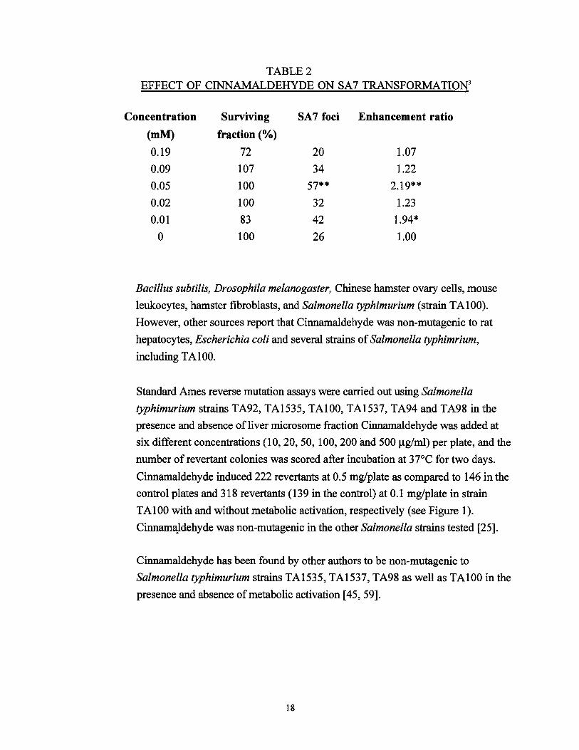
TABLE2 EFFECT OF CINNAMALDEHYDE ON SA7 TRANSFORMATION3
Concentration Surviving SA7 foci Enhancement ratio
(mM) fraction (%)
0.19 72 20 1.07
0.09 t07 34 1.22
0.05 100 57** 2.t9**
0.02 tOO 32 1.23
O.Ot 83 42 t.94*
0 tOO 26 1.00
Bacillus subtilis, Drosophila melanogaster, Chinese hamster ovary cells, mouse
leukocytes, hamster fibroblasts, and Salmonella typhimurium (strain TAtOO).
However, other sources report that Cinnamaldehyde was non-mutagenic to rat
hepatocytes, Escherichia coli and several strains ofSalmonella typhimrium,
including TA100.
Standard Ames reverse mutation assays were carried out using Salmonella
typhimurium strains TA92, TAt535, TAtOO, TAt537, TA94 and TA98 in the
presence and absence of liver microsome fraction Cinnamaldehyde was added at
six different concentrations (tO, 20, 50, tOO, 200 and 500 J..Lg/ml) per plate, and the
number of revertant colonies was scored after incubation at 3 7°C for two days.
Cinnamaldehyde induced 222 revertants at 0.5 mg/plate as compared to 146 in the
control plates and 318 revertants (139 in the control) at 0.1 mg/plate in strain
TAtOO with and without metabolic activation, respectively (see Figure t).
CinnamaJdehyde was non-mutagenic in the other Salmonella strains tested [25].
Cinnamaldehyde has been found by other authors to be non-mutagenic to
Salmonella typhimurium strains TAt535, TAt537, TA98 as well as TAtOO in the
presence and absence ofmetabolic activation [45, 59].
18
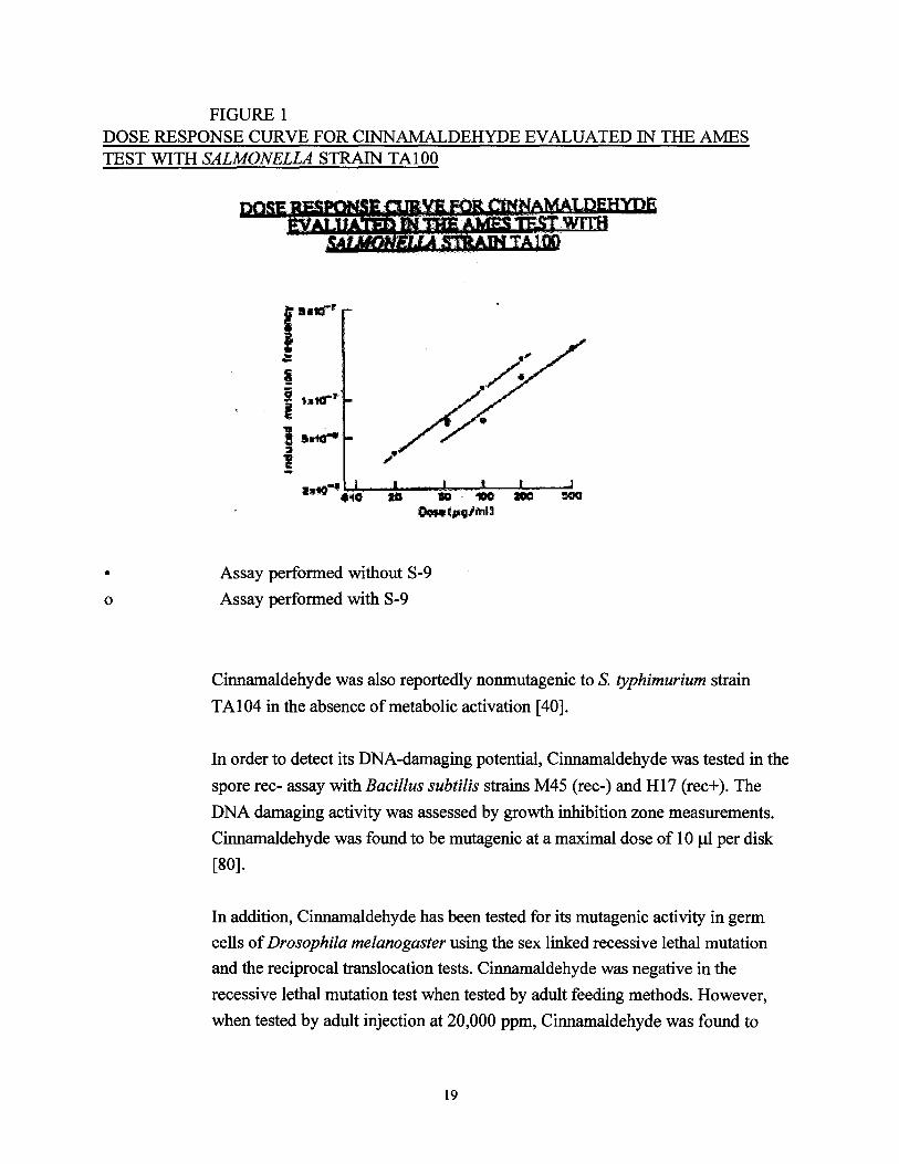
0
FIGURE 1 DOSE RESPONSE CURVE FOR CINNAMALDEHYDE EVALUATED IN THE AMES TEST WITH SALMONELLA STRAIN TA100
,...... ~
Assay performed without S-9 •
Assay performed with S-9
Cinnamaldehyde was also reportedly nonmutagenic to S. typhimurium strain
TA104 in the absence of metabolic activation [40].
In order to detect its DNA-damaging potential, Cinnamaldehyde was tested in the
spore rec- assay with Bacillus subtilis strains M45 (rec-) and H17 (rec+ ). The
DNA damaging activity was assessed by growth inhibition zone measurements.
Cinnamaldehyde was found to be mutagenic at a maximal dose of 1 0 Jll per disk
[80].
In addition, Cinnamaldehyde has been tested for its mutagenic activity in germ
cells ofDrosophila melanogaster using the sex linked recessive lethal mutation
and the reciprocal translocation tests. Cinnamaldehyde was negative in the
recessive lethal mutation test when tested by adult feeding methods. However,
when tested by adult injection at 20,000 ppm, Cinnamaldehyde was found to
19
induce sex-linked recessive lethal mutations in meiotic and post-meiotic germ cell
stages. Cinnamaldehyde was negative in the reciprocal translocation test [78].
Cinnamaldehyde has been found to induce chromosomal aberrations in Chinese
hamster fibroblast cells at concentrations of0.01 mg/ml after a 48 hour exposure
and 0.015 mg/ml following a 24 or 48 hour exposure in the absence ofmetabolic
activation. In order to obtain a quantitative evaluation of the clastogenic potential
of Cinnamaldehyde, the D20 4 and TR5 values were calculated. Cinnamaldehyde was
determined to be mutagenic at relatively low dose levels (D20=0.01) and was found
to have the highest TR value (TR=2133) among a total of 190 food additives
tested. TR values are generally reported to be high for chemicals having
carcinogenic potential in animals [25].
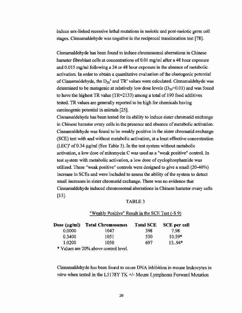
Cinnamaldehyde has been tested for its ability to induce sister chromatid exchange
in Chinese hamster ovary cells in the presence and absence of metabolic activation.
Cinnamaldehyde was found to be weakly positive in the sister chromatid exchange
(SCE) test with and without metabolic activation, at a least effective concentration
(LECt of 0.34 J.Lg/ml (See Table 3). In the test system without metabolic
activation, a low dose of mitomycin C was used as a "weak positive" control. In
test system with metabolic activation, a low dose of cyclophosphamide was
utilized. These "weak positive" controls were designed to give a small (20-40%)
increase in SCEs and were included to assess the ability of the system to detect
small increases in sister chromatid exchange. There was no evidence that
Cinnamaldehyde induced chromosomal aberrations in Chinese hamster ovary cells
[13]. TABLE 3
"Weakly Positive" Result in the SCE Test (-S 9)
Dose (J.Lg/ml) Total Chromosomes Total SCE SCE per cell 0.0000 1047 398 7.98 0.3400 1051 530 10.59* 1.0200 1050 697 13..94*
* Values are 20% above control level.
Cinnamaldehyde has been found to cause DNA inhibition in mouse leukocytes in
vitro when tested in the L5178Y TK +/-Mouse Lymphoma Forward Mutation
20
Assay assay. In the presence and absence ofactivation, Cinnamaldehyde
reportedly induced a "questionable" mutagenic response [57]. No additional
information was provided.
Cinnamaldehyde was not mutagenic in an in vivo test for the induction of
unscheduled DNA synthesis in rat hepatocytes following administration by
gavage [42]. In addition, Cinnamaldehyde did not cause micronucleus induction in
an in vivo micronucleus test with bone marrow mouse cells [22].
2. Human Data
There are no data available in the literature concerning the mutagenicity of
Cinnamaldehyde in humans.
E. Teratology/Reproductive Toxicology
1. Animal Data
The reproductive effects of Cinnamaldehyde have been examined in rats and mice,
and in both species Cinnamaldehyde was found to be negative for all parameters
tested. However, there are conflicting reports concerning the teratogenic effects of
Cinnamaldehyde.
Teratogenic parameters have been evaluated following administration of
Cinnamaldehyde to pregnant, CD-1 mice at a dose level of 1,200 mg/kg/day in
corn oil. Parameters included the number of females producing viable litters, the
number of females with resorbed or nonviable litters, the number ofproven
pregnant females and the reproductive index'. In addition, group litter and viability
data were evaluated, including the number of live pups per litter, the number of
dead pups per litter, the litter weight and the mean pup weight. No significant
differences from the control group were observed in any of the criteria examined
[23].
In another study, CD-I mice were dosed by gavage at 1,200 mg/kg/day of
Cinnamaldehyde during mid-pregnancy. Litter size, birth weight, neonatal growth
and survival to postnatal day three were recorded as indices of potential
developmental toxicity. Both the maternal response variables and the neonatal
21
response variables tested were not found to differ significantly from the control
[20].
Cinnamaldehyde was not found to affect body weight gain, reproductive ability,
or the development and viability of offspring following administration of2 mg on
alternate days to two generations of rats for 223 and 210 days respectively [56].
Suprablastodermic administration ofa single dose of Cinnamaldehyde to 3 day-old
chick embryos (white Leghorn x Rhode Island red strain) was reportedly
teratogenic. The Optimal Teratogenic Dose (OTDY was found to be 0.50 J!M per
embryo. At this concentration, the most common teratogenic effects observed
included limb malformations, primarily limb size reduction. Malformations of the
axial skeleton including spina bifida, anoura (tail absence) or haemisomia were
noted in several cases [ 1].
2. Human Data
There are no data available in the literature concerning the reproductive or
teratogenic effects of Cinnamaldehyde on humans.
F. Immunotoxicity
1. Animal Data
There are no data available in the literature concerning the lmmunotoxicity of
Cinnamaldehyde in animals.
2. Human Data
There are no data available in the literature concerning the immunotoxicity of
Cinnamaldehyde in humans.
22
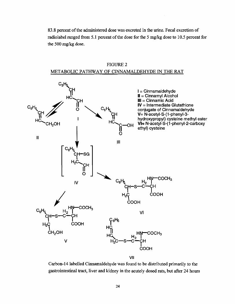
VI. CHEMICAL DISPOSITION
A. Animal Data
The elimination of Cinnamaldehyde has been studied in the female, Wistar rats after
administration of250 mg/kg daily for two weeks. Following this dosing regimen, two
sulphur-containing metabolites were isolated from the urine and identified by
synthesis, nuclear magnetic resonance (NMR) and mass spectrography as N-acetyl-S
(1-phenyl-3-hydroxypropyl) cysteine and N-acetyl-S-(1-phenyl-2-carboxy ethyl)
cysteine in a 4:1 ratio. The total thioether excretion, calculated as a percentage ofthe
dose ofCinnamaldehyde administered, was determined to be 14.8± 1.9%.
NMR spectra of the isolated mercapturic acids indicated that addition of a
nucleophilic Glutathione anion occurred to the B-carbon atom of the double bond of
Cinnamaldehyde. At some stage during the conversion of the intermediate Glutathione
conjugate ofCinnamaldehyde to a mercapturic acid, reduction of the carbonyl moiety
to a hydroxy group occurred. In addition, a small portion of the carbonyl moieties
were oxidized into a carboxylic group (see Figure 2) [9,10].
Cinnamaldehyde, which contains activated double bonds that are substrates for
Glutathione S-Alkenetransferases, has been found to depress liver Glutathione levels
markedly following intraperitoneal administration to rats at a dose of 0.5 mllkg.
Thirty minutes after administration, the Glutathione level had been reduced to 53
percent of the control, and after two hours, the Glutathione level had dropped to 35
percent of the control.
The absorption, distribution and excretion of Cinnamaldehyde labelled with Carbon
14 have been studied in male Fischer-344 rats following acute and subacute oral
administration. Cinnamaldehyde labelled with 5-10 J.LCilkg of Carbon-14 was
administered by gavage at dose levels of 5, 50 and 500 mg/kg. For the acute studies,
each rat was given a single, radioactive dose by gavage at one of the three dose levels.
In the subacute studies, one dose of unlabelled Cinnamaldehyde was administered to
groups of rats once a day for 7 days, followed by a single radioactive dose 24 hours
after administration of the last unlabelled dose.
Following acute administration, Cinnamaldehyde was found to be excreted primarily
in the urine, and within 72 hours after administration at the 50 and 500 mg/kg levels,
23
83.8 percent of the administered dose was excreted in the urine. Fecal excretion of
radiolabel ranged from 5.1 percent ofthe dose for the 5 mg/kg dose to 10.5 percent for
the 500 mg/kg dose.
FIGURE 2
METABOLIC PATHWAY OF CINNAMALDEHYDE IN THE RAT
I = Cinnamaldehyde II = Cinnamyl Alcohol Ill =Cinnamic Acid IV = Intermediate Glutathione conjugate of Cinnamaldehyde V= N-acetyi-S-(1-phenyl-3hydroxypropyl) cysteine methyl ester VI= N-acetyi-S-(1-phenyl-2-carboxy ethyl) cysteine
C H H HN-ICOCH3 IV 6 ~ 2
cr-s-C--rH
H21 COOH
COOH
VI
ysHs
Hll H~ H HrCOCHs
2 v H2c-s-c-yH
COOH
VII
Carbon-14labelled Cinnamaldehyde was found to be distributed primarily to the
gastrointestinal tract, liver and kidney in the acutely dosed rats, but after 24 hours
24
was reportedly cleared from the liver and kidney. An average of 5.2 percent of the
administered radiolabel was found in the gastrointestinal tract after 24 hours at all
dose levels. After 72 hours at the 50 and 500 mg/kg dose levels, the amount of
radiolable found in the gastrointestinal tract was 0.19 percent and 0.39 percent of the
administered dose respectively. Radio labelled Cinnamaldehyde was distributed to the
fat and was detectable in rats sacrificed 72 hours after dosing. Less than 0.1 percent of
the administered dose at all three dose levels tested was distributed to the brain, heart,
spleen, lung and testes. Estimated whole blood levels of Cinnamaldehyde averaged
less than 0.1 percent of the administered dose after 24 hours at all dose levels tested.
Similar tissue distribution and excretion patterns were found following subacute
dosing. A rapid clearance via the urine was observed 24 hours after administration,
with an average of 81 percent of the administered radioactivity recovered in the urine,
and an additional5.9 percent recovered in the feces at all dose levels tested.
The administered radioactivity was found to be distributed primarily in the fat and
gastrointestinal tract 24 hours after subacute administration at all dose levels. Liver
accumulation accounted for less than 0.15 percent of the administered doses after 24
hours. Carbon-14 labelled Cinnamaldehyde was detectable in the fat at the 500 mg/kg
dose level after three days. Less than 0.1 percent of the administered dose was
observed in other tissues after 24 hours. The estimated level of radio labelled
Cinnamaldehyde was less than 0.1 percent ofthe administered dose in whole blood
after 24 hours [63].
B. HumanData
There are no data available on the metabolism of Cinnamaldehyde in humans.
Presumably, Cinnamaldehyde is oxidized to cinnamic acid which is excreted in the
urine as benzoic and hippuric acids [16].
VII. BIOCHEMICAL TOXICOLOGY
A. Animal Data
Cinnamaldehyde has been found to be cytotoxic to L1210 mouse cells. The degree of
cytotoxicity of Cinnamaldehyde was found to be proportional to the amount of the
compound added to the cell culture medium. The ED 50 value9 of Cinnamaldehyde has
been determined to be 4.8 J..Lg/ml ofculture solution.
25
The mechanism by which Cinnamaldehyde inhibits L 1210 mouse cell growth was
examined by studying the effect of Cinnama1dehyde on RNA, DNA and protein
synthesis as well as its effect on glycolysis. Cinnamaldehyde at concentrations
ranging from 0 to 50 J.Lg/ml was added to cultures ofL1210 cells at various intervals,
and the resulting concentrations ofglucose and lactate in the culture solution were
determined enzymatically. The addition of Cinnamaldehyde to the culture media was
found to have only a slight effect on glycolysis by L1210 cells (see Figure 4).
The effect of Cinnamaldehyde on RNA, DNA and protein synthesis was determined
by measuring L1210 cell incorporation oftritiated Uridine, tritiated Thymidine, and
tritiated Leucine at various time intervals. Among the labelled isotopes tested, the
incorporation of tritiated Leucine was inhibited most strongly, indicating a preferential
inhibition of Cinnamaldehyde on protein (see Figure 5). The toxic effect of
Cinnamaldehyde on protein synthesis could be removed by transferring the cells to a
Cinnamaldehyde-free medium, suggesting that Cinnamaldehyde did not cause
irreversible cellular damage.
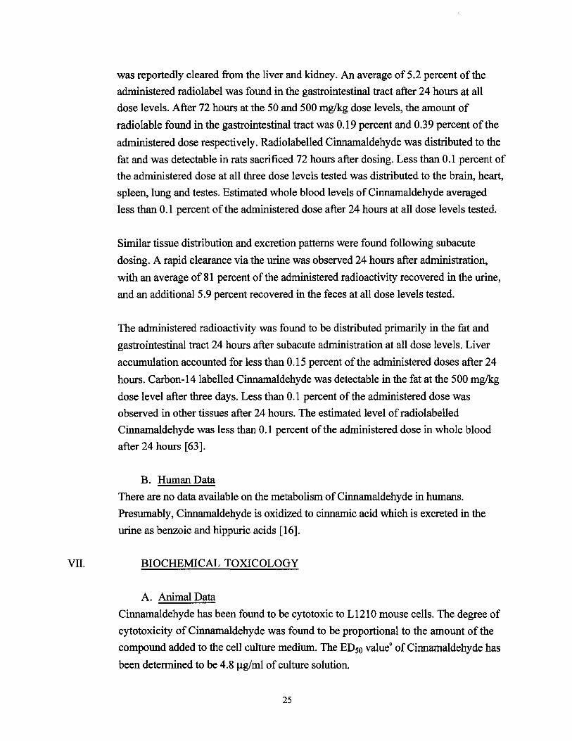
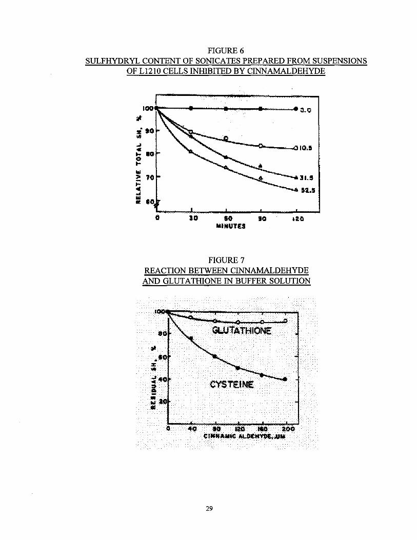
Cinnamaldehyde was subsequently found to inhibit the growth of L121 0 cells by
blocking protein synthesis through a direct interaction with sulfhydryl-containing
amino acids. Sonicates were prepared from suspensions ofL1210 cells inhibited by
Cinnamaldehyde and analyzed for their sulfhydryl content. Cinnamaldehyde was
found to reduce the sulfhydryl content of the sonicates in a dose-dependent manner
(see Figure 6), suggesting a direct chemical interaction between Cinnamaldehyde and
the sulfhydryl groups ofthe L1210 cell components.
This direct interaction was confirmed by the results of experiments in which Cysteine
or Glutathione was allowed to react with various concentrations of Cinnamaldehyde.
Glutathione, which was added to the reaction mixture as an additional source of
sulfhydryl groups, showed minimal reaction with Cinnamaldehyde based on the
concentration of residual sulfhydryl groups,
while Cinnamaldehyde was found to react directly with Cysteine (see Figure 7) [44].
The catecholamine-releasing effect of Cinnamaldehyde has been studied in male and
female Mongrel dogs following intravenous and intraduodenal administration of 20
mg/kg of Cinnamaldehyde. It was observed that the total catecholamine concentration
26
reached a maximal level two minutes after intravenous administration, before returning
to baseline, after approximately twenty minutes. No effect on blood pressure was
observed. Extraction of the catecholamines from the samples and analysis of content
revealed that the increased portion ofcatecholamines was epinephrine. Similarly,
intraduodenal administration of 50 mglkg of Cinnamaldehyde caused a dose-dependent
increase in catecholamine concentration. Epinephrine accounted for nearly all of the
increase in catecholamine, no significant change in norepinephrine concentration was
observed.
Ganglion blocking has not been found to affect the catecholamine releasing property
of Cinnamaldehyde. The influence ofganglion blocking on the catecholamine releasing
effect of Cinnamaldehyde was determined by monitoring blood pressure during the
co-administration ofganglion blocking agents (hexamethonium and atropine) and
Cinnamaldehyde intravenously. In addition, the influence of the adrenals on the
catecholamine releasing effect of Cinnamaldehyde was investigated. After surgically
blocking adrenal circulation, Cinnamaldehyde was administered intraduodenally. The
effect of
27

..J Q•~ !*. 0 0
~ tl
... ... c II:
100
LACTATE
aa 40 •o C Ullll AM.I C A....I.HYIDE, ' f./ Ml
a.o 8 / o_o !0.1
r:i w s :51)0
/ /10-' lot-I :52-:S
IE g lit.t
52-;! II.
.. ...w2DD
~ ~ "'ii! .,.I;
4
FIGURE 410
EFFECT OF CINNAMALDEHYDE ON THE GLYCOLYSIS OF L1210 CELLS
FIGURE 511
MODE OF INHIBITION OF CINNAMALDEHYDE ON GLYCOLYSIS AND MACROMOLECULE BIOSYNTHESIS
OF L1210 CELLS
28
-..... %
j CYSTEINE ii
•Iii
0 40
..
... :> :!i.! t=
!11.! .oil Ill lilt
10 to to MUIUTI:I
FIGURE 6 SULFHYDRYL CONTENT OF SONICATES PREPARED FROM SUSPENSIONS
OF L1210 CELLS INHIBITED BY CINNAMALDEHYDE
FIGURE 7 REACTION BETWEEN CINNAMALDEHYDE
AND GLUTATHIONE IN BUFFER SOLUTION
29
Cinnamaldehyde was observed to disappear significantly after ligation of the adrenals,
and the basal catecholamine level dropped to approximately half the baseline level
before ligation. Based on this observation, it is believed that plasma catecholamines
released following systemic administration ofCinnamaldehyde originate
predominately from the adrenals. The finding that the catecholamine releasing effect of
Cinnamaldehyde was not influenced by ganglion blocking indicates that this
compound increases plasma catecholamine concentration through a mechanism
independent of an increase in androgenic nervous activity [ 19].
The kinetics of impulse blocking by Cinnamaldehyde in frog sciatic nerve have been
tested at various temperatures and Cinnamaldehyde concentrations. The frog sciatic
nerves taken from male specimens of Rana temporaria were ligatured to prevent
inactivation by sodium before immersion in buffer solution. Varying concentrations of
Cinnamaldehyde (0.01-0.10%) were introduced into a stimulation chamber12 •
Stimulating square pulses were delivered by a generator through a pulse separation
unit and full size nervograms were obtained in order to assess the maximal action
potential.
Cinnamaldehyde was found to decrease the amplitude of the nervogram in frog nerve
up to complete blockage of the action potential, the rate of this effect depended on
temperature and Cinnamaldehyde concentration. It was found that nerve excitement
could be restored almost completely upon immersion in buffer solution without
Cinnamaldehyde, so that the effect ofa second treatment on the same nerve could be
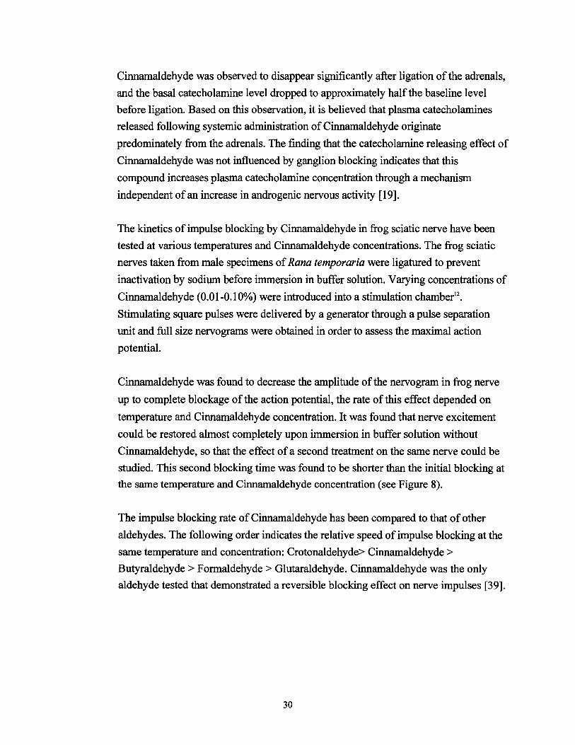
studied. This second blocking time was found to be shorter than the initial blocking at
the same temperature and Cinnamaldehyde concentration (see Figure 8).
The impulse blocking rate ofCinnamaldehyde has been compared to that of other
aldehydes. The following order indicates the relative speed of impulse blocking at the
same temperature and concentration: Crotonaldehyde> Cinnamaldehyde >
Butyraldehyde > Formaldehyde > Glutaraldehyde. Cinnamaldehyde was the only
aldehyde tested that demonstrated a reversible blocking effect on nerve impulses [39].
30
"' .. ~ ..... r----··- 1---·
~--· r--=::
~ ~
...... ... ~ ... : -·'&Ill!
..~ ---.... _ ·- ..... ~
fi.W. - .... -... -.......... .......... ~.::v. :ill.;; r.-..._.-·
• 1111 •
FIGURE 8 IMPULSE BLOCKING EFFECT OF CINNAMALDEHYDE
The blocking time ofnerve impulses as a function of temperature at two Cinnamaldehyde concentrations at a first treatment ( o) and a second one ( o) following recovery
B. Human Data
Cinnamaldehyde has been reported to have anti-platelet aggregating and vasodilatory
action in vitro . Thromboxane A2 (TxA2), an Arachidonic Acid metabolite which is
produced in platelets, is known to be a potent pro-aggregatory agent, and therefore
the alteration of TxA2 synthesis is believed to affect platelet aggregation.
In order to test the effect of Cinnamaldehyde on TxA2 formation, platelet rich human
plasma was incubated with various concentrations of Cinnamaldehyde and then
stimulated with the aggregant, collagen. Cinnamaldehyde was observed to inhibit
collagen-induced platelet aggregation in a dose dependent manner. A prolongation of
the lagtime before the initiation ofcollagen-induced platelet aggregation was observed
by the addition of increasing doses ofCinnamaldehyde. At a dose of750 J.LM,
Cinnamaldehyde almost completely suppressed collagen-induced platelet aggregation.
The effect of Cinnamaldehyde on a preparation ofwashed human platelets ( 5 x 1 05
J.Ll) was examined. Again, the addition ofCinnamaldehyde was found to decrease
collagen-induced platelet aggregation in a dose-dependent manner and nearly complete
suppression was observed when platelets were pretreated with 300 J.LM
Cinnamaldehyde.
31
In order to examine the effect ofCinnamaldehyde on Arachidonic Acid (AA)
metabolism in human platelets, washed platelets were stimulated with collagen in the
presence ofCinnamaldehyde and the concentration of AA-derived metabolites was
measured. The addition of Cinnamaldehyde was found to dose dependent decrease the
formation ofThromboxane B2 (TxB2), 12-Hydroxyheptadecatrienoic acid (HHT), and
12-hydroxy-eicosatetrgenoic acid (12-HETE)13• In addition, a positive correlation
between reduced platelet aggregation and decreased TxB2 formation in
Cinnamaldehyde pretreated platelets was observed. The addition of Cinnamaldehyde
at concentrations up to 300 J.LM to washed human platelets prelabelled with [14C]
Arachidonic Acid (AA) had no significant effect on the conversion of [14C]-AA to
either [14C]-TxB2, e4C]-HETE or [14C]-HHT, indicating that Cinnamaldehyde does
not affect the metabolism of Arachidonic Acid by either the cyclooxygenase or the
lipoxygenase pathways. The action of Cinnamaldehyde was therefore believed to be
proximal to the cyclooxygenase and lipoxygenase level.
In order to assess this possibility, the effect of Cinnamaldehyde on the collagen
stimulated release and metabolism of e4C]-AA from washed human platelets was
examined. The addition of Cinnamaldehyde was found to cause a dose-dependent
decrease in collagen-induced prelabelled platelet aggregation as well as a dose
dependent reduction in the percentage of [14C]-AA released, and the percentage of
[ 14C]-TxB2 formed, from prelabelled platelets. In addition, there was a positive
correlation between decreased platelet aggregation and reduced release of [14C]-AA, as
well as a positive correlation between decreased platelet aggregation and reduced
formation of TxB2• A positive correlation was also noted between the decreased
release of [14C]-AA and reduced formation of [14C]-TxB2•
These results indicate that the reduced production of Thromboxane B2 (TxB2) in
Cinnamaldehyde pretreated platelets may most likely be a result of impaired
Arachidonic Acid liberation from platelet membrane phospholipids, and not a result
of the inhibition ofAA metabolism via the cyclooxygenase pathway [ 66].
Several studies have been conducted to investigate the mechanism by which
Cinnamaldehyde causes skin sensitization. It is generally agreed that Cinnamaldehyde,
a low molecular weight substance, cannot induce contact allergy in the skin unless it is
bound to a protein. However, it is uncertain which proteins react with
32
Cinnamaldehyde, and it is unclear which reacting groups are involved.
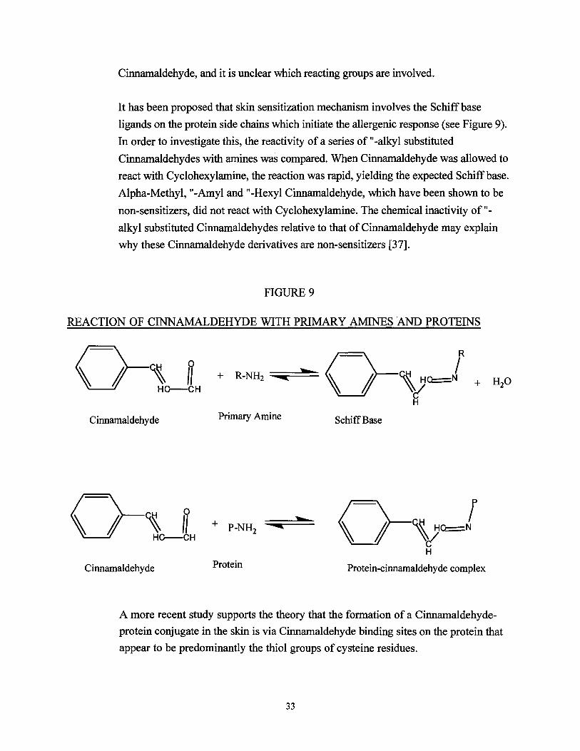
It has been proposed that skin sensitization mechanism involves the Schiff base
ligands on the protein side chains which initiate the allergenic response (see Figure 9).
In order to investigate this, the reactivity of a series of "-alkyl substituted
Cinnamaldehydes with amines was compared. When Cinnamaldehyde was allowed to
react with Cyclohexylamine, the reaction was rapid, yielding the expected Schiff base.
Alpha-Methyl, "-Amyl and "-Hexyl Cinnamaldehyde, which have been shown to be
non-sensitizers, did not react with Cyclohexylamine. The chemical inactivity of"
alkyl substituted Cinnamaldehydes relative to that ofCinnamaldehyde may explain
why these Cinnamaldehyde derivatives are non-sensitizers [37].
FIGURE 9
REACTION OF CINNAMALDEHYDE WITH PRIMARY AMINES AND PROTEINS
Primary Amine Cinnamaldehyde Schiff Base
o-VO==Nr H
ProteinCinnamaldehyde Protein-cinnamaldehyde complex
A more recent study supports the theory that the formation of a Cinnamaldehyde
protein conjugate in the skin is via Cinnamaldehyde binding sites on the protein that
appear to be predominantly the thiol groups of cysteine residues.
33

Reactions between Cinnamaldehyde and various nucleophiles have been carried out
using model compounds containing thiol nucleophiles such as Cysteine, N
Acetylcysteine and Thioethanol, as well as model compounds containing amine
nucleophiles including Lysine, Alanine, Glycine, Propylamine and Imidazole. The
reactions were performed at pHs ranging from 7.4 to 10.5 with the total nucleophilic
concentration in excess of the Cinnamaldehyde concentration. By monitoring the
concentration of Cinnamaldehyde spectrophotometrically and by high performance
liquid chromatography (HPLC), the reactions were determined to follow pseudo-first
order kinetics. The observed pseudo-first-order rate constants (l<obs) were found to
follow the rate expression:
Kobs = ko + kr (nucleophile)
where ko represents the rate constant at zero nucleophile concentration and kr the
nucleophilic attack of nucleophiles respectively on Cinnamaldehyde.
Considerably higher second order rate constants were found for the reaction of
Cinnamaldehyde with thiol nucleophiles than for reaction of Cinnamaldehyde with
amine nucleophiles, indicating that the free thiol groups of Cysteine residues are the
sites to which the Cinnamaldehyde molecule is primarily bound (see Table 3.1) [74].
The passage of Cinnamaldehyde through human skin has been investigated by in vitro
penetration studies using full thickness human skin. Abdominal skin samples were
obtained and stripped of adipose tissue, yielding a skin membrane of epidermis and
dermis ofapproximately 2 mm. Cinnamaldehyde, at a concentration of200 mg/ml,
was added to the epidermal side ofthe skin which had been enclosed by a glass
diffusion cell. Samples taken from the receptor phase were analyzed by HPLC after
precipitation of the protein. This analysis revealed that Cinnamyl alcohol and
Cinnamic Acid were found in the receptor phase at a higher concentration than
Cinnamaldehyde (see Figure 10). Only a small amount of unchanged Cinnamaldehyde
was detected in the receptor phase, suggesting a loss ofcinnamaldehyde either by
degradation in the receptor phase or by an enzyme/non enzyme mediated conversion
during diffusion of the Cinnamaldehyde through the skin.
34

TABLE3.1
SECOND-ORDER RATE CONSTANTS (Kr) FOR FORMATION OF CONJUGATES BETWEEN DIFFERENT NUCLEOPHILES AND CINNAMALDEHYDE
pH Nucleophile kr (M-1 min-1)
6.4 cysteine 13 7.4 cysteine 107 8.3 cysteine 440 6.4 N-acetyl cysteine 0.12 7.4 N-acetyl cysteine 1.0 8.2 N-acetyl cysteine 2.0 7.4 thioethanol -1.7 7.4 lysine n.d. 7.4 glycine 5.4 X 10⁴
8.6 glycine 7.2 X 10⁻³
7.4 alanine n.d. 8.8 alanine 1.4 X 10⁻³
10.5 propylamine 7.2 X 10⁴
7.4 phenol n.d. 7.4 imidazole n.d. 10.5 imidazole n.d.
n.d. = no detectable reaction FIGURE 10
IN VITRO PERCUTANEOUS PENETRATION OF CINNAMALDEHYDE
.. -I; • ;I
I..Ill
.... ....
,
-• = Jlmol Cinnamon (alcohol + aldehyde + acid) 0 = Jlmol Cinnamyl Alcohol a = J..Lmol Cinnamic Acid
IJ = Jlmol Cinnamaldehyde
35
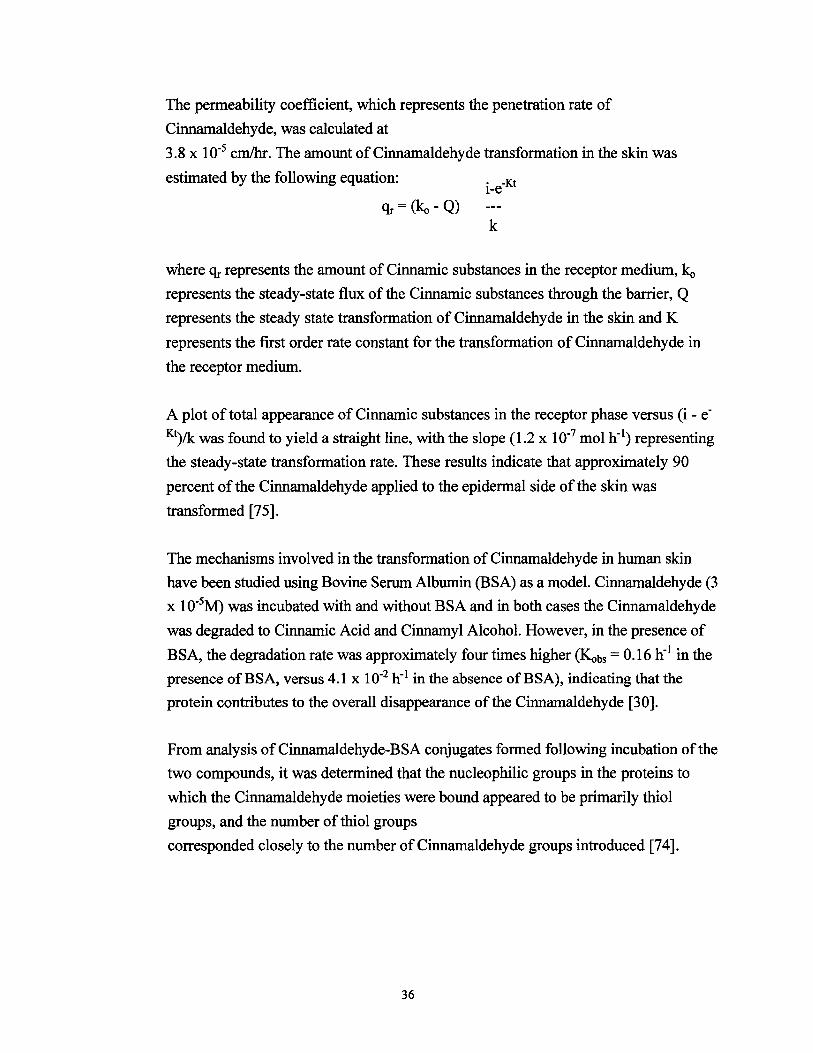
The permeability coefficient, which represents the penetration rate of
Cinnamaldehyde, was calculated at
3.8 x w-s em/hr. The amount ofCinnamaldehyde transformation in the skin was
estimated by the following equation: •1-e-Kt
k
where 'lr represents the amount of Cinnamic substances in the receptor medium, ko represents the steady-state flux of the Cinnamic substances through the barrier, Q
represents the steady state transformation of Cinnamaldehyde in the skin and K
represents the first order rate constant for the transformation of Cinnamaldehyde in
the receptor medium.
A plot oftotal appearance ofCinnamic substances in the receptor phase versus (i- e
Kt)/k was found to yield a straight line, with the slope (1.2 x 10⁻⁷ mol h-1) representing
the steady-state transformation rate. These results indicate that approximately 90
percent of the Cinnamaldehyde applied to the epidermal side of the skin was
transformed [75].
The mechanisms involved in the transformation of Cinnamaldehyde in human skin
have been studied using Bovine Serum Albumin (BSA) as a model. Cinnamaldehyde (3
x 1o-5M) was incubated with and without BSA and in both cases the Cinnamaldehyde
was degraded to Cinnamic Acid and Cinnamyl Alcohol. However, in the presence of
BSA, the degradation rate was approximately four times higher (Kobs = 0.16 h-1in the
presence ofBSA, versus 4.1 x 10⁻² h-1 in the absence ofBSA), indicating that the
protein contributes to the overall disappearance ofthe Cinnamaldehyde [30].
From analysis ofCinnamaldehyde-BSA conjugates formed following incubation ofthe
two compounds, it was determined that the nucleophilic groups in the proteins to
which the Cinnamaldehyde moieties were bound appeared to be primarily thiol
groups, and the number of thiol groups
corresponded closely to the number ofCinnamaldehyde groups introduced [74].
36
VIII. STRUCTURE/ ACTIVITY CONSIDERATIONS
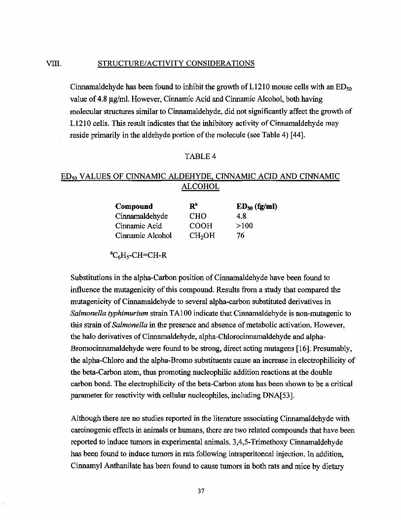
Cinnamaldehyde has been found to inhibit the growth ofL1210 mouse cells with an ED50
value of4.8 Jlg/ml. However, Cinnamic Acid and Cinnamic Alcohol, both having
molecular structures similar to Cinnamaldehyde, did not significantly affect the growth of
L 1210 cells. This result indicates that the inhibitory activity of Cinnamaldehyde may
reside primarily in the aldehyde portion of the molecule (see Table 4) [44].
TABLE4
ED50 VALUES OF CINNAMIC ALDEHYDE, CINNAMIC ACID AND CINNAMIC ALCOHOL
Compound ED50 (fglml) Cinnamaldehyde 4.8 Cinnamic Acid >100 Cinnamic Alcohol 76
Substitutions in the alpha-Carbon position of Cinnamaldehyde have been found to
influence the mutagenicity of this compound. Results from a study that compared the
mutagenicity of Cinnamaldehyde to several alpha-carbon substituted derivatives in
Salmonella typhimurium strain TA100 indicate that Cinnamaldehyde is non-mutagenic to
this strain ofSalmonella in the presence and absence of metabolic activation. However,
the halo derivatives of Cinnamaldehyde, alpha-Chlorocinnamaldehyde and alpha
Bromocinnamaldehyde were found to be strong, direct acting mutagens [16]. Presumably,
the alpha-Chloro and the alpha-Bromo substituents cause an increase in electrophilicity of
the beta-Carbon atom, thus promoting nucleophilic addition reactions at the double
carbon bond. The electrophilicity of the beta-Carbon atom has been shown to be a critical
parameter for reactivity with cellular nucleophiles, including DNA[53].
Although there are no studies reported in the literature associating Cinnamaldehyde with
carcinogenic effects in animals or humans, there are two related compounds that have been
reported to induce tumors in experimental animals. 3,4,5-Trimethoxy Cinnamaldehyde
has been found to induce tumors in rats following intraperitoneal injection. In addition,
Cinnamyl Anthanilate has been found to cause tumors in both rats and mice by dietary
37

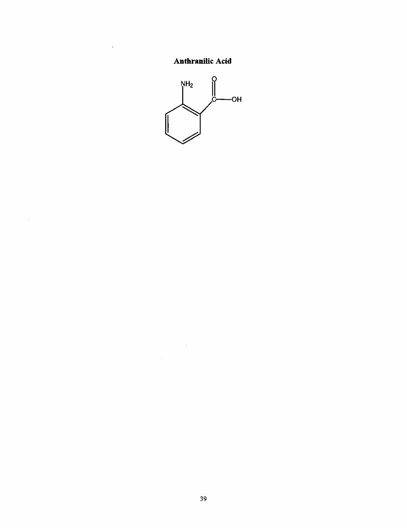
administration at 15,000 or 30,000 ppm [3]. Because Anthranilic Acid was not found to
be carcinogenic when tested in mice or rats it is believed that the Cinnamyl moiety may
play a role in the carcinogenicity ofCinnamyl Anthranilate [3] (see Figure 11).
As an aldehyde, Cinnamaldehyde is a potential alkylating agent. Through its reaction with
amino groups in cellular macromolecules, this compound forms Schiff base intermediates.
Cinnamaldehyde is also a potential alkylating agent via epoxidation of the double bond.
FIGURE 11
CINNAMALDEHYDE AND STRUCTURALLY RELATED COMPOUNDS
Cinnamaldehyde ~-"""
3,4,5-Trimethoxycinnamaldehyde
Cinnamyl Anthranilate
0
~ HC--C--o---0
\\ H2
38
Anthranilic Acid
C--OH ~
39
ON-LINE DATABASES SEARCHED
MEDLARS Chemline Chemlist RTECS Hazardous Substance Databank (HSDB) HZDB Toxline 1981-Present Toxline 65 1965-Present Toxlit 1981-Present Toxlit65 1965-1980
DIALOG Agricola Aquatic Science Abstracts Biosis Previews
1970-Present 1978-Present 1969-Present
CA Search 1967-Present Chemical Regulations and Guidelines system CHRIS USDA Compendex Plus EMBASE
November 1989 September 1989 1970-Present 197 4-Present
Environmental Bibliography FDC Reports Federal Register Foods Adlibra
1974-Present 1987-Present 1979-Present 1974-Present
FSTA 1969-Present Life Sciences Collection 1978-Present MEDLINE 1966- Present NTIS 1964-Present Occupational Safety and Health Pharmaceutical News INdex
1973-Present 1974-Present
Pollution Abstracts 1970-Present PTS PROMPT 1972-Present Trade and Industry Trade and Industry Index
1983-Present 1981-Present
40
IX. REFERENCES
1. Abramovici, A., and Rachmuth-Roizman, P., "Molecular Structure-Teratogenicity
Relationships of Some Fragrance Additives." Toxicology, Vol. 29 (1983), pp. 143-156.
2. Aldrich Chemical Company, Aldrich Catalog/Handbook ofFine Chemicals. 1988-1989.
3. Blakemore, W. and Thompson, H., "Trace Analysis ofCinnamaldehyde in Animal Feed,
Human Urine, and Wastewater by Electron Capture Gas Chromatography." Journal of
Agriculture and Food Chemistry, Vol. 31 (1983), pp. 1047-1052.
4. Boyland, E. and Chasseaud, L.F., "The Effect of Some Carbonyl Compounds on Rat
Liver Glutathione Levels." Biochemical Pharmacology, Vol. 19 (1970), pp. 1526-1528.
5. Bretherick, L., Handbook ofReactive Chemical Hazards, Third Edition. Boston:
Butterworths, 1985.
6. Broeckx, W., et al., "Cosmetic Intolerance." Contact Dermatitis, Vol. 16 (1987), pp. 189
194.
7. "Chemicals, Raw Materials & Specialties." Chemical Week, Buyers' Guide Issue
(October, 1989), p. 195.
8. Clayton, G.D. and Clayton, F.E., eds., Patty's Industrial Hygiene and Toxicology, Vol.
11, Second Revised Edition. New York: Wiley-Interscience, 1963.
9. Delbressine, L.P.C., et al., "Identification ofTwo Sulphur Containing Urinary
Metabolites ofCinnamic Aldehyde in the Rat." British Journal of Pharmacology, Vol. 68
(1980), p. 165p.
10. Delbressine, L.P.C., et al., "Isolation amd Identification of Mercapturic Acids of
Cinnamic Aldehyde and Cinnamyl Alcohol from Urine ofFemale Rats." Archives of
Toxicology, Vol. 49 (1981), pp. 57-64.
11. Estrin, N .F., Crosley, P .A., and Haynes, C.R., eds., CTF A Cosmetic Ingredient
Dictionary, Third Edition. Washington, D.C.: The Cosmetic Toiletry and Fragrance
Association, Inc., 1981.
41
13
12. Furia, T.E. and Bellanca, N., eds., Fenaroli's Handbook ofFlavor Ingredients, Second
Edition. Cleveland: CRC Press, 1975.
Galloway, S.M., et al., "Chromosome Aberrations and Sister Chromatid Exchanges in
Chinese Hamster Ovary Cells: Evaluations of 108 Chemicals." Environmental and
Molecular Mutagenesis, Vol. 10 (1987) pp. 1-36, 54-55, 109, 127.
14. Gennaro, A.R., ed., Remington's Pharmaceutical Sciences, Seventeenth Edition, Easton,
Pennsylvania: Mack Publishing Company, 1985.
15. Goodfield, M.J.D. and Saihan, E.M., "Fragrance Sensitivity in Coal Miners." Contact
Dermatitis, Vol. 18 (1988), pp. 81-83.
16. Gosselin, R.E., et al., Clinical Toxicology of Commercial Products: Acute Poisoning, Fifth
Edition. Baltimore: William & Wilkins, 1984.
17. Haley, T.J., "A Review of the Literature on Cinnamaldehyde." Dangerous Properties of
Industrial Materials Report, Vol. 1, No.5 (1981), pp. 5-7.
18. Harada, M. and Y ano, S., "Pharmacological Studies on Chinese Cinnamon. II. Effects of
Cinnamaldehyde on the Cardiovascular and Digestive Systems." Chemical and
Pharmaceutical Bulletin, Vol. 23, No.5 (May, 1975), pp. 941-947.
19. Harada, M., Hirayama, Y, and Yamazaki, R., "Pharmacological Studies on Chinese
Cinnamon V. Catecholamine Releasing Effect ofCinnamaldehyde in Dogs." Journal of
Pharmacological Dynamics, Vol. 5 (1982), pp. 539-546.
20. Hardin, B.D., et al., "Evaluation of 60 Chemicals in a Preliminary Developmental Toxicity
Test," Teratogenesis, Carcinogenesis, and Mutagenesis, Vol. 7 (1987), pp. 29-48.
21. Hatch, G.G., et al., "Chemical Enhancement ofSA7 Virus Transformation of Hamster
Embryo Cells: Evaluation by Interlaboratory Testing ofDiverse Chemicals."
Environmental Mutagenesis, Vol. 8 (1986), pp. 515-531.
22. Hayashi, M., et al., "Micronucleus Tests in Mice on 39 Food Additives and Eight
42
Miscellaneous Chemicals." Food Chemical Toxicology, Vol. 26, No.6 (1988), pp. 487
500.
23. Hazleton Laboratories America, Inc., Screening ofPriority Chemicals for Potential
Reproductive Hazard. Hazleton Study No. 6125-101-6125-110. NIOSH Contract No.
200-82-2542. National Technical Information Service. December 1983.
24. Hoskins, J.A., "The Occurence, Metabolism and Toxicity ofCinnamic Acid and Related
Compounds." Journal ofApplied Toxicology, Vol. 4, No.6 (1984), pp. 283-292.
25. Ishidate, M., "Primary Mutagenicity Screening of Food Additives Currently Used in
Japan." Food Chemical Toxicology, Vol. 22., No.8 (1984), pp. 623-636.
26. Jenner, P.M., et al., "Food Flavourings and Compounds of Related Structure I. Acute
Oral Toxicity." Food and Cosmetics Toxicology, Vol. 2 (1964), pp. 327-343.
27. Kasamaki, A. Yasuhara, T., and Urasawa, S., "Transforming Potency of Flavoring Agents
in Mammalian Cells." Journal ofToxicological Sciences, Vol. 9, No.3 (1984), p. 314.
28. Kasamaki, A., Y asuhara, T., and Urasawa, S., "Tumorigenicity of Chinese Hamster Cells
Transformed With Flavoring Agents in Nude Mice." Toxicology Letters, Vol. 31 (1986)
p. 198.
29. Kasamaki, A., Yasuhara, T., and Urasawa, S., "Neoplastic Transformation of Chinese
Hamster Cells In Vitro After Treatment with Flavoring Agents." The Journal of
Toxicological Sciences, Vol. 12. (1987), pp. 383-396.
30. Kirk-Othmer Concise Encyclopedia of Chemical Technology, Third Edition. New York:
Wiley-Interscience, 1979.
31. Klaasen, C.D., Amdur, M.O., and Doull, J., Casarett and Doull's Toxicology: The Basic
Science ofPoisons, Third Edition. New York: Macmillan, 1986.
32. Lahti, A., Maibach, H., "Species Specificity ofNonimmunologic Contact Urticaria."
Journal ofthe American Academy ofDermatology, "Vol. 13 (1985), pp. 66-69.
43

33. Leung, A.Y., Encyclopedia of Common Natural Ingredients Used in Food, Drugs and
Cosmetics. New York: Wiley-Interscience, 1980.
34. Lynde, C.W. and Mitchell, J.C., "Patch Test Results in 66 Hairdressers." Contact
Dermatitis, Vol. 8 (1982), pp. 302-307.
35. Maibach, H., "Cheilitis: Occult Allergy to Cinnamic Aldehyde." Contact Dermatitis, Vol.
15, No.2 (1986), pp. 106-107.
36. Maisey, J., and Miller, K., "Assessment of the Ability of Mice Fed on Vitamin A
Supplemented Diet to Respond to a Variety of Potential Contact Sensitizers." Contact
Dermatitis, Vol. 15 (1986), pp. 17-23.
37. Majeti, V. and Suskind, R., "Mechanism ofCinnamaldehyde Sensitization." Contact
Dermatitis, Vol. 3 (1977), pp. 16-18.
38. Malten, K.E., et al., "Reactions in Selected Patients to Twenty Two Fragrance
Materials." Contact Dermatitis, Vol. 11 (1984), pp. 1-10.
39. Margineanu, D., Katona, E., and Popa, J., "Kinetics ofNerve Impulse Blocking by
Protein Cross-Linking Aldehydes Apparent Critical Thermal Points." Biochemica and
Biophysica Acta, Vol. 649 (1981), pp. 581-586.
40. Mamett, L. "Naturally Occurring Carbonyl Compounds Are Mutagens in Salmonella
Tester Strain TA104." Mutation Research, Vol. 148 (1985), pp 25-34.
41. Mathias, C.G.T., Maibach, H., and Conant, M. "Perioral Leukoderma Simulating Vitiligo
From Use of a Toothpaste Containing Cinnamic Aldehyde." Archives ofDermatology,
Vol. 116 (October, 1980), pp. 1172-1173.
42. Mirsalis, J., et al., "Induction of Unscheduled DNA Synthesis (UDS) in Hepatocytes
Following In Vitro and In Vivo Treatment." Environmental Mutagenesis, Vol. 5,
No.3(1983), p. 482.
43. Mochida, K., et al., "Toxicity of Allyl Isothiocyanate and Cinnamic Aldehyde Assessed
Using Cultured Human KB Cells and Yeast, Saccharomyces Cerevisiae." Bulletin of
44
Environmental Contamination and Toxicology, Vol40 (1988), pp. 339-342.
44. Moon, K.H. and Pack, M.Y., "Cytotoxicity ofCinnamic Aldehyde on Leukemia L1210
Cells." Drug and Chemical Toxicology, Vol. 6, No.6 (1983), pp. 521-535.
45. Mortelmans, K., et al., "Salmonella Mutagenicity Tests" II. Results From the Testing of
270 Chemicals." Environmental Mutagenesis, Vol. 8, Supp. 7 (1986), pp. 1-27, 29, 39,
58.
46. Nater, J.P. and deGroot, A.C., Unwanted Effects of Cosmetics and Drugs Used in
Dermatology, Second Edition. Amsterdam: Elsevier, 1985.
47. National Library ofMedicine, Hazardous Substances Databank (HSDB). Maintained,
reviewed and updated on the National Library ofMedicine's Toxicology Data Network
(TOXNET) available through MEDLARS system.
48. National Library of Medicine, Registry ofToxic Effects of Chemical Substances
(RTECS). Maintained, reviewed and updated on the National Library of Medicine's
Toxicology Data Network (TOXNET) available through the MEDLARS system.
49. National Research Council. Commitee on Codex Specifications, Food Chemicals Codes,
Third Edition, Supplement 1. Washington, D.C.: National Academy Press, 1981.
50. National Toxicology Program, Fiscal Year 1988 Annual Plan. NTP 88-200, U.S.
Department of Health and Human Services, 1988.
51. National Toxicology Program, Review of Current DHHS, DOE, and EPA Research
Related to Toxicology. NTP 87-200, U.S. Department ofHealth and Human Services,
June 1988.
52. Nethercott, J., et al., "Contact Dermatitis Due to Cinnamic Aldehyde Induced In a
Deodorant Manufacturing Process." Contact Dermatitis, Vol. 9, No.3 (1983), pp. 241
242. ~·
53. Neudecker, T., et al., "Effect of Methyl and Halogen Substitutions in the "-C Position on
the Mutagenicity ofCinnamaldehyde.: Mutation Research, Vol. 110 (1983), pp. 1-8.
45
54. Office of the Federal Register, National Archives and Records Administration. Code of
Federal Regulations, Title 21, Food and Drugs, Parts 170-199. U.S. Government Printing
Office. Washington, D.C. April1, 1989.
55. Office of the Federal Register, National Archives and Records Administration. Code of
Federal Regulations, Title 15, Commerce and Foreign Trade, Parts 300-399. U.S.
Government Printing Office. Washington, D.C. January 1, 1987.
56. Opdyke, D.L.J., "Monographs in Fragrance Raw Materials." Food and Cosmetics
Toxicology, Vol. 17, No.3 (1979), pp 241-275.
57. Palmer, K. "L5178Y TK +/-Assay ofCinnamaldehyde and Several Related Compounds."
Environmental Mutagenesis, Vol. 6, No.3 (1984), pp. 423-424.
58. Plunkett, E.R., Handbook oflndustrial Toxicology Third Edition. New York: Chemical
Publishing Company, Inc., 1987:
59. Prival, M., Sheldon, A., Popkin, D, "Evaluations, Using Salmonella Typhimurium ofthe
Mutagenicity of Seven Chemicals Found in Cosmetics." Food Chemcial Toxicology, Vol.
20, (1982), pp. 427-432.
60. Rademaker, M. and Forsyth, A., "Contact Dermatitis in Children." Contact Dermatitis,
Vol. 20 (1989), pp. 104-107.
61. Reynolds, J.E.F. and Prasad, A.B., eds., Martindale: the Extra Pharmacopei~ Twenty
ninth Edition. London: The Pharmaceutical Press, 1989.
62. Roytech Publications, Suspect Chemicals Sourcebook. California: Roytech Publications,
1989.
63. Sapienza, P., et al., Tissue Distribution and Excretion of 14C-Labeled Cinnamic Aldehyde
Following Acute and Subacute Oral Administration In Male Fischer-344 Rats.
Unpublished Results, Division of Toxicological Studies, Food and Drug Administration,
1989.
64. Sax, N.l. and Lewis, R.J. Sr., Hawley's Condensed Chemical Dictionary, Eleventh Edition.
46
New York: Van Nostrand Reinhold, 1987.
65. Sax, N.l. and Lewis, R.J. Sr., Dangerous Properties oflndustrial Materials, Volumes II,
Seventh Edition. New York: Van Nostrand Reinhold, 1989.
66. Takenaga, M., et al., "In Vitro Effects of Cinnamic Aldehyde, A Main Component of
Cinnamomi Cortex, On Human Platelet Aggregation and Arachidonic Acid Metabolism."
Journal ofPharmacobiological Dynamics, Vol10 (1987), pp. 201-208.
67. Thiele, B. and lppen, H., "Zimtaldehyd und Seine Bedeutung Als Kontaktallergen."
Arztliche Kosmetologie, Vol. 15 (1985) pp. 108-113.
68. Trease, G.E. and Evans, W.C., Pharmacognosy, Eleventh Edition. London: Bailliere
Tindall, 1978.
69. United States National Institute For Occupational Safety and Health, Health Hazard
Evaluation Report No. HETA-83-453-1488, Chef Pierre, Incorporated, Traverse City,
Michigan, July, 1984.
70. Uragoda, C. "Asthma and Other Symptoms in Cinnamon Workers." British Journal of
Industrial Medicine, Vol. 41 (1984) pp. 224-227.
71. Verschueren, K., Handbook ofEnvironmental Data on Organic Chemicals, Second
Edition. New York: Van Nostrand Reinhold, 1983.
72. Watanabe, H., et al., "Central Effects ofCinnamaldehyde." Yakugaku Zasshi, Vol. 104,
No. 10 (1984), pp. 1095-1100.
73. Weast, R.C.,ed., CRC Handbook of Chemistry and Physics, Seventieth Edition. Boca
Raton, Florida: CRC Press, Inc., 1989.
74. Weibel, H. and Hansen, J., "Interaction ofCinnamaldehyde (a Sensitizer in Fragrance)
With Protein." Contact Dermatitis, Vol. 202 (1989), pp. 161-166.
75. Weibel, H. and Hansen, J., :"Penetration of the Fragrance Compounds, Cinnamaldehyde
and Cinnamyl Alcohol Through Human Skin In Vitro." Contact Dermatitis, Vol 20 (1989)
pp. 167-172.
47
76. Windholz, M., et al., eds., The Merck Index: An Encyclopedia of Chemicals and Drugs,
Tenth Edition. Rahway, New Jersey: Merck, 1983.
77. Wiseman, R., et al., "Structure-Activity Studies of the Hepatocarcinogenicities of
Alkenylbenzene Derivatives Related to Estragole and Safrole on Administration to
Preweanling Male C57BL/6J x C3H!HeJ F1 Mice." Cancer Research, Vol. 47 (May 1,
1987), pp. 2275-2283.
78. Woodruff, R., et al., "Chemical Mutagenesis Testing in Drosophila V. Results of 53
Coded Compounds Tested for the National Toxicology Program." Environmental
Mutagenesis, Vol. 7 (1985), pp. 677-702.
79. World Health Organization, Evaluation of Certain Food Additives and Contaminants.
Twenty-eighth Report of the Joint FAOIWHO Expert Committee on Food Additives.
World Health Organization, Geneva, 1984.
80. Yoo, Y., "Mutagenic and Antimutagenic Activities of Flavoring Agents Used in
Foodstuffs." Journal of Osaka City Medical Center, Vol. 34 (1986), pp. 267-288.
1 The ID50 value represents the dose required to inhibit cell growth by 50%.
2 Vitamin A acetate was added to the diet in order to amplify the presentation of immunogenic agents.
3 A statistically significant increase in the absolute number of foci and in the enhancement ratio is indicated by** (P#.Ol) or* (P#.05)
4 The D20 value represents the dose (mg/ml) at which structural abberations, including gaps, were detected in 20% of the metaphase chromosomes observed.
5 The TR value indicates the frequency of cells with exchange type aberrations per unit dose (mg/ml).
48
6 The least effective concentration (LEC) represents the lowest dose to give a statistically significant increase (P# 0.05) in aberrations or a 20% increase in SCEs.
7 The reproductive index is a measurement of the number of females that produced viable litters, divided by the number ofproven pregnant females (multiplied by 1 00).
8 The Optimal Teratogenic Dose (OTD) is defined as the concentration that induces a maximum teratogenic effect beyond the limits of the embryonic LD50•
9 The ED 50 value represents the concentration of test compound that inhibits cell growth by 50%.
10 Numbers next to the plotted lines indicate Cinnamaldehyde concentrations (Fg/ml) in cell culture medium.
11 Numbers indicate amount ofCinnamaldehyde added (Fg/ml)
, 12 The stimulation chamber has 7 compartments formed by plexiglass septa sealed
with vasoline. In the first 2 chambers from both ends, there are stimulating and recording electrodes. The entire chamber is thermostat controlled and a Cuconstantan thermocouple is located in the central compartment.
13 TxB2 and HHT are metabolites of Arachidonic Acid produced via the cyclooxygenase pathway. The Arachidonic Acid metabolite 12-HETE is produced via the lipoxygenase pathway.
49