``dr.khaled abd el-salam developmental disturbances ... · pdf file``dr.khaled abd el-salam...
TRANSCRIPT
``DR.Khaled Abd El-Salam DEVELO PMENTAL DISTURBANCES AFFECTING TEET
1
DEVELOPMENTAL DISTURBANCES
AFFECTING TEETH
A) DISTURBANCES DURING INTIATION OF
TOOTH GERMS
Abnormalities in the number
A – Reduced number of teeth (ANODONTIA)
I – Total anodontia It is a very rare condition
Associated with hereditary ectodermal dysplasia
II- Partial anodontia It classified into (a- true b- pseudo c- false )
A ) True anodontia : It means absence of teeth fail to develop
True anodontia due to :
1. Hereditary factor (Familial),
2. Fever during development.
3. X- ray radiation .
N.B. The most affected tooth with true anodontia is the maxillary lateral
incisor, mandibular lateral incisor and mandibular cuspids .
B) Pseudo anodontia : It means clinical absence of teeth but fail to erupt e.g embedded or
impacted teeth
C ) False anodontia : It means absence of teeth due to extraction N.P
Absence of 1( one) tooth or mores mean (Hypodontia)
Absence of 6 (six) tooth or more means (hyperdontia)
``DR.Khaled Abd El-Salam DEVELO PMENTAL DISTURBANCES AFFECTING TEET
2
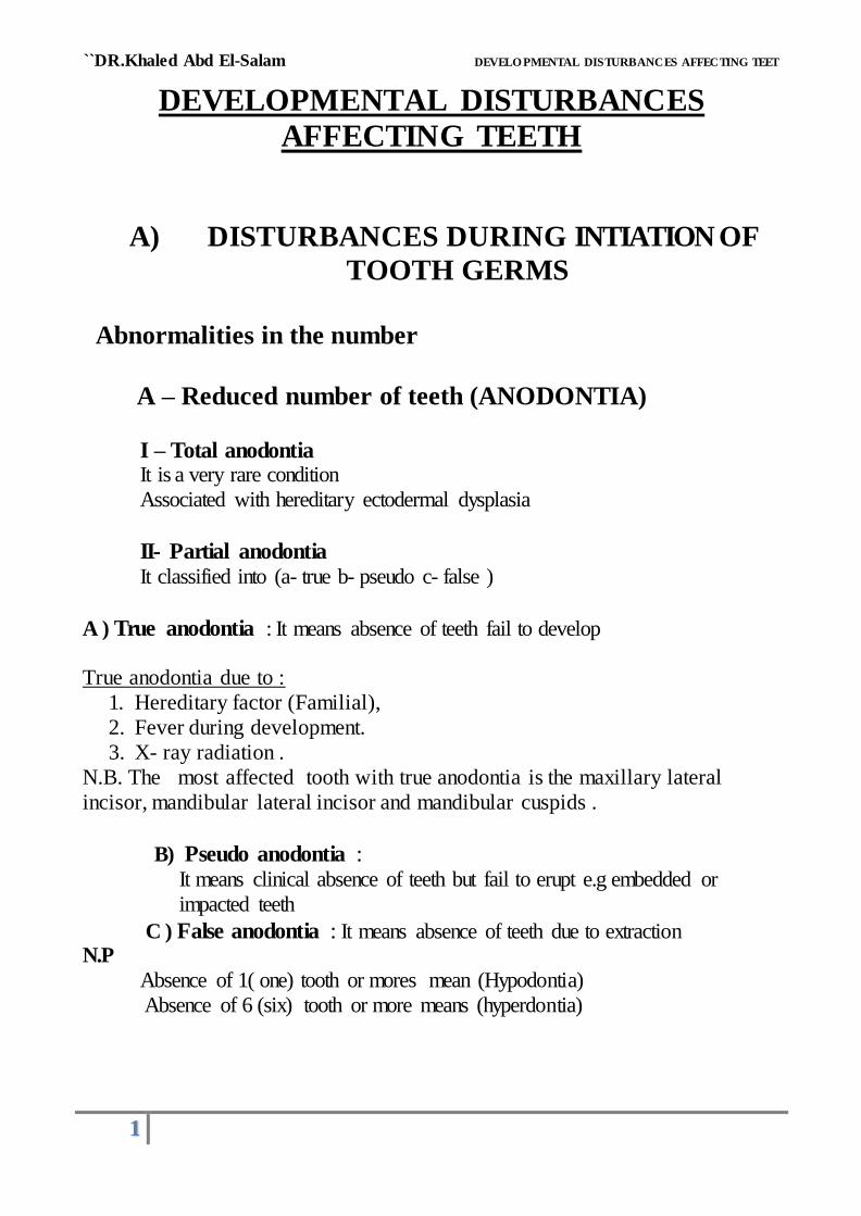
ECTODERMAL DYSPLASIA • It is a hereditary disease which involves all structures which are derived
from the ectoderm .
• It is characterized by (general manifestation) :
1- Skin ( thin, smooth, Dry skin)
2- Hair (Absence or reduction (hypotrichosis).
3- Sweat-gland (Absence anhydrosis).
4- sebaceous gland ( absent lead to dry skin)
5-Temperature elevation (because of anhydrosis)
6- Depressed bridge of the nose
7- Defective mental development
8- Defective of finger nail
Oral manifestation include teeth and salivary glands
Teeth: 1. Complete or partial anodontia ( deciduous and permanent ).
• incisor crowns usually appear peg-shaped or conical • defect in the structure of enamel
2- Retarded eruption
4- High arched cleft palate
5- Protrusion of the lip
6- Alveolar process is deficient in height
Salivary glands: Partial or complete absence leads to (Xerostomia)

``DR.Khaled Abd El-Salam DEVELO PMENTAL DISTURBANCES AFFECTING TEET
3
B- Additional number of teeth
These are extra teeth of the normal complement as referred to as Supplement teeth and
supernumerary teeth
According to morphology as
I- Supplement teeth These are extra teeth similar morphologically to the normal tooth
It classify according to time of eruption into
1- PREDECIDUOUS DENTITION
It means that presence of teeth preceding the deciduous dentition , it
usually present at birth or they erupt soon after birth before eruption of
deciduous teeth
ETIOLOGY :
l. May arises from a bud of an accessory dental lamina.
• This is very rare condition
• It is due to accessory buds being given before the formation of enamel
organ of the deciduous teeth
• They are rudimentary in size
• It present at birth usually erupt in the mandibular incisor and are weakly
attached to the gum ( natal teeth)
Clinical significance
• Can be inhaled into the air ways
• Pain during nursing feeding
Treatment extracted this tooth due to
o Difficult fading and may be Aspirated
N.P
it should be differentiated from premature
erupted deciduous teeth
which may erupt at birth (neonatal teeth )
``DR.Khaled Abd El-Salam DEVELO PMENTAL DISTURBANCES AFFECTING TEET
4
2- POST PERMANENT DENTITION
• It means that the teeth may erupt after the loss of permanent dentition .
• Usually these are impacted accessory teeth that erupt after the insertion of
denture.
• They may be developed from a bud of the dental lamina beyond the
permanent tooth germ.
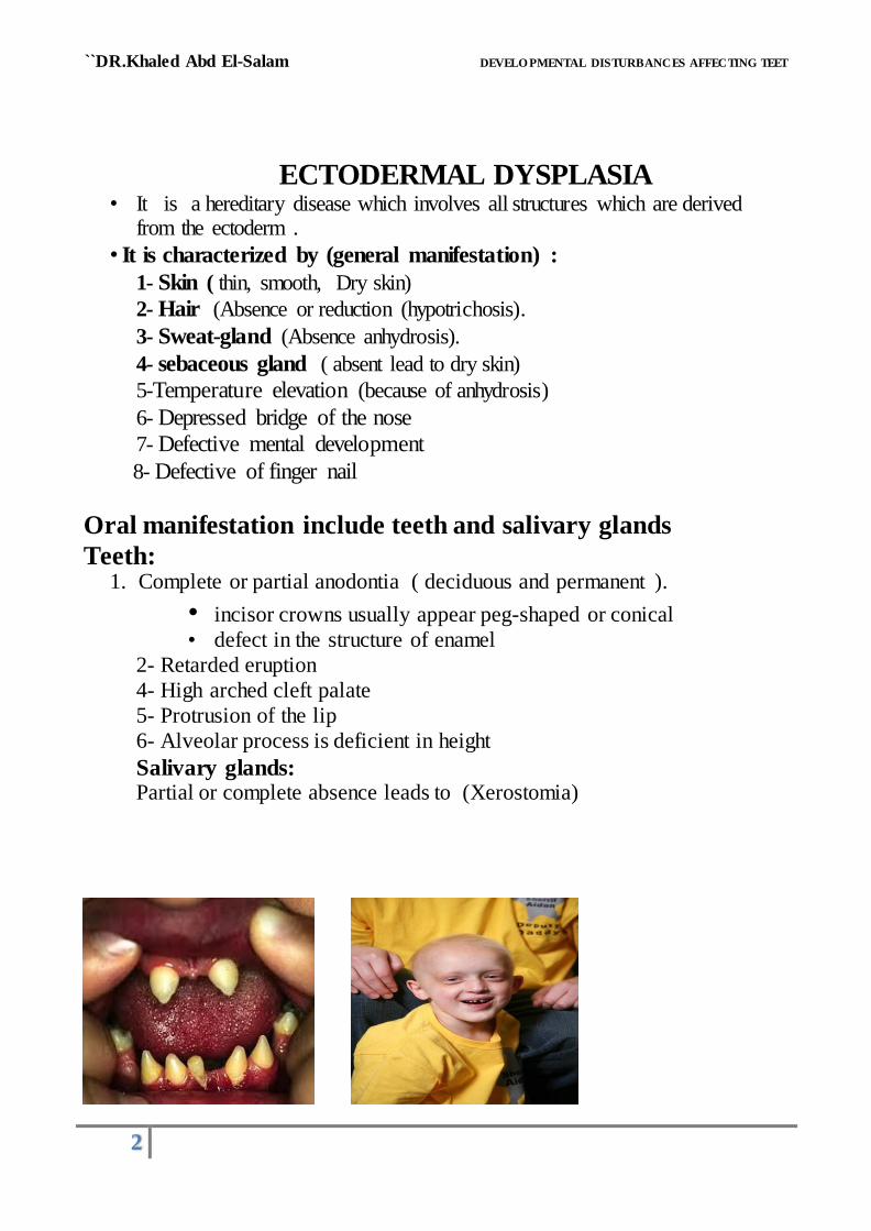
II- SUPERNUMERARY TEETH.
These are extra teeth not similar morphologically to the normal tooth
It classify according to site of eruption into
1. Mesiodens : When accessory tooth located between the maxillary
central incisors .
2. Paramolar : The accessory tooth located buccal or lingual to the molars
3. Distomolar : The accessory tooth located distal to the third molar .
Mesiodens Paramolar Distomolar
ETIOLOGY : 1. Third tooth bud arising from the dental lamina.
2. Splitting of the permanent tooth bud.
3. Hereditary.
N.B.
1- The accessory teeth far more common in the maxilla more than the
mandible (9:1) and the most common are the mesiodens and distmolar .
``DR.Khaled Abd El-Salam DEVELO PMENTAL DISTURBANCES AFFECTING TEET
5
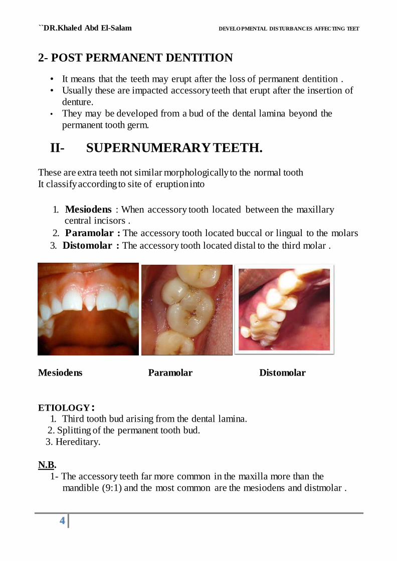
2- Multiple impacted supernumerary teeth associated with
1- Gardner's syndrome
2- Cleidocranial dysostosis
Gardner's syndrome
Multiple impacted Supernumerary tooth
Multiple osteoma of the jaw
Multiple polyposis of large intestine
Multiple epidermoid cyst
Desmoid tumor
Cleidocranial dysostosis : (Marie and Sainton's disease )
It is characterized by :
A) Abnormalities of the skull and jaws:
• Delayed closure of the fontanels. • Delayed closure of the cranial sutures.
• Presence of wormian bones (small, irregular bones between the chief bones of the skull).
• Underdevelopment of the upper face , particularly of the maxilla . • Underdevelopment of paranasal sinuses.
• High and narrow palate. • Prognathism or relative prominence of the mandible.
B) Abnormalities of the teeth :
• Delayed eruption of teeth
• Malocclusion . • Presence of supernumerary teeth .
• Impacted teeth. Which may be associated with the follicular cysts. C) Abnormalities of the clavicles :
• There is an absence or hypoplasia of the clavicles so that the patient can
approximate the shoulders with ease.
``DR.Khaled Abd El-Salam DEVELO PMENTAL DISTURBANCES AFFECTING TEET
6
B) DISTURBANCES DURING
MORPHODIFFERENTIATION OF TOOTH
GERM
I – Abnormalities in the size
1-MACRODONTIA
1- generalized macrodontia: A) True
It is very rare condition, the teeth are
larger than normal as shown associated with pituitary gigantism.
generalized macrodontia : B) Relative It is somewhat more common,
the teeth are normal in size or slightly larger than normal teeth in small
jaw,it is due to hereditary factor.
2- Macrodontia of single tooth (localized) : It is of unknown etiology and uncommon condition,
but may be seen in cases of hemihypertrophy of the face
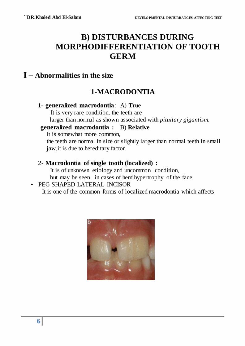
• PEG SHAPED LATERAL INCISOR
It is one of the common forms of localized macrodontia which affects
``DR.Khaled Abd El-Salam DEVELO PMENTAL DISTURBANCES AFFECTING TEET
7
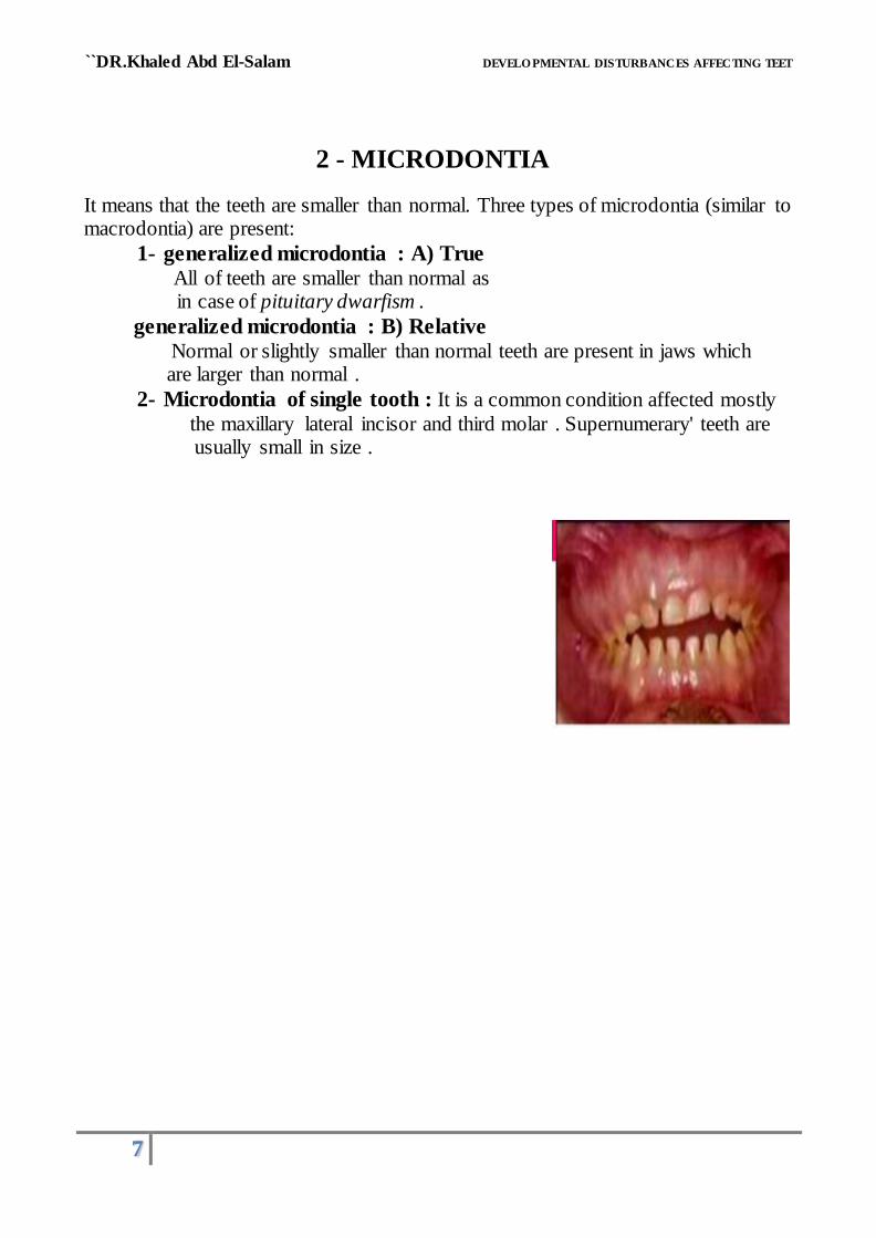
2 - MICRODONTIA
It means that the teeth are smaller than normal. Three types of microdontia (similar to macrodontia) are present:
1- generalized microdontia : A) True All of teeth are smaller than normal as
in case of pituitary dwarfism .
generalized microdontia : B) Relative
Normal or slightly smaller than normal teeth are present in jaws which are larger than normal .
2- Microdontia of single tooth : It is a common condition affected mostly
the maxillary lateral incisor and third molar . Supernumerary' teeth are usually small in size .
``DR.Khaled Abd El-Salam DEVELO PMENTAL DISTURBANCES AFFECTING TEET
8
II-Abnormalities in the shape
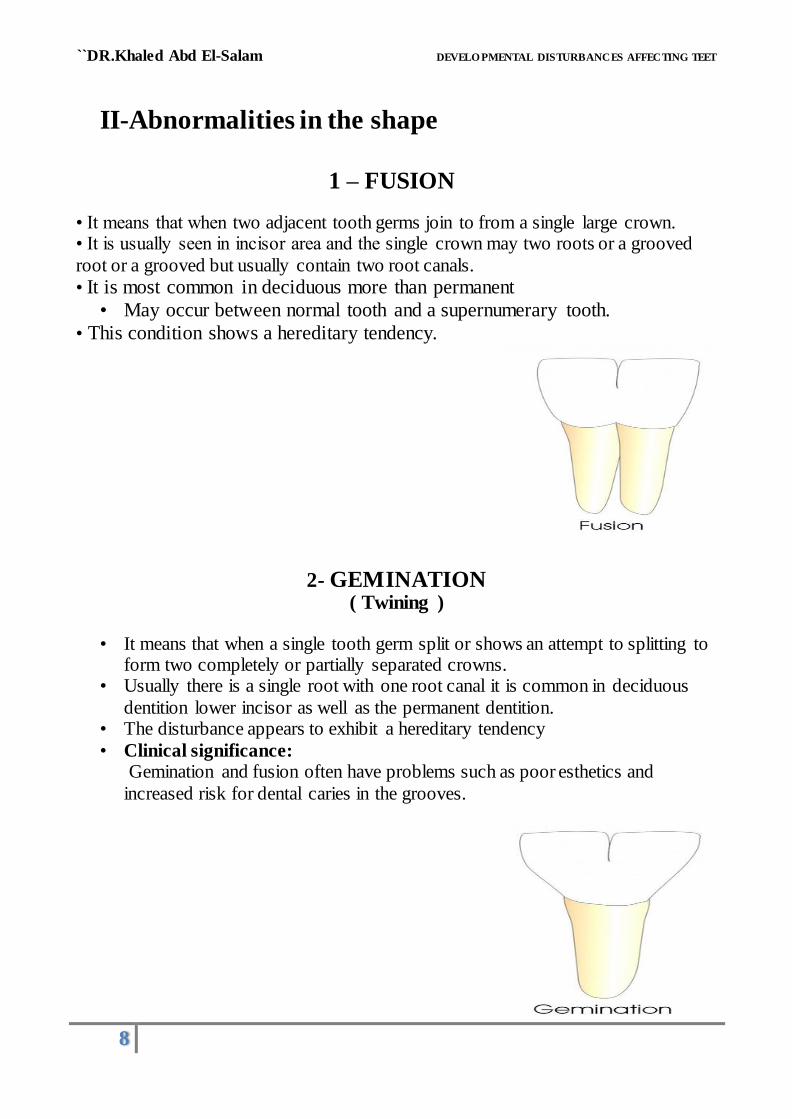
1 – FUSION
• It means that when two adjacent tooth germs join to from a single large crown. • It is usually seen in incisor area and the single crown may two roots or a grooved
root or a grooved but usually contain two root canals.
• It is most common in deciduous more than permanent
• May occur between normal tooth and a supernumerary tooth.
• This condition shows a hereditary tendency.
2- GEMINATION ( Twining )
• It means that when a single tooth germ split or shows an attempt to splitting to form two completely or partially separated crowns.
• Usually there is a single root with one root canal it is common in deciduous
dentition lower incisor as well as the permanent dentition. • The disturbance appears to exhibit a hereditary tendency
• Clinical significance: Gemination and fusion often have problems such as poor esthetics and
increased risk for dental caries in the grooves.
``DR.Khaled Abd El-Salam DEVELO PMENTAL DISTURBANCES AFFECTING TEET
9
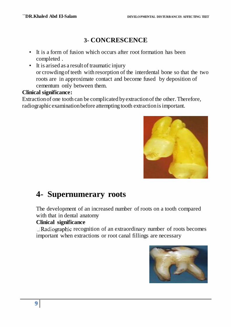
3- CONCRESCENCE
• It is a form of fusion which occurs after root formation has been
completed .
• It is arised as a result of traumatic injury
or crowding of teeth with resorption of the interdental bone so that the two
roots are in approximate contact and become fused by deposition of
cementum only between them.
Clinical significance:
Extraction of one tooth can be complicated by extraction of the other. Therefore,
radiographic examination before attempting tooth extraction is important.
4- Supernumerary roots
The development of an increased number of roots on a tooth compared
with that in dental anatomy
Clinical significance
recognition of an extraordinary number of roots becomes
important when extractions or root canal fillings are necessary
``DR.Khaled Abd El-Salam DEVELO PMENTAL DISTURBANCES AFFECTING TEET
10
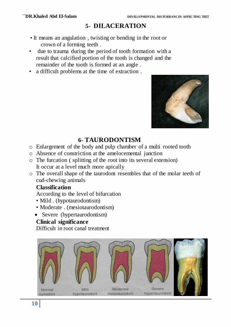
5- DILACERATION
• It means an angulation , twisting or bending in the root or
crown of a forming teeth .
• due to trauma during the period of tooth formation with a
result that calcified portion of the tooth is changed and the
remainder of the tooth is formed at an angle .
• a difficult problems at the time of extraction .
6- TAURODONTISM o Enlargement of the body and pulp chamber of a multi rooted tooth
o Absence of constriction at the amelocemental junction
o The furcation ( splitting of the root into its several extension)
It occur at a level much more apically
o The overall shape of the taurodont resembles that of the molar teeth of
cud-chewing animals
Classification According to the level of bifurcation
• Mild . (hypotaurodontism)
• Moderate . (mesiotaurodontism)
Severe (hypertaurodontism)
Clinical significance Difficult in root canal treatment
``DR.Khaled Abd El-Salam DEVELO PMENTAL DISTURBANCES AFFECTING TEET
11
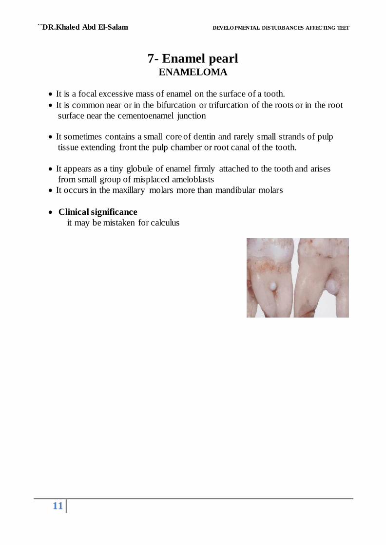
7- Enamel pearl
ENAMELOMA
It is a focal excessive mass of enamel on the surface of a tooth.
It is common near or in the bifurcation or trifurcation of the roots or in the root
surface near the cementoenamel junction
It sometimes contains a small core of dentin and rarely small strands of pulp
tissue extending front the pulp chamber or root canal of the tooth.
It appears as a tiny globule of enamel firmly attached to the tooth and arises
from small group of misplaced ameloblasts
It occurs in the maxillary molars more than mandibular molars
Clinical significance
it may be mistaken for calculus
``DR.Khaled Abd El-Salam DEVELO PMENTAL DISTURBANCES AFFECTING TEET
12
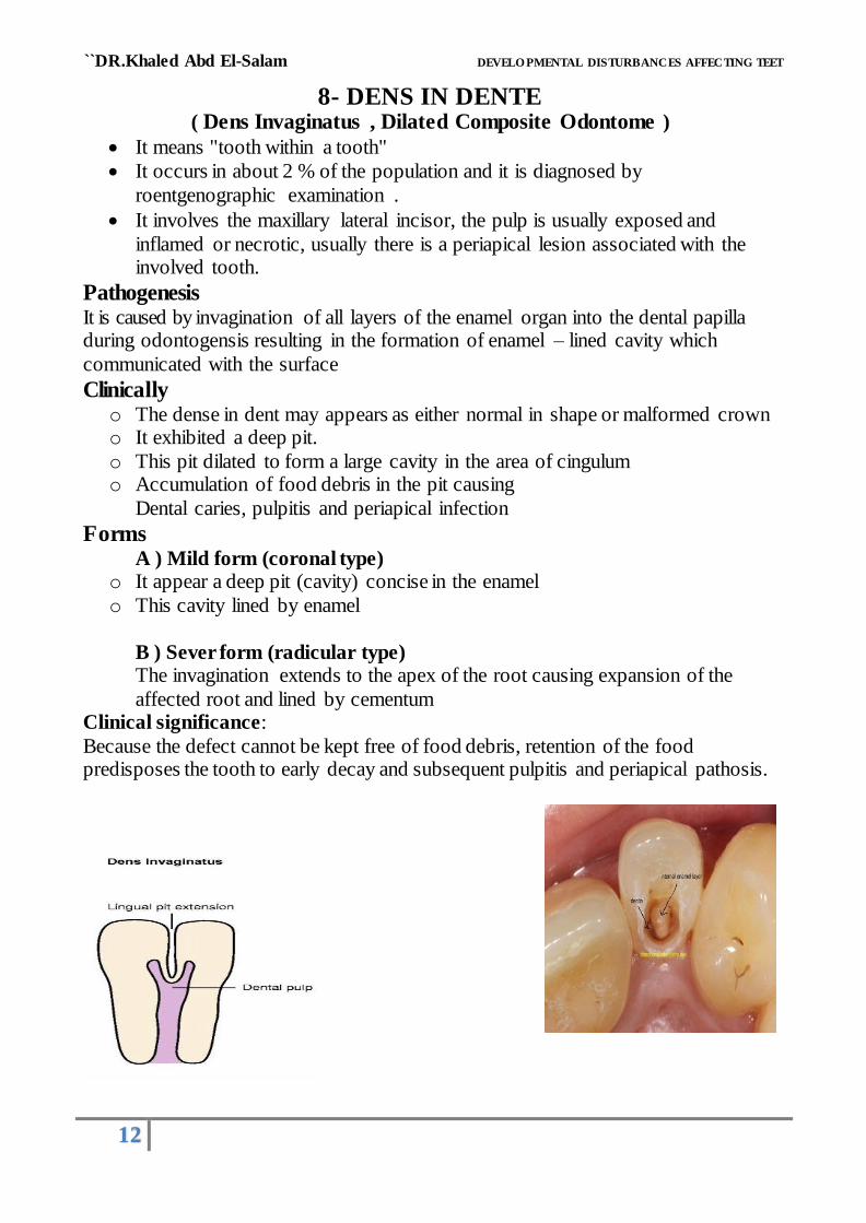
8- DENS IN DENTE ( Dens Invaginatus , Dilated Composite Odontome )
It means "tooth within a tooth"
It occurs in about 2 % of the population and it is diagnosed by
roentgenographic examination .
It involves the maxillary lateral incisor, the pulp is usually exposed and
inflamed or necrotic, usually there is a periapical lesion associated with the involved tooth.
Pathogenesis It is caused by invagination of all layers of the enamel organ into the dental papilla during odontogensis resulting in the formation of enamel – lined cavity which
communicated with the surface
Clinically o The dense in dent may appears as either normal in shape or malformed crown o It exhibited a deep pit.
o This pit dilated to form a large cavity in the area of cingulum o Accumulation of food debris in the pit causing
Dental caries, pulpitis and periapical infection
Forms A ) Mild form (coronal type)
o It appear a deep pit (cavity) concise in the enamel
o This cavity lined by enamel
B ) Sever form (radicular type) The invagination extends to the apex of the root causing expansion of the
affected root and lined by cementum Clinical significance:
Because the defect cannot be kept free of food debris, retention of the food predisposes the tooth to early decay and subsequent pulpitis and periapical pathosis.
``DR.Khaled Abd El-Salam DEVELO PMENTAL DISTURBANCES AFFECTING TEET
13
9- Teeth abnormalities associated with accessory cusps
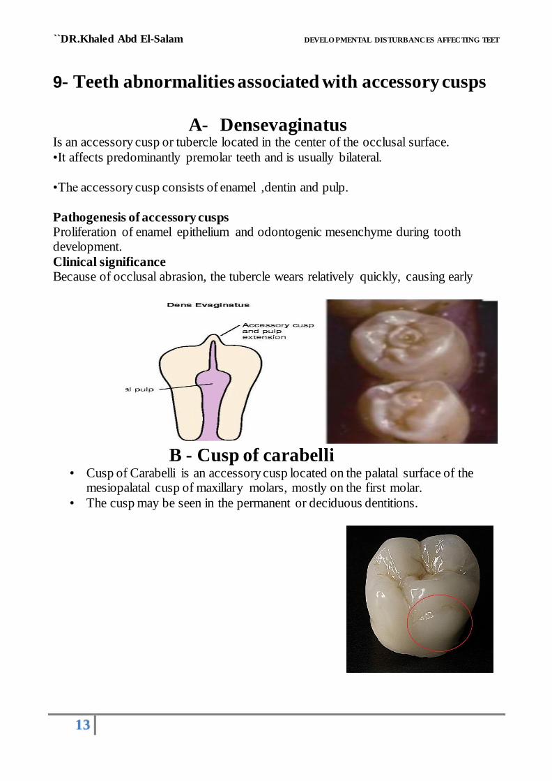
A- Densevaginatus Is an accessory cusp or tubercle located in the center of the occlusal surface.
•It affects predominantly premolar teeth and is usually bilateral.
•The accessory cusp consists of enamel ,dentin and pulp.
Pathogenesis of accessory cusps Proliferation of enamel epithelium and odontogenic mesenchyme during tooth development.
Clinical significance Because of occlusal abrasion, the tubercle wears relatively quickly, causing early
B - Cusp of carabelli
• Cusp of Carabelli is an accessory cusp located on the palatal surface of the mesiopalatal cusp of maxillary molars, mostly on the first molar.
• The cusp may be seen in the permanent or deciduous dentitions.
``DR.Khaled Abd El-Salam DEVELO PMENTAL DISTURBANCES AFFECTING TEET
14
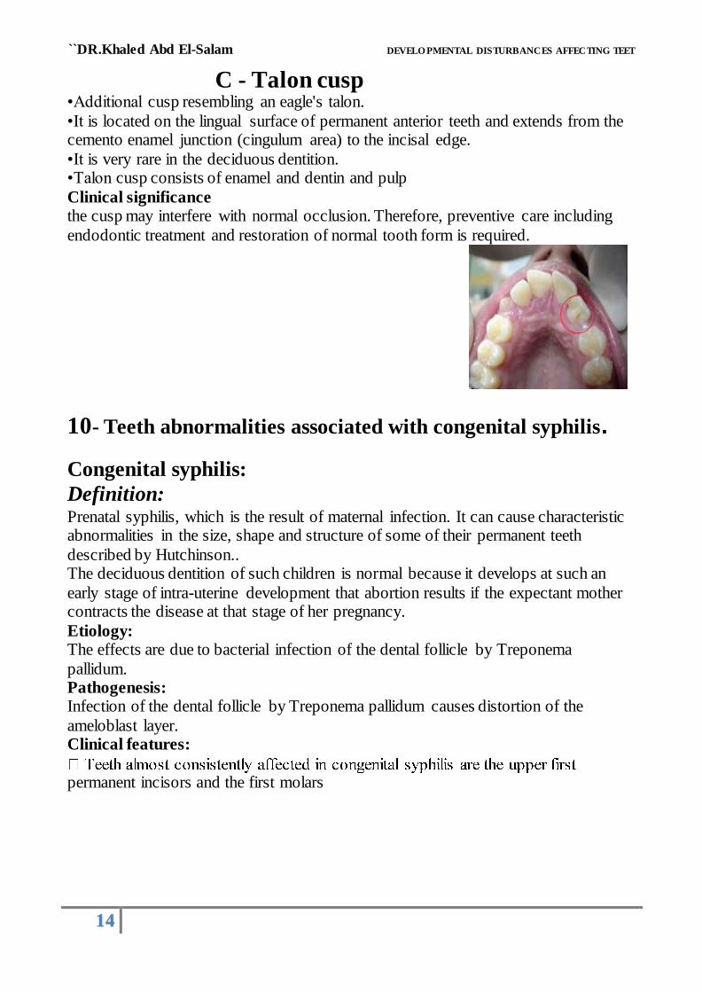
C - Talon cusp •Additional cusp resembling an eagle's talon.
•It is located on the lingual surface of permanent anterior teeth and extends from the cemento enamel junction (cingulum area) to the incisal edge.
•It is very rare in the deciduous dentition. •Talon cusp consists of enamel and dentin and pulp
Clinical significance the cusp may interfere with normal occlusion. Therefore, preventive care including
endodontic treatment and restoration of normal tooth form is required.
10- Teeth abnormalities associated with congenital syphilis.
Congenital syphilis:
Definition: Prenatal syphilis, which is the result of maternal infection. It can cause characteristic abnormalities in the size, shape and structure of some of their permanent teeth
described by Hutchinson.. The deciduous dentition of such children is normal because it develops at such an
early stage of intra-uterine development that abortion results if the expectant mother contracts the disease at that stage of her pregnancy.
Etiology: The effects are due to bacterial infection of the dental follicle by Treponema
pallidum. Pathogenesis: Infection of the dental follicle by Treponema pallidum causes distortion of the
ameloblast layer. Clinical features:
permanent incisors and the first molars
``DR.Khaled Abd El-Salam DEVELO PMENTAL DISTURBANCES AFFECTING TEET
15
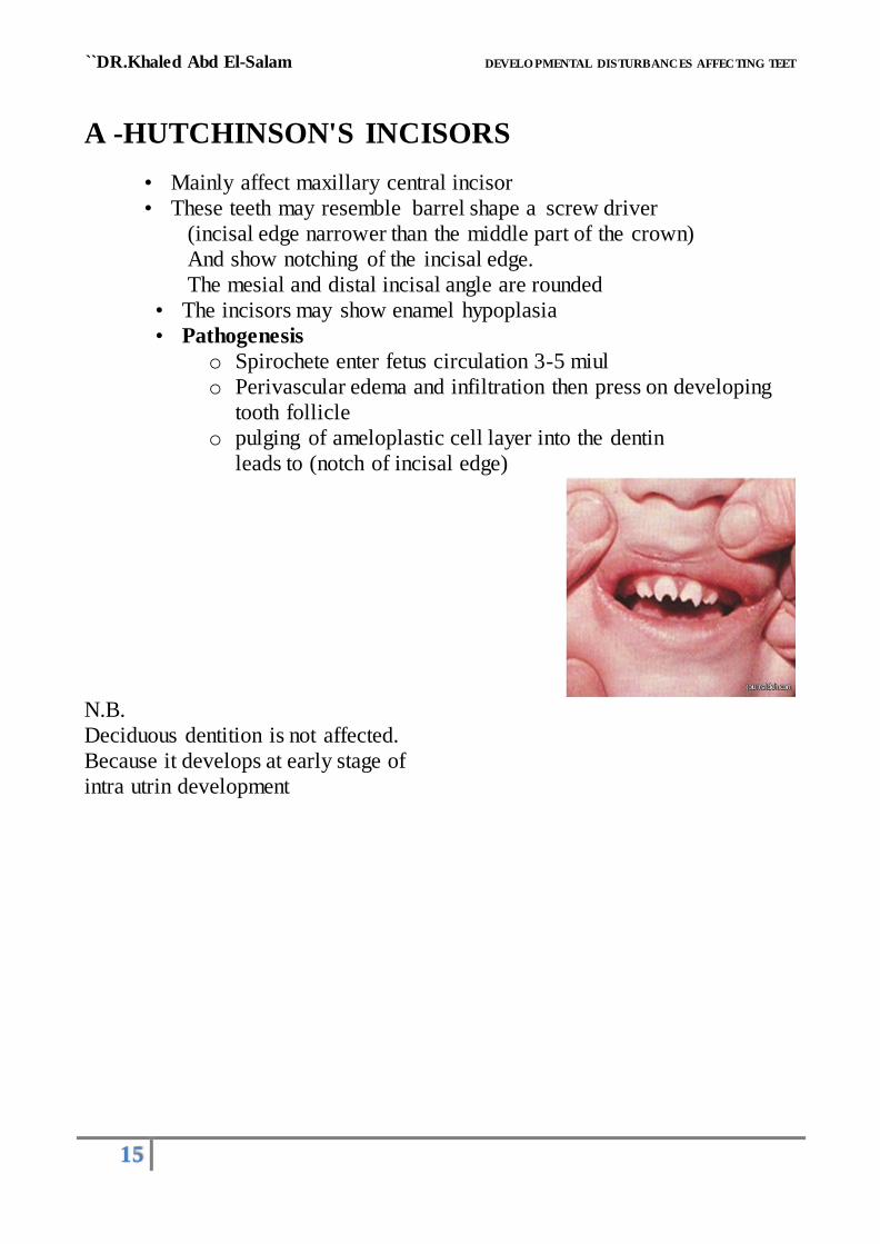
A -HUTCHINSON'S INCISORS
• Mainly affect maxillary central incisor
• These teeth may resemble barrel shape a screw driver
(incisal edge narrower than the middle part of the crown)
And show notching of the incisal edge.
The mesial and distal incisal angle are rounded
• The incisors may show enamel hypoplasia
• Pathogenesis
o Spirochete enter fetus circulation 3-5 miul
o Perivascular edema and infiltration then press on developing
tooth follicle
o pulging of ameloplastic cell layer into the dentin
leads to (notch of incisal edge)
N.B.
Deciduous dentition is not affected.
Because it develops at early stage of
intra utrin development
``DR.Khaled Abd El-Salam DEVELO PMENTAL DISTURBANCES AFFECTING TEET
16
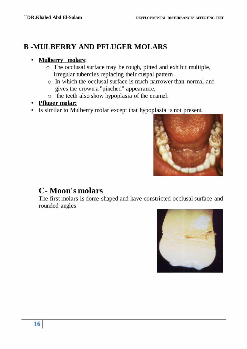
B -MULBERRY AND PFLUGER MOLARS
• Mulberry molars:
o The occlusal surface may be rough, pitted and exhibit multiple,
irregular tubercles replacing their cuspal pattern
o In which the occlusal surface is much narrower than normal and
gives the crown a "pinched" appearance,
o the teeth also show hypoplasia of the enamel.
• Pfluger molar:
• Is similar to Mulberry molar except that hypoplasia is not present.
C- Moon's molars The first molars is dome shaped and have constricted occlusal surface and
rounded angles
``DR.Khaled Abd El-Salam DEVELO PMENTAL DISTURBANCES AFFECTING TEET
17
C) DISTURBANCE DURING APPOSITION OF HARD DENTAL TISSUES
A. Defects of enamel B. Defects of dentin C. Defects of enamel and dentin
A. Defects of enamel
The defect of enamel may be due to Hereditary factors or environmental
(Acquired) factors ( 1- Local factors 2- systemic factors)
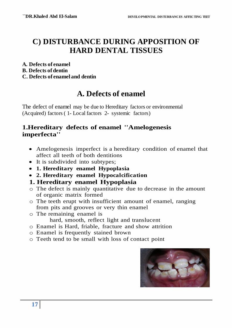
1.Hereditary defects of enamel ''Amelogenesis
imperfecta''
Amelogenesis imperfect is a hereditary condition of enamel that
affect all teeth of both dentitions
It is subdivided into subtypes;
1. Hereditary enamel Hypoplasia
2. Hereditary enamel Hypocalcification
1. Hereditary enamel Hypoplasia o The defect is mainly quantitative due to decrease in the amount
of organic matrix formed
o The teeth erupt with insufficient amount of enamel, ranging from pits and grooves or very thin enamel
o The remaining enamel is hard, smooth, reflect light and translucent
o Enamel is Hard, friable, fracture and show attrition o Enamel is frequently stained brown
o Teeth tend to be small with loss of contact point
``DR.Khaled Abd El-Salam DEVELO PMENTAL DISTURBANCES AFFECTING TEET
18
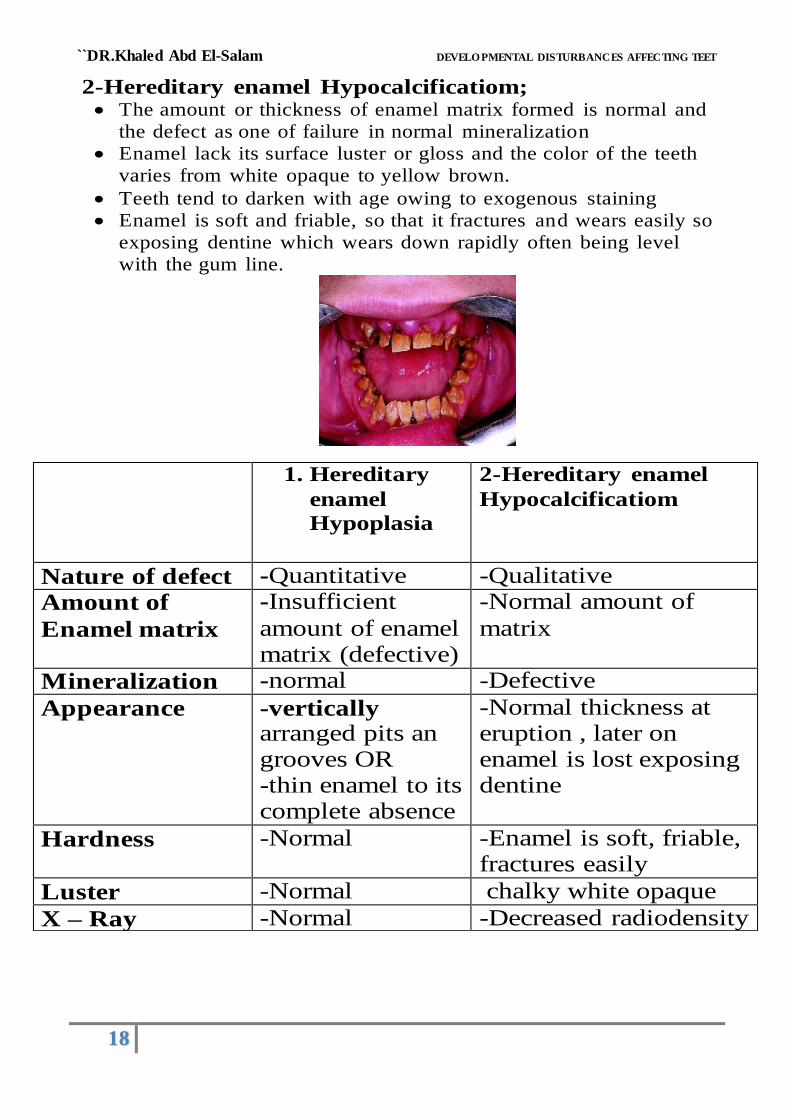
2-Hereditary enamel Hypocalcificatiom;
The amount or thickness of enamel matrix formed is normal and
the defect as one of failure in normal mineralization
Enamel lack its surface luster or gloss and the color of the teeth
varies from white opaque to yellow brown.
Teeth tend to darken with age owing to exogenous staining
Enamel is soft and friable, so that it fractures and wears easily so
exposing dentine which wears down rapidly often being level
with the gum line.
1. Hereditary
enamel
Hypoplasia
2-Hereditary enamel
Hypocalcificatiom
Nature of defect -Quantitative -Qualitative
Amount of
Enamel matrix
-Insufficient
amount of enamel matrix (defective)
-Normal amount of
matrix
Mineralization -normal -Defective
Appearance -vertically arranged pits an grooves OR -thin enamel to its complete absence
-Normal thickness at eruption , later on enamel is lost exposing dentine
Hardness -Normal -Enamel is soft, friable, fractures easily
Luster -Normal chalky white opaque
X – Ray -Normal -Decreased radiodensity

``DR.Khaled Abd El-Salam DEVELO PMENTAL DISTURBANCES AFFECTING TEET
19
II- ACQUIRED ENAMEL HYPOPLASIA
1- Local factors: It affects a tooth or a part of a tooth and is due to local cause e.g.
a) Trauma b) Periapical infection
c) Radiotherapy
N.B. Turner's tooth : It is tooth with enamel hypoplasia due to local factor.
• It is usually confined to single tooth , • the mandibular premolars are the most common affected teeth .
A ) INFECTION;
o Hypoplasia is caused by periapical infection and inflammation involving a primary tooth that spread to the enamel organ of underlying permanent
tooth germ before development of enamel is completed o damage of ameloblasts lead to defect of enamel.
o The enamel of the affected tooth may be thin, irregular, or even be entirely missing over an area of the crown.
B ) TRAUMA; o Traumatic injuries to deciduous incisors may path them deeply into their
sockets
o resulting in their root impinging upon the developing teeth germ of their permanent successors.
o The resulting injury may be manifested as yellowish or brownish stain or pigmentation of the enamel ,
usually on the labial surface ,or as a true hypoplastic pitting defect or deformity affecting the permanent incisors
``DR.Khaled Abd El-Salam DEVELO PMENTAL DISTURBANCES AFFECTING TEET
20
2. Systemic factors : 1- Chemicals ( fluoride, tetracycline)
2- RH hemolytic disease. 3- Infection (Chicken pox, Measles, syphilis, Scarlet fever)
4- Malnutrition (Vitamin A,C and D deficiency) 5- Hormonal disturbance (hypoparathyrodism)
6- Birth related trauma 7- Associated with generalized disease (cledocanial dysostosis , ectodermal
dysplasia, osteopetrosis, mongolism)
( Example of systemic hypocalcification):
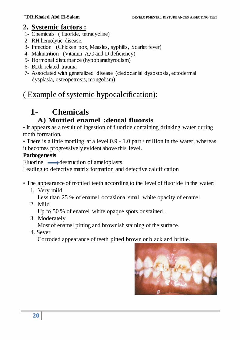
1- Chemicals A) Mottled enamel :dental fluorsis
• It appears as a result of ingestion of fluoride containing drinking water during
tooth formation.
• There is a little mottling at a level 0.9 - 1.0 part / million in the water, whereas
it becomes progressively evident above this level.
Pathogenesis
Fluorine destruction of ameloplasts
Leading to defective matrix formation and defective calcification
• The appearance of mottled teeth according to the level of fluoride in the water:
1. Very mild
Less than 25 % of enamel occasional small white opacity of enamel.
2. Mild
Up to 50 % of enamel white opaque spots or stained .
3. Moderately
Most of enamel pitting and brownish staining of the surface.
4. Sever
Corroded appearance of teeth pitted brown or black and brittle.

``DR.Khaled Abd El-Salam DEVELO PMENTAL DISTURBANCES AFFECTING TEET
21
B ) Tetracycline pigmentation Tetracycline binds to calcifying tissues and thus stains developing teeth and bone
and¸ in higher doses may cause enamel hypoplasia.
Tetracycline is deposited along the incremental line of dentine and enamel with
the result that the whole tooth maybe discolored. Affected teeth exhibit
fluorescence under ultraviolet light.
It depending of
1- Time of administration
2- Dose, duration and frequency of intake
Bind to calcium during calcification stage along the incremental lines
2) Erythroplastosis fetalis
-if ''RH+'' father maries ''RH-'' mother Hemolysis of baby's RBCs
CLINICALY; Teeth pigmentation; Narrow band of green or bluish pigments in dentin at (Neonatal line)
Enamel; show hypoplasia
SITE;
Crown of A&B, Half-crown of C & cusps of D&E.
``DR.Khaled Abd El-Salam DEVELO PMENTAL DISTURBANCES AFFECTING TEET
22
N.P
I- Teeth discoloration
I-Discoloration by exogenous pigments .
II-Discoloration by endogneous pigments . Exogenous stains
(stains on the surface of teeth that can be removed with abrasives) may be caused by:
a- Diet. b- Coffee and tea. c- Tobacco.
d- Excessive use of chlorohexidine.
e- Chromogenic bacteria
Endogneous stains
(Discoloration during tooth development) may be caused by:
a- Fluorosis .
b- Tetracycline .
c-Erythroblastosis fetalis .
d- Hemolytic jaundice of newborn
e- Porphyries (red or purple pigmentation, pink tooth, lavender tooth).
N.P
Differences between Hereditary and Acquired Defects
HEREDITARY DEFECTS ACQUIRED DEFECTS
Affect both the deciduous and
permanent dentition
Affect only one dentition
usually the permanent
Usually affect all teeth Usually affect single or group
of teeth
Affect enamel or dentine Affect both enamel and dentine
Produce diffuse or vertically or randomly oriented pits or
defects
Produce horizontally oriented pits or defects
``DR.Khaled Abd El-Salam DEVELO PMENTAL DISTURBANCES AFFECTING TEET
23
B. Defects of dentin
1- DENTINOGENESIS IMPERFECTA ( Hereditary Opalescent Dentin )
It is a hereditary disturbance affects development of dentin and may be accompanied by similar disturbance in the bones (osteogenesis imperfecta).
Classification Type I Associated with (osteogenesis imperfecta). Type II Not associated with (osteogenesis imperfecta
Type III Shell tooth
CLINICAL FEATURES : • It affects both deciduous and permanent dentition ,
The teeth appear opalescent or gray .
The crown of the teeth slightly smaller than normal constricted neck that give the bulbous appearance .
• The enamel is normal and attrition is rapid , chips off easily
The dentin undergoes rapid attrition
The occlusal surface are often severely flattened .
The root is short and stunted
ROENTGENOGRAPHS FEATURES : • The roots are short and conical with blunt apex . Sometimes the apices of teeth show radiolucent areas resembling dental
granulomas or cysts
• The pulp chambers are wide in the early stages but are narrowed or obliterated later .
The cementum, periodontal ligament and supporting alveolar bone is normal
HISTOLOGIC FEATURES :
The enamel is normal
The dentinoenamel junction appears smooth (not scalloped)
The mantle dentin is normal (first layer of dentin just below the
dentinoenamel junction). '
The deeper dentin shows the following
dentinal tubules is fewer in number but large in diameter and irregular give a tortuous course
``DR.Khaled Abd El-Salam DEVELO PMENTAL DISTURBANCES AFFECTING TEET
24
Degeneration of odontoblast or entrapped in the dentine
Poor calcification with large area of interglobular spaces
Irregular incremental line
The pulp shows
Numerous pulp stone Obliteration of the pulp chambers and canals
Absence of odontoblastic layer
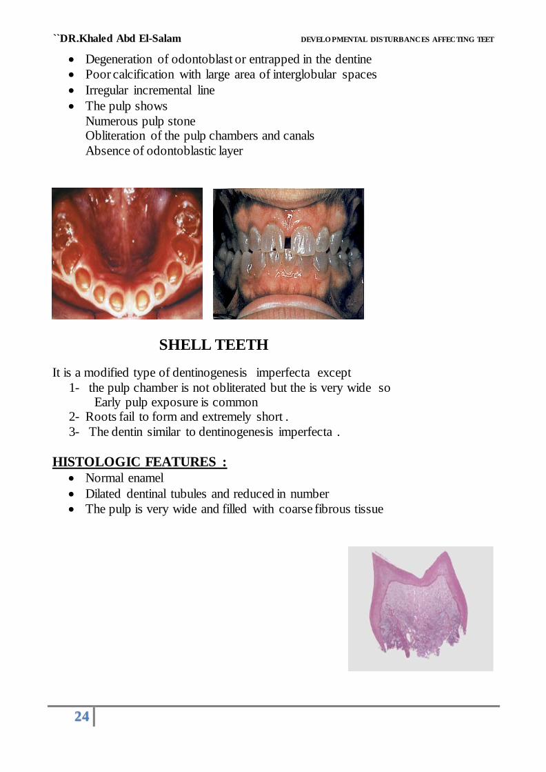
SHELL TEETH
It is a modified type of dentinogenesis imperfecta except
1- the pulp chamber is not obliterated but the is very wide so Early pulp exposure is common
2- Roots fail to form and extremely short .
3- The dentin similar to dentinogenesis imperfecta .
HISTOLOGIC FEATURES :
Normal enamel
Dilated dentinal tubules and reduced in number
The pulp is very wide and filled with coarse fibrous tissue
``DR.Khaled Abd El-Salam DEVELO PMENTAL DISTURBANCES AFFECTING TEET
25
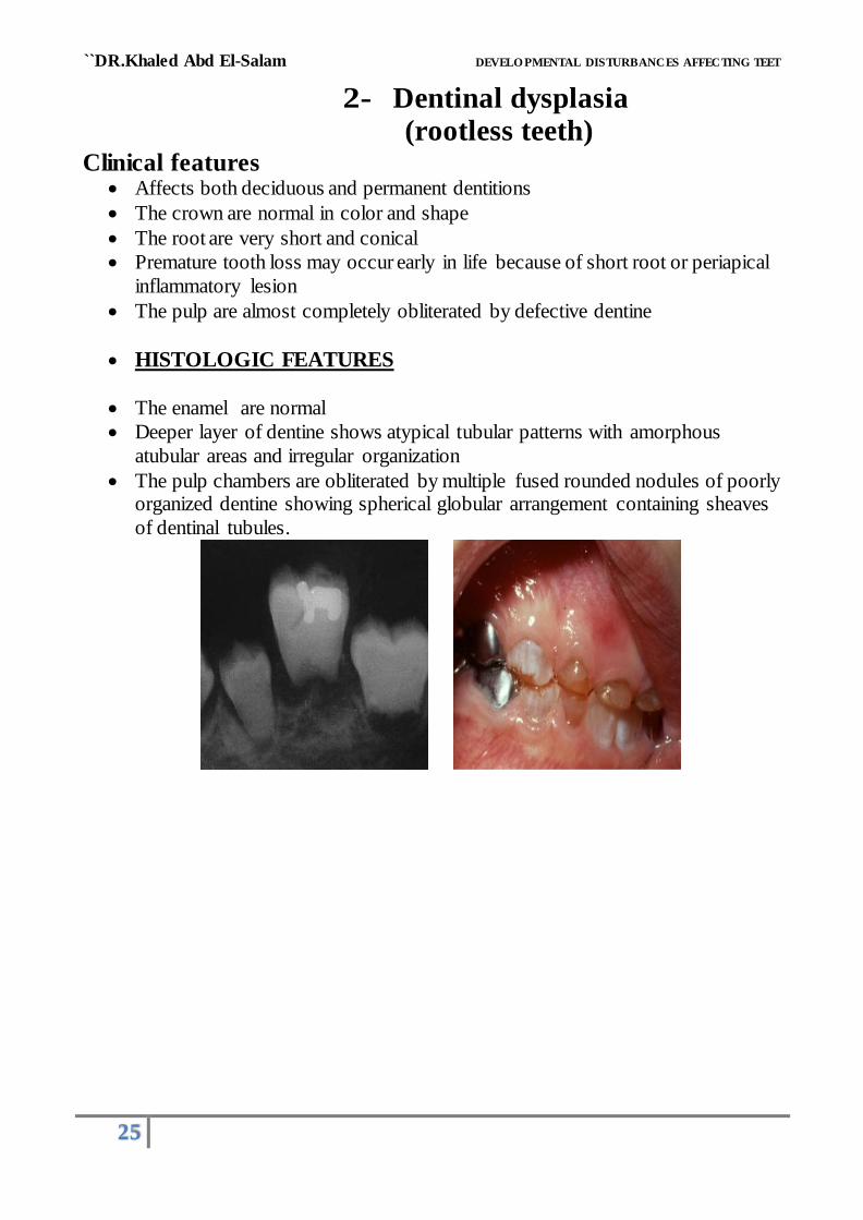
2- Dentinal dysplasia (rootless teeth)
Clinical features Affects both deciduous and permanent dentitions
The crown are normal in color and shape
The root are very short and conical
Premature tooth loss may occur early in life because of short root or periapical
inflammatory lesion
The pulp are almost completely obliterated by defective dentine
HISTOLOGIC FEATURES
The enamel are normal
Deeper layer of dentine shows atypical tubular patterns with amorphous
atubular areas and irregular organization
The pulp chambers are obliterated by multiple fused rounded nodules of poorly organized dentine showing spherical globular arrangement containing sheaves
of dentinal tubules.

``DR.Khaled Abd El-Salam DEVELO PMENTAL DISTURBANCES AFFECTING TEET
26
C. Defects of enamel and dentin
1- ODONTODYSPLASIA (Ghost Teeth , Odontogenic dysplasia, Odontogenesis Imperfecta)
Clinical feature It affect both the deciduous and permanent dentition
The maxillary teeth are commonly involved than the mandibular teeth . The most affected teeth are the central, lateral and cuspid teeth .
It is characterized by : a) Small , distorted discolored crowns
reduction in thickness of enamel, pitted, pigmented and rough give (moth-eaten appearance).
b) The teeth usually fail to erupt. c) One or several teeth in a localized areas are affected .
ROENTGENOGRAPHS FEATURES :
• Reduction in the radiodensity of the enamel and dentine Due to lack of calcification so the tooth assume
(ghost tooth). • The crown may be surrounded by radiolucent area .
• The pulp chambers are wide . • Both enamel and dentin appear to be very thin .
HISTOLOGIC FEATURES :
• Enamel : a) Very thin and hypoplastic due to distortion of enamel matrix .
b) Absence of enamel rods , and focal calcification in the connective tissue around the crown .
• Dentin : Reduction in the amount of dentine and widening of predentin Presence large area of interglobular dentine and irregular tubular pattern
• Pulp : It may show calcification
``DR.Khaled Abd El-Salam DEVELO PMENTAL DISTURBANCES
AFFECTING TEET
27
E) DISTURBANCE DURING ERUPTION
OF TEETH
1- PREMATURE ERUPTION
• Deciduous dentition : Natal teeth (as mentioned before).
• Permanent dentition : Premature eruption of permanent teeth
• Etiology
a- due to premature loss of deciduous teeth or
b- associated with hyperthyroidism .
c- associated with facial hemihypertrophy
d- genetic familial predilection
2- DELAYED ERUPTION (RETARDED)
1-Systemic
factors :
a) Endocrine disturbance (hypothyroidism). b) Vitamin deficiency (Vitamin D and rickets. c) Cleidocranialdysostos
d) cherubism
2- Local factors :
a) Fibromatosis gingivae . b) Lack of space .
c) Malposition . d) Dentigerous and eruption cyst.
e) Presence of - Odontoma
- Supernumerary teeth
- Retained deciduous teeth
3- EMBEDDED AND IMPACTED TEETH
``DR.Khaled Abd El-Salam DEVELO PMENTAL DISTURBANCES
AFFECTING TEET
28
• Embedded teeth : Teeth do not erupted due to lack of eruptive
force .
• Impacted teeth : Teeth can not eruption due to physical barrier in eruption path (Lack of space).
The most affected teeth are :
1. Maxillary third molars and cuspids .
2. Mandibular third molars . 3. Premolars and supernumerary teeth .
Complication affecting impacted teeth :
1- Pericoronitis 2- resorption of roots of adjacent teeth .
3- a periodic pain and even truisms , pain may referred to other areas 4- Dentigerous cyst may develop around the coronal portion of
impacted teeth 5- The impacted tooth may undergo resorption and replaced by bone
(hypercementosis)
N.B. Caries is impossible in completely impacted tooth .
4- Ankylosed deciduous teeth
``DR.Khaled Abd El-Salam DEVELO PMENTAL DISTURBANCES
AFFECTING TEET
29
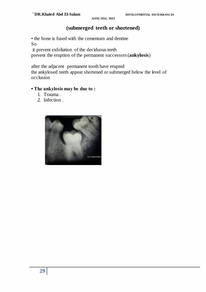
(submerged teeth or shortened)
• the bone is fused with the cementum and dentine So
it prevent exfoliation of the deciduous teeth prevent the eruption of the permanent successors (ankylosis)
after the adjacent permanent tooth have erupted
the ankylosed teeth appear shortened or submerged below the level of occlusion
• The ankylosis may be due to :
1. Trauma . 2. Infection .