dr.lakshmu naidu reader, department of …
TRANSCRIPT

Dr.LAKSHMU NAIDU
READER,
DEPARTMENT OF PROSTHODONTICS,
SUBHARTI DENTAL COLLEGE & HOSPITAL

Indications:
1.Extensive destruction from caries or trauma.
2. Endodontically treated teeth
3. Existing restoration
4. Non Vital teeth
5. Necessity for maximum retention and strength (Long span
bridges)
Indications:
6. To provide contours to receive a removable appliance
7.Other recontouring of axial surfaces (minor corrections of
malinclinations).
8. Correction of occlusal plane.
9. Rampant Caries

Contraindications:
1. Less than maximum retention necessary.
2. Esthetics
3. Poor Periodontal health
4. High DMF index
5. Teeth with internal resorption
6. Young individuals
Advantages:
1. Increases strength to the remaining tooth structure.
2. High retentive qualities
3. Usually easy to obtain adequate resistance from
4. Option to modify form and occlusion.
5. Favorable contours, guide plane for RPD.
6. Easiest procedure (So common used and misused)
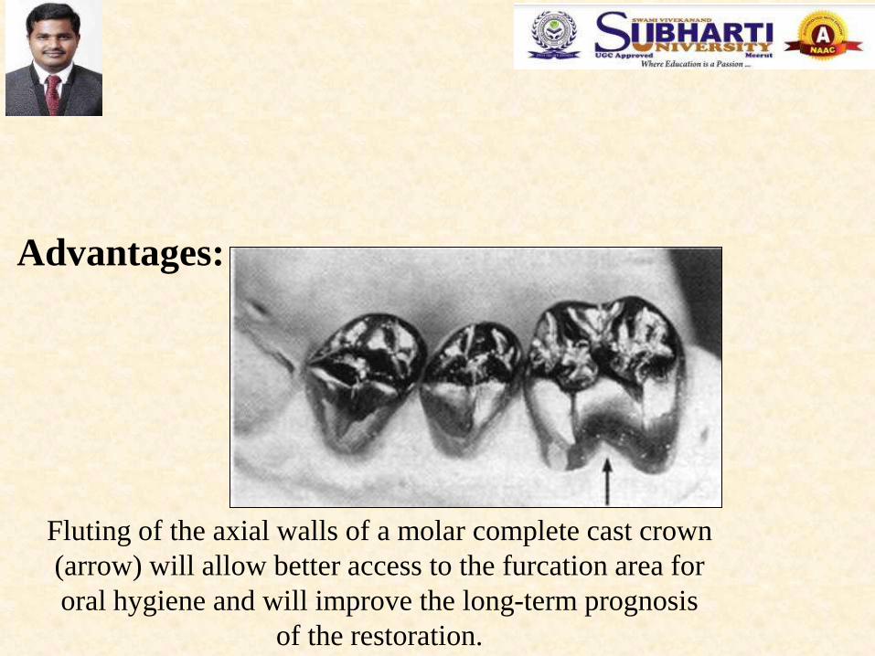
Fluting of the axial walls of a molar complete cast crown
(arrow) will allow better access to the furcation area for
oral hygiene and will improve the long-term prognosis
of the restoration.
Advantages:

Complete cast crowns used as retainers to accommodate a
mandibular removable partial denture. Metal-ceramic crowns
have been placed on the mandibular left canine (A) and the
maxillary first molar (B). Note the occlusal rest seats (A, arrows)
and the survey contours (B), which extent to form reciprocation
guide plane.
A
B

Disadvantages:
1. Removal of large amount of tooth structure.
2. Adverse effects on tissue.
3. Vitality testing not readily feasible
4. Display of metal.
5. Esthetics (it needs facing)
6. Undermined/secondary caries and periodontal diseases

Recommended dimensions for a complete cast crown. On functional cusps
(buccal mandibular and lingual maxillary) the occlusal clearance should be
equal to 1.5mm. On nonfunctional cusps, a clearance of at least 1mm is
needed. The chamfer should allow for approximately 0.5mm of metal
thickness at the margin.
Dimensions:
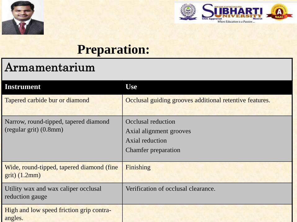
Armamentarium
Instrument Use
Tapered carbide bur or diamond Occlusal guiding grooves additional retentive features.
Narrow, round-tipped, tapered diamond
(regular grit) (0.8mm)
Occlusal reduction
Axial alignment grooves
Axial reduction
Chamfer preparation
Wide, round-tipped, tapered diamond (fine
grit) (1.2mm)
Finishing
Utility wax and wax caliper occlusal
reduction gauge
Verification of occlusal clearance.
High and low speed friction grip contra-
angles.
Preparation:

Preparation step:
- Depth grooves for occlusal reduction
Recommended Armamentarium:
- Tapered carbide or diamond
Criteria :
- Minimum clearance on noncentric cusps: 1mm
- Minimum clearance on centric cusps: 1.5mm

Guiding grooves are placed on the occlusal surface. They are deeper on the
functional cusp, and for the functional cusp bevel they diminish in depth for the cusp
tip to the cervical margin.
Note that the grooves are
deeper for the functional
cups
Step by Step procedure:
- Guiding Grooves for Occlusal Reduction:

A complete cast crown is indicated on
this mandibular second molar with
occlusal, proximal, and cervical lesions
as well as a buccal longitudinal fracture.
Initial depth grooves placed for occlusal
reduction. Note that they have not yet
been extended onto the buccal surface,
where the functional cusp bevel will be
place.
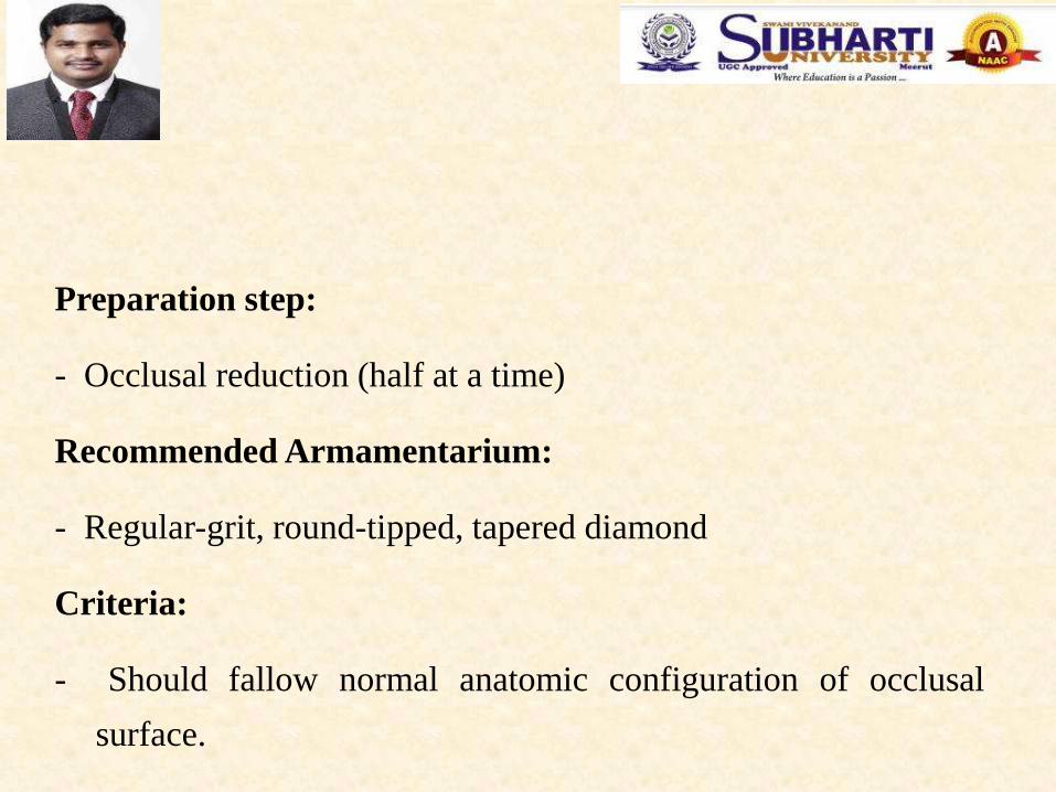
Preparation step:
- Occlusal reduction (half at a time)
Recommended Armamentarium:
- Regular-grit, round-tipped, tapered diamond
Criteria:
- Should fallow normal anatomic configuration of occlusal
surface.

Half of the occlusal reduction is
performed; the other half is
maintained for reference
purposes.
After the guiding grooves are placed, the occlusal
reduction is performed. Either the mesial or the distal
half is maintained initially as a reference.
Occlusal Reduction:

The functional cusp bevel is
prepared by slanting the bur at a
flatter angle than the cuspal
angulation. This will ensure
additional reduction for the
functional cusp.
- Functional (centric Cusp Bevel):
Recommended Armamentarium:
- Tapered carbide or diamond
Criteria:
- Flatter than cuspal plane, to allow additional reduction
at functional cusp.

Note the angulations of the bur as
the functional cusp bevel is placed.
Completed occlusal reduction.
Note that it follows normal
occlusal form. Three distinct
planes can be seen
buccolingually.

The patient closes into softened wax
The thickness of the wax is
assessed visually and measured
with a wax caliper after it has
been removed from the mouth.
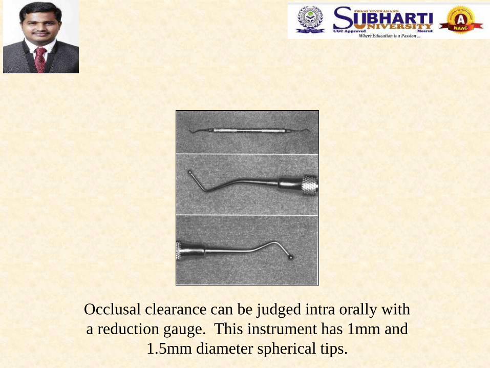
Occlusal clearance can be judged intra orally with
a reduction gauge. This instrument has 1mm and
1.5mm diameter spherical tips.
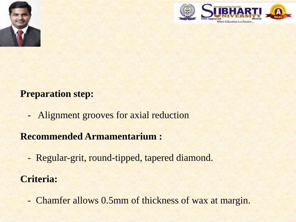
Preparation step:
- Alignment grooves for axial reduction
Recommended Armamentarium :
- Regular-grit, round-tipped, tapered diamond.
Criteria:
- Chamfer allows 0.5mm of thickness of wax at margin.

Alignment Grooves for Axial Reduction:
The diamond is aligned parallel
to the long axis of the tooth as
the buccal guiding grooves for
axial alignment are placed.
After all six grooves have been
placed. Note that they are deep
occlusally but shallower
towards the cervical margin.

Preparation step:
- Axial reduction (half at a time)
Recommended Armamentarium
- Regular-grit, round-tipped, tapered diamond.
Criteria
- Reduction performed parallel to long axis.

If axial reduction is completed first on either the distal or the
mesial half of the tooth, evaluation is simplified because the
remaining intact tooth can serve as a reference.
Axial Reduction:

Note the alignment of the diamond
as tooth structure between the
alignment grooves is removed.
Axial reduction. The
distobuccal axial reduction
has been completed.

As the mesiobuccal axial
reduction is performed, a
cervical chamfer is placed.
Make the chamfer of relatively even
width and maintain the somewhat
angular preparation outline form to
maximum resistance form.

A lip of enamel (arrow) protects the adjacent tooth from
iatrogenic damage as the axial reduction is completed.

Preparation of the proximal contact area.
As the axial reduction is performed,
eventually a small island of tooth
structure will remain in the
interproximal area. When
removing this, maintain a narrow
“lip” of tooth structure between the
diamond and the adjacent tooth to
protect the latter from damage.

Note that a adequate
clearance) exists between the
external surface of the
proximal chamfer and the
adjacent tooth.
Occlusal view of the preparation
≥ 0.6mm ≥ 0.6mm

Preparation step:
- Finishing of chamfer
Recommended Armamentarium
- Wide, round-tipped diamond or carbide
Criteria:
Smooth mesiodistally and buccolingually; resistance to
vertical displacement by tip of explorer or periodontal probe.

Preparation step:
- Finishing
Recommended Armamentarium
- Fine-grit diamond or carbide
Criteria:
Rounding of all sharp line angles to facilitate impression
making, die pouring, waxing and casting.

The transition from lingaul to occlusal is
rounded with a fine-grit diamond.
All sharp line angles between occlusal reduction and functional cusp bevel are similarly rounded.
The margin is refined, and any
minor irregularities are remove.
Finishing:
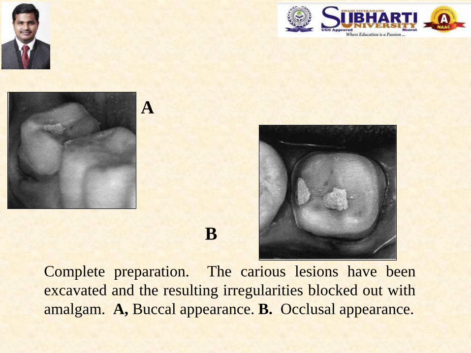
Complete preparation. The carious lesions have been
excavated and the resulting irregularities blocked out with
amalgam. A, Buccal appearance. B. Occlusal appearance.
A
B

Preparation step:
- Additional retentive features if needed
Recommended Armamentarium
- Tapered carbide
Criteria:
Grooves, boxes, pinholes as described for partial coverage
restorations.

Mesially tipped molars and
short premolars often benefit
from grooves and/or boxes
incorporated in the preparation
design.
When opposing axial walls are excessively
tapered, internal features such as this buccal
groove can be used to improve retention and
resistance from.
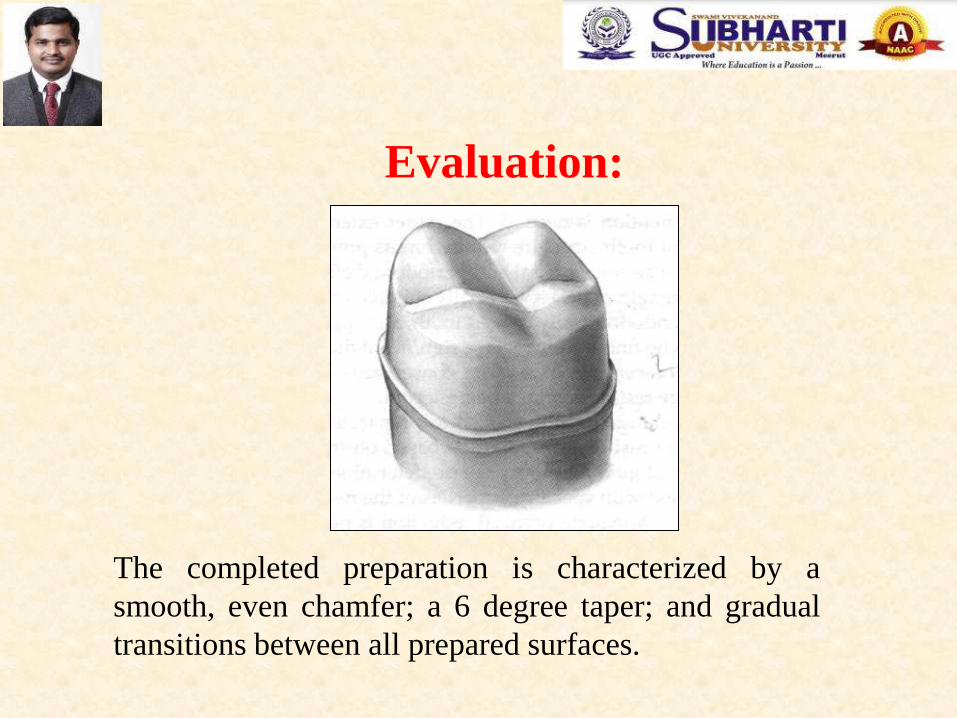
The completed preparation is characterized by a
smooth, even chamfer; a 6 degree taper; and gradual
transitions between all prepared surfaces.
Evaluation:
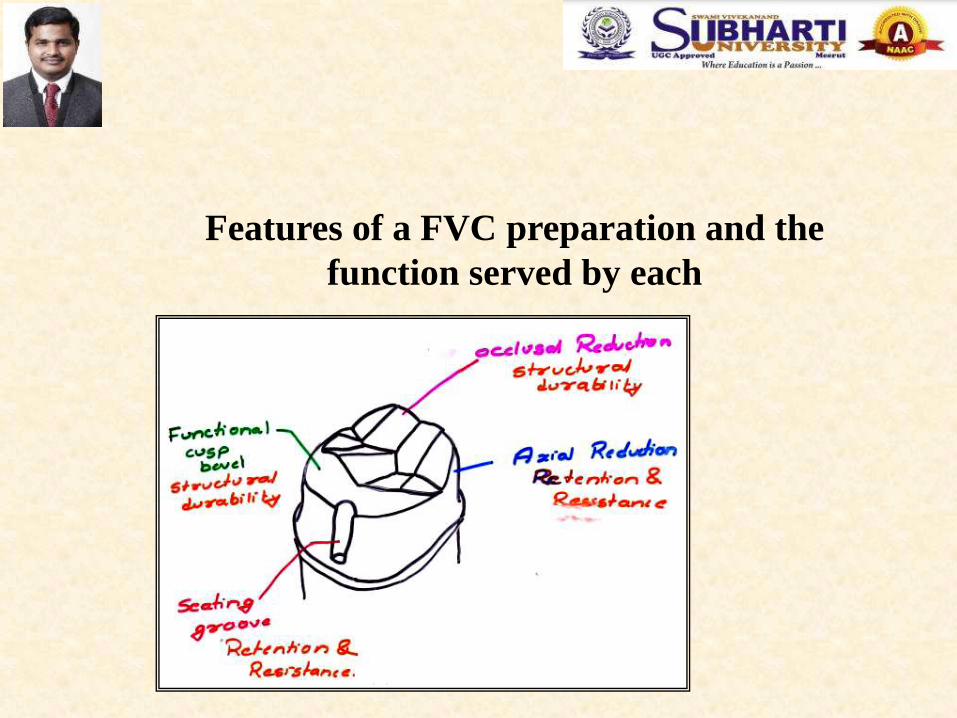
Features of a FVC preparation and the
function served by each

The configuration of the facial wall of the maxillary
molars may require slight additional reduction in the
occlusal third to prevent an over contoured restoration.
Special Considerations
- Nonfunctional (Noncentric) cusp Bevel:

Common Errors in the preparation:
- Under reduction
- Over reduction
- Undercut
- Inadequate occlusal clearance
- Damage to adjacent tooth
- Pulpal exposure.